inflammatory leukocytic recruitment and diffuse · pdf fileinflammatory leukocytic recruitment...
TRANSCRIPT

The Journal of Neuroscience, December 1995, 75(12): 8223-8233
Inflammatory Leukocytic Recruitment and Diffuse Neuronal Degeneration Are Separate Pathological Processes Resulting from Traumatic Brain Injury
Holly D. Soares,’ Ramona R. Hicks,2 Douglas Smith,3 and Tracy K. McIntosh3
‘Department of Developmental Biology, SJCRH, Memphis, Tennessee 38101-0318, 2Division of Physical Therapy, University of Kentucky, Lexington, Kentucky 40536-0079, and 3Division of Neurosurgery, University of Pennsylvania, Philadelphia, Pennsylvania 19104
The present study characterized whether inflammatory leu- kocytic infiltration is temporally and regionally correlated with neuronal degeneration and/or blood brain barrier (BBB) breakdown resulting from traumatic brain injury. Adult rats were sacrificed at 5 min, 2, 4, 12, 24, and 72 hr after lateral fluid percussion brain injury. BBB breakdown, neuronal de- generation and leukocyte infiltration were assessed using immunocytochemistry, silver impregnation and toluidine blue and eosin staining. BBB breakdown and neuronal de- generation occurred concomitantly in injured cortex, hip- pocampus, and along the dorsolateral quadrant of the di- encephalon. However, neuronal degeneration within deep diencephalic structures transpired in the absence of IgG ex- travasation. Neutrophils were observed only in regions ex- hibiting BBB damage and were first apparent in injured cor- tex and hippocampus between 2-12 hr posttrauma lining the vasculature and filling subarachnoid/subduraI spaces. Neu- trophils then migrated from damaged vasculature into trau- matized cortical and hippocampal parenchyma by 24 hr after lateral fluid percussion injury. Macrophages were also ob- served within cortical parenchyma at 24 hr and completely filled the cortical lesion site by 72 hr after injury. Macro- phages were not as abundant throughout hippocampal pa- renchyma and were found only in hippocampal regions ex- hibiting focal hemorrhage at 72 hr. Finally, neutrophils did not migrate to deep diencephalic structures that showed no BBB damage despite extensive neuronal degeneration. In- deed, lateral fluid percussion elicits inflammatory leukocytic recruitment only in regions experiencing concomitant BBB damage and neuronal degeneration. In summary, inflam- matory leukocytic recruitment and diffuse neuronal degen- eration are separate pathological processes resulting from traumatic brain injury.
[Key words: traumatic brain injury, inflammation, fluid
Receive Apr. 27, 1995; revised Aug. 14, 1995; accepted Aug. 21, 1995.
This study was supported, in part, by NIH Grants NS26818, NS08803, and a Merit Review grant from the Veterans Administration. Many thanks to Dr. Fujiro Sendo from the Department of Immunology and Parasitology, Yamagata University School of Medicine-Japan for his generous gift of the RP-1 anti- body. Special thanks to Brain Perry and Drs. D. Kent Morest, Elisa Barbarese, Marion Frank, Gerald Maxwell, and William Shoemaker for help in prepara- tions of the manuscript.
Correspondence should be addressed to Holly Soares, Department of De- velopmental Biology, St. Jude Children’s Hospital, 322 North Lauderdale, Memphis, TN 38101-0318.
Copyright 0 1995 Society for Neuroscience 0270-6474/95/158223-l 1$05.00/O
percussion, leukocytes, neuronal degeneration, RP-1, neu- trophils, macrophages, rat]
Although inflammatory leukocytes have long been implicated as pathological mediators in myocardial ischemic and pulmonary damage (Tate and Repine, 1983; Lucchesi and Mullane, 1986), the involvement of inflammatory cascades in CNS trauma are poorly understood. It is known that both neutrophils and mac- rophages appear in damaged brain following ibotenic acid in- jections (Coffey et al., 1990), intracerebral stab injury (Persson, 1976; Giulian et al., 1989; Moreno-Flores et al., 1993) spinal cord weight drop or compression (Means and Anderson, 1983; Holtz et al., 1990; Xu et al., 1990; Blight, 1992), spinal cord dorsal hemisection (Dusart and Schwab, 1994) brain weight drop contusion (Kochanek et al., 1990; Schoettle et al., 1990; Biagas et al., 1992; Clark et al., 1994), middle cerebral arterial occlusion (Barone et al., 1991; de1 Zoppo et al., 1991; Garcia et al., 1994; Matsuo et al., 1994), carotid air embolism (Hallen- beck et al., 1986) and after combined cryogenic brain injury/ hemorrhagic shock (Zhuang et al., 1993). However, it is not clear whether inflammatory leukocytes significantly contribute to CNS neuropathological damage.
Recent studies have suggested a pathological role for leuko- cytes during CNS injury. Clark et al. (1991a,b) described im- provement in neurological deficits if antibodies capable of blocking neutrophil adhesion and transendothelial migration were administered prior to spinal cord ischemia. Matsuo et al. (1994) reported neutrophil depletion significantly attenuated edema formation and infarct size following middle cerebral ar- terial occlusion. Finally, treatment of rabbits with antiserum to neutrophils improved cerebral blood flow, infarct size, and ICP 4 hours after cerebral embolism.(Bednar et al., 1991). Neutro- phils hypothetically augment pathological processes by (1) se- creting lysosomal enzymes, (2) releasing free radicals, (3) de- creasing blood flow by direct physical microvascular occlusion, and (4) increasing vascular permeability (Harlan, 198.5; Lucchesi and Mullane, 1986; Kochanek and Hallenbeck, 1992). In addi- tion, activated macrophages may actually release a variety of cytokines known to exhibit both neurotrophic and neurotoxic properties (Lindholm et al., 1987; Hama et al., 1989; David et al., 1990; Mori et al., 1990; Taupin et al., 1993; Ott et al., 1994). Thus, neutrophils and macrophages may greatly influence patho- logical sequelae of traumatic brain injury.
Few studies have examined inflammatory events resulting from traumatic CNS damage. To further examine the role of

8224 Soares et al. - Inflammatory Response after Brain Trauma
inflammatory leukocytes in traumatic brain injury, the present study employed lateral fluid percussion in rats to elicit a clini- cally relevant form of brain trauma. The lateral fluid percussion brain injury model in the rat has been extensively characterized (Dixon et al., 1987; Vink et al., 1988; Cortez et al., 1989; Mc- Intosh et al., 1989; Hovda et al., 1991; Smith et al., 1991; Ya- makami and McIntosh, 199 1; Yoshino et al., 199 1; Lowenstein et al., 1992; Soares et al., 1992; Hicks et al., 1993; Dietrich et al., 1994) and reproduces much of the pathophysiology associ- ated with human closed head injuries such as focal cortical con- tusion, blood brain barrier (BBB) breakdown and diffuse neu- ronal degeneration (Cortez et al., 1989; Dietrich et al., 1994; Hicks et al., 1995). However, it is not clear whether the patho- logical events transpiring during traumatic brain injury are also associated with inflammatory leukocytic recruitment.
In the present study, lateral fluid percussion was employed to characterize the spatiotemporal patterns of neutrophils and mac- rophages within contused brain. Our results demonstrate that inflammatory leukocytic recruitment is a significant neuropath- ological component of experimental traumatic brain injury. In- terestingly, inflammatory leukocytes were localized only in regions exhibiting blood brain barrier (BBB) damage and not in diencephalic regions showing extensive neuronal degeneration without concomitant BBB injury. Thus, diffuse neuronal degen- eration can transpire in the absence of inflammatory leukocytic recruitment and these two pathological processes appear to be distinct events resulting from traumatic brain injury.
Materials and Methods
Animal surgery All protocols used in these studies were approved by the Animal Re- search Committee at the University of Pennsylvania (IACUC), and were in accordance with guidelines set by the National Institutes of Health for the humane treatment of animals. Male albino Sprague-Dawley rats (350-400 gm; n = 36) were anesthetized with 60 mg/kg sodium pen- tobarbital administered intraperitoneally. Prior to the initial scalp inci- sion, rats received subcutaneous scalp injections of 1% lidocaine hy- drochloride. Anesthetized animals were placed into a stereotaxic frame and a midline incision made to expose the skull. A stainless steel screw was secured to the skull over left anterior cortex as an anchor for the Luer-Lok connector. A 5 mm diameter craniotomy, centered between bregma, lambda, and the sagittal suture, was drilled over left parietal cortex leaving the dura intact. Ninety minutes following the initial an- esthetic bolus, the anesthetized animals were then attached to the fluid percussion injury device via the female Luer-Lok connector and sub- jected to moderate contusive brain injury (2.3-2.4 atmospheres; 12 = 30). Sham control animals underwent surgical procedure, but did not receive any injury (n = 6). Anesthetized animals were kept on a heating pad both prior and following injury to ensure normothermic tempera- tures were maintained (Okiyama et al., 1994). This model of fluid per- cussion has been previously described in detail (McIntosh et al., 1989).
Tissue preparation At5min(n=7),2hr(n=6),4hr(n=4),12hr(n=4),24hr(n = 7), and 72 hr (n = 4) after injury, animals were reanesthetized with 200 mg/kg sodium pentobarbital intraperitoneally and transcardially per- fused with 100 ml 0.9% heparinized saline followed by 400 ml 4% para- formaldehyde in 0.1 M phosphate buffer pH 7.6. Following perfusion, brains were removed and postfixed an additional 4 hr at 4”C, then trans- ferred to 0.1 M phosphate buffer; 35 pm vibratome or 20 pm cryostat sections were cut and mounted onto gelatin coated slides. Adjacent al- ternate sections were either stained with toluidine blue and eosin, im- pregnated with silver, or immunocytochemically processed for the detec- tion of extravasated IgG and neutrophilic/macrophagic antigens.
Histological stains
To&dine blue and eosin. Sections were cleared in Xylene, rehydrated, and stained for 30 set in 0.05% toluidine blue in acetate buffer pH 4.4.
They were then rinsed in ddH,O, and immersed in 0.05% eosin Y in ddH,O for 2 min. Sections were then dehydrated and coverslipped using Permount.
Silver impregnation for degenerating neurons (Gallyas et al., 1980; Nadler and Evenson, 1983). Sections were rinsed in glycerol/ddH,O 3 X 5 min, and then pretreated 2 X 5 min in 4.5% sodium hydroxide and 0.6% ammonium nitrate. Sections were then impregnated for lo-15 min in 5.4% sodium hydroxide, 6.4% ammonium nitrate, and 0.3% silver nitrate. Sections were then washed 3 X 2 min in 0.012% ammonium nitrate, 95% ethanol, and 0.5% anhydrous sodium carbonate Silver pre- cipitates on sections were developed for 1-2 min in 0.012% ammonium nitrated, 0.55% formalin, 95% ethanol, and 0.5% anhydrous citric acid. Sections were then washed 5 X 5 min in 0.5% acetic acid in Tris-HCl 0.05 M pH 7.4, dried, and coverslipped.
IgG immunocytochemistry. Free floating vibratome sections or slide mounted cryostat sections were blocked for 1 hr with 10% normal goat serum (NGS) and 0.3% Triton-X in 0.05 M Tris-HCl pH 7.6. Sections were then rinsed in 2% NGS and incubated with biotinylated goat anti- rat IgG (1:50) 12-24 hr at 4°C. Controls included incubation with non- immune biotinylated goat immunoglobulins and preadsorbed antibodies. All antibodies were obtained commercially from Accurate. Sections were then rinsed with 2% NGSiTris 4 X 15 min, and then incubated for 24 hr in FITC-streptavidin (Accurate 1:lOOO) in 2% NGS. Follow- ing 3 X 10 min rinses, sections were mounted onto gelatin coated slides and coversliuued with a 2.5% DABCO (Siama)/glvcerol solution.
Neutrophi&and macrophage immuno&t~che’m%y. Ten micrometer cryostat sections were blocked with 5% normal goat serum in 0.1 M
phosphate buffer pH 7.4 for 30 min and then incubated overnight with either a monoclonal antibody for neutrophils or macrophages. The neu- trophil-specific antibody RP-1 (Gotoh et al., 1986) was a generous gift from Dr. Fujiro Sendo (Yamagata University School of Medicine, Ja- pan) and was used at a 1:500 dilution. The ED1 and OX-42 (which recognizes the CDllb or complement receptor 3 antigen) monoclonal antibodies were utilized to detect macrophages/activated microglia (Ser- otec) and were used at a 1:lOO and 1:5000 dilution, respectively. Sec- tions were then rinsed 4 X 10 min in buffer and incubated with FITC labeled goat-anti mouse IgG (Cappel; no cross-reaction to rat) for 6 hr. Finally, sections were again rinsed 4 X 15 min in phosphate buffer and coverslipped with a 2.5% DABCO (Sigma)/glycerol solution.
Histological analysis
Adjacent sections from six coronal regions spaced throughout the anterior to posterior axis were analyzed for IgG extravasation (i.e., indicating BBB breakdown), the presence of neutrophils and macrophages, and neuronal degeneration (Fig. 1A). Using conventional histology, macrophages were identified as having a foamy cytoplasmic phenotype with an eccentrically placed ovoid/kidney shaped nuclei and large cell body diameters ranging from 2WlO km (Fig. lB, arrow). The macrophage phenotype was con- firmed with immunocytochemistry for macrophage antigens (Fig. lF, ar row). Following toluidine blue and eosin staining, neutrophils exhibited faintly eosinophilic cytoplasmic granules, multilobed nuclei, and cell body diameters ranging from lG15 pm (Fig. lC, arrow). The neutrophil phenotype within damaged tissue was confirmed using the RI-1 antibody which specifically recognizes neutrophils (Fig. IE, arrowhead). Injured neurons appeared intensely argyrophilic with arborizations clearly visible during early stages of degeneration (Fig. lD, arrow). At later stages of degeneration (i.e., more than 24 hr), injured neuronal cell bodies remained intensely argyrophilic (Fig. 1, dl). Uninjured neurons appeared round with only the nucleoli showing slight argyrophilia (Fig. 1, d2). Neutro- phils, macrophages, degenerating neurons and IgG extravasation were plotted onto spatiotemporal coronal maps utilizing an Ml Imaging Re- search image analysis system (Imaging Research Inc, Canada) connected to a Leitz Diaplan microscope. The symbol for each cell type represent cell numbers of more than five.
Results
Experimental paradigm Figure 2 illustrates the gross pathology of lateral fluid percussion in rats. Twenty-four hours after trauma, a focal contusion forms within parietal and temporal cortices (Fig. 2A,B, arrows). Al- though lateral fluid percussion elicits extensive damage in hip- pocampal and diencephalic structures, only contused cortex ex-

The Journal of Neuroscience, December 1995, 75(12) 8225
Figure 1. Experimental paradigm and gross pathology of lateral fluid percussion. A, Adjacent sections from injured brain were subjected to either RP-1, EDI, or OX-42 immunocytochemistry or toluidine blue and eosin staining to detect leukocytes, silver impregnation to detect neuronal degeneration, and IgG immunocytochemistry to detect BBB breakdown. Individual cell types mapped in the present study are shown in B-F. Toluidine blue and eosin staining of a: B, foamy macrophage (arrow), and C, polymorphonuclear neutrophil (arrow). Silver impregnation of a: D, degenerating cortical neuron at 2 hr after injury (arrow); dl, Degenerating cortical neuron at 24 hr after injury; d2, Uninjured cortical neuron impregnated with silver. E, RI-1 immunocytochemistry of neutrophils within injured cortex at 12 hr after trauma (arrowhead demarcates an RP-1 immunopositive cell). F, OX-42 immunocytochemistry of a large foamy macrophage (arrow) in injured cortex at 24 hr after injury. Scale bars: C, 15 pm (for B and C); dl, 2.5 pm (for dl and d2); F, 30 km (for E and F).
hibits profound cavitation by 4 weeks after injury (Fig. 2C,D, arrowheads).
Leukocytes within contused brain
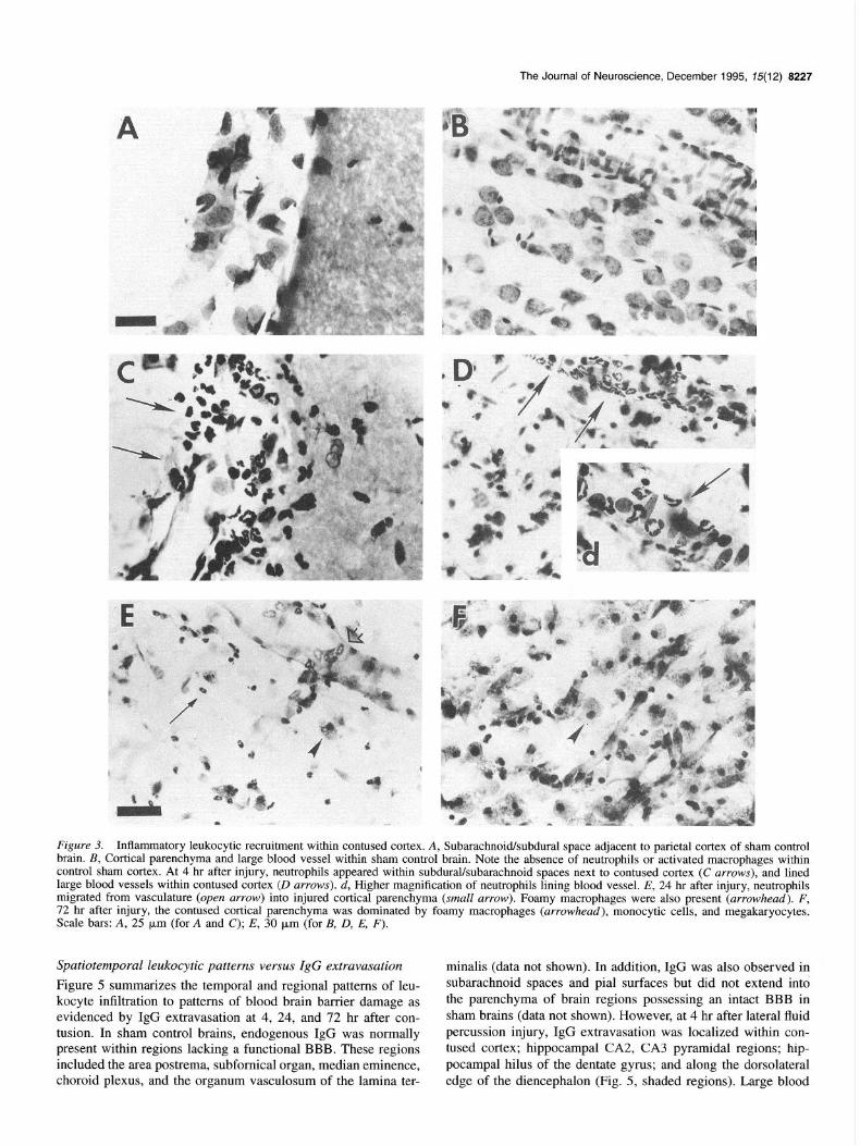
the rat. Under normal conditions, leukocytes do not fill the sub- arachnoid/subdural spaces (Fig. 3A) nor are they present in cor- tical parenchyma of a sham control brain (Fig. 3B). Leukocytes were not observed at 5 min after trauma (data not shown). How-
Figure 3 illustrates the pattern of leukocytic infiltration within ever, between 2-4 hr after injury, neutrophils filled subarach- contused cortex following lateral fluid percussion brain injury in noidfsubdural spaces adjacent to contused cortex (Fig. 3C, ar-

8226 Soares et al. - lnflammatoty Response after Brain Trauma
Figure 2. Gross pathology resulting from moderate levels of lateral fluid percussion brain injury in the rat. Moderate injury produces a focal contusion within parietal/temporal cortex (A and B, arrows). By 4 weeks after injury, the original cortical lesion has formed a glial lined cavity (C and D, arrowheads). Scale bar in D, 2 mm (for A-D).
rows) and were evident lining large blood vessels of injured cortex at 4 hr (Fig. 30 and d, arrows). Neutrophils were not observed outside of blood vessels, nor where any neutrophils observed within contused cortical parenchyma at 4 hr post in- jury. Monocytic cell types were also observed lining the cortical vasculature of injured cortex at 12 hours (data not shown). By 24 hr, neutrophils had progressed from damaged blood vessels into cortical parenchyma (Fig. 3E, small arrows) and the first foamy macrophages were observed (Fig. 3E, arrowhead). Paren- chymal damage was extensive in cortex and, by 72 hr after trau- ma, very few neurons remained and the contused cortical region was comprised primarily of foamy macrophages (Fig. 3F, ar- rowheads), blood vessels, monocytic cells, and megakaryocytes. Thus, inflammation is a significant pathological process within contused cortex following lateral fluid percussion brain injury.
Leukocytes within contused subcortical structures
Subcortical structures did not display the same distribution or temporal patterns of inflammatory leukocytic infiltration as did contused cortex. Figure 4 shows inflammatory leukocytes within the hippocampus and thalamus ipsilateral to the site of cortical trauma. Neutrophils were observed within subarachnoid spaces between hippocampus and dorsolateral thalamus (data not shown) and in large blood vessels of ipsilateral hippocampus between 2 and 12 hr after trauma. Figure 4A illustrates neutro- phils lining an injured blood vessel within ipsilateral hippocam- pus at 12 hr after lateral fluid percussion (arrows). At 12 and 24 hr after trauma, neutrophils were abundantly present throughout ipsilateral hippocampal parenchyma especially along ipsilateral
dentate gyrus at 12 hr (Fig. 4B, arrows) and around CA3. Al- though macrophages completely filled the contusion within in- jured cortex, macrophages did not completely fill hippocampal parenchyma at 72 hr after fluid percussion brain injury. Indeed, macrophages were only observed within small hemorrhagic foci around the CAl, CA2, CA3, and dentate hippocampal regions at 72 hr posttrauma (Fig. 4C, arrowheads). Thus, inflammatory leukocytic infiltration within ipsilateral hippocampus was dom- inated by neutrophils during the acute postinjury period while macrophages with foamy cytoplasmic phenotypes remained lo- calized to small hemorrhagic foci. .
Inflammatory leukocytic recruitment to thalamus and deep diencephalic structures appeared to be confined to left superficial dorsolateral nuclei. Neutrophils did not line the vasculature of diencephalic structures as observed in injured cortex and hip- pocampus during the acute postinjury period, nor did they ac- cumulate within deep diencephalic parenchyma at any of’ the time points examined. Figure 40 shows ipsilateral posterior thal- amus at 24 hr after injury. Despite extensive gliosis and neuronal degeneration, neutrophils were not observed in ipsilateral pos- terior thalamus at either 24 (Fig. 40) or 72 hr after injury (see also Fig. 5). A few cells in the dorsolateral thalamus were ED1 and OX-42 positive and tended to exhibit morphological char- acteristics of microglia such as ramified processes (data not shown). Nevertheless, neutrophils were not localized in dien- cephalic structures and the classical sequence of acute inflam- matory leukocytic recruitment (i.e., neutrophils followed by macrophages) occurred only within contused cortex and hippo- campus and not within deep diencephalic areas.
The Journal of Neuroscience, December 1995, 75(12) 8227
Figure 3. Inflammatory leukocytic recruitment within contused cortex. A, SubarachnoWsubdural space adjacent to parietal cortex of sham control brain. B, Cortical parenchyma and large blood vessel within sham control brain. Note the absence of neutrophils or activated macrophages within control sham cortex. At 4 hr after injury, neutrophils appeared within subdural/subarachnoid spaces next to contused cortex (C arrows), and lined large blood vessels within contused cortex (D arrows). d, Higher magnification of neutrophils lining blood vessel. E, 24 hr after injury, neutrophils migrated from vasculature (open arrow) into injured cortical parenchyma (small arrow). Foamy macrophages were also present (arrowhead). F, 72 hr after injury, the contused cortical parenchyma was dominated by foamy macrophages (arrowhead), monocytic cells, and megakaryocytes. Scale bars: A, 25 pm (for A and C); E, 30 pm (for B, 0, E, F).
Spatiotemporal leukocytic patterns versus IgG extravasation
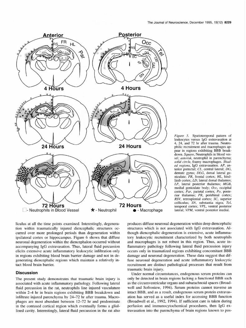
Figure 5 summarizes the temporal and regional patterns of leu- kocyte infiltration to patterns of blood brain barrier damage as evidenced by IgG extravasation at 4, 24, and 72 hr after con- tusion. In sham control brains, endogenous IgG was normally present within regions lacking a functional BBB. These regions included the area postrema, subfornical organ, median eminence, choroid plexus, and the organum vasculosum of the lamina ter-
minalis (data not shown). In addition, IgG was also observed in subarachnoid spaces and pial surfaces but did not extend into the parenchyma of brain regions possessing an intact BBB in sham brains (data not shown). However, at 4 hr after lateral fluid percussion injury, IgG extravasation was localized within con- tused cortex; hippocampal CA2, CA3 pyramidal regions; hip- pocampal hilus of the dentate gyrus; and along the dorsolateral edge of the diencephalon (Fig. 5, shaded regions). Large blood

8228 Soares et al. l inflammatory Response after Brain Trauma
Figure 4. Leukocytes in contused hippocampus and thalamus after lateral fluid percussion. A, Neutrophils lined large blood vessels within contused hippocampus at 12 hr after injury (arrows). B, Neutrophils were also present within hippocampal parenchyma surrounding dentate gyms at 12 hr after injury (arrows). C, A few foamy macrophages were found within small hemorrhagic foci of CA3 at 72 hr after trauma (arrowhead). D, Gliosis and neuronal degeneration in the posterior nucleus of thalamus 24 hr after injury. Note the absence of neutrophils or foamy macrophages. Scale bar in A, 25 p.rn (for A-D).
vessels within ipsilateral hippocampus also demonstrated IgG extravasation at 4 hours after trauma (data not shown). By 24 hr, IgG extravasation had spread throughout contused cortical and injured hippocampal parenchyma (Fig. 5, shaded region). Diencephalic IgG extravasation remained confined to the dor- solateral quadrant. In addition, small focal hemorrhagic regions were often observed in the substantia nigra, posterior pons and brainstem (data not shown). Neutrophils, as identified using RP-1 immunocytochemistry and conventional histology, first ap- peared in cortical and hippocampal blood vessels at 4 hr (Fig. 5, squares) and were later observed (Fig. 5, asterisk) in hippo- campal and cortical parenchymal regions 24-72 hr. Large foamy macrophages (Fig. 5, solid circles), as identified using immu- nocytochemistry and conventional histology, were observed within contused cortical parenchyma at 24 hr and eventually filled the original cortical contusion by 72 hr. However, such a dramatic accumulation of large foamy macrophages was not ob- served in either hippocampal or diencephalic structures. Within the injured hippocampus and diencephalon, large foamy mac- rophages remained confined to small hemorrhagic foci. Neutro- phils and monocytic cell types did not appear to line dience- phalic vasculature. In addition, neutrophils did not appear to migrate throughout diencephalic parenchyma except in regions exhibiting IgG extravasation and/or hemorrhage. Thus, acute in- flammatory leukocytic infiltration was confined to regions show-
ing concomitant BBB damage as evidenced by IgG extravasa- tion.
IgG extravasation versus neuronal degeneration
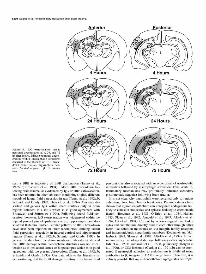
Figure 6 compares the temporal and regional patterns of IgG extravasation to those of neuronal degeneration resulting from lateral fluid percussion brain injury in the rat. Sham controls exhibited no agryophilic neurons and endogenous IgG was con- fined to regions lacking a functional BBB (data not shown). Between 2-24 hr post injury, argyrophilic neurons and extravas- ated IgG were both observed within parietal, temporal, and re- trosplenial cortices; and in hippocampalCA2, CA3, dentate gy- rus, and dentate hilus (Fig. 6). Although cortical IgG extravas- ation persisted throughout the 72 hr observation period, most of the cortical neurons had degenerated by 72 hr. Interestingly, neu- ronal degeneration within the hippocampus was localized to spe- cific regions despite widespread IgG extravasation (Fig. 6). Ip- silateral thalamic degeneration predominated throughout dorsal, lateral posterior, ventral posterior lateral, ventral posterior me- dial, and centromedial nuclei at 4, 24, and 72 hr postinjury (Fig. 6). Very few degenerating neurons were observed within the lateral geniculate, although the medial geniculate possessed nu- merous argyrophilic neurons at 4, 24, and 72 hr after injury. A few degenerating neurons were observed within the ipsilateral superficial gray and in the optic nerve layer of the superior col-
The Journal of Neuroscience, December 1995, 75(12) 8229
- - __
24 H ours
l - Macrophage
Figure 5. Spatiotemporal pattern of leukocytes versus IgG extravasation at 4, 24, and 72 hr after trauma. Neutro- philic recruitment and macrophages ap- pear in regions exhibiting BBB break- down. Square, Neutrophils in blood ves- sel; asterisk, neutrophil in parenchyma; solid circle, foamy macrophages. Shad- ed regions, IgG extravasation. AP, an- terior pretectal; CL, central lateral; DG, dentate gyms; DLG, dorsal lateral ge- niculate; FR, frontal cortex; HL, hind- limb cortex; LD, lateral dorsal thalamus; LP, lateral posterior thalamus; MGB, medial geniculate body; Occ, occipital cortex; Par, parietal cortex; PO, poste- rior thalamus; PR, perirhinal cortex; RSV, retrosplenial cortex; SC, superior colliculus; SN, substantia nigra; Tel, temporal cortex; VPL, ventral posterior lateral; VPM, ventral posterior medial.
liculus at all the time points examined. Interestingly, degenera- tion within traumatically injured diencephalic structures oc- curred over more prolonged periods than degeneration within ipsilateral cortex or hippocampus. Figure 6 shows that diffuse neuronal degeneration within the diencephalon occurred without accompanying IgG extravasation. Thus, lateral fluid percussion elicits extensive acute inflammatory leukocytic infiltration only in regions exhibiting blood brain barrier damage and not in de- generating diencephalic regions which maintain a relatively in- tact blood brain barrier.
produces diffuse neuronal degeneration within deep diencephalic structures which is not associated with IgG extravasation. Al- though diencephalic degeneration is extensive, acute inflamma- tory leukocytic recruitment characterized by both neutrophils and macrophages is not robust in this region. Thus, acute in- flammatory pathology following lateral fluid percussion injury occurs only in traumatized regions exhibiting concomitant BBB damage and neuronal degeneration. These data suggest that dif- fuse neuronal degeneration and acute inflammatory leukocytic recruitment are distinct pathological processes that result from
Discussion
The present study demonstrates that traumatic brain injury is associated with acute inflammatory pathology. Following lateral fluid percussion in the rat, neutrophils line injured vasculature within 24 hr in brain regions exhibiting BBB breakdown and infiltrate injured parenchyma by 24-72 hr after trauma. Macro- phages are most abundant between 12-72 hr and predominate in the contused cortical region which eventually forms a glial-
traumatic brain injury. Under normal circumstances, endogenous serum proteins can
only be detected in brain regions lacking a functional BBB such as the circumventricular organs and subarachnoid spaces (Broad- well and Sofroniew, 1994). Serum proteins cannot traverse an intact BBB, and as a result, endogenous serum protein extravas- ation has served as a useful index for accessing BBB function (Broadwell et al., 1992, 1994). If sufficient care is taken during perfusion and immunocytochemical procedures, then IgG ex-
lined cavity. Interestingly, lateral fluid percussion in the rat also travasation into the parenchyma of brain regions known to pos-
8230 Soares et al. * inflammatory Response after Brain Trauma
Figure 6. IgG extravasation versus neuronal degeneration at 4, 24, and 72 hr after injury. Diffuse neuronal degen- eration within diencephalic structures occurred in the absence of BBB break- down. Solid circles, argyrophilic neu- rons. Shaded regions, IgG extravasa- tion.
sess a BBB is indicative of BBB dysfunction (Tanno et al., percussion is also associated with an acute phase of neutrophilic 1992a,b; Broadwell et al., 1994). Indeed, BBB breakdown fol- infiltration followed by macrophagic activation. Thus, acute in- lowing brain trauma, as evidenced by IgG or HRP extravasation, flammatory mechanisms may profoundly influence secondary has been reported in other laboratories utilizing slightly different posttraumatic sequelae following brain trauma. models of lateral fluid percussion in rats (Tanno et al., 1992a,b; It is not clear why neutrophils were recruited only to regions Schmidt and Grady, 1993; Dietrich et al., 1994). Our data de- exhibiting blood brain barrier breakdown. Previous studies have scribed endogenous IgG within sham controls only in brain shown that injured endothelium can upregulate endogenous leu- regions deficient in a BBB which is in good agreement with kocytic adhesion molecules and release leukocytic chemotactic Broadwell and Sofroniew (1994). Following lateral fluid per- factors (Bowman et al., 1983; O’Brien et al., 1984; Harlan, cussion, however, IgG extravasation was widespread within the 1985; Sloan et al., 1992; Arnould et al., 1993; Albelda et al., injured parenchyma of ipsilateral cortex, hippocampus, and dor- 1994; Ott et al., 1994). Current hypotheses suggest that leuko- solateral thalamus. Indeed, similar patterns of BBB breakdown cytes and endothelium directly bind to each other through either have also been reported in other laboratories utilizing lateral lectin-like adhesion molecules or, via integrin family receptors fluid percussion especially in injured cortical and hippocampal and immunoglobulin superfamily members (Kochanek and Hal- regions (Tanno et al., 1992a,b; Schmidt and Grady, 1993). In lenbeck, 1992; Sloan et al., 1992; Albelda et al., 1994). In fact, general, studies from the above mentioned laboratories showed inflammatory pathological damage following either myocardial that BBB damage within diencephalic structures was not as ex- (Ma et al., 1991; Yamazaki et al., 1993), pulmonary (Horgan et tensive as in ipsilateral cortex or hippocampus which is in good al., 1990), or CNS ischemia (Clark et al., 1991a,b) can be atten- agreement with the present observations (Tanno et al., 1992a,b; uated if neutrophil adhesion to endothelium is inhibited using Schmidt and Grady, 1993). Our data adds to the literature by antibodies to p2 integrin or CAM-like proteins. Therefore, it is demonstrating that the BBB damage resulting from lateral fluid entirely possible that injured endothelium upregulates neutrophil
The Journal of Neuroscience, December 1995, 75(12) 8231
adhesion factors after brain trauma. Such a phenomena may per- haps explain why neutrophils were only observed in traumati- cally injured regions exhibiting blood brain barrier damage.
Neutrophils could greatly exacerbate the pathological sequel- ae of brain trauma by altering vascular permeability (Wedmore and Williams, 1981) contributing to oxidative damage (Tonne- sen et al.. 1988) inducing further neuronal damage via the se- cretion of lysosomal enzymes (Tonnesen et al., 1988) or by altering cerebral vascular blood how. Indeed, Uhl et al. (1994) showed that acute inflammation resulting from weight drop brain injury could contribute to the observed level of cerebral blood flow (CBF) in injured brain regions especially in those regions which experienced increases in CBE In addition, polymorpho- nuclear leukocyte recruitment has been correlated with increased cerebral edema in a weight drop paradigm of brain contusion (Schoettle et al., 1990) and neutrophil depletion has been re- ported to attenuate increases in brain water content and decrease infarct size following middle cerebral arterial occlusion (Matsuo et al.. 1994). It is intriguing to note that the same regions ex- hibiting neutrophil infiltration in our model of fluid percussion do proceed to develop edema (Soares et al., 1992). Thus, neu- trophils may contribute to edematous sequelae following lateral fluid percussion.
Acute inflammation is characterized by the early appearance of neutrophils followed by macrophages. The present study shows extensive recruitment of neutrophils to injured cortex. hippocampus, and dorsolateral thalamus, but not to deep dien- cephalic structures. Macrophages are identifiable after the ap- pearance of neutrophils within injured cortex, hippocampus, and dorsolateral thalamus suggesting that acute inflammation does occur in these regions as a result of lateral fluid percussion. However. it is not clear whether macrophages are indeed re- cruited, or whether they arise from resident populations such as microglia. Our data is in good agreement with the work of Ai- ham et al. (1995) which described the timecourse and localiza- tion of EDI and OX-42 immunopositive cells following fluid percussion injury. Unfortunately, once microglia become acti- vated, they express all macrophagic markers including EDI (Flaris et al., 1993; Ling and Wong, 1993) and it is not a trivial matter distinguishing recruited macrophages from activated mi- croglia. Although it is likely that many of the macrophages with- in contused cortex were recruited, our results do not directly demonstrate that the macrophages we observed within injured brain regions actually originated from outside the CNS. Indeed, it is quite probable the macrophagic population consisted of both resident and recruited cells. Nevertheless, our data does dem- onstrate that neutrophils were recruited to some, but not all trau- matically injured brain regions. Specifically, neutrophils were not observed in deep diencephalic structures. This is an impor- tant observation in that blood vessel damage is minimal within diencephalic structures despite extensive neuronal degeneration. Indeed, it would suggest that neutrophils may be responding more to vascular damage than to neuronal degeneration. Clearly, neutrophil recruitment and diffuse neuronal degeneration are separate events following lateral fluid percussion brain injury.
The most likely mechanism by which neutrophils and acti- vated macrophages/microglia can alter the pathological milieu is probably through the secretion of soluble factors such as cyto- kines. For example, IL-6, IL-I, and tumor necrosis factor alpha (TNF-a) levels increase and remain elevated in the brain follow- ing traumatic injury (Goodman et al., 1990; McClain et al., 1991: Taupin et al., 1993). Although IL-6 and IL-I have been
reported to exert neurotrophic effects in vitro (Lindholm et al., 1987; Hama et al., l989), pathological events could be exacer- bated through abnormal endothelial effects. Indeed, both IL-l and TNF-(w are capable of altering vascular permeability (Bey- non et al., 1993; Burker-Gaffney and Keenan, 1993) and these cytokines could exacerbate BBB breakdown and subsequent brain edema formation. In addition, traumatic brain injury has been reported to elicit elevations in plasma levels of cytokines (McClain et al., 1991) which could, in turn, contribute to such systemic complications as stress-related mucosal disease and pulmonary edema experienced by many severely head injured patients (Ott et al., 1994). Thus, secretory factors from activated leukocytes within the brain or circulating systemically may pro- foundly contribute to secondary pathological cascades following traumatic brain injury.
In addition to inflammation and BBB breakdown, lateral tluid percussion produced extensive neuronal degeneration. Although neuronal degeneration within fluid percussion injured cortex and hippocampus was associated with BBB breakdown, the present study showed that much of the neuronal degeneration within regions of the diencephalon transpired in the absence of IgG extravasation. Neuronal degeneration without associated vascu- lar damage has been observed in other experimental models of brain trauma (Povlishock, 1986; Povlishock et al., 1992) and may account for human head injuries which present as coma in the absence of mass lesions (Strich, 1956; Sahuquillo et al., 1989; Graham et al., 1993). It is interesting to note that, in our model, neutrophils appear only in contused brain regions expe- riencing BBB breakdown. Neutrophils did not infiltrate damaged regions failing to exhibit IgG extravasation despite extensive neuronal degeneration. Indeed, the present study suggests that diffuse diencephalic degeneration is a pathological process dis- tinct from traumatically induced acute inflammatory leukocytic recruitment in the brain.
In conclusion, lateral fluid percussion elicits complex neuro- pathology involving both inflammatory cascades and diffuse neuronal degeneration. Leukocytes are known to act as patho- genic mediators in experimental model of myocardial and CNS ischemia (Lucchesi and Mullane, 1986; Kochanek and Hallen- beck, 1992) and may serve similar pathological functions fo- lowing traumatic brain injury. In fact, a number of pharmaco- logical agents which possess immunosuppressive characteristics have been reported to improve outcome following contusive CNS injuries (McIntosh, 1993; Okiyama et al., 1994). Further- more, recent attempts at inhibiting leukocytic inllammatory pro- cesses through the blockage of cell adhesion molecules or by neutrophil depletion has shown promise in some models of isch- emia (Albelda et al., 1994; Matsuo et al., 1994). Although the present study did not demonstrate a casual relationship between leukocyte recruitment and neuropathological events such as brain edema, it is intriguing that neutrophils were localized Hurst along blood vessels within injured regions which go on to de- velop BBB breakdown. Clearly, further studies need to be pur- sued regarding the leukocytic contribution to traumatic brain in- jury. Our results do emphasize, however, that traumatic brain injuries may contain a diffuse degenerative component which can be quite distinct from leukocytic recruitment. Thus, separate pharmacological strategies may prove critical in the effective management of inflammatory sequelac and diffuse neuronal pa- thology resulting from traumatic brain in.jury.

8232 Soares et al. - Inflammatory Response after Brain Trauma
References
Aihara N, Hall JJ, Pitts LH, Fukuda K, Noble LJ (1995) Altered im- munoexpression of microglia and macrophages after mild head in- jury. J Neurotrauma 12:53-63.
Albelda SM, Smith CW, Ward PA (1994) Adhesion molecules and inflammatory injury. FASEB J 8504-5 12.
Arnould T, Michiels C, Remacle J (1993) Increased PMN adherence on endothelial cells after hypoxia: involvement of PAF, CD18/ CD1 lb, and ICAM-1. Am J Physiol 264:C1102-ClllO.
Barone FC, Hillegass LM, Price WJ, White RF, Lee EV, Feuerstein GZ, Sarau HM, Clark RK, Griswold DE (1991) Polymorphonuclear leu- kocyte infiltration into cerebral focal ischemic tissue: myeloperoxi- dase activity assay and histologic verification. J Neurosci Res 29: 336-345.
Bednar MM, Raymond S, McAuliffe T, Lodge PA, Gross CE (1991) The role of neutrophils and platelets in a rabbit model of throm- boembolic stroke. Stroke 22:44-50.
Beynon HL, Haskard DO, Davies KA, Haroutunian R, Walport MJ (1993) Combinations of low concentrations of cytokines and acute agonists synergize in increasing the permeability of endothelial monolayers. Clin Exp Immunol 91:314-319.
Biagas KV, Uhl M, Schiding JK, Nemoto EM, .Kochanek PM (1992) Assessment of posttraumatic polymorphonuclear leukocyte accumu- lation in rat brain using tissue myeloperoxidase assay and vinblastine treatment. J Neurotrauma 9:363-371.
Blight AR (1992) Macrophages and inflammatory damage in spinal cord injury. J Neurotrauma 9(Suppl l):S83-S91.
Bowman CM, Butler EM, Repine JE (1983) Hyperoxia damages cul- tured endothelial cells causing increased neutrophil adherence. Am Reb Respir Dis 128:469-472.
Broadwell RD, Sofroniew MV (1994) Serum proteins bypass the blood-brain fluid barriers for extracellular entry to the central nervous system. Exp Neurol 120:245-263.
Broadwell RD, Baker BJ, Ebert PS, Hickey WE Villegas J (1992) In- tracerebral grafting of solid tissues and cell suspensions: the blood- brain barrier and host immune response. Prog Brain Res 91:95-102.
Broadwell RD, Baker BJ, Ebert PS, Hickey WF (1994) Allografts of CNS tissue possess a blood-brain barrier. III. Neuropathological, methodological, and immunological considerations. Microsc Res Tech 271471-494.
Burker-Gaffney A, Keenan A (1993) Modulation of human endothelial cell permeability by combinations of the cytokines interleukin-1 al- pha/beta, tumor necrosis factor-alpha and interferon-gamma. Immu- nopharmacology 25:1-9.
Clark RSB, Schiding JK, Kaczorowski SL, Marion DW, Kochanek PM (1994) Neutrophil accumulation after traumatic brain injury in rats: comparison of weight drop and controlled cortical impact models. J Neurotrauma 11:499-506.
Clark WM, Madden Kp, Rothlein R, Zivin JA (1991) Reduction of central nervous system ischemic‘injury by monoclonal antibody to intercellular adhesion molecule. J Neurosurg 75:623-627.
Clark WM, Madden KP, Rothlein R, Zivin JA (1991) Reduction of central nervous system ischemic injury in rabbits using leukocyte adhesion antibodv treatment. Stroke 22:877-883.
Coffey PJ, Perry V’H, Rawlins JNP (1990) An investigation into the early stages of the inflammatory response following ibotenic acid induced neuronal degeneration. Neurosci 35: 121-l 32.
Cortez S, McIntosh TK, Noble LJ (1989) Experimental fluid percussion brain injury: vascular disruption and neuronal glial alterations. Brain Res 482:271-282.
David S, Bouchard G, Tsatas 0, Giftochristos N (1990) Macrophages can modify the nonpermissive nature of the adult mammalian central nervous system. Neuron 5:463-469.
de1 Zoppo GJ, Schmid-Schonbein GW, Mori E, Copeland BR, Chang CM (199 1) Polymorphonuclear leukocytes occlude capillaries fol- lowing middle cerebral artery occlusions and reperfusion in babbons. Stroke 22: 1276-1283.
Dietrich W, Alonso 0, Busto R, Globus M, Ginsberg M (1994) Post- traumatic brain hypothermia reduces histopathological damage fol- lowing concussive brain injury in the rat. Acta Neuropathol (Berl) 87:250-258.
Dixon CE, Lyeth BG, Polvishock JT, Findling RI, Hamm RJ, Marmarou A, Young HE Hayes RL (1987) A fluid percussion model of exper- imental brain injury in the rat: neurological, physiological, and his- topathological characteristics. J Neurosurg 67: 110-I 19.
Dusart I, Schwab ME (1994) Secondary cell death and the inflamma- tory reaction after dorsal hemisection of the rat spinal cord. Eur J Neurosci 6:712-724.
Flaris N, Densmore T, Molleston M, Hickey W (1993) Characterization of microglia and macrophages in the central nervous system of rats: definition of the differential expression of molecules using standard and novel monoclonal antibodies in normal CNS and in four models of parenchymal reaction. Glia 7:3+40.
Gallyas E Wolff JR, Bottcher H, Zoborsky L (1980) A reliable and sensitive method to localize terminal degeneration and lysosmes in the central nervous system. Stain Tech 55:299-306.
Garcia JH, Liu KF, Yoshida Y, Lian J, Chen S, de1 Zoppo GJ (1994) Influx of leukocytes and platelets in an evolving brain infarct (wistar rat). Am J Path01 144:188-199.
Gotoh S, Itoh M, Fujii Y, Arai S, Sendo F (1986) Enhancement of a rat neutrophil-specific cell surface antigen by activation with phorbol myristate acetate and concanavalin A. J Immunol 137:643-650.
Giulian D, Chen J, Ingeman JE, George JK, Noponen M (1989) The role of mononuclear phagocytes in wound healing after traumatic injury to adult mammalian brain. J Neurosci 9:4416-4429.
Goodman J, Robertson C, Grossman R, Narayan R (1990) Elevation of tumor necrosis factor in head injury. J Neuroimmunol 30:213- 217.
Graham DI, Adams JH, Gennarelli TA (1993) Pathology of brain dam- age in head injury. In: Head injury, 3d ed (Cooper E, ed), pp 91- 113. New York: Williams and Wilkins.
Hallenbeck JM, Dutka AM, Tanishima T, Kochanek PM, Kumaroo KK, Thompson CB, Obrenovitch TP, Contreras TJ (1986) Polymorpho- nuclear leukocyte accumulation in brain regions with low blood flow during the early postischemic period. Stroke 17:246-253.
Hama T, Miyamoto M, Tsukui H, Nishio C, Hatanaka H (1989) Inter- leukin-6 as a neurotrophic factor for promoting the survival of cul- tured basal forebrain cholinergic neurons from postnatal rats. Neu- rosci Lett 104.
Harlan JM (1985) Leukocyte-endothelial interactions. Blood 65:5 13- 525.
Hicks R, Soares HD, Smith DH, McIntosh TK (1995) Temporal and spatial characterization of neuronal injury following lateral fluid-per- cussion brain injury in the rat. Acta Neuropathol (Berl), in press.
Hicks RR, Smith DH, Lowenstein DH, Saint-Marie R, McIntosh TK (1993) Mild experimental brain injury in the rat induces cognitive deficits associated with regional neuronal loss in the hippocampus. J Neurotrauma 10:405414.
Holtz A, Nystrijm B, Gergin B, Olsson Y (1990) Neuropathological changes and neurological function after spinal cord compression in the rat. J Neurotrauma 7: 155-l 67.
Horgan MJ, Wright SD, Malik AB (1990) Antibody against leukocyte integrin (CD 18) prevents reperfusion-induced lung vascular injury. Am J Physiol 259:L315-L319.
Hovda DA, Yoshino A, Kawamata Y, Katayama Y, Becker DP (1991) Diffuse prolonged depression of cerebral oxidative metabolism fol- lowing concussive brain injury in the rat: a cytochrome oxidase his- tochemistry study. Brain Res 567: l-10.
Kochanek PM, Hallenbeck JM (1992) Polymorphonuclear leukocytes and monocytes/macrophages in the pathogenesis of cerebral ischemia and stroke. Stroke 23:1367-l 379.
Kochanek PM, Nemoto EM, Evans RW, Schoettle RJ (1990) Poly- morphonuclear leukocytes, platelets, and lipid mediators in the patho- genesis of ischemic and traumatic central nervous system injury. In: Lipid mediators in ischemic brain damage and experimental epilep- sy-new trends in lipid mediators research (Bazan NG, ed), pp 220- 240. Basel: Karger.
Lindholm D, Heumann R, Meyer M, Thoenen H (1987) Interleukin-1 regulates synthesis of nerve growth factor in non-neuronal cells of rat sciatic nerve. Nature 330:658-659.
Ling E, Wong W (1993) The origin and nature of ramified and amoe- goid microglia: a historical review and current concepts. Glia 7:9- 18.
Lowenstein D, Thomas M, Smith D, McIntosh T (1992) Selective vul- nerability of dentate hilar neurons following traumatic brain injury: a potential mechanistic link between head trauma and disorders of the hippocampus. J Neurosci 12:4864&4853.
Lucchesi BR, Mullane KM (1986) Leukocytes and ischemia-induced myocardial injury. Annu Rev Pharmacol Toxic01 26:201-224.
Ma XL, Tsao PS, Lefer AM (1991) Antibody to CD- 18 exerts endo-
The Journal of Neuroscience, December 1995, 15(12) 8233
theliai and cardiac protective effects in myocardial ischemia and re- perfusion. J Clin Invest 88:1237-1243.
Matsuo Y, Onodera H, Shiga Y, Nakamura M, Ninomiya M, Kihara T, Kogure K (1994) Correlation between myeloperoxidase-quantified neutrophil accumulation and ischemic brain injury in the rat: effects of neutrophil depletion. Stroke 25:1469-1475.
McClain C, Cohen D, Phillips R, Ott L, Young B (1991) Increased plasma and ventricular fluid interleukin 6 levels in patients with head injury. J Lab Clin Med 118:225-231.
McIntosh TK (1993) Novel pharmacologic therapies in the treatment of experimental traumatic brain injury: a review. J Neurotrauma 10: 215-261.
McIntosh TK, Vink R, Noble L, Yamakami I, Fernyak S, Soares HD, Faden AI (1989) Traumatic brain injury in the rat: characterization of a lateral fluid percussion model. Neuroscience 28:233-244.
Means ED, Anderson DK (1983) Neuronophagia by leukocytes in ex- perimental spinal cord injury. J Neuropathol Exp Neural 42:707-719.
Moreno-Flores MT Bovolenta I? Nieto-Samuedro M (1993) Polvmor- phonuclear leukocytes in brain parenchyma after injury and their in- teraction with purified astrocytes in culture. Glia 7:146-157.
Mori T, Iijima N, Kitabatake K, Kosaka S (1990) Alpha-2-macroglob- ulin is an astroglia derived neurite promoting factor for cultured neu- rons from rat central nervous system. Brain Res 527:55-61.
Nadler JV, Evenson DA (1983) Use of excitatory amino acids to make axon-sparing lesions of the hypothalamus. Methods Enzymol 103: 393400.
O’Brien RE Seton MP, Makarski JS, Center DM, Rounds S (1984) Thiourea causes endothelial cells in tissue culture to produce neutro- phi1 chemoattratant activity. Am Rev Respir Dis 130:103-109.
Okiyama K, Rosenkrantz T, Smith D, Gennarelli T, McIntosh T (1994) (S)-Emopamil attenuates acute reduction in cerebral blood flow fol- lowing experimental brain injury. J Neurotrauma 11:83-95.
Ott L. McClain CJ. Gillesnie M. Young B (1994) Cvtokines and met- abolic dysfunction after& severe head-injury. J Neurotrauma 11:447- 472.
Persson L (1976) Cellular reactions to small cerebral stab wounds in the frontal lobe. Virchows Arch 22:21-37.
Povlishock J (1986) Traumatically induced reactive axonal change without concomitant change in focally related neuronal somata and dendrites. Acta Neuropathol (Berl) 70:53-59,
Povlishock JT, Erb DE, Astruc J (1992) Axonal response to traumatic brain injury: reactive axonal change, deafferentation, and neuroplas- ticity. J‘Neurotrauma 9(Suppl l):S189-S200.
Sahuauillo J. Vilalta J. Lamarca J. Rubio E. Rodrinuez-Pazos M. Salva JAA(1989) Diffuse axonal injury after sever heavd trauma. A clinico- pathological study. Acta Neurochir 101: 149-158.
Schmidt RH, Grady MS (1993) Regional patterns of blood-brain bar- rier breakdown following central and lateral fluid percussion injury in rodents. J Neurotrauma 10:415+30.
Schoettle RJ, Kochanek PM, Magargee JM, Uhl MW, Nemoto EM (1990) Early polymorphonuclear leukocyte accumulation correlates with the development of post-traumatic cerebral edema in rats. J Neu- rotrauma 7:207-217.
Sloan DJ, Wood MJ, Charlton HM (1992) Leukocyte recruitment and inflammation in the CNS. Trends Neurosci 15:276-278.
Smith DH, Okiyama K, Thomas MJ, Claussen B, McIntosh TK (1991) Evaluation of memory dysfunction following experimental brain in- jury using the Morris Water Maze. J Neurotrauma 8:259-269.
Soares HD, Thomas M, Cloherty K, McIntosh TK (1992) Development of prolonged focal cerebral edema and regional cation changes fol- lowing experimental brain injury in the rat. J Neurochem 58:1845- 1852.
Strich SJ (1956) Diffuse degeneration of the cerebral white matter in severe dementia following head injury. J Neurol Neurosurg Psychi- atry 19:163-185.
Tanno H, Nockels RP, Pitts LH, Noble LJ (1992a) Breakdown of the blood brain barrier after fluid percussion injury in the rat. I. Distri- bution and time course of protein extravasation. J Neurotrauma 9:21- 32.
Tanno H, Nockels RP, Pitts LH, Noble LJ (1992b) Breakdown of the blood brain barrier after fluid percussion brain injury in the rat. 2. Effect of hypoxia on permeability to plasma proteins. J Neurotrauma 9:335-347.
Tate RM, Repine JE (1983) Neutrophils and the adult respiratory dis- tress syndrome. Am Rev Respir Dis 128:552-559.
Taupin V, Toulmond S, Serrano A, Benavides J, Zavala F (1993) In- crease in IL-6, IL-l, and TNF levels in rat brain following traumatic lesion. Influence of pre- and post- traumatic treatment with Ro54864, a peripheral-type (p site) benzodiazepine ligand. J Neuroimmuno142: 177-185.
Tonnesen MG, Worthen GS, Johnston RB (1988) Neutrophil emigra- tion, activation, and tissue damage. In: The molecular and cellular biology of wound repair (Clark RAE Hensen PM, eds), pp 149-184. New York: Plenum.
Uhl MW, Biagas KV, Grundl PD, Barmada MA, Schiding JK, Nemoto EM, Kochanek PM (1994) Effects of neutropenia on edema, histol- ogy, and cerebral blood flow after traumatic brain injury in rats. J Neurotrauma 11:303-3 15.
Vink R, McIntosh T, Yamakami I, Faden A (1988) Phosphorous 31 NMR characterization of graded traumatic brain injury in rats. Magn Reson Med 6:37-48.
Wedmore CV, Williams JT (198 1) Control of vascular permeability by polymorphonuclear leukocytes in inflammation. Nature 289:646-650.
Xu J, Hsu CY, Liu TH (1990) Leukotriene B, release and polymor- phonuclear cell infiltration in spinal cord injuiry. J Neurochem 55: 907-9 12.
Yamakami I, McIntosh TK (1991) Alterations in regional cerebral blood flow following brain injury in the rat. J Cereb Blood Flow Metab 11:655-660.
Yamazaki T, Seko Y, Tamatani T, Miyasaka M, Yagita H, Okumura K, Nagai R, Yazaki Y (1993) Expression of intercellular adhesion mol- ecule-l in rat heart with ischemia/reperfusion and limitation of infarct size by treatment with antibodies against cell adhesion molecules. Am J Pathol 143:410-108.
Yoshino A, Hovda D, Kawamata T Katayama Y, Becker D (1991) Dynamic changes in local cerebral glucose utilization following ce- rebral concussion in rats: evidence of a hyper- and subsequent hy- pometabolic state. Brain Res 561:106-l 19.
Zhuang J, Shackford SR, Schmoker JD, Anderson ML (1993) The association of leukocytes with secondary brain injury. J Trauma 35: 415422.