indian health diabetes best practice eye care - indian health service
TRANSCRIPT

INDIAN HEALTH DIABETES BEST PRACTICE
Eye Care
Revised April 2011
Note! Please review the Best Practice Addendum, which provides the most current information on the Required Key Measures along with examples of ways to obtain the measures. The Best Practice Addendum can be found here: http://www.ihs.gov/MedicalPrograms/Diabetes/HomeDocs/Tools/BestPractices/BP_2011_Table_RKM_508c.pdf
Indian Health Service Division of Diabetes Treatment and Prevention 5300 Homestead Road NE Albuquerque, New Mexico 87110 http://www.ihs.gov/MedicalPrograms/Diabetes/

Indian Health Diabetes Best Practice Eye Care 2
Table of Contents
Instructions for Using This Best Practice ..................................................................... 3
Summary of Key Recommendations and Key Measures ............................................. 4
PART 1 Essential Elements of Implementing This Best Practice ............................. 5
Purpose ....................................................................................................................... 6
Target Population ........................................................................................................ 6
Definition of Diabetes Eye Care ................................................................................... 6
Goals of This Best Practice ......................................................................................... 6
Key Recommendations ................................................................................................ 7
Planning for Your Program and Evaluation .................................................................. 8
Key Action Steps ...................................................................................................... 8
Key Measures .......................................................................................................... 8
PART 2 Key Recommendations ................................................................................. 10
Note! Part 2 provides important detail on the “why?” and “how?” of implementation of
each Key Recommendation. ...................................................................................... 10
PART 3 Appendices, Tools, and Resources ............................................................. 20
PART 4 References ..................................................................................................... 29
Indian Health Diabetes Best Practice Eye Care 3
Instructions for Using This Best Practice
The Best Practices are organized into topics on how to plan for and successfully implement a Best Practice in your community.
• Part 1 provides background information on planning for your program and evaluation, Key Recommendations, and Key Measures.
• Part 2 provides details on implementation of the Key Recommendations. • Part 3 includes appendices, tools, and resources. • Part 4 provides a list of references.
As you prepare to select, implement, and evaluate a Best Practice, consider these planning guidelines:
• Meet with your diabetes team to discuss which Best Practice(s) is best suited for your situation and resources.
• Use data from your Diabetes Care Outcomes and Audit and/or from a community needs assessment to guide your selection of the Best Practice(s).
• Determine your program goal(s) as a team. For example, your team may decide to work toward increasing the number of people who receive eye exams.
• Print out at least Part 1 of the Best Practice(s) your team feels is most appropriate to implement.
• Work with your diabetes team to review and discuss the Best Practice(s). You may choose to read it together as a team.
• Choose at least one Best Practice after carefully considering your goals and resources (funding, staff, and time).
• Review the entire Best Practice(s) you have selected with your diabetes team. Confirm that you have selected a Best Practice(s) appropriate for your community
needs and resources and that you are confident that your team can successfully implement, evaluate (measure), and document progress and outcomes.
Target the population your team wants to improve outcomes for with the Best Practice(s). Remember, you probably do not have resources to do everything for everyone.
Carefully consider the Key Recommendations. The recommendations are based on evidence and have been proven to be effective. You may already be doing some of the recommendations and can easily fit these into your plan, or you may want to consider some new recommendations to enhance and strengthen your program. Identify those your team can implement.
Carefully review the Key Measures. Choose those that best fit with your goals and the Key Recommendations you have chosen to implement.
If one Best Practice does not fit, then review another Best Practice until you find one that fits.
Throughout the document you will find links that draw your attention to important items within the Best Practice pdf. Here is a list of the items:
• Action! Indicates a link. Please use the link to access more detailed descriptions. • Note! Indicates an important item. Pay special attention to this important item.

Indian Health Diabetes Best Practice Eye Care 4
Summary of Key Recommendations and Key Measures These are evidence-based actions that will lead to improved outcomes in the community. Action! See Part 2 for details on the implementation of each Key Recommendation.
1. Provide a diabetic retinopathy (DR) education component in all diabetes education
programs for patients and family. 2. Adhere to the evidence-based accepted standards of care for DR surveillance and use a
qualifying examination for DR surveillance: • Dilated eye examination by an optometrist or ophthalmologist • Qualifying photographic retinal examination
Dilated seven standard field stereoscopic photographs (Early Treatment Diabetic Retinopathy Study (ETDRS) photos)
Other photographic method formally validated to ETDRS 3. Recognize early when to refer patient for consideration of treatment. 4. Monitor risk factors and treatments. 5. Provide ophthalmology referral for all cases determined to be at risk for vision loss and
possible candidates for treatment and provide visual rehabilitation for patients with vision loss.
These are specific measures that can be used to document changes in outcomes related to implementing the Best Practice. Note! All SDPI grant programs that choose this Best Practice must report as required in the terms and conditions attached to the notice of award on the indicated Measures. Programs may report on other measures as well. * The following measures are of primary importance: 1. *Percent of diabetes patients with a documented qualifying eye exam in the past twelve
months.
2. * Percentage of diabetes patients with abnormal retinal screening exam who received appropriate specialty follow up in the past twelve months.

Indian Health Diabetes Best Practice Eye Care 5
PART 1 Essential Elements of Implementing This Best
Practice
Indian Health Diabetes Best Practice Eye Care 6
Purpose This Best Practice describes guidelines for programs that seek to improve individual’s diabetic eye health status and to enhance the delivery of effective diabetic eye care. Target Population The target population to be covered by this Best Practice is persons with type 1 or type 2 diabetes. Action! See Part 3 – Appendix A. for the importance of eye care. Intended Users of this Best Practice The intended users of this Best Practice are all clinical providers serving all persons with type 1 or 2 diabetes, regardless of the patient’s age or duration of diabetes, or presence/absence of comorbidities.
Action! See Part 3 – Appendix A. Supplemental Information for discussion of the benefits and risks of implementing this Best Practice. Definition of Diabetes Eye Care People with diabetes mellitus (DM) are at lifelong risk for special problems with their eyes and vision. The complications associated with diabetes can affect visual function and virtually every component of the visual system, from the ocular adnexa and precorneal tear layer, throughout every structure in the eye, and finally to the occipital cortex in the brain. These changes can cause several ocular disorders such as retinopathy, cataracts, and glaucoma that may lead to vision loss or blindness, Because effective treatments for these disorders exist, eye care is an essential element of a diabetes program. Goals of This Best Practice
• Improve diabetic eye care and services for people with diabetes to prevent diabetic retinopathy.
• Decrease vision loss due to diabetic retinopathy.

Indian Health Diabetes Best Practice Eye Care 7
Key Recommendations These are evidence-based actions that can lead to improved outcomes for persons with type 1 or type 2 diabetes.
These are evidence-based actions that will lead to improved outcomes in the community.
1. Provide a diabetic retinopathy (DR) education component in all diabetes education programs for patients and family.
2. Adhere to the evidence-based accepted standards of care for DR surveillance and use a
qualifying examination for DR surveillance: • Dilated eye examination by an optometrist or ophthalmologist • Qualifying photographic retinal examination
Dilated seven standard field stereoscopic photographs (Early Treatment Diabetic Retinopathy Study (ETDRS) photos)
Other photographic method formally validated to ETDRS 3. Recognize early when to refer patient for consideration of treatment. 4. Monitor risk factors and treatments. 5. Provide ophthalmology referral for all cases determined to be at risk for vision loss and
possible candidates for treatment and provide visual rehabilitation for patients with vision loss.
Action! See Part 2 for details on the implementation of each key recommendation.

Indian Health Diabetes Best Practice Eye Care 8
Planning for Your Program and Evaluation Key Action Steps Key action steps in program and evaluation planning include:
1. Identify your program’s goal(s). There are many program goals consistent with the Key Recommendations of this practice. Choose program goals that fit with the Key Recommendations and your resources. Examples of program goals include: • Increase the number of people who receive annual eye exams. • Increase the number of people needing retinal treatment who receive it.
2. Define program objectives that will be met to reach the program goal(s) in the SMART format (specific, measurable, action-oriented, realistic, and time-bound).
Examples of SMART objectives for this Best Practice:
• Increase the number of people with diabetes with documented eye exams in the past twelve months from 70% to 90% by the end of the current fiscal year.
• By the end of the current fiscal year, we will increase the number of people with diabetes who were identified as needing retinal treatment who have documented treatment by 10%.
3. Use Key Measures. The following Key Measures can be used to monitor progress and
the effectiveness of implementing this Best Practice. Results of measures will indicate the degree of success in implementing the Key Recommendations and meeting program goals.
Measures of progress need to occur before the intervention (baseline) and at designated times thereafter. Measurement needs to be frequent enough to provide meaningful information for planning and evaluation.
Key Measures
These are specific measures that can be used to document changes in outcomes related to implementing the Best Practice. Note! All SDPI grant programs that choose this Best Practice must report as required in the terms and conditions attached to the notice of award on the indicated Measures. Programs may report on other measures as well.
*The following measures are of primary importance: 1. *Percent of diabetes patients with a documented qualifying eye exam in the past
twelve months.
2. *Percentage of diabetes patients with abnormal retinal screening exam who received appropriate specialty follow up in the past twelve months.
Indian Health Diabetes Best Practice Eye Care 9
4. Collect, record, and analyze data on an ongoing basis; share with the team and the organization leadership.
5. Use creative ways to display data and measure outcomes, such as graphs or charts. This helps the team understand the data and know whether there are improvements.
6. Think about what the data are telling you. What changes are you seeing? Are they improvements? Use data for planning next steps
.
Action! See online training and a workbook to get more ideas about setting goals and objectives and developing a program plan. Available from: http://www.ihs.gov/MedicalPrograms/Diabetes/HomeDocs/Training/WebBased/Basics/Creating/Workbook.pdf (pp. 23–28).
Team Notes:
Action! Link to the following resources to help your program improve.
See Part 3 – Appendix B. Key Measures Example to assist you with identifying ways to choose Key Measures that incorporate your community data.
See Part 3 – Appendix C. Improving Eye Care Programs Example to assist you with applying Key Recommendations and Key Measures to a program plan.

Indian Health Diabetes Best Practice Eye Care 10
PART 2 Key Recommendations
Note! Part 2 provides important detail on the “why?” and “how?” of implementation of each Key Recommendation.
Indian Health Diabetes Best Practice Eye Care 11
Key Recommendation 1. Provide a diabetes retinopathy (DR) education component in all diabetes education programs for patients and family. Why? Control of comorbidities can substantially reduce the onset and progression of diabetic retinopathy (DR), and timely diagnosis and treatment of high risk DR can nearly eliminate blindness due to DR. Because half of American Indian/Alaska Native people fail to achieve DR surveillance standard of care, even in clinical settings wherein the opportunity for this care is immediately available, this message is not being effectively delivered. Eye complications are frequently asymptomatic and go unrecognized by the patient (Giusti, 2001; Taylor et al., 2004), delaying detection and treatment of high risk DR when treatment is most effective. Patient awareness of their role and opportunities in management of their disease can increase compliance with standards of care and reduce vision loss due to diabetes.
How to Implement the Key Recommendation
A. Offer annual diabetic eye care/DR education to all patients with diabetes and reinforce this education during follow-up visits.
B. Use pre-printed material such as brochures and structured manuals during initial and follow-up visits (NEHEP, 2011).
C. Use the patient’s current retinal images as an educational tool and clinical demonstration aid following image acquisition. This education should occur at the time of image acquisition during a conventional eye or physical examination or telemedicine encounter, and should be repeated annually.
D. Educate patients and family members about eye guidelines and reinforce the education during visits. The goal and content of the education should emphasize: 1. The need to maintain blood glucose, blood pressure, and lipid levels as close to normal
as possible, 2. the importance of an annual dilated eye exam (or qualifying photographic retinal
surveillance) by an optometrist or ophthalmologist, 3. the importance of not smoking, 4. that DR can be totally asymptomatic even at advanced stages, and 5. that DR is treatable and vision loss preventable in most cases.
E. Advise when an individual should seek eye care beyond routine annual examination (e.g., any change in vision, blurred vision, difficulty reading signs or books, seeing double, seeing floaters or spots, apparent distortion or bending of straight lines, loss of side vision, eye pain in one or both eyes, prolonged eye redness, pressure feeling in the eyes, and pregnancy or planned pregnancy).
Team Notes:

Indian Health Diabetes Best Practice Eye Care 12
Key Recommendation 2. Adhere to the evidence-based accepted standards of care for DR surveillance and use a qualifying examination for DR surveillance.
Why? The risk of severe vision loss from diabetes can be reduced to less than 5% by timely diagnoses and treatment (ETDRS,1998).
How to Implement the Key Recommendation
A. Conduct a qualifying retinal examination for DR shortly after the diagnosis of diabetes (Office of Information Technology, IHS, 2010). 1. Dilated comprehensive eye examination by an optometrist or ophthalmologist 2. Seven standard field color stereoscopic photographic method using the Early Treatment
Diabetic Retinopathy Study (ETDRS) methodology 3. Validated photographic method based on ETDRS methodology
B. Repeat qualifying retinal surveillance annually.
C. Conduct eye examinations more frequently if retinopathy is progressing or risk factors for onset and progression of retinopathy are present.
D. Among women who have diabetes and become pregnant (i.e., women with pre-gestational DM, not gestational DM), conduct a comprehensive eye examination in the first trimester and follow closely throughout pregnancy.
Team Notes:

Indian Health Diabetes Best Practice Eye Care 13
Key Recommendation 3. Recognize early when to refer patients for consideration of treatment. Why? Controlled clinical trials demonstrate that appropriate treatment (e.g., laser photocoagulation, intravitreal injection, vitectomy) substantially reduces the risk of vision loss due to proliferative DR and diabetic macular edema. Early referral for patients with severe non-proliferative DR is critical because laser treatment at this stage is associated with a significant reduction in the risk of moderate and severe visual loss and need for more invasive procedures such as vitrectomy (ETDRS Research Group, 1991; ETDRS Research Group, 1985). Recently completed and ongoing clinical trials have elucidated the role of intravitreal injections of anti-VEGF agents and steroids (e.g., triamcinolone) in improving vision or preventing further loss of vision (Browning et al., 2009; DRCR Research Network, 2009, 2010).
How to Implement the Key Recommendation
A. Refer patients immediately if they have any level of diabetic macular edema, severe or more advanced non-proliferative DR, or any level of proliferative DR.
B. Provide care by an ophthalmologist knowledgeable and experienced in managing and treating DR.
Team Notes:
Indian Health Diabetes Best Practice Eye Care 14
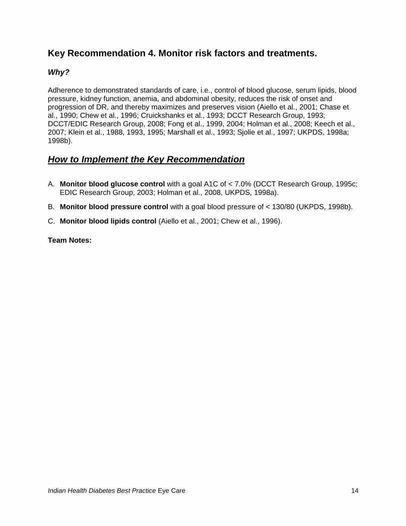
Key Recommendation 4. Monitor risk factors and treatments. Why? Adherence to demonstrated standards of care, i.e., control of blood glucose, serum lipids, blood pressure, kidney function, anemia, and abdominal obesity, reduces the risk of onset and progression of DR, and thereby maximizes and preserves vision (Aiello et al., 2001; Chase et al., 1990; Chew et al., 1996; Cruickshanks et al., 1993; DCCT Research Group, 1993; DCCT/EDIC Research Group, 2008; Fong et al., 1999, 2004; Holman et al., 2008; Keech et al., 2007; Klein et al., 1988, 1993, 1995; Marshall et al., 1993; Sjolie et al., 1997; UKPDS, 1998a; 1998b).
How to Implement the Key Recommendation
A. Monitor blood glucose control with a goal A1C of < 7.0% (DCCT Research Group, 1995c; EDIC Research Group, 2003; Holman et al., 2008, UKPDS, 1998a).
B. Monitor blood pressure control with a goal blood pressure of < 130/80 (UKPDS, 1998b).
C. Monitor blood lipids control (Aiello et al., 2001; Chew et al., 1996). Team Notes:

Indian Health Diabetes Best Practice Eye Care 15
Key Recommendation 5. Provide ophthalmology referral for all cases determined to be at risk for vision loss and possible candidates for treatment and provide vision rehabilitation for patients with vision loss. Why? Vision rehabilitation services can help maximize vision, allow for gainful employment, and help people perform daily living tasks (Goldzweig et al., 2004).
How to Implement the Key Recommendation
Refer the patient to an optometrist or ophthalmologist trained in vision rehabilitation and low-vision care. Team Notes:

Indian Health Diabetes Best Practice Eye Care 16
Additional Recommendations Working Together with Your Community and Organization In addition to implementing the Key Recommendations, programs need to work on broader community and organizational support of the goals they are trying to achieve. Community Recommendation Develop codified mechanisms for community-based DR surveillance and referral of persons with threshold DR to eye care and surgery. Why? Clinic-based eye care has been successful in achieving DR surveillance standard of care in approximately 50% of American Indian/Alaska Native people. Accessing the patient outside the eye clinic is an effective way to provide standard of care for the remaining 50%. (Wilson et al., 2005). In some settings community-based outreach using eye care providers can help close the gap; however, such programs are expensive in terms of human and logistic resources. Telemedicine provides an opportunity to provide cost-effective qualifying DR evaluations in Indian country (Whited et al., 2005).
How to Implement the Recommendation
A. Deploy a qualifying DR surveillance telemedicine program in the primary care clinic of a community-based health facility.
B. If the diabetes prevalence in the catchment area of the facilities serving a region is too small to support a dedicated telemedicine program, implement a portable or mobile program. If an existing qualified telemedicine program is located nearby, establish collaboration with the hosting facility to share DR surveillance capacity. This collaborative approach allows a virtual expansion of the catchment areas and user-based sharing of the operational costs.
C. Establish written or electronic referral mechanisms to optimize communication among clinic and community programs.
D. Evaluate outcomes regularly and modify referral mechanisms if needed. Team Notes:

Indian Health Diabetes Best Practice Eye Care 17
Organization Recommendations A health care organization that wants to improve diabetes eye care must be motivated and prepared for change throughout the entire organization. The organization’s leadership must identify diabetic eye care improvement as important work. This identification will encourage the entire organization to make changes that improve diabetic eye care. Organization Recommendation 1. Develop clearly articulated improvement goals, policies, and effective improvement strategies. Why? Improvements in the organization of the health care delivery system may improve the delivery of appropriate diabetes eye care.
A. Support a culture for quality evaluation and improvement.
How to Implement the Recommendation
B. Create incentives for improved eye care practices.
C. Provide programmatic time for continuous quality improvement.
D. Dedicate resources (human and financial).
E. Commit to improve eye health and reduce the burden of diabetic eye complications.
F. Develop specific eye care practice goals and objectives.
G. Adhere to the established and commonly accepted practice guidelines for DR (e.g., American Diabetes Association [ADA], American Academy of Ophthalmology [AAO], or American Optometric Association [AOA] guidelines) (Fong et al., 2004).
H. Incorporate validated, DR surveillance methods that include qualifying methodologies and have documented ability (Wilson et al., 2005) to improve on conventional clinical DR surveillance.
I. Support information technology with appropriate resources to document and track compliance with DR standards of care for diagnosis and treatment.
J. Commit to recruit, retain, and promote staff with the willingness and capacity to implement Best Practice programs effectively.
K. Increase capacity to conduct audits that monitor DR diagnostic and treatment practice. Team Notes:

Indian Health Diabetes Best Practice Eye Care 18
Organization Recommendation 2. Develop a diabetes team that includes diabetic eye care. Why? A team-based approach to diabetes care can improve outcomes for the patient and operational efficiencies for the health care organization.
A. Incorporate proper eye care into routine diabetes care.
How to Implement the Recommendation
B. Make diabetic eye care available on-site or through a referral mechanism that is convenient and efficient for the patient and provides feedback to the primary care provider.
C. Establish referral mechanisms if eye care services are not available in the facility and have the outcome of these extramural services available in the patient’s medical record.
Team Notes:

Indian Health Diabetes Best Practice Eye Care 19
Organization Recommendation 3. Cascade eye care objectives into annual performance plans. Why? Annual performance plans can provide a roadmap for clinical improvement goals and define accountability. Appropriate measures should be developed for clinical objectives related to these goals. Cascading these objectives into annual performance plans of the employees in the organization responsible for DR surveillance can enhance performance against improvement goals and serve as a basis for appropriate recognition.
How to Implement the Recommendation
A. Use Government Performance and Results Act (GPRA) performance goals for DR surveillance.
B. Collaborate with clinic staff involved on how changed clinical processes and provider roles will help meet these goals, and select appropriate objectives and measures to be included in their annual performance evaluation. Fully Successful should be considered the performance for meeting the GPRA goal, and Exceptional for exceeding it.
Team Notes:

Indian Health Diabetes Best Practice Eye Care 20
PART 3 Appendices, Tools, and Resources

Indian Health Diabetes Best Practice Eye Care 21
Appendix A. Supplemental Information 1. Importance of a Diabetes Eye Care Program The most common complications of diabetes that are associated with vision loss include retinopathy, cataracts, and glaucoma. These complications may lead to mild or moderate vision loss or even blindness. Diabetic retinopathy (DR) is the most common microvasculopathy associated with diabetes and the most common cause of new-onset blindness among working age adults in the United States and other industrialized countries. In almost all cases the disease is treatable, but the opportunity for preventing or reversing vision loss is associated with timely diagnosis and treatment. Approximately 15–40% of people with type 2 diabetes have retinopathy (i.e., damage to the small blood vessels in the retina) at the time diabetes is diagnosed. This early retinopathy may be due to the extended period of time these individuals had clinically significant diabetes, but remained undiagnosed and uncontrolled. The risk factors associated with the severity of retinopathy include high mean fasting blood sugar, elevated A1C level, elevated systolic blood pressure, elevated urinary albumin-to-creatinine ratio, kidney failure requiring dialysis, elevated cholesterol, dyslipidemia, abdominal obesity, anemia, and duration of diabetes.
American Indians and Alaska Natives (AI/AN) with diabetes are at increased risk of developing eye complications and vision loss due to retinopathy, cataracts, and glaucoma. The prevalence of retinopathy among AI/AN adults in Oklahoma and among Pima Indian adults with diabetes ranged from 18–49.3% (West et al., 1980). Among the Sioux Tribe in South Dakota, the prevalence of retinopathy among adults with diabetes was 45.3% (Berinstein et al., 1997). Overall, the prevalence of DR is 2.2 times greater than the non-Hispanic white U.S. population as a whole (CDC, 2011).
Consider these facts:
• The most dangerous eye diseases related to diabetes frequently cause no symptoms until they are advanced and less treatable.
• Diabetic eye disease is the leading cause of new-onset blindness in the United States for people between the ages of 20 and 74 (CDC, 1994).
• People with diabetes have 25 times the likelihood of becoming blind as compared with people without diabetes (CDC, 1991).
• The prevalence of retinopathy increases with duration of diabetes, and eventually almost all individuals with diabetes demonstrate DR. After approximately twenty years of the disease, more than 90% of patients with DM will have some degree of retinopathy (WHO, 1994).
• Diabetes causes 12,000–24,000 cases of new blindness every year. (CDC, 2007).
• People with diabetes have special issues with their eyes beyond DR, such as eye irritation due to abnormal tears and other ocular surface disease of the eye, blurred vision due to diabetes causing refractive changes in their eyes, and double vision.
Fortunately, these and other more severe eye complications of diabetes can be safely and effectively treated when identified early. Furthermore, controlled trials demonstrate that: (1) early and appropriate treatment substantially reduces the risk of vision loss due to diabetic macular edema and proliferative DR; and (2) control of blood sugar level, serum lipids, blood pressure, kidney function, anemia, and abdominal obesity reduces risk of onset and progression of DR.

Indian Health Diabetes Best Practice Eye Care 22
2. Benefits and Risks of Implementing This Best Practice
Diabetes eye care carries little or no risk beyond its cost, and successful implementation of this Best Practice will provide a favorable and cost-effective impact of the ocular and visual public health of the population served. 3. Health Questions Addressed by Best Practice
This Best Practice addresses the following questions: a. How many times a year do people with diabetes need a qualifying retinal examination? b. What is a qualifying DR examination; what are examples of qualifying and non-qualifying
retinal examinations? c. What interventions effectively protect against vision loss from DR? d. How can the effectiveness of referrals be enhanced? e. What new initiatives provide opportunities for enhancing standard of care for DR?
4. Sustaining a Diabetes Eye Care Program
It is common for new initiatives to require a certain level of maturity before care goals can be achieved. This maturational process may require more than a few years to produce the desired outcomes in a stable and self-sustaining fashion. Sustainability is a critical issue for programmatic success, and can be an elusive target. The following recommendations may be useful in fostering sustainability in newly implemented diabetic eye care programs:
• Obtain financial commitment from the organization’s administrative leadership for conventional and technology-based or other enhanced diabetes eye care programs.
• Obtain financial commitment from the organization’s administrative leadership for the maintenance and evolution of health care technology.
• Create and obtain support for the infrastructure needed to provide patients living in remote areas with technology-based solutions.
• Secure ongoing funding. Grants and other one-time funding opportunities are useful for program implementation, but ongoing revenue is needed for sustainability. Pursue reimbursement opportunities from all relevant third party payers. Newly implemented programs using innovative processes or technology may require a dialogue with the payer to provide a business case for reimbursement, including evidence supporting cost-effective clinical outcomes. Cost avoidance value should be showcased.
• Share successes with the community by making presentations to the Tribal health board and Tribal council and sharing news with Tribal media.
Indian Health Diabetes Best Practice Eye Care 23
Appendix B. Key Measures Example Remember—this is an example! Apply this process to your community using your data. Diabetes-related visual problems are increasing among our community. Our health care center and community are concerned about the risk of diabetes-related eye complications that can lead to blindness. Diabetes team takes action. Our diabetes team talked about addressing this problem and how the team could be more involved. We read the Diabetes Eye Care Best Practice
and talked about the key recommendations.
Identified sources of data. Local data included: • Audit data • RPMS data • Medical Record review • Contract Health data
o Data indicated: 70% of patients with diabetes are receiving annual eye exams. Current referral rate was unclear from data reviewed.
Selected suitable Best Practice. After thinking carefully about our goals and resources, and reviewing data, we decided the Diabetes Eye Care Best Practice was a good fit for us. We chose to work on two of the Key Recommendations: increasing annual eye examinations and making referrals to ophthalmology care for people who need retinal treatment. Identified target population. We decided to start implementing this Best Practice by including all current patients listed in diabetes registry. Identified program goals:
• To increase the number of people who have annual eye exams. • To increase the number of people who receive appropriate retinal treatment.
Identified SMART objectives based on our resources and data:
• The percent of diabetes patients that receive an annual eye exam will increase from 70% to 85% in the next twelve months.
• Eighty percent of patients with diabetes who are at risk for vision loss will be referred as possible candidates for appropriate retinal treatment in the next twelve months.
Indian Health Diabetes Best Practice Eye Care 24
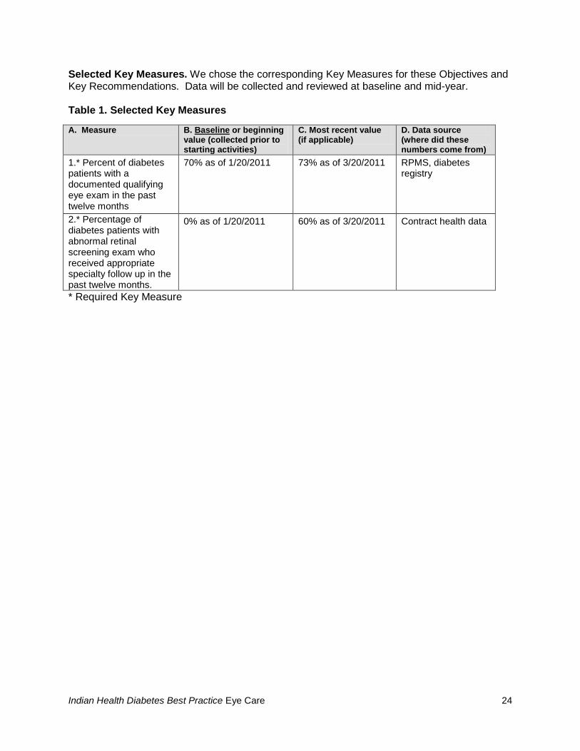
Selected Key Measures. We chose the corresponding Key Measures for these Objectives and Key Recommendations. Data will be collected and reviewed at baseline and mid-year. Table 1. Selected Key Measures
A. Measure B. Baseline or beginning value (collected prior to starting activities)
C. Most recent value (if applicable)
D. Data source (where did these numbers come from)
1.* Percent of diabetes patients with a documented qualifying eye exam in the past twelve months
70% as of 1/20/2011 73% as of 3/20/2011 RPMS, diabetes registry
2.* Percentage of diabetes patients with abnormal retinal screening exam who received appropriate specialty follow up in the past twelve months.
0% as of 1/20/2011 60% as of 3/20/2011 Contract health data
* Required Key Measure

Indian Health Diabetes Best Practice Eye Care 25
Appendix C. Improving Diabetes Eye Care Programs Example Remember—this is an example! Ask these questions in your community, thinking about your local needs, resources, and tracking systems. There are four fundamental questions to ask as you plan and implement your best practice. These questions (and sample answers) are:
1. Who is your target population? • The target population to be covered by this Best Practice is persons with type 1 or type 2
diabetes.
2. What are you trying to accomplish by implementing this Best Practice? • Improve diabetic eye care and services for people with diabetes to prevent DR. • Decrease vision loss due to DR in an efficient and cost effective manner.
3. How will you measure the impact of implementing this Best Practice?
Collect and display data on an ongoing basis. Analyze the data and use it to modify current activity and plan next steps.
4. What additional steps can you take to improve outcomes? • Provide leadership support to improve diabetic eye care. • Include DR education components in all diabetes education programs for individuals and
families.

Indian Health Diabetes Best Practice Eye Care 26
Tools and Resources Web-based Resources Agency for Healthcare Research and Quality. Diabetes Care Quality Improvement Resource Guide and Workbook. [Updated June 2009]. Online workbooks to help States assess the quality of DM care and create quality improvement strategies. http://www.ahrq.gov/qual/diabqualoc.htm American Academy of Ophthalmology. Preferred Practice Pattern: Diabetic Retinopathy [Updated September 2008; cited July 2009]. Provides detailed evidence-based recommendations for treatment by stage of retinopathy with and without macular edema and contains an extensive discussion of prevention and early detection of DR. http://one.aao.org/CE/PracticeGuidelines/PPP_Content.aspx?cid=d0c853d3-219f-487b-a524-326ab3cecd9a American Association of Diabetes Educators [Updated September 2008]. Online access to the AADE for tools, training, and other resources necessary to help patients with DM change their behavior and accomplish their DM self-management goals. http://www.diabeteseducator.org/
American Diabetes Association [Updated July 2009]. http://www.diabetes.org
American Optometric Association. Optometric Clinical Practice Guideline: Care of the Patient with Diabetes Mellitus. Reference Guide for Clinicians [Updated September 2009]. Detailed evidence-based recommendations for eye care for patients with DM, including non-retinal eye complications. http://www.aoa.org/documents/CPG-3.pdf Centers for Disease Control and Prevention (CDC). The National Diabetes Fact Sheet, 2011 [Updated January 2011]. Provides data on how many Americans have diabetes, as well as information on age, racial, and ethnic differences in diabetes, and on complications of the disease. http://www.cdc.gov/features/DiabetesFactSheet/ Diabetes National Plan for Action [Updated December 2004]. A DHHS publication that provides DM prevention, detection, and treatment information, and includes simple action steps for individuals, families, health practitioners, and others. It also provides screening tools, information on other federal DM programs, and listings of federally funded resources. http://aspe.hhs.gov/health/NDAP/NDAP04.pdf Division of Diabetes Treatment and Prevention (IHS) Creating Strong DM Programs: Plan a Trip to Success [38 pages with one page sample in appendix; Updated 2011]. A workbook (with online training course) on effective program planning and evaluation.
http://www.ihs.gov/MedicalPrograms/Diabetes/HomeDocs/Training/WebBased/Basics/Creating/Workbook.pdf
Indian Health Diabetes Best Practice Eye Care 27
Division of Diabetes Treatment and Prevention (IHS) [Updated 2011]. Information and resources to strengthen your clinical, public health, and community approach to diabetes treatment and prevention. http://www.ihs.gov/MedicalPrograms/Diabetes/index.cfm?module=trainingBasicsCreating Joslin Clinical Guidelines [Updated May 2010]. A broad range of DM management guidelines including specific reference to DR. http://www.joslin.org/managing_your_diabetes_joslin_clinical_guidelines.asp
National Eye Health Education Program (NEHEP) [Updated July 2009; cited July 2009]. An National Eye Institute site to increase awareness among healthcare professionals and the public of scientifically based health information that can be applied to preserving sight and preventing blindness. This is an online source for educational material and other resources to educate patients and the public about eye health and the importance of eye examinations. Several specialty sites are available, including one for DR. http://www.nei.nih.gov/NEHEP/
National Eye Institute Diabetes Retinopathy Homepage [Updated 2010]. An online source for patients and their families to search for general information about diabetic retinopathy. http://www.nei.nih.gov/health/diabetic/retinopathy.asp Native Diabetes Wellness Program [Updated 2010]. A CDC-sponsored program supporting AI/AN communities in developing effective strategies for DM care and prevention within their communities. http://www.cdc.gov/diabetes/projects/diabetes-wellness.htm Examples of Current Best Practice Programs Alaska Federal Health Care Access Network Christopher Patricoski, MD [email protected] This statewide telemedicine program in Alaska provides remote access to care using a broad range of store and forward and real-time modalities. This includes a portable program of DR surveillance using the IHS-JVN (Joslin Vision Network) Teleophthalmology Program. IHS/JVN (Joslin Vision Network) Teleophthalmology Program Mark Horton, OD, MD [email protected] This well-established, scalable, and centrally funded program uses validated telemedicine to increase compliance with DR surveillance standards of care in Indian country. Joslin Diabetes Center Jerry Cavallerano, OD, PhD [email protected] (617) 732-2554 This evidence-based program integrates web-based case management using patient data, patient behavioral health assessments, and education to optimize care.

Indian Health Diabetes Best Practice Eye Care 28
Phoenix Indian Medical Center Visiting Professional Program David Civic, MD [email protected] This program has a long history of successful outreach to bring specialty services to locations with documented barriers to access. Additional Contacts Contacting other people involved in diabetes eye care is important because they can help you get started. Your peers at other health care organizations can share their expertise, materials, and ideas, and can also tell you what has worked for them and what has not. This can help you avoid reinventing the wheel. Here are some tips on how to connect with others: Area Diabetes Consultants. Contact information can be viewed at: http://www.ihs.gov/MedicalPrograms/diabetes/index.cfm?module=peopleADCDirectory. Contact the Indian Health Service (IHS) Division of Diabetes Treatment and Prevention for ideas. http://www.ihs.gov/medicalprograms/diabetes/index.cfm?module=peopleDDTP Review resources from the National Diabetes Education Program (NDEP). NDEP offers materials that will help your program get started, including information specifically for American Indians and Alaska Natives. You can access these resources at the website: http://www.ndep.nih.gov/.

Indian Health Diabetes Best Practice Eye Care 29
PART 4 References

Indian Health Diabetes Best Practice Eye Care 30
References Aiello LP, Cahill MT, and Wong JS. Systemic considerations in the management of diabetic retinopathy. American Journal of Ophthalmology. 2001;132:760–66. American Academy of Ophthalmology. Preferred Practice Pattern: Diabetic Retinopathy. San Francisco: American Academy of Ophthalmology, 2003. Berinstein DM, Stahn RM, Welty TK, Leonardson GR, and Herlihy JJ. The prevalence of diabetic retinopathy and associated risk factors among Sioux Indians. Diabetes Care. 1997;20:757–79. Browning DJ, Miller KM, Aiello LP, Beck RW, Bressler NM, Davis MD, DiLoreto DA, Ferris FL, Friedman SM, Glassman AR, Glazer LC, Kollman C, Lauer AK, Marcus and DM, Starr J for the Diabetic Retinopathy Clinical Research Network. the course of response to focal/grid photocoagulation for diabetic macular edema. RETINA. 2009; Nov-Dec;29(10):1436-43. Cavallerano J and Cooppan R, editors. Care of the Patient with Diabetes Mellitus. St. Louis, MO: American Optometric Association, Revised: 2009. CDC. National Diabetes Fact Sheet, 2011. CDC. National Diabetes Fact Sheet, 2007. CDC. Prevalence of visual impairment and selected eye diseases among persons aged ≥ 50 years with and without diabetes—United States. MMWR Morbidity and Mortality Weekly Report. 2002;53:1069–71. CDC. The Prevention and Treatment of Complications of Diabetes Mellitus: A Guide for Primary Care Practitioners. Atlanta, GA: CDC, 1991. CDC (Centers for Disease Control and Prevention). Diabetes in the United States: A Strategy for Prevention. A Progress Report to the Technical Advisory Committee for Diabetes Translation and Community Control Programs. Atlanta, GA: CDC, 1994. Chase HP, Garg SK, Jackson WE, Thomas MA, Harris S, Marshall G, and Crews MJ. Blood pressure and retinopathy in type I diabetes. Ophthalmology. 1990;97(2):155–59. Chew EY, Klein ML, Ferris FL III, Remaley NA, Murphy RP, Chantry K, Hoogwerf BJ, and Miller D; ETDRS Research Group. Early Treatment Diabetic Retinopathy Study report number 22. Association of elevated serum lipid levels with retinal hard exudates in diabetic retinopathy. Archives of Ophthalmology. 1996;114:1079–84. Cruickshanks KJ, Ritter LL, Klein R, and Moss SE. The association of microalbuminuria with diabetic retinopathy. The Wisconsin Epidemiologic Study of Diabetic Retinopathy. Ophthalmology. 1993;100(6):862–67. Davis MD, Fisher MR, Gangnon RE, Barton F, Aiello LM, Chew EY, Ferris FL 3rd, and Knatterud GL. Risk factors for high-risk proliferative diabetic retinopathy and severe visual loss:

Indian Health Diabetes Best Practice Eye Care 31
Early Treatment Diabetic Retinopathy Study report number 18. Investigations in Ophthalmology and Visual Sciences. 1998;39:233–52. DCCT/EDIC (Diabetes Control and Complications Trial Research Group/Epidemiology of Diabetes Interventions and Complications Research Group). Design, implementation, and preliminary results of a long-term follow-up of the Diabetes Control and Complications Trial cohort. Diabetes Care. 1999;22(1):99–111. DCCT/EDIC. Retinopathy and nephropathy in patients with type 1 diabetes four years after a trial of intensive therapy. New England Journal of Medicine. 2000a;342(6):381–89. DCCT/EDIC. Retinopathy and nephropathy in patients with type 1 diabetes four years after a trial of intensive therapy. American Journal of Ophthalmology. 2000b;129(5);704–05. DCCT/EDIC. Prolonged effect of intensive therapy on the risk of retinopathy complications in patients with type 1 diabetes mellitus: 10 years after the DCCT. Arch Ophthalmol 2008;126:1707-15. DCCT (Diabetes Control and Complications Trial) Research Group. Early worsening of diabetic retinopathy in the diabetes control and complications trial. Archives of Ophthalmology. 1998;116(7):874–86. DCCT Research Group. The absence of a glycemic threshold for the development of long-term complications: The perspective of the Diabetes Control and Complications Trial. Diabetes. 1996a;45(10):1289–98. DCCT Trial Research Group. Influence of intensive diabetes treatment on quality-of-life outcomes in the diabetes control and complications trial. Diabetes Care. 1996b;19(3):195–203. DCCT Research Group. Lifetime benefits and costs of intensive therapy as practiced in the Diabetes Control and Complications Trial. Journal of the American Medical Association. 1996c;276:1409–15. DCCT Research Group. The effect of intensive diabetes treatment on the progression of diabetic retinopathy in insulin-dependent diabetes mellitus. Archives of Ophthalmology. 1995a;113(1):36–51. DCCT Research Group. Progression of retinopathy with intensive versus conventional treatment in the Diabetes Control and Complications Trial. Ophthalmology. 1995b;102:647–61. DCCT Research Group. The relationship of glycemic exposure (HbA1C) to the risk of development and progression of retinopathy in the Diabetes Control and Complications Trial. Diabetes. 1995c;44:968–83. DCCT Research Group. The effect of intensive treatment of diabetes on the development and progression of long-term complications in insulin dependent diabetes mellitus. New England Journal of Medicine. 1993;329:977–86. DCCT Research Group. Weight gain associated with intensive therapy in the Diabetes Control and Complications Trial. Diabetes Care. 1988;11(7)567–73.

Indian Health Diabetes Best Practice Eye Care 32
DRCR (Diabetic Retinopathy Clinical Research) Network. Manuscript Writing Committee: Elman MJ, Aiello LP, Beck RW, Bressler NM, Bressler SS, Edwards AR, Ferris FL, Friedman SM, Glassman AR, Miller KM, Scott IU, Stockdale CR, Sun JK. Randomized Trial evaluating Ranibizumab plus prompt or deferred lser or Triamcinolone plus prompt laser for diabetic macular edema. Ophthalmology 2010;117(60):1064–77. DRCR Network, Beck RW, Edwards AR, Aiello LP Bressler NM, Ferris F, Glassman AR, Hartnett E, Ip MS, Kim JE, Kollman C. Three-year follow up of a randomized trial comparing focal/grid photocoagulation and intravitreal triamcinolone for diabetic macular edema. Arch Ophthalmol 2009 Mar;127(3):245–51. Diabetic Retinopathy Study Research Group. Indications for photocoagulation treatment of diabetic retinopathy: Diabetic Retinopathy Study report number 14. International Ophthalmology Clinics. 1987;27:239–53. Diabetic Retinopathy Study Research Group. Photocoagulation treatment of proliferative diabetic retinopathy: Clinical application of Diabetic Retinopathy Study (DRS) findings, DRS report number 8. Ophthalmology. 1981a;88:583–600. Diabetic Retinopathy Study Research Group. Photocoagulation treatment of proliferative diabetic retinopathy: Relationship of adverse treatment effects to retinopathy severity, Diabetic Retinopathy Study report number 5. Developments in Ophthalmology. 1981b;2:248–61. Diabetic Retinopathy Study Research Group. Report 6: Design, methods, and baseline results. Report 7: A modification of the Airlie House classification of diabetic retinopathy. Investigative Ophthalmology and Visual Science. 1981;21:1–226. Diabetic Retinopathy Study Research Group. Four risk factors for severe visual loss in diabetic retinopathy: The third report from the Diabetic Retinopathy Study. Archives of Ophthalmology. 1979;97:654–55. Diabetic Retinopathy Study Research Group. Photocoagulation treatment of proliferative diabetic retinopathy: The second report of Diabetic Retinopathy Study findings. Ophthalmology. 1978;85:82–106. Diabetic Retinopathy Study Research Group. Preliminary report on effects of photocoagulation therapy. American Journal of Ophthalmology. 1976;81:383–96. Diabetic Retinopathy Vitrectomy Study Research Group. Early vitrectomy for severe vitreous hemorrhage in diabetic retinopathy. Four-year results of a randomized trial: Diabetic Retinopathy Study report number 5. Archives of Ophthalmology. 1990;108:958–64. Diabetic Retinopathy Vitrectomy Study Research Group. Early vitrectomy for severe proliferative diabetic retinopathy in eyes with useful vision: Clinical application of results of a randomized trial, Diabetic Retinopathy Vitrectomy Study report number 4. Ophthalmology. 1988a;95:1321–34. Diabetic Retinopathy Vitrectomy Study Research Group. Early vitrectomy for severe proliferative diabetic retinopathy in eyes with useful vision: Results of a randomized trial, Diabetic Retinopathy Vitrectomy Study report number 3. Ophthalmology. 1988b;95:1307–20.

Indian Health Diabetes Best Practice Eye Care 33
Diabetic Retinopathy Vitrectomy Study Research Group. Early vitrectomy for severe vitreous hemorrhage in diabetic retinopathy: Two-year results of a randomized trial, Diabetic Retinopathy Vitrectomy Study report number 2. Archives of Ophthalmology. 1985a;103:1644–52. Diabetic Retinopathy Vitrectomy Study Research Group. Two-year course of visual acuity in severe proliferative diabetic retinopathy with conventional management: Diabetic Retinopathy Vitrectomy Study (DRVS) report number 1. Ophthalmology. 1985b;92:492–502. Early Treatment Diabetic Retinopathy Study Group. Report number 19. Focal photocoagulation treatment of diabetic macular edema: Relationship of treatment effect to fluorescein angiographic and other retinal characteristics at baseline. Archives of Ophthalmology. 1995;113:1144–55. Early Treatment Diabetic Retinopathy Study Research Group. Aspirin effects on mortality and morbidity in patients with diabetes mellitus. ETDRS Report No. 14. Journal of the American Medical Association. 1992; 268:1292–300. Early Treatment Diabetic Retinopathy Study Research Group. Fluorescein angiographic risk factors for progression of diabetic retinopathy: ETDRS report number 13. Ophthalmology. 1991;98(5 Suppl):834–40. Early Treatment Diabetic Retinopathy Study Research Group. Fundus photographic risk factors for progression of diabetic retinopathy: ETDRS report number 12. Ophthalmology. 1991a;98(5 Suppl):823–33. Early Treatment Diabetic Retinopathy Study Research Group. Classification of diabetic retinopathy from fluorescein angiograms: ETDRS report number 11. Ophthalmology. 1991b;98(5 Suppl):807–22. Early Treatment Diabetic Retinopathy Study Research Group. Grading diabetic retinopathy from stereoscopic color fundus photographs—an extension of the modified Airlie House classification: ETDRS report number 10. Ophthalmology. 1991c;98(5 Suppl):786–806. Early Treatment Diabetic Retinopathy Study Research Group. Early photocoagulation for diabetic retinopathy: ETDRS report number 9. Ophthalmology. 1991d;98(5 Suppl):766–85. Early Treatment Diabetic Retinopathy Study Research Group. Effects of aspirin treatment on diabetic retinopathy: ETDRS report number 8. Ophthalmology. 1991e;98(5 Suppl):757–65. Early Treatment Diabetic Retinopathy Study Research Group. Early Treatment Diabetic Retinopathy Study design and baseline patient characteristics: ETDRS report number 7. Ophthalmology. 1991f;98(5 Suppl):741–56. Early Treatment Diabetic Retinopathy Study Research Group. Case reports to accompany Early Treatment Diabetic Retinopathy Study reports 3 and 4. International Ophthalmology Clinics. 1987a;27:273–333. Early Treatment Diabetic Retinopathy Study Research Group. Treatment techniques and clinical guidelines for photocoagulation of diabetic macular edema: Early Treatment Diabetic Retinopathy Study report number 2. Ophthalmology. 1987b;94:761–74.

Indian Health Diabetes Best Practice Eye Care 34
Early Treatment Diabetic Retinopathy Study Research Group. Photocoagulation for diabetic macular edema: Early Treatment Diabetic Retinopathy Study report number 4. International Ophthalmology Clinics. 1987c;27:265–72. Early Treatment Diabetic Retinopathy Study Research Group. Techniques for scatter and local photocoagulation treatment of diabetic retinopathy: Early Treatment Diabetic Retinopathy Study report number 3. International Ophthalmology Clinics. 1987d;27:254–64. Early Treatment Diabetic Retinopathy Study Research Group. Photocoagulation for diabetic macular edema: Early Treatment Diabetic Retinopathy Study report number 1. Archives of Ophthalmology. 1985;103:1796–806. Ferris FL, Chew EY, and Hoogwerf BJ. Serum lipids and diabetic retinopathy. Early Treatment Diabetic Retinopathy Study Research Group. Diabetes Care. 1996;19(11):1291–93. Ferris FL III, Podgor MJ, and Davis MD; the Diabetic Retinopathy Study Research Group. Macular edema in diabetic retinopathy study patients: Diabetic Retinopathy Study report number 12. Ophthalmology. 1987;94:754–60. Fong DS, Aiello L, Gardner TW, King GL, Blankenship G, Cavallerano JD, Ferris FL III, and Klein R. Retinopathy in diabetes. Diabetes Care. 2004;27:S84–87. Fong DS, Ferris FL 3rd, Davis MD, and Chew EY. Causes of severe visual loss in the early treatment diabetic retinopathy study: ETDRS report number 24. Early Treatment Diabetic Retinopathy Study Research Group. American Journal of Ophthalmology. 1999;127(2):137–41. Giusti C. Retinopathy in juvenile diabetes: a 10-year (1990–2000) review. Pediatric Diabetes. 2001;2(2):83–93. Goldzweig CL, Rowe S, Wenger NS, MacLean CH, and Shekelle PG. Preventing and managing visual disability in primary care: clinical applications. Journal of the American Medical Association. 2004;291(12):1497–502. Holman RR, Paul SK, Bethel MA, Matthew DR, Neil HAW. 10-year follow-up of intensive glucose control in type 2 diabetes. New England Journal of Medicine 2008;359:1577. Holman RR, Paul SK, Bethel MA, Neil HAW, Matthew DR. Long-term follow-up after tight control of blood pressure in type 2 diabetes. New England Journal of Medicine 2008;359:1565-76. Kaufman SC, Ferris FL III, et al.; the DRS Research Group. Factors associated with visual outcome after photocoagulation for diabetic retinopathy: Diabetic Retinopathy Study report number 13. Investigations in Ophthalmology and Visual Science. 1989;30:23–28. Keech A, Mitchell P, Summanen P, O’Day J, Davis T, Moffitt M, et al. Effect of fenofibrate on the need for laser treatment for diabetic retinopathy (FIELD study): a randomised controlled trial. Lancet. 2007;370:1687-97. Kinyoun JL, Martin DC, Fujimoto WY, and Leonetti DL. Ophthalmoscopy versus fundus photographs for detecting and grading diabetic retinopathy. Investigations in Ophthalmology and Visual Science. 1992;33(6):1888–93.

Indian Health Diabetes Best Practice Eye Care 35
Klein BE, Klein R, Moss SE, and Palta M. A cohort study of the relationship of diabetic retinopathy to blood pressure. Archives of Ophthalmology. 1995;113(5):601–06. Klein R. The epidemiology of diabetic retinopathy: Findings from the Wisconsin Epidemiologic Sstudy of Diabetic Retinopathy. International Ophthalmology Clinics. 1987;27(4):230–38. Klein R, Klein BE, Moss SE, and Cruickshanks KJ. The Wisconsin Epidemiological Study of Diabetic Retinopathy: XVII. The 14-year incidence and progression of diabetic retinopathy and associated risk factors in type 1 diabetes. Ophthalmology. 1998;105(10):1801–15. Klein R, Klein BE, and Moss SE. The Wisconsin Epidemiological Study of Diabetic Retinopathy: A review. Diabetes/Metabolism Reviews. 1989;5(7):559–70. Klein R, Klein BE, Moss SE, Davis MD, and DeMets DL. Glycosylated hemoglobin predicts the incidence and progression of diabetic retinopathy. Journal of the American Medical Association. 1988;260(19):2864–71. Klein R, Klein BE, Moss SE, Davis MD, and DeMets DL. The Wisconsin epidemiologic study of diabetic retinopathy. VI. Retinal photocoagulation. Ophthalmology. 1987;94(7):747–53. Klein R, Klein BEK, Moss SE, Davis MD, and DeMets DL. The Wisconsin Epidemiologic Study of Diabetic Retinopathy. II. Prevalence and risk of diabetic retinopathy when age at diagnosis is less than 30 years. Archives of Ophthalmology. 1984;102:520–26. Klein R, Klein BEK, Moss SE, Davis MD, and DeMets DL. The Wisconsin Epidemiologic Study of Diabetic Retinopathy. III. Prevalence and risk of diabetic retinopathy when age at diagnosis is 30 or more years. Archives of Ophthalmology. 1984;102:527–32. Klein R, Moss SE, and Klein BE. Is gross proteinuria a risk factor for the incidence of proliferative diabetic retinopathy? Ophthalmology. 1993;100(8):1140–46. Kohner EM, Stratton IM, Aldington SJ, Holman RR, and Mathews DR. Relationship between the severity of retinopathy and progression to photocoagulation in patients with type 2 diabetes mellitus in the UKPDS (UKPDS 52). Diabetic Medicine. 2001;18(3):178–94. Kohner EM, Aldington SJ, Stratton IM, Manley SE, Holman RR, Matthews DR, and Turner RC, for the United Kingdom Prospective Diabetes Study. Diabetic retinopathy at diagnosis of non-insulin-dependent diabetes mellitus and associated risk factors: United Kingdom Prospective Diabetes Study, 30. Archives of Ophthalmology. 1998;116:297–303. Marshall G, Garg SK, Jackson WE, Holmes DL, and Chase HP. Factors influencing the onset and progression of diabetic retinopathy in subjects with insulin-dependent diabetes mellitus. Ophthalmology. 1993;100(8):1133–39. Moss SE, Klein R, Kessler SD, and Richie KA. Comparison between ophthalmoscopy and fundus photography in determining severity of diabetic retinopathy. Ophthalmology. 1985;92(1):62–67.
Indian Health Diabetes Best Practice Eye Care 36
Moss SE, Klein R, and Klein BE. Factors associated with having eye examinations in persons with diabetes. Archives of Family Medicine. 1995;4(6):529–34. NEHEP (National Eye Health Education Program). Diabetes and Health Eyes Toolkit. 2011. http://www.nei.nih.gov/nehep/programs/ojo/toolkit.asp Office of Information Technology (OIT), IHS. National GPRA PART report, performance measure list and definitions. December 2010; V 11.0. http://www.ihs.gov/cio/crs/documents/GPRA%20PART%20Measures%20V11.pdf Orr P, Barron Y, Schein OD, Rubin GS, and West SK. Eye care utilization by older Americans: the SEE Project. Salisbury Eye Evaluation. Ophthalmology. 1999;106(5):904–09. Rand LI, Prud’homme GJ, Ederer F, and Canner PL; Diabetic Retinopathy Research Group. Factors influencing the development of visual loss in advanced diabetic retinopathy: Diabetic Retinopathy Study (DRS) Report No. 10. Investigations in Ophthalmology and Visual Science. 1985;26(7)983–91. Schachat AP, Hyman L, Leske MC, Connell AM, Hiner C, Javornik N, and Alexander J. Comparison of diabetic retinopathy detection by clinical examinations and photograph gradings. Barbados (West Indies) Eye Study Group. Archives of Ophthalmology. 1993;111(8):1064–70. Schoenfeld ER, Greene JM, Wu SY, and Leske MC. Patterns of adherence to diabetes vision care guidelines: Baseline findings from the Diabetic Retinopathy Awareness Program. Ophthalmology. 2001;108(3):563–71. Sjolie AK, Stephenson J, Aldington S, Kohner E, Janka H, Stevens L, and Fuller J. Retinopathy and vision loss in insulin-dependent diabetes in Europe. The EURODIAB IDDM Complications Study. Ophthalmology. 1997;104(2):252–60. Taylor HR, Vu HT, McCarty CA, and Keeffe JE. The need for routine eye examinations. Investigations in Ophthalmology and Visual Science. 2004;45:2539–42. Turner RC. The UK prospective diabetes study. A review. Diabetes Care. 1998;21(Suppl 3):C35–38. UKPDS (United Kingdom Prospective Diabetes Study) Group. Effect of intensive blood-glucose control with sulphonylureas or insulin compared with conventional treatment and risk of complications in patients with type 2 diabetes: UKPDS 33. Lancet. 1998a;352(9131):837–53. UKPDS Group. Tight blood pressure control and risk of macrovascular and microvascular complications in type 2 diabetes: UKPDS 38. British Medical Journal. 1998b;317(7160):703–13. West K, Edreich L, and Stober J. A detailed study of risk factors for retinopathy and nephropathy in diabetes. Diabetes. 1980;29:501–08. Wilson C, Horton M, Cavallerano J, Aiello LM. Addition of primary care–based retinal imaging technology to an existing eye care professional referral program increased the rate of surveillance and treatment of diabetic retinopathy. Diabetes. 2005; 28:318-322.
Indian Health Diabetes Best Practice Eye Care 37
Whited JD, Datta SK, Aiello LM, Aiello LP, Cavallerano JD, Conlin PR, Horton MB, Vigersky RA, Poropatich RK, Challa P, Darkins AW, Bursell SE. A modeled economic analysis of a digital teleophthalmology system as used by three federal healthcare agencies for detecting proliferative diabetic retinopathy. Telemedicine and e-Health. 2005;11:641-651. WHO (World Health Organization). Prevention of Diabetes Mellitus. Geneva: WHO, 1994.