incontinence of urine
TRANSCRIPT

Aboubakr Elnashar Benha university hospital, Egypt
ABOUBAKR ELNASHAR

DEFINITION
Involuntary escape of urine
ABOUBAKR ELNASHAR
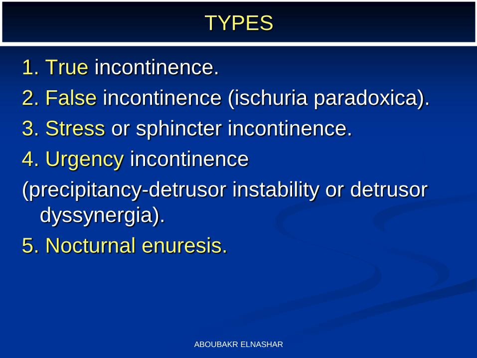
TYPES
1. True incontinence.
2. False incontinence (ischuria paradoxica).
3. Stress or sphincter incontinence.
4. Urgency incontinence
(precipitancy-detrusor instability or detrusor
dyssynergia).
5. Nocturnal enuresis.
ABOUBAKR ELNASHAR

1. True (continuous) incontinence
urine escapes continuously by day and by
night.
caused by:
(a) Urinary fistulae as vesicovaginal fistula.
(b) Ectopia vesica.
ABOUBAKR ELNASHAR

2. False incontinence
(Overflow incontinence)
Define: involuntary loss of urine following overdistension of the bladder.
usually short-term
Causes:
1. After vaginal delivery—especially if epidural anesthesia was used.
2. Other causes include diabetes, neurological diseases, severe genital prolapse, and post surgical obstruction.
ABOUBAKR ELNASHAR

4. Urgency incontinence
(precipitancy-detrusor instability or detrusor dyssynergia).
The woman feels the desire to micturate but
before she reaches the bathroom, urine passes
involuntarily.
It is due to irritability of the bladder muscle and so
the patient cannot inhibit it.
Causes:
1. Emotional disturbance,
2. Neurologic diseases, and
3. Bladder diseases as cystitis, stone or tumour. ABOUBAKR ELNASHAR

Detrusor instability (overactive bladder). It
was called detrusor dys-synergia
The bladder contracts involuntarily in
response to filling.
It commonly presents as urge incontinence
leakage of urine associated with a strong
desire to void.
ABOUBAKR ELNASHAR

Causes:
No cause is identified in more than 90% of these patients.
Advancing age is an important risk factor.
Detrusor instability caused by neurologic diseases (cerebrovascular disease, multiple sclerosis, or spinal cord injury) is called detrusor hyperreflexia.
Irritation of the bladder by inflammation (urinary tract infection) or prior pelvic surgery can also cause detrusor instability.
ABOUBAKR ELNASHAR

ABOUBAKR ELNASHAR

STRESS INCONTINENCE
)SPHINCTER INCONTINENCE-
GENUINE STRESS INCONTINENCE)
ABOUBAKR ELNASHAR

DEFINITION
involuntary escape of few drops of urine with
increased intra-abdominal pressure as during
straining, sneezing, coughing, laughing ... etc.
ABOUBAKR ELNASHAR

DEGREES OF STRESS INCONTINENCE
Grade I
Incontinence occurs only with severe stress, such as
coughing, sneezing, etc …
Grade II
Incontinence with moderate stress, such as rapid
movement or walking up and down stairs
Grade III
Incontinence with mild stress, such as standing. The
patient is continent in the supine position
ABOUBAKR ELNASHAR

PHYSIOLOGICAL ANATOMY
The bladder neck and upper third or half of the urethra are above the level of the pelvic floor.
With increased intra-abdominal pressure, the pressure is equally transmitted to the bladder and upper urethra and urine will not escape
ABOUBAKR ELNASHAR

ABOUBAKR ELNASHAR

ABOUBAKR ELNASHAR

ABOUBAKR ELNASHAR
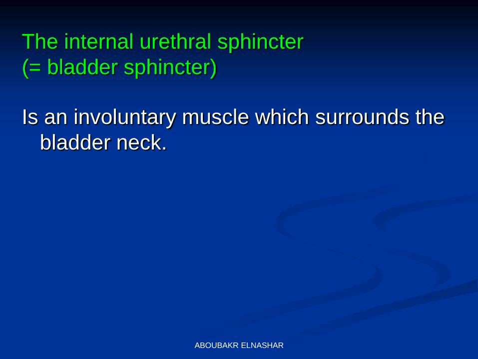
Is an involuntary muscle which surrounds the
bladder neck.
The internal urethral sphincter
(= bladder sphincter)
ABOUBAKR ELNASHAR

The external urethral sphincter
is a voluntary muscle found between the
superficial and deep perineal membranes
and surrounds the middle part of the urethra
(compessor urethrae muscle).
ABOUBAKR ELNASHAR

It empties the urethra after the act of micturition,
Interrupts the flow of urine on desire and
It acts as a secondary defensive mechanism against escape of urine.
ABOUBAKR ELNASHAR

At rest the urethra makes an angle of 90-100
degrees with the base of the urinary bladder
called the:
posterior urethrovesical angle.
The urethra also makes an angle of less
than 30 degrees with the vertical line.
ABOUBAKR ELNASHAR

During micturition the following changes occur:
1. Descent of the bladder neck with complete loss of
the posterior urethrovesical angle (angle
becomes 180 degrees).
2. Opening (funneling) of the bladder neck and
upper urethra.
3. Descent of the urethra leading to increase in the
angle between it and vertical line, so the angle
becomes more than 30 degrees.
. In stress incontinence, one or all of the above
changes occur with increased intra-abdominal
pressure. ABOUBAKR ELNASHAR

Incidence of Subtypes of Urinary Incontinence in
Women
Stress Incontinence 50%
Urge Incontinence 20%
Mixed 30%
ABOUBAKR ELNASHAR

TYPES OF STRESS INCONTINENCE
Type 1 : There is complete loss of the posterior
urethrovesical angle.
Type 2 : There is complete loss of the posterior
urethrovesical angle together with increase in the
angle between the urethra and vertical line to be
more than 30 degrees.
This type leads to severe stress incontinence
ABOUBAKR ELNASHAR

AETIOLOGY
It is due to either :
Weakness of the internal urethral sphincter
or
Descent of bladder neck below the level of
the pelvic floor.
ABOUBAKR ELNASHAR

1. Congenital weakness of the internal
urethral sphincter, seen in the young
nullipara.
2. Congenital defects as:
1. Epispadias,
2. Short urethra (less than 1 cm),
3. Wide bladder neck, and
4. Separation of symphysis pubis.
ABOUBAKR ELNASHAR

ABOUBAKR ELNASHAR

3. Trauma to the region of the bladder neck
due to vaginal delivery or operation.
The incidence of stress incontinence
increases with parity due to repeated birth
trauma.
In fact vaginal delivery is the commonest cause of stress incontinence.
ABOUBAKR ELNASHAR

4. Menopause: Lack of oestrogen leads to
atrophy of bladder neck supports.
5.Pregnancy and continuous administration
of oestrogen-progestogen preparation to
induce psuedopregnancy state to treat
endometriosis.
The hormonal imbalance with increased
progesterone weakens the internal urethral
sphincter.
ABOUBAKR ELNASHAR

6. Genital prolapse:
If the bladder neck descends below the level
of the pelvic floor, the increased intra-
abdominal pressure will be transmitted to
the bladder and not to the upper urethra
leading to escape of urine.
7. Organic nervous diseases
as disseminated sclerosis.
ABOUBAKR ELNASHAR

Pathophysiology of Stress
Incontinence
The basic pathology is urethral incompetence.
This can be either due to:
A) Urethral hypermobility
(80 - 90% of patients)
B) Intrinsic Sphincter Dysfunction
(10 - 20% of patients)
ABOUBAKR ELNASHAR

A) Urethral hypermobility
(80 - 90% of patients)
This results from loss of the normal pelvic support mechanism of the bladder and urethra
due to:
1. Trauma and stretching of vaginal delivery
2. Hysterectomy
3. Hormonal changes ( Menopause)
4. Pelvic denervation
5. Congenital weakness
ABOUBAKR ELNASHAR

As the bladder neck support is weakened,
the increase in intra-abdominal pressure is
no longer transmitted equally to the bladder
outlet, and therefore instantaneous leakage
occurs.
A) Urethral hypermobility
(80 - 90% of patients)
ABOUBAKR ELNASHAR

B) Intrinsic Sphincter Dysfunction
(10 - 20% of patients)
This results from damage to the sphincter due to:
1. Multiple prior operations
2. Trauma
3. Radiation
4. Neurogenic disorders including Diabetes Mellitus
5. Atrophic changes: lack of estrogen.
ABOUBAKR ELNASHAR

ABOUBAKR ELNASHAR

1. A detailed history differentiates between the different
types of incontinence.
2. Stress incontinence and detrusor instability
frequently occur together.
3. Gradual onset after menopause suggests oestrogen
deficiency.
4. History of vaginal repair or operation in the region of
the bladder neck and history of any neurologic
disease.
ABOUBAKR ELNASHAR

ABOUBAKR ELNASHAR

1. Stress Test
The bladder must be moderately full.
The patient in the lithotomy position, the two labia are
separated, and the patient is asked to cough.
If urine escapes, the patient is incontinent.
If no urine escapes, the test is repeated while the
index and middle fingers in the vagina press on the
perineum to abolish reflex contraction of the levator
ani muscles during straining.
If still no urine escapes, the test is repeated while the
patient is standing with the legs separated.
ABOUBAKR ELNASHAR

2. Bonney test
It is indicated in case of a positive stress test
associated with a cystocele.
To know if incontinence is due to descent of
bladder neck or weakness of the sphincter.
The index and middle fingers are placed on
both sides of the urethra to elevate the bladder
neck upwards.
If no urine escapes on stress it means that the
incontinence is due to descent of the bladder
neck, but if urine still escapes it means
weakness of the sphincter. ABOUBAKR ELNASHAR

Indicated in case of a negative stress test
associated with a large cystocele to diagnose
hidden stress incontinence.
The cystocele is reduced, the cervix is grasped
with a volsellum and pushed upward, then the
patient is asked to cough.
If urine escapes, it indicates that the patient
was continent because of kinking of the urethra.
3. Yousef Test
ABOUBAKR ELNASHAR

4. Examination of Urine
Urinalysis, culture and sensitivity to
exclude cystitis.
ABOUBAKR ELNASHAR

To exclude lesions in the urethra and bladder.
The bladder neck is examined.
It should close in response to straining.
However, it opens in case of stress
incontinence.
5. Cystourethroscopy
ABOUBAKR ELNASHAR

A radio-opaque dye is injected by a catheter
into the bladder.
On straining, the lateral view will show absence
of the posterior urethrovesical angle in more
than 90% of cases.
Funneling of the bladder neck in the antero-
posterior view may be seen in some cases.
The procedure is recorded on video tape
(video Cystourethrography) to facilitate
diagnosis and for education purposes.
6. Cystourethrography
ABOUBAKR ELNASHAR
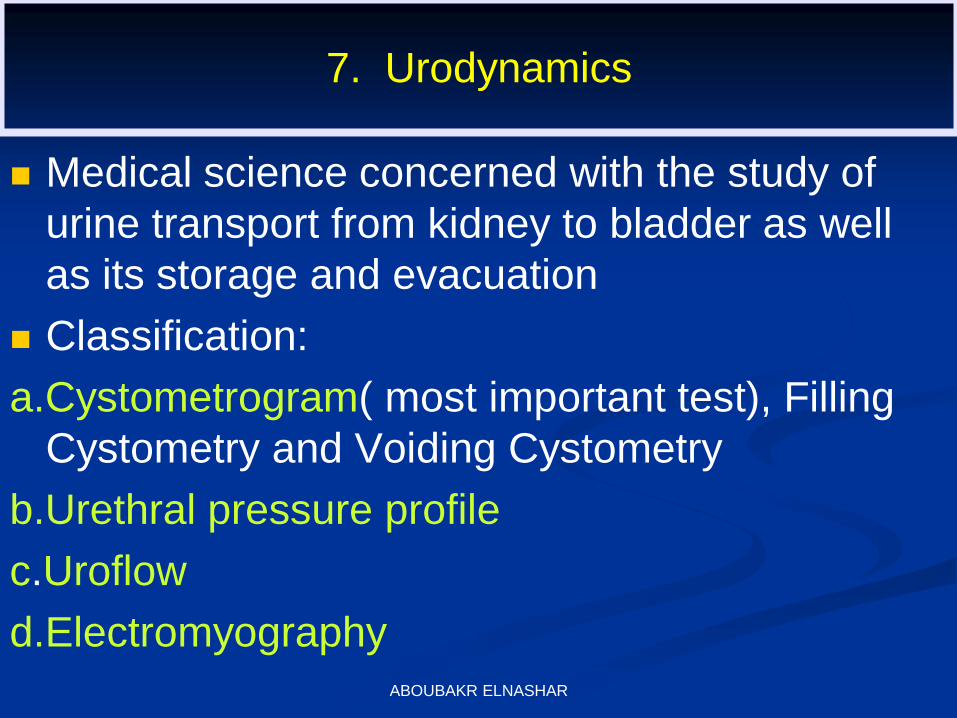
7. Urodynamics
Medical science concerned with the study of
urine transport from kidney to bladder as well
as its storage and evacuation
Classification:
a.Cystometrogram( most important test), Filling
Cystometry and Voiding Cystometry
b.Urethral pressure profile
c.Uroflow
d.Electromyography ABOUBAKR ELNASHAR
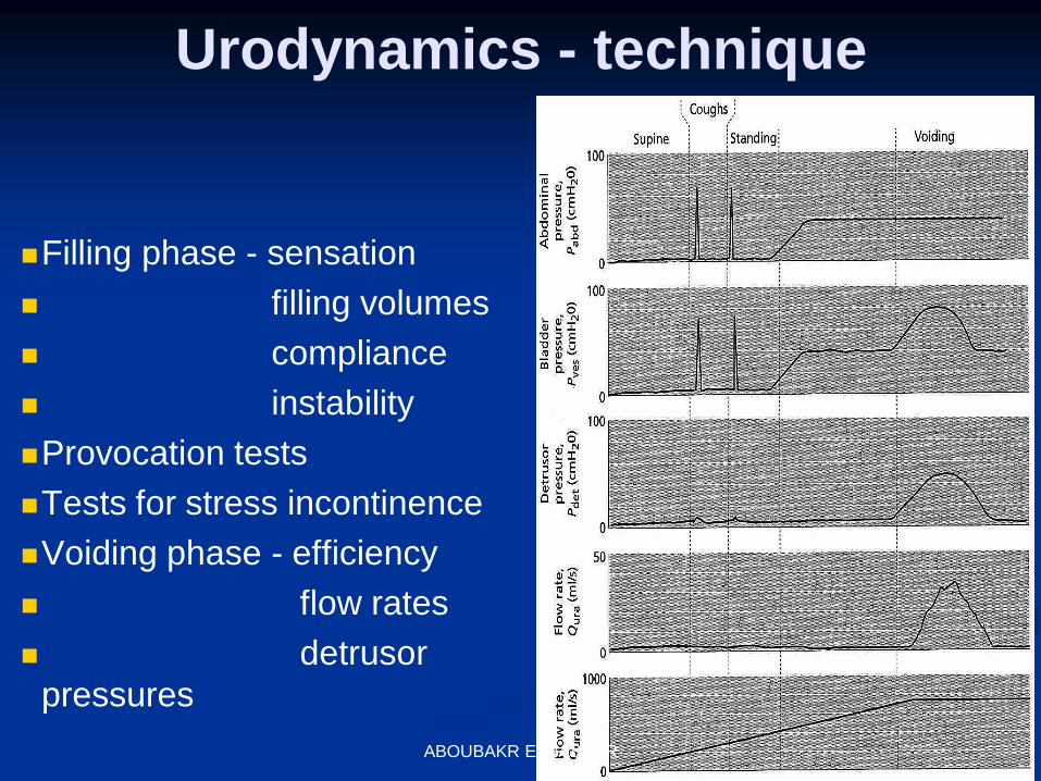
Urodynamics - technique
Filling phase - sensation
filling volumes
compliance
instability
Provocation tests
Tests for stress incontinence
Voiding phase - efficiency
flow rates
detrusor
pressures
ABOUBAKR ELNASHAR

Urodynamics
Lawrence Techniques in Urology 1999 ABOUBAKR ELNASHAR
To measure the intravesical pressure while the
bladder is filled with sterile water or carbon
dioxide gas.
It diagnoses stress incontinence and detrusor
instability.
The most important test.
a. Cystometrogram
ABOUBAKR ELNASHAR

Involves filling the bladder to measure
volume-pressure relationships.
As the bladder is filled to its normal capacity
of 300-500 ml, the pressure inside the
bladder should remain low.
The patient usually experiences the first
urge to void at 150-200 ml.
ABOUBAKR ELNASHAR

Patients with DI often have reduced bladder
capacity (< 300 ml) and demonstrate urinary
incontinence that is associated with involuntary
bladder contractions (pressure increase above
baseline)
ABOUBAKR ELNASHAR

In patients with GSI, incontinence is demonstrated when
the patients coughs or strains (e.g., Valsalva maneuver).
The intravesical pressure at which leakage is noted (leak
point pressure) is generally < 60 cm of water pressure if intrinsic sphincter deficiency is present.
ABOUBAKR ELNASHAR

To maintain continence, the urethral pressure
(100-120 cm water) must be higher than the
intravesical pressure (0-20 cm water).
A special catheter; is used which measures the
intravesical and intra-urethral pressure.
b. Measurement of Urethral Pressure
ABOUBAKR ELNASHAR

The urethral closing pressure
Equals the intraurethral pressure minus the intravesical pressure (normally 90-100 cm water).
The length of the urethra along which urethral pressure exceeds bladder pressure is termed functional length of the urethra which is 3-4 cm.
In stress incontinence the urethral closing pressure is reduced.
ABOUBAKR ELNASHAR

It records the rate of urine flow through the
urethra when the patient is asked to void
spontaneously while sitting on uroflow chair.
It is used to evaluate patients with stress
incontinence before surgery to exclude difficulty
in voiding which may be increased by bladder
neck surgery.
C. UROFLOWMETRY
ABOUBAKR ELNASHAR

UROFLOWMETRY
Lucus et al
Incontinence
ABOUBAKR ELNASHAR

The normal female voids by the rule of
"20"
that is urine is passed at a rate of 20
ml/second and the bladder is emptied in
less than 20 seconds.
ABOUBAKR ELNASHAR

8. The Cotton-Tip Applicator (Q-Tip) Test
A sterile applicator with a small piece of cotton
at its tip is introduced to reach the bladder neck.
The angle between the applicator and the
horizontal is measured.
The patient then strains maximally using the
Valsalva manoeuvre.
This causes descent of the bladder neck and
upward movement of the applicator producing a
new angle with the horizontal.
ABOUBAKR ELNASHAR

In normal patients the increase in the angle is less
than 30 degrees.
In stress incontinence the change is more than 30
degrees indicating poor support and abnormal
descent of bladder neck
The test is positive in more than 90% of cases with
stress incontinence.
ABOUBAKR ELNASHAR

Stress incontinence occurs if the length is
less than 1 cm.
9. Measurement of Urethral Length
ABOUBAKR ELNASHAR

It gives information about funneling of the
bladder neck, both at rest and with
Valsalva manoeuvre.
10. Sonographic
ABOUBAKR ELNASHAR

Three-dimension transvaginal ultrasound
The continent women have a thick wall internal
urethral sphincter which extends from the bladder
neck and along 60-80% of the whole urethra.
In stress incontinence, the sphincter is torn as proved
by appearance of areas of echolucency.
ABOUBAKR ELNASHAR

When rupture affects the upper part of the sphincter,
the urethra appears "funnel-shaped".
When damage affects the lower part, the urethra
appears "vase-shaped".
When rupture affects the whole length of the
sphincter, the urethra appears short and irregular.
ABOUBAKR ELNASHAR

laboratory tests helpful in evaluating incontinence?
Postvoid residual is an easy initial test to obtain.
After the patient voids, there should be less than 50 ml
of urine in the bladder.
Postvoid residual is measured by ultrasound or
catheterizing the patient in the office.
A patient with an elevated Postvoid residual (repeat
measurements greater than 100-200 ml) may have an
underlying neurologic disorder.
ABOUBAKR ELNASHAR

Catheterization also provides a good opportunity to
obtain urine for analysis and culture.
Urinalysis and urine culture help to diagnose urinary
tract infection.
Blood work is required only if compromised renal
function, diabetes, syphilis, or other systemic diseases
are suspected.
ABOUBAKR ELNASHAR

Differentiating between GSI and DI
Cystometrogram
Cystoscopy :
should be performed especially in patients with: irritative bladder symptoms such as
urgency, frequency, and hematuria
To rule out:
1. inflammation,
2. tumors, or
3. anatomic deformities
ABOUBAKR ELNASHAR

ABOUBAKR ELNASHAR

I. Prophylactic Treatment
1. During labour, the bladder should be kept empty.
2. Episiotomy is performed if necessary.
3. Physiotherapy.
Pelvic floor exercises are started after delivery.
These include repeated stoppage of the urinary
stream during micturition and repeated contractions
of the pelvic floor muscles.
ABOUBAKR ELNASHAR

Indications:
1.Mild stress incontinence.
2.The patient not completed her family as
vaginal delivery may damage a bladder neck
repair
3.Patient is unfit for surgery or refuses surgery.
4.When stress incontinence is combined with
detrusor instability.
The latter should be treated at first before
surgery is done for stress incontinence.
II. Conservative (non-surgical)
Treatment
ABOUBAKR ELNASHAR

Conservative treatment cures or
improves 50% of cases and include: 1. Physiotherapy: Kegl perineometer may be used.
2. Faradic current stimulation of the levator ani
muscles to improve their tone.
3. Vaginal cones:
A set consists of 5 or 9 cones.
Weight ranges from 20 to 100 grams.
Patient inserts the cone in the vagina and keeps it for
15 minutes twice daily.
If this succeeds she inserts the next cone.
This improves the tone of the pelvic floor muscles.
ABOUBAKR ELNASHAR

4.Oestrogen therapy for menopausal patients:
It causes thickening of the urethral mucosa and engorgement of the underlying blood vessels thus increasing the urethral pressure and resistance.
Oestrogen is given orally or as vaginal cream.
5. Alpha-adrenergic stimulants: which stimulate contraction of the internal urethral sphincter, e.g. ephedrine.
6.Large vaginal diaphragms, Hodge pessary to elevate ' and support the bladder neck.
ABOUBAKR ELNASHAR

7. Reduction of weight in obese patients to reduce intra-abdominal pressure.
8. Stop caffeine (to avoid diuresis) and smoking (to avoid coughing)
9. Injection of Teflon or bovine collagen in the submucosal layer in the region of the bladder neck.
This leads to narrowing of the urethral lumen and increased urethral resistance.
ABOUBAKR ELNASHAR

III. Surgical Treatment
It is the primary treatment of stress
incontinence.
The operation is done vaginally,
abdominally, or abdominovaginally.
Almost 200 operations have been
described.
ABOUBAKR ELNASHAR

1. Urehroplasty (Kelly,Kennedy,etc….)
2. Urethropexy (Retropubic urethropexy e.g.
Marchall-Marchitti-Krantz, etc….)
3. Colposuspension ( Burch operation, Preyera ,
etc….)
4. Urethral slings (Aldridge operation, etc…..)
5. Tension free Vaginal Tape (TVT)
ABOUBAKR ELNASHAR
ABOUBAKR ELNASHAR

It consists of repair of cystocele and/or urethrocele.
Vertical mattress sutures are then placed to plicate the
whole urethra and bladder neck.
This gives support to the urethra and restores the
normal posterior urethrovesical angle.
Operation is done for mild and moderate cases of
stress incontinence.
Long term success rate is 55-65%.
1. Kelly operation 1914
ABOUBAKR ELNASHAR

2. El-Hemaly urethrorrhaphy operation
A vertical incision is made in the anterior vaginal
wall.
The torn edges of the internal urethral sphincter
are sutured together to restore its integrity.
The repair restores the normal urethrovesical
angles seen in continent women.
ABOUBAKR ELNASHAR

3. Vaginal tape operation
a. TVT (1996)
The tape is made of prolene and has a curved needle
at each end.
Operation is done using local infiltration anaesthesia.
Two small transverse incisions 5 cm apart are made
in the suprapubic area.
A vertical incision is made in the anterior vaginal wall.
The needles of the tape are passed upward behind
the pubic bone and brought out through the
suprapubic incisions.
The tape is made to surround the mid-urethra.
ABOUBAKR ELNASHAR

ABOUBAKR ELNASHAR

The cystoscope is used by the assistant to make sure
that the bladder is not pierced by the needle.
The tape is adjusted by pulling on its ends, and
continence is confirmed by asking the patient to
cough.
The ends of the tape are cut off and left free and not
fixed to the tissues,
Finally the vaginal and suprapubic incisions are
closed.
When stress occurs ,the recti will contract and pull on
the tape to support the urethra and prevent escape of
urine ABOUBAKR ELNASHAR

Simple, easy, relatively safe with short recovery
& little pain.
Cure is 86% & improvement is 11%.
Operation takes 20-30 minutes.
Complications: urine retention, parautrethral &
paravesical hemorrhage, infection , bladder
&bowel injury.
ABOUBAKR ELNASHAR

b. ObTape transobturator sling
September 10, 2003 new surgical implant for
treatment of stress incontinence in women has been
approved by the FDA.
It was pioneered in 1999 by Emmanuel Delorme in
France.
Soon became popular because the procedure is
perceived to be simpler and faster, with less risk of
complications, than alternative procedures.
In the last 2 years over 11,000 women have been
successfully treated for stress incontinence with
transobturator sling.
ABOUBAKR ELNASHAR

ABOUBAKR ELNASHAR

The stitches are placed in the fascia on each side of
the bladder neck and upper half of the urethra and are
attached to the periosteum on the back of the
symphysis pubis.
This restores the normal intra-abdominal position of
the urethra.
Main complication is osteitis pubis (0.5-5%).
Nonabsorpable (as mersilene) or delayed absorbable
sutures (as Vicryl or Dexon) are used.
1. Mashall-Marchetti-Krantz 1949
ABOUBAKR ELNASHAR

2. Burch Operation 1968
Burch colposuspension is the operation of choice.
It corrects both stress incontinence and cystocele.
The stitches are placed in the fascia on each side of the bladder neck and the base of the bladder and are attached to the iliopectineal ligaments (Cooper Ligaments),
(The pectineal part of the inguinal ligament)
Nonabsorpable or delayed absorbable sutures are used.
Operation can be done through the laparoscope.
ABOUBAKR ELNASHAR

The success rate of the above abdominal
operations is 80-90%
ABOUBAKR ELNASHAR

ABOUBAKR ELNASHAR

1. Urethral Slings
In this condition, there is damage or paralysis of
the sphincteric unit which could even be in a
normal position.
The goal of surgery for Intrinsic Dysfunction is
coaptation, support, and compression of the
damaged sphincteric unit.
Simple suspension of the bladder neck is
unlikely to correct the problem.
Urethral Sling Procedures is the best to achieve
the goal. ABOUBAKR ELNASHAR
A sling is put around the urethra at the
bladder neck and either fixed around the
rectus muscles or to the pubic bone.
- The sling could be taken from the rectus
sheath "Aldridge operation".
- A nylon sling may be used "Pereyra
operation".
ABOUBAKR ELNASHAR

ABOUBAKR ELNASHAR

An incision is made in the vaginal wall to
expose the bladder neck.
A nylon suture is placed in the fascia on each
side of the bladder neck.
The two sutures are passed upward behind the
symphysis pubis and are attached to the
anterior rectus sheath.
The cystoscope is used to be sure that the
needle does not pass through the bladder
(endoscopic needle bladder neck suspension).
2. Needle Bladder Neck Suspension
Operations
ABOUBAKR ELNASHAR

An example is Stamey operation in which two Dacron
tubes (1 cm) are used to give support to the bladder neck and to
avoid the sutures cutting through the
tissues.
ABOUBAKR ELNASHAR

ABOUBAKR ELNASHAR

Indicated when surgery fails to correct stress
incontinence.
The device consists of a cuff which is placed
around the bladder neck.
A balloon reservoir, containing fluid is placed in
the peritoneal cavity or under the anterior rectus
sheath, and a small pump is situated in one
labium major.
D. Artificial Urinary Sphincter
ABOUBAKR ELNASHAR

Under normal conditions the cuff is full with
fluid thus closing the bladder neck.
When voiding is desired the pump is
pressed to force the fluid in the cuff to go
back into the balloon reservoir so that
voiding can occur.
The cuff then gradually refills over the next
few minutes.
ABOUBAKR ELNASHAR

ABOUBAKR ELNASHAR

The patient complains of urgency incontinence,
frequency and nocturia.
Involuntary loss of urine also occurs when the women
sits for a long time and stands to go to the bathroom.
She may pass urine with the sight or sound of water
ABOUBAKR ELNASHAR

Women typically complain of urgency
followed by a large loss of urine.
Cystometry confirms the diagnosis.
Involuntary detrusor contractions of 15 cm
of water or more occur during filling of the
bladder.
ABOUBAKR ELNASHAR

Detrusor instability
Lucus et al Incontinence 2000 ABOUBAKR ELNASHAR
TREATMENT of (DI)
1. Bladder retraining drills:
The patient is asked to pass urine every hour
during daytime and to increase the interval by
15 minutes every week until she passes urine
every 2-3 hours.
ABOUBAKR ELNASHAR

2. Drugs :
Which inhibit the contractions of detrusor muscle as
anticholinergic drugs, tricyclic antidepressants, and
ephedrine.
Ephedrine stimulates alpha-adrenergic receptors in the
internal urethral sphincter leading to contraction, and
stimulates beta-adrenergic receptors in the detrusor muscle leading to relaxation.
ABOUBAKR ELNASHAR

ABOUBAKR ELNASHAR