in-vivo and in-vitro toxicity eman i. *1abdel-gawad and ... · firstly, the characters of...
TRANSCRIPT

Journal of American Science, 2011;7(1) http://www.americanscience.org
In-vivo and in-vitro Prediction of the Efficiency of Nano-Synthesized Material in Removal of Lead Nitrate
Toxicity
Eman I. Abdel-Gawad*1 and Sameh A. Awwad2
1Radioisotopes Department, Atomic Energy Authority, 2Egyptian Army Forces, Egypt
Abstract: Due to large grain sizes, the biological properties of the conventional hydroxyapatite (HAp) is limited to a great extent. Progresses in nanotechnological approaches now allow the fabrication of nano-HAp. In this study, firstly, the characters of nano-hydroxyapatite gel was described and the interaction performance of the formed gel with lead nitrate Pb(NO3)2 in vitro was identified. Then, the biological efficiency of nano-HAp gel against Pb(NO3)2 toxicity in vivo was introduced. A polymeric matrix route was selected to synthesis nano- composite hydroxyapatite gel. The formed gel characterized using FTIR, XRD, SEM, TEM. Various volumes of the produced nano-HAp gel (10, 20, 30, 40, 50 and 60 µl) was adding to 4 ml of ECS solution. The clear supernatant was separated and analyzed by ICP-MS. The results showed a successful removal of lead ions by formed gel. A single dose of intravenous nano-hydroxyapatite at a level of 150 and 300 mg/kg b.w. was injected to male rats following intraperitoneal 93mg/kg b.w. (LD50) of lead nitrate Pb(NO3)2. The results revealed that nano- HAp composite had the ability to alleviate lead nitrate toxicity, to a great extent, in serum antioxidant status, liver and kidney function as well as corticosterone and calcium levels but phosphorus value was not affected among the all treated groups. However, most successful results were attributed to the treatment with high dose of formed nano-HAp particularly after 48 h more than the treatment with low dose. Histopathological observations confirmed the biochemical results, since nano-HAp into rats evident the recovery of lead nitrate cytotoxicity in liver and kidney cells. [Eman I. Abdel-Gawad*1 and Sameh A. Awwad. In-vivo and in-vitro Prediction of the Efficiency of Nano-Synthesized Material in Removal of Lead Nitrate Toxicity. Journal of American Science 2011;7(1):105-119]. (ISSN: 1545-1003). http://www.americanscience.org. Keywords: Nano-HAp, lead nitrate, antioxidant status, liver enzymes, kidney functions , corticosterone 1. Introduction:
The advent of nanoscience gives human a new perspective in biology; all biological systems have their most basic structures, properties and functions defined at the nanoscale from their first level of organization and are governed by the molecular behavior at nanometer scales (Christenson et al. 2007). These mysterious structures and functions of the nanoscale living organelles (e.g., ribosome) and non-living nanostructures (e.g., tooth) formed within living organisms have always attracted attention and fascination. With the development of molecular and nanoscale technique and engineering, the methodologies provide tools and platforms for the creation and manipulation of complex structures and functions on the scale of nanometers (Xu et al. 2007). At the same time, biology also serves as the source of inspiration for creating new devices and systems integrated from the nanoscale. Thus, the fabricated nano biomaterials with biomimetic structures and functions are expected to bear high bioactivities and unexpected biological effects, and ultimately can well serve as biomedicinal devices for human disease treatment. Many commercial substitute materials now have been developed, including natural and synthetic polymers, human bones, synthetic ceramics and composites (LeGeros,2008) especially hydroxyapatite
(Descamps et al. 2008). The reason for the development of the HAp based biomaterials is their similarity in composition to the bone mineral, and has good biocompatibility, bioactivity, osteoconductivity (Bauer et al. 2008 and Abel-Gawad & Awwad, 2010). Because lead has a major health hazard throughout the world due to its disruption effect on biological systems (Sharma et al. 2010) and considering as carcinogenic agent (Silbergeld, 2003), it is important to explore the possibility of minimizing its effects on the body. The currently approved treatment for lead intoxication is to give chelating agents but, these chelators are potentially toxic (Flora et al. 2007) and often fail to remove lead from all body tissues (Sharma et al. 2010). For this, several methods have been investigated for the removal of lead ions using calcium phosphates specially hydroxyapatite structure (Ciobanu et al. 2000 and Xu et al. 2007). Great researches on HAp have been carried out to understand the immobilization mechanism of heavy metals from aqueous solutions and to evaluate its usage for environmental remediation (Mavropoulos et al. 2002). Modification of the HAp surface is a technique available for developing catalysts and adsorbents with novel functions. However, HAp is
http://www.americanscience.org [email protected] 105

Journal of American Science, 2011;7(1) http://www.americanscience.org
usually provided in powder or calcined pellets form, which might be a disadvantage to recover this material after removing heavy metal ions from wastewater (Janga et al. 2008). In light of the long history of therapeutic application of HAp, it hypothesized that are of this compound may be of interest in the management of heavy metal-induced disease. On the other hand, progresses in nanotechnological approaches now allow the fabrication of nanocrystalline HAp and could conceive the biological properties of nano-HAp are able to be improved evidently. Hence, in the present study, nano- composite HAp gel was prepared and characterized, then it was investigated its efficiency in removal of lead ions from aqueous solution and finally evaluated the biological effects of intravenous nano-HAp against lead toxicity in male rats including biochemical and pathological investigations. 2. Materials and methods Chemicals: The chemicals used for nano-HAp preparation were calcium nitrate tetrahydrate (Ca (NO3)2.4H2O, Mwt. 236.15 g/mole, Merk, Germany), diammonium hydrogen ortho phosphate anhydrous ((NH4)2HPO4, 132.06g/mole, S.D. Fine Chem. Ltd. Mumbai, India), poly vinyl alcohol (PVAL) (Mwt. � 160000 g/mole), and ammonium hydroxide (NH4OH, Mwt. 35.5g/mole, May & Baker, England). Environmental calibration standard (ECS) containing 300 ppb of Pb element, Agilent, USA). The range of the elements to be tested was within a concentration of ppb depending on their natural environmental presence. All chemicals were used in the experimental work without further purification. Also, commercial kits were used for biochemical analyses as mentioned below in materials and methods section. In vitro experimental design: Nano-HAp composite gel was synthesis according to technique of Abdelfattah et al., (2006) and characterized using FTIR, TEM and XRD to confirm the synthesis of hydroxyapatite structure. The formed nano composite HAp gel was used for removal of lead ions from environmental calibration standard solution ECS contained 300 ppb of lead ions. The effect of nano-HAp gel amount on the removal of lead ions from ECS solution was followed by adding various volumes of the produced nano-HAp gel (10, 20, 30, 40, 50 and 60 µl) to 4 ml of ECS solution. The clear supernatant was separated and analyzed by ICP-MS. The effect of time was studied by preparing seven vials, each vial contained 4 ml of ECS solution then the optimum amount of the gel (50 µl) was added to each vial. The supernatant was
decanted at different times (10, 15, 20, 25, 30, 40 45 and 50 min) and analyzed by ICP-MS. The supernatant solution was decanted and analyzed by ICP-MS at constant time interval and the results were recorded. The formed nano gel ion mixture were analyzed using TEM and FTIR. In vivo experimental design: Animals and treatments: Adult healthy male rats (140-160 g) were allowed to acclimate at the animal facilities for two weeks before use. They were housed under standard environmental conditions with free access food and water throughout the experiment. The rats were divided into four groups, control and three treated groups (each of 15 rats). Each of the treated groups received intravenous (through tail vein) single dose (93 mg/kg b.w., LD50) of Pb(NO3)2 according to (ATSDR, 1993). Following three hours, the third and fourth groups received single dose of intravenous nano hydeoxyapatite (HAp) at a dose of 150 and 300 mg/kg b.w., respectively (Abel-Gawad and Awwad, 2010). Saline solution was administered to control group in the same manner as in the treated groups as a solvent for lead nitrate and nano-HAp. Blood samples were collected from orbital venous plexus at a time intervals of 3 and 48 hours for all biochemical parameters except calcium and phosphorus were estimated at times intervals 3, 24 and 48 h after nano-HAp injection. The blood centrifuged at 3000 r.p.m. and obtained serum was stored at -20ºC till analyses through a week. The abdomen of all rats were dissected immediately to remove livers and kidneys and stored in 10% formalin solution for histopathological examination. Biochemical investigation: Obtained serum was analyzed for the following biochemical parameters: superoxide dismutase (SOD), catalase (CAT) and glutathion peroxidase (GPx) activity were measured using commercial kits purchased from (BioVesion Research Products, USA) according to McCord & Fridovich (1988), Chelikani et al. (2004) and Ran et al. (2007) technique, respectively. Malondialdehyde (MDA) was determined (Northwest, Life Science Specialties,LLC, USA) by using Nair et al. (2008) method. Arginase (Gentaur Comp., Europe), gamma glutamyle transferase (GGT) (Shenzhen Mindray Bio-Medical Electronics Co.Ltd, Germany), aspartate aminotransferase (AST) and alanine aminotransferase (ALT) (Right Choice Diagnostics, Ltd, Germany) were analyzed according to the methods of Crombez & Cederbaum (2005), Thomas, (1998) and Young (1990), respectively. Also, kidney function was estimated through the evaluation of urea
http://www.americanscience.org [email protected] 106

Journal of American Science, 2011;7(1) http://www.americanscience.org
and creatinine levels in serum by using (Vitro Scient. Diagnostics, Egypt) according to the method of Tietz, (1990) and Tegar-Nelsson (1961). Calcium and phosphorus levels were determined using kits purchased from (Biotron Diagnostics, INC., USA) according to Woo and Cannon (1984) and Young (1990) techniques, respectively. Corticoesterone value was estimated using solid phase RIA technique according to Saino et al. (1988). Histopathological investigation: Small pieces of liver 3-5 mm thick were fixed in 10% formalin solution for 24 h. They were washed in running water for 24 h. They were then dehydrated by passing through ascending grades of alcohol: 50, 70, 90 and 100% for 2-3 days following which they were cleared in benzene to remove alcohol till the tissues became more or less transparent. They were later passed through three cups containing molten paraffin at 58C and finally embedded in a cubical block of paraffin made by the L moulds. From the embedded samples, sections of 6 microns thick were cut using the microtone and fixed on a slide by Mayer's albumen. The sections were stained with hematoxylin to cover the section and kept for 6 min. Excess stain was removed with tap water. Eosin was added to cover the stem for 2 min. Excess eosin was poured away and removed with tap water. This was covered with a cover glass to avoid air bubbles, and viewed with the use of both low and high power microscope (Banchroft et al., 1996). Statistical Analysis: All values were expressed as mean ± SE. Statistical analysis was performed with two way analysis of variance (ANOVA) followed by Duncan's
t ' test. P values < 0.05 were considered to be statistically significant. 3. Results In vitro experimental results: Characterization of the formed gel: The XRD analysis of the dried powder at 100°C for 24 h identified the presence of nano-HAp crystal structure. The characteristic XRD pattern of nano-HAp (main peak d=2.81 Å and secondary peaks at d=2.78 Å, d= 2.72 Å) still exist (Fig. 1) with a small shift due to the presence of lead nitrate. It is to be noticed that the presence of lead nitrate didn’t change the crystal structure of nano-HAp. The decrease of the peaks intensity is mainly due to reduced content of the nano-HAp concentration, and confirmed with IR spectrum which proved the formation of nano-HAp structure with no any other calcium phosphate structure. The microstructure analysis by SEM/EDAX of 0.1 M lead nitrate with nano-HAp gel dried at 100o for 24 h was shown a presence of lead and nitrogen while the Ca/P ratio is approved the presence of nano-HAp crystals structure (Fig. 2). The FTIR analysis of the formed HAp-gel without drying and nano-HAp–gel with lead nitrate (Fig. 3) were shown the of OH-1at 3570 cm-1 has disappeared while the of OH-1 at 630 cm-1 has decreased. The broadening of characteristic bands of PO4
3- at 1091 and 1036 cm-1 compared to the sharp beak of HAp could be due to the decrease of the crystallinity of HAp by the presence of lead ions. Due to the gel content in the OH band The FTIR analysis of the formed gel shows a brooded OH band due to gel content.
Fig.1 : XRD patterns of the nano-HAp gel with lead nitrate dried at 100°C for 24 h
http://www.americanscience.org [email protected] 107

Journal of American Science, 2011;7(1) http://www.americanscience.org
2 4 6 8 10 12 14
KeV Fig.2 : EDAX analysis of 0.1M lead nitrate with nano-HAp gel dried at 100°C for 24h
Tra
nsm
ittan
ce %
4000 3500 3000 2500 2000 1500 1000 500
Wave numbers (cm-1)
Fig. 3: IR spectra of a) nano-HAp-gel and b) nano-HAp gel with lead nitrate. The transmission electron microscope (TEM) showed a nano-HAp of ultra small crystals distributed in matrix as shown in figure 4 with average grain size of 40 nm. TEM micrograph depicted the precipitation of hydroxyapatite
aggregates in porous poly (vinyl-alcohol)–gelatin matrix. TEM studies showed a uniform distributed of nano-HAp with self-assembled and aggregates of uniform size and morphology.
http://www.americanscience.org [email protected] 108
Journal of American Science, 2011;7(1) http://www.americanscience.org
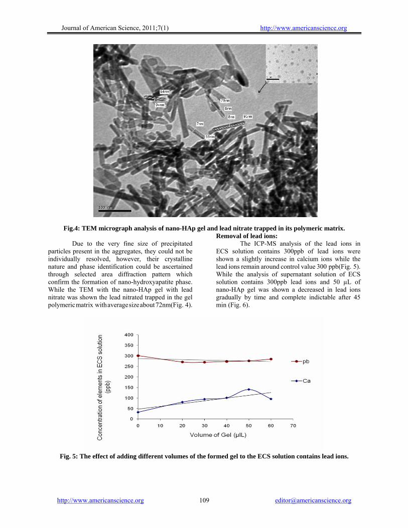
Fig.4: TEM micrograph analysis of nano-HAp gel and lead nitrate trapped in its polymeric matrix. Due to the very fine size of precipitated particles present in the aggregates, they could not be individually resolved, however, their crystalline nature and phase identification could be ascertained through selected area diffraction pattern which confirm the formation of nano-hydroxyapatite phase. While the TEM with the nano-HAp gel with lead nitrate was shown the lead nitrated trapped in the gel polymeric matrix with average size about 72nm(Fig. 4).
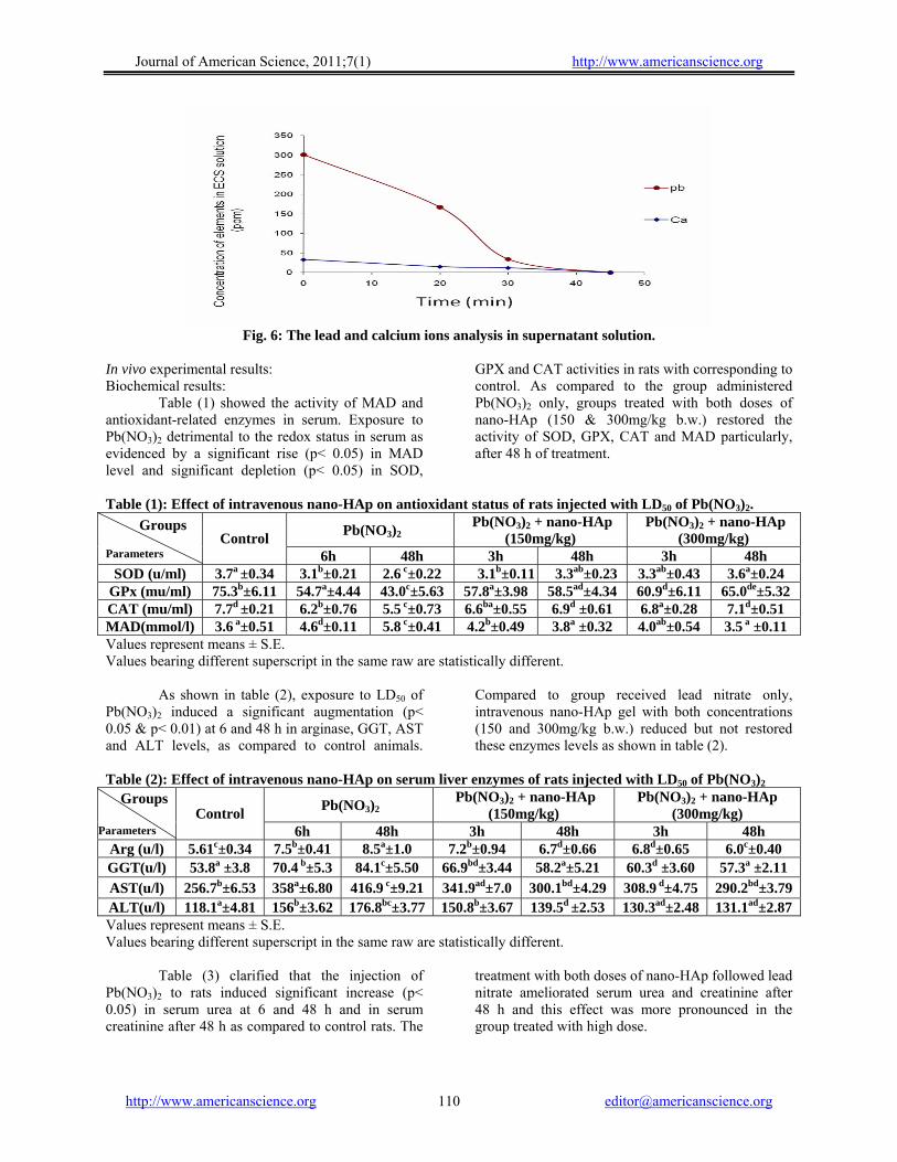
Removal of lead ions: The ICP-MS analysis of the lead ions in ECS solution contains 300ppb of lead ions were shown a slightly increase in calcium ions while the lead ions remain around control value 300 ppb(Fig. 5). While the analysis of supernatant solution of ECS solution contains 300ppb lead ions and 50 µL of nano-HAp gel was shown a decreased in lead ions gradually by time and complete indictable after 45 min (Fig. 6).
Fig. 5: The effect of adding different volumes of the formed gel to the ECS solution contains lead ions.
http://www.americanscience.org [email protected] 109
Journal of American Science, 2011;7(1) http://www.americanscience.org
Fig. 6: The lead and calcium ions analysis in supernatant solution.
In vivo experimental results: Biochemical results: Table (1) showed the activity of MAD and antioxidant-related enzymes in serum. Exposure to Pb(NO3)2 detrimental to the redox status in serum as evidenced by a significant rise (p< 0.05) in MAD level and significant depletion (p< 0.05) in SOD,
GPX and CAT activities in rats with corresponding to control. As compared to the group administered Pb(NO3)2 only, groups treated with both doses of nano-HAp (150 & 300mg/kg b.w.) restored the activity of SOD, GPX, CAT and MAD particularly, after 48 h of treatment.
Table (1): Effect of intravenous nano-HAp on antioxidant status of rats injected with LD50 of Pb(NO3)2.
Pb(NO3)2 Pb(NO3)2 + nano-HAp
(150mg/kg) Pb(NO3)2 + nano-HAp
(300mg/kg) Groups
Parameters
Control 6h 48h 3h 48h 3h 48h
SOD (u/ml) 3.7a ±0.34 3.1b±0.21 2.6 c±0.22 3.1b±0.11 3.3ab±0.23 3.3ab±0.43 3.6a±0.24 GPx (mu/ml) 75.3b±6.11 54.7a±4.44 43.0c±5.63 57.8a±3.98 58.5ad±4.34 60.9d±6.11 65.0de±5.32 CAT (mu/ml) 7.7d ±0.21 6.2b±0.76 5.5 c±0.73 6.6ba±0.55 6.9d ±0.61 6.8a±0.28 7.1d±0.51 MAD(mmol/l) 3.6 a±0.51 4.6d±0.11 5.8 c±0.41 4.2b±0.49 3.8a ±0.32 4.0ab±0.54 3.5 a ±0.11 Values represent means ± S.E. Values bearing different superscript in the same raw are statistically different. As shown in table (2), exposure to LD50 of Pb(NO3)2 induced a significant augmentation (p< 0.05 & p< 0.01) at 6 and 48 h in arginase, GGT, AST and ALT levels, as compared to control animals.
Compared to group received lead nitrate only, intravenous nano-HAp gel with both concentrations (150 and 300mg/kg b.w.) reduced but not restored these enzymes levels as shown in table (2).
Table (2): Effect of intravenous nano-HAp on serum liver enzymes of rats injected with LD50 of Pb(NO3)2
Pb(NO3)2 Pb(NO3)2 + nano-HAp
(150mg/kg) Pb(NO3)2 + nano-HAp
(300mg/kg) Groups
Parameters Control
6h 48h 3h 48h 3h 48h Arg (u/l) 5.61c±0.34 7.5b±0.41 8.5a±1.0 7.2b±0.94 6.7d±0.66 6.8d±0.65 6.0c±0.40 GGT(u/l) 53.8a ±3.8 70.4 b±5.3 84.1c±5.50 66.9bd±3.44 58.2a±5.21 60.3d ±3.60 57.3a ±2.11 AST(u/l) 256.7b±6.53 358a±6.80 416.9 c±9.21 341.9ad±7.0 300.1bd±4.29 308.9 d±4.75 290.2bd±3.79 ALT(u/l) 118.1a±4.81 156b±3.62 176.8bc±3.77 150.8b±3.67 139.5d ±2.53 130.3ad±2.48 131.1ad±2.87 Values represent means ± S.E. Values bearing different superscript in the same raw are statistically different. Table (3) clarified that the injection of Pb(NO3)2 to rats induced significant increase (p< 0.05) in serum urea at 6 and 48 h and in serum creatinine after 48 h as compared to control rats. The
treatment with both doses of nano-HAp followed lead nitrate ameliorated serum urea and creatinine after 48 h and this effect was more pronounced in the group treated with high dose.
http://www.americanscience.org [email protected] 110

Journal of American Science, 2011;7(1) http://www.americanscience.org
Table (3): Effect of intravenous nano-HAp on serum kidneys function of rats injected with LD50 of Pb(NO3)2.
Pb(NO3)2 Pb(NO3)2 +nano-HAp
(150mg/kg) Pb(NO3)2 +nano-HAp
(300mg/kg) Groups parameters Control
6h 48h 3h 48h 3h 48h Urea (mg/dL) 28c±3.71 41.5b±4.23 65.3a±2.98 45b±2.83 41.5b±3.22 40b±3.67 35c±2.94
Creatinine(mg/dL) 0.9a±0.06 1.1a.±0.05 1.5b±0.09 1.1a±0.16 0.8c±0.21 1.1a±0.01 1a±0.02 Values represent means ± S.E. Values bearing different superscript in the same raw are statistically different. As shown in table (4), administration of Pb(NO3)2 induced significant increase (p< 0.01) in serum corticostrone level in rats after 6 and 48 h as compared to control rats. Intravenous injection of
nano-HAp (150 &300mg/kg) followed exposure to LD50 of Pb(NO3)2 inhibit the level of serum corticosterone and such effect was more evident in rats received low dose of nano-HAp.
Table (4): Effect of intravenous nano-HAp on corticoesterone level of rats injected with LD50 of Pb(NO3)2.
Pb(NO3)2Pb(NO3)2+ nano-HAp
(150mg/kg) Pb(NO3)2+ nano-HAp
(300mg/kg) Groups Control 6h 48h 3h 48h 3h 48h
Corticoesterone 160a±4.32 268b±5.23 218.5c±6.82 150a±3.11 155a±3.84 178ad±3.47 185ad±5.14
Values represent means ± S.E. Values bearing different superscript in the same raw are statistically different. As shown in table (5), rats exposed to LD50 of Pb(NO3)2 showed significant decrease (p< 0.05) in serum calcium level after 24 and 48 h., but the level of phosphorus not affected. Both doses of nano-HAp
followed Pb(NO3)2 did not affect either calcium nor phosphorus levels among all treated groups.
Table (5): Effect of intravenous nano-HAp on serum calcium and phosphorus value of rats injected with LD50
of Pb(NO3)2. Pb(NO3)2 Pb(NO3)2+nano-HAp (150mg/kg) Pb(NO3)2+nano-HAp (300mg/kg) Groups
parameters
Control 6h 24h 48h 3h 24h 48h 3h 24h 48h
Ca (mg/dL) 10.9a±1.2 10.7a±0.9 9.4b±0.4 8.3c±0.1 10.4a±1.2 9.0 b±0.1 8.7c±0.4 10.4a±0.9 9.7b±0.3 8.9cb±0.7
P (mg/dL) 7.6c±0.6 7.5c±0.5 7.3c±0.2 7.4c±0.1 7ac±0.1 6.9ac±0.1 7.3c±0.05 7.1ac±0.04 7.2c±0.2 7.4c±0.3
Values represent means ± S.E. Values bearing different superscript in the same raw are statistically different. Histopathological results: Liver tissue: Severity of the reaction in liver of different groups according to the histopathological alterations treatment.
Pb(NO3)2Pb(NO3)2 + nano-HAp
(150mg/kg) Pb(NO3)2 + nano-HAp
(300mg/kg) Control 3h 48 h 3h 48h 3h 48h
- ++ ++++ ++ + ++ + Very sever ++++, sever +++, moderate ++, mild +, nil -
Transverse section of control rat liver showed normal histological structure of the central vein (C) and surrounding hepatocytes (H) with centrally located nuclei were recorded (Fig.7a). Rats
exposure to lead nitrate (93mg/kg b.w.) induced sever congestion in the portal veins (pv) and sinusoids (arrow) with inflammatory cells infiltration (m) in portal area through 6 h (Fig. 7b). After 48 h, this
http://www.americanscience.org [email protected] 111

Journal of American Science, 2011;7(1) http://www.americanscience.org
congestion associated with other histopathological changes including disruption of the normal structural organization of the hepatic lobules, loss of the characteristic cord-like arrangement of the normal liver cells, vacuolization and infiltration of inflammatory cells (Fig. 7c). According to histopathological results, treatment with both doses of nano-HAp post Pb(NO3)2 administration diminished the majority of histopathological changes in the portal area after 3 h. There was a moderate congestion in the central vein (p v) and sinusoids associated with thickening (h) in periductal surrounding the bile duct (b d) in rats
received low dose (Fig. 7 d & e) and few inflammatory cells infiltration (m) in the portal area in rats received high dose (Fig.7 f). On the other hand, treatment with nano-HAp post Pb(NO3)2 administration restored normal arrangement of hepatocytes through 48 h. But, there was a congestion in the central vein (c) in rats received 150mg/kg nano-HAp (Fig.7 g) and proliferation of diffuse kupffer cells (arrow) with infiltration of inflammatory cells in between the hepatocytes in rats received 300mg/kg nano-HAp (Fig.7 h).
(Fig. 7), (H&E) Fig. (7): Section of rat's liver obtained as follows: (a) control rats (x 64) (b): after 6h of Pb(NO3)2 administration (x 80). (c): after 48 h of Pb(NO3)2 administration (H&E x 40). (d &7e): after 3 h of low dose of nano-HAp post Pb(NO3)2 administration ( x40 &80). (f): after 3 h of high dose of nano-HAp post Pb(NO3)2 administration (x64) (g): after 48 h of low dose of nano-HAp post Pb(NO3)2 administration (x64). (h): after 48 h of high dose of nano-HAp post Pb(NO3)2 administration (x64). Kidney tissue:
http://www.americanscience.org [email protected] 112

Journal of American Science, 2011;7(1) http://www.americanscience.org
Severity of the reaction in kidney of different groups according to the histopathological alterations treatment.
Pb(NO3)2 Pb(NO3)2+ nano-HAp (150mg/kg) Pb(NO3)2+ nano-HAp (300mg/kg) Control
3h 48 h 3h 48h 3h 48h
- ++ ++++ ++ ++ ++ + Very sever ++++, sever +++, moderate ++, mild +, nil - Kidney of control rat showed normal histological structure of the glomeruli (g) and tubules (r) in the cortex (Fig. 8a) as well as the tubules in the medulla (Fig. 8b). Histopathological investigation of kidney obtained from rats after 6 h of Pb(NO3)2 administration showed congestion in the cortical blood vessels(v) as shown in (Fig. 8c) associated with focal hemorrhages in the corticomedullary and medulla portions (h) (Fig. 8d&e). After 48 h of Pb(NO3)2 administration, focal inflammatory cells infiltration surrounding the cortical blood vessels (Fig 8f) associated with swelling and vacuolization of the endothelial cells lining the tuft of the glomeuli (g) were observed (Fig.8g). In addition to focal hemorrhages (h) in the corticomedullary portion (Fig.8h).
According to histopathological results, kidney collected from rats after 3 h of nano-HAp treatment revealed infiltration of focal inflammatory cells in the perivascular (m) area in the cortex associated with congestion in the tufts of the glomeruli (g). But, the focal hemorrhage (h) in the medulla was reduced (Fig 8 I, j & k). On the other hand, after 48 of 150mg/kg nano-HAp treatment, the congestion in the cortical blood vessels as well as focal haemorrhages (h) in the corticomedullary and medullary portions disappeared. But, there was a proliferation of the endothelial cells lining the hyperemic tuft of the glomeruli (g) (Fig8 l). Regarding to the rats received 300mg/kg nano-HAp, there was a proliferation in the endothelial cells lining the hyperemic tufts of the glomeruli associated with focal inflammatory cells infiltration in between the tubules (m) at the cortex (Fig 8 m) as well as in the medulla (Fig 8 n).
http://www.americanscience.org [email protected] 113

Journal of American Science, 2011;7(1) http://www.americanscience.org
(Fig.8), (H&E) Fig. (8): Section of rat's liver obtained as follows: (a): kidney cortex of control rats (x 64). (b): Kidney medulla of control rats (x 64). (c & d & e ): after 6h of Pb(NO3)2 administration (x64) (f, g , h): after 48h of Pb(NO3)2 administration (x64, x160, x64). (i): after 3 h 3 h of low dose of nano-HAp post Pb(NO3)2 (x80). (j & k): after 3 h of high dose of nano-HAp post Pb(NO3)2 administration (x64, x 80). (l): after 48 h of low dose of nano-HAp post Pb(NO3)2 administration (x80) (m & n): after 48 h of high dose of nano-HAp post Pb(NO3)2 administration (x80). 4. Discussion: Hydroxyapatite is a unique substance because of its high capacity in removal of heavy-metal ions and high biological compatibility (Ozawa and Kanahara 2005). Several investigations concerned the metal ions chelating mechanisms suggested that HAp removed lead ions by ions exchange or by absorption (Admassu and Breese, 1999) or adsorption (Stötzel et al. 2009). It was reported that synthetic hydroxyapatite could reduce aqueous pb from 1000 mg/l to less than 1 mg/l (Shashkova et al. 1988).
The present results showed that no presence of Pb2+ or Ca2+ in the solution (Fig. 6) and according to ICP-MS analysis, the increased of nano-HAp concentration had no effect in the decreased of Pb2+. The TEM analyses clarified the trapping of lead nitrate in the polymeric matrix of the formed nano-HAp gel which decreased its crystallinity in FTIR analysis (Fig.3). The same results had been investigated in details for the removal of another divalent ions (Ni+2) and its anion using nano-HAp by Abdelfattah et al (2006). Also nano size of the used gel enhanced the capacity of ions removal besides the
http://www.americanscience.org [email protected] 114

Journal of American Science, 2011;7(1) http://www.americanscience.org
presence of PVAL work as steric entrapment for cations leading to complete removal of the elements in the solution. Thus, it could be suggested that nano-HAp gel achieved success in chelating lead ions with its anion and HAp kept its crystals structure with no replacement of calcium ions. The performance of Pb(NO3)2 toxicity on oxidant/antioxidant system in vivo was manifested in the present study by increase in MDA level and decrease in the endogenous antioxidant enzymes such as SOD, CAT, and GPx. One general mechanism was proposed for lead intoxication through generation of free radicals by either or both of the following events: depletion of glutathione or inhibition of sulfhydryl-dependent enzymes (Silbergeld et al. 2000) or interfering with some essential metals needed for antioxidant enzyme activities (Slater, 1985). Subsequently, cellular concentrations of hematoproteins lowered and the redox buffering capacity of cells reduced. Under such conditions, ROS generated by other events may not be neutralized and thereby the likelihood of oxidative damage to DNA may be increased (Caylak et al. 2007). The maximum level of Pb in the liver reached rapidly within the first hour after administration which in turn, increases the susceptibility of cells to oxidative attack by altering the fatty acid composition and membrane permeability resulting in escaping of enzymes from cells into blood (Bechara, 2004). Lead oxidative hepatocytes damage had been responsible for the elevation of serum arginase, GGT, AST and ALT levels observed in the present study. These enzymes considered as biomarkers for liver function and integrity (O'Brien, 2002) and usually elevated in acute hepatotoxicity or mild hepato-cellular injury (Sharma et al. 2010). As urea production in mammals occurs essentially in the liver, the plasma concentration of this compound resulting from amino acids catabolism, could also be used as an indicative of hepatic function (Araύjo et al., 2005). In addition, kidney cortex and medulla followed the liver among the soft tissues affected by lead exposure (Nolan and Shaikh 1992). However, kidney disease (nephropathy) is a characteristic manifestation of lead toxicity. Thus, elevation of creatinine level observed in Pb(NO3)2 treated group may be due to loss of 50% of kidney function and referable to functional evidence of lead induced nephrotoxicity (Qu et al. 2002). Accordingly, it could be postulated that lead-induced oxidative stress and lipid peroxidation with concomitant inhibition of several antioxidant enzymes in blood (Bolin et al. 2006 and Arif et al. 2008) has been to be one of the possible mechanisms of its toxic effects (Pande and Flora, 2002) and major
contributors to lead-exposure related diseases (Patrick, 2006).
Regarding to corticosterone, the elevation in its level after exposure to Pb(NO3)2 was a detrimental effect related also to oxidative stress. This is because exposure to lead is well known by association with inflammation mediated by oxidative stress (Songdej et al. 2010) which induced hyperactivity of hypothalamo-pituitary-adrenal axis yielding elevated corticoesterone level (Parthasarathy et al. 2006). On the other wise, lead has adverse effect on immune system and decreased antibody for matrix inflammatory response (Sin and Woo, 1992). These findings support the concept that susceptibility of rats to lead was regulated by HPA axis-immune system feedback loop responsiveness to inflammatory stress.
It is pertinent to mention that the peak concentration of intravenous lead in blood is reached about one hour after injection (35 to 40% of the administered dose) in the rats. The level in blood declined fairly rapidly thereafter and only about 5% of the dose remained in blood after two days (Poirier et al. 2006). Lead reacted rapidly with calcium and phosphors in plasma and caused a withdrawal of these ions indirectly. But calcium and phosphorus are immediately withdrawn from extravascular sources returning plasma concentrations to their initial value (Talmage et al. 1978). At the same time, the most concentration of intravenous nano-HAp detected in liver and spleen at 1 h after administration and decreased significantly after 72h (Tang et al. 2009 and Xie, 2010). Thereby, calcium and phosphorus levels monitored in the present study at times intervals of 6, 24 and 48 h post Pb(NO3)2 administration to clearly detect the performance interaction of i.v. nano-HAp with Pb(NO3)2 related to concentration of both metals in the blood. The results revealed gradual decrease in calcium level after 24 h forward to 48 h post Pb(NO3)2 administration while phosphorus level not affected. Such decrease in calcium level was probably because lead often shared calcium transport mechanisms, through the physicochemical interactions at the injection site or through biochemical competition at critical macromolecular binding sites (Poirier et al., 2006). The most important renderings of nano-HAp gel bioavailability observed in the present study was improvement of the antioxidant enzymes and lipid peroxidation status because the oxidative stress is the manager for internal and/or external factors induced adverse effect. Such antioxidant effect of nano-HAp may be attributed to the scavenging of superoxide, which is the main component of oxidative stress (Scherbart et al., 2009) or to inhibition of oxidative and nitrooxidative species formation (Fouda et al.,
http://www.americanscience.org [email protected] 115
Journal of American Science, 2011;7(1) http://www.americanscience.org
2009). However, elevated activity of catalase is more beneficial than increase in SOD activity alone because without a simultaneous increase in catalase activity, increased SOD activity may lead to intracellular accumulation of H2O2 with detrimental effects (Das et al., 1995). On the other hand, the results of liver and kidney functions contained a lot of important information. Since, all the nano-HAp treated animals have normal kidney function due to normal creatinine levels. Also, the survival of all the animals through the experiment indicated that the liver was not seriously damaged or not fatal (Jayabalan et al., 2010). In this aspect, Xie (2010) investigated the quantitative tissue distribution of intravenous nano-HAp in rats using125I radiolabeling and reported that nano-HAp accumulated in the soft tissues mainly liver and spleen. Apparently, nano-HAp having antioxidant activity effective in treating Pb(NO3)2 induced hepatotoxicity by scavenging the free radicals, thereby preventing the liver and kidney damage induced by both Pb(NO3)2 as well as subsequent depletion of glutathione. There is inconsistency between the general view of literature and the present results because the degradability of intravenous nano-HAp not as stated before when injected after intraperitoneal lead nitrate. This proposal based on the mentioned results referable to similarity of serum calcium level among the rats treated with either nano-HAp and Pb(NO3)2 or lead nitrate only treated rats. Additionally, internalization of i.v. nano-HAp inside the body was confirmed by the recovery of liver and kidney function and structure as well as corticosterone level. Thus, it could be in the light of the in vitro investigation data suggested that nano-HAp uptake lead by capture it in the blood, liver and kidney. Histopathological examination revealed that induction of hepatic and renal cellular proliferation occurred with single dose of lead nitrate administration. A growing body of evidence suggests that oxidative stress is the key player in the pathogenesis of lead-induced toxicity (Adamis et al., 1999). It was shown that, lead nitrate induced a synchronized wave of hepatocyte proliferation in rat liver without accompanying histopathological necrosis (Abdel-Aal et al., 1989). Moreover, Proliferation on non parenchymal cells reflect a direct mitogenic effect of lead nitrate and that hepatocyte proliferation follows the nonparenchymal cell reaction to lead nitrate (Kubo et al., 1996). In this view Pezzatini et al. (2006) showed that lead-induced liver hyperplasia followed by apoptosis mediated by oxidative stress in kupffer cells. Rijhsinghani et al., (1993) have reported that 8 hours after an intravenous injection of lead nitrate, DNA synthesis was detected
in a few scattered hepatocytes and in nonparenchymal cells, including bile duct epithelial cells, fibroblast, macrophage, an nondescript periductular cells. The histopathological results as biochemical results confirmed the biological efficacy of nano-HAp since, liver and kidney restored most of normal structure after the treatment with nano-HAp. It was clear that the nano-HAp particle size could influence the pathway of internalization, the increase of the inhibition effect on the proliferation activity could be reduced (Bauer et al., 2008). General observations favoring enzyme release during reversible cell damage include the apparent lack of histologic evidence of necrosis in spite of increased serum enzyme activity. Conceivably however, this may also reflect a lack of sensitivity of histologic techniques to detect cell necrosis when it is patchy, early on, or involves only a small number of cells. Nevertheless, additional findings made over the previous decades have led to a growing acknowledgement that the escape of enzymes from cells likely includes mechanisms other than cell death. While increased membrane permeability is a well-known outcome of irreversible cell damage, it is unlikely that cells sustaining the formation of perforations or tears of adequate size to allow the leakage of macromolecules such as enzymes could maintain adequate viability to recover. The mechanism proposed to explain the appearance of cytosolic enzymes in blood with reversible damage is by the formation of membrane blebs that detach and allow the cell membrane to reseal without cell death (Mair, 1999).
5. Conclusion: Nano-HAp gel was synthesized and tested as of prospective chelating agent of one of very toxic heavy metal. The nano-composite gel successfully removed lead ions and its anion from its solution. The lead ions were completely chelated and intervened with the polymeric gel reducing the degree of HAp crystallinity as being completely adsorbed on the HAp structure with its anion. The newly-formed nano-HAp gel have shown good biocompatibility and little toxicity when injected intravenously. It showed significant and fast therapeutic effect of lead nitrate toxicity within 48h. Apparently, the biological risks, cytotoxicity of the HAp nanoparticles are size-and dose-dependent. The future application of nano-HAp gel in the human biomedical was expected. Further study is needed to be carried out in order to proof its safety and efficacy in the human body.
http://www.americanscience.org [email protected] 116

Journal of American Science, 2011;7(1) http://www.americanscience.org
Acknowledgment: The authors are grateful to Dr. Adel M. Bakeer Kholoussy, Professor of Pathology, Faculty of Veterinary Medicine, Cairo University for his helping in the examination of the histopathological slides and for his valuable comments. Corresponding author Eman I. Abdel-Gawad Radioisotopes Department, Atomic Energy Authority, Egypt [email protected] 6. References: 1. Abdel-Aal S.F., Shalaby S.A., Badawy A.H.,
Sammour S.A. (1989): Effect of lead nitrate administration on liver and kidney structure in rats. J Egypt Soc Parasitol.,19: 689-99.
2. Abdelfattah W., Fayed M., Gouda Sh., and Awwad S. (2006): Nano Hydroxyapatite gel for removal of nickel ions for environmental applications. Isotope & Rad. Res., 38: 417 – 427.
3. Abdel-Gawad E. I. and Awwad S. (2010): Biocompatibility of Intravenous Nano Hydroxyapatite in Male Rats. Nat. and Sci., 8: 60-68.
4. Admassu W. and Breese T. (1999): Feasibility of using natural fishbone apatite as a substitute for hydroxyapatite in remediating aqueous heavy metals. J. Hazardous Materials 69: 187–196.
5. Adamis Z, Ta´trai E, Honma K and Ungva´ry G. (1999): Effects of Lead(II) Nitrate and a Dithiocarbamate Fungicide on the Rat Lung. J. Appl. Toxicol. 19, 347–350.
6. Araύjo E.J.A.; Sant’Ana D.M.G., Molinal, S.L. and Neto M.H.M. (2005). Hematologic and biochemical parameters of rats subjected to hypoproteic and hipercaloric diet. Arq. ciên. vet. zool. UNIPAR, 8: 139-146.
7. Arif M., Kabir F., Hassan F., Zaved Waise T.M., and Ehsanul Md., Mazumder H.and Rahman S. (2008): Increased DNA damage in blood cells of rat treated with lead as assessed by comet assay. Bangladesh J Pharmacol, 3: 97-101.
8. ATSDR (Agency for Toxic Substances and disease Registry), (1993): Toxicological Profile for Lead. Update. Prepared by Clement International Corporation under contract No. 205 – 88 - 0608 for ATSDR, U.S. Public Health Service, Atlanta, GA.
9. Banchroft J.D., Stevens A. and Turner D.R. (1996). Theory and practice of histological
techniques. Fourth Ed. Churchil Livingstone, New York, London, San Francisco, Tokyo.
10. Bauer I W, Li S. P., Han Y. C. and Yin L. M. (2008): Internalization of hydroxyapatite nanoparticles in liver cancer cells. J Mater Sci: Mater Med, 19:1091–1095
11. Bechara E.J.H. (2004): Lead poisoning and oxidative stress. Free Radic Biol Med., 36: 22.
12. Bolin C.M.,Basha R.,Cox D.,Zawia N.H., Maloney B.,Lahiri D.K. and Cardozo-Pelaez F. (2006): Exposure to lead and exposed to lead using micronucleus assay, comet assay and TCR gene mutation test. Toxicology, 223: 219-226.
13. Caylak E., Halifeoiglu I., Aydin S., Telo S., Bulmus O. and Celik H. (2007): The effects of sulfur-containing compounds on total antioxidant capacity levels of liver, kidney and brain in lead-exposed rats. Turkiye Klinikleri J Med Sci., 27: 823–828.
14. Chelikani P., Fita I. and Loewen P.C. (2004): Diversity of structures and properties among catalases. Cell. Mol. Life sci., 61: 192-208.
15. Christenson, E.M., K.S. Anseth, J.J.J.P. vanden Beucken, C.K. Chan, B. Ercan, J.A. Janson, C.T. Laurencin, W.J. Li, R. Murugan, L.S. Nair, S. Ramakrishna, R.S. Tuan, T.H. Webster, and Mikos A.G.(2007): Nanobiomaterial applications in orthopedics. Inc. J Orthop Res., 25:11-22.
16. Ciobanu G., Ignat D., Carja G. and C. Luca C. (2000): Hydroxyapatite /polyurethane composite membranes for lead ions removal. Env. Eng. and Manag. J., 8:1347-1350.
17. Crombez E. A and Cederbaum S. D. (2005): Hyperargininemia due to liver arginase deficiency. Mol. Genet Metab. 84: 243-251.
18. Das, D.K., Maulik, N. and Moraru, I.I. (1995): Gene expression in acute myocardial stress. Induction by hypoxia, ischemia/ reperfusion, hyperthermia and oxidative stress. J Mol. Cell Cardiol. , 27: 181-193.
19. Descamps M., Richart O., Hardouin P., Hornez J.C. and Leriche A. (2008): Synthesis of macroporous b-tricalcium phosphate with controlled porous architectural. Ceramics Int., 34: 1131-1137.
20. Flora S.J.S., Saxena G. and Mehta A. (2007): Reversal of lead-induced neuronal apoptosis by chelation treatment in rats: role of ROS and intracellular Ca2+. J Pharmacol Exp Ther.; 322: 108–16.
21. Fouda M.F.A., Nemat A., Gawish A. and Ayman R and Baiuomy A.R. (2009): Does the Coating of Titanium Implants by Hydroxyapatite affect the Elaboration of Free
http://www.americanscience.org [email protected] 117

Journal of American Science, 2011;7(1) http://www.americanscience.org
Radicals. An Experimental Study Aust J. Basic and Appl Sci, 3: 1122-1129.
22. Janga, S. H., Jeonga,Y.G., Mina, B.G., Lyoob, W.S. and Leea, S.L. (2008): Preparation and lead ion removal property of hydroxyapatite/polyacrylamide composite hydrogels. J. Hazardous Materials 159: 294–3002.
23. Jayabalan M., Shalumon K.T., Mitha M.K., K. Ganesan K. and Epple
M. (2010): Effect of hydroxyapatite on the biodegradation and biomechanical stability of polyester nanocomposites for orthopaedic applications. Acta Biomaterialia, 6: 763-775.
24. Kubo Y.,Yasunaga M.,Masuhara M.,Terai S., Nakamura T. and Okita K. (1996): Hepatocyte Proliferation Induced in Rats by Lead Nitrate Is Suppressed by Several Tumor Necrosis Factor a Inhibitors. Hepatology, 23:104-114.
25. LeGeros, R.Z. (2008): Calcium Phosphate-Based Osteoinductive Materials. Chem. Rev., 108 (11): 4742–4753.
26. Mair J. (1999): Tissue release of cardiac markers: from physiology to clinical applications. Clin Chem Lab Med., 37:1077-84.
27. Mavropoulos E., Rossi A. M., Costa A. M., Perez. A. C., Moreira J. C. and Saldanha M., (2002): Studies on the mechanisms of lead immobilization by hydroxyapatite. Environ. Sci. Technol., 36: 1625 –1629.
28. McCord J.M. and Fridovich I. (1988): Superoxide dismutase: the twenty first years (1968-1988). Free Radic. Biol. Med. 5: 363-369.
29. Nair V., O'Neil C.L. and Wang P.G. (2008): "Malondialdehyde" Encyclopedia of Reagents for Organic Synthesis. Jon Wiley and Sons, New York.
30. Nolan C.V. and Shaikh Z.A. (1992): Lead nephrotoxicity and associated disorders: biochemical mechanism. Toxicol. 73: 127-140.
31. O'Brien P.J., Slaughter M. R., Polley S.R. and Kramer K. (2002): Advantages of glutamate dehydrogenase as a blood biomarker of acute hepatic injury in rats. Lab Anim., 36: 313 - 321.
32. Ozawa M. and Kanahara S. (2005): Removal of aqueous lead by fish-bone waste hydroxyapatite powder. J Mat. sci., 40:1037 – 1038.
33. Pande M. and Flora S.J.S. (2002): Lead induced oxidative damage and its response to combined administration of alipoic acid and succimers in rats. Toxicology, 177: 187-196.
34. Patrick L. (2006): Lead toxicity part II: The role of free radical damage and the use of
antioxidants in the pathology and treatment of lead toxicity. Altern Med Rev., 11: 114-127.
35. Parthasarathy N.J., Kumar R.S., Manikandan S., Narayanan G.S., Kumar R.V. and Devi R.S. (2006): Effect of methanol-induced oxidative stress on the neuroimmune system of experimental rats. Chem Biol Interct., 161: 14 -25.
36. Pezzatini S., Solito R., Morbidelli L., Lamponi S., Boanini E., Bigi A. and Ziche M.(2006): The importance of the implant surface with respect to tissue reaction has been recognized with regard to maintain a controlled tissue interface. J Biomed Mater Res,76: 656-663
37. Poirier L. A., Jeffrey C., Arnold L. J., and Shimkin M. B. (1984): Inhibition by Magnesium and Calcium Acetates of Lead Subacetate- and Nickel Acetate-induced Lung Tumors in Strain A Mice. Cancer Research 44, 1520 -1522.
38. Poirier L.A., Jeffrey C., Arnold L.J., and Michael B. and Shimkin M.B. (2006): Effect of methanol-induced oxidative stress on the neuroimmune system of experimental rats. CHem. Biol. Interact., 161:14-25.
39. Qu W., Diwan B.A., Liu J., Goyer R.A., Dawson T., Horton J.L., Cherian M.G. and Waalkes M.P. (2002): The metallothionein-null phenotype is associated with heightened sensitivity to lead toxicity and an inability to form inclusion bodies. Am. J. Pathol. 160: 1047-1056.
40. Ran Q., Liang H. and Ikeno Y. (2007): Reduction in glutathione peroxidase for increases life spane through increased sensitivity to apoptosis. Biol. Sci. Med. Sci., 62: 932-942.
41. Rijhsinghani K, Choi HH, Burton LA, Paronetto F. and Tavoloni N. (1993): Immunoelectron microscopy identification of early proliferating cells in rat liver tissue during hyperplasia induced by lead niscription. Hepatolog , 17: 685 -692.
42. Saino E.L., Lehtola T. and Roininen P. (1988): Radioimmunoassay of total and free corticosterone in rat plasma: measurement of the effect of different doses of corticosterone. Steroids, 51: 609 - 622.
43. Scherbart C.A., Agnes M., van Berlo, Braunbarth D., Schins C.M., Roel P.F. and Julia S. (2009): Evaluation of cytotoxic effects and oxidative stress with hydroxyapatite dispersions of different physicochemical properties in rat NR8383 cells and primary macrophage. Toxicology in Vitro, 23: 520 - 530.
http://www.americanscience.org [email protected] 118

Journal of American Science, 2011;7(1) http://www.americanscience.org
44. Sharma A., Sharma V. and Kansal L. (2010):
Amelioration of lead-induced hepatotoxicity by Allium sativum extracts in Swiss albino mice. Libyan J. of Med., 5: 4621. incl Supplements.
45. Shashkova I., Rat’ko A. and Kitikova N. (1988): Removal of heavy metal ions from aqueous solutions by alkaline-earth metal phosphates. Colloids and Surfaces Bionterfaces 160: 207–215.
46. Silbergeld E. K. (2003): Facilitative mechanisms of lead as a carcinogen. Mut. Res./Fund. and Mol. Mechanisms of Mutagenesis. 533 : 121-133.
47. Silbergeld E. K., Waalkes M. and Jerry M. and Rice J.M. (2000): Lead as a Carcinogen: Experimental Evidence and Mechanisms of Action. Am. J Ind. Med. 38: 316-323.
48. Sin Y.M. and Woo P.T. (1992): Inflammatory response and resistance in lead-treated mice. Bull. Environ. Contam. Toxicol., 48: 502-508.
49. Slater T.S. (1985): Free radical mechanism in tissue injury. Biochem J., 222: 1–25.
50. Songdej N., Winters P.C., McCabe M.J. Jr and Van Wijngaarden E. (2010): A population-based assessment of blood lead levels in relation to inflammation. Environ Res.,110: 272-277.
51. Stötzel C., Müller F. A., Reinert F., Niederdraenk F., Barralet J. and Gbureck U. (2009): Ion adsorption behaviour of hydroxyapatite with different crystallinities, Colloids and surfaces Bionterfaces, 74: 91-95.
52. Talmage R. V. , CWiel C.J.V. and H. Norimatsu H. (1978): A reevaluation of the cause of acute hypercalcemia following intravenous administration of lead acetate. Calcified Tissue International, 26: 149-153
53. Tegar-Nilsson, A.C. (1961): Quantitative colorimetric determination of creatinine in serum or urine. Scand. J. Clin, Lab. Invest. 13: 326.
54. Thomas, L. (1998): Alanine aminotransferase (ALT), Aspartate aminotransferase (AST) In: Thomas L., Editor. Clinical Laboratories Diagnostics, 1st Edi. Frankfurt: TH-Boos Verlagsgesellschaft; p. 55-65 and 208-214.
55. Tietz, N.W. (1990): Clinical guide to laboratory tests. 2nd ed. Philadelphia: WB Saunders; 566.
56. Woo J. and Cannon D.C. (1984): Metabolic intermediates and inorganic ions. "Clinical Diagnosis and Management by Laboratory Methods" 17th ed.JB Henry, RA McPherson, Philadelphia, 133.
57. Xie J.S.G. (2010): Tissue distribution of intravenously administrated hydroxyapatite nanoparticles labeled with 125I. Nanoelectronics
Conference (INEC), 3rd International 1415 – 1416.
58. Xu H., YANG L., WANG P., LIU Y. and PENG M. (2007): Removal Mechanism of Aqueous Lead by a Novel Eco-material: Carbonate Hydroxyapatite. J mater. and sci. technol., 23: 417-422.
59. Young D. S. (1990): Effects of drugs on clinical laboratory tests 3rd ed. Washington DC, AACC Press.
http://www.americanscience.org [email protected] 119