in vivo amelioration of endogenous antitumor … › content › pnas › 113 › 48 ›...
TRANSCRIPT

In vivo amelioration of endogenous antitumorautoantibodies via low-dose P4N through theLTA4H/activin A/BAFF pathwayYu-Ling Lina,b, Nu-Man Tsaic,d, Cheng-Hao Hsieha, Shu-Yi Hoa, Jung Changa, Hsin-Yi Wue, Ming-Hua Hsuf,Chia-Ching Changa, Kuang-Wen Liaoa,e,g,1,2, Tiffany L. B. Jacksonh, David E. Moldh, and Ru Chih C. Huanga,h,1,2
aDepartment of Biological Science and Technology, National Chiao Tung University, Hsinchu, 30068, Taiwan, Republic of China; bCenter for BioinformaticsResearch, National Chiao Tung University, Hsinchu, 30068, Taiwan, Republic of China; cSchool of Medical Laboratory and Biotechnology, Chung ShanMedical University, Taichung, 40201, Taiwan, Republic of China; dClinical Laboratory, Chung Shan Medical University Hospital, Taichung 40201, Taiwan,Republic of China; eInstitute of Molecular Medicine and Bioengineering, National Chiao Tung University, Hsinchu, 30068, Taiwan, Republic of China;fNuclear Science & Technology Development Center, National Tsing Hua University, Hsinchu, 30013, Taiwan, Republic of China; gGraduate Institute ofMedicine, College of Medicine, Kaohsiung Medical University, Kaohsiung, 80708, Taiwan, Republic of China; and hDepartment of Biology, Johns HopkinsUniversity, Baltimore, MD 21218-2685
Edited by Jason G. Cyster, University of California, San Francisco, CA, and approved October 24, 2016 (received for review March 30, 2016)
Cancer progression is associated with the development of antitu-mor autoantibodies in patients’ sera. Although passive treatmentwith antitumor antibodies has exhibited remarkable therapeuticefficacy, inhibitory effects on tumor progression by endogenousantitumor autoantibodies (EAAs) have been limited. In this study,we show that P4N, a derivative of the plant lignan nordihydro-guaiaretic acid (NDGA), enhanced the production of EAAs andinhibited tumor growth at low noncytotoxic concentrations viaits immunoregulatory activity. Intratumoral injection of P4N im-proved the quantity and quality of EAAs, and passive transfer ofP4N-induced EAAs dramatically suppressed lung metastasis forma-tion and prolonged the survival of mice inoculated with metastaticCT26 tumor cells. P4N-induced EAAs specifically recognized twosurface antigens, 78-kDa glucose-regulated protein (GRP78) andF1F0 ATP synthase, on the plasma membrane of cancer cells. Ad-ditionally, P4N treatment led to B-cell proliferation, differentiationto plasma cells, and high titers of autoantibody production. Byserial induction of autocrine and paracrine signals in monocytes,P4N increased B-cell proliferation and antibody production via theleukotriene A4 hydrolase (LTA4H)/activin A/B-cell activating factor(BAFF) pathway. This mechanism provides a useful platform forstudying and seeking a novel immunomodulator that can be ap-plied in targeting therapy by improving the quantity and qualityof the EAAs.
endogenous antitumor autoantibody | P4N | B-cell proliferation |colorectal cancer | cancer immunotherapy
Colorectal cancer (CRC) is the second most prevalent cancerin the western world and is also rapidly increasing in Asia (1).
It is well known that multiple genetic events involved in the de-velopment of this disease lead to the generation of tumor-associatedantigens (TAAs) against which patients with CRC develop auto-antibodies (2). More than 100 TAAs have been identified by theseendogenous antitumor autoantibodies (EAAs), including 78-kDaglucose-regulated protein [GRP78, also known as binding Ig protein(BiP)], p53, carcinoembryonic acid (CEA), and mucin 1 (MUC1)(2). The use of these autoantibody signatures as biomarkers in theearly detection of CRC has been proposed (3–5). Typically, EAAshave not had a significant effect on tumor elimination, most likelydue to immune tolerance induction by the tumor (6, 7). However,extraction of EAAs from the sera of patients with cancer to activatethe humoral immune response against some malignant tumors hasbeen considered. A few EAAs selected from patients, such as SC-1(anti-CD55), PAM-1 [anti–cysteine-rich fibroblast growth factor(anti-CFR1)], and PAT-SM6 (anti-GRP78), act directly againsttumors and effectively kill them via antibody-mediated cellular cy-totoxicity (8). In addition, a natural human IgM autoantibody(PAT-SM6) selected from patients’ sera against the cell surface
GRP78 protein provides therapeutic effects for patients with cancer(9, 10). Although the therapeutic effects of EAAs are ill-defined,these studies display their potential for clinical therapy.Alternatively, passive immune therapeutics composed of anti-
bodies ligated to targeted molecules (11) and directed against tumorgrowth factors (12) have been used clinically to induce apoptosis oftumor cells directly. Moreover, these passive therapeutic antibodiestrigger complement-dependent cytotoxicity (CDC) or antibody-dependent cellular cytotoxicity (ADCC) (12, 13), promote phago-cytosis by dendritic cells (DCs) (14), induce cross-talk betweenimmune cells [natural killer (NK) cells and DCs], produce immu-nomodulatory cytokines (type I and type II interferons) (12), andenhance the cross-presentation of antigen-presenting cells (APCs)for the priming of CD8+ cytotoxic T lymphocytes (CTLs) (12, 14).By these reactions, passive therapeutic antibodies can be effectiveagents for tumor inhibition. The effectiveness of therapeutic anti-tumor antibodies portends the potential of enhanced or improvedEAAs to function as effective therapeutic entities.
Significance
This study finds that a small-molecule drug (P4N) is able to in-hibit tumor growth by augmentation of endogenous antitu-mor autoantibodies (EAAs). We show that the enhancement ofEAA activity by P4N is mediated through activation of theleukotriene A4 hydrolase (LTA4H)/activin A/B-cell activatingfactor (BAFF) pathway, revealing a valuable method for de-veloping new immune modulators of tumor growth via hu-moral immunity. Typically, the effects of the humoral responseon tumor inhibition are modest; however, the results of thisstudy demonstrate that by removing the impediment to cancercell destruction posed by low-activity autoantibodies, the re-alization of new, more potent immunotherapies for cancertreatment may be possible.
Author contributions: Y.-L.L., K.-W.L., and R.C.C.H. designed research; Y.-L.L., N.-M.T., C.-H.H.,S.-Y.H., J.C., H.-Y.W., C.-C.C., T.L.B.J., and D.E.M. performed research; M.-H.H. and R.C.C.H.contributed new reagents/analytic tools; Y.-L.L. analyzed data; and Y.-L.L., K.-W.L., D.E.M.,and R.C.C.H. wrote the paper.
The authors declare no conflict of interest.
This article is a PNAS Direct Submission.
Freely available online through the PNAS open access option.
Data deposition: Gene Expression Profiling has been deposited and published in Gene Expres-sion Omnibus at National Center for Biotechnology Information (accession no. GSE89659).1K.-W.L. and R.C.C.H. contributed equally to this work.2To whom correspondence may be addressed. Email: [email protected] or [email protected].
This article contains supporting information online at www.pnas.org/lookup/suppl/doi:10.1073/pnas.1604752113/-/DCSupplemental.
E7798–E7807 | PNAS | Published online November 17, 2016 www.pnas.org/cgi/doi/10.1073/pnas.1604752113

Recently, low-dose chemotherapy (metronomic chemotherapy)has been shown to induce an antitumor immune response andenhance the efficacy of cancer therapy. For example, the anti-microtubule taxanes (paclitaxel and docetaxel) were found to triggerthe production of cytokines by macrophages to activate other im-mune cells, such as DCs (15), NK cells (16), and CTLs, againsttumors (16, 17). Paclitaxel also reduced the number of regulatory T(Treg) cells and myeloid-derived suppressor cells (MDSCs), and ledto the augmentation of the functions of CD4 and CD8 T cells (16,17). In other cases, DNA alkylating agents, such as cyclophospha-mide and mafosfamide, in low doses selectively depleted Treg cells(18, 19), caused an increase in effector T cells (Teff)/Treg cell ratiosvia up-regulation of the T helper (Th) 17 pathway (20), and im-proved the outcome of tumor vaccinations against cancer (21–23).Furthermore, doxorubicin, mitomycin C, vinblastine, and metho-trexate in low doses have been found to up-regulate DCmaturation,antigen processing, and antigen presentation, which led to syner-gistic antitumor effects of low-dose chemotherapy combined with aDC vaccine (24–26). These various antitumor drugs in low dosescan induce cell-mediated immunity against tumors, but they haveless of a contribution to humoral immunity. Therefore, they havenot been used to raise EAAs against tumor growth in patients.P4N, a derivative of nordihydroguaiaretic acid (NDGA), a nat-
ural product from the creosote bush, Larrea tridentate, is composedof two phenolic rings connected by long and flexible –CH2–CH2–
linkers to piperidines (27). Like methylated derivatives of NDGA,P4N has shown noteworthy antivirus and anticancer effects viacompetition with the transcription factor Sp1 for its DNA-bindingsite (27, 28). P4N’s parent compound, NDGA, is a potent antioxi-dant and has been shown to have promising applications in thetreatment of multiple diseases, including cardiovascular disease andneurological disorders (29). It also inhibits the growth of varioustumors (30), although it is substantially less potent than P4N (SIAppendix, Fig. S1) and likely has a different mechanism of action(28). NDGA has immunoregulatory activities as well. It inhibits5-lipoxygenase, suppresses the production of leukotriene B4 (LTB4),and activates macrophages (29). The effect of P4N on immunefunction is unknown.In this study, the immunoregulatory activity of low-dose P4N was
investigated. Unlike previously described antitumor drugs, low-doseP4N contributes to humoral immunity by raising the titers and ac-tivities of autoantibodies against GRP78 and F1F0 ATP synthase onthe surface of CT26 cells and inducing B-cell proliferation anddifferentiation of plasma cells. We show that low-dose P4N inducesB-cell proliferation by activating leukotriene A4 hydrolase (LTA4H),thereby enhancing the production of LTB4 and stimulatingmonocytes to release proinflammatory cytokines. The release ofactivin A acts as an autocrine signal, stimulating B-cell activatingfactor (BAFF) production via activation of the ALK4/Smad3pathway. BAFF then enhances B-cell proliferation and activation.
ResultsP4N-Activated Humoral Immune Response Suppresses Tumor Growthin Vivo. To examine whether low-dose P4N causes immunoregu-latory activity for the suppression of tumor growth, the effects ofa single intratumoral injection with 2.5 mg/kg or 5 mg/kg of P4Ninto CT26 tumors in BALB/c or nude mice were monitored.Both doses of P4N significantly inhibited the growth of CT26tumors in BALB/c mice, but had no effect on the growth of CT26tumors in the immunodeficient nude mice (Fig. 1A). As shown inFig. 1B, CD8+ T-cell depletion attenuated the effect of P4N-induced tumor inhibition. In contrast, B-cell depletion abolishedthe effect of P4N-induced tumor inhibition (Fig. 1B). The tumor-suppressive effects of P4N were not the result of direct cytotoxicityof the drug, because there was no significant difference in tu-mor growth between P4N and PBS treatments in the B-cell–depleted mice. These results suggested that a humoral immuneresponse may play a major role in P4N-induced tumor inhibition.
Using an immunohistochemistry (IHC) assay, it was shown thatP4N treatments led to the infiltration of immune cells, such asmacrophages, DCs, and T or B cells, into the tumor area (Fig. 1C).There was no significant difference between the two mouse strainsin terms of the levels of macrophages and DCs in the tumors, butthe levels of T and B cells in the tumors were higher in BALB/cmice than in nude mice (SI Appendix, Fig. S2). In addition, it wasfound that P4N treatments enhanced the expression levels of TNF-αand IL-8 in both strains of mice (Fig. 1D). Taken together, theseresults indicate that low-dose P4N elicits certain immune re-sponses in tumors, but only suppresses tumor growth in BALB/cmice, where adaptive immunity may play an important role.To determine whether humoral immunity is involved in the an-
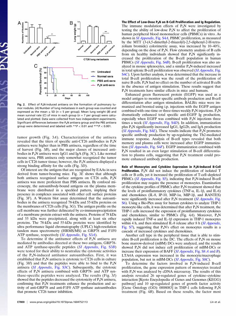
titumor activity of low-dose P4N, sera isolated from P4N- or vehicle(PBS)-treated CT26 tumor-bearing mice were passively transferredinto BALB/c mice inoculated with metastatic CT26 cells. Within 18d after tumor cell injection, untreated mice developed lung me-tastases, with metastatic nodules surrounding the lungs. Adminis-tration with antisera derived from PBS-treated mice decreased thenumber of metastatic nodules and the total weight of the lungs (Fig.2 A and B). P4N-derived antisera displayed an even stronger effect,dramatically inhibiting tumor colonization, as evidenced by thenearly complete lack of metastatic nodules and total lung weightsapproaching the total lung weights of normal mice. All mice treatedwith P4N antisera survived more than 60 d, a survival time threefoldthe survival time of the control groups (Fig. 2C). These resultssuggested that the inhibition of tumor colonization by P4N antiseramight be the result of P4N-induced EAAs in the sera.
The Effect of P4N on Production and Activity of AntitumorAutoantibodies. To eliminate the influence of T cells, the anti-sera were injected into CT26 tumor-containing immunodeficientmice. P4N antisera still significantly suppressed tumor growth inthese mice, whereas PBS antisera had no significant effect on
Fig. 1. Antitumor effects of low-dose P4N in immune-competent and-deficient mice. (A) BALB/c (n = 10 per group) and nude mice (n = 10 per group)bearing CT26 tumors were treated with a single intratumoral injection of 2.5or 5.0 mg/kg of P4N. (B) B-cell– or CD8
+ T-cell–depleted BALB/c mice (n = 9 pergroup) bearing CT26 tumors were treated with 5 mg/kg of P4N by intra-tumoral injection every week. Tumor volumes were measured every 2 d aftertreatment. Significant differences between the P4N groups and the PBS groupwere identified and labeled with *P < 0.05 and **P < 0.01. (C) IHC stainingwas used to monitor the infiltration of macrophages (F4/80), DCs (CD11c), Tcells (CD3), and B cells (CD20) in the tumor area after a single intratumoralinjection of PBS or 5 mg/kg of P4N. The photographs are representative of themice killed on day 7 after the treatments. (D) Expression of TNF-α and IL-8 inthe tumors was also observed by IHC staining. (Magnification: C and D, 400×.)
Lin et al. PNAS | Published online November 17, 2016 | E7799
IMMUNOLO
GYAND
INFLAMMATION
PNASPL
US
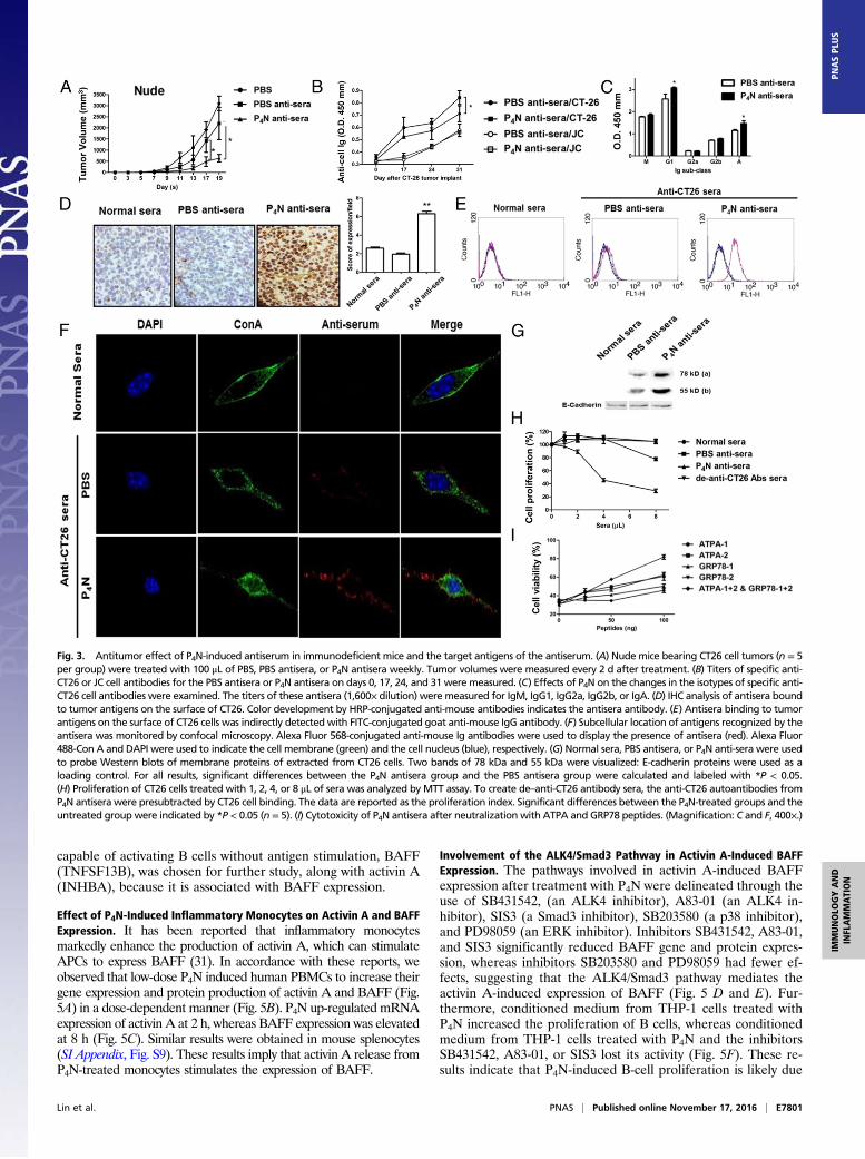
tumor growth (Fig. 3A). Characterization of the antiserarevealed that the titers of specific anti-CT26 antibodies in P4Nantisera were higher than in PBS antisera, regardless of the timeof harvest (Fig. 3B), and the major classes of increased anti-bodies in P4N antisera were IgG1 and IgA (Fig. 3C). Like normalmouse sera, PBS antisera only somewhat recognized the tumorcells in CT26 tumor tissue; however, the P4N antisera displayed astrong binding affinity for the cells (Fig. 3D).Of interest are the antigens that are recognized by EAAs in sera
derived from tumor-bearing mice. Fig. 3E shows that althoughboth antisera recognized surface antigens on CT26 cells, P4Nantisera was more proficient than PBS antisera. By confocal mi-croscopy, the autoantibody-bound antigens on the plasma mem-brane were distributed in a speckled pattern, implying theirpresence in complexes associated with other cell surface proteins(Fig. 3F). A Western blot assay determined that the autoanti-bodies in the antisera recognized 78-kDa and 55-kDa proteins onthe membranes of CT26 cells (Fig. 3G). The antigen profile on thecell membrane was further delineated by co-immunoprecipitationof a membrane protein extract with the antisera. Proteins of 78 kDaand 55 kDa were precipitated, along with at least six otherproteins. The 78-kDa and 55-kDa proteins were identified byultra performance liquid chromatography (UPLC) high-resolutiontandem mass spectrometry (HRMS/MS) as GRP78 and F1F0ATP synthase, respectively (SI Appendix, Fig. S3A).To determine if the antitumor effects of P4N antisera are
mediated by antibodies directed at these two antigens, GRP78-and ATP synthase-specific peptides (SI Appendix, Fig. S3B)were tested for their ability to neutralize the cytotoxic activitiesof the P4N-induced antitumor autoantibodies. First, it wasestablished that P4N antisera is cytotoxic to CT26 cells in culture(Fig. 3H) and that the peptides were able to bind to the P4Nantisera (SI Appendix, Fig. S3C). Subsequently, the cytotoxiceffects of P4N antisera combined with GRP78- and ATP syn-thase-specific peptides were analyzed. The results (Fig. 3I)showed that the peptides decreased the cytotoxicity of P4N antisera,confirming that P4N treatments enhance the production and ac-tivity of anti-GRP78 and anti-F1F0 ATP synthase autoantibodiesthat promote CT26 tumor inhibition.
The Effect of Low-Dose P4N on B-Cell Proliferation and Ig Regulation.The immune modulation effects of P4N were investigated bytesting the ability of low-dose P4N to affect the proliferation ofhuman peripheral blood mononuclear cells (PBMCs) in vitro. Asshown in SI Appendix, Fig. S4A, PBMC proliferation, as measuredby the MTT (3-(4,5-dimethyl-2-thiazolyl)-2,5-diphenyl-2-H-tetra-zolium bromide) colorimetric assay, was increased by 10–40%,depending on the dose of P4N. Flow cytometry analysis of B cellsfrom six healthy individuals showed that P4N significantly in-creased the proliferation of the B-cell population in humanPBMCs (SI Appendix, Fig. S4B). B-cell proliferation was also an-alyzed in mouse splenocytes, and a similar P4N-induced increasedin total splenic B-cell proliferation was observed (SI Appendix, Fig.S4C). Upon further analysis, it was determined that the increase intotal B-cell proliferation was the result of the proliferation ofnaive B cells. P4N had no effect on the number of activated B cellsin the absence of antigen stimulation. These results suggest thatP4N treatments have similar effects in mice and humans.Enhanced green fluorescent protein (EGFP) was used as a
model antigen to monitor specific antibody production and B-celldifferentiation after antigen stimulation. BALB/c mice were im-munized and boosted using i.p. injections with the EGFP antigencombined with one-time or three-times weekly P4N injections. P4Ndramatically enhanced total specific anti-EGFP Ig production,especially when EGFP was combined with P4N injections threetimes a week (SI Appendix, Fig. S4D). In addition, immunizationplus P4N significantly increased IgG1, IgG2b, and IgA production(SI Appendix, Fig. S4E). These results indicate that P4N promotesspecific antibody production by up-regulating the Th2-mediatedimmune response. Analysis of B-cell types revealed that bothmemory and plasma cells were increased after EGFP immuniza-tion (SI Appendix, Fig. S4F). EGFP immunization combined withP4N resulted in an even larger stimulation of B-cell differentia-tion to plasma cells, suggesting that P4N treatment could pro-mote enhanced antibody production.
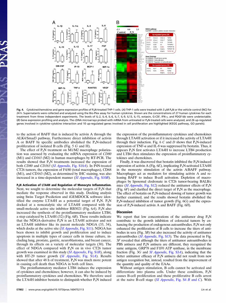
Role of Monocytes and Cytokine Expression in P4N-Induced B-CellProliferation. P4N did not induce the proliferation of isolated Tcells or B cells, yet it increased the proliferation of T-cell–depletedPBMCs (SI Appendix, Fig. S5), indicating that P4N-induced B-cellproliferation may require the presence of monocytes. Examinationof the cytokine profiles of PBMCs after P4N treatment showed thatthe levels of proinflammatory cytokines (TNF-α, IL-1β, and IL-6)and chemokines (IL-8, IP-10, MIP-1α, MIP-1β, and RANTES)were significantly increased after P4N treatment (SI Appendix, Fig.S6). Using a Bio-Plex assay for human cytokines to analyze THP-1monocyte-like cells, it was determined that after P4N treatment, theTHP-1 cells increased the expression of proinflammatory cytokinesand chemokines, similar to PBMCs (Fig. 4A). Moreover, P4Nrapidly induced TNF-α and IL-1β expression in THP-1 monocyteswithin 4 h, and then stimulated IL-8 release after 8 h (SI Appendix,Fig. S7), suggesting that P4N’s effect on monocytes results in acascade of increased cytokines and chemokines.Another cell type in the peripheral tissue that is able to stim-
ulate B-cell proliferation is the DC. The effects of P4N on mousebone marrow-derived (mBM) DCs were analyzed, and the resultsshowed P4N did not induce cell proliferation of mBM-DCs orincrease their expression of BAFF (SI Appendix, Fig. S8 A and B).LTA4A expression was increased in the monocyte/macrophagepopulation, but not in mBM-DCs (SI Appendix, Fig. S8C).To determine the factors involved in P4N-induced B-cell
proliferation, the gene expression pattern of monocytes treatedwith P4N was analyzed by cDNA microarray. The results of thisanalysis revealed 26 up-regulated genes of cytokine–cytokineinteraction [Kyoto Encyclopedia of Genes and Genomes (KEGG)pathway] and 10 up-regulated genes of growth factor activity[Gene Ontology (GO): 0008083] in THP-1 cells following P4Ntreatment (Fig. 4B). Based on these results, the factor most
Fig. 2. Effect of P4N-induced antisera on the formation of pulmonary tu-mor nodules. (A) Number of lung metastases in each group was counted andexpressed as the mean ± SD (n = 5 per group). Mean lung weight (B) andmean survival rate (C) of mice in each group (n = 7 per group) were calcu-lated and plotted. Data were collected from two independent experiments.Significant differences between the P4N antisera group and the PBS antiseragroup were determined and labeled with **P < 0.01 and ***P < 0.001.
E7800 | www.pnas.org/cgi/doi/10.1073/pnas.1604752113 Lin et al.
capable of activating B cells without antigen stimulation, BAFF(TNFSF13B), was chosen for further study, along with activin A(INHBA), because it is associated with BAFF expression.
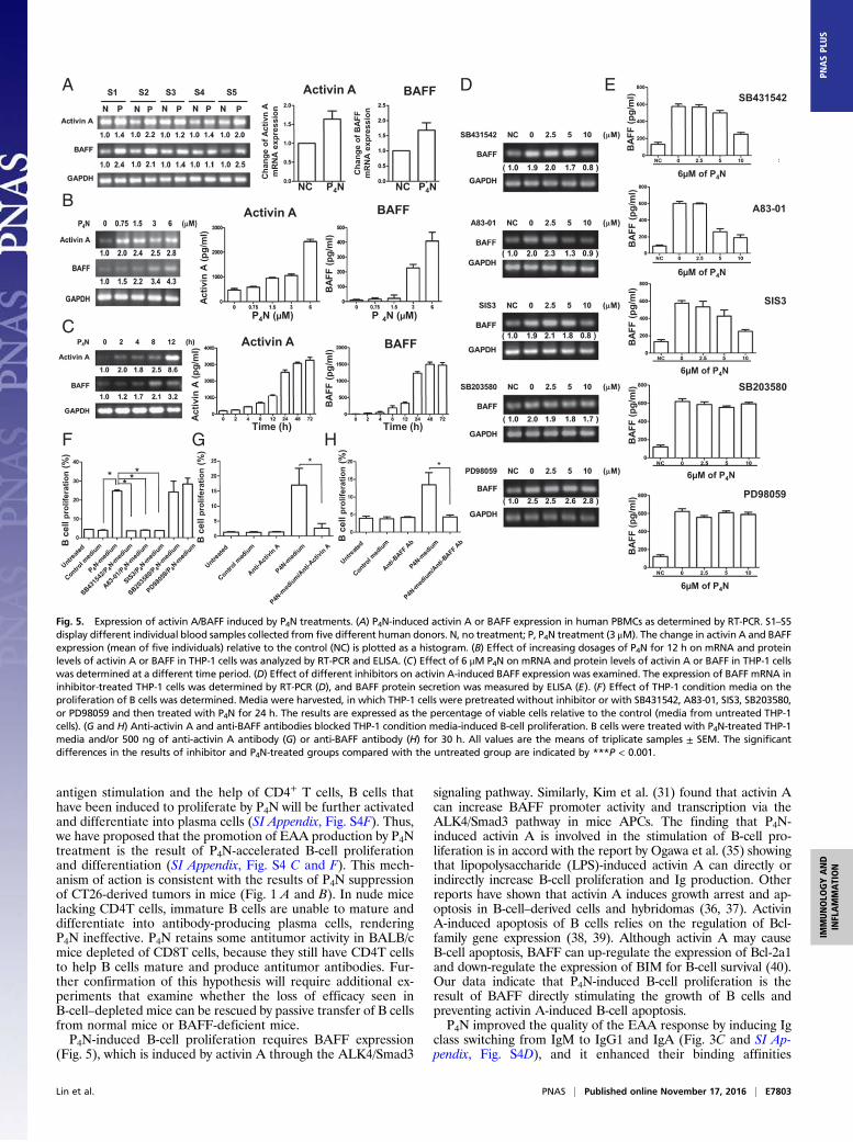
Effect of P4N-Induced Inflammatory Monocytes on Activin A and BAFFExpression. It has been reported that inflammatory monocytesmarkedly enhance the production of activin A, which can stimulateAPCs to express BAFF (31). In accordance with these reports, weobserved that low-dose P4N induced human PBMCs to increase theirgene expression and protein production of activin A and BAFF (Fig.5A) in a dose-dependent manner (Fig. 5B). P4N up-regulated mRNAexpression of activin A at 2 h, whereas BAFF expression was elevatedat 8 h (Fig. 5C). Similar results were obtained in mouse splenocytes(SI Appendix, Fig. S9). These results imply that activin A release fromP4N-treated monocytes stimulates the expression of BAFF.
Involvement of the ALK4/Smad3 Pathway in Activin A-Induced BAFFExpression. The pathways involved in activin A-induced BAFFexpression after treatment with P4N were delineated through theuse of SB431542, (an ALK4 inhibitor), A83-01 (an ALK4 in-hibitor), SIS3 (a Smad3 inhibitor), SB203580 (a p38 inhibitor),and PD98059 (an ERK inhibitor). Inhibitors SB431542, A83-01,and SIS3 significantly reduced BAFF gene and protein expres-sion, whereas inhibitors SB203580 and PD98059 had fewer ef-fects, suggesting that the ALK4/Smad3 pathway mediates theactivin A-induced expression of BAFF (Fig. 5 D and E). Fur-thermore, conditioned medium from THP-1 cells treated withP4N increased the proliferation of B cells, whereas conditionedmedium from THP-1 cells treated with P4N and the inhibitorsSB431542, A83-01, or SIS3 lost its activity (Fig. 5F). These re-sults indicate that P4N-induced B-cell proliferation is likely due
Fig. 3. Antitumor effect of P4N-induced antiserum in immunodeficient mice and the target antigens of the antiserum. (A) Nude mice bearing CT26 cell tumors (n = 5per group) were treated with 100 μL of PBS, PBS antisera, or P4N antisera weekly. Tumor volumes were measured every 2 d after treatment. (B) Titers of specific anti-CT26 or JC cell antibodies for the PBS antisera or P4N antisera on days 0, 17, 24, and 31 were measured. (C) Effects of P4N on the changes in the isotypes of specific anti-CT26 cell antibodies were examined. The titers of these antisera (1,600× dilution) were measured for IgM, IgG1, IgG2a, IgG2b, or IgA. (D) IHC analysis of antisera boundto tumor antigens on the surface of CT26. Color development by HRP-conjugated anti-mouse antibodies indicates the antisera antibody. (E) Antisera binding to tumorantigens on the surface of CT26 cells was indirectly detected with FITC-conjugated goat anti-mouse IgG antibody. (F) Subcellular location of antigens recognized by theantisera was monitored by confocal microscopy. Alexa Fluor 568-conjugated anti-mouse Ig antibodies were used to display the presence of antisera (red). Alexa Fluor488-Con A and DAPI were used to indicate the cell membrane (green) and the cell nucleus (blue), respectively. (G) Normal sera, PBS antisera, or P4N anti-sera were usedto probe Western blots of membrane proteins of extracted from CT26 cells. Two bands of 78 kDa and 55 kDa were visualized: E-cadherin proteins were used as aloading control. For all results, significant differences between the P4N antisera group and the PBS antisera group were calculated and labeled with *P < 0.05.(H) Proliferation of CT26 cells treated with 1, 2, 4, or 8 μL of sera was analyzed by MTT assay. To create de–anti-CT26 antibody sera, the anti-CT26 autoantibodies fromP4N antisera were presubtracted by CT26 cell binding. The data are reported as the proliferation index. Significant differences between the P4N-treated groups and theuntreated group were indicated by *P < 0.05 (n = 5). (I) Cytotoxicity of P4N antisera after neutralization with ATPA and GRP78 peptides. (Magnification: C and F, 400×.)
Lin et al. PNAS | Published online November 17, 2016 | E7801
IMMUNOLO
GYAND
INFLAMMATION
PNASPL
US
to the action of BAFF that is induced by activin A through theALK4/Smad3 pathway, Furthermore direct inhibition of activinA or BAFF by specific antibodies abolished the P4N-inducedproliferation of isolated B cells (Fig. 5 G and H).The effect of P4N treatment on M1/M2 macrophage polariza-
tion was assessed by evaluating the mRNA expression of CD80(M1) and CD163 (M2) in human macrophages by RT-PCR. Theresults showed that P4N treatments increased the expression ofboth CD80 and CD163 (SI Appendix, Fig. S10A). In P4N-treatedCT26 tumors, the expression of F4/80 (total macrophages), CD68(M1), and CD163 (M2), as determined by IHC staining, was alsoincreased in a time-dependent manner (SI Appendix, Fig. S10B).
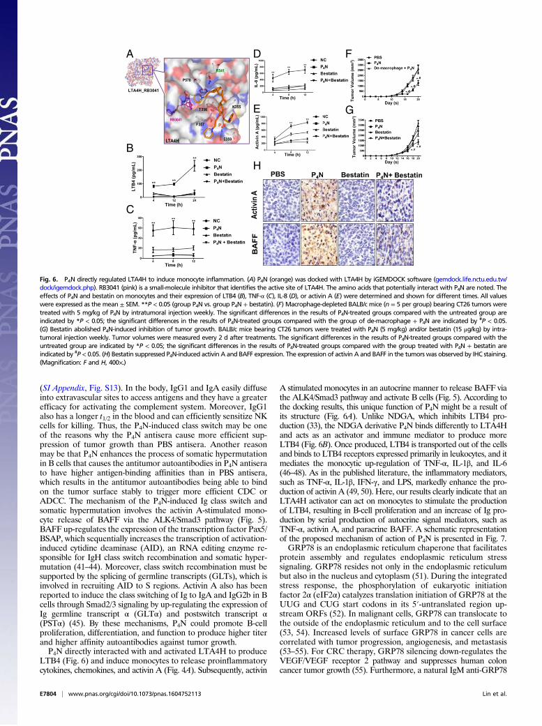
P4N Activation of LTA4H and Regulation of Monocyte Inflammation.Next, we sought to determine the molecular targets of P4N thatmediate the response observed in this study. Docking analysisusing Swiss Target Prediction and iGEMDOCK software iden-tified the enzyme LTA4H as a potential target of P4N. P4Ndocked at a noncatalytic site of LTA4H compared with thesmall-molecule active site inhibitor RB3021 (Fig. 6A). P4N alsoincreased the synthesis of the proinflammatory mediator LTB4,a step catalyzed by LTA4H (32) (Fig. 6B). These results indicatethat the NDGA-derivative P4N is an LTA4H activator and notan LTA4H inhibitor like its parent molecule NDGA (33, 34),which docks at the active site (SI Appendix, Fig. S11). NDGA hasbeen shown to inhibit growth and proliferation and to induceapoptosis in multiple types of cancer cells in tissue culture, in-cluding lung, prostate, gastric, neuroblastoma, and breast cancer,through its effects on a variety of molecular targets (30). Theeffect of NDGA compared with P4N on in vivo CT26 tumorgrowth was examined in this study (SI Appendix, Fig. S1B), alongwith HT-29 tumor growth (SI Appendix, Fig. S1A). Resultsshowed that after 48 h of treatment, P4N was much more potentin causing cell death than NDGA in both cell lines.The proinflammatory mediator LTB4 induces the expression
of cytokines and chemokines; however, it can also be induced byproinflammatory cytokines and chemokines. We therefore usedthe LTA4H inhibitor bestatin to distinguish whether P4N induced
the expression of the proinflammatory cytokines and chemokinesthrough LTA4H activation or if it increased the activity of LTA4Hthrough their induction. Fig. 6 C and D shows that P4N-inducedexpression of TNF-α and IL-8 was suppressed by bestatin. Thus, itappears P4N first activates LTA4H to increase LTB4 productionand LTB4 then stimulates the expression of proinflammatory cy-tokines and chemokines.Finally, it was discovered that bestatin inhibited the P4N-induced
expression of activin A (Fig. 6E), implicating P4N-activated LTA4Hin the monocyte stimulation of the activin A/BAFF pathway.Macrophages act as mediators for stimulating activin A and re-leasing BAFF to induce B-cell activation. Depletion of macro-phages by liposomal clodronate in CT26 tumor-bearing BALB/cmice (SI Appendix, Fig. S12) reduced the antitumor effects of P4N(Fig. 6F) and clarified the direct target of P4N as the macrophage.The effect of bestatin on P4N-induced slowing of tumor growth wasfurther examined, and the results showed bestatin abolished theP4N-induced inhibition of tumor growth (Fig. 6G) and the expres-sion of P4N-induced activin A and BAFF (Fig. 6H).
DiscussionWe report that low concentrations of the antitumor drug P4Ncontribute to the growth inhibition of colorectal tumors by en-hancing the production of endogenous autoantibodies. P4N not onlyenhanced the proliferation of B cells to increase the titers of anti-bodies in sera (Fig. 3B) but also increased the activity of antitumorautoantibodies (SI Appendix, Fig. S13). The data presented in Fig.3F revealed that although the titers of antitumor autoantibodies inPBS antisera and P4N antisera are different, they recognized thesame antigens, GRP78 and F1F0 ATP synthase, in the membranefraction (Fig. 3G and SI Appendix, Fig. S3A), indicating that thebetter antitumor efficacy of P4N antisera did not result from newantigen recognition but, instead, resulted from the improvement ofthe quantity and quality of the EAAs.Without antigen stimulation, B cells cannot be activated and
differentiate into plasma cells. Under these conditions, P4Ncauses B-cell proliferation and these proliferative B cells arrestat the naive B-cell stage (SI Appendix, Fig. S4 B and C). With
Control P4N-2.3 2.30.0
IL-1β IL-1ra IL-5 IL-8
IL-10 IL-12 IL-17 FGF
GM-CSF IP-10 MCP-1 MIP-1a
MIP-1b TNF-α RANTES VEGF
IL-1
β(p
g/m
L)IL
-10
(pg/
mL)
GM
-CSF
(pg/
mL)
MIP
-1b
(pg/
mL)
IL-1
ra (p
g/m
L)IL
-12
(pg/
mL)
IP-1
0 (p
g/m
L)TN
F-α
(pg/
mL)
IL-5
(pg/
mL)
IL-1
7 (p
g/m
L)M
CP-
1 (p
g/m
L)R
ANTE
S (p
g/m
L)
IL-8
(pg/
mL)
FGF
(pg/
mL)
MIP
-1a
(pg/
mL)
VEG
F (p
g/m
L)
A B
Fig. 4. Cytokine/chemokine and gene expression profiles of P4N-treated THP-1 cells. (A) THP-1 cells were treated with 3 μM P4N or the vehicle control (NC) for24 h. Supernatants were collected and analyzed using the Bio-Plex assay for human cytokines. Shown are the concentrations of 27 human cytokines for eachtreatment from three independent experiments. The levels of IL-2, IL-4, IL-6, IL-7, IL-9, IL13, IL-15, eotaxin, G-CSF, IFN-γ, and PDGF-bb were undetectable.(B) Gene expression profiling and analysis. The cDNA microarrays probed with mRNA from untreated or P4N-treated cells were analyzed, and 26 up-regulatedgenes involved in cytokine–cytokine interaction and 10 up-regulated genes involved in cell proliferation are highlighted (KEGG pathway, GO panels).
E7802 | www.pnas.org/cgi/doi/10.1073/pnas.1604752113 Lin et al.
antigen stimulation and the help of CD4+ T cells, B cells thathave been induced to proliferate by P4N will be further activatedand differentiate into plasma cells (SI Appendix, Fig. S4F). Thus,we have proposed that the promotion of EAA production by P4Ntreatment is the result of P4N-accelerated B-cell proliferationand differentiation (SI Appendix, Fig. S4 C and F). This mech-anism of action is consistent with the results of P4N suppressionof CT26-derived tumors in mice (Fig. 1 A and B). In nude micelacking CD4T cells, immature B cells are unable to mature anddifferentiate into antibody-producing plasma cells, renderingP4N ineffective. P4N retains some antitumor activity in BALB/cmice depleted of CD8T cells, because they still have CD4T cellsto help B cells mature and produce antitumor antibodies. Fur-ther confirmation of this hypothesis will require additional ex-periments that examine whether the loss of efficacy seen inB-cell–depleted mice can be rescued by passive transfer of B cellsfrom normal mice or BAFF-deficient mice.P4N-induced B-cell proliferation requires BAFF expression
(Fig. 5), which is induced by activin A through the ALK4/Smad3
signaling pathway. Similarly, Kim et al. (31) found that activin Acan increase BAFF promoter activity and transcription via theALK4/Smad3 pathway in mice APCs. The finding that P4N-induced activin A is involved in the stimulation of B-cell pro-liferation is in accord with the report by Ogawa et al. (35) showingthat lipopolysaccharide (LPS)-induced activin A can directly orindirectly increase B-cell proliferation and Ig production. Otherreports have shown that activin A induces growth arrest and ap-optosis in B-cell–derived cells and hybridomas (36, 37). ActivinA-induced apoptosis of B cells relies on the regulation of Bcl-family gene expression (38, 39). Although activin A may causeB-cell apoptosis, BAFF can up-regulate the expression of Bcl-2a1and down-regulate the expression of BIM for B-cell survival (40).Our data indicate that P4N-induced B-cell proliferation is theresult of BAFF directly stimulating the growth of B cells andpreventing activin A-induced B-cell apoptosis.P4N improved the quality of the EAA response by inducing Ig
class switching from IgM to IgG1 and IgA (Fig. 3C and SI Ap-pendix, Fig. S4D), and it enhanced their binding affinities
Activin A BAFF
BAFF
P4N (μM) P 4N (μM)
Activin A BAFF
SB431542
A83-01
SIS3
SB203580
PD98059
6μM of P4N
6μM of P4N
6μM of P4N
6μM of P4N
6μM of P4N
Activ
inA
(pg/
ml)
Activ
inA
(pg/
ml)
BAF
F (p
g/m
l)B
AFF
(pg/
ml)
Time (h) Time (h)
P4NP4NNC NC
BAF
F (p
g/m
l)B
AFF
(pg/
ml)
BAF
F (p
g/m
l)B
AFF
(pg/
ml)
BAF
F (p
g/m
l)
B c
ell p
rolif
erat
ion
(%)
B c
ell p
rolif
erat
ion
(%)
B c
ell p
rolif
erat
ion
(%)
Cha
nge
of B
AFF
mR
NA
expr
essi
on
Cha
nge
of A
ctiv
nA
mR
NA
expr
essi
on
Activin AA
B
C
D E
F G H
Fig. 5. Expression of activin A/BAFF induced by P4N treatments. (A) P4N-induced activin A or BAFF expression in human PBMCs as determined by RT-PCR. S1–S5display different individual blood samples collected from five different human donors. N, no treatment; P, P4N treatment (3 μM). The change in activin A and BAFFexpression (mean of five individuals) relative to the control (NC) is plotted as a histogram. (B) Effect of increasing dosages of P4N for 12 h on mRNA and proteinlevels of activin A or BAFF in THP-1 cells was analyzed by RT-PCR and ELISA. (C) Effect of 6 μM P4N on mRNA and protein levels of activin A or BAFF in THP-1 cellswas determined at a different time period. (D) Effect of different inhibitors on activin A-induced BAFF expression was examined. The expression of BAFF mRNA ininhibitor-treated THP-1 cells was determined by RT-PCR (D), and BAFF protein secretion was measured by ELISA (E). (F) Effect of THP-1 condition media on theproliferation of B cells was determined. Media were harvested, in which THP-1 cells were pretreated without inhibitor or with SB431542, A83-01, SIS3, SB203580,or PD98059 and then treated with P4N for 24 h. The results are expressed as the percentage of viable cells relative to the control (media from untreated THP-1cells). (G and H) Anti-activin A and anti-BAFF antibodies blocked THP-1 condition media-induced B-cell proliferation. B cells were treated with P4N-treated THP-1media and/or 500 ng of anti-activin A antibody (G) or anti-BAFF antibody (H) for 30 h. All values are the means of triplicate samples ± SEM. The significantdifferences in the results of inhibitor and P4N-treated groups compared with the untreated group are indicated by ***P < 0.001.
Lin et al. PNAS | Published online November 17, 2016 | E7803
IMMUNOLO
GYAND
INFLAMMATION
PNASPL
US
(SI Appendix, Fig. S13). In the body, IgG1 and IgA easily diffuseinto extravascular sites to access antigens and they have a greaterefficacy for activating the complement system. Moreover, IgG1also has a longer t1/2 in the blood and can efficiently sensitize NKcells for killing. Thus, the P4N-induced class switch may be oneof the reasons why the P4N antisera cause more efficient sup-pression of tumor growth than PBS antisera. Another reasonmay be that P4N enhances the process of somatic hypermutationin B cells that causes the antitumor autoantibodies in P4N antiserato have higher antigen-binding affinities than in PBS antisera,which results in the antitumor autoantibodies being able to bindon the tumor surface stably to trigger more efficient CDC orADCC. The mechanism of the P4N-induced Ig class switch andsomatic hypermutation involves the activin A-stimulated mono-cyte release of BAFF via the ALK4/Smad3 pathway (Fig. 5).BAFF up-regulates the expression of the transcription factor Pax5/BSAP, which sequentially increases the transcription of activation-induced cytidine deaminase (AID), an RNA editing enzyme re-sponsible for IgH class switch recombination and somatic hyper-mutation (41–44). Moreover, class switch recombination must besupported by the splicing of germline transcripts (GLTs), which isinvolved in recruiting AID to S regions. Activin A also has beenreported to induce the class switching of Ig to IgA and IgG2b in Bcells through Smad2/3 signaling by up-regulating the expression ofIg germline transcript α (GLTα) and postswitch transcript α(PSTα) (45). By these mechanisms, P4N could promote B-cellproliferation, differentiation, and function to produce higher titerand higher affinity autoantibodies against tumor growth.P4N directly interacted with and activated LTA4H to produce
LTB4 (Fig. 6) and induce monocytes to release proinflammatorycytokines, chemokines, and activin A (Fig. 4A). Subsequently, activin
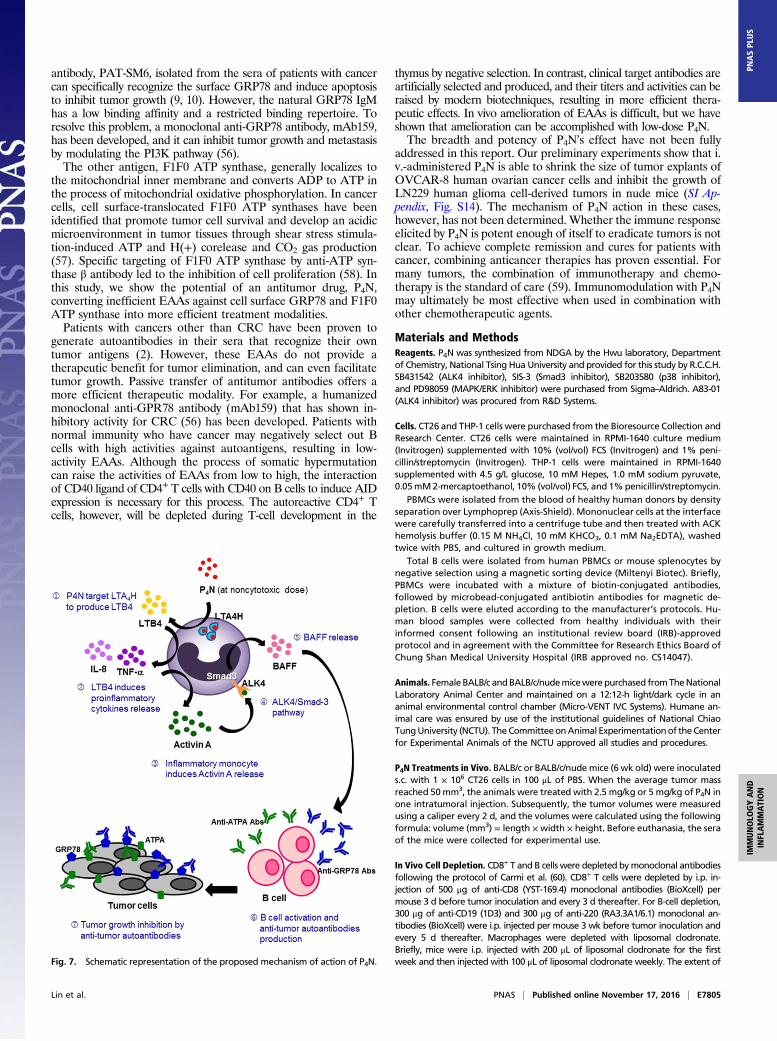
A stimulated monocytes in an autocrine manner to release BAFF viathe ALK4/Smad3 pathway and activate B cells (Fig. 5). According tothe docking results, this unique function of P4N might be a result ofits structure (Fig. 6A). Unlike NDGA, which inhibits LTB4 pro-duction (33), the NDGA derivative P4N binds differently to LTA4Hand acts as an activator and immune mediator to produce moreLTB4 (Fig. 6B). Once produced, LTB4 is transported out of the cellsand binds to LTB4 receptors expressed primarily in leukocytes, and itmediates the monocytic up-regulation of TNF-α, IL-1β, and IL-6(46–48). As in the published literature, the inflammatory mediators,such as TNF-α, IL-1β, IFN-γ, and LPS, markedly enhance the pro-duction of activin A (49, 50). Here, our results clearly indicate that anLTA4H activator can act on monocytes to stimulate the productionof LTB4, resulting in B-cell proliferation and an increase of Ig pro-duction by serial production of autocrine signal mediators, such asTNF-α, activin A, and paracrine BAFF. A schematic representationof the proposed mechanism of action of P4N is presented in Fig. 7.GRP78 is an endoplasmic reticulum chaperone that facilitates
protein assembly and regulates endoplasmic reticulum stresssignaling. GRP78 resides not only in the endoplasmic reticulumbut also in the nucleus and cytoplasm (51). During the integratedstress response, the phosphorylation of eukaryotic initiationfactor 2α (eIF2α) catalyzes translation initiation of GRP78 at theUUG and CUG start codons in its 5′-untranslated region up-stream ORFs (52). In malignant cells, GRP78 can translocate tothe outside of the endoplasmic reticulum and to the cell surface(53, 54). Increased levels of surface GRP78 in cancer cells arecorrelated with tumor progression, angiogenesis, and metastasis(53–55). For CRC therapy, GRP78 silencing down-regulates theVEGF/VEGF receptor 2 pathway and suppresses human coloncancer tumor growth (55). Furthermore, a natural IgM anti-GRP78
LTA4H_RB3041
Tum
or V
olum
e (m
m3 )
Tum
or V
olum
e (m
m3 )
Day (s)
Day (s)
Time (h)
Time (h)
Time (h)
Time (h)
IL-8
(pg/
mL)
TNF-
α(p
g/m
L)LT
B4
(pg/
mL)
Act
ivin
A (p
g/m
L)
A
B
C
D
E
F
G
H
Fig. 6. P4N directly regulated LTA4H to induce monocyte inflammation. (A) P4N (orange) was docked with LTA4H by iGEMDOCK software (gemdock.life.nctu.edu.tw/dock/igemdock.php). RB3041 (pink) is a small-molecule inhibitor that identifies the active site of LTA4H. The amino acids that potentially interact with P4N are noted. Theeffects of P4N and bestatin on monocytes and their expression of LTB4 (B), TNF-α (C), IL-8 (D), or activin A (E) were determined and shown for different times. All valueswere expressed as the mean ± SEM. **P < 0.05 (group P4N vs. group P4N + bestatin). (F) Macrophage-depleted BALB/c mice (n = 5 per group) bearing CT26 tumors weretreated with 5 mg/kg of P4N by intratumoral injection weekly. The significant differences in the results of P4N-treated groups compared with the untreated group areindicated by *P < 0.05; the significant differences in the results of P4N-treated groups compared with the group of de-macrophage + P4N are indicated by #P < 0.05.(G) Bestatin abolished P4N-induced inhibition of tumor growth. BALB/c mice bearing CT26 tumors were treated with P4N (5 mg/kg) and/or bestatin (15 μg/kg) by intra-tumoral injection weekly. Tumor volumes were measured every 2 d after treatments. The significant differences in the results of P4N-treated groups compared with theuntreated group are indicated by *P < 0.05; the significant differences in the results of P4N-treated groups compared with the group treated with P4N + bestatin areindicated by #P < 0.05. (H) Bestatin suppressed P4N-induced activin A and BAFF expression. The expression of activin A and BAFF in the tumors was observed by IHC staining.(Magnification: F and H, 400×.)
E7804 | www.pnas.org/cgi/doi/10.1073/pnas.1604752113 Lin et al.
antibody, PAT-SM6, isolated from the sera of patients with cancercan specifically recognize the surface GRP78 and induce apoptosisto inhibit tumor growth (9, 10). However, the natural GRP78 IgMhas a low binding affinity and a restricted binding repertoire. Toresolve this problem, a monoclonal anti-GRP78 antibody, mAb159,has been developed, and it can inhibit tumor growth and metastasisby modulating the PI3K pathway (56).The other antigen, F1F0 ATP synthase, generally localizes to
the mitochondrial inner membrane and converts ADP to ATP inthe process of mitochondrial oxidative phosphorylation. In cancercells, cell surface-translocated F1F0 ATP synthases have beenidentified that promote tumor cell survival and develop an acidicmicroenvironment in tumor tissues through shear stress stimula-tion-induced ATP and H(+) corelease and CO2 gas production(57). Specific targeting of F1F0 ATP synthase by anti-ATP syn-thase β antibody led to the inhibition of cell proliferation (58). Inthis study, we show the potential of an antitumor drug, P4N,converting inefficient EAAs against cell surface GRP78 and F1F0ATP synthase into more efficient treatment modalities.Patients with cancers other than CRC have been proven to
generate autoantibodies in their sera that recognize their owntumor antigens (2). However, these EAAs do not provide atherapeutic benefit for tumor elimination, and can even facilitatetumor growth. Passive transfer of antitumor antibodies offers amore efficient therapeutic modality. For example, a humanizedmonoclonal anti-GPR78 antibody (mAb159) that has shown in-hibitory activity for CRC (56) has been developed. Patients withnormal immunity who have cancer may negatively select out Bcells with high activities against autoantigens, resulting in low-activity EAAs. Although the process of somatic hypermutationcan raise the activities of EAAs from low to high, the interactionof CD40 ligand of CD4+ T cells with CD40 on B cells to induce AIDexpression is necessary for this process. The autoreactive CD4+ Tcells, however, will be depleted during T-cell development in the
thymus by negative selection. In contrast, clinical target antibodies areartificially selected and produced, and their titers and activities can beraised by modern biotechniques, resulting in more efficient thera-peutic effects. In vivo amelioration of EAAs is difficult, but we haveshown that amelioration can be accomplished with low-dose P4N.The breadth and potency of P4N’s effect have not been fully
addressed in this report. Our preliminary experiments show that i.v.-administered P4N is able to shrink the size of tumor explants ofOVCAR-8 human ovarian cancer cells and inhibit the growth ofLN229 human glioma cell-derived tumors in nude mice (SI Ap-pendix, Fig. S14). The mechanism of P4N action in these cases,however, has not been determined. Whether the immune responseelicited by P4N is potent enough of itself to eradicate tumors is notclear. To achieve complete remission and cures for patients withcancer, combining anticancer therapies has proven essential. Formany tumors, the combination of immunotherapy and chemo-therapy is the standard of care (59). Immunomodulation with P4Nmay ultimately be most effective when used in combination withother chemotherapeutic agents.
Materials and MethodsReagents. P4N was synthesized from NDGA by the Hwu laboratory, Departmentof Chemistry, National Tsing Hua University and provided for this study by R.C.C.H.SB431542 (ALK4 inhibitor), SIS-3 (Smad3 inhibitor), SB203580 (p38 inhibitor),and PD98059 (MAPK/ERK inhibitor) were purchased from Sigma–Aldrich. A83-01(ALK4 inhibitor) was procured from R&D Systems.
Cells. CT26 and THP-1 cells were purchased from the Bioresource Collection andResearch Center. CT26 cells were maintained in RPMI-1640 culture medium(Invitrogen) supplemented with 10% (vol/vol) FCS (Invitrogen) and 1% peni-cillin/streptomycin (Invitrogen). THP-1 cells were maintained in RPMI-1640supplemented with 4.5 g/L glucose, 10 mM Hepes, 1.0 mM sodium pyruvate,0.05mM2-mercaptoethanol, 10% (vol/vol) FCS, and 1%penicillin/streptomycin.
PBMCs were isolated from the blood of healthy human donors by densityseparation over Lymphoprep (Axis-Shield). Mononuclear cells at the interfacewere carefully transferred into a centrifuge tube and then treated with ACKhemolysis buffer (0.15 M NH4Cl, 10 mM KHCO3, 0.1 mM Na2EDTA), washedtwice with PBS, and cultured in growth medium.
Total B cells were isolated from human PBMCs or mouse splenocytes bynegative selection using a magnetic sorting device (Miltenyi Biotec). Briefly,PBMCs were incubated with a mixture of biotin-conjugated antibodies,followed by microbead-conjugated antibiotin antibodies for magnetic de-pletion. B cells were eluted according to the manufacturer’s protocols. Hu-man blood samples were collected from healthy individuals with theirinformed consent following an institutional review board (IRB)-approvedprotocol and in agreement with the Committee for Research Ethics Board ofChung Shan Medical University Hospital (IRB approved no. CS14047).
Animals. FemaleBALB/c andBALB/c/nudemicewerepurchased fromTheNationalLaboratory Animal Center and maintained on a 12:12-h light/dark cycle in ananimal environmental control chamber (Micro-VENT IVC Systems). Humane an-imal care was ensured by use of the institutional guidelines of National ChiaoTungUniversity (NCTU). TheCommitteeonAnimal Experimentationof theCenterfor Experimental Animals of the NCTU approved all studies and procedures.
P4N Treatments in Vivo. BALB/c or BALB/c/nude mice (6 wk old) were inoculateds.c. with 1 × 10⁶ CT26 cells in 100 μL of PBS. When the average tumor massreached 50mm3, the animals were treated with 2.5 mg/kg or 5 mg/kg of P4N inone intratumoral injection. Subsequently, the tumor volumes were measuredusing a caliper every 2 d, and the volumes were calculated using the followingformula: volume (mm3) = length ×width × height. Before euthanasia, the seraof the mice were collected for experimental use.
In Vivo Cell Depletion. CD8+ T and B cells were depleted bymonoclonal antibodiesfollowing the protocol of Carmi et al. (60). CD8+ T cells were depleted by i.p. in-jection of 500 μg of anti-CD8 (YST-169.4) monoclonal antibodies (BioXcell) permouse 3 d before tumor inoculation and every 3 d thereafter. For B-cell depletion,300 μg of anti-CD19 (1D3) and 300 μg of anti-220 (RA3.3A1/6.1) monoclonal an-tibodies (BioXcell) were i.p. injected per mouse 3 wk before tumor inoculation andevery 5 d thereafter. Macrophages were depleted with liposomal clodronate.Briefly, mice were i.p. injected with 200 μL of liposomal clodronate for the firstweek and then injected with 100 μL of liposomal clodronate weekly. The extent ofFig. 7. Schematic representation of the proposed mechanism of action of P4N.
Lin et al. PNAS | Published online November 17, 2016 | E7805
IMMUNOLO
GYAND
INFLAMMATION
PNASPL
US

depletion of CD8T, B cells, and macrophages was evaluated by IHC staining. Tumortissue was randomly selected from depleted animals; embedded in paraffin; andprobed with rat monoclonal anti-CD20, anti-CD8, and anti-F4/80 antibodies (resultsare shown in SI Appendix, Figs. S12 and S15).
Sera Transfer in Vivo. In a metastatic model, BALB/c mice were i.v. injected with1 × 106 CT26 cells in 100 μL of PBS via the tail vein, and then treated with 100 μLof antisera once every week. Upon the death of the mice, the lungs were dis-sected, observed, and photographed. Three mice in the P4N antisera groupwere killed 24 d after injection. Tumor nodules on the lungs were counted, andlung weights were determined. Survival of the mice was noted, and rates ofsurvival were calculated. In an s.c. tumormodel, the nudemice bearing∼50-mm3
CT26 tumors were injected i.v. with 100 μL of the different antisera onceevery week. The tumor volumes were measured as described above.
IHC Staining. Tumors isolated from P4N- and vehicle-treated mice were em-bedded in paraffin, and thin sections were made and processed for IHCstaining. The sections were probed with rat monoclonal anti-F4/80 (1:50dilution; GeneTex) and anti-CD20 (1:200 dilution; Santa Cruz Biotechnology),hamster monoclonal anti-CD11c (1:25 dilution; GeneTex), rabbit monoclonalanti-CD3 (1:50 dilution; GeneTex), and rabbit polyclonal anti–TNF-α andanti–IL-8 antibodies (1:50 dilution; Assay BioTech) at 4 °C overnight, and thedetection antibodies were recognized using a horseradish peroxidase (HRP)-conjugated anti-rat, anti-hamster, or anti-rabbit IgG antibody (1:1,500 di-lution; Santa Cruz Biotechnology). The immune complexes in the sectionswere visualized using the LSAB2 System (DAKO). The sections were coun-terstained with hematoxylin, mounted, observed under a light microscopeat a magnification of 400×, and photographed.
Sections of CT26 tumors from untreated mice were incubated with a 200-fold dilution of normal sera, PBS antisera, or P4N antisera at 4 °C overnight.The sections were then incubated with an HRP-conjugated anti-mouse IgGantibody (1:1,500 dilution; Santa Cruz Biotechnology), developed, counter-stained, and photographed as described above.
Titers and Ig Classes of Antitumor Autoantibodies. Blood was collected weeklyfrom CT26-bearing mice treated with PBS or 5 mg/kg P4N, and the titers ofCT26-specific antibodies in the sera were measured by the following method.The wells of 96-well culture plates were seeded with CT26 (106 cells per well) ormouse mammary gland adenocarcinoma JC cells (106 cells per well). On thefollowing day, the cells were fixed with 4% (wt/vol) paraformaldehyde,washed, and blocked with 300 μL of 2% (wt/vol) skim milk in PBST (PBS bufferwith 0.05% Tween-20) for 1 h. One hundred microliters of 6,400-fold dilutedsera in PBS containing 0.5% skim milk was loaded into each well and in-cubated at room temperature for 2 h. After washing three times, 100 μL ofHRP-conjugated anti-mouse Ig antibody (1:10,000 dilution; Sigma–Aldrich) wasadded to each well and incubated for 1 h. After washing three times, 100 μL ofNeA-Blue (Clinical Science Products, Inc.) was added to each well, incubated for20 min, and stopped using 100 μL of 1-N HCl. The optical density was measured at450 nm using an ELISA reader (Tecan). The isotypes of specific anti-CT26 cellantibodies in antisera (1:1,600 dilution) were determined by using HRP-con-jugated specific anti-mouse IgM, IgG1, IgG2a, IgG2b, or IgA antibodies (Acris).
Immunofluorescence Analysis of Cell Surface Tumor Antigens. CT26 cells wereseeded on glass cover slides, and fixed with 4% (wt/wt) paraformaldehydeThe fixed cells were then incubated with normal sera, PBS antisera, or P4Nantisera for 1 h and detected with secondary antibodies conjugated to AlexaFluor 568 (Molecular Probes). Alexa Fluor 488-conjugated Con A (MolecularProbes) and DAPI (Molecular Probes) were used to label the plasma mem-brane and nucleus, respectively. The images of tumor antigens recognizedby the antisera were photographed at a magnification of 400× using a ZeissLSM 510 META confocal microscope (Carl Zeiss).
Western Blot Analysis. Themembrane proteins of 3×10⁷ CT26 cellswere extractedand harvested with the Mem-PER Eukaryotic Membrane Protein Extraction Re-agent Kit (Thermo Fisher Scientific), following the manufacturer’s instructions. Fiftymicrograms of total membrane protein was subjected to SDS/PAGE; transferred toNitrocellulose Blotting Membrane (General Electric); and probed with normal sera,PBS antisera, or P4N antisera. The membranes were then blocked with 2% (wt/vol)milk in PBST, incubated with HRP-conjugated anti-mouse Ig antibody (1:10,000dilution; Sigma–Aldrich), and developed with the WesternBright ECL Westernblotting detection kit (Advansta). Antibody-bound proteins were visualized by theHansor Luminescence Image System (Hansor). E-cadherin proteins in all sampleswere probed by rabbit polyclonal anti–E-cadherin antibody (1:1,000 di-lution; GeneTex) and HRP-conjugated anti-rabbit Ig antibody (1:10,000dilution; Sigma–Aldrich).
Co-Immunoprecipitation Assay. Five microliters of normal sera, PBS antisera, orP4N antisera was mixed with 100 μL of protein G agarose (Merck Millipore),and then covalently linked with disuccinimidyl suberate (Thermo Fisher Sci-entific). After washing, 5 μL of protein G agarose-conjugated sera was in-cubated with 400 μL of membrane proteins at room temperature. The immunecomplexes were then washed three times with lysis buffer and eluted withelution buffer (Thermo Fisher Scientific). The precipitated samples were thenheated in reducing sample buffer and resolved by the SDS/PAGE.
Antigen Identification by UPLC/HRMS/MS. Proteins were excised from SDS poly-acrylamide gels, digested with the In-Gel Tryptic Digestion Kit (Thermo Fisher Sci-entific), and identified by UPLC/HRMS/MS (Bruker BioSpin). Peptide sequenceinformation was used to search sequences in the protein database of the NationalCenter for Biotechnology InformationusingBLAST (blast.ncbi.nlm.nih.gov/blast.cgi).
Cytokine Multiplex Assay. THP-1 cells (5 × 10⁵ cells per milliliter) in 1 mL ofculture medium were seeded in each well of a 24-well microplate andtreated with 3 μM P4N for 24 h. Cell culture supernatants were collected andanalyzed by a cytokine multiplex assay following the Bio-Plex Pro HumanCytokine Standard Group I 27-Plex, according to the manufacturer’s protocol(Bio-Rad). The change levels of cytokine were calculated as follows: Changelevel = (value of the P4N-treated cells)/(value of the untreated cells) × 100%.
Gene Expression Profiling and Analysis. Amplification and labeling of RNAwereperformed with the IlluminaTotalPrep RNA Amplification Kit from Life Tech-nologies (Ambion; Applied Biosystems) using 150 ng of RNA per sample. La-beled RNA (750 ng) was hybridized to Illumina HT-12 v4 Expression BeadChips(∼48,000 probes) and processed according to the manufacturer’s protocol.Expression data underwent quality control and normalization by GenomeStudio (Illumina). Genes differentially expressed in the cells treated with P4Nrelative to the untreated cells were identified, with special emphasis given togenes involved in cytokine–cytokine receptor interaction (KEGG pathway).Genes with a P value <0.05 and a fold change ≥0.4 were considered to bedifferentially expressed, up-regulated genes. The identified genes were sub-jected to the Database for Annotation, Visualization, and Integrated Discovery(https://david.ncifcrf.gov/) for GO and KEGG pathway enrichment analysis. AP value <0.05 was set as the threshold of enrichment analysis.
RT-PCR. Human PBMCs or THP-1 cells were treated with P4N, and the mRNAexpression of activin A and BAFF in these cells was then measured by RT-PCR.Briefly, total cellular RNA was extracted with TRIzol reagent (Invitrogen) andreverse-transcribed into cDNA using the SuperScript RT-Kit (Invitrogen). ThecDNA of activin A and BAFF was then amplified by PCR. The primers for humanactivin A were forward primer 5′-GCCGAGTCAGGAACAGCCAG-3′ and reverseprimer 5′-TTTCTTCTTCTTCTTGCCCAGGA-3′, and the primers for human BAFFwere forward primer 5′-ATGGATGACTCCACAGAAAGG-3′ and reverse primer5′-TGGTAGAAAGACACCACCG-3′. All PCR reagents used to amplify the cDNAwere purchased from Promega. GAPDH cDNA in the samples was used tonormalize the loading amounts in each reaction. Finally, PCR products wereresolved by electrophoresis on 2% agarose gels, stained with ethidium bro-mide, and photographed using the Uni-photo band tool (EZ laboratory).
Cell Proliferation Assay. Purified B cells (2 × 10⁵ cells per well) were prestainedwith DiI fluorescent dye, seeded, and treated with THP-1–conditioned media.After 30 h, the number of viable cells was determined by flow cytometry.
Activin A and BAFF Neutralization. To examination the roles of activin A andBAFF involved in P4N-induced B-cell proliferation, purified B cells (2 × 10⁵
cells per well) were treated with P4N-treated THP-1 media and/or 500 ng ofneutralized anti-activin A antibody or anti-BAFF antibody for 30 h. Then,B-cell proliferation was determined as previously described.
ELISAs. THP-1 cells (1 × 10⁶ cells per milliliter per well) in a 24-well culture platewere pretreated with 10 μM bestatin (LTA4H inhibitor; Sigma–Aldrich) for 2 hand then treated with 3 μM P4N for various time intervals. The levels of LTB4 inthe culture media were determined by the LTB4 ELISA Kit (Enzo Life Sciences).The amounts of TNF-α, IL-8, and activin A in the cultured media were mea-sured in a similar manner.
Statistical Analyses. The results are presented as the mean ± SEM. The sta-tistical significance was evaluated using Student’s t test, and P < 0.05 wasconsidered significant.
E7806 | www.pnas.org/cgi/doi/10.1073/pnas.1604752113 Lin et al.

ACKNOWLEDGMENTS. We thank Prof. Jinn-Moon Yang for kind support indocking technology. We thank the core facility of the multiphoton andconfocal microscope system and UPLC/HRMS/MS of the NCTU. This research
was supported by Grants NCTU 102W976 and NCTU 103W976 (to R.C.C.H.)and Ministry of Science and Technology of Taiwan (MOST) Grants MOST 104-2627-M-009-007 and MOST 103-2112-M-009-011-MY3 (to C.C.C.).
1. International Agency for Research on Cancer, WHO (2015) GLOBOCAN 2012: Esti-mated Cancer Incidence, Mortality and Prevalence Worldwide in 2012. Available atglobocan.iarc.fr/Default.aspx. Accessed August 10, 2015.
2. Chen H, Werner S, Tao S, Zörnig I, Brenner H (2014) Blood autoantibodies againsttumor-associated antigens as biomarkers in early detection of colorectal cancer.Cancer Lett 346(2):178–187.
3. Babel I, et al. (2009) Identification of tumor-associated autoantigens for the diagnosisof colorectal cancer in serum using high density protein microarrays. Mol CellProteomics 8(10):2382–2395.
4. Ran Y, et al. (2008) Profiling tumor-associated autoantibodies for the detection ofcolon cancer. Clin Cancer Res 14(9):2696–2700.
5. Babel I, et al. (2009) Identification of tumor-associated autoantigens for the diagnosisof colorectal cancer in serum using high density protein microarrays. Mol CellProteomics 8(10):2382–2395.
6. Bei R, Masuelli L, Palumbo C, Modesti M, Modesti A (2009) A common repertoire ofautoantibodies is shared by cancer and autoimmune disease patients: Inflammationin their induction and impact on tumor growth. Cancer Lett 281(1):8–23.
7. Zhang Y, Gallastegui N, Rosenblatt JD (2015) Regulatory B cells in anti-tumor im-munity. Int Immunol 27(10):521–530.
8. Díaz-Zaragoza M, Hernández-Ávila R, Viedma-Rodríguez R, Arenas-Aranda D, Ostoa-Saloma P (2015) Natural and adaptive IgM antibodies in the recognition of tumor-associated antigens of breast cancer (Review). Oncol Rep 34(3):1106–1114.
9. Rasche L, et al. (2015) GRP78-directed immunotherapy in relapsed or refractorymultiple myeloma - results from a phase 1 trial with the monoclonal immunoglobulinM antibody PAT-SM6. Haematologica 100(3):377–384.
10. Rasche L, et al. (2013) The natural human IgM antibody PAT-SM6 induces apoptosis inprimary human multiple myeloma cells by targeting heat shock protein GRP78. PLoSOne 8(5):e63414.
11. Oflazoglu E, Audoly LP (2010) Evolution of anti-CD20 monoclonal antibody thera-peutics in oncology. MAbs 2(1):14–19.
12. Bianchini G, Gianni L (2014) The immune system and response to HER2-targetedtreatment in breast cancer. Lancet Oncol 15(2):e58–e68.
13. Taylor RP, Lindorfer MA (2008) Immunotherapeutic mechanisms of anti-CD20monoclonal antibodies. Curr Opin Immunol 20(4):444–449.
14. Selenko N, et al. (2001) CD20 antibody (C2B8)-induced apoptosis of lymphoma cellspromotes phagocytosis by dendritic cells and cross-priming of CD8+ cytotoxic T cells.Leukemia 15(10):1619–1626.
15. He Q, et al. (2011) Low-dose paclitaxel enhances the anti-tumor efficacy of GM-CSFsurface-modified whole-tumor-cell vaccine in mouse model of prostate cancer. CancerImmunol Immunother 60(5):715–730.
16. Sevko A, et al. (2012) Application of paclitaxel in low non-cytotoxic doses supportsvaccination with melanoma antigens in normal mice. J Immunotoxicol 9(3):275–281.
17. Zhong H, et al. (2007) Low-dose paclitaxel prior to intratumoral dendritic cell vaccinemodulates intratumoral cytokine network and lung cancer growth. Clin Cancer Res13(18 Pt 1):5455–5462.
18. Heylmann D, et al. (2013) Human CD4+CD25+ regulatory T cells are sensitive to lowdose cyclophosphamide: Implications for the immune response. PLoS One 8(12):e83384.
19. Dimeloe S, et al. (2014) Human regulatory T cells lack the cyclophosphamide-extruding transporter ABCB1 and are more susceptible to cyclophosphamide-inducedapoptosis. Eur J Immunol 44(12):3614–3620.
20. Lutz ER, et al. (2014) Immunotherapy converts nonimmunogenic pancreatic tumorsinto immunogenic foci of immune regulation. Cancer Immunol Res 2(7):616–631.
21. Sharabi A, Haran-Ghera N (2011) Immune recovery after cyclophosphamide treat-ment in multiple myeloma: Implication for maintenance immunotherapy. BoneMarrow Res 2011:269519.
22. Peng S, et al. (2013) Low-dose cyclophosphamide administered as daily or single doseenhances the antitumor effects of a therapeutic HPV vaccine. Cancer ImmunolImmunother 62(1):171–182.
23. Sharabi A, Laronne-Bar-On A, Meshorer A, Haran-Ghera N (2010) Chemoimmunotherapyreduces the progression of multiple myeloma in a mouse model. Cancer Prev Res(Phila) 3(10):1265–1276.
24. Kaneno R, et al. (2011) Chemotherapeutic agents in low noncytotoxic concentrationsincrease immunogenicity of human colon cancer cells. Cell Oncol (Dordr) 34(2):97–106.
25. Shurin GV, Tourkova IL, Kaneno R, Shurin MR (2009) Chemotherapeutic agents innoncytotoxic concentrations increase antigen presentation by dendritic cells via an IL-12-dependent mechanism. J Immunol 183(1):137–144.
26. Kaneno R, Shurin GV, Tourkova IL, Shurin MR (2009) Chemomodulation of humandendritic cell function by antineoplastic agents in low noncytotoxic concentrations.J Transl Med 7:58.
27. Dohm JA, et al. (2005) Influence of ions, hydration, and the transcriptional inhibitorP4N on the conformations of the Sp1 binding site. J Mol Biol 349(4):731–744.
28. Hwu JR, Hsu MH, Huang RC (2008) New nordihydroguaiaretic acid derivatives as anti-HIV agents. Bioorg Med Chem Lett 18(6):1884–1888.
29. Lü JM, et al. (2010) Molecular mechanisms and clinical applications of nordihydroguaia-retic acid (NDGA) and its derivatives: An update. Med Sci Monit 16(5):RA93–RA100.
30. Zhang Y, et al. (2012) mTORC1 is a target of nordihydroguaiaretic acid to preventbreast tumor growth in vitro and in vivo. Breast Cancer Res Treat 136(2):379–388.
31. Kim JH, Seo GY, Kim PH (2011) Activin A stimulates mouse APCs to express BAFF viaALK4-Smad3 pathway. Immune Netw 11(4):196–202.
32. Haeggström JZ (2004) Leukotriene A4 hydrolase/aminopeptidase, the gatekeeper ofchemotactic leukotriene B4 biosynthesis. J Biol Chem 279(49):50639–50642.
33. Conti P, et al. (1993) Human recombinant IL-1 receptor antagonist (IL-1Ra) inhibitsleukotriene B4 generation from human monocyte suspensions stimulated by lipo-polysaccharide (LPS). Clin Exp Immunol 91(3):526–531.
34. Maloff BL, Fefer D, Cooke GM, Ackerman NR (1987) Inhibition of LTB4 binding tohuman neutrophils by nordihydroguaiaretic acid. Agents Actions 21(3-4):358–360.
35. Ogawa K, Funaba M, Tsujimoto M (2008) A dual role of activin A in regulating im-munoglobulin production of B cells. J Leukoc Biol 83(6):1451–1458.
36. Hashimoto O, et al. (1998) The role of activin type I receptors in activin A-inducedgrowth arrest and apoptosis in mouse B-cell hybridoma cells. Cell Signal 10(10):743–749.
37. Zipori D, Barda-Saad M (2001) Role of activin A in negative regulation of normal andtumor B lymphocytes. J Leukoc Biol 69(6):867–873.
38. Wang B, et al. (2009) Involvement of ERK, Bcl-2 family and caspase 3 in recombinanthuman activin A-induced apoptosis in A549. Toxicology 258(2-3):176–183.
39. Koseki T, et al. (1998) Correlation between Bcl-X expression and B-cell hybridomaapoptosis induced by activin A. Cell Signal 10(7):517–521.
40. Craxton A, Draves KE, Gruppi A, Clark EA (2005) BAFF regulates B cell survival bydownregulating the BH3-only family member Bim via the ERK pathway. J Exp Med202(10):1363–1374.
41. Anderson KS, et al. (2015) Autoantibody signature for the serologic detection ofovarian cancer. J Proteome Res 14(1):578–586.
42. Xu Z, et al. (2007) Regulation of aicda expression and AID activity: relevance to somatichypermutation and class switch DNA recombination. Crit Rev Immunol 27(4):367–397.
43. Stavnezer J, Schrader CE (2014) IgH chain class switch recombination: Mechanism andregulation. J Immunol 193(11):5370–5378.
44. Muramatsu M, et al. (2000) Class switch recombination and hypermutation requireactivation-induced cytidine deaminase (AID), a potential RNA editing enzyme. Cell102(5):553–563.
45. Lee HJ, Seo GY, Kim HA, Kim PH (2008) Activin A stimulates IgA expression in mouse Bcells. Biochem Biophys Res Commun 366(2):574–578.
46. Rola-Pleszczynski M, Lemaire I (1985) Leukotrienes augment interleukin 1 productionby human monocytes. J Immunol 135(6):3958–3961.
47. Poubelle PE, Stankova J, Grassi J, Rola-Pleszczynski M (1991) Leukotriene B4 up-regu-lates IL-6 rather than IL-1 synthesis in human monocytes. Agents Actions 34(1-2):42–45.
48. Xu S, Lu H, Lin J, Chen Z, Jiang D (2010) Regulation of TNFalpha and IL1beta in rheu-matoid arthritis synovial fibroblasts by leukotriene B4. Rheumatol Int 30(9):1183–1189.
49. Yoshino O, et al. (2011) Activin-A is induced by interleukin-1β and tumor necrosis factor-α and enhances the mRNA expression of interleukin-6 and protease-activated receptor-2 and proliferation of stromal cells from endometrioma. Fertil Steril 96(1):118–121.
50. Jones KL, et al. (2007) Activin A is a critical component of the inflammatory response,and its binding protein, follistatin, reduces mortality in endotoxemia. Proc Natl AcadSci USA 104(41):16239–16244.
51. Ni M, Zhang Y, Lee AS (2011) Beyond the endoplasmic reticulum: Atypical GRP78 incell viability, signalling and therapeutic targeting. Biochem J 434(2):181–188.
52. Starck SR, et al. (2016) Translation from the 5′ untranslated region shapes the in-tegrated stress response. Science 351(6272):aad3867.
53. Yao X, et al. (2015) Cell surface GRP78 accelerated breast cancer cell proliferation andmigration by activating STAT3. PLoS One 10(5):e0125634.
54. Li Z, et al. (2013) Cell-surface GRP78 facilitates colorectal cancer cell migration andinvasion. Int J Biochem Cell Biol 45(5):987–994.
55. Kuo LJ, Hung CS, Chen WY, Chang YJ, Wei PL (2013) Glucose-regulated protein 78silencing down-regulates vascular endothelial growth factor/vascular endothelialgrowth factor receptor 2 pathway to suppress human colon cancer tumor growth.J Surg Res 185(1):264–272.
56. Liu R, et al. (2013) Monoclonal antibody against cell surface GRP78 as a novel agent insuppressing PI3K/AKT signaling, tumor growth, and metastasis. Clin Cancer Res19(24):6802–6811.
57. Kawai Y, Kaidoh M, Yokoyama Y, Ohhashi T (2013) Cell surface F1/FO ATP synthasecontributes to interstitial flow-mediated development of the acidic microenviron-ment in tumor tissues. Am J Physiol Cell Physiol 305(11):C1139–C1150.
58. Fliedner SM, et al. (2015) Potential therapeutic target for malignant paragangliomas:ATP synthase on the surface of paraganglioma cells. Am J Cancer Res 5(4):1558–1570.
59. Mahoney KM, Rennert PD, Freeman GJ (2015) Combination cancer immunotherapyand new immunomodulatory targets. Nat Rev Drug Discov 14(8):561–584.
60. Carmi Y, et al. (2015) Allogeneic IgG combined with dendritic cell stimuli induceantitumour T-cell immunity. Nature 521(7550):99–104.
Lin et al. PNAS | Published online November 17, 2016 | E7807
IMMUNOLO
GYAND
INFLAMMATION
PNASPL
US