in situ microtomography investigation of microstructural
TRANSCRIPT

In situ microtomography investigation of microstructural evolution in
Al-Cu alloys during holding in semi-solid state
S. TERZI1,L. SALVO2, M. SUERY2, A. DAHLE1, E. BOLLER3
1. Australian Research Council Centre of Excellence for Design in Light Metals, Materials Engineering, The University of Queensland, Brisbane Queensland 4072, Australia;
2. Université de Grenoble, Science et Ingénierie des Matériaux et Procédés, Génie Physique et Mécanique des Matériaux, UMR CNRS 5266, Grenoble INP, Université Joseph Fourrier, BP46, 38402 Saint-Martin d’Hères Cedex, France;
3. European Synchrotron Radiation Facility, 6 rue Jules Horowitz, BP220, 38043 Grenoble Cedex, France
Received 13 May 2010; accepted 25 June 2010
Abstract: The aim of this paper is to report the results of experiments carried out on Al-Cu alloys with different Cu contents, studying the microstructure evolution during holding in the semi-solid state. The 3-D microstructure was observed by in situ X-ray microtomography carried out at ESRF Grenoble, France. The variation of the solid-liquid interface area per unit volume during holding was determined. In addition, local observations show that two coarsening mechanisms of the solid particles occur simultaneously: dissolution of small particles to the benefit of larger ones by an Ostwald-type mechanism and the growth of necks between solid particles due to coalescence. These observations confirm that in situ X-ray tomography is a very powerful tool to study the microstructure evolution in the semi-solid state and the influencing mechanisms in real-time. Key words: Al-Cu alloys; microtomography; remelting; microstructure 1 Introduction
The main microstructural requirement for forming alloys in the semi-solid state is the presence of solid globules suspended in the liquid with a volume fraction close to 50%. This kind of structure can be generated either during solidification under particular conditions or during partial remelting and holding for some time in the semi-solid state. During holding, the solid globules grow and usually also agglomerate in order to reduce the total solid/liquid interface energy. Coarsening laws have been proposed based on measurements of the size of the globules or the solid/liquid interface area on metallographic sections after the alloy has been held for various times in the semi-solid state and quenched. These measurements assume that the quenching is sufficiently effective so that the microstructure is representative of that which existed in the semi-solid state, but this is only the case if the specimen is very small or the liquid is of near-eutectic composition.
It was found that the variation of the solid/liquid interface area with time for a globular Al-15.8%Cu alloy does not follow the classical law with t−1/3, but rather a law with t−1/7.5 [1]. In addition, it has been reported that the coarsening law depends on the volume fraction of solid, although the results in the literature show different trends. Some authors found that the coarsening law with an exponent of −1/3 is valid for various solid fractions with a pre-exponential constant depending on the solid fraction[2−3]. Increasing coefficients with increasing solid fractions has been reported[2], but the opposite trend has also been observed[3]. MANSON-WHITTON et al[4] reported that the coarsening rate increases with solid fraction for solid fractions less than approximately 0.75 and then decreases again with further increasing solid fraction. Other authors found that the coarsening exponent is smaller than 1/3 when the solid fraction increases which tends to indicate that coalescence mechanisms of the solid globules are favored at high solid fractions[5].
In situ tomography experiments offer the opportunity
Corresponding author: S.TERZI; Tel: +61-4-1559-8053; E-mail: [email protected]
Trans. Nonferrous Met. Soc. China 20(2010) s734-s738

S. TERZI, et al/Trans. Nonferrous Met. Soc. China 20(2010) s734-s738 s735
to carry out microstructural characterization of the measurements in 3-D and real-time without quenching the specimen, thus observing the true semi-solid microstructure. In this work, the microstructure of Al-Cu alloys with different copper contents were characterized by in situ X-ray microtomography carried out at ESRF Grenoble during holding in the semi-solid state. The variations of solid/liquid interface area as a function of holding time were determined. Characterization of the local coarsening mechanisms was also carried out. 2 Experimental
The alloys used in the present investigation were prepared from a base Al-8%Cu (mass fraction) alloy and pure Cu by melting the constituents in a ceramic crucible and casting the molten alloy in a steel mold of 12 mm in diameter and 150 mm in length. Two compositions were prepared, Al-13%Cu (alloy 1) and Al-20%Cu (alloy 2). Small specimens with 1.5 mm in diameter and 7 mm in length were machined from the ingots for the microtomography experiments. The experimental set-up for the in situ microtomography is similar to that used in previous work[6]. While in the X-ray beam line, the sample was introduced in the furnace at 540 °C and then gradually heated at 25 K/min to 555 °C. It was held at this temperature for about 80 min and then removed from the furnace.
During the isothermal holding of the sample at 555 °C, scans were performed every 57 s. This time corresponds to a 180° rotation of the sample during which 600 projections were taken, recording dark field images (i.e. images without X-rays) and flat field images (i.e. images with X-rays but without the sample) and the return of the sample to the original position prior to the next scan. Classical image corrections and a ring-artifact correction algorithm were applied before the final 3-D reconstruction. The method used to obtain 3-D volumes from the projections was described elsewhere[1, 7].
3 Results and discussion
Fig.1 shows the microstructures of the as-cast alloys. It is equiaxed dendritic with the primary Al-rich phase in light grey and the eutectic mixture in dark grey. As expected, increasing the Cu content leads to an increase in the volume of eutectic. Owing to the large cooling rate in the steel mould, the eutectic mixture cannot be resolved and the secondary dendrite arm spacing of the primary phase is small (15−20 µm).
Fig.2 shows the variation of the volume fraction of solid, measured directly in the volume of the samples, with holding time in the semi-solid state at 555 °C. It is close to 75% for alloy 1 and 50% for alloy 2 and it
remains approximately constant during holding. This means that the alloys are close to equilibrium with the solid fraction given by the lever rule.
Fig.1 Optical micrographs of alloys in as-cast condition:
(a) Al-13%Cu (alloy 1); (b) Al-20%Cu (alloy 2)
Fig.2 Variations of volume fraction of solid with holding time
for alloys
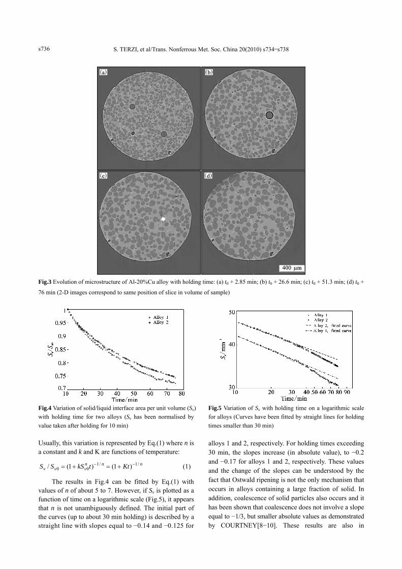
Fig.3 shows a sequence of 2-D images extracted
from the volume of the Al-20%Cu sample after various holding times in the semi-solid state. Spheroidisation of those equiaxed dendrites and coarsening have occurred owing to the reduction of the solid/liquid interface area. In order to quantify this evolution, the solid/liquid interface area per unit volume, Sv was measured directly in the volume of the samples and its variation with holding time is shown in Fig.4 for the two alloys. In Fig.4, Sv was normalized by the Sv0 value taken after holding for 10 min. Sv decreases with increasing time.
S. TERZI, et al/Trans. Nonferrous Met. Soc. China 20(2010) s734-s738 s736
Fig.3 Evolution of microstructure of Al-20%Cu alloy with holding time: (a) t0 + 2.85 min; (b) t0 + 26.6 min; (c) t0 + 51.3 min; (d) t0 +
76 min (2-D images correspond to same position of slice in volume of sample)
Fig.4 Variation of solid/liquid interface area per unit volume (Sv) with holding time for two alloys (Sv has been normalised by value taken after holding for 10 min) Usually, this variation is represented by Eq.(1) where n is a constant and k and K are functions of temperature:
nnnvvv KttkSSS /1/100 )1()1(/ −− +=+= (1)
The results in Fig.4 can be fitted by Eq.(1) with values of n of about 5 to 7. However, if Sv is plotted as a function of time on a logarithmic scale (Fig.5), it appears that n is not unambiguously defined. The initial part of the curves (up to about 30 min holding) is described by a straight line with slopes equal to −0.14 and −0.125 for
Fig.5 Variation of Sv with holding time on a logarithmic scale for alloys (Curves have been fitted by straight lines for holding times smaller than 30 min) alloys 1 and 2, respectively. For holding times exceeding 30 min, the slopes increase (in absolute value), to −0.2 and −0.17 for alloys 1 and 2, respectively. These values and the change of the slopes can be understood by the fact that Ostwald ripening is not the only mechanism that occurs in alloys containing a large fraction of solid. In addition, coalescence of solid particles also occurs and it has been shown that coalescence does not involve a slope equal to −1/3, but smaller absolute values as demonstrated by COURTNEY[8−10]. These results are also in

S. TERZI, et al/Trans. Nonferrous Met. Soc. China 20(2010) s734-s738 s737
agreement with those obtained previously on an Al-15.8%Cu alloy[1]. The in situ tomography experiments allow observation of these local mechanisms.
Fig.6 shows a sequence of 2-D images extracted from a region of the volume of the Al-13%Cu sample showing the Ostwald ripening mechanism. In Fig.6, a small globule connected to other ones disappears to the benefit of the surrounding larger globules. Another sequence extracted from the volume of the same alloy (Fig.7) shows the coalescence mechanism. Necks between solid particles grow in size during holding. As shown in Fig.3, the samples contain some pores which originate from the preparation of the ingots. The evolution of these pores can also be monitored during the holding of the alloys. Fig.8 shows a sequence of 2-D images showing an example of pore evolution. Each image corresponds to the section in which the pore has its maximum diameter, i.e. they are not from exactly the same position in the specimen.
Most pores, such as the one shown in Fig.8, experience an expansion stage and then shrink until they completely disappear. No significant movements of the pores are observed. It should be pointed out that once the pores disappear, the space where they are located is replaced by a liquid region due to liquid flow.
Mechanisms which control pore evolution are very complex. They involve hydrogen exchanges among the pores, the liquid and the atmosphere, Hydrogen diffusion through the mushy zone and liquid flow may also influence the evolution of pores.
These observations demonstrate that in situ tomography is a very interesting and powerful tool to follow the various microstructural events that occur during holding of samples in the semi-solid state. 4 Conclusions
1) In situ tomography experiments have been carried out to study the evolution of the microstructure during holding in the semi-solid state.
2) The solid/liquid interface area per unit volume decreases with holding time, but its variation is not represented by a unique value of the slope on a logarithmic scale. This result can be explained by the fact that coarsening involves not only Ostwald ripening of small globules to the benefit of their neighbors, but also growth of necks between globules by coalescence. These mechanisms have been observed in situ.
3) It is observed that pores originating from the casting process of the alloy can shrink and disappear completely.
Fig.6 Sequence of 2-D images extracted from volume of Al-13%Cu alloy showing shrinkage of a small globule at expense of its neighbors: (a) t=13.3 mm; (b) t=27.6 min; (c) t=41.8 min
Fig.7 Sequence of 2-D images extracted from volume of Al-13%Cu alloy showing coalescence of several globules: (a) t=27.6 min;
(b) t=41.8 min; (c) t=56.1 min

S. TERZI, et al/Trans. Nonferrous Met. Soc. China 20(2010) s734-s738 s738
Fig.8 Sequence of 2-D images extracted from volume of Al-13%Cu alloy showing evolution of a pore during holding of alloy in semi-solid state: (a) t=13.3 min; (b) t=27.6 min; (c) t=41.8 min; (d) t=56.1 min Acknowledgements
This work was carried out in the framework of the project ANR-05-BLAN-0286-01 TOMOSOLIDAL supported by the French Agence Nationale de la Recherche, which is gratefully acknowledged. The authors wish to thank all sta� members of the ID19 beam line of ESRF Grenoble for their technical support. A.K.D. wishes to thank PHELMA, Grenoble INP for the appointment as Invited Professor for three months and the Australian Academy of Science for the Bede Morris Fellowship which enabled his travel to Grenoble. References [1] LIMODIN N, SALVO L, SUERY M, DIMICHIEL M. In situ
investigation by X-ray tomography of the overall and local microstructural changes occurring during partial remelting of an Al-15.8 wt.% Cu alloy [J]. Acta Materialia, 2007, 55: 3177−3191.
[2] FERRANTE M, de FREITAS E. Rheology and microstructural development of a Al-4wt%Cu alloy in the semi-solid state [J]. Materials Science and Engineering A, 1999, 271: 172−180.
[3] ANNAVARAPU S, DOHERTY R D. Inhibited coarsening of solid-liquid microstructures in spray casting at high volume fractions
of solid [J]. Acta Metall Mater, 1995, 43(8): 3207−3230. [4] MANSON-WHITTON E D, STONE I C, JONES J R, GRANT P S,
CANTOR B. Isothermal grain coarsening of spray formed alloys in the semi-solid state [J]. Acta Materialia, 2002, 50: 2517−2535.
[5] ZABLER S, RUEDA A, RACK A, RIESEMEIR H, ZASLANSKY P, MANKE I, GARCIA-MORENO F, BANHART J. Coarsening of grain-refined semi-solid Al-Ge32 alloy: X-ray microtomography and in situ radiography [J]. Acta Materialia, 2007, 55: 5045−5055.
[6] TERZI S, SALVO L, SUERY M, DAHLE A K, BOLLER E. Coarsening mechanisms in a dendritic Al-10% Cu alloy [J]. Acta Materialia, 2010, 58: 20−30.
[7] LUDWIG O, DIMICHIEL M, SALVO L, SUERY M, FALUS P. In-situ three-dimensional microstructural investigation of solidification of an Al-Cu alloy by ultrafast X-ray microtomography [J]. Metall Mater Trans A, 2005, 36, 1515−1525.
[8] COURTNEY TH. A reanalysis of the kinetics of neck growth during liquid phase sintering [J]. Met Trans A, 1977, 8: 671−678.
[9] COURTNEY T H. Microstructural evolution during liquid phase sintering: Part I. Development of microstructure [J]. Met Trans A, 1977, 8: 679−684.
[10] COURTNEY T H. Microstructural evolution during liquid phase sintering: Part II. Microstructural coarsening [J]. Met Trans A, 1977, 8: 685−689.
(Edited by YANG Hua)