in pursuit of scientific breakthroughs - utsouthwestern.edu · a prion, an infectious protein that...
TRANSCRIPT

UT Southwestern’s cryo-EM
facility advances science
with its solutions of important
protein structures.
In pursuit of scientific breakthroughsWhat leads to a breakthrough? Asking tough questions. Digging deeper. Refusing to stop investigating, despite setbacks, until an answer validates a hypothesis. For UT Southwestern researchers, these are ingrained skills that have produced notable findings such as the point at which a protein in the brain turns toxic or the revelation of how a liver hormone affects cravings for alcohol and sugar.


Tau
Tubules
Tau protein (inert) Tau protein (pathological)
Neuron
In pathological tau,
reactive amino
acids are exposed,
enabling tau to
stack up.
Shape-shifting tau identified as genesis of Alzheimer’s diseaseUT Southwestern scientists have discovered the exact point at which a protein called tau
becomes toxic but has not yet begun forming deadly tangles in the brain characteristic of
Alzheimer’s disease.
This revolutionary investiga-
tion – from UT Southwestern’s Peter
O’Donnell Jr. Brain Institute – provides
new insight into the shape-shifting
nature of tau just before the molecule
begins sticking to itself to form the
larger aggregates seen in Alzheimer’s
cases. The findings reveal a new
strategy for detecting the devastating
disease before it takes hold, and they
also set in motion intense efforts to
develop treatments that may stabilize
tau proteins before they change shape.
“This is perhaps the biggest finding we have made to date,” said Dr. Marc Diamond,
Director of UT Southwestern’s Center for Alzheimer’s and Neurodegenerative Diseases. “It has
completely changed how we think about the problem.”
The research, published in eLife, contradicts the belief that tau has no distinct shape and
is only harmful after beginning to assemble with other tau proteins. Scientists in the Diamond
lab made their discovery after extracting tau from human brains and isolating the proteins
as single molecules. They found that the harmful form of tau exposes a part of itself that is
normally folded inside. This exposed portion causes it to stick to other tau proteins, enabling
the formation of tangles that kill neurons.

2726 ><
A Professor of Neurology and Neurother-
apeutics, and Neuroscience, Dr. Diamond
also holds the Distinguished Chair in
Basic Brain Injury and Repair. In this study,
he collaborated with Dr. Lukasz Joachimiak,
an Assistant Professor in the Center for
Alzheimer’s and Neurodegenerative Diseases,
and in the Department of Biochemistry, who
is also an Effie Marie Cain Scholar in Medical
Research. Their research was supported
by the Rainwater Charitable Foundation, the
“We think of this as the Big Bang of
tau pathology,” said Dr. Diamond, referring
to the prevailing scientific theory about
the formation of the universe. “This is a way
of peering into the very beginning of the
disease process.”
Dr. Diamond is a leading dementia expert
credited with determining that tau acts like
a prion, an infectious protein that can self-
replicate. Prions became notorious as the cause
of the 1980s outbreak of mad cow disease.
Dr. Marc Diamond’s studies focus on a molecule linked to the beginning of Alzheimer’s.

Growing up, Dr. Marc
Diamond pulled apart
watches to see how they
worked, built small boats
and rockets, and held a
general fascination with
the natural world. The
neurologist’s leanings
toward a future career in
science and medicine were
inherited as well to some
extent – his father, also
a neurologist, founded
a center for addiction
research and two uncles
were physicians.
During college, Dr.
Diamond worked for two
summers in the lab of
Dr. Stanley B. Prusiner,
a famed University of
California, San Francisco
researcher who later
won the Nobel Prize for
discovering infectious
proteins called prions.
Dr. Diamond graduated
from Princeton University
with a history degree –
about as far afield from
science as you’d imagine –
but then headed to
UCSF School of Medicine.
He took a two-year break
as a medical student to
work as a Howard Hughes
Medical Institute Student
Research Fellow in the
lab of Dr. Keith Yamamoto,
studying how nuclear
receptors sense hormones
in the body and regulate
transcription.
“After working with
Keith, I knew I wanted to be
a lab scientist,” said
Dr. Diamond, Director of
UT Southwestern’s Center
for Alzheimer’s and Neuro-
degenerative Diseases, who
holds the Distinguished
Chair in Basic Brain Injury
and Repair. “I decided to
focus on neurodegener-
ative diseases because
I recognized that they
represent the single most
mysterious and awful
problem in neurology.” At
that point, the path to tau
research was clear.
After deciding in 2003
to test whether the brain’s
tau proteins might work
like prions to cause neu-
rodegenerative diseases
such as Alzheimer’s, it
took Dr. Diamond seven
years to get National
Institutes of Health (NIH)
funding for his work.
“The ideas were
very revolutionary at
the time, and we needed
tremendous amounts of
preliminary data to
convince reviewers that
this could be true,”
Dr. Diamond said. Back
then, beta-amyloid tan-
gles were the trending
Alzheimer’s research topic.
“Fortunately, we were able
to get funding from the
Sandler Foundation, a
philanthropic supporter
of science, to carry on
this work.”
Later, when he was
ready to publish his lab’s
first work, which reported
that assemblies of tau
can journey into cells and
between them to spread
pathology, he spent 18
months getting rejections
before his study was finally
accepted for publication
in the Journal of Biologi-
cal Chemistry in 2009. It is
now the most highly cited
work from his lab.
The path toward tau research

2928 ><
Uncharted territory: UT Southwestern joins global effort to map human cellsUT Southwestern’s Peter O’Donnell Jr. Brain
Institute is taking part in an international
effort to map and characterize all cells in the
human body, an ambitious project designed
to gain insight into how cellular changes
cause disease.
Dr. Genevieve Konopka, a neuroscientist
with the O’Donnell Brain Institute, leads
a seven-member team that is evaluating
which technologies are best for determin-
ing how genes are expressed in the brain at a
single-cell level. The team is discovering how
cells turn specific genes on and off to
generate the dozens of cell types found in the
human brain.
The project is part of the Human Cell
Atlas, an effort involving scientists from
around the world to create comprehensive
maps of all human cells. The goal is to
understand how healthy cells work and what
malfunctions when people get sick.
Dr. Konopka’s brain team is funded by
the Chan Zuckerberg Initiative.
“Our project could have a major impact
on how we understand and ultimately treat
brain disorders with complex genomic under-
pinnings such as autism and schizophrenia,”
said Dr. Konopka, Associate Professor of
National Institutes of Health, and the Effie
Marie Cain Endowed Scholarship.
Despite billions of dollars spent on clini-
cal trials through the decades, Alzheimer’s
remains one of the most devastating and
baffling diseases in the world, affecting more
than 5 million Americans.
But now Dr. Diamond is hopeful the
field has turned a corner, noting that
identifying the genesis of the disease provides
scientists a vital target to diagnose the
condition at its earliest stage, before
the symptoms of memory loss and cognitive
decline become apparent.
His team’s next step is developing a
clinical test that can examine a patient’s blood
or spinal fluid to detect the first biological
signs of the abnormal tau protein.
Just as important, efforts are underway
to develop a treatment for those diagnosed.
Dr. Diamond cites a compelling reason for
cautious optimism here: Tafamidis, a recently
approved drug, stabilizes a different shape-
shifting protein that causes deadly protein
accumulation in the heart, similar to how tau
overwhelms the brain.
“The hunt is on to build on this finding
and make a treatment that blocks the neuro-
degeneration process where it begins,”
Dr. Diamond said. “If it works, the incidence
of Alzheimer’s disease could be substantially
reduced. That would be amazing.”
What is the
Human Cell Atlas?
In 2016, prominent
world scientists
launched a project
that would describe
and define all
human cells. As a
result, the Human
Cell Atlas consor-
tium was founded.
The group’s goal is
to create compre-
hensive reference
maps of all human
cells as a basis for
both understanding
human health and
diagnosing, moni-
toring, and treating
disease.
For more information,
visit humancellatlas.org.

that are vulnerable in autism. In 2017, she
published a study identifying more than
100 genes linked to memory.
“Dr. Konopka is a rising star in the
field of human gene expression in the
brain,” said Dr. Joseph Takahashi, Chair
of Neuroscience and an Investigator
Neuroscience and a Jon Heighten Scholar in
Autism Research, who estimated this work
would take at least a decade.
Dr. Konopka has researched various
aspects of the brain, including the genetic
pathways involved in language development
Dr. Genevieve Konopka, a neuroscientist with the Peter O’Donnell Jr. Brain Institute at UT Southwestern, leads a team working to identify
genes and cells within the brain as part of an international project – the Human Cell Atlas – aiming to map all cells in the body.

3130 ><
Mind bender: Researchers identify 100-plus genes linked to memoryScientists at UT Southwestern’s Peter O’Donnell Jr. Brain Institute
have identified more than 100 genes linked to memory, including many
previously thought to have no brain process connections.
The findings could ultimately advance treatment of various brain
disorders and unravel some of the mysteries behind the body’s most
complex organ.
Dr. Genevieve Konopka, Associate Professor of Neuroscience and a
Jon Heighten Scholar in Autism Research, teamed up with Dr. Bradley
Lega, Assistant Professor of Neurological Surgery, Neurology and
Neurotherapeutics, and Psychiatry, who was conducting memory
research on epilepsy patients. Dr. Lega works with epilepsy patients
to map their brain waves and pinpoint patterns related to memory
formation. In her research, Dr. Konopka studies the link between specific
genes and resting-state brain behavior.
Combining their techniques, the researchers found that a different
group of genes is used in memory processing than those involved when
the brain is in a resting state.
The researchers are hopeful these findings can help scientists
better understand and treat a range of conditions involving memory
impairment, from epilepsy to Alzheimer’s disease. Follow-up studies
are currently underway to refine and add to this list of memory
genes by analyzing the expression of genes in the brains of patients
undergoing memory assessments.
for the Howard Hughes Medical Institute.
Dr. Takahashi also holds the Loyd B. Sands
Distinguished Chair in Neuroscience.
The Chan Zuckerberg Initiative, founded
by Facebook creator Mark Zuckerberg
and his wife Priscilla Chan in 2015, supports
various initiatives in scientific research.
The illustration shows genes involved in memory.

an Assistant Professor of Neurology and
Neurotherapeutics with the Peter
O’Donnell Jr. Brain Institute and a former
UTSW fellow and resident.
The research focused on white matter,
a type of brain tissue comprised of millions
of bundles of nerve fibers used by neurons
to communicate across the brain.
Dr. Ding’s team enrolled older patients
at high risk of developing Alzheimer’s
who showed early signs of memory loss, or
mild cognitive impairment. They measured
the patients’ cardiorespiratory fitness and
Poor fitness linked to weaker brain fiber, higher dementia riskScientists have more evidence that exercise
improves brain health and could be a
lifesaving ingredient that prevents Alzheim-
er’s disease.
This UT Southwestern research, pub-
lished in the Journal of Alzheimer’s Disease,
suggests that the lower the fitness level,
the faster the deterioration of vital nerve
fibers in the brain. What happens then is
cognitive decline, including memory issues
characteristic of dementia patients.
“This work supports the hypothesis
that improving people’s fitness may improve
their brain health and slow down the aging
process,” said the study’s author, Dr. Kan Ding,
Brain imaging shows
yellow and reddish pixels
representing areas where
the functionality of white
matter is associated with
higher fitness levels.

3332 ><
levels preserves brain function. This trial
involves more than 600 older adults who are
at risk to develop Alzheimer’s.
Dr. Rong Zhang, Professor of Neurology
and Neurotherapeutics and Internal Medicine,
is overseeing the trial. The Dallas arm of
the study is being carried out by the Institute
for Exercise and Environmental Medicine,
a joint program between UT Southwestern
and Texas Health Resources. Dr. Zhang
is Director of the Cerebrovascular Laboratory
at the Institute.
imaged the brain to test the functionality of
their white matter. Memory and other
cognitive tests on the patients followed, to
evaluate brain function.
The researchers found that lower fitness
levels were associated with weaker white
matter, which in turn correlated with lower
brain function.
In related work, researchers at the
O’Donnell Brain Institute are leading a five-
year multicenter clinical trial designed to
determine whether participating in aerobic
exercise regularly and taking medications
to reduce high blood pressure and cholesterol
Dr. Kan Ding (left) and Dr. Rong Zhang examine images of human brains as part of research at UT Southwestern looking into whether
exercise can help ward off dementia.

While the hormone remained stable
in those drinking only juice, in response to
alcohol, FGF21 levels peaked after about
two hours.
“This suggests that FGF21 might
someday be used as a drug to limit alcohol
consumption and protect against its effects in
people,” said Dr. Mangelsdorf, Chair of Phar-
macology, Professor of Pharmacology and
Biochemistry, and a Howard Hughes Medical
Institute Investigator. Dr. Mangelsdorf also
holds the Alfred G. Gilman Distinguished
Chair in Pharmacology, and the Raymond
and Ellen Willie Distinguished Chair in
Molecular Neuropharmacology in Honor of
Harold B. Crasilneck, Ph.D.
The scientists knew that exposure to
alcohol or sugar turned on production of
FGF21 in the liver, added Dr. Kliewer, Profes-
sor of Molecular Biology and Pharmacology.
Why alcohol, sugar lead to thirstTexas and European researchers uncover liver hormone’s role in the brainWhy does drinking alcohol or consum-
ing sugar make us thirsty? An international
study reveals an unexpected anti-dehydration
mechanism that may help in finding new
treatments to prevent intoxication.
The study, published in Cell Metabolism,
identifies a hormone that acts on the brain
to increase the desire to drink water
under conditions that can cause dehydra-
tion, such as alcohol consumption. The study
involved research in mice and data from
human study participants in Europe.
UT Southwestern researchers Dr. David
Mangelsdorf and Dr. Steven Kliewer have
long studied the liver hormone FGF21, also
known as fibroblast growth factor 21. In
earlier mouse studies, they found that the
hormone acts via the brain’s reward pathway
to suppress the desire for sugar and alcohol
in favor of drinking water.
An important finding in the latest study
is a strong response to the hormone in
humans as well, Dr. Kliewer said. Validating
the researchers’ work in mice, 21 study
participants at the Medical University of Graz
in Austria were randomly assigned to drink
either a mixture of alcohol and juice, or juice
alone. Hourly over four hours, researchers
measured their FGF21 blood levels.
UT Southwestern
researchers
identified a hormone
that acts on the
brain to increase
the desire to drink
water in response
to specific nutrient
stresses that can
cause dehydration.

A high-fat/low-carbohydrate diet stimulated
water drinking in normal mice, but mice
genetically unable to produce the hormone
did not drink more in response to this
nutritional stress. The findings confirm the
hormone’s role in signaling the hydration
pathway, the researchers said.
For a long time, feeding behavior has
been emphasized in metabolic research rather
than hydration. This study suggests a change
may be on the table, the researchers said.
“To put this in context, we always look
at food intake, and the metabolic field has
spent comparatively little time studying water
intake. This study suggests that we should
think more about hydration and how it might
contribute to metabolism,” Dr. Kliewer said.
Until this study, however, they did not
know that the hormone then travels in the
blood to a specific part of the brain – the
hypothalamus – to stimulate thirst and
prevent dehydration.
“Unexpectedly, FGF21 works through
a new pathway that is independent of the
classical renin-angiotensin-aldosterone thirst
pathway in the kidneys,” said Dr. Kliewer,
who holds the Diana K. and Richard C.
Strauss Distinguished Chair in Develop-
mental Biology.
The hormone-induced thirst response
appears to depend on another signaling
pathway in the hypothalamus, the β-adrener-
gic circuit, the researchers said.
In another part of the study, done in
mice, FGF21 was shown to regulate water
consumption in response to nutrient stress.
UT Southwestern researchers found that the FGF21 hormone appears to reduce cravings for sweets and alcohol.
From left: Dr. Parkyong Song, Dr. David Mangelsdorf, Dr. Steven Kliewer, and Dr. Yuan Zhang.

Medical Center, UCLA Health, and Duke
University Health System.
“As one of the world’s foremost research
institutions, UT Southwestern has long
cultivated an environment where the pursuit
of rigorous scientific research blends seam-
lessly with multidisciplinary collaboration,
resulting in a strong record of leading-edge
discoveries and consistent translation into
new treatment development,” said Dr. Dwain
Thiele, Vice Provost and Senior Associate
Dean for Faculty Affairs and Initiatives, and
Professor, Department of Internal Medicine,
who holds the Jan and Henri Bromberg
Chair in Internal Medicine.
“This ranking is a testament to the
research being conducted every day in the
hundreds of labs across campus, where senior
faculty, early career researchers, postdoctoral
fellows, and graduate students tirelessly
work on discovering the underlying causes
of disease and the ways in which we can
improve health and extend life,” he said.
The Nature Index 2018 rankings are
based on primary research articles published
in a group of 82 high-quality science journals,
as selected by a panel of active scientists
independently of Nature Research. The list
of publications includes both multidisci-
plinary journals and some of the most highly
selective journals within the main disciplines
of the natural sciences.
UT Southwestern recognized for quality of scientific researchThe quality of scientific investigation
underway daily at UT Southwestern and its
potential impact is unquestionably high,
particularly given the large number
of esteemed researchers such as Nobel
Laureates, Breakthrough Prize winners,
members of the National Academies, and
Howard Hughes Medical Institute
Investigators who comprise the faculty.
In one of the latest affirmations of this
attribute, Nature Index last year ranked
UT Southwestern as the top institution within
the health care category internationally for
publishing high-quality scientific research.
In the Nature Index 2018 Annual Tables,
UTSW is ranked first in this category among
peer institutions that include Columbia
University Medical Center, Memorial Sloan
Kettering Cancer Center, Massachusetts
General Hospital, and UC San Diego Health.
Others rounding out the top 10 are the
University of Michigan Health System, MD
Anderson Cancer Center, NYU Langone
The Nature Index
2018 Annual Tables
ranked institutions
based on the quality
of primary research
articles published in
2017, as selected by
a panel of scientists
independently of
Nature Research.
UT Southwestern ranked first among peer
institutions in the health care institution category
on the Nature Index 2018 Annual Tables.


In a study published in Nature, researchers
at UT Southwestern’s Peter O’Donnell Jr.
Brain Institute provide a detailed description
of the structure of the GABAA receptor – the
receptor in the brain targeted by many med-
ications, including the benzodiazepines used
for anesthesia during surgery and prescribed
to treat epilepsy, anxiety, and insomnia.
The report shows the first 3D atomic
structures of the receptor bound to its
neurotransmitter GABA and to the drug flu-
mazenil, which is used to reverse anesthesia
and to treat benzodiazepine overdoses.
Knowing the structure of the receptor
could someday lead to better treatments,
said Dr. Ryan Hibbs, Assistant Professor
UTSW researchers solve structure of brain receptor using cryo-EMUsing some of the most advanced cryo-
electron microscopy equipment in the world,
UT Southwestern scientists have deciphered
the atomic structure of an important
neurotransmitter receptor in the brain.
Dr. Shaotong Zhu (left) and Dr. Ryan Hibbs look over an image of the GABAA receptor, whose molecular atomic structure they
deciphered using cryo-electron microscopy.

3938 ><
of Neuroscience and Biophysics with the
O’Donnell Brain Institute.
“This study reveals the first high-
resolution structural information for one
of the most abundant and important neuro-
transmitter receptors in the brain,” said
Dr. Hibbs, an Effie Marie Cain Scholar in
Medical Research. “We are tremendously
excited about it.”
Many drugs – both legal and illegal –
work on the GABAA receptor. The receptor
binds to GABA (γ-aminobutyric acid),
the major calming neurotransmitter in the
adult brain.
To function properly, the brain needs
a balance of stimulating and calming signals,
Dr. Hibbs said. Dysfunction of the GABAA
receptor is found in conditions marked
by excessive excitation in the brain, such
as epilepsy.
The GABAA receptor has been notori-
ously resistant to structural characterization.
X-ray crystallography – a structural
biology approach long considered the stan-
dard – requires the crystallization of proteins
to determine their structures, Dr. Hibbs
explained.
Dr. Shaotong Zhu, lead author of the
study and a postdoctoral researcher in
Neuroscience, tried that method on GABAA
but with inferior results. At the same time,
she also tried to unveil GABAA’s structure
using cryo-electron microscopy (cryo-EM).
That approach was successful, providing the
first 3D atomic structures of the receptor
bound to GABA and to the drug flumazenil.
This work was made possible using the
University’s $22.5 million cryo-EM facility,
where samples are rapidly frozen to prevent
the formation of damaging ice crystals
and then viewed at around minus 300 degrees
Fahrenheit (cryogenic temperatures).
UT Southwestern’s facility – which runs
around the clock – is one of the world’s top
facilities for cryo-EM structural biology.
“We were able to define how GABA
binds so selectively to the receptor and to
explain why drugs like benzodiazepines and
flumazenil – the agent that competes with
those drugs at the same binding site to
reverse their effects – act specifically on this
receptor,” Dr. Hibbs said.
Daniel Stoddard, a Core
Facilities Manager, shows
the Titan Krios cryo-electron
microscope to graduate
student researcher DaNae
Woodard.
Dr. Shaotong Zhu (left) and Dr. Ryan Hibbs look over an image of the GABAA receptor, whose molecular atomic structure they
deciphered using cryo-electron microscopy.
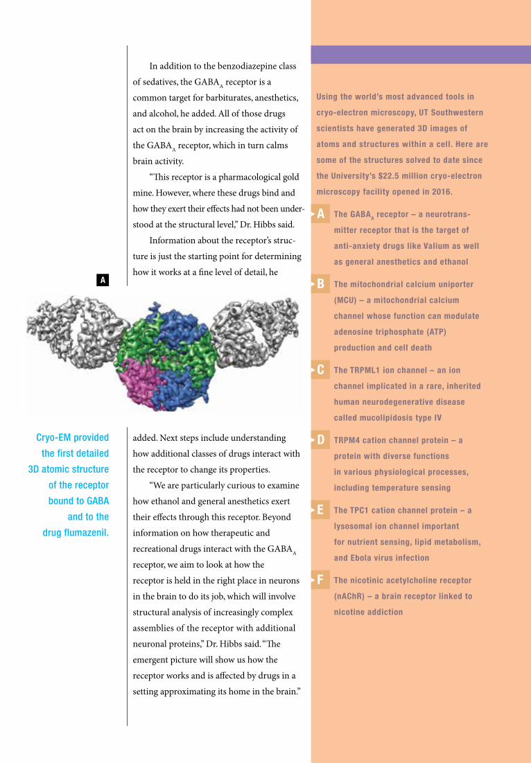
Using the world’s most advanced tools in
cryo-electron microscopy, UT Southwestern
scientists have generated 3D images of
atoms and structures within a cell. Here are
some of the structures solved to date since
the University’s $22.5 million cryo-electron
microscopy facility opened in 2016.
The GABAA receptor – a neurotrans-
mitter receptor that is the target of
anti-anxiety drugs like Valium as well
as general anesthetics and ethanol
The mitochondrial calcium uniporter
(MCU) – a mitochondrial calcium
channel whose function can modulate
adenosine triphosphate (ATP)
production and cell death
The TRPML1 ion channel – an ion
channel implicated in a rare, inherited
human neurodegenerative disease
called mucolipidosis type IV
TRPM4 cation channel protein – a
protein with diverse functions
in various physiological processes,
including temperature sensing
The TPC1 cation channel protein – a
lysosomal ion channel important
for nutrient sensing, lipid metabolism,
and Ebola virus infection
The nicotinic acetylcholine receptor
(nAChR) – a brain receptor linked to
nicotine addiction
A
B
C
D
E
F
Cryo-EM provided
the first detailed
3D atomic structure
of the receptor
bound to GABA
and to the
drug flumazenil.
In addition to the benzodiazepine class
of sedatives, the GABAA receptor is a
common target for barbiturates, anesthetics,
and alcohol, he added. All of those drugs
act on the brain by increasing the activity of
the GABAA receptor, which in turn calms
brain activity.
“This receptor is a pharmacological gold
mine. However, where these drugs bind and
how they exert their effects had not been under-
stood at the structural level,” Dr. Hibbs said.
Information about the receptor’s struc-
ture is just the starting point for determining
how it works at a fine level of detail, he
added. Next steps include understanding
how additional classes of drugs interact with
the receptor to change its properties.
“We are particularly curious to examine
how ethanol and general anesthetics exert
their effects through this receptor. Beyond
information on how therapeutic and
recreational drugs interact with the GABAA
receptor, we aim to look at how the
receptor is held in the right place in neurons
in the brain to do its job, which will involve
structural analysis of increasingly complex
assemblies of the receptor with additional
neuronal proteins,” Dr. Hibbs said. “The
emergent picture will show us how the
receptor works and is affected by drugs in a
setting approximating its home in the brain.”
A

Through the looking glass: The microscopic world of cryo-EM
F
B C
D E

Dr. Gaudenz Danuser, Chair of the Lyda Hill Department of Bioinformatics, wants to build a computer science department within a
medical center to help manage and analyze the masses of data now important in biomedical research.

4342 ><
of creating high-resolution, 3D images of
living cancer cells in realistic microenviron-
ments. To do so, he recruited Dr. Reto Fiolka,
now Assistant Professor of Cell Biology
and Bioinformatics, from the Howard
Hughes Medical Institute’s Janelia Research
Campus. The two co-authored a study in
2016 that describes the design of their unique
microscope.
Dr. Danuser also initiated the launch
of UT Southwestern’s Biomedical High
Performance Computing (BioHPC) initiative.
Today, the BioHPC has grown into a
consortium of 16 UT Southwestern depart-
ments that share thousands of
processors and 11 petabytes of
data storage to perform data-
driven basic and clinical science
investigations.
“Besides assembling
increasingly more sophisticated
computer algorithms, including
artificial intelligence systems,
to discover more and more
refined information in growing piles of data
– which are truly meaningful to biomedical
research and clinical practice – I predict that
one of the most exciting expansions of bio-
informatics moving forward will involve the
science of perception,” said Dr. Danuser,
also Professor of Cell Biology, holder of the
Patrick E. Haggerty Distinguished Chair in
Basic Biomedical Science, and a Cancer Preven-
tion and Research Institute of Texas Scholar.
“We need to think about how we present
the essence of all data we are generating in
an intuitive way. Bioinformatics may more
and more become the art of finding and seeing
the important.”
Advancing breakthroughs with the tools of bioinformaticsDr. Gaudenz Danuser, an internationally
recognized leader in engineering and
computational biology, has spent the past
five years helping UT Southwestern harness
the power of bioinformatics – a rapidly
growing area of computer science concerned
with the collection, organization, and analysis
of biomedical data.
Dr. Danuser’s goal is to create a computer
science department within a medical center
– to invent the computational procedures
needed to manage and analyze the extremely
large data sets now important for biomedical
research. Within 10 years, he hopes to recruit
16-20 new faculty involved in this research.
“I see informatics as the backbone of
everything we do in biomedical science,” said
Dr. Danuser, Chair of the Lyda Hill Depart-
ment of Bioinformatics since its founding. In
2015, a remarkable $25 million gift from
Dallas entrepreneur and philanthropist Lyda
Hill established the Department.
One of Dr. Danuser’s first major
studies at UT Southwestern involved designing
and building a new microscope capable
In 2015, a
$25 million gift
from Dallas
philanthropist
Lyda Hill
established
the Lyda Hill
Department of
Bioinformatics.