highly infectious prions generated by a single round of ... · agent, termed a prion, propagates by...
TRANSCRIPT
Highly Infectious Prions Generated by a Single Round of Microplate-Based Protein Misfolding Cyclic Amplification
Mohammed Moudjou,a Pierre Sibille,a Guillaume Fichet,a,b Fabienne Reine,a Jérôme Chapuis,a Laetitia Herzog,a Emilie Jaumain,a
Florent Laferrière,a Charles-Adrien Richard,a Hubert Laude,a Olivier Andréoletti,c Human Rezaei,a Vincent Béringuea
INRA (Institut National de la Recherche Agronomique), UR892, Virologie Immunologie Moléculaires, Jouy-en-Josas, Francea; Franklab, Montigny-le-Bretonneux, Franceb;UMR INRA ENVT 1225, Interactions Hôtes Agents Pathogènes, École Nationale Vétérinaire de Toulouse, Toulouse, Francec
M.M. and P.S. contributed equally to this work.
ABSTRACT Measurements of the presence of prions in biological tissues or fluids rely more and more on cell-free assays. Al-though protein misfolding cyclic amplification (PMCA) has emerged as a valuable, sensitive tool, it is currently hampered by itslack of robustness and rapidity for high-throughput purposes. Here, we made a number of improvements making it possible toamplify the maximum levels of scrapie prions in a single 48-h round and in a microplate format. The amplification rates and theinfectious titer of the PMCA-formed prions appeared similar to those derived from the in vivo laboratory bioassays. This en-hanced technique also amplified efficiently prions from different species, including those responsible for human variantCreutzfeldt-Jakob disease. This new format should help in developing ultrasensitive, high-throughput prion assays for cognitive,diagnostic, and therapeutic applications.
IMPORTANCE The method developed here allows large-scale, fast, and reliable cell-free amplification of subinfectious levels ofprions from different species. The sensitivity and rapidity achieved approach or equal those of other recently developed prion-seeded conversion assays. Our simplified assay may be amenable to high-throughput, automated purposes and serve in a com-plementary manner with other recently developed assays for urgently needed antemortem diagnostic tests, by using bodily fluidscontaining small amounts of prion infectivity. Such a combination of assays is of paramount importance to reduce the transfu-sion risk in the human population and to identify asymptomatic carriers of variant Creutzfeldt-Jakob disease.
Received 8 November 2013 Accepted 25 November 2013 Published 31 December 2013
Citation Moudjou M, Sibille P, Fichet G, Reine F, Chapuis J, Herzog L, Jaumain E, Laferrière F, Richard C-A, Laude H, Andréoletti O, Rezaei H, Béringue V. 2013. Highly infectiousprions generated by a single round of microplate-based protein misfolding cyclic amplification. mBio 5(1):e00829-13. doi:10.1128/mBio.00829-13.
Invited Editor Byron Caughey, NIH/NIAID Rocky Mountain Laboratories Editor Reed Wickner, National Institutes of Health
Copyright © 2013 Moudjou et al. This is an open-access article distributed under the terms of the Creative Commons Attribution-Noncommercial-ShareAlike 3.0 Unportedlicense, which permits unrestricted noncommercial use, distribution, and reproduction in any medium, provided the original author and source are credited.
Address correspondence to Vincent Béringue, [email protected], or Mohammed Moudjou, [email protected].
Prion diseases are infectious neurodegenerative disorders af-fecting a broad range of mammalian species, including
Creutzfeldt-Jakob disease (CJD) in humans, scrapie in sheep andgoats, bovine spongiform encephalopathy (BSE) in cattle, andchronic wasting disease in cervids (1). The causal proteinaceousagent, termed a prion, propagates by converting the �-helix-richhost-encoded prion protein PrPC into a misfolded, �-sheet-enriched conformer designated PrPSc. The abnormal form PrPSc isbelieved to replicate by recruiting and converting PrPC intohigher-order aggregates, through a so-called seeded polymeriza-tion process (2, 3). Fragmentation of PrPSc assemblies is thoughtto generate new PrPSc seeds to sustain the conversion (4). Distinctstrains of prions are recognized phenotypically, based on differentincubation times, neuropathological features, and PrPSc biochem-ical properties in experimentally infected rodents. Strain biologi-cal properties are believed to be encoded in different, specificPrPSc conformers (5–8). Several lines of evidence support PrPSc asthe main molecular determinant of prion replication and infectiv-ity (2, 3). Among them is the possibility to generate prion infec-tivity by PrPSc-templated conversion of PrPC under cell-free con-ditions using protein misfolding cyclic amplification (PMCA)assays. This technique has emerged in the last decade as a very
efficient procedure to amplify prions in a test tube (9). PMCAexploits the ability of PrPSc to template the conversion of PrPC byrepetitive cycles of incubation and sonication, leading to the am-plification of minute amounts of PrPSc. The sensitivity achievedallows detection of PrPSc present at low levels in biological tissueor fluid samples, including blood, urine, feces, or cerebrospinalfluid (10–14). The mechanisms by which such efficient in vitroamplification is achieved are essentially unknown. By analogywith the nucleation/polymerization process, incubation of PrPSc
seeds with PrPC-containing substrate is thought to favor PrPC
conversion and growth of PrPSc aggregates. Sonication is thoughtto fragment the polymers, thus providing new seeds for conver-sion. The PMCA-generated products are infectious and share(in general) similar biochemical and structural properties andbiological strain properties with the prion strain seed that servesfor amplification (15–19). There are, however, some clear discrep-ancies in the amounts of prion infectivity generated among thestudies (16, 18–20). Significant variations in the laboratory-specific methodologies employed to amplify PrPSc could explainthe discrepancies observed. Importantly, the PMCA reaction mix-tures employed were not always supplemented with beads, whichwere shown to significantly improve the level and the reproduc-
RESEARCH ARTICLE
January/February 2014 Volume 5 Issue 1 e00829-13 ® mbio.asm.org 1
on April 1, 2019 by guest
http://mbio.asm
.org/D
ownloaded from

ibility of the amplification (21, 22), by putatively favoring thefragmentation of the generated polymers. The infectious titer ofthe amplicon was also frequently measured after several roundsof amplification. Repetitive, long-term incubations and sonica-tions may alter infectivity. Besides, the higher number of roundsincreases the probability of generating false-positive amplifica-tions (23).
In this study, we first report significant simplification and im-provements of the PMCA technique, leading to high throughputand highly efficient amplification of PrPSc from several prionstrains from different species in a single 48-h round. We thendemonstrate by endpoint dilution quantitation in reporter micethat this method restores an infectivity titer comparable to that ofin vivo brain-derived prions, whatever the input dilution seedingthe PMCA reaction.
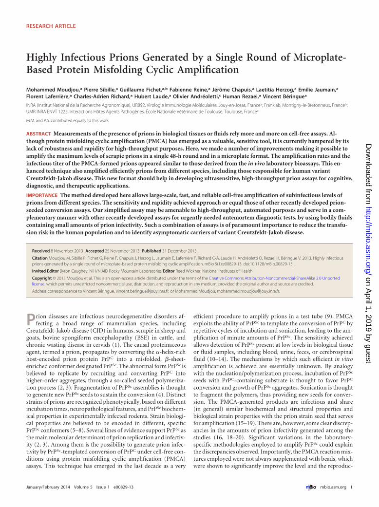
RESULTSEndpoint titration of 127S prions using mb-PMCA. The experi-mental conditions leading to miniaturized bead-PMCA (mb-PMCA) were established with brain material from transgenictg338 mice overexpressing ovine PrPC. These mice were infectedor not with the 127S scrapie strain, a prototypal “fast” strain kill-ing the mice within 2 months (24, 25). The procedure was per-formed by seeding tg338 mouse brain lysate containing ovinePrPC substrate with serial 10-fold dilutions of 127S-infected brainhomogenate and running 96 sonication/incubation cycles for around of 48 h. The amplicons were treated with proteinase K (PK)to eliminate PrPC before detection of PK-resistant PrPSc (PrPres)signals by dot blotting and Western blotting. Without amplifica-tion, PrPres is detected until the brain homogenate is diluted 103-fold (Fig. 1a). A series of technical changes were employed, im-
-3 -4 -5 -6 -7 -3 -4 -5 -6 -7 -5 -6 -7
38
28
17
-3 -4 -5
b
c
38
28
17
38
28
17
14
-5 -6 -7 -8 -9 -10 -11 -12 -13 -14 -15 U U U U U
100 μL in individual PCR tubes 100 μL in 8 PCR tubes strip
36 μL in 96 PCR microplate
a
Log10 (Brain
No beads + beads
Log10 (Brain
+ beads
FIG 1 Endpoint titration of 127S prions by a single round of mb-PMCA. Brain homogenate from tg338 mice infected with 127S prions was serially diluted intg338 healthy brain lysate as indicated. Each dilution was directly analyzed by Western blotting (a) or served as seed for a single 48-h round of PMCA usingdifferent experimental conditions (b and c). Samples were amplified in the absence or presence of 3 ceramic beads (as indicated) in individual PCR tubes or an8-PCR-tube strip containing 100 �l of PMCA mixtures (b) or in a PCR microplate containing 36 �l of PMCA mixture (c). Unseeded samples (U) were run onthe same microplate. All samples were digested with PK before Western blot analysis using Sha31 anti-PrP antibody. Undigested normal brain homogenate(PrPC) and PK-digested, infected brain homogenate (PrPres) are provided as electrophoretic references. Molecular mass markers (kDa) are indicated to the leftof each panel.
Moudjou et al.
2 ® mbio.asm.org January/February 2014 Volume 5 Issue 1 e00829-13
on April 1, 2019 by guest
http://mbio.asm
.org/D
ownloaded from

proving by 107-fold the sensitivity of the technique. “Standard”PMCA protocols (26) were initially followed using individualPCR tubes, brain from perfused tg338 mice to prepare the sub-strate, phosphate-buffered saline (PBS)–Triton as PMCA buffer,no beads added, and a final reaction volume of 100 �l. In ourhands, this often resulted in variations in the amplification effi-ciency and difficulties in completely digesting PrPC with PK, evenin the unseeded control samples (data not shown). When PBS wasreplaced with Tris in the PMCA buffer, better PrPSc amplificationand reduced PrPC undigested background were obtained. Underthese conditions, PrPres was detected in PMCA reaction mixturesseeded with 10�4-diluted 127S brain homogenate (Fig. 1b). Addi-tion of 3 ceramic beads during the reaction markedly increased thesensitivity and the reproducibility of the amplification; PrPres wasdetected routinely from reaction mixtures seeded with 10�6-diluted inoculum (Fig. 1b). Addition of one Teflon bead was alsobeneficial, as previously described (21). Use of strips of 8 PCRtubes instead of individual tubes placed in rigorously defined po-sitions in the cup horn led to systematic and robust amplificationof PrPSc in 10�7-diluted inoculum (Fig. 1b). Encouraged by theseobservations, we shifted to 96-well PCR microplates. The finalPMCA volume was reduced to 36 �l. Using this new experimentaldesign (referred to as mb-PMCA), highly efficient and reproduc-ible detection of PrPres was achieved in reaction mixtures seededwith 10�11-diluted brain material (Fig. 1c). PrPres was occasion-ally detected from amplification of 10�12 dilutions (see, for exam-ple, Fig. 2). Reactions seeded with higher dilutions (up to 10�15)did not show any positive PrPres signal (Fig. 1c). Specificity wasfurther assessed by submitting the mb-PMCA samples seededwith 10�13-diluted material to a second round with fresh sub-strate. No PrPres could be detected (data not shown). Two to sixunseeded control samples per plate were systematically includedin each mb-PMCA. No PrPres was detected in these control sam-ples in a total of more than 220 mb-PMCA reactions (i.e., theequivalent of 21,000 individual tubes) (for example, Fig. 1c), thushighlighting the high specificity of the reaction. Taken together,these findings suggest that the input seed limiting dilution of 127Sprions should be established at 10�11 to 10�12 and can be ampli-fied by mb-PMCA in one 48-h round of 96 cycles of sonication/incubation.
PrPC and other brain factors are limiting mb-PMCA efficacy.We next examined whether the high sensitivity of the mb-PMCAprocedure was linked to the amount of PrPC present in the sub-strate lysate, as tg338 mouse brain overexpresses approximately
8-fold PrPC compared to sheep brain (27). Performing mb-PMCAwith serial 10-fold dilutions of 127S input seeds mixed with sub-strate containing 100%, 50%, or 25% tg338 brain lysate (the latterdilutions were performed in 10% PrP-knockout brain lysatesmade in PMCA buffer) led to a 2-log10 reduction of the efficacy ofthe amplification (Fig. 2). In marked contrast, diluting tg338 brainlysate by 1:2 in PMCA buffer reduced by 6 log10 the PMCA effi-ciency (Fig. 2), suggesting that PrPC was not the sole limitingfactor in PMCA conversion efficacy.
We estimated the PrPC conversion yield during the 48-h mb-PMCA procedure. 127S PrPSc, but not PrPC, resists thermolysindigestion (28). Measuring the ratio of thermolysin-resistant PrPSc
to total PrP would thus provide information on the percentage ofmb-PMCA-converted PrPC. Samples from two independent mb-PMCAs, seeded with serial dilutions of 127S prions, were treatedwith 200 �g/ml thermolysin, a concentration degrading PrPC inunseeded PMCA samples. We observed substantial variations inthe amounts of PrPC converted among the mb-PMCA products,regardless of the dilution of the input seed. Approximately 20% ofPrPC was converted in one round as shown in the representativeFig. 3. Taken together, these data suggest that all the PrPC sub-strate is not converted and that non-PrP brain components maybe involved in the PrP conversion process.
Efficient amplification of mouse, hamster, and human pri-ons by mb-PMCA. To examine whether mb-PMCA sensitivitywas specific to 127S prions, we applied this single-round protocolto other prions and PrP species combinations. Previously, all theprions tested have been serially passaged and typed phenotypicallyon the ad hoc transgenic mice. As summarized in Table 1, mouseprions 139A, 22L, RML, and Chandler were efficiently amplifiedusing transgenic tga20 mouse brain (29) as the substrate formouse PrPC. PrPres was routinely detected from input seeds di-luted up to 109-fold (n � 6 experiments). ME7 prion amplifica-tion was 1,000 times less efficient. The PrPres glycoprofile of theamplified products resembled that of the inoculum, with a prom-inent monoglycosylated PrP form typical of these mouse prions(Fig. 4 and data not shown). PrPres from 263K, Sc237, and HYhamster prions was amplified from 10�7-diluted input seeds inone round using hamster PrP transgenic mouse brain (tg7 line[25]) as the substrate (n � 10 experiments). The DY hamsterstrain was less efficiently amplified (10�5). The PrPres electropho-retic and glycoform profiles of the nonamplified and mb-PMCA-amplified prions were similar (Fig. 4 and data not shown). Finally,we assessed the efficacy of mb-PMCA for human variant CJD
-5 -6 -7 -8 -9 -10 -11 -12 -5 -6 -7 -8 -9 -10 -5 -6 -7 -8 -9 -10 -5 -6 -7 -8 -9 -10
50% tg338 + 50% PrP0/0 BH 50% tg338 + 50% PMCA buffer
38
28
17
100 % tg338 BH
Log10 (Brain n)
25% tg338 + 75% PrP0/0 BH
FIG 2 mb-PMCA of 127S prions by using different concentrations of PrPC substrate. Brain homogenate from tg338 mice infected with 127S prions was seriallydiluted in substrate containing either pure tg338 mouse brain homogenate (BH) or tg338 BH mixed with PrP0/0 BH or PMCA buffer, as indicated. The mixturewas then subjected to a single round of mb-PMCA. All samples were digested with PK before Western blot analysis. Undigested normal brain homogenate (PrPC)and PK-digested, infected brain homogenate (PrPres) are provided as electrophoretic references. Molecular mass markers (kDa) are indicated on the left.
Maximal Prion Amplification in a Single Round of PMCA
January/February 2014 Volume 5 Issue 1 e00829-13 ® mbio.asm.org 3
on April 1, 2019 by guest
http://mbio.asm
.org/D
ownloaded from

(vCJD) prions using human PrP transgenic mice (Met129 allele;tg650 line [30]). In one round, the limiting dilution of the inputseed was reproducibly established at 10�8 (n � 6 experiments)(Fig. 4). The glycoform ratio of the mb-PMCA PrPvCJD productsclosely resembled that of the input seed (Fig. 4). However, themb-PMCA PrPvCJD of vCJD exhibited a slightly lower electropho-retic mobility than did brain-derived PrPres (Fig. 4a), as previouslyreported for hamster prion strains (31). Taken together, thesefindings highlight mb-PMCA as a versatile protocol to amplifyminute amounts of PrPSc from different prion strains in a singleround.
mb-PMCA generates highly infectious prions. We finallysought to determine whether highly efficient mb-PMCA of PrPSc
was associated with similarly efficient amplification of infectivity.This was done with 127S prions using the tg338 mouse bioassay.
According to endpoint titration by the intracerebral (IC) route,the infectious titer of 127S prions in tg338 mouse brain is 109.2
lethal dose 50 (LD50 IC) per gram (25). These experiments allowedus to determine that the lowest dose resulting in positive transmis-sion (as based on the appearance of clinical signs and detection ofPrPSc in brain) was observed at the 10�7 dilution of 127S braininoculum (Table 2; Fig. 5). mb-PMCA products were generatedfrom reaction mixtures seeded with this limiting dilution or with100-fold-more-diluted or -more-concentrated seeds. The ampli-con generated with the 10�9 seed was 10-fold diluted up to a 10�7
dilution for complete endpoint titration while the amplicons ob-tained with the 10�5 and 10�7 seeds were diluted 101-, 103-, 105-,and 107-fold. Two unseeded samples run in parallel were 10-folddiluted. All the dilutions were prepared separately and immedi-ately inoculated into recipient tg338 mice by the intracerebralroute. The results are summarized in Table 2. At the time of writ-ing, more than 400 days after the experimental infection, miceinoculated with unseeded controls were still alive and healthy.Whatever the initial dilution of the 127S seeds that served formb-PMCA, an attack rate of 100% was observed in tg338 miceinoculated with all the amplicons diluted up to 105- to 106-fold. Atthe 10�7 dilution, 1/5 (10�7 and 10�9 seed) or 2/5 (10�5 seed)tg338 mice were still infected, an attack rate reminiscent of thatobserved for nonamplified 127S-infected brain. In other words,one round of PMCA was sufficient to regenerate infectivity tolevels identical to those reached in the brains of intracerebrallyinoculated tg338 mice at the terminal stage of disease. For the fullytitrated amplicon generated with the 10�9 input seed, the relation-ship between prion concentration and mouse incubation periodappeared superimposable on that observed with prions derivedfrom terminally sick mouse brains (Fig. 5), suggesting similarmultiplication rates between cell-free and brain-derived prionsupon injection in the tg338 mouse brain.
The clinical signs of tg338 mice inoculated with serially dilutedamplicons were identical to those of mice infected intracerebrallywith 127S prions, including notably hyperexcitability, waddling,and rolling gait. Brain and spleen samples from these animalsaccumulated PrPres with a glycosylation and tissue-specific (28)mobility profile similar to that found in 127S-infected brain andspleen, respectively (Fig. 6a and b and data not shown). The re-gional distribution of PrPres in the brains of tg338 mice infectedwith the amplicons resembled that observed with 127S inoculatedintracerebrally at equivalent dilutions (24) (Fig. 6c and data notshown). The PrPres staining was pronounced in the septum, cor-
25 28 20 14 10 16 15
-5 -6 -7 -8 -9 -10 -11
+TL
- +
U
+PK
% PrPSc
38
28
17
38
28
FIG 3 Western blot analysis of thermolysin-resistant PrPSc generated bymb-PMCA. Brain homogenate from tg338 mice infected with 127S prions wasserially diluted in uninfected tg338 brain lysate, as indicated, and submitted toa single round of mb-PMCA. The resulting products were treated with eitherPK (�PK) or thermolysin (�TL) before Western blot analysis. Thethermolysin-resistant signals were quantified so as to measure the ratio ofthermolysin-resistant PrPSc to total PrP present in the unseeded samples (U).The ratios are indicated (% PrPSc). Unseeded sample has been 10-fold dilutedbefore loading on the Western blot. The figure is representative of the ratiosobserved in two independent experiments. Molecular masses (kDa) are indi-cated at left.
TABLE 1 Endpoint titration of mouse, hamster, and human prions by a single round of mb-PMCA
Species Prion strain PrPC substrate (mouse line) Limiting dilution of brain material
Mouse 139A Mouse (tga20) 10�8
RML 10�9
Chandler 10�7
22L 10�9
BSE 10�10
ME7 10�6
Hamster Sc237 Hamster (tg7) 10�7
263K 10�7
HY 10�7
DY 10�5
Human vCJD Human M129 (tg650) 10�8
Moudjou et al.
4 ® mbio.asm.org January/February 2014 Volume 5 Issue 1 e00829-13
on April 1, 2019 by guest
http://mbio.asm
.org/D
ownloaded from

pus callosum, habenula, hypothalamus, lateral and posterior hy-pothalamic area, and brain stem.
Together, our data led to the conclusion that the mb-PMCA127S amplicons are strongly related, if not identical, to 127S pri-ons phenotypically and that one round of mb-PMCA restoresinfectivity levels similar to that of 127S original brain material.
DISCUSSION
The sensitivity of PMCA in amplifying prions has long been rec-ognized. By using 96-well microplates, beads, and a reduced reac-
tion volume, we now show for the first time specific and highlysensitive prion amplification in a single round, thus greatly limit-ing the probability of false-positive samples. We also provide evi-dence that the PMCA-amplified products can be highly infectious,exhibiting a titer similar to that found in the parental brain mate-rial.
The PMCA method is constantly under continuous adaptationand improvements, due to inconsistent robustness and reproduc-ibility, thus compromising its use for large-scale purposes. Twoimportant technical limitations were circumvented in our study:
FIG 4 Highly efficient amplification of murine, hamster, and human prions by a single round of mb-PMCA. Western blot analysis (a) and ratio (b) of high- andlow-molecular-mass PrPres glycoforms of mb-PMCA products. Brain homogenates from tga20 mice infected with Chandler prions, tg7 mice infected with 263Kprions, and tg650 mice infected with vCJD prions were serially diluted in homotypic brain lysates as indicated. Each dilution was directly analyzed by Westernblotting or was submitted to a single round of mb-PMCA, as indicated (a). All samples were treated with PK before SDS-PAGE and Western blotting. Data inpanel b are plotted as means � standard errors of the means. Nonamplified, brain prions are represented by red triangles; mb-PMCA amplicons generated fromthe indicated input seed are indicated by blue symbols. The ratios were established from 3 independent mb-PMCA experiments.
TABLE 2 Incubation times of tg338 mice inoculated with serial 10-fold dilutions of brain-derived or mb-PMCA-generated 127S prionsa
Dilution
Incubation time in days � SEM (n/n0)
PMCA generated
Brain derived10�5 seed 10�7 seed 10�9 seed
10�1 63 � 2 (5/5) 60 � 1 (4/4) 59 � 1 (5/5) 64 � 1 (5/5)10�3 74 � 1 (5/5) 72 � 2 (5/5) 74 � 3 (5/5) 79 � 2 (5/5)10�4 ND ND 82 � 2 (5/5) 85 � 1 (5/5)10�5 91 � 2 (5/5) 98 � 3 (5/5) 111 � 11 (5/5) 99 � 2 (5/5)10�6 ND ND 117 � 11 (5/5) 119 � 7 (4/5)10�7 134; 147 (2/5) 124 (1/5) 139 (1/5) 157 (1/5)Unseeded no. 1 �400Unseeded no. 2 �400a n/n0, number of affected/number of inoculated tg338 mice; ND, not done.
Maximal Prion Amplification in a Single Round of PMCA
January/February 2014 Volume 5 Issue 1 e00829-13 ® mbio.asm.org 5
on April 1, 2019 by guest
http://mbio.asm
.org/D
ownloaded from

(i) the repetition of incubation/sonication cycles and substraterefreshment over several rounds, which appeared necessary to at-tain high amplification levels (17, 32, 33) but was shown to be oneof the main factors responsible for inadvertent cross-contamination events or so-called spontaneous de novo genera-tion of prions (23, 34), and (ii) the position of the tubes within thesonicator and their distance from the center of the horn (21, 26,35), which greatly limited the number of amplified samples perround. These improvements were achieved by using ceramicbeads during the incubation/sonication cycles and by decreasingthe volume necessary to the reaction with the PCR microplateformat. This led to the use of the maximum surface offered by thesonicator, allowing more than 110 samples to be run at the sametime (see Fig. S1 in the supplemental material). The observationthat ceramic beads greatly improved the efficiency of PMCA iscongruent with previous observations made with Teflon beads(21, 22). Beads are believed to promote efficient fragmentation ofPrPSc polymers, thus increasing the number of seeds available forconversion (21). The observation that the number and the size ofbeads added to the conversion appeared less important than theirchemical composition (our unpublished data and reference 21)might suggest a more complex physical involvement of the beadsin the PMCA reaction. This effect appears optimal in a reducedreaction volume, suggesting a link with the distribution of sonica-tion energy.
Using the so-called mb-PMCA protocol, a single 48-h roundwas sufficient to achieve amplification of the highest dilution of127S prions to levels detectable by conventional Western blotting.The sensitivity reached by the mb-PMCA assay exceeded that ofthe tg338 bioassay by 104- to 105-fold. This difference is consistentwith other studies (17, 33). A significant part of the inoculated or
generated prions might be degraded in animal bioassays (36, 37),a phenomenon that would be limited in cell-free assays. Remark-ably, the technical improvements employed were beneficial toother prion strains, such as mouse, hamster, and human vCJDprions, without, at variance with previous studies (35, 38, 39), anapparent need to adapt the conditions to each prion strain. Vari-ation in the power amplitude of the sonicator (10 to 80%) waswithout notable influence on the sensitivity achieved with 127S,263K, Chandler, and vCJD prions (data not shown), suggestingthat these samples received optimal sonication energy in the mb-PMCA format. However, some prion sources, notably those re-sponsible for certain CJD subtypes, fairly resist mb-PMCA (un-published observations), suggesting that certain cell-free prionpolymerizations would necessitate cofactors or different experi-mental conditions. The maximum dilution of prion brain inocu-lum detected by mb-PMCA varied with certain strain types (Ta-ble 1). This may reflect differences in infectious titers amongstrains. Comparatively, ME7 and DY are at least 100-fold less in-fectious than RML/Chandler and HY prions in tga20 mice (40)and hamsters (41), respectively. The sensitivity of detectionachieved with vCJD prions would be compatible with that neces-sary for reliable detection of this agent in blood (42), an importantpublic health concern given the current uncertainties about thenumber of individuals incubating the disease (43–45).
We confirm here that PMCA sensitivity is dependent on theconcentration of PrPC in the PMCA substrate (46–48). However,despite its remarkable sensitivity, the PrPC conversion yieldachieved by the mb-PMCA assay was approximately 20%. Thislow conversion yield might suggest that all brain PrPC species arenot convertible, a hypothesis consistent with the deposition ofPrPSc in (strain-dependent) specific brain areas in infected ani-mals (49–51). Alternatively but not exclusively, other brain factorsmight be a limiting factor. Supporting this hypothesis, a 2-folddilution of PMCA substrate in PMCA buffer instead of PrP0/0
brain lysate dramatically decreased the mb-PMCA efficiency.RNAs or poly(A) and lipids have been shown to be instrumental inefficient PMCA conversion (52–54). The high-throughputscreening capacities of mb-PMCA will be helpful to investigatethis further and to search for potent cellular factors involved in thePrPC conversion on a large scale.
Our 20% conversion yield sharply contrasted with the 100%conversion yield previously reported by using beads in the PMCAreaction (21). These differences might be due to different prionsources/PrP substrate combinations and/or to the quantificationmethods used. Here, we quantified the densitometric ratio ofthermolysin-resistant PrPSc generated by mb-PMCA to the totalamount of PrP. In a mixture such as a PMCA product containingboth PrPSc and PrPC, thermolysin is known to preserve protease-sensitive and protease-resistant PrPSc species while destroyingPrPC (55, 56) and thus may allow reliable assessment of the con-version yield.
Remarkably, the rates of multiplication and terminal prion ti-ters appeared similar between mb-PMCA-generated and brain-derived 127S prions when injected in reporter tg338 mice. The useof mb-PMCA has immediate potential application as a surrogatefor 127S infectivity quantitation. The infectivity of the mb-PMCAamplicons was determined by endpoint titration in tg338 mice,which is regarded as the most accurate method for measuringprion infectious titer in infected samples (57, 58). Some titratedamplicons were generated with input seeds diluted 100-fold more
FIG 5 Endpoint titration of nonamplified and mb-PMCA-generated 127Sprion infectivity in tg338 mice. Brain homogenates from infected tg338 miceand mb-PMCA-generated amplicons were 10-fold diluted (y axis) and inocu-lated intracerebrally into groups of tg338 mice. Individual survival times areshown on the x axis as days postinoculation. tg338 mice were inoculated withbrain-derived 127S prions (filled triangles) or mb-PMCA amplicons generatedfrom 10�5 (open circles), 10�7 (open squares), and 10�9 (open triangles)input seed.
Moudjou et al.
6 ® mbio.asm.org January/February 2014 Volume 5 Issue 1 e00829-13
on April 1, 2019 by guest
http://mbio.asm
.org/D
ownloaded from

than the 127S prion limiting dilution in the tg338 bioassay, thusexcluding any contribution of the parental seed to the infectivitymeasured. This tight relationship between prion replication dy-namics in cell-free and animal assays contrasts with previouslypublished results. Shikiya and Bartz (18) found, as we did, thattheir PMCA protocol generated high-titer prions; however, therate of prion amplification was significantly altered. Notably, theytitrated PMCA-generated HY prions after the 10th round of am-plification, which may have altered the amplicon infectious prop-erties. Other contradictory studies, which were also based on mul-tiround PMCA, divided the infectivity titer by the amount ofPrPres generated by the PMCA reaction, so as to calculate PrPres
specific infectivity (16, 19). A number of pieces of experimentalevidence suggest a quantitative disconnection between infectivityand PrPres in the brains of prion-infected animals (59). Specificallyfor 127S and the extensively used Sc237 hamster prions, a subpop-ulation of PrPSc assemblies would support most of the infectivity,making a significant proportion of PrPres assemblies relatively in-nocuous (25, 60). We may have regenerated high-titer 127S prionsby a single round of mb-PMCA because this “most infectious”PrPSc subpopulation was preferentially amplified. Accordingly,we demonstrated with another “fast” ovine prion strain, exhibit-ing a behavior similar to that of 127S in tg338 mice (25), that thesubset of PrPSc assemblies that carried the major part of prioninfectivity also exhibited by far the highest templating activity bymb-PMCA (61).
In summary, the mb-PMCA assay allows large-scale, fast, andreliable cell-free amplification of subinfectious levels of prionsfrom different species. The sensitivity and rapidity achieved ap-proach or equal those of other prion-seeded conversion assays(62), such as the quaking-induced conversion (QuIC) assay (63).At variance with the latter (62, 64), mb-PMCA can regeneratelarge amounts of prion infectivity. Such a simplified assay may beamenable to high-throughput, automated purposes and serve, in acomplementary manner with QuIC-like (62) or solid-phase bind-ing (42) assays, for urgently needed preclinical diagnostic tests, byusing bodily fluids containing small amounts of prion infectivity.
MATERIALS AND METHODSEthics statement. All animal experiments have been performed in strictaccordance with EU directive 2010/2063 and were approved by the localethics committees of the authors’ institutions (Comethea; permit number12/034).
Transgenic mice and prion strains. The ovine (tg338 line; Val136-Arg154-Gln171 allele), human (tg650 line; Met129 allele), hamster (tg7line), and mouse (tga20) PrP transgenic lines have been described previ-ously (24, 25, 29, 30, 43, 65). These lines are homozygous and overexpress
FIG 6 PrPres glycopattern and PrPres regional deposition in the brains oftg338 mice inoculated with mb-PMCA-generated 127S prions. Western blotanalysis (a) and ratios (b) of high- and low-molecular-mass PrPres glycoformsin the brains and spleens of tg338 mice following challenge with brain-derivedor mb-PMCA-generated 127S prions. Samples in panel a are from mice inoc-
(Continued)
Figure Legend Continued
ulated with 10-fold-diluted amplicons. Data in panel b are plotted as means �standard errors of the means. Brains and spleen are represented by filled andopen symbols, respectively. Brain-derived 127S prions are represented by redtriangles; mb-PMCA amplicons generated from 10�5, 10�7, and 10�9 inputseed are indicated by blue circles, squares, and triangles, respectively. Theratios were established from tissue samples of mice inoculated with all theamplicon dilutions. As no differences were observed among the amplicondilutions, the ratios were combined for the sake of clarity. Histoblotting ofrepresentative coronal sections of tg338 mice inoculated with mb-PMCA am-plicons at the level of the septum (c), hippocampus (d), midbrain (e), andbrain stem (f) (10�9 input seed) and hippocampus (g and i) and midbrain (hand j) (10�7 and 10�5 input seed).
Maximal Prion Amplification in a Single Round of PMCA
January/February 2014 Volume 5 Issue 1 e00829-13 ® mbio.asm.org 7
on April 1, 2019 by guest
http://mbio.asm
.org/D
ownloaded from

about 8-, 6-, 4-, and 10-fold the heterologous PrPC level on a mousePrP-null background, respectively.
The 127S scrapie prion strain has been obtained through serial trans-mission and subsequent biological cloning by limiting dilutions of PG127field scrapie isolate to tg338 mice (24, 27). The 127S infectious titer is 109.2
50% lethal doses (LD50)/g of tg338 brain (25). Pools of 127S-infectedtg338 mouse brains were prepared as 20% (wt/vol) homogenate in 5%glucose by use of a tissue homogenizer (Precellys 24 Ribolyzer; Ozyme;Bertin Technologies, France). The homogenate was diluted half to 10% inPMCA buffer (see below) to obtain the 10�1 dilution of the inoculum andstored at �80°C. All subsequent dilutions refer to this 10% homogenatestarting material.
Mouse prion strains 139A, 22L, RML, Chandler, ME7, and mouse-adapted bovine spongiform encephalopathy (BSE); hamster prion strains263K, Sc237, HY, and DY; and human vCJD prions have been seriallypassaged on tga20, tg7 (25), and tg650 mice (30), respectively.
PMCA. Mouse brain lysate from uninfected tg338 mice was used as thesubstrate for 127S scrapie prions. Mouse brain lysates from tga20 mice,tg7 mice, and tg650 mice were used as the substrates for mouse, hamster,and human prions, respectively. Two- to 12-month-old mice were eutha-nized by CO2 exposure. Brains were rapidly removed and washed twice inCa2�/Mg2�-free PBS containing 5 mM EDTA. They were either usedimmediately to prepare the PrPC substrate lysate or stored at �80°C untiluse. PrPC substrate (10% brain lysate) was prepared using cold PMCAbuffer (50 mM Tris-HCl, pH 7.4, 5 mM EDTA, 300 mM NaCl, 1% TritonX-100) and Dounce cooled in ice with 20 to 30 strokes to completelyhomogenize the brain tissue. The lysate was left at 4°C for 30 min andbriefly clarified by centrifugation at 1,000 � g for 2 min at 4°C. Theresulting supernatant, corresponding to the PrPC substrate lysate, wascollected, aliquoted, and stored at �80°C. Protein misfolding cyclic am-plification (PMCA) performed with either young (6- to 10-week) or old(1-year) mouse brain homogenates yielded the same results. Moreover,brain perfusion prior to collection and homogenization appeared not tobe of any influence on the performance of the mb-PMCA (data notshown).
PMCA was performed in a final volume of either 100 �l or 36 �l oflysate per well, in either single PCR tubes (conventional method); in 2-, 4-,or 8-PCR-tube strips; or with a 96-well PCR microplate (Axygen, UnionCity, CA, USA). Each tube or well was first filled with ceramic beads (3beads of 1.23 mm in diameter; Mineralex, France). Two ceramic beads of2.4 mm or one Teflon bead of 2.381 mm in diameter (Marteau et Lemarié,Pantin, France) was also efficient. A 4-�l aliquot of the analyte inoculum(10�2 dilution) was suspended in 36 �l of healthy tg338 brain lysate toobtain the 10�3 dilution. Then, a series of 10-fold dilutions was made byadding 4 �l from the previous inoculum dilution to the next 36-�l-containing tube or well. Individual tubes, tube strips, or microplates wereplaced on a Plexiglas rack designated for the cup horn of the S3000 orQ700 sonicator (Misonix, Farmingdale, NY, USA, or Delta Labo, Colom-belles, France) and subjected to 96 cycles of 30 s of sonication at 220- to240-W power (level 6 to 7 for the S3000 or 30% amplitude of the Q700sonicator) followed by 29 min 30 s of incubation at 37°C. The cup hornwas filled with 300 ml of water (or 4 M guanidium hydrochloride solu-tion) circulating with rubber tubing in a water bath maintained at a tem-perature of 35 to 36°C. When needed, subsequent rounds of PMCA wererealized using a 1/10 dilution of the products of the previous PMCA roundas the template. At the end of the PMCA, the tubes or microplates wereremoved and aliquots from each sample were taken to be analyzed fortheir PrPres content.
Protease digestion of PMCA products. To analyze the production ofproteinase K (PK)-resistant PrPSc species during PMCA, 10 to 18 �l ofeach sample was supplemented with SDS (0.3 to 0.6% final concentration)and treated with PK (115-�g/ml final concentration) at 37°C for 1 h. ThePK digestion was stopped by adding an equal volume of 2� Laemmlidenaturation sample buffer and heating at 100°C for 5 min. The sampleswere then stored at �20°C until dot blotting and Western blotting.
To analyze the levels of thermolysin-resistant species, PMCA productswere first diluted 1/5 in TNT buffer (50 mM Tris-HCl, pH 7.4, 5 mMEDTA, 150 mM NaCl, 1% Triton X-100) to decrease the EDTA concen-tration which prevents thermolysin digestion. The samples were thentreated with 200 �g/ml of thermolysin (Sigma, St. Louis, MO, USA) at37°C for 1 h before denaturation as described above.
Dot blotting analysis of PrPSc. Given the significant number of sam-ples generated with the PMCA procedure, a quick dot blot analysis was setup and turned out to be practical and efficient in getting an overview of thePMCA results. Ten to 20 �l of each PK-digested PMCA sample was re-moved and transferred into a 96-well microplate containing 50 �l of 1%SDS and 20% glycerol. The samples were then transferred onto a nitro-cellulose membrane placed on the dot blot apparatus (Whatman, France)connected to a vacuum system. The membrane was removed and rinsedtwice with Tris-buffered saline (TBS)– 0.1% Tween 20 before incubationwith the primary antibody (biotinylated Sha31 [66] anti-PrP monoclonalantibody) for 15 to 30 min at room temperature. After 3 washes of 5 mineach, the membrane was incubated with streptavidin-coupled horserad-ish peroxidase for 15 to 30 min at room temperature and processed fordetection with the enhanced chemiluminescence (ECL) reagent (GEHealthcare, Saclay, France).
SDS-PAGE and Western blotting. PMCA samples were run on either4 to 12% or 12% Bis-Tris NuPAGE precast gels (Invitrogen, Cergy-Pontoise, France) or on Criterion XT 12% Bis-Tris precast gels (Bio-Rad,Hercules, CA, USA), electrotransferred onto nitrocellulose membraneswith the semidry electrotransfer system (Bio-Rad), and probed with bio-tinylated Sha31 anti-PrP monoclonal antibody. Secondary antibody incu-bation and ECL detection were performed as described above. When nec-essary, the PrPres content of PMCA samples was determined withGeneTools software after acquisition of the signals with a GeneGnomedigital imager (Syngene, Frederick, MD, USA).
Endpoint titration of PMCA products in tg338 mice. A strict proto-col based on the use of disposable equipment and preparation of all inoc-ula in a class II microbiological cabinet was followed to avoid any cross-contamination. Serial 10-fold dilutions of PMCA products were preparedin sterile 5% glucose containing 5% bovine serum albumin, and 20 �l ofeach dilution was immediately inoculated into individually identified 6-to 10-week-old tg338 recipient mice (n � 5 mice per dilution) by theintracerebral route. The inoculated animals were observed daily for theappearance of prion disease. Animals at the terminal stage of disease wereeuthanized. The survival time was defined as the number of days frominoculation to euthanasia. Their brains and spleens were removed forPrPres analysis by Western blotting and histoblotting as previously de-scribed (24, 25). For the histoblotting procedure, brains were rapidly re-moved from euthanized mice and frozen on dry ice. Cryosections were cutat 8 to 10 mm, transferred onto Superfrost slides, and kept at �20° C untiluse. Histoblot analyses were performed on 3 brains per dilution per am-plicon, using the 12F10 anti-PrP antibody (67).
SUPPLEMENTAL MATERIALSupplemental material for this article may be found at http://mbio.asm.org/lookup/suppl/doi:10.1128/mBio.00829-13/-/DCSupplemental.
Figure S1, PDF file, 0.1 MB.
ACKNOWLEDGMENTS
We thank J. Castilla and C. Soto (University of Texas Medical Branch,Galveston, TX, USA) for introducing us to PMCA and the staff of Ani-malerie Rongeurs (INRA, Jouy-en-Josas, France) for excellent animalcare.
This work was supported by fellowship (F.L.) and grants from theIle-de-France region (DIM MALINF, France).
REFERENCES1. Collinge J. 2001. Prion diseases of humans and animals: their causes and
molecular basis. Annu. Rev. Neurosci. 24:519 –550.
Moudjou et al.
8 ® mbio.asm.org January/February 2014 Volume 5 Issue 1 e00829-13
on April 1, 2019 by guest
http://mbio.asm
.org/D
ownloaded from

2. Diaz-Espinoza R, Soto C. 2012. High-resolution structure of infectiousprion protein: the final frontier. Nat. Struct. Mol. Biol. 19:370 –377.
3. Colby DW, Prusiner SB. 2011. Prions. Cold Spring Harb. Perspect. Biol3:a006833. http://dx.doi.org/10.1101/cshperspect.a006833.
4. Knowles TP, Waudby CA, Devlin GL, Cohen SI, Aguzzi A, VendruscoloM, Terentjev EM, Welland ME, Dobson CM. 2009. An analytical solu-tion to the kinetics of breakable filament assembly. Science 326:1533–1537.
5. Bessen RA, Marsh RF. 1994. Distinct PrP properties suggest the molec-ular basis of strain variation in transmissible mink encephalopathy. J.Virol. 68:7859 –7868.
6. Sim VL, Caughey B. 2009. Ultrastructures and strain comparison ofunder-glycosylated scrapie prion fibrils. Neurobiol. Aging 30:2031–2042.
7. Spassov S, Beekes M, Naumann D. 2006. Structural differences betweenTSEs strains investigated by FT-IR spectroscopy. Biochim. Biophys. Acta1760:1138 –1149.
8. Telling GC, Parchi P, DeArmond SJ, Cortelli P, Montagna P, GabizonR, Mastrianni J, Lugaresi E, Gambetti P, Prusiner SB. 1996. Evidence forthe conformation of the pathologic isoform of the prion protein encipher-ing and propagating prion diversity. Science 274:2079 –2082.
9. Saborio GP, Permanne B, Soto C. 2001. Sensitive detection of patholog-ical prion protein by cyclic amplification of protein misfolding. Nature411:810 – 813.
10. Atarashi R, Moore RA, Sim VL, Hughson AG, Dorward DW, Onwu-biko HA, Priola SA, Caughey B. 2007. Ultrasensitive detection of scrapieprion protein using seeded conversion of recombinant prion protein. Nat.Methods 4:645– 650.
11. Bannach O, Birkmann E, Reinartz E, Jaeger KE, Langeveld JP, RohwerRG, Gregori L, Terry LA, Willbold D, Riesner D. 2012. Detection ofprion protein particles in blood plasma of scrapie infected sheep. PLoSOne 7:e36620. http://dx.doi.org/10.1371/journal.pone.0036620.
12. Castilla J, Saa P, Soto C. 2005. Detection of prions in blood. Nat. Med.11:982–985.
13. Lacroux C, Vilette D, Fernandez-Borges N, Litaise C, Lugan S, Morel N,Corbiere F, Simon S, Simmons H, Costes P, Weisbecker JL, Lantier I,Lantier F, Schelcher F, Grassi J, Castilla J, Andreoletti O. 2012. Prion-emia and leukocyte-platelet-associated infectivity in sheep transmissiblespongiform encephalopathy models. J. Virol. 86:2056 –2066.
14. Safar JG, Lessard P, Tamguney G, Freyman Y, Deering C, Letessier F,Dearmond SJ, Prusiner SB. 2008. Transmission and detection of prionsin feces. J. Infect. Dis. 198:81– 89.
15. Castilla J, Gonzalez-Romero D, Saa P, Morales R, De Castro J, Soto C.2008. Crossing the species barrier by PrP(Sc) replication in vitro generatesunique infectious prions. Cell 134:757–768.
16. Klingeborn M, Race B, Meade-White KD, Chesebro B. 2011. Lowerspecific infectivity of protease-resistant prion protein generated in cell-free reactions. Proc. Natl. Acad. Sci. U. S. A. 108:E1244 –E1253.
17. Saa P, Castilla J, Soto C. 2006. Ultra-efficient replication of infectiousprions by automated protein misfolding cyclic amplification. J. Biol.Chem. 281:35245–35252.
18. Shikiya RA, Bartz JC. 2011. In vitro generation of high-titer prions. J.Virol. 85:13439 –13442.
19. Weber P, Giese A, Piening N, Mitteregger G, Thomzig A, Beekes M,Kretzschmar HA. 2006. Cell-free formation of misfolded prion proteinwith authentic prion infectivity. Proc. Natl. Acad. Sci. U. S. A. 103:15818 –15823.
20. Daus ML, Wagenfuhr K, Thomzig A, Boerner S, Hermann P, Her-melink A, Beekes M, Lasch P. 25 October 2013. Infrared microspectros-copy detects protein misfolding cyclic amplification (PMCA)-inducedconformational alterations in hamster scrapie progeny seeds. J. Biol.Chem. http://dx.doi.org/10.1074/jbc.M113.497131.
21. Gonzalez-Montalban N, Makarava N, Ostapchenko VG, Savtchenk R,Alexeeva I, Rohwer RG, Baskakov IV. 2011. Highly efficient proteinmisfolding cyclic amplification. PLoS Pathog. 7:e1001277. http://dx.doi.org/10.1371/journal.ppat.1001277.
22. Johnson CJ, Aiken JM, McKenzie D, Samuel MD, Pedersen JA. 2012.Highly efficient amplification of chronic wasting disease agent by proteinmisfolding cyclic amplification with beads (PMCAb). PLoS One 7:e35383.http://dx.doi.org/10.1371/journal.pone.0035383.
23. Cosseddu GM, Nonno R, Vaccari G, Bucalossi C, Fernandez-Borges N,Di Bari MA, Castilla J, Agrimi U. 2011. Ultra-efficient PrP(Sc) amplifi-cation highlights potentialities and pitfalls of PMCA technology. PLoSPathog. 7:e1002370. http://dx.doi.org/10.1371/journal.ppat.1002370.
24. Langevin C, Andreoletti O, Le Dur A, Laude H, Beringue V. 2011.Marked influence of the route of infection on prion strain apparent phe-notype in a scrapie transgenic mouse model. Neurobiol. Dis. 41:219 –225.
25. Tixador P, Herzog L, Reine F, Jaumain E, Chapuis J, Le Dur A, LaudeH, Beringue V. 2010. The physical relationship between infectivity andprion protein aggregates is strain-dependent. PLoS Pathog. 6:e1000859.http://dx.doi.org/10.1371/journal.ppat.1000859.
26. Castilla J, Saa P, Morales R, Abid K, Maundrell K, Soto C. 2006. Proteinmisfolding cyclic amplification for diagnosis and prion propagation stud-ies. Methods Enzymol. 412:3–21.
27. Vilotte JL, Soulier S, Essalmani R, Stinnakre MG, Vaiman D, LepourryL, Da Silva JC, Besnard N, Dawson M, Buschmann A, Groschup M,Petit S, Madelaine MF, Rakatobe S, Le Dur A, Vilette D, Laude H. 2001.Markedly increased susceptibility to natural sheep scrapie of transgenicmice expressing ovine PrP. J. Virol. 75:5977–5984.
28. Dron M, Moudjou M, Chapuis J, Salamat MK, Bernard J, Cronier S,Langevin C, Laude H. 2010. Endogenous proteolytic cleavage of disease-associated prion protein to produce C2 fragments is strongly cell- andtissue-dependent. J. Biol. Chem. 285:10252–10264.
29. Fischer M, Rulicke T, Raeber A, Sailer A, Moser M, Oesch B, BrandnerS, Aguzzi A, Weissmann C. 1996. Prion protein (PrP) with amino-proximal deletions restoring susceptibility of PrP knockout mice toscrapie. EMBO J. 15:1255–1264.
30. Beringue V, Le Dur A, Tixador P, Reine F, Lepourry L, Perret-LiaudetA, Haik S, Vilotte JL, Fontes M, Laude H. 2008. Prominent and persis-tent extraneural infection in human PrP transgenic mice infected withvariant CJD. PLoS One 3:e1419. http://dx.doi.org/10.1371/journal.pone.0001419.
31. Gonzalez-Montalban N, Baskakov IV. 2012. Assessment of strain-specific PrP(Sc) elongation rates revealed a transformation of PrP(Sc)properties during protein misfolding cyclic amplification. PLoS One7:e41210. http://dx.doi.org/10.1371/journal.pone.0041210.
32. Kim C, Haldiman T, Surewicz K, Cohen Y, Chen W, Blevins J, Sy MS,Cohen M, Kong Q, Telling GC, Surewicz WK, Safar JG. 2012. Smallprotease sensitive oligomers of PrPSc in distinct human prions determineconversion rate of PrP(C). PLoS Pathog. 8:e1002835. http://dx.doi.org/10.1371/journal.ppat.1002835.
33. Makarava N, Savtchenko R, Alexeeva I, Rohwer RG, Baskakov IV. 2012.Fast and ultrasensitive method for quantitating prion infectivity titre. Nat.Commun. 3:741. http://dx.doi.org/10.1038/ncomms1730.
34. Barria MA, Mukherjee A, Gonzalez-Romero D, Morales R, Soto C.2009. De novo generation of infectious prions in vitro produces a newdisease phenotype. PLoS Pathog. 5:e1000421. http://dx.doi.org/10.1371/journal.ppat.1000421.
35. Morales R, Duran-Aniotz C, Diaz-Espinoza R, Camacho MV, Soto C.2012. Protein misfolding cyclic amplification of infectious prions. Nat.Protoc. 7:1397–1409.
36. Safar JG, DeArmond SJ, Kociuba K, Deering C, Didorenko S,Bouzamondo-Bernstein E, Prusiner SB, Tremblay P. 2005. Prion clear-ance in bigenic mice. J. Gen. Virol. 86:2913–2923.
37. Safar JG, Kellings K, Serban A, Groth D, Cleaver JE, Prusiner SB,Riesner D. 2005. Search for a prion-specific nucleic acid. J. Virol. 79:10796 –10806.
38. Deleault NR, Kascsak R, Geoghegan JC, Supattapone S. 2010. Species-dependent differences in cofactor utilization for formation of theprotease-resistant prion protein in vitro. Biochemistry 49:3928 –3934.
39. Murayama Y, Yoshioka M, Masujin K, Okada H, Iwamaru Y, ImamuraM, Matsuura Y, Fukuda S, Onoe S, Yokoyama T, Mohri S. 2010.Sulfated dextrans enhance in vitro amplification of bovine spongiformencephalopathy PrP(Sc) and enable ultrasensitive detection of bovinePrP(Sc) . PLoS One 5:e13152 . ht tp : / /dx .doi .org/10 .1371/journal.pone.0013152.
40. Thackray AM, Klein MA, Aguzzi A, Bujdoso R. 2002. Chronic subclin-ical prion disease induced by low-dose inoculum. J. Virol. 76:2510 –2517.
41. Bessen RA, Marsh RF. 1992. Identification of two biologically distinctstrains of transmissible mink encephalopathy in hamsters. J. Gen. Virol.73:329 –334.
42. Edgeworth JA, Farmer M, Sicilia A, Tavares P, Beck J, Campbell T,Lowe J, Mead S, Rudge P, Collinge J, Jackson GS. 2011. Detection ofprion infection in variant Creutzfeldt-Jakob disease: a blood-based assay.Lancet 377:487– 493.
43. Beringue V, Herzog L, Jaumain E, Reine F, Sibille P, Le Dur A, Vilotte
Maximal Prion Amplification in a Single Round of PMCA
January/February 2014 Volume 5 Issue 1 e00829-13 ® mbio.asm.org 9
on April 1, 2019 by guest
http://mbio.asm
.org/D
ownloaded from

JL, Laude H. 2012. Facilitated cross-species transmission of prions inextraneural tissue. Science 335:472– 475.
44. Collinge J. 2012. Cell biology. The risk of prion zoonoses. Science 335:411– 413.
45. Gill ON, Spencer Y, Richard-Loendt A, Kelly C, Dabaghian R, Boyes L,Linehan J, Simmons M, Webb P, Bellerby P, Andrews N, Hilton DA,Ironside JW, Beck J, Poulter M, Mead S, Brandner S. 2013. Prevalentabnormal prion protein in human appendixes after bovine spongiformencephalopathy epizootic: large scale survey. BMJ 347:f5675. http://dx.doi.org/10.1136/bmj.f5675.
46. Kurt TD, Perrott MR, Wilusz CJ, Wilusz J, Supattapone S, Telling GC,Zabel MD, Hoover EA. 2007. Efficient in vitro amplification of chronicwasting disease PrPRES. J. Virol. 81:9605–9608.
47. Mays CE, Titlow W, Seward T, Telling GC, Ryou C. 2009. Enhancementof protein misfolding cyclic amplification by using concentrated cellularprion protein source. Biochem. Biophys. Res. Commun. 388:306 –310.
48. Segarra C, Bougard D, Moudjou M, Laude H, Beringue V, Coste J.2013. Plasminogen-based capture combined with amplification technol-ogy for the detection of PrP(TSE) in the pre-clinical phase of infection.PLoS One 8:e69632. http://dx.doi.org/10.1371/journal.pone.0069632.
49. Bruce ME, McBride PA, Jeffrey M, Scott JR. 1994. PrP in pathology andpathogenesis in scrapie-infected mice. Mol. Neurobiol. 8:105–112.
50. DeArmond SJ, Sanchez H, Yehiely F, Qiu Y, Ninchak-Casey A, DaggettV, Camerino AP, Cayetano J, Rogers M, Groth D, Torchia M, TremblayP, Scott MR, Cohen FE, Prusiner SB. 1997. Selective neuronal targetingin prion disease. Neuron 19:1337–1348.
51. Hecker R, Taraboulos A, Scott M, Pan KM, Yang SL, Torchia M,Jendroska K, DeArmond SJ, Prusiner SB. 1992. Replication of distinctscrapie prion isolates is region specific in brains of transgenic mice andhamsters. Genes Dev. 6:1213–1228.
52. Deleault NR, Walsh DJ, Piro JR, Wang F, Wang X, Ma J, Rees JR,Supattapone S. 2012. Cofactor molecules maintain infectious conforma-tion and restrict strain properties in purified prions. Proc. Natl. Acad. Sci.U. S. A. 109:E1938 –E1946.
53. Deleault NR, Harris BT, Rees JR, Supattapone S. 2007. Formation ofnative prions from minimal components in vitro. Proc. Natl. Acad. Sci.U. S. A. 104:9741–9746.
54. Wang F, Wang X, Yuan CG, Ma J. 2010. Generating a prion withbacterially expressed recombinant prion protein. Science 327:1132–1135.
55. Cronier S, Gros N, Tattum MH, Jackson GS, Clarke AR, Collinge J,Wadsworth JD. 2008. Detection and characterization of proteinase
K-sensitive disease-related prion protein with thermolysin. Biochem. J.416:297–305.
56. Owen JP, Maddison BC, Whitelam GC, Gough KC. 2007. Use of ther-molysin in the diagnosis of prion diseases. Mol. Biotechnol. 35:161–170.
57. Dickinson AG, Fraser H. 1969. Genetical control of the concentration ofME7 scrapie agent in mouse spleen. J. Comp. Pathol. 79:363–366.
58. Dickinson AG, Meikle VM, Fraser H. 1969. Genetical control of theconcentration of ME7 scrapie agent in the brain of mice. J. Comp. Pathol.79:15–22.
59. Barron RM, Campbell SL, King D, Bellon A, Chapman KE, WilliamsonRA, Manson JC. 2007. High titers of transmissible spongiform encepha-lopathy infectivity associated with extremely low levels of PrPSc in vivo. J.Biol. Chem. 282:35878 –35886.
60. Silveira JR, Raymond GJ, Hughson AG, Race RE, Sim VL, Hayes SF,Caughey B. 2005. The most infectious prion protein particles. Nature437:257–261.
61. Laferriere F, Tixador P, Moudjou M, Chapuis J, Sibille P, Herzog L,Reine F, Jaumain E, Laude H, Rezaei H, Beringue V. 2013. Quaternarystructure of pathological prion protein as a determining factor of strain-specific prion replication dynamics. PLoS Pathog. 9:e1003702. http://dx.doi.org/10.1371/journal.ppat.1003702.
62. Orru CD, Wilham JM, Vascellari S, Hughson AG, Caughey B. 2012.New generation QuIC assays for prion seeding activity. Prion 6:147–152.
63. Wilham JM, Orru CD, Bessen RA, Atarashi R, Sano K, Race B, Meade-White KD, Taubner LM, Timmes A, Caughey B. 2010. Rapid end-pointquantitation of prion seeding activity with sensitivity comparable to bio-assays. PLoS Pathog. 6:e1001217. http://dx.doi.org/10.1371/journal.ppat.1001217.
64. McGuire LI, Peden AH, Orru CD, Wilham JM, Appleford NE, Mallin-son G, Andrews M, Head MW, Caughey B, Will RG, Knight RS, GreenAJ. 2012. Real time quaking-induced conversion analysis of cerebrospinalfluid in sporadic Creutzfeldt-Jakob disease. Ann. Neurol. 72:278 –285.
65. Cronier S, Beringue V, Bellon A, Peyrin JM, Laude H. 2007. Prionstrain- and species-dependent effects of antiprion molecules in primaryneuronal cultures. J. Virol. 81:13794 –13800.
66. Feraudet C, Morel N, Simon S, Volland H, Frobert Y, Creminon C,Vilette D, Lehmann S, Grassi J. 2005. Screening of 145 anti-PrP mono-clonal antibodies for their capacity to inhibit PrPSc replication in infectedcells. J. Biol. Chem. 280:11247–11258.
67. Krasemann S, Groschup MH, Harmeyer S, Hunsmann G, Bodemer W.1996. Generation of monoclonal antibodies against human prion proteinsin PrP0/0 mice. Mol. Med. 2:725–734.
Moudjou et al.
10 ® mbio.asm.org January/February 2014 Volume 5 Issue 1 e00829-13
on April 1, 2019 by guest
http://mbio.asm
.org/D
ownloaded from