improved safety for molecular diagnosis of classical ... · virus (rabv) is also referred to as...
TRANSCRIPT

JOURNAL OF CLINICAL MICROBIOLOGY, Nov. 2010, p. 3970–3978 Vol. 48, No. 110095-1137/10/$12.00 doi:10.1128/JCM.00612-10Copyright © 2010, American Society for Microbiology. All Rights Reserved.
Improved Safety for Molecular Diagnosis of Classical Rabies Virusesby Use of a TaqMan Real-Time Reverse Transcription-PCR
“Double Check” Strategy�†B. Hoffmann,1‡ C. M. Freuling,2‡ P. R. Wakeley,3 T. B. Rasmussen,4 S. Leech,3 A. R. Fooks,3
M. Beer,1* and T. Muller2
Institute of Diagnostic Virology, Friedrich-Loeffler-Institut, D-17493 Greifswald-Insel Riems, Germany1; Institute ofEpidemiology, Friedrich-Loeffler-Institut, D-16868 Wusterhausen, Germany2; Rabies and Wildlife Zoonoses Group,
WHO Collaborating Centre for the Characterisation of Rabies and Rabies-Related Viruses,Veterinary Laboratories Agency, Weybridge, Woodham Lane, Addlestone, Surrey, KT15 3NB,
United Kingdom3; and DTU National Veterinary Institute, Technical University ofDenmark, Lindholm, DK-4771 Kalvehave, Denmark4
Received 23 March 2010/Returned for modification 29 April 2010/Accepted 18 August 2010
To improve the diagnosis of classical rabies virus with molecular methods, a validated, ready-to-use,real-time reverse transcription-PCR (RT-PCR) assay was developed. In a first step, primers and 6-car-boxyfluorescien-labeled TaqMan probes specific for rabies virus were selected from the consensus se-quence of the nucleoprotein gene of 203 different rabies virus sequences derived from GenBank. Theselected primer-probe combination was highly specific and sensitive. During validation using a sample setof rabies virus strains from the virus archives of the Friedrich-Loeffler-Institut (FLI; Germany), theVeterinary Laboratories Agency (VLA; United Kingdom), and the DTU National Veterinary Institute(Lindholm, Denmark), covering the global diversity of rabies virus lineages, it was shown that both thenewly developed assay and a previously described one had some detection failures. This was overcome bya combined assay that detected all samples as positive. In addition, the introduction of labeled positivecontrols (LPC) increased the diagnostic safety of the single as well as the combined assay. Based on thenewly developed, alternative assay for the detection of rabies virus and the application of LPCs, animproved diagnostic sensitivity and reliability can be ascertained for postmortem and intra vitam real-timeRT-PCR analyses in rabies reference laboratories.
Rabies, the oldest and one of the most feared lethal zoonoticdiseases known to humanity, is caused by different Lyssavirusspecies of the family Rhabdoviridae (31). The classical rabiesvirus (RABV) is also referred to as genotype 1 and is widelydistributed across the globe, with only certain regions andcountries (e.g., western Europe, the United Kingdom, NewZealand, Hawaii, Australia, Japan, and Antarctica) being freefrom the disease, either historically or through successful erad-ication programs. The transmission of RABV is maintained inmost parts of the world by omni- or carnivorous mammals,such as dogs, foxes, jackals, mongoose, skunks, raccoon dogs,and raccoons; in the Americas, RABV also is transmitted by awide variety of bat species (24). However, most human rabiescases occur in Africa and Asia, with an estimated 55,000 deathsper year (40). Definitive rabies diagnoses in both human andanimal samples rely on postmortem laboratory findings. Thegold standard of both the WHO and the International Office ofEpizootics (OIE) is the detection of lyssavirus antigen by thefluorescent antibody test (FAT) (8). In animal samples fromhuman contacts or with epidemiological relevance, virus isola-
tion should be used as a confirmatory test for FAT-inconclu-sive and -negative samples (40). With the advent of modernmolecular techniques, reverse transcription-PCR (RT-PCR)was applied for virus characterization and the diagnosis oflyssaviruses based on the detection of virus-specific geneticmaterial (28). Classical PCR assays proved to be a sensitiveand specific tool for routine diagnostic purposes (32, 33), par-ticularly in decomposed samples (7, 1, 39) or archival speci-mens (22, 3). The rapidity and high sensitivity of the PCRtechnique also has offered new prospects of the intra vitamdiagnosis of humans being superior to conventional techniques(6). Viral RNA can be detected in several biological fluids andsamples (e.g., saliva, cerebrospinal fluid, tears, skin biopsy, andurine), but serial sampling is needed because of the intermit-tent shedding of virus.
Various conventional, gel-based PCR protocols for the am-plification of lyssavirus RNA fragments are available (11). Ingeneric pan-lyssavirus approaches, heminested (15) or fullynested assays are used (9, 13, 35). However, gel-based systemsused for the detection of amplified PCR products bear the riskof cross-contamination, especially when using (hemi)nestedPCRs and do not allow the exact quantification of genomecopies. Furthermore, there is no test for specificity included inthe assay (2). To overcome this, several methods have beendeveloped for rabies diagnosis, including hybridization (28),restriction fragment length polymorphism (RFLP), PCR-en-zyme-linked immunosorbent assay (ELISA) (5), in situ hybrid-
* Corresponding author. Mailing address: Institute of DiagnosticVirology, Sudufer 10, 17493 Greifswald-Insel Riems, Germany. Phone:49383517200. Fax: 49383517151. E-mail: [email protected].
† Supplemental material for this article may be found at http://jcm.asm.org/.
‡ These authors contributed equally.� Published ahead of print on 25 August 2010.
3970
on May 15, 2019 by guest
http://jcm.asm
.org/D
ownloaded from
ization (10), and sequencing. The latter has become widelyused, as the sequences obtained can be used for further geneticcharacterization (21).
Only the use of fluorogenic probes allows the detection ofsequence-specific templates in real time. Also, hybridizationreactions provide specificity, and cross-contaminations can belargely avoided (14, 16). Consequently, TaqMan RT-PCR as-says were developed for various lyssavirus genotypes otherthan RABV (13, 19, 27, 37).
In North America, TaqMan PCR assays to detect RABVeither were comparable (29) or had a considerably reduceddetection limit compared to that of heminested PCR (20). ASYBR green real-time PCR also has been used as a method toimprove intra vitam rabies diagnosis by testing the saliva ofdiseased persons (26). Similarly, a real-time RT-PCR assaywith TaqMan technology was developed by using sequences ofrabies variants prevalent in Thailand, Myanmar, Cambodia,Indonesia, and India (36). Orlowska and coworkers (27) com-pared a real-time PCR for RABV to a heminested RT-PCRmethod and found comparable sensitivity and specificity. Thebroadest approach was a real-time TaqMan RT-PCR for thedetection and differentiation of several lyssavirus genotypes(4); however, there were limitations in the rapidity and speci-ficity and the need for separate reactions. The technical con-straints of this assay were overcome by a later approach usinggeneric primers and genotype-specific probes in one reaction.This assay is able to detect and distinguish RABV and Euro-pean bat lyssaviruses 1 and 2 (37).
In this study, a different modified assay solely targetingRABV was developed. The designed assay was subject to val-idation as a single and combined assay with the protocol de-scribed by Wakeley and coworkers (37).
To respond to demands in quality assurance and to test thefunctionality and robustness of the assays, different innovativeinternal process controls were included in this study. Artificiallabeled positive controls (LPC) were designed, generated, andassessed to replace conventional positive controls that bringthe risk of cross-contamination.
MATERIALS AND METHODS
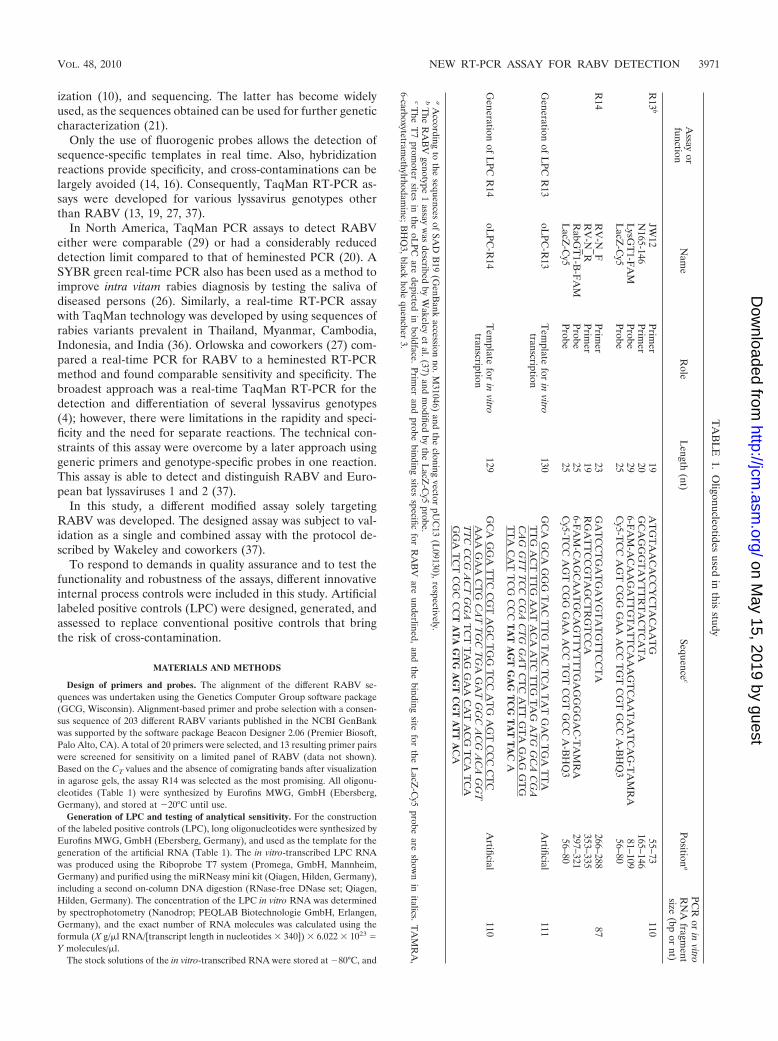
Design of primers and probes. The alignment of the different RABV se-quences was undertaken using the Genetics Computer Group software package(GCG, Wisconsin). Alignment-based primer and probe selection with a consen-sus sequence of 203 different RABV variants published in the NCBI GenBankwas supported by the software package Beacon Designer 2.06 (Premier Biosoft,Palo Alto, CA). A total of 20 primers were selected, and 13 resulting primer pairswere screened for sensitivity on a limited panel of RABV (data not shown).Based on the CT values and the absence of comigrating bands after visualizationin agarose gels, the assay R14 was selected as the most promising. All oligonu-cleotides (Table 1) were synthesized by Eurofins MWG, GmbH (Ebersberg,Germany), and stored at �20°C until use.
Generation of LPC and testing of analytical sensitivity. For the constructionof the labeled positive controls (LPC), long oligonucleotides were synthesized byEurofins MWG, GmbH (Ebersberg, Germany), and used as the template for thegeneration of the artificial RNA (Table 1). The in vitro-transcribed LPC RNAwas produced using the Riboprobe T7 system (Promega, GmbH, Mannheim,Germany) and purified using the miRNeasy mini kit (Qiagen, Hilden, Germany),including a second on-column DNA digestion (RNase-free DNase set; Qiagen,Hilden, Germany). The concentration of the LPC in vitro RNA was determinedby spectrophotometry (Nanodrop; PEQLAB Biotechnologie GmbH, Erlangen,Germany), and the exact number of RNA molecules was calculated using theformula (X g/�l RNA/[transcript length in nucleotides � 340]) � 6.022 � 1023 �Y molecules/�l.
The stock solutions of the in vitro-transcribed RNA were stored at �80°C, and
TA
BL
E1.
Oligonucleotides
usedin
thisstudy
Assay
orfunction
Nam
eR
oleL
ength(nt)
Sequencec
Positiona
PCR
orin
vitroR
NA
fragment
size(bp
ornt)
R13
bJW
12Prim
er19
AT
GT
AA
CA
CC
YC
TA
CA
AT
G55–73
110N
165-146Prim
er20
GC
AG
GG
TA
YT
TR
TA
CT
CA
TA
165–146L
ysGT
1-FA
MProbe
296-F
AM
-AC
AA
GA
TT
GT
AT
TC
AA
AG
TC
AA
TA
AT
CA
G-T
AM
RA
81–109L
acZ-C
y5Probe
25C
y5-TC
CA
GT
CG
GG
AA
AC
CT
GT
CG
TG
CC
A-B
HQ
356–80
R14
RV
-N_F
Primer
23G
AT
CC
TG
AT
GA
YG
TA
TG
TT
CC
TA
266–28887
RV
-N_R
Primer
19R
GA
TT
CC
GT
AG
CT
RG
TC
CA
353–335R
abGT
1-B-F
AM
Probe25
6-FA
M-C
AG
CA
AT
GC
AG
TT
YT
TT
GA
GG
GG
AC
-TA
MR
A297–321
LacZ
-Cy5
Probe25
Cy5-T
CC
AG
TC
GG
GA
AA
CC
TG
TC
GT
GC
CA
-BH
Q3
56–80
Generation
ofL
PCR
13oL
PC-R
13T
emplate
forin
vitrotranscription
130G
CA
GC
AG
GG
TA
CT
TG
TA
CT
CA
TA
TG
AC
TG
AT
TA
TT
GA
CT
TT
GA
AT
AC
AA
TC
TT
GT
AG
AT
GG
CA
CG
AC
AG
GT
TT
CC
CG
AC
TG
GA
TC
TC
AT
TG
TA
GA
GG
TG
TT
AC
AT
TC
GC
CC
TA
TA
GT
GA
GT
CG
TA
TT
AC
A
Artificial
111
Generation
ofL
PCR
14oL
PC-R
14T
emplate
forin
vitrotranscription
129G
CA
GG
AT
TC
CG
TA
GC
TG
GT
CC
AT
GA
GT
CC
CC
TC
AA
AG
AA
CT
GC
AT
TG
CT
GA
GA
TG
GC
AC
GA
CA
GG
TT
TC
CC
GA
CT
GG
AT
CT
TA
GG
AA
CA
TA
CG
TC
AT
CA
GG
AT
CT
CG
CC
CT
AT
AG
TG
AG
TC
GT
AT
TA
CA
Artificial
110
aA
ccordingto
thesequences
ofSA
DB
19(G
enBank
accessionno.M
31046)and
thecloning
vectorpU
C13
(L09130),respectively.
bT
heR
AB
Vgenotype
1assay
was
describedby
Wakeley
etal.(37)
andm
odifiedby
theL
acZ-C
y5probe.
cThe
T7
promoter
sitesin
theoL
PCare
depictedin
boldface.Prim
erand
probebinding
sitesspecific
forR
AB
Vare
underlined,and
thebinding
sitefor
theL
acZ-C
y5probe
areshow
nin
italics.T
AM
RA
,6-carboxytetram
ethylrhodamine;B
HQ
3,blackhole
quencher3.
VOL. 48, 2010 NEW RT-PCR ASSAY FOR RABV DETECTION 3971
on May 15, 2019 by guest
http://jcm.asm
.org/D
ownloaded from

the diluted working solutions were stored at �20°C. Using the working solutionsthe analytical sensitivity of the real-time RT-PCR assays was determined bytesting R13 and R14 LPC standard RNA diluted serially 10-fold.
Samples, viruses, and cells. RABVs, Lagos bat virus (LBV), Mokola virus(MOKV), Duvenhage virus (DUVV), European bat lyssavirus 1 and 2 (EBLV-1and EBLV-2), and Australian bat lyssavirus (ABLV) were obtained from thearchive of the National Reference Laboratory for Rabies, located at theFriedrich-Loeffler-Institut, Wusterhausen, Germany. Brain homogenates fromoriginal samples or from mice after inoculation were used. For a few archivedviruses, tissue culture supernatant was used (Table 2). For the latter, viruses werecultured using murine neuroblastoma cells (NA-cells; cat. no. 229; Collection ofCell Lines in Veterinary Medicine [CCLV], Insel Riems, Germany) as describedpreviously (38). Brain samples from 12 different species that tested negative byFAT were used as negative controls.
RNA extraction. Viral RNA was extracted from cell culture using the QIAampviral RNA kit (Qiagen, Hilden Germany) or RNeasy kit (Qiagen, Hilden, Ger-many) according to the manufacturer’s instructions. After the column waswashed twice with the appropriate buffer, RNA was eluted using 50 �l elutionbuffer and stored at �80°C until use.
TaqMan real-time RT-PCR. To minimize the risk of cross-contamination, aone-step reverse transcription-PCR (RT-PCR) (RT-PCR) protocol was carriedout using the commercially available QuantiTect probe RT-PCR kit (Qiagen,Hilden, Germany) or the RNA UltraSense one-step quantitative RT-PCR sys-tem (Invitrogen, Carlsbad, CA). The real-time RT-PCR assay with the Quanti-Tect probe RT-PCR kit was optimized using a total volume of 25 �l. Briefly, fora single well, 5.25 �l RNase-free water, 12.5 �l 2� QuantiTect probe RT-PCRmaster mix, 0.25 �l RT mix, 5.0 �l of prediluted RNA, and 2.0 �l primer-probemix R13 or R14 was mixed. In combined assays, 1 �l of each primer-probe mixR13 and R14 was used. Based on stock solutions of the primer and probes of 100pmol/�l (100 �M), the two different primer-probe mixes were created. For thenewly developed assay R14, 20 �l of the relevant forward and reverse primer, 2.5�l of the 6-carboxyfluorescein (FAM)-labeled R14 rabies probe, and 2.5 �l of theCy5-labeled positive-control probe were mixed in 155 �l 0.1� Tris-EDTA (TE)buffer. Thus, a final concentration of 10 �M each primer and 1.25 �M each probewas combined in the R14 primer-probe mix. For the R13 primer-probe mix,identical concentrations with the relevant primers and probes were established.
The reactions were carried out either on an Mx3000P/Mx3005P multiplexquantitative PCR system (Stratagene, La Jolla) or on a LightCycler (RocheDiagnostics, Mannheim, Germany). The RNAs were reverse transcribed andamplified according to the following heating and cooling program: 1 cycle of 50°Cfor 30 min and 95°C for 15 min, followed by 42 cycles of 94°C for 30 s, 55°C for30 s, and 72°C for 30 s. In the LightCycler, while keeping the same temperatureregimen, the number of cycles was set at 45 and the duration time for each cyclestep was 20 s. For each RT-PCR, a critical threshold cycle number (CT) wasdetermined corresponding to the PCR cycle number at which the fluorescence ofthe reaction exceeded a value determined to be statistically higher than thebackground by the respective software associated with each system.
To estimate the reproducibility of the assays, most samples (N � 69) were runtwice with R13, R14, and the combined assay on an Mx3000P (FLI, Riems,Germany), LightCycler 1.0 (FLI, Wusterhausen, Germany), and Mx3005P(DTU, Lindholm, Denmark). The data were transferred into an Excel spread-sheet (Microsoft), and the resulting graphs and correlation coefficients (of de-termination) were calculated.
Sequencing. A selection of samples that tested negative in real-time RT-PCRwith either the developed assay (R14) or the previously described one, R13, withthe RABV protocol from Wakeley et al. (37), were retested with a conventionalRT-PCR as described before (15). Resulting amplicons were subject to thesequence analysis of the partial N gene essentially as described previously (21).
RESULTS
Development and validation of the new R14 real-time RT-PCR for RABV. (i) Primers and probe. In a first step, 203sequences of the conserved N gene of different rabies virusstrains were used for the creation of a consensus sequence andthe identification of highly conserved sequence regions. Be-sides the genome region used by Wakeley et al. (37), only a fewnucleotides downstream make a suitable region for the designof alternative primers, and a respective hybridization probe
was chosen. The newly selected primers amplified a fragmentof 87 bp in size (Table 1).
(ii) Construction of the LPC. Long oligonucleotides (oLPC)were used as the template for the preparation of labeled invitro-transcribed artificial positive control RNA (LPC). TheLPC RNAs were tagged by an additional binding sequence foran extra probe (LacZ-Cy5). The LacZ-Cy5 probe was inte-grated into the R13 and R14 primer-probe mixes (Table 1). Inthe case of the amplification and detection of RABV RNA, nobinding of the LacZ-Cy5 probe can be observed. In contrast,the analyses of LPC-RNA produce RABV-specific FAM sig-nals as well as the tag-specific Cy5 fluorescence. The principleof the LPC is depicted in Fig. 1. Based on the equimolarconcentration of the probe binding sites for the RABV andLacZ probes, nearly identical CT values can be observed(Fig. 2).
(iii) Analytical sensitivity and specificity. Using a series of10-fold dilutions of LPC RNA, it could be verified that the R13and R14 real-time RT-PCR systems, as well as the combinedassay, amplified the control RNA in a linear fashion from 107
copies per well down to less than 10 copies per well with anPCR efficiency of more than 90% (Fig. 2). Identical high PCRefficiencies were calculated using an RNA dilution series ofdifferent RABV strains (see Fig. S1a and b in the supplementalmaterial).
The analytical specificity of the R14 assay was assessed by insilico BlastN searches. Only the forward primer of the R14systems showed partial identity with EBLV-2, whereas thereverse primer and the R14 probe did not show any significantsimilarity with other pathogens or housekeeping genes. Thespecificity of the R14 assay was confirmed by the analysis ofrelated lyssaviruses.
(iv) Diagnostic sensitivity and specificity. Of 93 brain sam-ples or virus isolations from initially FAT-positive animals, 80tested positive by the R14 assay. Samples that tested negativeoriginated from India, Pakistan, Afghanistan, and Nigeria (Ta-ble 2). Furthermore, none of the negative-control samples, orany of genotypes other than RABV, gave a result above thethreshold indicative for the presence of RABV-specific RNA(data not shown).
(v) Reproducibility. The reproducibility was very high (Fig.3). The corresponding coefficient of determination (R2) was0.9904.
(vi) Integration of internal controls. For checking the func-tionality of the RNA extraction as well as the inhibition-freeamplification of the R14 fragment, two different internal con-trol (IC) systems were assessed, i.e., the beta-actin housekeep-ing gene system described by Wakeley et al. (37) and theexternal universal control system based on in vitro-transcribedenhanced green fluorescent protein RNA (18). Both IC sys-tems used HEX (hexachloro-6-carboxyfluorescein)-labeled hy-drolysis probes for the detection. The comparison of the singleR14 assay to the duplex R14/IC assays showed similar analyt-ical sensitivities for both systems (data not shown).
Comparison of the developed assay (R14) to a publishedassay (R13). The specificity of the R13 assay (37) was evalu-ated using the same panel of samples as that used before. Inthis study, of 93 samples 85 tested positive by the R13 assay.Eight samples were negative or gave inconclusive results in thedifferent laboratories (did not score as reproducibly positive),
3972 HOFFMANN ET AL. J. CLIN. MICROBIOL.
on May 15, 2019 by guest
http://jcm.asm
.org/D
ownloaded from

TABLE 2. Diagnostic sensitivity and specificity using the R13, R14, and combined assay for the detection of RABV with prediluted RNAa
Lab ID Material Yr Host Continent Country R13 assay (CT) R14 assay (CT) R13-R14 combinationassay (CT)
13118 BS* 1983 Dog Africa Algeria 39.20 30.96 30.7320827 BS 1991 Duiker Africa Botswana 27.77 23.28 24.2913130 BS 1974 Africa Egypt 29.27 28.24 26.8120829 BS 1998 Dog Africa Egypt 22.57 20.75 21.3520830 BS 1998 Dog Africa Egypt 28.39 26.97 27.3413136 BS 1989 Africa Nigeria 22.79 22.9113137 BS 1988 Dog Africa Nigeria 25.30 24.4120825 BS 2005 Dog Africa Nigeria 28.86 28.9420826 BS 1996 Human Africa Nigeria 22.79 21.3220828 BS 1990 Cat Africa South Africa 26.79 22.41 23.713176 BS 1992 Dog Africa Sudan 32.17 32.4213116 BS Dog Africa Tunesia 30.00 25.21 24.4413230 BS Skunk Americas Canada 34.36 30.09 30.2113231 BS Fox Americas Canada 30.02 32.02 26.6113239 BS Bat Americas Canada 24.11 23.9713240 BS* 1986 Americas Canada 25.37 25.513250 BS 1973 Human Americas Chile 28.79 28.7613254 BS* 1979 Human Americas Chile 20.45 20.4613196 BS 1981 Dog Americas Mexico 26.21 22.88 23.2113242 BS* 1966 Bat Americas South America 27.45 23.64 24.4913199 BS* 1980 Skunk Americas United States 35.46 32.76 33.6813213 TCS 1981 Skunk Americas United States 27.98 25.75 25.1613216 TCS Bat Americas United States 28.55 28.7320823 BS 1988 Bat Americas United States 27.59 28.9 28.4620824 BS 1988 Raccoon Americas United States 22.31 24.76 22.7713091 BS 1994 Camel Asia Abu Dhabi 17.47 16.69 16.7320277 BS 2005 Dog Asia Afghanistan 18.10 19.5820278 BS 2005 Pig Asia Afghanistan 22.91 24.5920280 BS 2006 Dog Asia Afghanistan 15.72 17.0420281 BS 2006 Dog Asia Afghanistan 14.87 17.2620282 BS 2006 Dog Asia Afghanistan 26.32 23.7520287 BS 2008 Dog Asia Afghanistan 17.85 18.8120292 BS 2008 Dog Asia Afghanistan 14.55 15.795989 BS 2002 Dog Asia Aserbaidshan 29.12 24.21 24.7613081 TCS 1985 Asia China 26.09 22.99 22.6820833 BS 1989 Dog Asia China 29.01 22.23 19.2920834 BS 1989 Cow Asia China 28.57 25.41 22.6113181 BS 1982 Dog Asia India 26.33 24.5113184 BS 1993 Monkey Asia India 27.89 22.44 23.1813101 BS 1988 Dog Asia Indonesia 31.09 25.77 26.6413151 BS 1991 Wolf Asia Iran 30.80 30.19 29.5213160 TCS 1991 Sheep Asia Iran 18.39 16.54 16.3413161 TCS 1991 Sheep Asia Iran 19.86 19.51 19.2613164 TCS 1991 Hyena Asia Iran 25.01 26.3 24.3420276 BS 2004 Dog Asia Iraq 16.81 18.25 22.5920279 BS 2005 Dog Asia Iraq 15.43 12.82 12.6120283 BS 2007 Dog Asia Iraq 17.55 13.83 12.9920284 BS 2007 Dog Asia Iraq 16.31 13.67 13.7620285 BS 2007 Mongoose Asia Iraq 17.62 17.9 16.7820288 BS 2008 Cow Asia Iraq 21.77 21.49 20.8220289 BS 2008 Cow Asia Iraq 18.85 18.25 17.8120290 BS 2008 Cow Asia Iraq 24.20 25.18 24.2820291 BS 2008 Dog Asia Iraq 22.56 18.47 19.120293 BS 2008 Dog Asia Iraq 16.55 12.62 12.9720294 BS 2008 Dog Asia Iraq 16.58 12.48 12.520295 BS 2009 Dog Asia Iraq 17.41 12.86 13.3820296 BS 2009 Dog Asia Iraq 18.84 15.01 13.920297 BS 2009 Horse Asia Iraq 14.90 11.92 11.8120298 BS 2009 Cow Asia Iraq 20.05 15.73 15.9920299 BS 2008 Dog Asia Iraq 17.52 13.68 13.6213109 BS 1974 Dog Asia Malaysia 26.91 21.91 22.1913141 BS Asia Oman 30.15 29.56 28.1513088 BS* 1979 Dog Asia Pakistan 28.69 28.2213042 BS* 1987 Fox Asia Saudi Arabia 26.98 22.35 22.5213044 TCS 1990 Fox Asia Saudi Arabia 20.48 19.42 19.513096 BS 1974 Dog Asia Singapore 29.81 25.1 25.2113099 BS 1974 Dog Asia Taiwan 33.56 29.02 29.23
Continued on following page
VOL. 48, 2010 NEW RT-PCR ASSAY FOR RABV DETECTION 3973
on May 15, 2019 by guest
http://jcm.asm
.org/D
ownloaded from

comprising samples from Europe, Chile, North America, theSudan, and Greenland (Table 2). Overall, a high reproducibil-ity was observed, with R2 � 0.9963. None of the negative-control samples gave a result above the threshold indicative forthe presence of virus-specific RNA. In addition, the R13 assayshowed reactivity with one ABLV isolate (data not shown);however, these data have to be verified using additional iso-lates of the same genotype.
When combining both assays in a single mix, all initiallypositive samples with either method also scored positive (Table2). The reproducibility of the novel combined R13/14 assay wasdetermined as R2 � 0.98 (Fig. 3).
Sequence comparison. Selected sequences of samples thattested negative in either assay were aligned and compared tothe sequences of primers and probes. The R14 assay did notdetect strains belonging to the Arctic-like viruses from India,Pakistan, and Afghanistan (23). Also, two Nigerian RABVs
belonging to the lineage Africa 2 were not detected with R14.Sequence comparison showed that Arctic-like sequences hadup to three nucleotide differences compared to the sequence ofthe reverse primer RABV-N_R and also three nucleotide dif-ferences from the probe (Fig. 4b). For the same isolates, theprimer and probe binding regions for the R13 assays revealedonly one mismatch in the probe binding region (Fig. 4a).
An isolate from Chile (13250) that did not yield a CT valuewith the R13 assay had two mismatches in the probe bindingregion (Fig. 4a), while the nucleotide sequence of the sameisolate only has one mismatch within the forward primer of theR14 assay (Fig. 4b).
DISCUSSION
In this study, we developed and validated a specific andsensitive real-time RT-PCR for the detection of RABV, com-pared it to a published assay (37), and improved the perfor-mance of the assays by the combination of both primer andprobe sets as well as by the introduction of labeled positivecontrols (LPC).
Generally, the use of fluorogenic probes as detection sys-tems for PCRs have improved the analytical sensitivity due tovery short amplification products, which result in increasedspecificity and the prevention of cross-contaminations throughthe absence of post-PCR handling. The rapidity of the test,with results obtained in real time by obviating gel electro-phoresis, offers the possibility to provide timely information tothe relevant authorities.
As the R14 diagnostic assay can detect less than 10 genomecopies/well, it represents a valuable tool for intra vitam diag-nosis. A further advantage of real-time PCR techniques is the
TABLE 2—Continued
LabID Material Yr Host Continent Country R13 assay (CT) R14 assay (CT) R13-R14 combination
assay (CT)
20831 BS Asia Thailand 14.07 25.86 23.6520832 BS Asia Thailand 25.87 26.8 25.2813056 BS* 1984 Dog Asia Turkey 26.45 23.47 23.8913077 BS 1995 Fox Europe Bulgaria 27.02 24.28 24.3413078 BS 1995 Human Europe Bulgaria 13.8 12.68 12.7713079 BS 1995 Fox Europe Bulgaria 29.66 26.9 26.7412952 BS 2001 Fox Europe Estonia 31.01 27.39 27.813001 TCS 1991 Europe Finland 30.87 27.25 27.713000 BS 1990 Raccoon dog Europe Finland 33.05 29.39 30.01390 TCS 1998 Fox Europe Germany 29.98 30.98 29.2611164 BS 2005 Fox Europe Germany 23.43 23.911240 BS 2005 Human Europe Germany 16.9 18.27 16.6611251 BS 2005 Human Europe Germany 20.03 21.71 20.2812542 BS 2005 Human Europe Germany 24.99 24.53 24.213057 BS 2005 Human Europe Germany 17.01 17.25 16.64SAD B19 TCS Vaccine strain Europe Germany 18.22 19.00 18.32GRA 11/04 BS 2004 Polar fox Europe Greenland 15.67 16.20 14.56GRA 16/06 BS 2006 Polar fox Europe Greenland 13.90 14.71GRA 2/07 BS 2007 Polar fox Europe Greenland 12.58 12.68 10.72GRA 2/08 BS 2008 Polar fox Europe Greenland 15.33 16.41 15.24GRA 4/07 BS 2007 Polar fox Europe Greenland 12.14 12.54 12.54GRA 5/03 BS 2003 Dog Europe Greenland 15.31 16.76 14.67GRA 5/04 BS 2004 Polar fox Europe Greenland 15.15 17.06 13.94GRA 6/05 BS 2005 Polar fox Europe Greenland 16.71 16.99 15.70GRA 6/08 BS 2008 Polar fox Europe Greenland 12.79 12.25 12.71GRA 7/05 BS 2005 Polar fox Europe Greenland 17.93 18.50 17.13
a BS, brain suspension; BS*, brain suspension from inoculated mice; TCS, tissue culture supernatant.
FIG. 1. Graphical depiction of the principle of the labeled positivecontrol (LPC).
3974 HOFFMANN ET AL. J. CLIN. MICROBIOL.
on May 15, 2019 by guest
http://jcm.asm
.org/D
ownloaded from

possibility to quantify viral RNA in real time, giving a relativelyquick and reliable method for measuring the levels of viralRNA. Since the assay has proven reproducibility (Fig. 3), it issuitable for experimental infection studies with RABV inves-
tigating the spread of the virus and its accumulation in varioustissues.
Both investigated assays had limits to detect some of theRABVs in the panel. Specifically, the newly developed RT-PCR (R14) did not detect RABVs of the Arctic-like lineagefrom Asia and viruses from Nigeria (Africa 2) (Table 2). It wasdemonstrated before that the number of sequence mismatchesbetween primer and probe sets and target sequences of rabiesviruses significantly affects amplification and detection (20, 37,25). In this case, sequence analysis showed that both probe andreverse primer sequences were different from the sequences ofthis lineage (Fig. 4). In contrast, the R13 assay had only onenucleotide difference from the Arctic-like viruses and henceresulted in good performance. The opposite is the case forRABVs from Chile. These samples exemplify that the assayscomplement each other in combination, which could be shownpractically with the combined R13/14 assay. Overall, all FAT-positive samples were confirmed as positive using the com-bined real-time RT-PCR assay. During the development andvalidation of the combined assay, an RABV and sequencepanel with wide geographical and phylogenetic variation wereused. Also, with the previously published R13 RT-PCR beingrevalidated with a different virus panel, in total more than 130virus isolates were successfully tested. In conclusion, it appearslikely that this combined assay will detect RABV strains oc-curring worldwide.
Generally, a combination of two assays targeting differentlocations on the genome will limit the chance of false negativestremendously. Furthermore, by choosing a similar fragmentsize and similar specifications for primers and probes (Table1), a combination as multiplex or parallel assays gives diagnos-ticians many options for implementation on various PCR ma-chines.
The approach of a combined version of primer and probesets to overcome the diversity among RABVs previously hasbeen applied to improve intra vitam human rabies diagnosis(25). Using a collection of representatives of the world-widediversity of RABV, all three primer-probe sets were shown todetect a wide range of RABV strains with very few detectionfailures (25). In fact, the most successful primer-probe combi-nation was a slightly modified version of the Wakeley protocol
FIG. 2. Amplification plot for the definition of the analytical sen-sitivity of the R13 assay (A) and the newly designed R14 system (B),based on in vitro-transcribed LPC RNA. Nearly similar CT values wereobserved for the FAM-labeled rabies probe (blue line with dots) aswell as for the Cy5-labeled LacZ probe (red line with open square).
FIG. 3. Reproducibility shown by the CT values for each sample tested twice using the R13 assay (a), the R14 assay (b), and the combination(c). The regression line is indicated, and the coefficients of determination (R2) are provided.
VOL. 48, 2010 NEW RT-PCR ASSAY FOR RABV DETECTION 3975
on May 15, 2019 by guest
http://jcm.asm
.org/D
ownloaded from

for RABV (25), indicating that assays can and should be im-proved and revalidated when more sequence data are avail-able.
The diversity of RABV is important, especially for referencelaboratories for rabies. International travel leads to possibili-ties for people to have contact with various RABV variants(12). Also, companion animals often accompany passengers,thus there is a need for reference laboratories to provide fastand reliable results for subsequent decision-making by publicand veterinary health authorities. For this purpose, sensitive,specific-but-broad-spectrum assays such as the one describedhere should be applied. Other published real-time RT-PCRassays have been validated using small numbers of RABVstrains only (29, 20, 27, 36), thus their use for internationalreference laboratories needs additional evaluation.
A further advantage of RT-PCR is that RABV RNA can bedetected in a range of biological fluids and samples (e.g., saliva,cerebrospinal fluid, tears, and skin biopsy samples). Generally,RT-PCR has been reported to confirm rabies diagnosis intravitam in suspect human cases, when conventional diagnosticmethods have failed and postmortem material is not available(11). Owing to the intermittent shedding of virus, negativeresults should not be used to exclude a diagnosis of rabies.However, diagnostic sensitivity is of eminent importance forintra vitam diagnosis. Because of the high sensitivity of thereal-time PCR described here, this technique offers a potentialuse in this diagnostic scheme, as demonstrated earlier with aSYBR green real-time PCR assay (26).
At present, no recommended standard protocol for rabiesdiagnosis using RT-PCR has been published by the OIE orWHO. Since both generic approaches as well as specific PCRassays targeting lyssavirus genotypes or even variants have ad-vantages and limitations, a cascade-type diagnostic procedurefor rabies PCRs would be preferable, as described for thediagnosis of other viral diseases (17). A pan-lyssavirus or evena pan-rhabdovirus PCR assay could be combined with more-specific and -sensitive genotype-specific real-time PCRs or
even variant specific real-time PCRs to confirm each other,allow genotyping, and obtain epidemiologically relevant infor-mation in real time. In this respect each single assay must bevalidated.
Besides the fully validated target assays, the functionalityand robustness of molecular genetic tests can be increased bythe application of innovative control systems. Internal processcontrols (e.g., beta-actin gene) check the efficiency of totalRNA extraction as well as the inhibition of free amplification(37, 34). The disadvantage of such housekeeping gene systemsis the inconsistent RNA load in different sample materials.Thus, a reduced sensitivity of the target assay based on partialinhibition is unlikely to be identified. In such cases the ampli-fication of an external control using a defined amount of het-erologous in vitro-transcribed RNA (18) can be helpful. Thenewly designed R14 assay was successfully combined with twopublished control assays (37, 18), and the combined systemscan be used routinely. Furthermore, the R14 system seemssufficiently robust for the implementation of alternative inter-nal control systems generally used in the different laboratories(e.g., commercial IC).
An important advantage of the real-time RT-PCR technol-ogy is the minimizing of cross-contamination based on theamplicon detection without opening the lid of the reactiontube. Nevertheless, the accidental removal of the lid or seal canbe responsible for the spreading of amplicons. Furthermore,during pipetting, handling positive controls or standards isnecessary. Therefore, an easy way to generate a labeled posi-tive control without any cloning steps was developed. Based ona synthetic oligonucleotide, including the primers and probefor the target assay and a T7 promoter site, extensive quantitiesof in vitro-transcribed RNA was produced. To tag the in vitrotranscript, an additional probe binding site was introduced intothe LPC. The tag-specific probe integrated in the master mixcan hybridize only with the LPC, and only then can the tag-specific fluorophore be detected during amplification. A simi-lar approach has been described by Snow et al. (30). In general
FIG. 4. Details of binding sites for two isolates, 13250 (Chile; GenBank accession no. HQ116829) and 20277 (Afghanistan; GenBank accessionno. HQ116830), that were not detected by the R13 assay (a) or by the R14 assay (b). Differences are indicated. The sequence of SAD B19 (M31046)was included for better visualization.
3976 HOFFMANN ET AL. J. CLIN. MICROBIOL.
on May 15, 2019 by guest
http://jcm.asm
.org/D
ownloaded from

these authors used the same strategy, but they cloned thetagged positive control prior to in vitro transcription. Here, asimplified protocol for the more-rapid generation of LPC with-out a cloning procedure is presented. This simple protocolshould support the construction and application of LPC forincreasing the diagnostic safety of molecular rabies diagnostics.In general, the implementation of LPC can play an importantrole for the further harmonization and standardization of real-time PCR assays.
The lack of standardization, quality issues like contamina-tion or false-negative results, and the varying reliability of PCRresults in many laboratories, especially in developing countries,have been obstacles to the general use of PCR for rabiesdiagnosis. Therefore, the WHO does not currently recommendthis technique for the routine postmortem diagnosis of rabies(40). However, with the accreditation of quality control mea-sures being implemented in a growing number of laboratoriesworldwide, recommended PCR methods may become avail-able. Such quality controls for diagnostic rabies PCRs shouldencompass several measures, e.g., the inclusion of appropriatepositive (LPC), negative, and inhibition controls in assay runs.The consistency and the interassay reproducibility also shouldbe ensured over time by monitoring the performance of theassay rigorously. If PCR is applied for epidemiological surveys,positive results should be confirmed by conventional virologi-cal techniques. In most national and international legislatures,only the detection of virus or viral antigen leads to the confir-mation of a rabies case and thus to subsequent control mea-sures. Also, the isolation of viable virus provides further pos-sibilities to study the characteristics of this particular strain.
Only if laboratories meet the required standard (40) can thePCR and, especially, real-time PCR offer its full potential as aconfirmatory diagnostic test, especially in decomposed or intravitam samples.
ACKNOWLEDGMENTS
We thank Christian Korthase and Jeanette Kliemt at the Friedrich-Loeffler-Institute for excellent technical assistance.
This work was partially supported by EPIZONE, the EU Network ofExcellence for Epizootic Disease Diagnosis and Control (FOOD-CT-2006-016236), under work package 4.1, designated real-time PCR di-agnostics, and was partially funded from the project European Man-agement Platform for Emerging and Re-emerging Infectious Disease(EMPERIE) (EC grant agreement 223.498).
REFERENCES
1. Araujo, D. B., H. Langoni, M. F. Almeida, and J. Megid. 2008. Heminestedreverse-transcriptase polymerase chain reaction (hnRT-PCR) as a tool forrabies virus detection in stored and decomposed samples. BMC Res. Notes1:17.
2. Belak, S., and P. Thoren. 2001. Molecular diagnosis of animal diseases: someexperiences over the past decade. Expert. Rev. Mol. Diagn. 1:434–443.
3. Biswal, M., R. Ratho, and B. Mishra. 2007. Usefulness of reverse tran-scriptase-polymerase chain reaction for detection of rabies RNA in archivalsamples. Jpn. J. Infect. Dis. 60:298–299.
4. Black, E. M., J. P. Lowings, J. Smith, P. R. Heaton, and L. M. McElhinney.2002. A rapid RT-PCR method to differentiate six established genotypes ofrabies and rabies-related viruses using TaqMan technology. J. Virol. Meth-ods 105:25–35.
5. Black, E. M., L. M. McElhinney, J. P. Lowings, J. Smith, P. Johnstone, andP. R. Heaton. 2000. Molecular methods to distinguish between classicalrabies and the rabies-related European bat lyssaviruses. J. Virol. Methods87:123–131.
6. Crepin, P., L. Audry, Y. Rotivel, A. Gacoin, C. Caroff, and H. Bourhy. 1998.Intravitam diagnosis of human rabies by PCR using saliva and cerebrospinalfluid. J. Clin. Microbiol. 36:1117–1121.
7. David, D., B. Yakobson, D. Rotenberg, N. Dveres, I. Davidson, and Y. Stram.
2002. Rabies virus detection by RT-PCR in decomposed naturally infectedbrains. Vet. Microbiol. 87:111–118.
8. Dean, D. J., M. K. Abelseth, and P. Athanasiu. 1996. The fluorescenceantibody test, p. 88–93. In F.-X. Meslin, M. M. Kaplan, and H. Koprowski(ed.), Laboratory techniques in rabies. World Health Organization, Geneva,Switzerland.
9. Echevarría, J. E., A. Avellon, J. Juste, M. Vera, and C. Ibanez. 2001. Screen-ing of active lyssavirus infection in wild bat populations by viral RNA de-tection on oropharyngeal swabs. J. Clin. Microbiol. 39:3678–3683.
10. Finnegan, C. J., S. M. Brookes, and A. R. Fooks. 2004. Detection of Euro-pean bat lyssavirus mRNA in mouse brain by employing in situ hybridisation.J. Virol. Methods 121:221–227.
11. Fooks, A. R., N. Johnson, C. M. Freuling, P. Wakeley, A. Banyard, L. M.McElhinney, D. A. Marston, A. Dastjerdi, E. Wright, R. Weiss, and T.Muller. 2009. Emerging technologies for the detection of rabies virus: chal-lenges and hopes in the 21st century. PLoS Neglect. Trop. D 3:e530.
12. Fooks, A. R., N. Johnson, S. M. Brookes, G. Parsons, and L. M. McElhinney.2003. Risk factors associated with travel to rabies endemic countries. J. Appl.Microbiol. 94:31–36.
13. Foord, A. J., H. Heine, L. I. Pritchard, R. A. Lunt, K. M. Newberry, C. L.Rootes, and B. D. Boyle. 2006. Molecular diagnosis of lyssaviruses andsequence comparison of Australian bat lyssavirus samples. Aust. Vet. J.84:225–230.
14. Gibson, U. E., C. A. Heid, and P. M. Williams. 1996. A novel method for realtime quantitative RT-PCR. Genome Res. 6:995–1001.
15. Heaton, P. R., P. Johnstone, L. M. McElhinney, R. Cowley, E. O’Sullivan,and J. E. Whitby. 1997. Heminested PCR assay for detection of six genotypesof rabies and rabies-related viruses. J. Clin. Microbiol. 35:2762–2766.
16. Heid, C. A., J. Stevens, K. J. Livak, and P. M. Williams. 1996. Real timequantitative PCR. Genome Res. 6:986–994.
17. Hoffmann, B., T. Harder, E. Starick, K. Depner, O. Werner, and M. Beer.2007. Rapid and highly sensitive pathotyping of avian influenza A H5N1virus by using real-time reverse transcription-PCR. J. Clin. Microbiol. 45:600–603.
18. Hoffmann, B., K. Depner, H. Schirrmeier, and M. Beer. 2006. A universalheterologous internal control system for duplex real-time RT-PCR assaysused in a detection system for pestiviruses. J. Virol. Methods 136:200–209.
19. Hughes, G. J., I. V. Kuzmin, A. Schmitz, J. Blanton, J. Manangan, S.Murphy, and C. E. Rupprecht. 2006. Experimental infection of big brownbats (Eptesicus fuscus) with Eurasian bat lyssaviruses Aravan, Khujand, andIrkut virus. Arch. Virol. 151:2021–2035.
20. Hughes, G. J., J. S. Smith, C. A. Hanlon, and C. E. Rupprecht. 2004.Evaluation of a TaqMan PCR assay to detect rabies virus RNA: influence ofsequence variation and application to quantification of viral loads. J. Clin.Microbiol. 42:299–306.
21. Johnson, N., L. M. McElhinney, J. Smith, P. Lowings, and A. R. Fooks. 2002.Phylogenetic comparison of the genus Lyssavirus using distal coding sequencesof the glycoprotein and nucleoprotein genes. Arch. Virol. 147:2111–2123.
22. Kulonen, K., M. Fekadu, S. Whitfield, and C. K. Warner. 1999. An evalua-tion of immunofluorescence and PCR methods for detection of rabies inarchival Carnoy-fixed, paraffin-embedded brain tissue. Zentralbl. Veteri-narmed. B 46:151–155.
23. Kuzmin, I. V., G. J. Hughes, A. D. Botvinkin, S. G. Gribencha, and C. E.Rupprecht. 2008. Arctic and Arctic-like rabies viruses: distribution, phylog-eny and evolutionary history. Epidemiol. Infect. 136:509–519.
24. Kuzmin, I. V., and C. E. Rupprecht. 2007. Bat rabies, p. 259–307. In A. C.Jackson and W. Wunner. (ed.), Rabies. Academic Press, New York, NY.
25. Nadin-Davis, S. A., M. Sheen, and A. I. Wandeler. 2009. Development ofreal-time reverse transcriptase polymerase chain reaction methods for hu-man rabies diagnosis. J. Med. Virol. 81:1484–1497.
26. Nagaraj, T., J. P. Vasanth, A. Desai, A. Kamat, S. N. Madhusudana, and V.Ravi. 2006. Ante mortem diagnosis of human rabies using saliva samples:comparison of real time and conventional RT-PCR techniques. J. Clin.Virol. 36:17–23.
27. Orlowska, A., M. Smreczak, P. Trebas, and J. F. Zmudzinski. 2008. Com-parison of realtime PCR and heminested RT-PCR methods in the detectionof rabies virus infection in bats and terrestrial animals. Bull. Vet. Inst.Pulawy. 52:313–318.
28. Sacramento, D., H. Bourhy, and N. Tordo. 1991. PCR technique as analternative method for diagnosis and molecular epidemiology of rabies virus.Mol. Cell Probes 5:229–240.
29. Shankar, V., R. A. Bowen, A. D. Davis, C. E. Rupprecht, and T. J. O’Shea.2004. Rabies in a captive colony of big brown bats (Eptesicus fuscus). J.Wildl. Dis. 40:403–413.
30. Snow, M., P. McKay, and I. Matejusova. 2009. Development of a widelyapplicable positive control strategy to support detection of infectious salmonanaemia virus (ISAV) using Taqman real-time PCR. J. Fish Dis. 32:151–156.
31. Tordo, N., A. Benmansour, C. Calisher, R. G. Dietzgen, R. X. Fang, A. O.Jackson, G. Kurath, S. Nadin-Davis, R. B. Tesh, and P. J. Walker. 2004.Rhabdoviridae, p. 623–644. In C. M. Fauquet, M. A. Mayo, J. Maniloff, U.Desselberger, and L. A. Ball (ed.), Virus taxonomy, VIIIth report of theICTV. Elsevier/Academic Press, London, United Kingdom.
VOL. 48, 2010 NEW RT-PCR ASSAY FOR RABV DETECTION 3977
on May 15, 2019 by guest
http://jcm.asm
.org/D
ownloaded from
32. Tordo, N., D. Sacramento, and H. Bourhy. 1996. The polymerase chainreaction (PCR) technique for diagnosis, typing and epidemiological studies,p. 157–170. In F.-X. Meslin, M. M. Kaplan, and H. Koprowski (ed.), Labo-ratory techniques in rabies, 4th ed. World Health Organization, Geneva,Switzerland.
33. Tordo, N., H. Bourhy, and D. Sacramento. 1995. PCR technology for lyssa-virus diagnosis, p. 125–145. In J. P. Clewley (ed.), Polymerase chain reaction(PCR) for human viral diagnosis. CRC, Boca Raton, FL.
34. Toussaint, J. F., C. Sailleau, E. Breard, S. Zientara, and K. De Clercq. 2007.Bluetongue virus detection by two real-time RT-qPCRs targeting two dif-ferent genomic segments. J. Virol. Methods 140:115–123.
35. Vazquez-Moron, S., A. Avellon, and J. E. Echevarria. 2006. RT-PCR fordetection of all seven genotypes of Lyssavirus genus. J. Virol. Methods135:281–287.
36. Wacharapluesadee, S., J. Sutipanya, S. Damrongwatanapokin, P. Phumesin,
P. Chamnanpood, C. Leowijuk, and T. Hemachudha. 2008. Development ofa TaqMan real-time RT-PCR assay for the detection of rabies virus. J. Virol.Methods 151:317–320.
37. Wakeley, P. R., N. Johnson, L. M. McElhinney, D. Marston, J. Sawyer, andA. R. Fooks. 2005. Development of a real-time, TaqMan reverse transcrip-tion-PCR assay for detection and differentiation of lyssavirus genotypes 1, 5,and 6. J. Clin. Microbiol. 43:2786–2792.
38. Webster, W. A., and G. A. Casey. 1996. Virus isolation in neuroblastoma cellculture, p. 93–104. In F.-X. Meslin, M. M. Kaplan, and H. Koprowski (ed.),Laboratory techniques in rabies. World Health Organization, Geneva, Switzerland.
39. Whitby, J. E., P. R. Heaton, E. M. Black, M. Wooldridge, L. M. McElhinney,and P. Johnstone. 2000. First isolation of a rabies-related virus from aDaubenton’s bat in the United Kingdom. Vet. Rec. 147:385–388.
40. World Health Organisation. 2005. Expert consultation on rabies, first report.World Health Organ. Tech. Rep. Ser. 931:1–121.
3978 HOFFMANN ET AL. J. CLIN. MICROBIOL.
on May 15, 2019 by guest
http://jcm.asm
.org/D
ownloaded from