improved amplification efficiency on stool samples … amplification efficiency on stool samples by...
TRANSCRIPT

Roperch et al. BMC Biotechnology (2015) 15:41 DOI 10.1186/s12896-015-0148-6
METHODOLOGY ARTICLE Open Access
Improved amplification efficiency on stoolsamples by addition of spermidine and its usefor non-invasive detection of colorectal cancerJean-Pierre Roperch1,2*, Karim Benzekri3, Hicham Mansour4 and Roberto Incitti5
Abstract
Background: Using quantitative methylation-specific PCR (QM-MSP) is a promising method for colorectal cancer(CRC) diagnosis from stool samples. Difficulty in eliminating PCR inhibitors of this body fluid has been extensivelyreported. Here, spermidine is presented as PCR facilitator for the detection of stool DNA methylation biomarkersusing QM-MSP. We examined its effectiveness with NPY, PENK and WIF1, three biomarkers which we have previouslyshown to be of relevance to CRC.
Results: We determined an optimal window for the amplification of the albumin (Alb) gene (100 ng of bisulfite-treatedstool DNA added of 1 mM spermidine) at which we report that spermidine acts as a PCR facilitator (AE = 1680%) for SGRT-PCR. We show that the amplification of methylated PENK, NPY and WIF1 is considerably facilitated by QM-MSP asmeasured by an increase of CMI (Cumulative Methylation Index, i.e. the sum of the three methylation values) by a factorof 1.5 to 23 fold in individual samples, and of 10 fold in a pool of five samples.
Conclusions: We contend that spermidine greatly reduces the problems of PCR inhibition in stool samples. Thisobserved feature, after validation on a larger sampling, could be used in the development of stool-based CRC diagnosistests.
Keywords: Spermidine, Colorectal cancer, QM-MSP, Methylated biomarkers
BackgroundColorectal cancer (CRC) is one of the most common formsof cancer in the world [1]. CRC can be cured if diagnosedat early stage using endoscopic examination [2,3], makingearly non-invasive screening a crucial aim. The develop-ment of CRC results from the progressive accumulation ofgenetic and epigenetic alterations leading to the transform-ation of normal colonic epithelium to colon adenocarcin-oma [4-6]. Fecal-occult blood test (FOBT) is the mostwidely used method of screening for CRC [7,8]. However,FOBT lacks sensitivity as well as specificity for screeningan average risk population. Epigenetic alterations havebeen found frequently in neoplastic diseases [9,10]. It hasbeen reported that the analysis of DNA methylation car-ried out in body fluids represents a valuable source for thediscovery of cancer biomarkers [11]. Prior studies showed
* Correspondence: [email protected], Paris Biotech 24 rue du Faubourg St Jacques, Paris 75014, France2OncoDiag, Agoranov 96 Bis, Boulevard Raspail, Paris 75006, FranceFull list of author information is available at the end of the article
© 2015 Roperch et al.; licensee BioMed CentraCommons Attribution License (http://creativecreproduction in any medium, provided the orDedication waiver (http://creativecommons.orunless otherwise stated.
that the hypermethylation can be detected in tumor-derived DNA found in the serum [12-14] and stool [14-17]of patients with CRC. More recently, we proposed a panelof three hypermethylated genes (NPY, PENK and WIF1) aspotential biomarkers for the early diagnosis of CRC in tis-sue and blood samples, based on QM-MSP assay [18].However, these studies show that the sensitivity of detec-tion must be improved for the application in diagnosticroutine. For this reason, the analysis of aberrant methyla-tion in stool DNA might provide a novel strategy for earlydetection of CRC. The Ahquist’s team demonstrated thatblood invasion is more common in advanced stages ofCRC where an earlier exfoliation of adenoma and/or tumorcells into the colonic lumen [19]. Moreover, Davies RJ andcolleagues showed that the number of colonocytes in thestool following exfoliation from malignant lesions is about4–5 fold greater than from normal tissue [20] with a meanconcentration of 100 ng/g stool, corresponding to 0.01% ofthe total DNA [21]. However, the composition of feces ishighly complex including PCR inhibitors (i.e., bile salts and
l. This is an Open Access article distributed under the terms of the Creativeommons.org/licenses/by/4.0), which permits unrestricted use, distribution, andiginal work is properly credited. The Creative Commons Public Domaing/publicdomain/zero/1.0/) applies to the data made available in this article,

Roperch et al. BMC Biotechnology (2015) 15:41 Page 2 of 8
polysaccharides). It has been reported that the presence ofinhibitors can dramatically reduce the sensitivity and amp-lification efficiency of PCR [22]. As a consequence, consist-ent extraction of high-quality DNA from fecal samples canbe quite challenging, because of the presence of PCR inhib-itors that are co-extracted with DNA. Spermidine is a poly-amine that has previously been reported to facilitate stoolDNA amplification by inhibiting PCR inhibitors [23,24].Here, we investigated the stimulating effect of spermi-
dine as PCR facilitator for detecting tumor-specific meth-ylated markers in stool DNA.
ResultsUsing the Alb gene to test PCR specificity to bisulfitesequencing; checking about absence of interference withPCR ampilication of the NPY, PENK and WIF genesWe performed bisulfite sequencing of the entire amplifi-cation products in presence and absence of spermidineusing nucleic sequences obtained from the albumin(Alb) gene (data not shown). In Figure 1A, we have rep-resented the sequencing electrophoregram of the Albpromoter, as assessed by using SG RT-PCR from C1 andS1 with 1 mM spermidine. We observed that thymidineare detected instead of cytosine, as expected after DNAbisulfite modification of unmethylated amplicon prod-ucts, since they correspond to a region of Alb whichdoes not contain CpG sites. Those findings indicate thatall cytosine are converted to thymine as a result of theDNA modification step being performed successfullyand that the spermidine do not interferes in the specifi-city of PCR. We also verified that spermidine does not
Figure 1 Verification and comparison of PCR amplification products of the albisulfite sequencing electrophoregram of the Alb promoter using SG RT-PCR i(C1) and stool DNA sample (S1). All cytosine are converted to thymine noted ibisulfite treatment (Bis) when referring to wild-type (WT) Alb gene sequence aelectrophoresis and revealed single amplification fragment of the predicted siNTC as negative control.
interferes the PCR amplification of NPY, PENK andWIF1 genes into of CpG rich regions (data not shown).
Comparing Alb PCR products with and without utilizationof spermidineOn agarose gel electrophoresis, we observed, in Control(C) and Sample (S) DNA, the 76 bp band confirming theexistence of Alb gene with or without the presence ofspermidine (data not shown). Figure 1B, shows the DNAmigration from C (C0-C1) and S (S0-S1) with and with-out 1 mM spermidine addition to the reaction mixture.As expected, we noted a correspondence between band in-tensities and Ct values with S0 (Ct = 28.16) and S1 (Ct =25.11) and not with C0 (Ct = 20.05) and C1 (Ct = 20.11)(Table 1, 1st serial). The negative template control (NTC)was negative, indicating that it was not nonspecific primerbinding or contamination using 1 mM spermidine andalso in presence of various concentrations of spermidine,ranging from 1 mM to 10 mM (data not shown).
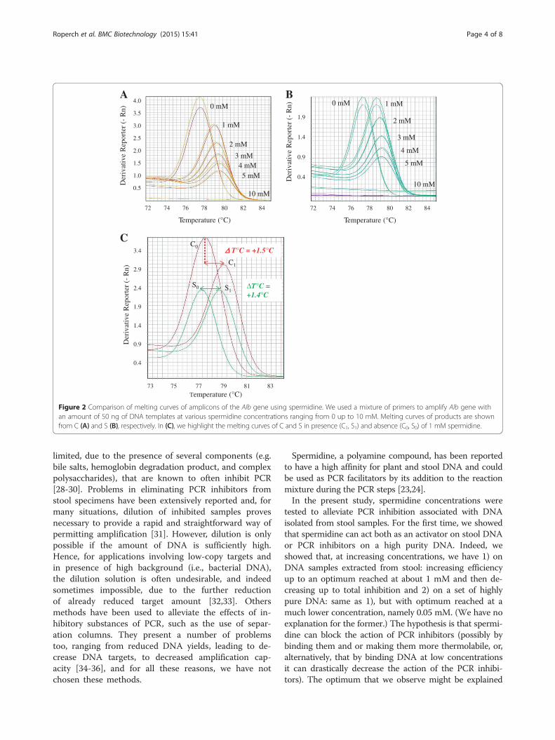
Impact of spermidine on the melting curves of PCRproducts of Alb geneIn Figure 2 are represented the melting curves of ampli-cons of the Alb gene using spermidine in SG RT-PCRfrom C (Figure 2A) and S (Figure 2B). We highlight inFigure 2C the melting curves of C and S in presence (C1,S1) and absence (C0, S0) of 1 mM spermidine. Both, C0
and S0 showed a similar temperature of melting (Tm) of77.7°C and 77.4°C as expected, while for C1 and S1, weobtained a Tm near 79.2°C (ΔTm = +1.5°C) and 78.9°C(ΔTm = +1.4°C), respectively. (The full results are
bumin gene in presence and absence of spermidine. A: representativen presence of 1 mM spermidine from universal methylated human DNAn red resulting entirely from DNA modification. This follows after sodiumnd B: the same PCR products of the Alb were analysed by agarose gelze (76 pb) when spermidine is present (C1, S1) and absence (C0, S0);
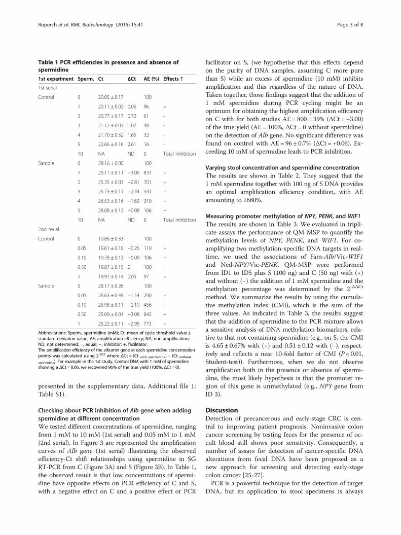
Table 1 PCR efficiencies in presence and absence ofspermidine
1st experiment Sperm. Ct ΔCt AE (%) Effects ?
1st serial
Control 0 20.05 ± 0.17 100
1 20.11 ± 0.02 0.06 96 =
2 20.77 ± 0.17 0.72 61 -
3 21.12 ± 0.03 1.07 48 -
4 21.70 ± 0.32 1.65 32 -
5 22.66 ± 0.16 2.61 16 -
10 NA ND 0 Total inhibition
Sample 0 28.16 ± 0.85 100
1 25.11 ± 0.11 −3.06 831 +
2 25.35 ± 0.03 −2.81 701 +
3 25.73 ± 0.11 −2.44 541 +
4 26.53 ± 0.16 −1.63 310 +
5 28.08 ± 0.13 −0.08 106 +
10 NA ND 0 Total inhibition
2nd serial
Control 0 19.86 ± 0.33 100
0.05 19.61 ± 0.10 −0.25 119 +
0.10 19.78 ± 0.13 −0.09 106 +
0.50 19.87 ± 0.15 0 100 =
1 19.91 ± 0.14 0.05 97 =
Sample 0 28.17 ± 0.26 100
0.05 26.63 ± 0.49 −1.54 290 +
0.10 25.98 ± 0.11 −2.19 456 +
0.50 25.09 ± 0.01 −3.08 843 +
1 25.22 ± 0.11 −2.95 773 +
Abbreviations: Sperm., spermidine (mM); Ct, mean of cycle threshold value ±standard deviation value; AE, amplification efficiency; NA, non amplification;ND, not determined; =, equal; −, inhibitor; +, facilitator.The amplification efficiency of the albumin gene at each spermidine concentrationpoints was calculated using 2-ΔCt where ΔCt = (Ct with spermidine) – (Ct without
spermidine). For example in the 1st study, Control DNA with 1 mM of spermidineshowing a ΔCt = 0.06, we recovered 96% of the true yield (100%, ΔCt = 0).
Roperch et al. BMC Biotechnology (2015) 15:41 Page 3 of 8
presented in the supplementary data, Additional file 1:Table S1).
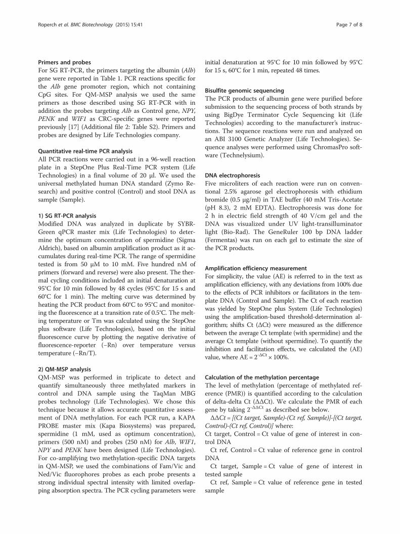
Checking about PCR inhibition of Alb gene when addingspermidine at different concentrationWe tested different concentrations of spermidine, rangingfrom 1 mM to 10 mM (1st serial) and 0.05 mM to 1 mM(2nd serial). In Figure 3 are represented the amplificationcurves of Alb gene (1st serial) illustrating the observedefficiency-Ct shift relationships using spermidine in SGRT-PCR from C (Figure 3A) and S (Figure 3B). In Table 1,the observed result is that low concentrations of spermi-dine have opposite effects on PCR efficiency of C and S,with a negative effect on C and a positive effect or PCR
facilitator on S, (we hypothetise that this effects dependon the purity of DNA samples, assuming C more purethan S) while an excess of spermidine (10 mM) inhibitsamplification and this regardless of the nature of DNA.Taken together, those findings suggest that the addition of1 mM spermidine during PCR cycling might be anoptimum for obtaining the highest amplification efficiencyon C with for both studies AE = 800 ± 39% (ΔCt = −3.00)of the true yield (AE = 100%, ΔCt = 0 without spermidine)on the detection of Alb gene. No significant difference wasfound on control with AE = 96 ± 0.7% (ΔCt = +0.06). Ex-ceeding 10 mM of spermidine leads to PCR inhibition.
Varying stool concentration and spermidine concentrationThe results are shown in Table 2. They suggest that the1 mM spermidine together with 100 ng of S DNA providesan optimal amplification efficiency condition, with AEamounting to 1680%.
Measuring promoter methylation of NPY, PENK, and WIF1The results are shown in Table 3. We evaluated in tripli-cate assays the performance of QM-MSP to quantify themethylation levels of NPY, PENK, and WIF1. For co-amplifying two methylation-specific DNA targets in real-time, we used the associations of Fam-Alb/Vic-WIF1and Ned-NPY/Vic-PENK. QM-MSP were performedfrom ID1 to ID5 plus S (100 ng) and C (50 ng) with (+)and without (−) the addition of 1 mM spermidine and themethylation percentage was determined by the 2-ΔΔCt
method. We summarize the results by using the cumula-tive methylation index (CMI), which is the sum of thethree values. As indicated in Table 3, the results suggestthat the addition of spermidine to the PCR mixture allowsa sensitive analysis of DNA methylation biomarkers, rela-tive to that not containing spermidine (e.g., on S, the CMIis 4.65 ± 0.67% with (+) and 0.51 ± 0.12 with (−), respect-ively and reflects a near 10-fold factor of CMI (P < 0.01,Student-test)). Furthermore, when we do not observeamplification both in the presence or absence of spermi-dine, the most likely hypothesis is that the promoter re-gion of this gene is unmethylated (e.g., NPY gene fromID 3).
DiscussionDetection of precancerous and early-stage CRC is cen-tral to improving patient prognosis. Noninvasive coloncancer screening by testing feces for the presence of oc-cult blood still shows poor sensitivity. Consequently, anumber of assays for detection of cancer-specific DNAalterations from fecal DNA have been proposed as anew approach for screening and detecting early-stagecolon cancer [25-27].PCR is a powerful technique for the detection of target
DNA, but its application to stool specimens is always
72 74 76 78 80 82 84
Temperature (°C)
1.9
1.4
0.9
0.4Der
ivat
ive
Rep
orte
r (-
Rn) 1 mM
2 mM
3 mM
4 mM
5 mM
0 mM
10 mM
4.0
3.5
3.0
2.5
2.0
1.5
1.0
0.5
Der
ivat
ive
Rep
orte
r (-
Rn)
1 mM
2 mM
3 mM4 mM5 mM
0 mM
10 mM
72 74 76 78 80 82 84
Temperature (°C)
A B
3.4
2.9
2.4
1.9
1.4
0.9
0.4
73 75 77 79 81 83Temperature (°C)
Der
ivat
ive
Rep
orte
r (-
Rn)
ΔΔ T°C = +1.5°C
ΔT°C = +1.4°C
C0
S1S0
C1
C
Figure 2 Comparison of melting curves of amplicons of the Alb gene using spermidine. We used a mixture of primers to amplify Alb gene withan amount of 50 ng of DNA templates at various spermidine concentrations ranging from 0 up to 10 mM. Melting curves of products are shownfrom C (A) and S (B), respectively. In (C), we highlight the melting curves of C and S in presence (C1, S1) and absence (C0, S0) of 1 mM spermidine.
Roperch et al. BMC Biotechnology (2015) 15:41 Page 4 of 8
limited, due to the presence of several components (e.g.bile salts, hemoglobin degradation product, and complexpolysaccharides), that are known to often inhibit PCR[28-30]. Problems in eliminating PCR inhibitors fromstool specimens have been extensively reported and, formany situations, dilution of inhibited samples provesnecessary to provide a rapid and straightforward way ofpermitting amplification [31]. However, dilution is onlypossible if the amount of DNA is sufficiently high.Hence, for applications involving low-copy targets andin presence of high background (i.e., bacterial DNA),the dilution solution is often undesirable, and indeedsometimes impossible, due to the further reductionof already reduced target amount [32,33]. Othersmethods have been used to alleviate the effects of in-hibitory substances of PCR, such as the use of separ-ation columns. They present a number of problemstoo, ranging from reduced DNA yields, leading to de-crease DNA targets, to decreased amplification cap-acity [34-36], and for all these reasons, we have notchosen these methods.
Spermidine, a polyamine compound, has been reportedto have a high affinity for plant and stool DNA and couldbe used as PCR facilitators by its addition to the reactionmixture during the PCR steps [23,24].In the present study, spermidine concentrations were
tested to alleviate PCR inhibition associated with DNAisolated from stool samples. For the first time, we showedthat spermidine can act both as an activator on stool DNAor PCR inhibitors on a high purity DNA. Indeed, weshowed that, at increasing concentrations, we have 1) onDNA samples extracted from stool: increasing efficiencyup to an optimum reached at about 1 mM and then de-creasing up to total inhibition and 2) on a set of highlypure DNA: same as 1), but with optimum reached at amuch lower concentration, namely 0.05 mM. (We have noexplanation for the former.) The hypothesis is that spermi-dine can block the action of PCR inhibitors (possibly bybinding them and or making them more thermolabile, or,alternatively, that by binding DNA at low concentrationsit can drastically decrease the action of the PCR inhibi-tors). The optimum that we observe might be explained

NTC10 mM
1 mM 2 mM
3 mM
4 mM
5 mM0 mM
B
NTC 10 mM
0 mM 1 mM
2 mM
3 mM4 mM
5 mM
12 14 16 18 20 22 24 26 28 30 32 34
Cycle
16 18 20 22 24 26 28 30 32 34 36
Cycle
10
1
0.1
0.01
0.001
ΔR
n
1
0.1
0.01
0.001
ΔR
n
A
Figure 3 Interference of PCR amplification by spermidine. From 50 ng of DNA, the amplification curves of PCR products of the Alb gene usingspermidine in SG RT-PCR are shown from C (A) and S (B). The spermidine has been used at various concentrations, ranging from 1 mM to 10 mM.
Roperch et al. BMC Biotechnology (2015) 15:41 Page 5 of 8
by the existence, for each specific sample type, of a con-centration threshold above which spermidine no longerblocks PCR inhibitors, may be due to steric effects or sat-uration, and starts massively binding to DNA, so inhibit-ing PCR.Interesting, we observed that the addition of spermi-
dine causes a positive shift of the melting temperature ofthe stool and control DNA. This observation may bedue to the interaction of the spermidine with DNA as ithas been described previously by Ahokas and Erkkila[23]. We have determined a range of spermidine concen-trations that counteract the PCR inhibitors co-extractedwith DNA, so facilitating the amplification efficiency ofmethylation markers. We applied that finding to asses-sing the methylation of NPY, PENK, and WIF1, (whosedetection is of interest in CRC, as shown in [17]) whilemaintaining sufficient DNA yield. We showed the ad-vantage of our method in the quantification of methyla-tion values of CRC markers NPY, PENK and WIF1,where, on undiluted stool DNA (100 ng) and by usingthe QM-MSP in presence of 1 mM spermidine, we glo-bally enhanced detection by a near 10-fold factor, asassessed by summing up the three values. In the future,
it would be interesting to evaluate our method withother biomarkers such as Septin 9, which is used as amarker of blood-based methylation requiring improvedaccuracy for a clinical practise [12].In summary, we performed a comparative study on
the effect of spermidine onto PCR efficiency reportingthat spermidine addition is easier and more useful thandilution or purification methods and that it can dramat-ically improve the quantification of methylation values.We also highlighted a possible mechanism for its action.Our QM-MSP using the presence of 1 mM spermidineand 100 ng of stool DNA could be used as a potentialPCR facilitator for stool-based detection of CRC. Ourmethodology is also a serious candidate for being devel-oped into a robust technology, as it has been optimizedwith several primer pair and reaction buffer.
ConclusionsIn this study, we present a proof of principle for usingspermidine to allow alleviation of the PCR inhibitors fre-quently encountered in DNA amplification from stoolsamples. We also demonstrated that spermidine, an in-expensive chemical, is useful for sensitive stool-based
Table 2 Relationship between spermidine concentrationand amount of stool DNA
Sperm. Ct ΔCt AE (%)
2nd experiment
50 ng 0 28.32 ± 0.15 100
0.5 25.04 ± 0.11 −3.28 968
1 25.23 ± 0.17 −3.09 849
5 28.39 ± 0.21 0.07 95
100 ng 0 27.04 ± 0.16 100
0.5 23.74 ± 0.12 −3.31 988
1 22.97 ± 0.24 −4.07 1680
5 25.49 ± 0.17 −1.55 293
250 ng 0 NA ND ND
0.5 28.67 ± 0.27 ND ND
1 23.70 ± 0.37 ND ND
5 23.65 ± 0.10 ND ND
500 ng 0 NA ND ND
0.5 NA ND ND
1 28.00 ± 0.56 ND ND
5 26.92 ± 0.30 ND ND
Abbreviations: Sample, stool DNA pooled; Sperm, spermidine (mM); Ct, meanof cycle threshold value ± standard deviation value; AE, amplification efficiency;NA, non amplification; ND, not determined.
Roperch et al. BMC Biotechnology (2015) 15:41 Page 6 of 8
detection of methylation-specific markers for CRC tu-mors using QM-MSP. These results, after corroborationin a large cohort, can lead to the elaboration of amethod to be used in clinical practice as a aid in prese-lecting the patients for colonoscopy.
Table 3 Quantitative DNA methylation analysis
Methylation (%) CMI (%)
3rd experiment NPY PENK WIF1
ID1 - 0.01 ± 0.01 0.13 ± 0.03 NA 0.14 ± 0.04
+ 0.86 ± 0.26 1.10 ± 0.08 1.22 ± 0.41 3.18 ± 0.75
ID2 - 1.22 ± 0.21 1.31 ± 0.53 0.07 ± 0.18 2.60 ± 0.92
+ 1.86 ± 0.11 1.35 ± 0.09 0.76 ± 0.15 3.97 ± 0.35
ID3 - NA NA 0.17 ± 0.07 0.17 ± 0.07
+ NA 0.10 ± 0.04 1.59 ± 0.22 1.69 ± 0.26
ID4 - 0.17 ± 0.08 0.32 ± 0.10 NA 0.49 ± 0.18
+ 2.33 ± 0.32 1.88 ± 0.44 1.71 ± 0.27 5.65 ± 0.67
ID5 - 0.09 ± 0.08 0.17 ± 0.05 0.07 ± 0.01 0.33 ± 0.14
+ 1.33 ± 0.22 1.44 ± 0.16 1.01 ± 0.07 3.78 ± 0.45
Sample (pooling - 0.21 ± 0.06 0.27 ± 0.06 0.03 ± 0.03 0.51 ± 0.12
of five patients) + 1.74 ± 0.30 2.10 ± 0.18 0.81 ± 0.1 4.65 ± 0.67
MethodsHuman stool samplesWe analyzed human stool samples from 5 colonoscopy-negative subjects from the Valihybritest’s collection, regis-tered under the number NCT01270360 (Clinical Trials.gov). All of the patients provided informed consent forthe research use of their samples.Use of these samples for this study was approved by the
ethical committee of the Val de Marne Paris-Est medicaldistrict, registered under code CCP-IDF-IX-11-010.
Experimental designThis study included three related experiments: the firstexperiment was designed to study the interference ofPCR amplification by spermidine addition at variousconcentrations ranging from 0.05 mM to 10 mM both50 ng of universal methylated DNA (Control, C) and50 ng of stool DNA pooled (Sample, S), each DNA pre-viously modified by sodium bisulfite. The second experi-ment determined the optimum condition giving the bestperformance of PCR amplification by modulating boththe amount of stool DNA (from 50 ng to 500 ng ofmodified DNA) and the concentration of spermidine(from 0.5 mM to 5 mM). In these studies, we usedspermidine-containing reaction solutions to assess theamplification of the albumin (Alb) gene using SYBERGreen real-time PCR (SG RT-PCR). In the third experi-ment, the quantitative methylation-methylation specificPCR (QM-MSP) was performed, with the optimum con-dition previously defined in 1st and 2nd experiments, tomeasured the degree of methylation of a three-genepanel consisting of NPY, PENK, and WIF1 and calcu-lated the cumulative methylation index (CMI) value ofeach extract of stool DNA, i.e., ID 1 to ID 5 and Sample.
Stool DNA isolation and quantificationAbout 5 g stool were collected from each individual.DNA was isolated from stool samples (200 mg) usingthe QiAamp DNA stool mini kit (Qiagen) according tothe manufacturer’s protocol. DNA concentrations weredetermined by measurement at 260 nm using BioPho-tometer (Eppendorf ). Isolated DNA was stored at −20°C.
Bisulfite modification1 μg of each DNA (C and S) were modified by sodiumbisulfite overnight at 50°C using the EZ DNA Methyla-tion kit (Zymo Research) and eluted in 100 μl of TEbuffer (10 mM Tris–HCl (pH 8.0), 1 mM EDTA). Bisul-fite treatment converts all unmethylated cytosine resi-dues to uracil (later replicated as thymidine during PCRcycling), while leaving methylcytosines unchanged.
Roperch et al. BMC Biotechnology (2015) 15:41 Page 7 of 8
Primers and probesFor SG RT-PCR, the primers targeting the albumin (Alb)gene were reported in Table 1. PCR reactions specific forthe Alb gene promoter region, which not containingCpG sites. For QM-MSP analysis we used the sameprimers as those described using SG RT-PCR with inaddition the probes targeting Alb as Control gene, NPY,PENK and WIF1 as CRC-specific genes were reportedpreviously [17] (Additional file 2: Table S2). Primers andprobes are designed by Life Technologies company.
Quantitative real-time PCR analysisAll PCR reactions were carried out in a 96-well reactionplate in a StepOne Plus Real-Time PCR system (LifeTechnologies) in a final volume of 20 μl. We used theuniversal methylated human DNA standard (Zymo Re-search) and positive control (Control) and stool DNA assample (Sample).
1) SG RT-PCR analysisModified DNA was analyzed in duplicate by SYBR-Green qPCR master mix (Life Technologies) to deter-mine the optimum concentration of spermidine (SigmaAldrich), based on albumin amplification product as it ac-cumulates during real-time PCR. The range of spermidinetested is from 50 μM to 10 mM. Five hundred nM ofprimers (forward and reverse) were also present. The ther-mal cycling conditions included an initial denaturation at95°C for 10 min followed by 48 cycles (95°C for 15 s and60°C for 1 min). The melting curve was determined byheating the PCR product from 60°C to 95°C and monitor-ing the fluorescence at a transition rate of 0.5°C. The melt-ing temperature or Tm was calculated using the StepOneplus software (Life Technologies), based on the initialfluorescence curve by plotting the negative derivative offluorescence-reporter (−Rn) over temperature versustemperature (−Rn/T).
2) QM-MSP analysisQM-MSP was performed in triplicate to detect andquantify simultaneously three methylated markers incontrol and DNA sample using the TaqMan MBGprobes technology (Life Technologies). We chose thistechnique because it allows accurate quantitative assess-ment of DNA methylation. For each PCR run, a KAPAPROBE master mix (Kapa Biosystems) was prepared,spermidine (1 mM, used as optimum concentration),primers (500 nM) and probes (250 nM) for Alb, WIF1,NPY and PENK have been designed (Life Technologies).For co-amplifying two methylation-specific DNA targetsin QM-MSP, we used the combinations of Fam/Vic andNed/Vic fluorophores probes as each probe presents astrong individual spectral intensity with limited overlap-ping absorption spectra. The PCR cycling parameters were
initial denaturation at 95°C for 10 min followed by 95°Cfor 15 s, 60°C for 1 min, repeated 48 times.
Bisulfite genomic sequencingThe PCR products of albumin gene were purified beforesubmission to the sequencing process of both strands byusing BigDye Terminator Cycle Sequencing kit (LifeTechnologies) according to the manufacturer’s instruc-tions. The sequence reactions were run and analyzed onan ABI 3100 Genetic Analyzer (Life Technologies). Se-quence analyses were performed using ChromasPro soft-ware (Technelysium).
DNA electrophoresisFive microliters of each reaction were run on conven-tional 2.5% agarose gel electrophoresis with ethidiumbromide (0.5 μg/ml) in TAE buffer (40 mM Tris-Acetate(pH 8.3), 2 mM EDTA). Electrophoresis was done for2 h in electric field strength of 40 V/cm gel and theDNA was visualized under UV light-transilluminatorlight (Bio-Rad). The GeneRuler 100 bp DNA ladder(Fermentas) was run on each gel to estimate the size ofthe PCR products.
Amplification efficiency measurementFor simplicity, the value (AE) is referred to in the text asamplification efficiency, with any deviations from 100% dueto the effects of PCR inhibitors or facilitators in the tem-plate DNA (Control and Sample). The Ct of each reactionwas yielded by StepOne plus System (Life Technologies)using the amplification-based threshold-determination al-gorithm; shifts Ct (ΔCt) were measured as the differencebetween the average Ct template (with spermidine) and theaverage Ct template (without spermidine). To quantify theinhibition and facilitation effects, we calculated the (AE)value, where AE = 2-ΔCt × 100%.
Calculation of the methylation percentageThe level of methylation (percentage of methylated ref-erence (PMR)) is quantified according to the calculationof delta-delta Ct (ΔΔCt). We calculate the PMR of eachgene by taking 2-ΔΔCt as described see below.ΔΔCt = [(Ct target, Sample)-(Ct ref, Sample)]-[(Ct target,
Control)-(Ct ref, Control)] where:Ct target, Control = Ct value of gene of interest in con-trol DNACt ref, Control = Ct value of reference gene in control
DNACt target, Sample = Ct value of gene of interest in
tested sampleCt ref, Sample = Ct value of reference gene in tested
sample

Roperch et al. BMC Biotechnology (2015) 15:41 Page 8 of 8
Additional files
Additional file 1: Figure S1. Effects of spermidine on the temperaturemelting of the albumin products.
Additional file 2: Figure S2. Oligonucleotides.
Competing interestsThe authors declare that they have no competing interests.
Authors’ contributionsParticipated in research design: All authors. Conducted experiments: JPR.Performed data analysis: JPR and RI. Wrote or Contributed to the writing ofthe manuscript: all authors. All authors read and approved the finalmanuscript to be published.
AcknowledgementsWe wish to thank Pr. Iradj Sobhani and Assistance Publique-Hôpitaux de Parisfor providing the samples; Hanane El Asri and Amine Amoura for valuableadvice (Department of Gastroenterology, Laboratoire d’Investigation Clinique,Henri Mondor Hospital, Créteil, France).
Author details1Profilome, Paris Biotech 24 rue du Faubourg St Jacques, Paris 75014, France.2OncoDiag, Agoranov 96 Bis, Boulevard Raspail, Paris 75006, France. 3Centred’Investigation Clinique (CIC), Henri Mondor Hospital, Créteil, France. 4KingAbdullah University of Science and Technology (KAUST), Bioscience CoreLaboratory Research Department, Thuwal 23955-6900, Saudi Arabia. 5KingAbdullah University of Science and Technology (KAUST), ComputationalBiology Research Center, Thuwal 23955-6900, Saudi Arabia.
Received: 18 October 2014 Accepted: 22 April 2015
References1. Jemal A, Siegel R, Xu J, Ward E. Cancer Statistics, 2010. Cancer J Clin.
2010;60(5):277–300.2. Desch CE, Benson AB, Somerfield MR, Flynn PJ, Krause C, Loprinzi CL, et al.
Colorectal cancer surveillance: 2005 update of an American Society ofClinical Oncology practice guideline. J Clin Oncol. 2005;23(33):8512–9.
3. Winawer S, Fletcher R, Bond RD, Desch CE, Benson AB, Somerfield MR, et al.Colorectal cancer surveillance: 2005 update of an American Society ofClinical Oncology practice guideline. J Clin Oncol. 2005;23(33):8512–9.
4. Esteller M. DNA methylation and cancer therapy: new developments andexpectations. Curr Opin Oncol. 2005;17(1):55–60.
5. Nosho K, Yamamoto H, Takahashi T, Mikami M, Taniguchi H, et al. Geneticand epigenetic profiling in early colorectal potential in pT1 (early invasive)colorectal cancers. Carcinogenesis. 2007;28(6):1364–70.
6. Wong JJ, Hawkins NJ, Ward RL. Colorectal cancer: a model for epigenetictumorigenesis. Gut. 2007;56(1):140–8.
7. Osborn NK, Ahlquist DA. Stool screening for colorectal cancer: molecularapproaches. Gastroenterology. 2005;128(1):192–206.
8. Sobhani I, Alzahouri K, Ghout I, Charles DJ, Durand-Zaleski I. Cost-effectiveness of mass screening for colorectal cancer: choice of fecal occultblood test and screening strategy. Dis Colon Rectum. 2011;54(7):876–86.
9. Wong JJ, Hawkins NJ, Ward RL. Colorectal cancer: a model for epigenetictumorigenesis. Gut. 2007;56(1):140–8.
10. Jones PA. DNA methylation and cancer. Oncogene. 2002;21(35):5358–60.11. Muller HM, Widschwendter M. Methylated DNA as a possible screening
marker for neoplastic disease in several body fluids. Expert Rev Mol Diagn.2003;3(4):443–58.
12. Grützmann R, Molnar T, Devos C, Pilarsky JK, Habermann PM, Schlag HD,et al. Sensitive detection of colorectal cancer in peripheral blood by septin9 DNA methylation assay. Plos One. 2008;3:3759.
13. Lofton-Day C, Model T, Devos R, Tetzner J, Distler M, Schuster X, et al. DNAmethylation biomarkers for blood-based colorectal cancer screening. ClinChem. 2008;54:414–23.
14. Mansour H. Cell-free nucleic acids as noninvasive biomarkers for colorectalcancer. Front Genet. 2014;5:182.
15. Model F, Osborn N, Ahlquist D, Gruetzmann R, Molnar B, Sipos F, et al.Identification and validation of colorectal neoplasia-specific methylationmarkers for accurate classification of disease. Mol Cancer Res. 2007;5:153–63.
16. Lenhard K, Bommer GT, Asutay S, Schauer R, Brabletz T, Goke B, et al.Analysis of promoter methylation in stool: a novel method for the detectionof colorectal cancer. Clin Gastroenterol Hepatol. 2005;23:142–9.
17. Huang ZH, Li LH, Yang F, Wang JF. Detection of aberrant methylation infecal DNA as a molecular screening tool for colorectal cancer andprecancerous lesions. World J Gastroenterol. 2007;13:954–4.
18. Roperch JP, Incitti R, Forbin S, Bard F, Mansour H, Maesli F, et al. Aberrantmethylation of NPY, PENK, and WIF1 as a promising marker for blood-baseddiagnosis of colorectal cancer. BMC Cancer. 2013;13:566.
19. Ahlquist DA. Molecular detection of colorectal neoplasia. Gastroenterology.2010;138(6):2127–39.
20. Davies RJ, Miller R, Coleman N. Colorectal cancer screening: prospects formolecular stool analysis. Nat Rev Cancer. 2005;5(3):199–2009.
21. Zou H, Harrington J, Rego RL, Ahlquist DA. A novel method to capturemethylated human DNA from stool: implications for colorectal cancerscreening. Clin Chem. 2007;53(9):1646–51.
22. Ramakers CJ, Ruijter JM, Deprez RH, Moorman AF. Assumption-free analysisof quantitative real-time polymerase chain reaction (PCR) data. NeurosciLett. 2003;339:62–6.
23. Ahokas H, Erkkila MJ. Interference of PCR amplification by the polyamines,spermine and spermidine. PCR Methods Appl. 1993;3:65–8.
24. Kikuchi A, Sawamura T, Kawase N, Kitajima Y, Yoshida T, et al. Utility ofSpermidine in PCR Amplification of Stool Samples. Biochem Genet.2010;48:428–32.
25. Sidransky D, Tokino T, Hamilton S, Kinzler K, Levin B, Frost P, et al.Identification of ras oncogene mutations in the stool of patients withcurable colorectal tumors. Science. 1992;256:102–5.
26. Ahlquist DA, Shuber AP. Stool screening for colorectal cancer: evolutionfrom occult blood to molecular markers. Clin Chim Acta. 2002;315:157–68.
27. Ahlquist DA. Molecular detection of colorectal neoplasia. Gastroenterology.2010;138(6):2127–39.
28. Lantz PG, Matsson M, Wadström T, Rådstrom P. Removal of PCR inhibitorsfrom human faecal samples through the use of an aqueous two-phase systemfor sample preparation prior to PCR. J Microbiol Methods. 1997;28:159–67.
29. Nagasaka T, Tanaka N, Cullings HM, et al. Analysis of fecal DNA methylationto detect gastrointestinal neoplasia. J Natl Cancer Inst. 2009;101(18):1244–58.
30. Monteiro L, Bonnemaison D, Vekris A, Petry KG, Bonnet J, Vidal R, et al.Complex polysaccharides as PCR inhibitors in feces: Helicobacter pylorimodel. J Clin Microbiol. 1997;35:995–8.
31. Makristathis A, Pasching E, Schütze K, Wimmer M, Rotter ML, Hirschl AM.Detection of Helicobacter pylori in stool specimens by PCR and antigenenzyme immunoassay. J Clin Microbiol. 1998;36:2772–4.
32. King CE, Debruyne R, Kuch M, Schwarz C, Poinar HN. A quatitative approachto detect and overcome PCR inhibition in ancient DNA extracts.Biotechniques. 2009;47:941–9.
33. Wilson IG. Inhibition and facilitation of nucleic acid amplification. ApplEnviron Microbiol. 1997;63:3741–51.
34. Olive DM. Detection of enterotoxigenic Escherichia coli after polymerasechain reaction amplification with a thermostable DNA polymerase. J ClinMicrobiol. 1989;27:261–5.
35. Dowd SE, Gerba CP, Enriquez FJ, Pepper IL. PCR amplification and speciesdetermination of microsporidia in formalin-fixed feces after immunomagneticseparation. Appl Environ Microbiol. 1998;64:333–6.
36. Ahlquist DA, Skoletsky JE, Boynton KA, Harrington JJ, Mahoney DW. al.Colorectal cancer screening by detection of altered human DNA stool:feasibility of a multitarget assay panel. Gastroenterology. 2000;119:1219–27.