impaired response fibroblastsfrom patients ... · maslowska,* andallan d. sniderman*...
TRANSCRIPT

Impaired Response of Fibroblasts from Patientswith Hyperapobetalipoproteinemia to Acylation-stimulating ProteinKatherine M. Cianflone,** Magdalena H. Maslowska,* and Allan D. Sniderman**McGill Unit for the Prevention of Cardiovascular Disease, Royal Victoria Hospital, Montreal, Quebec H3A JAJ; andtDepartment of Chemistry and Biochemistry, Concordia University, Montreal, Quebec II3G IM8, Canada
Abstract
Acylation-stimulating protein (ASP) is a small, basic, humanplasma protein that markedly stimulates triglyceride synthesisin human adipocytes and cultured human skin fibroblasts. Thepresent studies examine the response to ASPof cultured skinfibroblasts from normal subjects patients with hyperapobetali-poproteinemia, patients with familial hypercholesterolemia,and patients with hypertriglyceridemia without hyperapobeta-lipoproteinemia. Triglyceride synthesis induced by ASP didnot differ significantly among the normals, the patients withfamilial hypercholesterolemia, and the patients with hyper-triglyceridemia with normal low density lipoprotein (LDL)apolipoprotein B levels; however, on average, it was markedlyreduced in the patients with hyperapobetalipoproteinemia. Inall groups studied, evidence of specific saturable binding ofradioiodinated ASPwas present. Binding, however, was signif-icantly reduced in the groups with hyperapobetalipoprotein-emia whereas the other three groups were indistinguishable.By contrast, LDL-specific binding was reduced' only' in thepatients with famili'al'hypercholesterolemia. There was a sig-nificant direct relation between the degree of ASPbinding andthe triglyceride synthesis inducible by ASP. In addition, withthe exception of the patients with familial hypercholesterol-emia, there was an inverse relation between both ASP-specificbinding and ASP-induced triglyceride synthesis in fibroblaststo LDL levels in plasma whereas no relation was evident toplasma high density lipoprotein and very low density lipopro-tein. (J. Clin. Invest. 1990. 85:722-730.) hyperapobetalipo-proteinemia - acylation-stimulating protein * regulation of tri-glyceride synthesis
Introduction
Increased numbers of low-density lipoprotein (LDL) particlesin plasma are a commonabnormality in patients with prema-ture coronary artery disease. Plasma LDL particle number can
increase either because of impaired catabolism of LDL such as
occurs, for example, in familial hypercholesterolemia (1), or
because of increased production such as occurs in hyperapo-betalipoproteinemia (hyperapoB)' (2). The specific faults in
Address reprint requests to Dr. Sniderman, Cardiology Division,Royal Victoria Hospital, 687 Pine Avenue West, Montreal, QuebecH3A IA I, Canada.
Receivedfor publication 5 June 1989 and in revisedform 29 Sep-tember 1989.
1. Abbreviations used in this paper: hyperapoB, hyperapobetalipopro-teinemia.
the LDL pathway that result in familial hypercholesterolemiahave been explicated in considerable detail whereas the basis,or bases, for the increased hepatic synthesis of apolipoproteinB (apoB) 100 lipoprotein particles in hyperapoB are unknown.
In the course of studies comparing fatty acid metabolism inperipheral tissues from normal subjects and patients with hy-perapoB, we serendipitously recognized the presence inhuman plasma of a factor which markedly stimulated triglyc-eride -synthesis in skin fibroblasts cultured from normals (3).However, cells from patients with hyperapoB were much lessresponsive to this factor (4). The protein responsible for thisactivity has now been purified to homogeneity and namedacylation-stimulating protein (ASP) (5). The purpose of thepresent studies is to compare the response to ASP of skinfibroblasts from normals with those from patients with hyper-apoB, as well as with those from patients with familial hyper-cholesterolemia and hypertriglyceridemia with normal LDLapoB levels.
Methods
Lipid and apolipoprotein assays. Plasma total cholesterol and triglycer-ide, high density lipoprotein (HDL) cholesterol, LDL cholesterol, andplasma LDL apoB were measured in fasting samples obtained from allsubjects. Plasma cholesterol and triglyceride were measured by enzy-matic determination (Nos. 237574 and 701904, respectively,Boehringer-Mannheim Diagnostics, Houston, TX). HDL was mea-sured by the heparin-manganese method (6) and LDL cholesterol wascalculated according to the method of Friedewald et al. (7). PlasmaLDL apoB was measured by radial immunodiffusion (8). The normalranges for plasma and lipoprotein lipids were taken from the LipidResearch Clinics Prevalence Study (9). A value of plasma LDL apoB> 120 mg/dl was taken to be elevated (10) and the diagnosis of hyper-apoB made on the basis of an elevated LDL apoB with a LDL choles-terol to apoB ratio < 1.5:1 (1 1). The diagnosis of familial hypercholes-terolemia was made clinically (12) but also was based on the presenceof an elevated LDL cholesterol with an LDL cholesterol to apoB ratioof 1.8:1 or greater ( 1 ).
ASPpurification. ASPwas purified by a modification of the origi-nal method (5). Frozen human plasma was thawed at 4VC overnight.The plasma was centrifuged at 2,000 rpm for 20 min and the superna-tant was then filtered to remove the precipitate. Plasma was applied toa Fast-Flow-S-Sepharose column (Pharmacia, Uppsala, Sweden) (14ml/ml gel) and washed with seven column volumes of buffer A (0.02 Mphosphate, pH 7.1, with 0.02% sodium azide). The bound proteinfraction was eluted with seven column volumes of 3 MNaCl in bufferA with absorbance monitored at OD280. The fractions were pooledand concentrated by dialysis against 50% polyethylene glycol in buf-fer A.
This was then fractionated by gel filtration chromatography onSephadex G-75 (Pharmacia) applied in a sample volume of 4-5% ofthe column volume (column volume = 1.7 ml/ml of starting plasma).The column was eluted with buffer A and the fraction of interest (thesecond peak) emerged at Ve/Vo of 3.4. The fractions were pooled anddialyzed against buffer A to remove any NaCl, then applied to a secondFast-Flow-S-Sepharose column (20 ml of column volume/liter ofstarting plasma). The bound protein peak was again eluted, collected,
722 K. M. Cianflone, M. H. Maslowska, and A. D. Sniderman
J. Clin. Invest.© The American Society for Clinical Investigation, Inc.0021-9738/90/03/0722/09 $2.00Volume 85, March 1990, 722-730

and concentrated as described above using buffer A without sodiumazide. The ASP was dialyzed overnight and centrifuged to removeprecipitate, and the protein concentration was measured by themethod of Bradford using bovine serum albumin as a standard (13).Purification was monitored by SDS-1 5% PAGEelectrophoresis (14).
Tissue culture of human skin fibroblasts. Fibroblasts were obtainedfrom forearm skin biopsies of all subjects. Primary cultures were es-tablished (15) and maintained in minimum essential medium with10% fetal calf serum supplemented with penicillin-streptomycin (100IU/ml). Fibroblasts were subcultured every 7-10 d with a split ratio of1:1 or 1:2 after a 10-min incubation with 0.25% trypsin in magnesiumand calcium-free phosphate-buffered saline to detach cells from theflask. Cells were used for experiments between passages 5 and 15. Forexperiments, cells were plated out at a concentration of 1 X 10⁵ cellsper 60-mm dish in 2 ml of medium. On day 6, cells were changed to aserum-free hormone medium of Dulbecco's minimum essential me-dium and Ham's F- 12 (1: 1) supplemented with insulin, biotin, calciumpantothenate, transferrin, triiodothyronine, and hydrocortisone (16).
Measurement of fibroblast triglyceride synthesis. On day 7, ASPand [1-'4C]oleic acid (sp act = 52.6 Ci/mol, Du Pont-New EnglandNuclear, Boston, MA) were added to the medium at the indicatedconcentrations (average sp act = 10 dpm/pmol) and incubated for thespecified times at 370C in a 5%CO2humid incubator. The cells werethen placed on ice and the medium removed, after which the cells werewashed three times with I ml of phosphate-buffered saline to removedexcess free ['4Cloleic acid. The cells were harvested by scraping with 2X I ml of phosphate-buffered saline using a rubber policeman andvortexed. Aliquots of the cell suspension were removed for proteindetermination as described above and for scintillation counting. Theremaining suspension was extracted with a fivefold volume of chloro-form/methanol/ I NHCI (20:10:0.025 vol/vol/vol) (17). After 30 min,the sample was centrifuged (2,000 rpm) for 10 min and the two phaseswere separated. The organic phase was evaporated under a flow ofnitrogen and redissolved in chloroform/methanol (2:1), and applied toa silica gel plate for thin-layer chromatography in hexane/ether/aceticacid (85:15:1 vol/vol/vol) with reference lipids. The lipid spots werevisualized by exposure to iodine vapor, and the spots were then scrapedinto vials with scintillation fluid and counted in a liquid scintillationcounter.
Examination of ASP-fibroblast interactions. For these experi-ments, ASPwas radioiodinated with iodine 125 by the chloramine-Tmethod of Elder (18). The average specific activity obtained was 79cpm/ng protein and 95% of the radioactivity was precipitable by incu-bation with 10% trichloroacetic acid. To study specific binding of ASP,aliquots of fibroblasts were precooled on ice (4°C) for 15 min and1251 -ASP added to the medium at the desired concentrations and incu-bated at 4°C for 2 h. The medium was removed and the cells were thenwashed three times with 2 mg/ml bovine serum albumin in phos-phate-buffered saline, and then three times with phosphate-bufferedsaline. The samples were collected by scraping, or by dissolution in 0.1N NaOH for I h. An aliquot of the cell suspension was taken forcounting in a gammacounter and for protein determination. Nonspe-cific binding was measured by adding a 20-fold excess of unlabelledASP to the media before the addition of labeled ASP (19). Specificbinding was calculated as the difference between total and nonspeci-fic binding. Results are expressed as nanograms of ASP bound permilligram of cell protein. Scatchard analyses were performed as de-scribed (20).
Internalization and degradation of ASPwas also determined. To doso, cells were preincubated in serum-free hormone medium (as de-scribed above) for 24 h, followed by incubation at 37°C for 4-24 h inthe presence of [1 -'4C]oleic acid and '251I-ASP at the indicated concen-trations. After the incubation the media was removed and the cellswere washed and harvested by scraping as described above. Aliquots ofcells were counted for 1251I radioactivity and their protein content wasdetermined. '251 degradation products in the media were measuredafter precipitation with 60% trichloroacetic acid (final concentration10%) with the addition of 1 mg/ml bovine serum albumin (final con-
centration) as carrier (21). The medium was centrifuged for 15 min at2,000 rpm. Free iodine in the supernatant was then precipitated with5% AgNO3 (final AgNO3 concentration 1.7%). An aliquot from thesupernatant containing the degradation products was then counted ina gammacounter for 1251 radioactivity.
Measurement of LDL binding. LDL was isolated between densities1.019 and 1.063 by standard ultracentrifugal techniques (22), and theprotein was measured as described above. LDL was iodinated by theiodine monochloride method of MacFarlane as modified by Bilheimeret al. (23). The average specific activity obtained was 216 cpm/ng, and95% of the radioactivity was precipitable by incubation with 10% tri-chloroacetic acid. LDL binding was performed in the same manner asdescribed for ASPbinding in the absence (total binding) and the pres-ence (nonspecific binding) of a 20-fold excess of LDL. Specific bindingwas calculated as the difference between total and nonspecific binding.
Statistics. All results are expressed as average±standard error of themean (SEM). Student's t test for two means was used for comparisonbetween the different cell groups. Linear regression was used for therelations between cell bioactivity and lipid values and the significancewas calculated by Pearson's correlation coefficient.
Results
Patients studied. The plasma lipid, lipoprotein lipid, and LDLapoB levels in all the subjects studied are listed in Table I.Cultured skin fibroblasts were obtained from eight normals. Ofthese, six were male, and two female, with ages ranging be-tween 27 and 66 yr. 12 patients with hyperapoB were studied:2 were female and 10 male; their ages ranged between 33 and62 yr. Six had levels of LDL cholesterol less than the 95 per-centile whereas six exceeded it. Of the first six patients, onealso had elevated fasting plasma triglyceride levels whereas, ofthe other six, plasma triglyceride was increased in three. Thusthe lipoprotein phenotypes present within the hyperapoBgroup include five normal, three type II A, and three II B, andone type IV hyperlipoproteinemia. Three patients with hetero-zygous familial hypercholesterolemia were studied, and, in ad-dition, four cell lines obtained from the American Tissue Cul-ture Collection, Rockville, MD, cell bank were also examined.Of these, all were from patients said to be homozygous forfamilial hypercholesterolemia. The final group consisted offour patients with type IV hyperlipoproteinemia with a normalLDL apoB.
ASP stimulation of triglyceride synthesis. The results areshown in Fig. 1. In Fig. 1 A are the results obtained with thenormal cells. It is evident that as ASP concentration was in-creased, cellular triglyceride synthesis, expressed as percentstimulation above baseline, increased until a plateau wasreached. In the next three panels, the results in the other threegroups are presented.
In all cases, baseline activity, that is, the rate of triglyceridesynthesis in the absence of ASP, did not differ significantly.There were no significant differences in the response of triglyc-eride synthesis to ASP in the cells from patients with familialhypercholesterolemia (Fig. 1 C) and those from the patientswith type IV hyperlipoproteinemia without hyperapoB (Fig. 1D) compared to the normals, the average increase being 58%and 53% respectively above baseline.
By contrast, the response of the hyperapoB cells to ASPwas considerably blunted (Fig. 1 B) in that it was significantlyless at all concentrations of ASP, with an average maximalincrease of 13%. Moreover, it should be noted that there wasno significant difference in the response of the hyperapoB cells
Response of HumanFibroblasts to Acylation-stimulating Protein 723
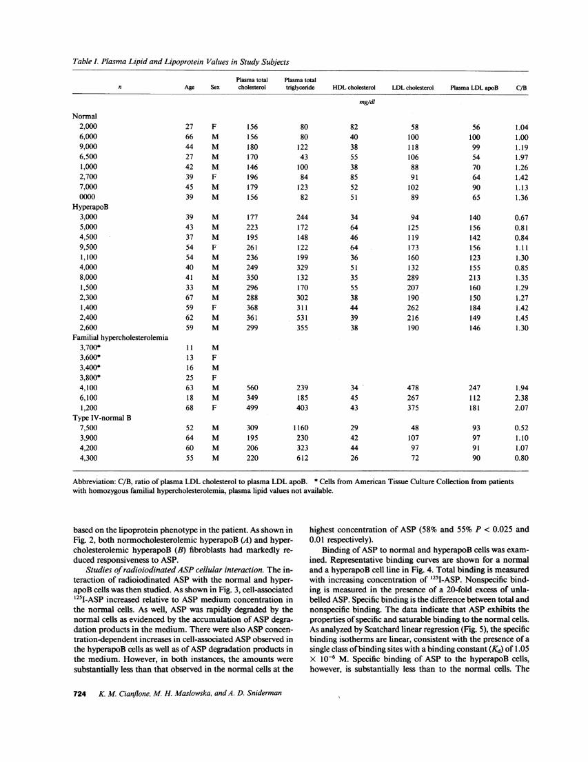
Table L Plasma Lipid and Lipoprotein Values in Study Subjects
Plasma total Plasma totaln Age Sex cholesterol triglyceride HDLcholesterol LDL cholesterol Plasma LDL apoB C/B
mg/dl
Normal2,0006,0009,0006,5001,0002,7007,0000000
HyperapoB3,0005,0004,5009,5001,1004,0008,0001,5002,3001,4002,4002,600
Familial hypercholesterolemia3,700*3,600*3,400*3,800*4,1006,1001,200
Type IV-normal B7,5003,9004,2004,300
27 F 15666 M 15644 M 18027 M 17042 M 14639 F 19645 M 17939 M 156
39 M 17743 M 22337 M 19554 F 26154 M 23640 M 24941 M 35033 M 29667 M 28859 F 36862 M 36159 M 299
11 M13 F16 M25 F63 M 56018 M 34968 F 499
8080
12243
10084
12382
244172148122199329132170302311531355
239185403
52 M 309 116064 M 195 23060 M 206 32355 M 220 612
8240385538855251
346446643651355538443938
344543
29424426
581001181068891
10289
94125119173160132289207190262216190
478267375
481079772
56 1.04100 1.0099 1.1954 1.9770 1.2664 1.4290 1.1365 1.36
140 0.67156 0.81142 0.84156 1.11123 1.30155 0.85213 1.35160 1.29150 1.27184 1.42149 1.45146 1.30
247 1.94112 2.38181 2.07
93 0.5297 1.1091 1.0790 0.80
Abbreviation: C/B, ratio of plasma LDL cholesterol to plasma LDL apoB. * Cells from American Tissue Culture Collection from patientswith homozygous familial hypercholesterolemia, plasma lipid values not available.
based on the lipoprotein phenotype in the patient. As shown inFig. 2, both normocholesterolemic hyperapoB (A) and hyper-cholesterolemic hyperapoB (B) fibroblasts had markedly re-duced responsiveness to ASP.
Studies of radioiodinated ASPcellular interaction. The in-teraction of radioiodinated ASP with the normal and hyper-apoB cells was then studied. As shown in Fig. 3, cell-associated1251I-ASP increased relative to ASP medium concentration inthe normal cells. As well, ASP was rapidly degraded by thenormal cells as evidenced by the accumulation of ASPdegra-dation products in the medium. There were also ASPconcen-tration-dependent increases in cell-associated ASPobserved inthe hyperapoB cells as well as of ASPdegradation products inthe medium. However, in both instances, the amounts weresubstantially less than that observed in the normal cells at the
highest concentration of ASP (58% and 55% P < 0.025 and0.01 respectively).
Binding of ASP to normal and hyperapoB cells was exam-ined. Representative binding curves are shown for a normaland a hyperapoB cell line in Fig. 4. Total binding is measuredwith increasing concentration of 125I-ASP. Nonspecific bind-ing is measured in the presence of a 20-fold excess of unla-belled ASP. Specific binding is the difference between total andnonspecific binding. The data indicate that ASP exhibits theproperties of specific and saturable binding to the normal cells.As analyzed by Scatchard linear regression (Fig. 5), the specificbinding isotherms are linear, consistent with the presence of asingle class of binding sites with a binding constant (Kd) of 1.05X 10⁻⁶ M. Specific binding of ASP to the hyperapoB cells,however, is substantially less than to the normal cells. The
724 K. M. Cianflone, M. H. Maslowska, and A. D. Sniderman

n% Triglyceride Stimulation
40
20-
{E
ASP concentration (tig/ml)
0 2 4 e 8 10 12 14 16
ASP concentration (Vg/ml)
2 4 d 8 10 12ASP concentration (ig/ml)
4 e 8 10 12ASP concentration (ig/ml)
Figure 1. ASPstimulation of triglyceride synthesis in human skin fibroblasts. Cells were incubated with 10 IAM ['4C]oleate and increasing con-centrations of ASPfor 20 h and the amount of radioactive triglyceride was determined. Results are expressed as percentage increase over base-line (mean±SEM) triglyceride synthesis which is taken as 0%. (A) Eight normal cell lines (o); (B) 12 hyperapoB cell lines (*); (C) seven FH celllines (x); (D) four type IV normal LDL apoB cell lines (o).
slopes of the two lines are virtually identical [-0.068±0.01(gg/ml)-' normal vs. -0.08±0.02 hyperapoB, P NS], whereasthe x-intercept in the hyperapoB cells is almost exactly halfthat in the normal cells (1.313±0.13 ng/mg cell protein normalvs. 0.76±0.10 ng/mg cell protein hyperapoB, P < 0.0025).These results point to a single class of receptors for ASPon thecell surface of both the normal and hyperapoB cells. However,the number of these receptors, on average, appear to be re-duced by half in the hyperapoB cells.
The individual results of ASP specific binding in the nor-mals and hyperapoB patients studied are shown in Fig. 6. Theaverage ASP binding in the normals was calculated based onthe x-intercept from the Scatchard linear regression analysis(1.313 ng/mg cell protein) which was taken as 100%. Individ-ual Scatchard results for each cell line were then compared tothe average in normals. A range in ASPbinding in both groupsis evident. The ASP binding in the normals was 103%±10.3and in the hyperapoB 64%±7. 1. However, in none of the hy-perapoB patients did binding of ASPexceed 100%. In eight, itwas 80% or less and in five it was 50% or less. Although,overall, the hyperapoB cell lines show reduced responsivenessto ASP, this specific defect appears to be more characteristic ofsome HyperapoB cell lines than others.
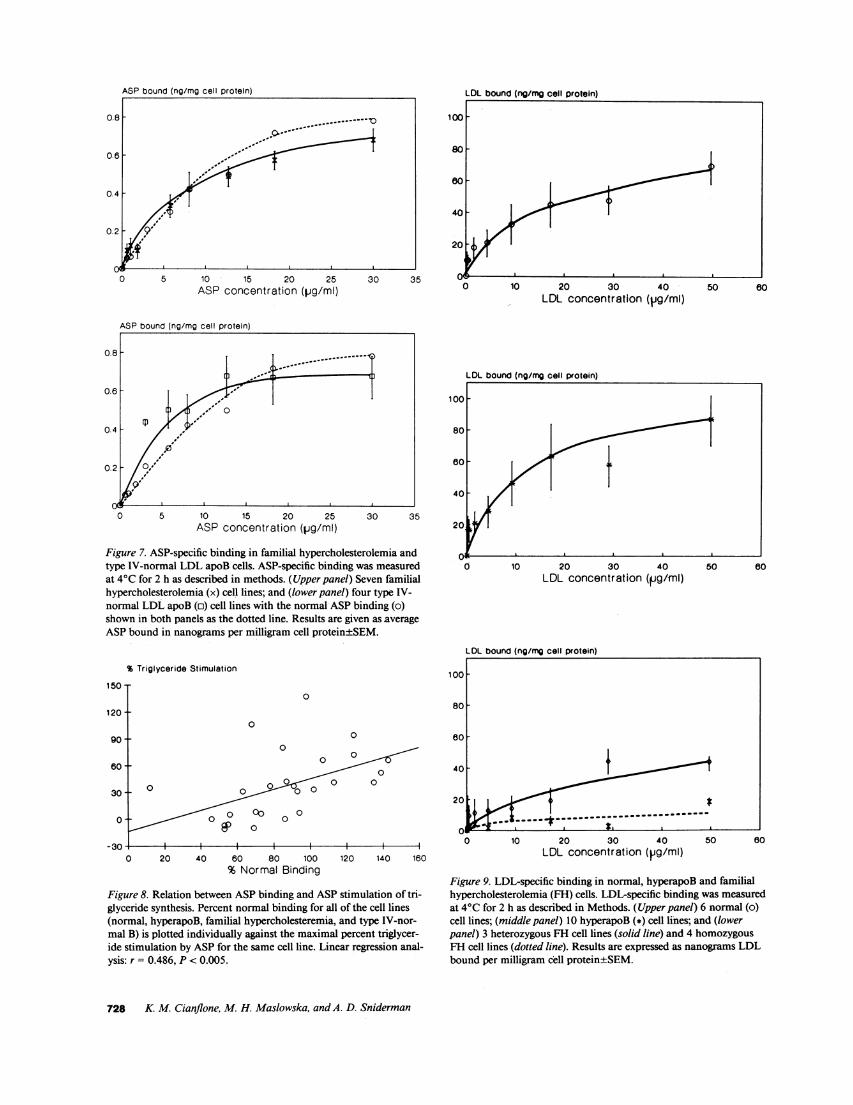
The results of ASP-specific binding for the familial hyper-cholesterolemia cells and the type IV-normal apoB cells areshown in Fig. 7, A and B. Neither differs significantly from the
normal. These findings, of course, are consistent with the ear-lier observations of a normal increase in triglyceride synthesisinduced by ASP (Fig. 1). The relation between ASP bindingand subsequent stimulation of triglyceride synthesis is shownin Fig. 8 for all of the cell lines studied. A significant directrelation, with however substantial scatter, between ASPbind-ing and triglyceride synthetic response is evident.
The results for LDL binding are shown in Fig. 9. The nor-mal cells are depicted in the top panel; the hyperapoB and thefamilial hypercholesterolemic group are shown in the succeed-ing panels. Except for those known to have familial hypercho-lesterolemia (bottom panel), the results are normal. As antici-pated, LDL binding was markedly reduced in the cell linesfrom patients with heterozygous familial hypercholesterolemiaand severely reduced in the cell lines from the patients withhomozygous familial hypercholesterolemia.
Finally, the relation between the ASP-specific bindingmeasured in vitro and the in vivo plasma lipid, lipoproteinlipid, and LDL apoB levels were examined using linear regres-sion and Pearson's correlation coefficient. Because the meta-bolic defect which produces the elevated LDL particle numberin plasma in familial hypercholesterolemia has been delin-eated, this group was excluded. For all the rest there was nosignificant relation between ASP-specific binding to humanfibroblasts and the corresponding plasma triglyceride or HDLcholesterol levels. However, there were significant inverse re-
Response of HumanFibroblasts to Acylation-stimulating Protein 725
B: hyperapoB
14 16

0 2 4 6 8 10 12ASP concentration (Vg/ml)
4 6 8 10 12ASP concentration (jig/ml)
Figure 2. ASPstimulation of triglyceride synthesis in normocholes-terolemic and hypercholesterolemic hyperapoB. Cells were incubatedas described in Fig. 1. (A) Six normocholesterolemic hyperapoB (*)and (B) six hypercholesterolemic hyperapoB (*). In both panels, eightnormals (o) are shown in the dotted lines. Results are expressed aspercentage stimulation over baseline (0%)±SEM.
0 2 4 6 8 10 12 14 16
ASP concentration (pig/ml)
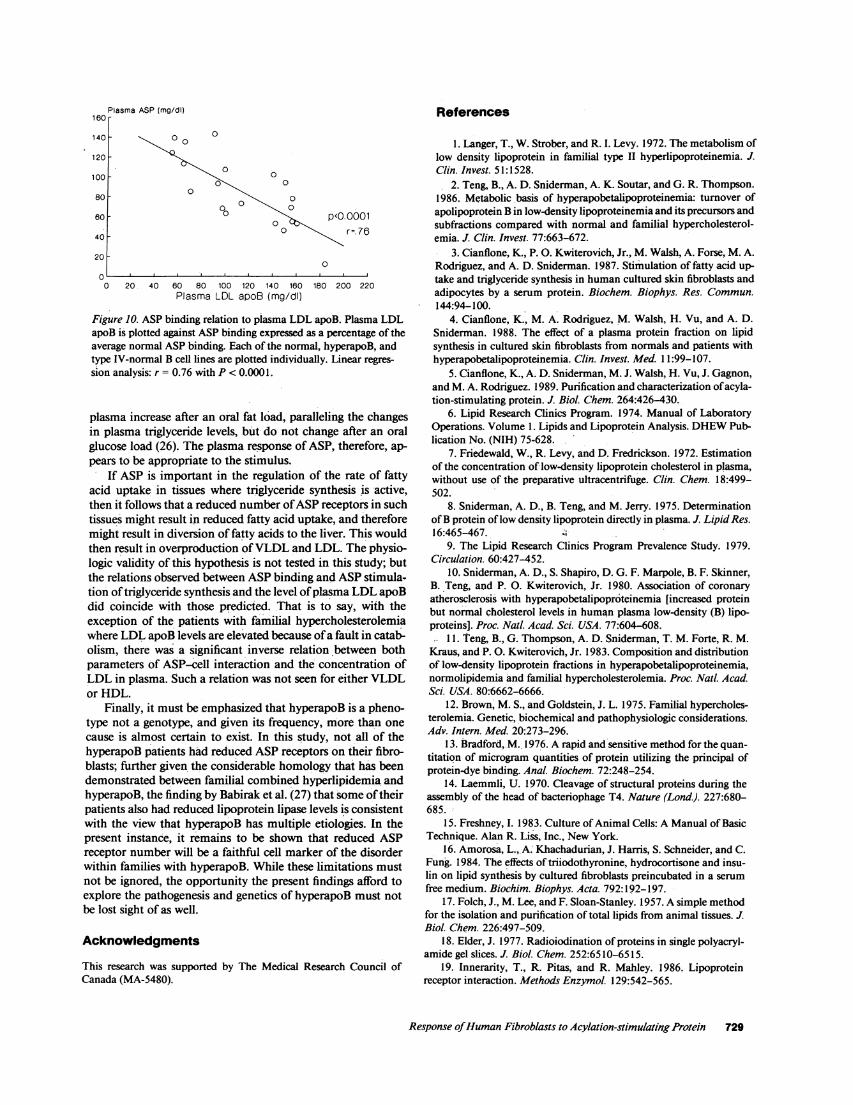
lations between ASP specific binding and total and LDL cho-lesterol with correlation coefficients of 0.56 and 0.71 and Pvalues of < 0.005 and 0.0005 respectively. The highest corre-....o lation coefficient, 0.76 P < 0.0001, was to LDL apoB, whichalso demonstrated the most negative slope (-0.632, Fig. 10). Itis of interest that, if the analysis is confined to the normolipid-emic patients with hyperapoB and the normals, then the onlyplasma variable that correlates with ASP-specific binding is thelevel of LDL apoB. Though not shown, the same findingsresult if the ASP-induced increase in cell triglyceride synthesisis correlated against plasma levels of the total lipids, lipopro-tein lipids, and LDL apoB.
14 16 Discussion
Hyperapobetalipoproteinemia is defined as the lipoproteinphenotype characterized by an increased LDL particle numberin plasma owing to overproduction of hepatic apoB lipopro-teins (2). However, no specific cause for the hepatic overpro-duction of apoB particles has yet been recognized; nor has anycell-specific marker of the disorder been suggested. The pres-ent study has examined the capacity of ASP to bind to, andstimulate triglyceride synthesis in human cultured skin fibro-blasts. The results in the patients with hyperapoB stand apartfrom those obtained in normals, in patients with familial hy-percholesterolemia, and in patients with type IV hyperlipo-proteinemia with normal LDL apoB.
The data confirm that ASP markedly stimulates triglycer-ide synthesis in skin fibroblasts cultured from normals. How-ever, the same degree of stimulation was observed in fibro-blasts obtained from patients with familial hypercholesterol-emia and patients with type IV hyperlipoproteinemia withouthyperapoB. Furthermore, the binding of ASPwas specific andsaturable with a Kd of 1.05 X 10⁻⁶ M in the normal cells.Similarly, in familial hypercholesterolemia and type IV hyper-lipoproteinemia without hyperapoB, specific saturable bindingof ASP to a single class of receptors was demonstrated and themaximal binding obtained was essentially the same as in thenormal group. By contrast, the increase in triglyceride synthe-sis induced by ASP in skin fibroblasts cultured from patients
ded (ng/mg cell protein)
Figure 3. Cell associated andcell degraded ASPin normaland hyperapoB cells. Cellswere incubated with 10 uM['4C]oleate and increasingconcentrations of '251-ASPfor 20 h, and the amount ofASPassociated with the celland the degradation products
/.i/ in the media were measured.Results are the average of 8normal (o) and 12 hyper-apoB (*) cell lines expressedas nanograms of ASPper
______________. ___. ____. ___. milligram of cell pro-4 6 8 10 12 14 16 tein±SEM. P values: *P
ASP concentration (pg/mI) < 0.025, **P < 0.01.
726 K. M. Cianflone, M. H. Maslowska, and A. D. Sniderman
V- - I

10 16 20 26ASP concentration (pg/ml)
ASP bound (ng/rng cell protein)
HYPERAPOB
0.8
0.8
0.4-
0.2-
0 10 15 20 28ASP concentration (Vg/ml)
Figure 4. ASPbinding to a representative normal and hyperapoBcell. '25I-ASP total, nonspecific, and specific binding was measured at40C for 2 h as described in methods. (*) Total binding; (A) nonspe-cific binding; (o) specific binding. Results are expressed as nano-
grams of ASPper milligram of cell protein.
0 0.2 0.4 0.6 0.8 1 1.2 1.4
ASP Bound (ng/mg cell protein)
Figure 5. Scatchard analysis of ASPspecific binding. Scatchard anal-ysis of bound ASP vs. bound/free ASP. Normal (o): n = 7, slope= -0.068±0.01 (Ag/ml)-', x-intercept = 1.313±0.13 ng/mg cell pro-
tein, r = 0.610; hyperapoB (*): n = 10, slope = -0.080±0.02 (gIg/ml)-', x-intercept = 0.76±0.10 ng/mg cell protein, r = 0.863.
The present study establishes that the ASP-induced increase incellular triglyceride synthesis is related to the degree of ASPbinding to the cell. Similarly, although the in vitro effective-ness of ASPappears clear, much remains to be done to estab-lish whether or not it plays an important metabolic role invivo. In this regard, it should be noted that ASP levels in
%of Normal Binding160F140
with hyperapoB was markedly less. At the same time, althoughspecific saturable binding to ASP was evident, the maximalspecific binding of ASPto these cells was considerably reducedbecause, it seems, of a reduced number of specific receptors inthe cell membrane.
This line of research began with the hypothesis that the rateof uptake of fatty acids into key peripheral tissues such as
adipocytes might be reduced in patients with hyperapoB. As a
consequence of this, the flux of fatty acids to the liver mightincrease, resulting in turn in increased hepatic apoB synthesisand secretion. In support of this hypothesis were the observa-tions that chylomicron triglyceride clearance was reduced inpatients with hyperapoB (24) and the in vitro observations thatthe rate of triglyceride synthesis in adipose tissue obtainedfrom patients with hyperapoB was less than in similar tissueobtained from normals (25). It was these findings that sug-gested the experiments examining triglyceride synthesis innormal and hyperapoB fibroblasts which led to the recognitionof ASP (5).
The mechanism of action of ASPremains to be elucidated.
120 F
100
80
60F
40
20
80
0
0
0
0
*>*s
Figure 6. Distribution of ASPbinding in normal and hyperapoB.Percentage of normal binding was calculated for each cell line as
Scatchard x-intercept for each cell line divided by the average Scat-chard x-intercept (1.313 ng/mg cell protein) X 100 for 7 normals (o)and 10 hyperapoB (*).
Response of HumanFibroblasts to Acylation-stimulating Protein 727
Bound/Free
0
0 30 38
20 30 40LDL concentration (1jg/ml)
0.8
0.6
0.4
0.2
0 5 10 15 20 25ASP concentration (pg/ml)
30 35
Figure 7. ASP-specific binding in familial hypercholesterolemia andtype IV-normal LDL apoB cells. ASP-specific binding was measuredat 40C for 2 h as described in methods. (Upper panel) Seven familialhypercholesterolemia (x) cell lines; and (lower panel) four type IV-normal LDL apoB (c) cell lines with the normal ASP binding (o)shown in both panels as the dotted line. Results are given as averageASPbound in nanograms per milligram cell protein±SEM.
% Triglyceride Stimulation
150
120
0
0
go0±
0
-30
0
0 20 40 60 80 100 120 140 160% Normal Binding
Figure 8. Relation between ASPbinding and ASP stimulation of tri-glyceride synthesis. Percent normal binding for all of the cell lines(normal, hyperapoB, familial hypercholesteremia, and type IV-nor-mal B) is plotted individually against the maximal percent triglycer-ide stimulation by ASP for the same cell line. Linear regression anal-ysis: r = 0.486, P < 0.005.
20 30 40LDL concentration (1g/ml)
50
LDL bound (ng/mg cell protein)
0 10 20 30 40LDL concentration (pg/ml)
50 60
Figure 9. LDL-specific binding in normal, hyperapoB and familialhypercholesterolemia (FH) cells. LDL-specific binding was measuredat 4VC for 2 h as described in Methods. (Upper panel) 6 normal (o)cell lines; (middle panel) 10 hyperapoB (*) cell lines; and (lowerpanel) 3 heterozygous FH cell lines (solid line) and 4 homozygousFH cell lines (dotted line). Results are expressed as nanograms LDLbound per milligram cell protein±SEM.
728 K. M. Cianflone, M. H. Maslowska, and A. D. Sniderman
00
0
0
00
0
0 CO 00EP o
60 -
30 -000-- t
- - - - - - - - - -*
----------------1.
P
160
140
120
100 _
80
60
40
20
0
:lasma ASP (mg/dl)
0 0
0
0
00
0-', 0
p<0.0001r=.76
0
0 20 40 60 80 100 120 140 160 180 200 220Plasma LDL apoB (mg/dl)
Figure 10. ASP binding relation to plasma LDL apoB. Plasma LDLapoB is plotted against ASPbinding expressed as a percentage of theaverage normal ASP binding. Each of the normal, hyperapoB, andtype IV-normal B cell lines are plotted individually. Linear regres-sion analysis: r = 0.76 with P < 0.0001.
plasma increase after an oral fat load, paralleling the changesin plasma triglyceride levels, but- do not change after an oralglucose load (26). The plasma response of ASP, therefore, ap-
pears to be appropriate to the stimulus.If ASP is important in the regulation of the rate of fatty
acid uptake in tissues where triglyceride synthesis is active,then it follows that a reduced number of ASPreceptors in suchtissues might result in reduced fatty acid uptake, and thereforemight result in diversion of fatty acids to the liver. This wouldthen result in overproduction of VLDL and LDL. The physio-logic validity of this hypothesis is not tested in this study; butthe relations observed between ASPbinding and ASPstimula-tion of triglyceride synthesis and the level of plasma LDL apoBdid coincide with those predicted. That is to say, with theexception of the patients with familial hypercholesterolemiawhere LDL apoB levels are elevated because of a fault in catab-olism, there was a significant inverse relation between bothparameters of ASP-cell interaction and the concentration ofLDL in plasma. Such a relation was not seen for either VLDLor HDL.
Finally, it must be emphasized that hyperapoB is a pheno-type not a genotype, and given its frequency, more than one
cause is almost certain to exist. In this study, not all of thehyperapoB patients had reduced ASP receptors on their fibro-blasts; further given the considerable homology that has beendemonstrated between familial combined hyperlipidemia andhyperapoB, the finding by Babirak et al. (27) that some of theirpatients also had reduced lipoprotein lipase levels is consistentwith the view that hyperapoB has multiple etiologies. In thepresent instance, it remains to be shown that reduced ASPreceptor number will be a faithful cell marker of the disorderwithin families with hyperapoB. While these limitations must
not be ignored, the opportunity the present findings afford to
explore the pathogenesis and genetics of hyperapoB must not
be lost sight of as well.
AcknowledgmentsThis research was supported by The Medical Research Council ofCanada (MA-5480).
References
1. Langer, T., W. Strober, and R. I. Levy. 1972. The metabolism oflow density lipoprotein in familial type II hyperlipoproteinemia. J.Clin. Invest. 51:1528.
2. Teng, B., A. D. Sniderman, A. K. Soutar, and G. R. Thompson.1986. Metabolic basis of hyperapobetalipoproteinemia: turnover ofapolipoprotein B in low-density lipoproteinemia and its precursors andsubfractions compared with normal and familial hypercholesterol-emia. J. Clin. Invest. 77:663-672.
3. Cianflone, K., P.O. Kwiterovich, Jr., M. Walsh, A. Forse, M. A.Rodriguez, and A. D. Sniderman. 1987. Stimulation of fatty acid up-
take and triglyceride synthesis in human cultured skin fibroblasts andadipocytes by a serum protein. Biochem. Biophys. Res. Commun.144:94-100.
4. Cianflone, K., M. A. Rodriguez, M. Walsh, H. Vu, and A. D.Sniderman. 1988. The effect of a plasma protein fraction on lipidsynthesis in cultured skin fibroblasts from normals and patients withhyperapobetalipoproteinemia. Clin. Invest. Med. 11:99-107.
5. Cianflone, K., A. D. Sniderman, M. J. Walsh, H. Vu, J. Gagnon,and M. A. Rodriguez. 1989. Purification and characterization of acyla-tion-stimulating protein. J. Biol. Chem. 264:426-430.
6. Lipid Research Clinics Program. 1974. Manual of LaboratoryOperations. Volume 1. Lipids and Lipoprotein Analysis. DHEWPub-lication No. (NIH) 75-628.
7. Friedewald, W., R. Levy, and D. Fredrickson. 1972. Estimationof the concentration of low-density lipoprotein cholesterol in plasma,without use of the preparative ultracentrifuge. Clin. Chem. 18:499-502.
8. Sniderman, A. D., B. Teng, and M. Jerry. 1975. Determinationof B protein of low density lipoprotein directly in plasma. J. Lipid Res.16:465-467.
9. The Lipid Research Clinics Program Prevalence Study. 1979.Circulation. 60:427-452.
10. Sniderman, A. D., S. Shapiro, D. G. F. Marpole, B. F. Skinner,B. Teng, and P. 0. Kwiterovich, Jr. 1980. Association of coronary
atherosclerosis with hyperapobetalipoproteinemia [increased proteinbut normal cholesterol levels in human plasma low-density (B) lipo-proteins]. Proc. Natl. Acad. Sci. USA. 77:604-608.
11.Teng, B., G. Thompson, A. D. Sniderman, T. M. Forte, R. M.Kraus, and P.O. Kwiterovich, Jr. 1983. Composition and distributionof low-density lipoprotein fractions in hyperapobetalipoproteinemia,normolipidemia and familial hypercholesterolemia. Proc. Natl. Acad.Sci. USA. 80:6662-6666.
12. Brown, M. S., and Goldstein, J. L. 1975. Familial hypercholes-terolemia. Genetic, biochemical and pathophysiologic considerations.Adv. Intern. Med. 20:273-296.
13. Bradford, M. 1976. A rapid and sensitive method for the quan-
titation of microgram quantities of protein utilizing the principal ofprotein-dye binding. Anal. Biochem. 72:248-254.
14. Laemmli, U. 1970. Cleavage of structural proteins during theassembly of the head of bacteriophage T4. Nature(Lond.). 227:680-685.
15. Freshney, I. 1983. Culture of Animal Cells: A Manual of BasicTechnique. Alan R. Liss, Inc., New York.
16. Amorosa, L., A: Khachadurian, J. Harris, S. Schneider, and C.Fung. 1984. The effects of triiodothyronine, hydrocortisone and insu-lin on lipid synthesis by cultured fibroblasts preincubated in a serum
free medium. Biochim. Biophys. Acta. 792:192-197.17. Folch, J., M. Lee, and F. Sloan-Stanley. 1957. A simple method
for the isolation and purification of total lipids from animal tissues.J.Bio/. Chem. 226:497-509.
18. Elder, J. 1977. Radioiodination of proteins in single polyacryl-
amide gel slices. J. Bio/. Chem. 252:6510-6515.19. Innerarity, T., R. Pitas, and R. Mahley. 1986. Lipoprotein
receptor interaction. Methods Enzymol. 129:542-565.
Response of Human Fibrob/asts to Acy/ation-stimulating Protein 729
20. Scatchard, G. 1949. The attraction of proteins for small mole-cules and ions. Ann. N. Y. Acad. Sci. 55:660-672.
21. Goldstein, J., S. Baar, and M. Brown. 1983. Receptor mediatedendocytosis of low density lipoproteinemia in cultured cells. MethodsEnzymol. 98:241-261.
22. Havel, R., R. Eder, and J. Bragson. 1955. The distribution andchemical composition of ultracentrifugally separated lipoproteins inhuman serum. J. Clin. Invest. 34:1345-1353.
23. Bilheimer, D. W., S. Eisenberg, and R. I. Levy. 1972. Themetabolism of very low density lipoprotein proteins. I. Preliminary invitro and in vivo observations. Biochim. Biophys. Acta. 260:212-221.
24. Genest, J., A. D. Sniderman, K. Cianflone, B. Teng, S. Wach-
older, Y. L. Marcel, and P. O. Kwiterovich, Jr. 1986. Hyperapobetali-poproteinemia: plasma lipoprotein responses to oral fat load. Arterio-sclerosis. 6:297-304.
25. Teng, B., A. Forse, M. A. Rodriguez, and A. D. Sniderman.1988. Adipose tissue glyceride synthesis in patients with hyperapobe-talipoproteinemia. Can. J. Pharmacol. Physiol. 66:239-242.
26. Cianflone, K., H. Vu, M. Walsh, A. Baldo, and A. D. Snider-man. 1989. The metabolic response of acylation stimulating protein toan oral fat load. J. Lipid Res. 30:1727-1733.
27. Babirak, S. P., P. H. Iverius, W. Y. Fujimoto, and J. D. Brun-zell. 1989. Detection and characterization of the heterozygote state forlipoprotein lipase deficiency. Arteriosclerosis. 9:326-334.
730 K. M. Cianflone, M. H. Maslowska, and A. D. Sniderman