immunotoxicity of dermal permethrin and cis … · cuca caused diminished splenic macrophage...
TRANSCRIPT

i
IMMUNOTOXICITY OF DERMAL PERMETHRIN AND CIS-UROCANIC ACID:EFFECTS OF CHEMICAL MIXTURES IN ENVIRONMENTAL HEALTH
By
Mary Renée Prater
Dissertation submitted to the faculty of the Virginia Polytechnic Institute and StateUniversity in partial fulfillment of the requirements for the degree of
Doctor of Philosophy
In
Veterinary Medical Sciences
APPROVED BY:
___________________________________Steven D. Holladay, Committee Chairperson
_____________ ____________ _____________S. Ansar Ahmed Eric A. Wong Holly S. Bender
_________________ ________________Robert M. Gogal, Jr. Benny L. Blaylock
8 March 2002
Blacksburg, VA
Keywords: Permethrin, Cis-urocanic acid, Ultraviolet irradiation, Immunotoxicity,Contact hypersensitivity, Skin, Thymus, Spleen, Mouse, TNFα, IFNγ, CD1a

ii
IMMUNOTOXICITY OF DERMAL PERMETHRIN AND CIS-UROCANIC ACID:EFFECTS OF CHEMICAL MIXTURES IN ENVIRONMENTAL HEALTH
M. Renée Prater
Abstract
The present study examined adverse effects of sunlight exposure (mimicked by
intradermal cis-urocanic acid, cUCA) on local and systemic immune responses, with or
without co-exposure to the immunotoxic insecticide permethrin. A single exposure to
cUCA caused diminished splenic macrophage phagocytosis that was persistent up to 30
days post-exposure. Five-day exposure to cUCA subtly increased splenocyte
proliferation in response to the T cell mitogen Concanavalin A. Four-week exposure to
cUCA caused increased splenic lymphocyte cellularity, thymic hypocellularity, and
enhanced hydrogen peroxide production by splenic leukocytes. Single exposure to topical
permethrin resulted in decreased thymic and splenic weight and cellularity, and inhibited
antibody production by splenic B cells. cUCA worsened the negative effect of permethrin
on both thymic weight and cellularity, and depressed splenocyte blastogenesis, hydrogen
peroxide production, and antibody production. Five-day exposure to either cUCA or
permethrin also caused persistent decreased contact hypersensitivity responses, an effect
that became more than additive when the chemicals were administered concurrently.
Defects in antigen processing and presentation by cutaneous Langerhans cells were
evaluated as possible contributing mechanisms to the cutaneous immunosuppression,
using mice with deleted genes. Vehicle-exposed IFNγ knockout mice displayed
approximately a 22.1% depression in the ear swelling response as compared to control
C57BL/6N mice, suggesting that this cytokine may be required for mounting a control-

iii
level hypersensitivity response. Ear swelling in cUCA-exposed IFNγ knockout mice
displayed a 21.4% depressed response as compared to cUCA-exposed wild-type
C57BL/6N mice, again suggesting that IFNγ is an important cytokine in the contact
hypersensitivity (CH) response. TNFαR knockout mice exposed to cUCA displayed
33.9% greater ear swelling than cUCA-exposed wild-type C57BL/6N mice, suggesting
that increased TNFα may be involved in inhibited CH by cUCA. TNFαR knockout mice
exposed to permethrin displayed 33.9% greater ear swelling than permethrin-exposed
C57BL/6N mice, suggesting that increased TNFα may also be involved in inhibited CH
by permethrin. C57BL/6N mice exposed to cUCA + permethrin displayed severe
reduction of the CH response to 8.7% of the control level. IFNγ knockout mice exposed
to permethrin + cUCA showed essentially identical depression of the CH response as
IFNγ knockout mice exposed to either permethrin or cUCA alone. These results suggest
that IFNγ is required for the greater than additive immunotoxic effect that occurred when
these two agents were co-administered. TNFαR knockout mice exposed to cUCA +
permethrin displayed 8.7 fold greater ear swelling than similarly exposed C57BL/6N
mice, again suggesting that increased TNFα is involved in inhibited CH by both cUCA
and permethrin.

iv
DEDICATION
I dedicate this effort to my daughters, Katherine and Christine, my mother Jean, my sister
Lisa, and my dear friends and family, for their unwavering support and sense of humor as
I completed the last in a long series of educational endeavors. I would like to extend my
most sincere appreciation and respect for my major advisor, Dr. Steven Holladay, for his
patience and guidance in the successful completion of this project, and to the members of
my graduate committee, Drs. Ahmed, Wong, Bender, and Gogal, and external examiner,
Dr. Blaylock for their expertise and input throughout my dissertation research.

v
TABLE OF CONTENTS
CHAPTER 1: INTRODUCTION AND LITERATURE REVIEW 1
A. Chemical Immunotoxicity 1
B. Permethrin 1
C. Urocanic Acid 5
D. Chemical Mixtures 11
E. References 13
CHAPTER 2: SINGLE-DOSE TOPICAL EXPOSURE TO THE PYRETHROID
INSECTICIDE, PERMETHRIN IN C57BL/6N MICE: EFFECTS ON THYMUS
AND SPLEEN 25
A. Abstract 27
B. Introduction 28
C. Materials and Methods 31
1. Mice 31
2. Permethrin Preparation and Treatment Protocols 31
3. Organ Weights, Cell Preparation, and Cellularity 31
4. Surface Antigen Expression by Flow Cytometry 32
5. Thymocyte Apoptosis and Necrosis: Flow Cytometric And
Cytologic Evaluation 33
6. Proliferation of Splenocytes and Thymocytes 33
a. In-Vitro Permethrin Exposure 33
b. In-Vivo Permethrin Exposure 34
7. Splenic Macrophage Phagocytosis 35
8. Chemiluminescence Response in Splenic Macrophages 36
9. B Lymphocyte Antibody Production: Plaque-Forming
Cell Assay 36
10. 51Cr Release Cytotoxicity Assay 37
11. Statistical Analysis 38
D. Results 39

vi
E. Discussion 43
F. Acknowledgement 47
G. References 48
CHAPTER 3: IMMUNOTOXIC EFFECTS OF CIS-UROCANIC ACID
EXPOSURE IN C57BL/6N MICE 67
A. Abstract 69
B. Introduction 70
C. Materials and Methods 73
1. Mice 73
2. Cis-Urocanic Acid 73
3. Organ Weights, Cell Preparation, and Cellularity 73
4. Thymocyte Surface Antigen Expression by Flow Cytometry 74
5. Proliferation of Splenocytes and Thymocytes 75
6. Splenic Macrophage Phagocytosis 76
7. Chemiluminescence Response in Splenic Macrophages 76
8. B Lymphocyte Antibody Production: Plaque-Forming
Cell Assay 77
9. Cytotoxicity Assay 78
10. Statistical Analysis 79
D. Results 80
E. Discussion 83
F. Acknowledgement 88
G. References 89
CHAPTER 4: CIS-UROCANIC ACID INCREASES BOTH
IMMUNOTOXICITY AND LETHALITY OF DERMALLY
ADMINISTERED PERMETHRIN IN C57BL/6N MICE 103
A. Abstract 105
B. Introduction 106
C. Materials and Methods 109

vii
1. Mice 109
2. Permethrin and Cis-Urocanic Acid Preparation and
Treatment Protocols 109
3. Organ Weights, Cell Preparation, and Cellularity 110
4. Proliferation of Splenocytes and Thymocytes 110
5. Splenic Macrophage Phagocytosis 111
6. Chemiluminescence Response in Splenic Macrophages 112
7. B Lymphocyte Antibody Production: PFC Assay 112
8. Statistical Analysis 113
D. Results 115
E. Discussion 118
F. Acknowledgement 120
G. References 121
CHAPTER 5: MOLECULAR MECHANISMS OF SUNLIGHT AND
PERMETHRIN-INDUCED ALTERATIONS IN CUTANEOUS IMMUNITY 134
A. Abstract 136
B. Introduction 138
C. Materials And Methods 141
1. Mice 141
2. Permethrin Treatment Protocol 141
3. Cis-Urocanic Acid 142
4. Contact Hypersensitivity 142
5. Statistical Analysis 143
D. Results 144
E. Discussion 147
F. Acknowledgement 152
G. References 153
CHAPTER 6: CONCLUSIONS 167

viii
APPENDICES 170
I. Splenic Macrophage Phagocytosis Response to Permethrin 170
II. Splenic Chemiluminescence Response to Permethrin 171
III. Splenic Cytotoxic T Lymphocyte Response to Permethrin 172
IV. Thymocyte Proliferative Response to Permethrin 173
V. Thymic Weight Response to Single Exposure to cUCA 174
VI. Thymic Cellularity Response to Single Exposure to cUCA 175
VII. Splenic Weight Response to Single Exposure to cUCA 176
VIII. Splenic Cellularity Response to Single Exposure to cUCA 177
IX. Thymic Weight Response to 5-Day Exposure to cUCA 178
X. Thymic Cellularity to 5-Day Exposure to cUCA 179
XI. Splenic Macrophage Phagocytosis Response to 5-Day cUCA 180
XII. Splenic Antibody Production Response to 5-Day cUCA 181
XIII. Splenic Chemiluminescence Response to 5-Day cUCA 182
XIV. Splenic Cytotoxic T Lymphocyte Response to 5-Day cUCA 183
XV. Splenic Macrophage Phagocytosis to cUCA + Permethrin 184
CURRICULUM VITAE 185

ix
LIST OF TABLES
2.1 Effect of Topical Permethrin on Absolute Thymocyte Subpopulations 58
2.2 Effect of Topical Permethrin on Thymocyte Viability and Apoptosis 59
3.1 Effect of Five-Day Cis-Urocanic Acid on Thymocyte Differentiation 97

x
LIST OF FIGURES
2.1 Thymic Weight Response to Permethrin 60
2.2 Thymic Cellularity Response to Permethrin 61
2.3 Splenic Weight Response to Permethrin 62
2.4 Splenic Cellularity Response to Permethrin 63
2.5 Antibody-mediated Response to Permethrin 64
2.6 Proliferation Response of Splenocytes to in-vivo Permethrin 65
2.7 Proliferation Response of Splenocytes to in-vitro Permethrin 66
3.1 Splenocyte and Thymocyte Proliferation Response to cUCA 98
3.2 Splenic Macrophage Phagocytosis Response to cUCA 99
3.3 Thymic Cellularity Response to cUCA 100
3.4 Splenic Cellularity Response to cUCA 101
3.5 Splenic Chemiluminescence Response to cUCA 102
4.1 Effect of cUCA and Permethrin on the Thymic Weight and Cellularity 128
4.2 Effect of cUCA and Permethrin on the Splenic Weight and Cellularity 129
4.3 Effect of cUCA and Permethrin on Splenocyte and Thymocyte Proliferation 130
4.4 Effect of cUCA and Permethrin on Splenic Chemiluminescence 131
4.5 Effect of cUCA and Permethrin on Antibody-Mediated Immunity 132
4.6 Effect of cUCA and Permethrin on Neurotoxic Deaths 133
5.1 CH Response to cUCA and Permethrin in C57BL/6N and SvImJ Mice 163
5.2 CH Response to cUCA and Permethrin in C57BL/6N and TNFαR KO Mice 164
5.3 CH Response to cUCA and Permethrin in C57BL/6N and IFNγ KO Mice 165
5.4 CH Response to cUCA and Permethrin in SvImJ and CD1a KO Mice 166

1
CHAPTER 1: INTRODUCTION AND LITERATURE REVIEW
1A. CHEMICAL IMMUNOTOXICITY
Toxicological assessment for approval of environmental chemicals and
pharmaceutical products typically does not include evaluation of the immune system.
The need to incorporate immunotoxicity testing has only been realized in recent years.
Risk assessment models for immunotoxicity evaluation have been studied as researchers
recognize it is temporally and economically impractical to complete a full evaluation of
all arms of the immune system for each compound in question (Luster et al. 1992, 1993).
Therefore, a risk assessment model was developed such that individual and pair-wise
predictive values were established for the various immunological assays in an attempt to
expediently quantitate the possibility of decreased host resistance to disease following
chemical challenge using a few selected immune tests. Earlier studies had outlined a
tiered approach to full evaluation of the immune system that included immunopathology,
cell-mediated and antibody-mediated immune evaluation, nonspecific immunity, and host
resistance challenge models (Luster et al. 1988) which supplied the background
information necessary for the development of these risk assessment models.
1B. PERMETHRIN
Permethrin (3-phenoxyphenyl methyl (+) cis, trans-3-(2,2-dichloroethenyl)-2,2
dimethylcyclopropane carboxylate) is a synthetic, third generation, type I pyrethroid
insecticide that is commonly used in shampoos and topical creams in human and
veterinary medicine to eliminate ectoparasites such as fleas, ticks, lice, and mites
(Asakawa et al. 1996, Llewellyn et al. 1996, Fuortes 1999). Permethrin is also used in

2
the prevention of ectoparasite infestation and possible resultant insect-borne disease via
direct topical application or permeation of military and hunting clothing (Schreck et
al.1986, Schreck and Kline 1989, Sholdt et al. 1989). The degree of topical absorption is
species-dependent, and ranged from 2% in human beings and rabbits, 10% in mice, 15%
in rhesus monkeys (variation according to isomer and site of application), to as much as
44% in rats. Urinary excretion follows hepatic hydrolysis and either oxidization or
glucuronide-conjugation (Miyamoto 1976, Glickman et al.1981, Shah et al. 1981, Taplin
and Meinking 1987, Sidon et al. 1988, Snodgrass 1992). The No Observable Effect
Level (NOEL) for oral permethrin in rabbits has been reported to be 5 mg/kg/day
(Snodgrass 1992). The median lethal oral dose in rats is 4 g/kg, and this level varies
slightly according to the age, nutrition, gender, and strain of rat. However, the median
lethal oral dose of permethrin in corn oil vehicle is 380 mg/kg. This finding raises
concern about the extent of systemic absorption and resulting toxicity with topically-
applied insecticides, since most commercially available insecticides are only available as
a mixture of active ingredients and inert solvents or vehicles (Metker et al. 1977, McCain
et al. 1997).
The type I pyrethroid insecticides, including permethrin, cause neurotoxic effects
that have been well characterized in the literature and include aggression, sparring,
tremor, and hyperthermia. These effects are very distinct from the effects of type II
pyrethroid insecticides such as cypermethrin and deltamethrin, which cause pawing,
burrowing, salivation, hypothermia, and lowered motor activity (McDaniel and Moser
1993). These effects of permethrin are thought to be the result of alterations in synaptic
membrane potential and resulting neuroexcitation (Eells et al. 1992). This

3
neuroexcitation has been demonstrated in laboratory rodents to result from increased
sodium influx through voltage-sensitive sodium channels into synaptic terminals (Lees
1998), producing membrane depolarization, excess glutamate uptake, and
neurotransmitter (acetylcholine) release (Eells et al. 1992, Vaccari et al. 1998).
Permethrin has also been shown to inhibit acetylcholinesterase (AchE) activity in rat
brain cortex by 97-98% within 45 minutes of in-vitro (5 ppm) exposure or by 50% within
30 minutes following in-vivo (250 mg/kg body weight) administration, which would
exacerbate permethrin’s neurotoxic effects (Bandyopadhyay 1982). The maximum
plasma concentrations are achieved at 3.52 hours (Anadon et al. 1991), peak neurotoxic
effect of permethrin is at 5 hours post oral administration in rodents, and result in changes
in biogenic amines and their metabolites: 3-methoxy-4-hydroxyphenylglycol (MHPG),
5-hydroxyindoleacetic acid (5-HIAA, a serotonin metabolite), and aspartate levels in
homogenized brain tissue (Hudson et al. 1986). Permethrin is also thought to diminish
energy coupling by inhibiting glutamate and succinate respiration in mitochondria
(Gassner et al. 1997). Larger (approximately a quarter of the acute oral LD50 value, 300
mg/kg/day) or more prolonged exposure (5-7 days) to oral permethrin and two type II
pyrethroid insecticides, cypermethrin and deltamethrin, results in sparse axonal
peripheral nerve demyelination and Wallerian degeneration, which results in abnormal
locomotion; the potential mechanism of action includes upregulation of β-glucuronidase
and β-galactosidase in laboratory rodents (Rose and Dewar 1983, Cavaliere et al. 1990).
Further, there was enhanced neurotoxicity when permethrin was administered
concurrently with other pesticides such as DEET (N,N-diethyl-m-toluamide) and
pyridostigmine bromide, which is likely attributable to competition for liver and plasma

4
esterases, leading to decreased insecticide breakdown and increased transport of the
parent compound to nervous tissues (Abou-Donia et al. 1996).
Low doses of permethrin cause mild neurological signs and immunomodulation.
Higher doses of permethrin result in more severe peripheral and central nervous system
clinical signs including hyperactivity, convulsions, paralysis, and even death (Hansen et
al.1994). Permethrin and several analogs (cypermethrin, bioallethrin, and deltamethrin)
have demonstrated immunomodulatory effects in murine and human models, including
diminished natural killer cell cytotoxicity, T cell and antibody-mediated
immunomodulation, alterations in class II MHC cells, modified cytokine levels, and
variations in thymocyte numbers, distribution and function (Puig et al. 1989, Enan et al.
1996, Santoni et al. 1997, 1998, Diel et al. 1998-1, 2, 1999, Murphy et al.1999, Zhang et
al. 1999). These data suggest that this class of insecticides may significantly alter local
and systemic immunity.
Limited information is available about precise mechanisms of immunomodulation
caused by permethrin. Recent studies have demonstrated that low dose subacute topical
exposure (10 days) to permethrin causes diminished antibody-mediated immunity in
mice, and oral cypermethrin decreases antibody production in both rats and mice (Desi et
al. 1985, Tamang et al. 1988, Punareewattana et al. 2001). A potential mechanism of
action has been demonstrated to be inhibited production of cytokines (IFNγ and IL-4)
necessary for antibody production (Diel et al. 1998). Further, subacute topical
permethrin exposure in mice depressed splenic macrophage hydrogen peroxide
production, and the speculated mechanism included permethrin-induced inhibition of
mitochondrial complex I in the electron transport chain, which may interfere with

5
leukocyte respiratory burst development (Gassner et al. 1997, Punareewattana et al.
2001). Other authors have noted that intraperitoneal administration of permethrin or
deltamethrin results in a calcium/calmodulin-dependent alteration of the protein kinase-
phosphatase cascade, leading to increased apoptosis of thymocytes and resulting thymic
atrophy (Rashatwar and Matsumura 1985, Enan et al. 1996). These reports have
demonstrated a few potential mechanisms by which permethrin affects the immune
system, but clearly more information is needed in order to more fully understand the
molecular mechanisms of permethrin’s immunotoxicity.
1C. UROCANIC ACID
Urocanic acid (2-propenoic acid, 3-[1H-imidazol-4(5)-yl] was originally found in
dog urine more than a century ago (Jaffe 1874) and was later detected in animal sweat
and skin (Zenisek and Kral 1953). This led to the proposal that UCA may act as an
endogenous sunscreen or photoprotectant against UVB-induced DNA damage. Urocanic
acid is found predominantly in the stratum corneum and the liver, and is synthesized via
deamination of histidine. Urocanic acid represents about 0.7% of the dry weight of the
epidermis, and is present predominantly in the trans isomer in skin protected from
sunlight (Tabachnick 1957). The concentration of cUCA in nonirradiated murine skin is
0.2 ug/cm2 (4% of total UCA in the skin), and rises to 15 µg/cm2 upon irradiation at 96
mJ/cm2 or 80 µg/mouse after 42 mJ/cm2 (Noonan et al. 1988). Only 1 µg is necessary to
reduce contact hypersensitivity (CH) responses, and it has been demonstrated in BALB/c
mice that only 21-26 minutes of sunlight exposure, regardless of latitude and with normal
stratospheric ozone concentrations, results in sufficient conversion of urocanic acid to

6
result in a 50% drop in skin immune responses (DeFabo et al. 1990). Urocanic acid is
produced by epidermal keratinocytes and is stored in the trans isomeric form (tUCA) in
keratohyalin granules in the stratum corneum. The poorly soluble tUCA is racemized to
the highly water soluble cis isomer when exposed to low-wavelength (280-320 nm) UVB
irradiation. Cis-urocanic acid is then absorbed systemically and is thought to be excreted
unmetabolized in the urine (Anglin and Batten 1968, Morrison and Deibel 1986).
Ultraviolet-B irradiation causes protein denaturation and DNA damage via
induction of cyclobutane pyrimidine formation, strand breaks, and production of adducts
which is thought to be an initiating step into the development of some cutaneous
neoplasms. Ultraviolet-B irradiation has been implicated as a risk factor for development
and progression of several skin cancers, including basal cell carcinoma, malignant
melanoma, and squamous cell carcinoma in human beings, and the percentage of UVB-
sensitive human beings is estimated to be approximately 40-45% in both the Caucasian
and non-Caucasian races (Streilein 1993). Ultraviolet-B irradiation exposure has been
known for over 20 years to predispose mice to development of immunogenic cutaneous
tumors, and it is thought that direct DNA damage (such as mutations to the p53 gene), in
conjunction with immune suppression, leads to the development and progression of
immunogenic skin cancers (Kripke 1983, Majewski and Jablonska 1995). Cis-urocanic
acid is thought to act as a DNA protectant by absorbing the majority of the UVB rays and
undergoing transformation from the trans isomer to the cis isomer (Norval et al. 1995).
Therefore, UCA has been proposed to act as a natural cutaneous photoprotecting agent
against DNA damage caused by UVB irradiation. Recent reports have examined the
intensity of UVB exposure necessary to evoke immune suppression in mice, and have

7
found that under normal stratospheric ozone concentrations at nearly any latitude, 50%
cutaneous depression of contact hypersensitivity immune response in the BALB/c mouse
would occur following 21-26 minutes of sunlight exposure. These data suggest that
induction of murine immune suppression requires low levels of sunlight that can be
experienced almost anywhere in the world (DeFabo et al. 1990).
However, increased epidermal concentrations of UCA increases sensitivity to
UVB immune suppression and resulting skin cancer development (DeFabo et al. 1997).
Consequently, the immunologic effects of sunlight exposure are elicited in the laboratory
by the intracutaneous administration of cUCA. The immunologic defects in animals
previously exposed to UVB or cUCA were discovered to be two-fold: deficiency of
conventional antigen-presenting cells in the spleen and altered populations of regulatory
T cells that selectively impaired anti-tumor immunity. Ultraviolet-B irradiation indirectly,
through the actions of cUCA, causes upregulation of suppressor T lymphocytes in spleen
and lymph nodes, which prevents immunologic rejection of UV tumors transplanted into
syngeneic recipients, and also significantly reduce the latent period of tumor appearance
during UV carcinogenesis (Fisher and Kripke 1978, 1982).
The beneficial immunomodulatory effects of cUCA have been applied to grafting,
and two reports have suggested that cUCA can prolong the survival of allogeneic MHC
disparate tail and corneal grafts in BALB/c mice (Gruner et al. 1992, Guymer and
Mandel 1993). These findings suggest future development of cUCA or an analog for
therapeutic use in the long-term survival of transplanted organs, or in the prevention of
graft-versus host disease. Fetal mice have been shown to contain a low quantity of UCA
in the skin that is all in the trans isomeric form. Within one day of birth, the quantity of

8
UCA increases 20 fold, and a high percentage is converted (11%) to the cis isomer. As
previously mentioned, older mice contain only 4% cUCA in UVB-protected skin. It is
thought that this high percentage of cUCA in neonatal mice may serve as a protective
role to suppress immune responses to epidermal antigens while the animal is regulating
immune responses to self-antigens, including epidermal antigens (Norval et al. 1989).
However, circulating cUCA has been shown to also confer harmful local and
systemic immunomodulation via diminished T cell-mediated immunity and decreased
antigen presentation, which may actually contribute to rather than protect against the
clinically relevant end-point of increased risk of infectious or neoplastic disease. It has
been well documented that exposure to acute, low-dose UVB that qualitatively and
quantitatively resembles typical human sun exposure results in diminished CH and
induces antigen-specific tolerance by increasing suppressor T cells and converting
immunogenic antigen presenting cells to tolerogenic cells (Streilein et al. 1994, Vink et
al. 1996). These effects can also be mimicked by intracutaneous injections of cUCA or
TNFα (Streilein et al. 1994), suggesting that there are chemical intermediaries at least
partially involved in sunlight-induced immunomodulation.
Immunosuppressive effects of cUCA include inhibition of CH (DeFabo and
Noonan 1983), delayed allograft rejection (Gruner et al. 1992), and prevention of graft
vs. host disease (Gruner et al. 1992). In-vivo UVB radiation or intracutaneous
administration of cUCA in human beings causes a defect in cutaneous antigen
presentation by abrogating the function of CD1+ Langerhans cells (LC), and inducing
CD1- macrophages that activate suppressor T lymphocytes and diminish contact
hypersensitivity (Baadsgaard et al. 1990). Another study substantiated these findings, and

9
showed that following a single exposure of 4 minimal erythemal dose in human beings,
CH was significantly reduced. While numbers of CD1a+ LC were significantly
diminished and remaining LC were rendered ineffective functionally, numbers of CD1a-
macrophages were increased in the skin (LeVee et al. 1997). Cis-urocanic acid also
induces e-selectin expression from endothelial cells of dermal microvessels, which causes
release of constitutive TNFα from mast cell granules. Therefore, DNA damage causes
direct mutagenesis to skin cells, and also induces the expression and release of TNFα
from many mammalian cells (Kibitel et al. 1998). Tumor necrosis factor-alpha decreases
vimentin expression in LC, which results in blunting of LC cytoplasmic processes and
decreased ability of the compromised cells to acquire and process antigens to present to T
lymphocytes. Tumor necrosis factor-alpha is also chemoattractant to LC, so increased
concentrations in the skin prevent migration of LC to local lymph nodes and thus
decrease antigen presentation.
UVB is thought to suppress cutaneous and systemic immune responses in an
indirect manner, following UVB exposure to the two cutaneous chromophores urocanic
acid and DNA. Several mediators have been implicated in this process including
prostaglandins, interleukin-10, and TNFα (Norval et al. 1995). Epidermal cell-derived
thymocyte activating factor (ETAF, or IL-1) is also depressed following UVB exposure,
which depresses the activation of T lymphocytes that are associated with antigen (Norval
et al. 1989). Ultraviolet-B irradiation -induced increased release of TNFα contributes to
failed CH induction (Alard et al. 2001). Tumor necrosis factor-alpha is also thought to
induce production and activation of matrix metalloproteinases that may contribute to
tumor progression and metastasis (Han et al. 2001). It is now known that for

10
development of the full extent of murine contact hypersensitivity, activation of both
tumor necrosis factor receptors (type I p55TNFR and type II p75TNFR) is necessary.
Mice deficient for p55TNFR had a defect in antigen uptake but showed normal migration
into regional lymph nodes, whereas mice dendritic cells lacking in p75TNFR showed
diminished migration into regional lymph nodes after antigen uptake, but the antigen
uptake itself was not affected (Becke et al. 2001).
Ultraviolet-B irradiation also increases macrophagic release of TGFβ1, which is
also involved in the release of TNFα from local mast cells and local mononuclear cells
and differentiation/maturation of CD1+ DC. (Majewski and Jablonska 1995). Signaling
mediators of the TGFβ superfamily, called SMADS, are normally expressed at high
levels in the epidermis, but during skin carcinogenesis, mutations in SMADS resulted in
loss of growth inhibition mediated by TGFβ, thus resulting in tumor progression (He et
al. 2001).
Prolonged exposure to cUCA in rodents results not only in cutaneous immune
suppression, but also systemic immunomodulation including thymic atrophy and thymic
hypocellularity, upregulation of systemic suppression T cells, and a shift of Th1/Th2
cytokine profiles (El-Ghorr et al. 1997). Several molecular mediators have been
implicated. Upregulation of IL-10 following low-dose UVB irradiation is thought to
mediate systemic tolerance. It is thought that cUCA also mediates inhibition of NK
activation, as a result of uncoupling of NK receptors from phospholipase C-mediated
phosphoinositide hydrolysis. Cis-urocanic acid down-regulates induction of cAMP,
which may affect the second messenger system of the NK cell (Norval et al. 1995).
Additionally, monocytes down-regulate NK cell activity by reducing the expression of

11
CD16 and CD56 surface antigens on NK cells, suggesting that cUCA acts indirectly on
NK cells via monocytes or dendritic cells. Also, monocytes normally produce IL-12 in
the development of Th1 responses and in the activation of NK cells. This causes NK
cells to synthesize IFNγ. Ultraviolet irradiation promotes Th2 responses and depresses
Th1 responses, and the cUCA immunomodulation of NK activity has been proposed to be
the result of IL-12 and IFNγ downregulation or UV-induced macrophage production of
IL-12p40 homodimer, a natural antagonist of biologically active IL-12, leading to IFNγ
suppression (Norval et al. 1995, Reeve et al. 1999, Schmitt and Ullrich 2000).
1D. CHEMICAL MIXTURES
Although the effect of concurrent exposure to permethrin and sunlight is a highly
relevant concern in human, especially pediatric medicine, there is currently a paucity of
information over how this chemical mixture specifically affects the cutaneous and
systemic immunity against the development of infectious or neoplastic disease. The
alterations in murine systemic immune competence that result from subacute to chronic
administration of cUCA seem to be localized primarily to cell-mediated immunity, and
are manifested as thymic atrophy, thymic hypocellularity, and altered CH. Preliminary
experiments have demonstrated immune modulation following subacute exposure to
topical permethrin in both the cellular and antibody-mediated immune systems.
However, the effects of shorter duration exposures are not known. Therefore, the present
study proposes that acute topical exposure to permethrin or 5-day intradermal cUCA
would also result in systemic modulation of cellular and antibody-mediated immunity.
Further, it is an attempt to determine potential molecular mechanisms of this modulation

12
with the use of combination therapy on genetically altered strains of mice to determine
whether cUCA and permethrin have similar or divergent molecular mechanisms. The
molecular mechanisms of the cutaneous and systemic immunotoxicity of permethrin and
cUCA are currently poorly understood, although reports in the literature suggest local
cutaneous alterations in cytokines (TNFα, TGFβ, IL-1, IL-2, IL-10) following chemical
exposure. The end-points to determine functional effects of the chemicals included
splenic and thymic organ weights and cellularity, splenic phagocyte function, splenic
leukocyte chemiluminescence assay, splenic and thymic cell surface antigen evaluation
and cytometry, plaque forming cell assay, cytotoxic T lymphocyte assay, in-vitro
proliferation of thymocytes, quantitation of apoptosis, and CH.

13
1E. REFERENCES
Abou-Donia MB, Wilmarth KR, Jensen KF, Oehme FW and Kurt TL. Neurotoxicity
resulting from coexposure to pyridostigmine bromide, DEET, and permethrin:
implications of Gulf War chemical exposures. J Toxicol Environ Health 48:35-56, 1996.
Alard P, Kurimoto I, Niizeki H, Doherty JM, Streilein JW. Hapten-specific tolerance
induced by acute, low-dose ultraviolet B radiation of skin requires mast cell
degranulation. Eur J Immunol 31:1736-1746, 2001.
Anadon A, Martinez-Larranaga MR, Diaz MJ, Bringas P. Toxicokinetics of permethrin in
the rat. Toxicol Appl Pharmacol. 110:1-8, 1991.
Anglin JH Jr., Batten WH. Studies on cis-urocanic acid. J Invest Dermatol 50:463-466,
1968.
Asakawa F, Jitsunari F, Miki K, Choi JO, Takeda N, Kitamoado T, Suna S, Manabe Y.
Agricultural worker exposure to and absorption of permethrin applied to cabbage. Bull
Environ Contam Toxicol 56:42-49, 1996.
Baadsgaard O, Salvo B, Mannie A, Dass B, Fox DA, Cooper KD. In-vivo ultraviolet-
exposed human epidermal cells activate T suppressor cell pathways that involve CD4+
CD45RA+ suppressor-inducer T cells. J Immunol 145:2854-2861, 1990.

14
Bandyopadhyay R. Inhibition of acetylcholine esterase by permethrin & its reversion by
acetylthiocholine. Indian J Exp Biol 20:488-491, 1982.
Becke FM, Hehlgans T, Borckhoff G, Mannel DN. Development of allergic contact
dermatitis requires activation of both tumor necrosis factor-receptors. Eur Cytokine
Netw 12:45-50, 2001.
Cavaliere MJ, Maeda MY, Shih LW, Puga FR. Changes in myelinated nerve fibers and
skeletal muscle of rats exposed to high doses of permethrin. Biomed Environ Sci 3:139-
145, 1990.
DeFabo EC, Noonan FP. Mechanism of immune suppression by ultraviolet irradiation
in-vivo. I. Evidence for the existence of a unique photoreceptor in skin and its role in
photoimmunology. J Exp Med 158:84-98, 1983.
DeFabo EC, Noonan FP, and Frederick JE. Biologically effective doses of sunlight for
immune suppression at various latitudes and their relationship to changes in stratospheric
ozone. Photochem Photobiol. 52:811-817, 1990.
DeFabo EC, Webber LJ, Ulman EA, and Broemeling LD. Dietary L-histidine regulates
murine skin levels of trans-urocanic acid, an immune-regulating photoreceptor, with an
unanticipated modulation: potential relevance to skin cancer. J Nutr 127:2158-2164,
1997.
15
Desi I, Varga L, Dobronyi I, Szklenarik G. Immunotoxicological investigation of the
effects of a pesticide; cypermethrin. Arch Toxicol Suppl 8:305-309, 1985.
Diel F, Detscher M, Borck H. Schrimpf D, Diel E, Hoppe HW. Effects of permethrin on
human basophils and lymphocytes in-vitro. Inflamm Res Suppl 1:S11-S12, 1998.
Diel F, Detscher M, Schock B, Ennis M. In-vitro effects of the pyrethroid S-bioallethrin
on lymphocytes and basophils from atopic and nonatopic subjects. Allergy 53:1053-
1059, 1998.
Diel F, Horr B, Borck H, Savtchenko H, Mitgsche T, Diel E. Pyrethroids and piperonyl-
butoxide affect human T-lymphocytes in-vitro. Toxicol Lett 107:65-74, 1999.
Eells JT, Bandettini PA, Holman PA, Propp JM. Pyrethroid insecticide-induced
alterations in mammalian synaptic membrane potential. J Pharmacol Exp Ther 262:1173-
1181, 1992.
El-Ghorr AA, Norval M. The effect of chronic treatment of mice with urocanic acid
isomers. Photochem Photobiol 65:866-872, 1997.
Enan E, Pinkerton KE, Peake J, Matsumura F. Deltamethrin-induced thymus atrophy in
male BALB/c mice. Biochem Pharmacol 51:447-454, 1996.

16
Fisher MS and Kripke ML. Further studies on the tumor-specific suppressor cells
induced by ultraviolet radiation. J Immunol 121:1139-1144, 1978.
Fisher MS and Kripke ML. Suppressor T lymphocytes control the development of
primary skin cancers in ultraviolet-irradiated mice. Science 216:1133-1134, 1982.
Fuortes L. Urticaria due to airborne permethrin exposure. Vet Hum Toxicol 41:92-93,
1999.
Gassner B, Wuthrich A, Scholtysik G, Solioz M. The pyrethroids permethrin and
cyhalothrin are potent inhibitors of the mitochondrial complex I. J Pharmacol Exp Ther
281:855-860, 1997.
Glickman AH, Hamid AA, Rickert DE, and Lech, JJ. Elimination and metabolism of
permethrin isomers in rainbow trout. Tox Appl Pharm 57:88-98, 1981.
Gruner S, Diezel W. Stoppe H, Oesterwitz H, and Henke W. Inhibition of skin allograft
rejection and acute graft-versus-host disease by cis-urocanic acid. J. Invest. Dermatol
98:459-462, 1992.
Guymer RH and Mandel TE. Urocanic acid as immunosuppressant in allotransplantation
in mice. Transplantation 55:36-43, 1993.

17
Han YP, Tuan TL, Hughes M, Wu H, and Garner WL. Transforming growth factor-β
and tumor necrosis factor-α-mediated induction and proteolytic activation of MMP-9 in
human skin. J Biol Chem 276:22341-22350, 2001.
Hansen SR, Stemme KA, Villar D, Buck WB. Pyrethrins and pyrethroids in dogs and
cats. Compend Contin Educ Pract Vet 16:707-713, 1994.
He W, Cao T, Smith DA, Myers TE, Wang XJ. SMADS mediate signaling of the TGF
beta superfamily in normal keratinocytes but are lost during skin chemical
carcinogenesis. Oncogene 20:471-483, 2001.
Hudson PM, Tilson HA, Chen PH, Hong JS. Neurobehavioral effects of permethrin are
associated with alterations in regional levels of biogenic amine metabolites and amino
acid neurotransmitters. Neurotoxicology 7:143-153, 1986.
Jaffe M. Concerning a new constituent in the urine of dogs. Ber Deut Chem Gest
7:1669-1673, 1874.
Kibitel J, Hejmadi V. Alas L, O’Connor A, Sutherland BM and Yarosh D. UV-DNA
damage in mouse and human cells induces the expression of tumor necrosis factor alpha.
Photochem Photobiol 67:541-546, 1998.

18
Kripke ML. Immunobiology of photocarcinogenesis. In: Parrish HA (ed.). The effect
of Ultraviolet Radiation on the Immune System. Johnson and Johnson Products. New
Brunswick, NJ, pp. 87-106, 1983.
Lees G. Effects of pyrethroid molecules on rat nerves in-vitro: potential to reverse
temperature-sensitive conduction block of demyelinated peripheral axons. Br J
Pharmacol 123:487-496, 1998.
LeVee GJ, Oberhelman L, Anderson T, Koren H and Cooper KD. UVAII exposure of
human skin results in decreased immunization capacity, increased induction of tolerance
and a unique pattern of epidermal antigen-presenting cell alteration. Photochem
Photobiol 65:622-629, 1997.
Llewellyn DM, Brazier A, Brown R, Cocker J, Evans ML, Hampton J, Nutley BP, White
J. Occupational exposure to permethrin during its use as a public hygiene insecticide.
Ann Occup Hyg 40:499-509, 1996.
Luster MI, Munson AE, Thomas PT, Holsapple MP, Fenters JD, White KL, Lauer LD,
Germolec DR, Rosenthal GJ, Dean JH. Methods evaluation: development of a testing
battery to assess chemical-induced immunotoxicity: national toxicology program’s
guidelines for immunotoxicity evaluation in mice. Fund Appl Toxicol 10:2-19, 1988.

19
Luster MI, Portier C, Pait DG, White KL, Gennings C, Munson AE, Rosenthal GJ. Risk
assessment in immunotoxicology I: sensitivity and predictability of immune tests. Fund
Appl Toxicol 18:200-210, 1992.
Luster MI, Portier C, Pait DG, Rosenthal GJ, Germolec DR, Corsini E, Blaylock BL,
Pollock P, Kouchi Y, Craig W, White KL, Munson AE, Comment CE. Risk assessment
in immunotoxicology II: relationships between immune and host resistance tests. Fund
Appl Toxicol 21:71-82, 1993.
Majewski S, Jablonska S. Epidermodysplasia verruciformis as a model of human
papillomavirus-induced genetic cancer of the skin. Arch Dermatol 131:1312-1318, 1995.
McCain WC, Lee R, Johnson MS, Whaley JE, Ferguson JW, Beall P. Acute oral toxicity
study of pyridostigmine bromide, permethrin, and DEET in the laboratory rat. J Toxicol
Environ Health 50:113-124, 1997
McDaniel KL, Moser VC. Utility of a neurobehavioral screening battery for
differentiating the effects of two pyrethroids, permethrin and cypermethrin. Neurotoxicol
Teratol 15:71-83, 1993.
Metker LW, Angerhofer RA, Pope CR, Swentzel KC. Toxicological evaluation of 3-
(phenoxyphenyl) methyl (+)-cis,trans-3-(2,2-dichloroethenyl)-2,2-
dimethylcyclopropanecarboxylate (permethrin). Study no. 51-0831-78. US

20
Environmental Hygiene Agency, Aberdeen Proving Ground, Md.
Miyamoto J. Degradation, metabolism, and toxicity of synthetic pyrethroids. Environ
Health Perspectives 14:15-28, 1976.
Morrison H, Deibel RM. Photochemistry and photobiology of urocanic acid. Photochem
Photobiol 43:663-665, 1986.
Murphy FM, Kang H, Dalager NA, Lee KY, Allen RE, Mather SH, Kizer KW. The
health status of Gulf War veterans: lessons learned from the Department of Veterans
Affairs Health Registry. Mil Med 164:327-331, 1999.
Noonan FP, DeFabo EC, Morrison H. Cis-urocanic acid, a product formed by ultraviolet
B irradiation of the skin, initiates an antigen presentation defect in splenic dendritic cells
in-vivo. J Invest Dermatol 90:92-99, 1988.
Norval M, Simpson TJ, Ross JA. Urocanic acid and immunosuppression. Photochem
Photobiol 50:267-275, 1989.
Norval M, Gibbs NK, Gilmour J. The role of urocanic acid in UV-induced
immunosuppression: recent advances (1992-1994). Photochem Photobiol 62:209-217,
1995.

21
Puig M, Carbonell E, Xamena N, Creus A, Marcos R. Analysis of cytogenetic damage
induced in cultured human lymphocytes by the pyrethroid insecticides cypermethrin and
fenvalerate. Mutagenesis 4:72-74, 1989.
Punareewattana K, Smith BJ, Blaylock BL, Gogal RM Jr., Prater MR, Longstreth J,
Holladay SD. Topical permethrin exposure inhibits antibody production and macrophage
function in C57Bl/6N mice. Food Chem Toxicol 39:133-139, 2001.
Rashatwar SS and Matsumura F. Interaction of DDT and pyrethroids with calmodulin
and its significance in the expression of enzyme activities of phosphodiesterase.
Biochem Pharmacol 34:1689-1694, 1985.
Reeve VE, Bosnic M, Nishimura N. Interferon-g is involved in photoimmunoprotection
by UVA (320-400 nm) radiation in mice. J Invest Dermatol 112:945-950, 1999.
Rose GP, Dewar AJ. Intoxication with four synthetic pyrethroids fails to show any
correlation between neuromuscular dysfunction and neurobiochemical abnormalities in
rats. Arch Toxicol 53:297-316, 1983.
Santoni G, Cantalamessa F, Mazzucca L, Romagnoli S, Piccoli M. Prenatal exposure to
cypermethrin modulates rat NK cell cytotoxic functions. Toxicol 120:231-242, 1997.
Santoni G, Cantalamessa F, Cavagna R, Romagnoli S, Spreghini E, Piccoli M.

22
Cypermethrin-induced alteration of thymocyte distribution and functions in prenatally-
exposed rats. Toxicol 125:67-78, 1998.
Schmitt DA, Ullrich SE. Exposure to ultraviolet radiation causes dendritic
cells/macrophages to secrete immune-suppressive IL-12p40 homodimers. J Immunol
165:3162-3167, 2000.
Schreck CE, Snoddy EL, Spielman A. Pressurized sprays of permethrin or DEET on
military clothing for personal protection against Ixodes dammini (Acari: Ixodidae). J
Med Entomol 2823:396-399, 1986.
Schreck CE, Kline DL. Personal protection afforded by controlled-release topical
repellents and permethrin-treated clothing against natural populations of Aedes
taeniorhynchus. J Am Mosq Control Assoc 5:77-80, 1989.
Shah PV, Monroe RJ, Guthrie FE. Comparative rates of dermal penetration of
insecticides in mice. Tox Appl Pharm 59:414-423, 1981.
Sholdt LL, Rogers EJ Jr., Gerberg EJ, Schreck CE. Effectiveness of permethrin-treated
military uniform fabric against human body lice. Mil Med 154:90-93, 1989.
Sidon EW, Moody RP, Franklin CA. Percutaneous absorption of cis and trans-
permethrin in rhesus monkeys and rats: anatomic site and interspecies variation. J

23
Toxicol Environ Health 23:207-216, 1988.
Snodgrass HL. Permethrin transfer from treated cloth to the skin surface: potential for
exposure in humans. J Tox Environ Health 35:91-105, 1992.
Streilein JW. Sunlight and skin-associated lymphoid tissues (SALT): If UVB is the
trigger and TNFα is its mediator, what is the message? J Invest Dermatol 100:47S-52S,
1993.
Streilein JW, Taylor JR, Vincek V, Kurimoto I, Richardson J, Tie C, Medema JP,
Golomb C. Relationship between ultraviolet radiation-induced immunosuppression and
carcinogenesis. J Invest Dermatol 103:107S-111S, 1994.
Tabachnick J. Urocanic acid, the major acid soluble, UV-absorbing compound in guinea
pig epidermis. Arch Biochem Biophys 70:295-298, 1957.
Tamang RK, Jha GJ, Gupta MK, Chauhan HV, Tiwary BK. In-vivo immunosuppression
by synthetic pyrethroid (cypermethrin) pesticide in mice and goats. Vet Immunol
Immunopath 19:299-305, 1988.
Taplin D and Meinking TL. Pyrethrins and pyrethroids for the treatment of scabies and
pediculosis. Semin Dermatol 6:125-135, 1987.

24
Vaccari A, Ruiu S, Mocci I, Saba P. Selected pyrethroid insecticides stimulate glutamate
uptake in brain synaptic vesicles. Neuroreport 9:3519-3523, 1998.
Vink AA, Strickland FM, Bucana C, Cox PA, Roza K, Yarosh DB and Kripke ML.
Localization of DNA damage and its role in altered antigen-presenting cell function in
ultraviolet-irradiated mice. J Exp Med 183:1491-1500, 1996.
Zenisek A and Kral JA. The occurrence of urocanic acid in sweat. Biochim Biophys
Acta 12:479-484, 1953.
Zhang Q, Zhou XD, Denny T, Ottenweller JE, Lange G, LaManca JJ, Lavietes MH,
Pollet C, Gause WC, Natelson BH. Changes in immune parameters seen in Gulf War
veterans but not in civilians with chronic fatigue syndrome. Clin Diagn Lab Immunol
6:6-13, 1999.

25
CHAPTER 2: SINGLE-DOSE TOPICAL EXPOSURE TO THE PYRETHROID
INSECTICIDE, PERMETHRIN IN C57BL/6N MICE: EFFECTS ON THYMUS
AND SPLEEN
MR Prater1, RM Gogal, Jr1., BL Blaylock2, J Longstreth3, SD Holladay1
Corresponding author:
Dr. M. Renee Prater
Department of Biomedical Sciences and Pathobiology
Virginia-Maryland Regional College of Veterinary Medicine
Virginia Polytechnic Institute and State University
Blacksburg, VA 24061-0442, USA
Phone: (540) 231-2275
FAX: (540) 231-6033
Electronic mail: [email protected]
1Department of Biomedical Sciences and Pathobiology
Virginia-Maryland Regional College of Veterinary Medicine
Virginia Polytechnic Institute and State University
Blacksburg, VA 24061-0442, USA
2College of Pharmacy and Health Sciences
University of Louisiana at Monroe

26
Monroe, LA 71209, USA
3The Institute for Global Risk Research
9119 Kirkdale Road
Bethesda, MD 20817
Running title: Immunotoxicity of dermal permethrin in mice
Keywords: permethrin, C57BL/6N mouse, pesticide, thymocyte, splenocyte,
chemiluminescence, apoptosis, proliferation, plaque forming cell assay, lymphocyte
phenotypes, immunomodulation

27
2A. ABSTRACT
Immunomodulatory effects of single topical exposure to permethrin were
evaluated in four-to-five-week-old female C57BL/6N mice. Mice exposed to 5-25
µL permethrin (equivalent to 220-1100 g/kg body weight) on the shaved interscapular
space were evaluated 48 hours later for: changes in body weight; splenic and thymic
organ weight and cellularity; thymocyte cell surface expression, cellular apoptosis,
and necrosis; splenic macrophage phagocytosis and H2O2 production by
chemiluminescence; splenic B cell antibody production and T cell cytolytic activity;
and mitogen-induced proliferation of splenocytes and thymocytes after in-vivo or in-
vitro permethrin exposure. Topical application of permethrin caused significant
inhibition of splenic T cell proliferation, but did not appear to affect leukocyte
function in the other assays evaluated. A dose-related decrease in thymic cellularity
was seen in the permethrin-exposed mice. The CD4+CD8+ thymocyte subpopulation
was most severely diminished, suggesting possible chemical-induced apoptosis as a
mechanism leading to thymic atrophy. Apoptosis was significantly increased in CD4-
8- and CD4-8+ thymocytes. Cellularity of the spleen was also reduced by permethrin,
an effect that may relate to inhibited proliferation or reduced seeding from the
hypocellular thymus.

28
2B. INTRODUCTION
Permethrin (3-phenoxyphenyl methyl (+) cis, trans-3-(2,2-dichloroethenyl)-
2,2 dimethylcyclopropane carboxylate) is a synthetic, third generation, type I
pyrethroid insecticide that is commonly used in human and veterinary medicine for
the prevention and treatment of ectoparasites such as fleas, ticks, lice, and scabies
(Schreck et al. 1986, Schreck and Kline 1989, Sholdt et al. 1989, Asakawa et al.
1996, Llewellyn et al. 1996, Fuortes 1999). This insecticide has been considered
relatively safe due to its high activity against a variety of insects and low mammalian
toxicity (Papadopoulou-Mourkidou 1983). Permethrin is degraded quickly in the liver
by hydrolysis and either oxidization or glucuronide-conjugation, and then the
metabolites are excreted in the urine (Miyamoto 1976, Glickman et al. 1981, Shah et
al. 1981, Taplin and Meinking 1987, Sidon et al. 1988, Snodgrass 1992). However,
despite its presumed relative safety as a topical insecticide, recent reports suggest that
low levels of permethrin (34 µg/kg/day topically in treated military clothing) may
contribute to the persistent local and systemic immunotoxicity referred to as the
“Persian Gulf Syndrome” (Snodgrass 1992). Furthermore, commercially available
permethrin is typically formulated as an insecticide/solvent mixture, which results in
log-fold higher median LD50 (380 mg/kg) versus pure permethrin (4 g/kg in rats),
suggesting greater systemic absorption and resulting increased risk of toxicity
(Metker et al. 1977, McCain et al. 1997).
Low doses of permethrin cause mild neurological signs; higher doses result in
more severe peripheral and central nervous system clinical signs including
hyperactivity, convulsions, paralysis, and even death (Hansen et al. 1994).

29
Mechanisms of such neurotoxicity have been studied for permethrin and include
prolonged sodium ion channel opening, which causes increased sodium influx and
protracted axonal depolarization. More recent reports suggest that pyrethroids also
disrupt mitochondrial energy coupling, which is thought to exacerbate permethrin’s
neurotoxic effects (Gassner et al. 1997). Beyond neurotoxicity, subacute to chronic
oral exposure to permethrin and its analogs cypermethrin, bioallethrin, and
deltamethrin have been reported to cause local and systemic immunomodulation in
mice. These immune effects include diminished natural killer cell cytotoxicity and
variations in thymocyte numbers, distribution and function (Enan et al. 1996, Santoni
et al. 1997, 1998). Preliminary observations in humans exposed by the topical route
to permethrin, as occurs with insecticide use or treatment for lice or mites, suggest
these individuals may also suffer immune modulation. Specifically, T cell and
antibody-mediated immunomodulation, alterations in class II MHC cells, and
modified cytokine levels, have recently been reported in permethrin-exposed humans
(Puig et al. 1989, Murphy et al. 1999, Zhang et al. 1999). Topical absorption of
permethrin is rapid in mice compared to other insecticides, as measurable quantities
can be detected in the blood within five minutes after topical application (Shah et al.
1981). Rate and extent of topical absorption is also species-dependent and ranges
from 2% in human beings and rabbits to 10-25% in mice, 15% in rhesus monkeys,
and 44% in rats (Shah et al. 1981, Grissom et al. 1987, Sidon et al. 1988, Franz et al.
1996). These data suggest that absorption of permethrin across the skin could result in
systemic immune effects, similar to oral exposure where uptake from the gut is
limited (McCain et al. 1997). In this regard, we recently reported immune modulation

30
in mice treated daily with permethrin for 10 - 30 days on the shaved interscapular
skin. These mice displayed reduced size and cellularity of the spleen and thymus, and
inhibited antibody production and contact hypersensitivity responses (Punareewattana
et al. 2000, 2001). In the present studies, a broader panel of assays was used to more
closely examine immune effects in mice receiving single-dose topical permethrin
treatment, which may be more comparable to typical human exposure. Specific
assays were selected based on Luster et al. (1992, 1993) to evaluate multiple arms of
immune system function, and included in spleen and thymus: organ weights and cell
counts; cell-surface antigen expression; and lymphocyte proliferation. Functional
assays were evaluated in splenic leukocytes and included: macrophage phagocytosis
and chemiluminescence; B cell antibody production; and lytic activity of cytotoxic T
cells. Similar organ weight effects occurred as were seen previously, including
significant thymic involution. Cytometric evaluation further indicated that CD4+8+
thymocytes were markedly depleted by permethrin, suggesting the possibility of
enhanced apoptosis. Direct chemical effects on thymocytes were observed following
in-vivo or in-vitro treatment with permethrin, including increased apoptotic and
necrotic cells, and may relate to thymic involution caused by topical permethrin.

31
2C. MATERIALS AND METHODS
1. MICE. Five-week-old female C57Bl/6N mice were used in these studies.
Mice were purchased from Charles River Laboratories (Portage, MI). Mice were
acclimated one week and maintained under controlled conditions of temperature
(22±1˚C), humidity (40-60%), and light (12/12-hour light/dark cycle). Food and
water were provided ad libitum. Mice in all experiments were humanely treated, in
accordance with the guidelines of the Virginia Tech Institutional Animal Care and
Use Committee (VT IACUC).
2. PERMETHRIN PREPARATION AND TREATMENT PROTOCOLS.
Permethrin was provided by the US Army Center for Health Promotion and
Preventive Medicine (Aberdeen Proving Ground, MD) from stock purchased
immediately before by the Army from Coulston Industries (Coulston Products,
Easton, PA). The permethrin consisted of a 91.6% pure mixture of 57.7% trans and
42.3% cis permethrin, respectively. Mice were exposed to a single dose of 5, 10, 15,
or 25 µL permethrin (which exceeds typical human exposure by approximately 10-
50x) on the shaved interscapular skin, to mimic the most common route of human
exposure at levels that typically did not result in neurotoxicity (Snodgrass 1992).
Control groups received identical volumes of vegetable oil (vehicle) in the
interscapular space, 48 hours prior to sacrifice. Mice were sacrificed by cervical
dislocation 48 hours following dosing.
3. ORGAN WEIGHTS, CELL PREPARATION, AND CELLULARITY.
The thymus and spleen from each mouse were collected by dissection and placed,

32
individually, into pre-weighed 60 x15 mm culture dishes (Fisher Scientific, Norcross,
GA). Wet organ weights were immediately obtained using an Ohaus analytical scale
(TS 120S, Florham Park, NJ). Eight mL cold incomplete culture medium (RPMI-
1640, Fisher) were added to petri dishes with organs, after which thymocytes and
splenic cells were gently dissociated into the culture medium using a wire mesh
screen (Sigma, St. Louis, MO) and curved forceps. Suspended cells were washed
twice in incomplete culture medium, resuspended in 2 mL culture medium, and
counted using a Scharfe CASY-1 electronic cell counter (Scharfe System, GmbH,
Germany). The accuracy of the cell counter was verified in each experiment by
counting 2-3 samples on a hemocytometer using Natt Herrick’s vital stain.
4. SURFACE ANTIGEN EXPRESSION BY FLOW CYTOMETRY.
Thymocytes from control and permethrin-exposed mice were suspended in PBS at 5 x
106 cells/mL. Expression of CD4 and CD8 surface antigens on thymocytes was
determined by incubating 100 µL cell suspension containing 5 x 10⁵ cells with 0.5 µg
fluorescein isothiocyanate (FITC)-conjugated anti-mouse CD8a (clone 536.7) and 0.5
µg phycoerythrin (PE)-conjugated anti-mouse CD4 (clone H129.19) monoclonal
antibodies (BD Pharmingen, San Diego, CA) in a 96-well round-bottom tissue culture
plate (Corning, NY). Following a 30-minute incubation in the dark at 4˚C while
mixing on a Gyrotary Shaker Model G2 orbital mixer (New Brunswick Scientific,
New Brunswick, NJ), cells were washed twice in PBS and 5,000 events from each
sample were analyzed using an Epics XL flow cytometer (Coulter, Hilaeh, FL). Cell
viability, cell size and granularity were verified by forward and side-angle light
scatter and viability was determined to be greater than 95% in all samples. Dead cells

33
were excluded from analysis with electronic gates, and data were analyzed using the
Immuno-4 software program.
5. THYMOCYTE APOPTOSIS AND NECROSIS: FLOW CYTOMETRIC
AND CYTOLOGIC EVALUATION. Thymocytes isolated from mice previously
treated with topical permethrin at 4, 8 and 12 hours prior to sacrifice were dissociated
according to the procedures outlined above, and then were cultured in 96 well
polystyrene plates, 5 x 10⁵ cells per well at 37˚C, 5% CO2. Cells were first stained
with the CD4 and CD8 monoclonal antibodies according to the procedure described
above. Cells were then incubated with 7-amino-actinomycin D (7-AAD, 1 µg/5 x 105
cells, Molecular Probes, Eugene OR) for identification of apoptotic cells, or annexin
V (Pharmingen) staining for late apoptosis/necrosis 30 minutes prior to analysis on
the flow cytometer. This procedure permitted quantitation of viable, early apoptotic,
and late apoptotic/necrotic cells in each thymocyte subpopulation.
6. PROLIFERATION OF SPLENOCYTES AND THYMOCYTES:
a. IN-VITRO PERMETHRIN EXPOSURE. Splenocytes and thymocytes
from untreated 5-week old female C57BL/6N mice were collected and dissociated
using sterile technique. Erythrocytes were removed from splenocyte cell suspensions
in ACK lysing solution (0.015 M NH4Cl, 1.0 mM NaHCO3, 0.1 mM EDTA) for five
minutes at room temperature. Splenocytes and thymocytes were then suspended in
the presence of either 25 or 100 µM permethrin in DMSO or vegetable oil control.
Blastogenesis of the splenocytes and thymocytes was quantified in the presence and
absence of the T cell mitogen, concanavalin A (ConA, Sigma) at 0.1 or 1.0 µg/well

34
using the nonradioactive colorimetric/fluorometric assay (Ahmed et al. 1994 and
Gogal et al. 1997). Briefly, 100 µL aliquots of 5 x 10⁶ lymphocytes/mL were added
to quadruplicate wells containing 100 µL of either medium alone or medium plus
ConA. After 24 hours of culture for splenocytes or 48 hours of culture for
thymocytes at 37˚C and 5% CO2, 20 µL of Alamar blue dye (Accumed International,
Inc., Westlake, OH) was added to each well of the plate and returned to the incubator.
Twenty-four hours after the dye was added, the absorbance at 570 nm and 600 nm
was measured with a kinetic microplate reader (Molecular Devices, Menlo Park, CA).
The dye, when added, is in an oxidized (blue color) form that is reduced (red color) as
the cells proliferate. The 570 nm absorbance measures the reduced form and 600 nm
measures the oxidized form. Because there is some degree of overlap between the
two absorbances, it is necessary to subtract the 600 nm absorbance from the 570 nm
absorbance to obtain the specific absorbance, which reflects the specific level of
proliferation. The specific absorbance of unstimulated cells (in media alone) was
compared to the specific absorbance of cells incubated with the mitogens, and was
expressed as percent of control.
b. IN-VIVO PERMETHRIN EXPOSURE. Splenocytes and thymocytes from
mice topically treated with either 25 µL topical permethrin or vegetable oil were
aseptically isolated and dissociated 48 hours after treatment, as described above.
Following resuspension in complete medium, 100 µL aliquots (5 x 10⁵ cells/100 µl)
was added to triplicate wells containing ConA at a concentration of 1.0 µg/well. A
ten-fold range-finding dose of ConA was used in the in-vitro studies, and the optimal
35
dose was assessed to be 1.0 µg/well; subsequent in-vivo studies used just this
concentration for optimal proliferation. Ability to proliferate in the presence of the
mitogen was assessed by the colorimetric change of Alamar blue by spectroscopy, as
described above. Cells cultured in wells containing complete medium without ConA
were classified as unstimulated wells and served to measure spontaneous
proliferation.
7. SPLENIC MACROPHAGE PHAGOCYTOSIS. Splenic macrophages
were dissociated and suspended as above in culture medium. Erythrocytes were
removed from splenocyte cell suspensions in ACK lysing solution, as described
above. Cells were washed twice in culture medium, resuspended in 2 mL standard
buffer (Hank's balanced salt solution, HBSS, Fisher), and counted electronically as
described above. The phagocytic capacity of splenic macrophages was determined by
a modification of the method of Dunn and Tylor (1981). Briefly, 10 µL of PBS-
washed and disaggregated (Ultrasonic Cell Disrupter, 30 sec at 35%, Misonix, Inc.,
Farmingdale, NY) Fluoresbrite microspheres (1.16 µm; Polysciences, Inc.,
Warrington, PA) were added to 5 x 10⁵ splenic cells/well to result in approximately
50 microspheres/cell per well. Following coincubation of microspheres and splenic
macrophage suspensions at 37˚ C and 5% CO2 for 18 hours, cells were washed twice
to remove non-phagocytosed microspheres and resuspended in PBS prior to flow
cytometric analysis. For each sample, 5,000 events were collected and the number of
cells ingesting fluorescent particles was expressed as a percentage of 5,000, as
previously described (Hart et al. 1997).

36
8. CHEMILUMINESCENCE RESPONSE IN SPLENIC MACROPHAGES.
The production of hydrogen peroxide (H2O2) in phorbol-12-myristate 13-acetate
(PMA) – stimulated splenic macrophages was determined by the method of Bass et
al. (1983). Briefly, splenic cell suspensions that were prepared as above and diluted
to 5 x 10⁵ cells in complete culture medium were incubated with 5 µL of 5 mM
dichlorofluorescein-diacetate (DCF-DA; Molecular Probes, Eugene, OR) for 15
minutes at 37˚C, 5% CO2. Following incubation with DCF-DA, cells were
stimulated by the addition of 10 µL of 100 ng/mL PMA (Sigma) in a subsequent 30-
minute incubation period at 37˚C and 5% CO2. Cells were then placed on ice to stop
the reaction and immediately analyzed by flow cytometry. Background fluorescence,
determined using unstained cells from each treatment group, was subtracted from
respective populations incubated with the fluorescent probe.
9. B LYMPHOCYTE ANTIBODY PRODUCTION: PLAQUE-FORMING
CELL ASSAY. The plaque forming cell assay (PFC) was used to quantify the ability
of splenic lymphocytes to mount an antibody-mediated immune response to the T-
dependent antigen, sheep red blood cells (sRBC) (Roitt and Delves 1992). Four days
prior to sacrifice, mice received by intraperitoneal injection 0.5 mL 10% sRBC that
had been previously washed twice and resuspended in PBS. Four days following
sRBC administration, mouse spleens were isolated and dissociated as described
above. After two washings, splenocytes were counted and suspended to a
concentration of 2 x 10⁷ cells/mL in RPMI. Agar was prepared from 500 mg agar
dissolved in 110 mL HBSS, with pH adjusted to 7.3, and 1.6 mL DEAE-dextran
37
solution obtained from a stock of 150 mg DEAE-dextran dissolved in 5 mL saline.
Agar was kept in a 47˚C water bath, and 0.8 mL was used per sample, into which the
following was added: 30 µL 30% sRBC, 100 µL splenic cell suspension, and 20 µL
guinea pig complement. This mixture was mixed and poured into a 35 mm petri dish
top, and immediately the lower part of the same dish was placed on top of the agar to
ensure even and thin spreading of the agar. Samples were performed in duplicate,
and the solidified plates were incubated (37˚C, 5% CO2) for a minimum of 4 hours.
Lysed red cells appeared as foci of clearing in the agar. These areas of clearing
(plaques) indicated the location of splenocytes that were producing IgM against the
foreign red cell antigens. The plaques were enumerated microscopically and
expressed as a ratio of number of IgM-producing splenocytes per 1,000 splenocytes
as an indicator of the animal’s ability to respond to foreign antigen with an antibody-
mediated immune response.
10. 51Cr RELEASE CYTOTOXICITY ASSAY. Cytotoxic T lymphocyte
activity was measured in splenocytes sensitized in vitro with mitomycin c-treated
P815 mastocytoma cells (Brunner et al. 1968). Following ACK lysis of erythrocytes,
splenocyte responder cells from experimental mice were suspended at 2 x 10⁷
cells/mL. Sensitizing cells were log phase P815 mastocytoma cells (American Type
Culture Collection, Manassas, VA) that had been raised in cell culture, washed, and
incubated at 37˚C for one hour with 100 µCi 51Cr (ICN Pharmaceutical Inc., Irvine,
CA) at a concentration of 5 x 10⁶ cells/mL in complete medium, and then mixed with
responder cells at ratios of 50:1, 25:1, and 12.5:1 to assess the level of splenocyte

38
cytotoxic T cell activity. Chromium labeled P815 cells were also added to
quadruplicate wells containing medium only to determine spontaneous release, and to
quadruplicate wells containing 0.1% triton x-100 (RPI, Elk Grove Village, IL) to
determine total release. The cultures were centrifuged and then incubated at 37˚C and
5% CO2 for 44 hr, after which 0.2 mL of the supernatant was harvested using a
Skatron harvester (Skatron, Sterling, VA). Determination of released label was made
using a Cobra gamma counter (Packard Instruments, Chicago, IL). CTL activity was
calculated from the formula:
cpm (experimental - cpm (spontaneous release)cpm (total release) - cpm (spontaneous release) X 100
11. STATISTICAL ANALYSIS. Data were expressed as arithmetic mean ±
SEM. For the thymocyte apoptosis study, a univariate 2-way analysis of variance was
performed and for the remaining experiments, a one-way analysis of variance was
completed; treatment structure for the thymocyte apoptosis study only was a two by
three way factorial design, with a simple, random sampling structure. A randomized
complete block design was used for error control, with restricted randomization in the
in-vitro studies. Dunnett’s post-hoc t-test was used to establish significant differences
in treatment groups versus controls, and a Bonferroni correction was utilized in the
in-vitro studies to account for relationships between the thymocyte subgroups.
Results described as different in this paper indicate significantly different at p< 0.05
(*) or p<0.01 (**).

39
2D. RESULTS
1. ORGAN WEIGHTS AND CELLULARITY. The single application of
topical permethrin at 5, 10, 15 or 25 µL (equivalent to approximately 220-1100
mg/kg permethrin) did not alter body weight in any of the aged-matched and weight-
matched treatment groups. However, topical permethrin caused a dose-related
decrease in thymic organ weight and cellularity 48 hours following exposure. Mean
thymic organ weight was numerically reduced at all permethrin concentrations and
significantly diminished at the 15 µL and 25 µL doses (p=0.0011 and p=0.0008,
respectively; n=10/treatment group, Fig. 2.1). Thymocyte cellularity diminished in a
manner parallel to thymic weight, with significance again occurring at 15 and 25 µL
permethrin (p=0.0028 and p=0.0023, respectively; n=10/treatment group, Fig. 2.2).
Topical exposure to the three higher doses of permethrin (10, 15 or 25 µL) caused a
similar dose-related decrease in spleen weight relative to control (p=0.007 at 10 µL,
p=0.00004 at 15µL and 0.0001 at 25 µL, n=19/treatment group). The lowest volume
of topical permethrin (5 µL) resulted in a numerically lower splenic organ weight that
was not statistically significant (p=0.24, n=5; Fig. 2.3). The effect of permethrin on
splenic leukocyte counts paralleled that of spleen weight, being dose-related and
significant at the higher doses (p=0.0021 at 15 µL and p=0.0001 at 25 µL,
n=19/treatment group; Fig. 2.4).
2. SURFACE ANTIGEN EXPRESSION BY FLOW CYTOMETRY.
Permethrin caused a significant depletion of thymocytes. Therefore, flow cytometric
evaluation of thymocyte subpopulations defined by CD4 and CD8 antigen expression
was examined. The percentage of cells in the major CD4+CD8+ positive (double

40
positive; DP) thymocyte population was markedly diminished at both 15 and 25 µL
permethrin (p=0.0162 for 15 µL and p=0.001 for 25 µL; n=5/treatment group). The
percentage of cells in each of the remaining populations defined by these antigens,
CD4-8- (double negative; DN) CD4-8+ (CD8 single positive; SP) and CD4+8- cells
(CD4 SP) increased as permethrin dose increased, possibly the result of the decreased
percentages of cells in the major DP population. The absolute numbers of cells in
each thymocyte population (calculated as total thymic cellularity X percentage in
each phenotype) was therefore calculated. The total number of cells in all phenotypes
was reduced by permethrin, with DP cells being most affected (Table 2.1).
3. THYMOCYTE APOPTOSIS AND NECROSIS. The decreased number of
thymocytes in phenotypes defined by CD4 and CD8 antigens, and more particularly
the decreased DP cells, suggested the possibility that permethrin may directly target
thymocytes, increasing apoptotic or necrotic cell death. Therefore, thymocytes from
mice exposed to 25 µL topical permethrin were isolated at several time points post-
exposure, and evaluated cytometrically for viability, apoptosis, and necrosis. Data
were expressed as percentages of viable, apoptotic, and necrotic cells from total
thymocytes. The proportion of viable, apoptotic, and necrotic cells from each
subpopulation was also considered to determine if sensitivity of thymocyte
subpopulations varied across phenotypes in response to permethrin exposure. Total
thymocyte numbers did not change over the 8-hour exposure. Permethrin treatment
caused a numerical decrease in total viable thymocyte numbers; however, statistical
significance was not achieved in any experiment. In contrast, the viability of DN and
CD4 and CD8 SP thymocytes was significantly decreased by permethrin (Table

41
2.2A). Apoptosis of total thymocytes, DN, and CD8 SP was significantly increased,
while apoptosis of CD4 SP and DP thymocytes increased numerically but not
statistically (Table 2.2B). Similar to 7-AAD, annexin-V staining demonstrated late
apoptosis/necrosis that was significant in CD4 and CD8 SP thymocytes, and
numerically increased in all thymocyte subpopulations (Table 2.2C).
4. FUNCTIONAL ASSAYS: MACROPHAGE ACTIVITY, B CELL
ANTIBODY PRODUCTION, LYMPHOCYTE PROLIFERATION, AND T CELL
LYTIC ACTIVITY. The initial experiments demonstrated that permethrin
diminished size and cellularity of the spleen and thymus. These results raised
questions regarding functionality of leukocytes in the spleen and thymus following
permethrin treatment. The ability of splenic macrophages to phagocytose fluorescent
microspheres was not significantly altered following permethrin treatment, nor were
significant differences seen in hydrogen peroxide production by splenocytes or
cytotoxic T cell-mediated lysis assays (Appendices I, II, and II, respectively). A
dose-related numerical decrease in the number of antibody-producing splenic B cells
from permethrin-treated animals was observed, but was not significant (p=0.117 at
the 25 µL dose, n=14/treatment group; Fig. 2.5). Splenocytes from mice previously
treated with topical permethrin (25 µL administered to the shaved interscapular
space) demonstrated highly significant decreased proliferative response to the T cell
mitogen, ConA (p=0.0002, n=8/treatment group; Fig. 2.6). Similarly, splenocytes
collected from untreated animals and cultured with permethrin displayed reduced
blastogenesis in response to ConA. Proliferation was inhibited significantly at both

42
25 and 100 µM permethrin, after 72 hours of culture (p=0.013 at 25 µM and
p=0.0034 at 100 µM, n=16/treatment group; Fig. 2.7). In repeated experiments,
thymocytes isolated from permethrin-treated animals demonstrated non-significant
trends toward reduced proliferative ability following 72 hr culture in the presence of
ConA (Appendix IV).

43
2E. DISCUSSION
The low neurotoxic risk and the efficacy of permethrin for ectoparasite control
have resulted in increased human exposure to this insecticide. A growing database
suggests, however, that permethrin and related pyrethroids may target the immune
system. The type II pyrethroid insecticide cypermethrin, which differs from
permethrin by a single cyano substitution, has been shown to suppress cellular and
antibody-mediated immune responses in mice, rats, rabbits, and goats (Desi et al.
1985, Tamang et al. 1988). Recent studies indicate that permethrin also negatively
affects the immune system through alterations in cell-mediated, antibody-mediated,
and natural killer cell immunity (McCorkle et al. 1980, Blaylock et al. 1995, Santoni
et al. 1997, 1998, Diel et al. 1998, 1999, Punareewattana et al. 2001). With limited
exceptions, these adverse immune effects have been reported in animals exposed by
the inhalational or oral route, rather than by topical exposure, which is the most
common route of exposure in humans.
Punareewattana et al. (2000, 2001) recently reported systemic and local
immunotoxicity in mice that were exposed by the topical route to permethrin. The
systemic immune effects observed by these authors occurred after subacute dosing
(10-30 consecutive days) were similar to effects reported in rodents after oral
permethrin exposure, and included thymic and splenic atrophy and hypocellularity,
and inhibited antibody production and T cell cytolytic activity. The local immune
effects persisted to 30 days post-dosing, and were manifested as diminished contact
hypersensitivity. It remains unclear if significant immunotoxicity may be caused by
single-dose topical exposure to permethrin, which may be more representative of

44
exposure in humans for control of lice (e.g., shampoo preparations) or mites (e.g.,
topical creams). Further, little information is currently available regarding
mechanisms by which oral or topical permethrin causes systemic immunotoxicity.
Results of the present studies demonstrate that single-dose topical permethrin
causes systemic immune changes in mice similar to those seen with longer topical
exposures, in the absence of overt toxicity. Decreased thymic weight and thymocyte
cellularity in treated mice was particularly noteworthy, with the greatest reduction in
thymocyte numbers being observed in the DP thymocyte subpopulation. Apoptotic
cell death naturally occurs in such DP thymocytes, as a part of selective processes for
recognition of antigen presenting molecules as well as for recognition of self antigens
(Cohen et al. 1992, Green and Cotter 1992, Hartley et al. 1993). Together, these
observations raised questions regarding the possibility that permethrin may cause
thymic hypocellularity, at least in part, by increased apoptosis of thymocytes. In this
regard, deltamethrin was reported to cause increased thymocyte apoptosis (Enan et al.
1996).
Thymocytes collected from mice previously exposed to topical permethrin
showed limited but significant decreases in viability, consistent with possible
increased apoptotic or necrotic cell death. Apoptosis was significantly increased in
the DN and SP phenotypes rather than in the DP cells that were most strongly
diminished by the topical permethrin treatment. The reason for this observation is
unclear, but may relate to relative sensitivity of the different thymocyte phenotypes to
chemical-induced apoptosis, as well as to timing of thymocyte collection and
apoptosis assay. In this regard, Kamath et al. (1997) recently made a similar

45
observation in dioxin-treated mice, and hypothesized that the apoptotic DP
thymocytes were rapidly cleared by resident macrophages and thus difficult to
demonstrate.
Antiproliferative effects may also contribute to thymic or splenic atrophy, thus
were also evaluated as mechanisms that may lead to hypocellularity. Splenocytes, but
not thymocytes, collected from permethrin-exposed mice displayed reduced
proliferative capacity to the mitogen, ConA. These cells were collected 48 hr after
permethrin exposure and in the case of thymocytes incubated for 48 hr before
proliferation was determined. Thymocytes have been described as metabolically
active cells (Holladay et al. 1993), raising questions about the possibility of recovery
from an antiproliferative effect of permethrin after dosing and during the incubation.
Therefore, proliferation was also examined in thymocytes and splenocytes following
in vitro exposure to permethrin, and was significantly diminished in splenocytes but
not thymocytes.
In addition to the antiproliferative immunotoxic effects of permethrin, the
current study also demonstrated trends towards decreased antibody production
following a single dose of permethrin. These data are similar to a previous report, in
which longer topical permethrin dosing caused diminished antibody production
(Punareewattana et al. 2001). Alterations in antibody-mediated immunity have also
been demonstrated with other pesticides such as propanil or pentachlorophenol in
human beings (Daniel et al. 2001, McClure et al. 2001), heptachlor in rats
(Smialowicz et al. 2001), malathion in fish, mice, rats, and rabbits (Banerjee et al.

46
1998, Beaman et al. 1999) and the organochlorines DDT and lindane in rats (Koner et
al. 1998).
In summary, single dose exposure to topical permethrin caused a variety of
immunological changes in mice, with the thymus being more profoundly affected
than the spleen. Thymic atrophy and hypocellularity appeared to result, at least in
part, from increased thymocyte apoptosis and from antiproliferative chemical effects
on thymocytes. Antiproliferative effects in splenic T lymphocytes and reduced cell
counts were demonstrated in this organ. In support of this hypothesis, Stelzer and
Gordon (1984) previously reported inhibited proliferation of murine T cells exposed
in vitro to pyrethroids. The recent report of inhibited cell division in an immortal
Chinese hamster lung cell line (V79) following exposure to permethrin (Hadnagy et
al. 1999) further supports the observation that this agent may inhibit thymocyte and
splenocyte proliferation.
Comparing permethrin doses used to cause thymic and splenic effects in mice
to human exposure is difficult, however some estimates can be made. Grissom et al.
(1987) calculated that 2.5% of topical permethrin in acetone crossed the skin in mice.
Baynes et al. (1997) estimated that 1.7% of permethrin in DMSO crossed mouse skin.
Studies using topical Elimite® cream (5% permethrin) for mite control in humans
suggest that about 1.25% of the permethrin is absorbed after an 8 - 14 hr treatment
(i.e., after treatment according to label) (COT 1994). Based on the estimated 1.25%
absorbed rate, Punareewattana et al. (2001) calculated an absorbed permethrin dose in
humans treated with Elimite® of about 558 µg/kg. The 5 - 25 µL topical permethrin
received by the present mice corresponded to about 220 - 1100 mg/kg topical

47
permethrin. Assuming the higher 2.5% absorbed rate across mouse skin, this indicates
5.5 - 27.5 mg/kg absorbed permethrin, or about 10 - 50 times estimated human
exposure. It is important to note that the absorption rate of permethrin across mouse
skin from the present corn oil vehicle is not known, thus the above calculations may
not accurately represent systemic permethrin exposure in the present mice.
In conclusion, the present study in mice demonstrates altered immune
responses after topical permethrin exposure. The immune responses that were
inhibited, including antibody production, have high concordance (predictive values)
for immunosuppression in mice (Luster et al. 1992). Beyond immunosuppression,
recent data suggest possible increased incidence of allergic disease associated with
chronic inhalation exposure to permethrin (Fuortes 1999). This observation may
relate to increased histamine release by basophils and decreased IFNγ and IL-4
release by peripheral lymphocytes caused by permethrin. These collective reports
verify the need for additional examination of the immunomodulatory effects of
permethrin.
2F. ACKNOWLEDGEMENT
Supported by NIH RO1 ES09642-02

48
2G. REFERENCES
Ahmed SA, Gogal RM Jr., Walsh JE. A new rapid and simple non-radioactive assay to
monitor and determine the proliferation of lymphocytes: an alternative to [3H] thymidine
incorporation assay. J Immunol Meth 170:211-224, 1994.
Asakawa F, Jitsunari F, Miki K, Choi JO, Takeda N, Kitamoado T, Suna S, Manabe
Y. Agricultural worker exposure to and absorption of permethrin applied to cabbage.
Bull Environ Contam Toxicol 56:42-49, 1996.
Banerjee BD, Pasha ST, Hussain QZ, Koner BC, Ray A. A comparative evaluation
of immunotoxicity of malathion after subchronic exposure in experimental animals.
Indian J Exp Biol 36:273-282, 1998.
Bass DA, Parce JW, Dechatelet LR, Szejda P, Seeds M, Thomas M. Flow cytometric
studies of oxidative product formation by neutrophils: a graded response to
membrane stimulation. J Immunol 130:1910-1917, 1983.
Baynes RE, Monteiro-Riviere NA, Qiao GL, Riviere JE. Cutaneous toxicity of the
benzidine dye direct red 28 applied as mechanistically-defined chemical mixtures
(MDCM) in perfused porcine skin. Toxicol Lett 93:159-169, 1997.

49
Beaman JR, Finch R, Gardner H, Hoffmann F, Rosencrance A, Zelikoff JT.
Mammalian immunoassays for predicting the toxicity of malathion in a laboratory
fish model. J Toxicol Environ Health A 56:523-542, 1999.
Blaylock BL, Abdel-Nasser M, McCarty SM, Knesel JA, Tolson KM, Ferguson PW,
Mehendale HM. Suppression of cellular immune responses in BALB/c mice
following oral exposure to permethrin. Bull Environ Contam Toxicol 54:68-774,
1995.
Brunner K, Mauel J, Cerottini JC, Chapus B. Quantitative assay of the lytic action of
immune lymphoid cells on 51Cr-labelled allogeneic target cells in vitro; inhibition by
isoantibody and by drugs. Immunology 14:181, 1968.
Cohen JJ. Programmed cell death in the immune system. Adv Immunol 50:55-85,
1992.
COT (Committee on Toxicology). Health Effects of Permethrin-Impregnated Army
Battle-Dress Uniforms. Subcommittee to Review Permethrin Toxicity from Military
Uniforms, Committee on Toxicology, Board on Environmental Studies and
Toxicology, Commission on Life Sciences, National Research Council. National
Academy Press, Washington, D.C, 1994.

50
Daniel V, Huber W, Bauer K, Suesal C, Mytilineos J, Melk A, Conradt C, Opelz G.
Association of elevated blood levels of pentachlorophenol (PCP) with cellular and
humoral immunodeficiencies. Arch Environ Health 56:77-83, 2001.
Desi I, Varga L, Dobronyi I, Szkenarik G. Immunotoxic investigation of the effects
of a pesticide; cypermethrin. Arch Toxicol Suppl 8:305-309, 1985.
Diel F, Detscher M, Borck H, Schrimpf D, Diel E, Hoppe,HW. Effects of permethrin
on human basophils and lymphocytes in vitro. Inflamm Res Suppl 1:S11-S12, 1998.
Diel F, Horr B, Borck H, Savtchenko H, Mitgsche T, Diel E. Pyrethroids and
piperonyl-butoxide affect human T-lymphocytes in vitro. Toxicol Lett 107:65-74, 1999.
Dunn PA, Tylor HW. Quantitation of neutrophil phagocytosis, using fluorescent latex
beads. Correlation of microscopy and flow cytometry. J Lab Clin Med 98:374-381,
1981.
Enan E, Pinkerton KE, Peake J, Matsumura F. Deltamethrin-induced thymus atrophy
in male BALB/c mice. Biochem Pharmacol 51:447-454, 1996.
Fuortes L. Urticaria due to airborne permethrin exposure. Vet Human Toxicol 41:92-
93, 1999.

51
Franz TJ, Lehman PA, Franz SF, Guin JD. Comparative percutaneous absorption of
lindane and permethrin. Arch Dermatol 132:901-905, 1996.
Gassner B, Wuthrich A, Scholtysik G, Solioz M. The pyrethroids permethrin and
cyhalothrin are potent inhibitors of the mitochondrial complex I. J Pharmacol Exp
Ther 281:855-860, 1997.
Glickman AH, Hamid AA, Rickert DE, Lech JJ. Elimination and metabolism of
permethrin isomers in rainbow trout. Toxicol Appl Pharmacol 57:88-98, 1981.
Gogal RM Jr., Ahmed SA, Larsen CT. Analysis of avian lymphocyte proliferation by
a new, simple, nonradioactive assay (Lympho-pro). Avian Dis 41:714-725, 1997.
Green DR, Cotter TG. Apoptosis in the immune system. Semin Immunol 4, 355-362,
1992.
Grissom RE Jr., Brownie C, Guthrie FE. In-vivo and in-vitro dermal penetration of
lipophilic and hydrophilic pesticides in mice. Bull Environ Contam Toxicol 38:917-
924, 1987.
Hadnagy W, Seemayer NH, Kuhn KH, Leng G, Idel H. Induction of mitotic cell
division disturbances and mitotic arrest by pyrethroids in V79 cell cultures. Toxicol
Lett 107:81-87, 1999.

52
Hansen SR, Stemme KA, Villar D, Buck WB. Pyrethrins and pyrethroids in dogs and
cats. Compend Cont Educ Pract Vet 16:707-713, 1994.
Hart LJ, Smith SA, Smith BJ, Robertson JL, Holladay SD. Exposure of tilapian fish
to the pesticide lindane results in hypocellularity of the primary hematopoietic organ
(pronephros) and the spleen without altering activity of phagocytic cells in these
organs. Toxicol 118:211-22, 1997.
Hartley SB, Cooke MP, Fulcher DA, Harris AW, Cory S, Basten A, Goodnow CC.
Elimination of self-reactive B lymphocytes proceeds in two stages: arrested
development and cell death. Cell 72:325-335, 1993.
Holladay S, Blaylock B, Smith B, Luster M. PNA lectin-based separation of
thymocytes into mature and immature subpopulations: CD4-8- double negative cells
display characteristics of PNAlo mature thymocytes. Immunol Invest 22:517-529, 1993.
Kamath AB, Xu H, Nagarkatti PS, Nagarkatti M. Evidence for the induction of
apoptosis in thymocytes by 2,3,7,8-tetrachlorodibenzo-p-dioxin in vivo. Toxicol
Appl Pharmacol 142:367-377, 1997.
Koner BC, Banerjee BD, Ray A. Organochlorine pesticide-induced oxidative stress
and immune suppression in rats. Indian J Exp Biol 36:395-398, 1998.
53
Llewellyn DM, Brazier A, Brown R, Cocker J, Evans ML, Hampton J, Nutley BP,
White J. Occupational exposure to permethrin during its use as a public hygiene
insecticide. Ann Occup Hygiene 40:499-509, 1996.
Luster MI, Portier C, Pait DG, White KL, Gennings C, Munson AE, Rosenthal GJ.
Risk assessment in immunotoxicology I: sensitivity and predictability of immune
tests. Fund Appl Toxicol 8:200-210, 1992.
Luster MI, Portier C, Pait DG, Rosenthal GJ, Germolec DR, Corsini E, Blaylock BL,
Pollock P, Kouchi Y, Craig W, White KL, Munson AE, Comment CE. Risk
assessment in immunotoxicology II: relationships between immune and host
resistance tests. Fund Appl Toxicol 21:71-82, 1993.
McCain WC, Lee R, Johnson MS, Whaley JE, Ferguson JW, Beall P. Acute oral
toxicity study of pyridostigmine bromide, permethrin, and DEET in the laboratory rat.
J Toxicol Environ Health 50:113-124, 1997.
McClure GY, Helm RM, Stine K, Burks AW, Jones SM, Gandy J. Evaluation of
immune parameters in propanil-exposed farm families. Arch Environ Contam
Toxicol 41:104-111, 2001.

54
McCorkle F, Taylor R, Martin D, Glick G. The effect of permethrin on the immune
response in chickens. Poultry Sci 59:1568, 1980.
Metker LW, Angerhofer RA, Pope CR, Swentzel KC. Toxicological evaluation of 3-
(phenoxyphenyl) methyl (+)-cis,trans-3-(2,2-dichloroethenyl)-2,2-
dimethylcyclopropanecarboxylate (permethrin). Study no. 51-0831-78. US
Environmental Hygiene Agency, Aberdeen Proving Ground, MD, 1977.
Miyamoto J. Degradation, metabolism, and toxicity of synthetic pyrethroids.
Environ Health Persp 14:15-28, 1976.
Murphy FM, Kang H, Dalager NA, Lee KY, Allen RE, Mather SH, Kizer KW. The
health status of Gulf War veterans: lessons learned from the Department of Veterans
Affairs Health Registry. Mil Med 164:327-331, 1999.
Papadopoulou-Mourkidou E. Analysis of established pyrethroid insecticides.
Residue Rev 89:179-298, 1983.
Puig M, Carbonell E, Xamena N, Creus A, Marcos R. Analysis of cytogenetic
damage induced in cultured human lymphocytes by the pyrethroid insecticides
cypermethrin and fenvalerate. Mutagenesis 4, 72-74, 1989.

55
Punareewattana K, Smith BJ, Blaylock BL, Robertson JR, Gogal RM Jr., Prater MR,
Longstreth J, Snodgrass HL, Holladay SD. Topical permethrin exposure causes
thymic atrophy and persistent inhibition of the contact hypersensitivity response in
C57Bl/6 mice. Int J Toxicol 19:383-389, 2000.
Punareewattana K, Smith BJ, Blaylock BL, Gogal RM Jr., Prater MR, Longstreth J,
Holladay SD. Topical permethrin exposure inhibits antibody production and
macrophage function in C57Bl/6N mice. Food Chem Toxicol 39:33-139, 2001.
Roitt IM, Delves PJ. Encyclopedia of Immunology. pp. 1236-1238. Academic Press,
San Diego, CA, 1992.
Santoni G, Cantalamessa F, Mazzucca L, Romagnoli S, Piccoli M. Prenatal exposure
to cypermethrin modulates rat NK cell cytotoxic functions. Toxicology 120:231-242,
1997.
Santoni G, Cantalamessa F, Cavagna R, Romagnoli S, Spreghini E, Piccoli M.
Cypermethrin-induced alteration of thymocyte distribution and functions in
prenatally-exposed rats. Toxicology 25:67-78, 1998.
Schreck CE, Snoddy EL, Spielman A. Pressurized sprays of permethrin or DEET on
military clothing for personal protection against Ixodes dammini (Acari: Ixodidae). J
Med Entomol 2823:396-399, 1986.

56
Schreck CE, Kline DL. Personal protection afforded by controlled-release topical
repellents and permethrin-treated clothing against natural populations of Aedes
taeniorhynchus. J Am Mosq Control Assoc 5:77-80, 1989.
Shah PV, Monroe RJ, Guthrie FE. Comparative rates of dermal penetration of
insecticides in mice. Toxicol Appl Pharmacol 59:414-423, 1981.
Shah PV, Fisher HL, Sumler MJ, Monroe RJ, Chernoff N, Hall LL. Comparison of
the penetration of 14 pesticides through the skin of young and adult rats. J Toxicol
Environ Health 21:353-366, 1987.
Sholdt LL, Rogers EJ Jr., Gerberg EJ, Schreck CE. Effectiveness of permethrin-
treated military uniform fabric against human body lice. Mil Med 154:90-93, 1989.
Sidon EW, Moody RP, Franklin CA. Percutaneous absorption of cis and trans-
permethrin in rhesus monkeys and rats: anatomic site and interspecies variation J
Toxicol Environ Health 23:207-216, 1988.
Smialowicz RJ, Williams WC, Copeland CB, Harris MW, Overstreet D, Davis BJ,
Chapin RE. The effects of perinatal/juvenile heptachlor exposure on adult immune
and reproductive system function in rats. Toxicol Sci 61:164-75, 2001.

57
Snodgrass HL. Permethrin transfer from treated cloth to the skin surface: potential
for exposure in humans. J Toxicol Environ Health 35:91-105, 1992.
Stelzer KJ, Gordon MA. Effects of pyrethroids on lymphocyte mitogenic
responsiveness. Res Comm Chem Pathol Pharmacol 46:137-150, 1984.
Tamang R. K., Jha G. J., Gupta M. K., Chauhan H. V. S. and Tiwary B. K. (1988) In
vivo immunosuppression by synthetic pyrethroid (cypermethrin) pesticide in mice
and goats. Vet Immunol Immunopathol 19:299-305, 1988.
Taplin D, Meinking TL. Pyrethrins and pyrethroids for the treatment of scabies and
pediculosis. Semin Dermatol 6:125-135, 1987.
Zhang Q, Zhou XD, Denny T, Ottenweller JE, Lange G, LaManca JJ, Lavietes MH,
Pollet C, Gause WC, Natelson BH. Changes in immune parameters seen in Gulf War
veterans but not in civilians with chronic fatigue syndrome. Clin Diag Lab Immunol
6:6-13, 1999.

58
Table 2.1. Effect of Topical Permethrin on Absolute ThymocyteSubpopulations
Permethrin( L)
Total Thymocytes(x106)
CD4-CD8-(x106)
CD4+8-(x106)
CD4-8+(x106)
CD4+CD8+(x106)
0 69.86±7.92 7.96±3.19 9.32±1.33 3.85±0.44 48.72±3.94
15 19.94±0.86** 4.70±0.94 3.85±0.37* 1.60±0.18* 9.77±1.66**
25 18.52±0.86** 6.08±0.96 4.14±0.33* 1.54±0.12* 6.73±1.07**
Each measurement is mean ± SEM (n=5 per treatment group). *p<0.05; **p<0.01.

59
Table 2.2. Effect of Topical Permethrin on Thymocyte Viability and Apoptosis
A. Thymocyte viabilityExposure Total CD4-CD8- CD4 CD8 CD4+CD8+
Vehicle 74.9±3.1 66.7±4.1 56.0±3.6 80.9±3.4 80.9±2.725 µL perm 72.4±3.1 62.8±3.9 52.0±3.3 74.7±3.3 79.0±2.8
B. Thymocyte apoptosis (7-AAD)Exposure Total CD4-CD8- CD4 CD8 CD4+CD8+
Vehicle 20.4±2.6 22.3±2.9 22.3±1.7 14.0±2.4 12.3±1.625 µL perm 22.7±2.7 27.2±2.7* 24.2±1.8 18.2±2.5* 13.7±1.6
C. Thymocyte apoptosis (annexin V)Exposure Total CD4-CD8- CD4 CD8 CD4+CD8+
Vehicle 4.7±0.8 8.9±1.5 21.5±2.5 5.0±1.3 6.6±1.425 µL perm 4.9±0.6 9.8±1.4 23.6±2.2 7.1±1.0 7.2±1.4
Numbers are expressed as percentages of viable and apoptotic cells from each thymocytephenotype. Each measurement is mean ± SEM (n=5 per treatment group). *p<0.05.

60
Figure 2.1. Thymic weight response to single exposure to topical permethrin in 5 week-old female C57BL/6N mice. Permethrin was applied to the shaved interscapular space at0 – 25 µl. Results represent a dose-related decrease in thymic weight at 48 hr followingpermethrin exposure. Each measurement is the mean ± SEM (n=10 per treatment group);**p<0.01.
0
0.02
0.04
0.06
0.08
0 5 10 15 25
Permethrin (uL)
**
**
Thy
mic
wei
ght
± SE
M (
g)

61
Figure 2.2. Thymic cellularity response to single exposure to topical permethrin in femaleC57BL/6N mice. Permethrin was applied to the shaved interscapular space at 0 – 25 µL.Results represent a dose-related decrease in thymic cellularity at 48 hr followingpermethrin exposure. Each measurement is the mean ± SEM (n=10 per treatment group);*p<0.05, ** p<0.01.
0
25
50
75
100
0 5 10 15 25
Permethrin (uL)
*
**
Thy
mic
cel
lula
rity
± S
EM
(x
106 )

62
Figure 2.3. Splenic weight response to single exposure to topical permethrin in femaleC57BL/6N mice. Permethrin was applied to the shaved interscapular space at 0 – 25 µL.Results represent a dose-related decrease in splenic weight at 48 hr following permethrinexposure. Each measurement is the mean ± SEM (n=19 per treatment group); **p<0.01.
0
0.025
0.05
0.075
0.1
0 5 10 15 25
Permethrin (uL)
** **
**
Sple
nic
wei
ght ±
SE
M (
g)

63
Permethrin (uL)
Figure 2.4. Splenic cellularity response to single exposure to topical permethrin in femaleC57BL/6N mice. Permethrin was applied to the shaved interscapular space at 0 – 25 µL.Results represent a dose-related decrease in splenic cellularity at 48 hr followingpermethrin exposure. Each measurement is the mean ± SEM (n=19 per treatment group);**p<0.01.
0
10
20
30
40
50
60
0 5 10 15 25
**
**
Sple
nic
cellu
lari
ty ±
SE
M (
x 10
6 )

64
Figure 2.5. Antibody-mediated immune response to single exposure to topical permethrinin female C57BL/6N mice. Permethrin was applied to the shaved interscapular space at0, 15, and 25 µL. Results represent a dose-related trend towards decreased ability ofsplenocytes to produce antibodies against allogeneic sheep red cell antigens. Eachmeasurement is the mean ± SEM (n=8 per treatment group).
0
20
40
60
0 15 25
Permethrin (uL)
Ant
ibod
y pr
oduc
ing
cells
/106 s
plen
ocyt
es ±
SE
M

65
Figure 2.6. Proliferation response of splenocytes from female C57BL/6N mice previouslyexposed to in-vivo 0 or 25 µL topical permethrin in the interscapular space. 72 hrproliferation was measured by colorimetric change in response to a T-cell mitogen,Concanavalin A, and is represented as a percent of control. Each measurement is themean ± SEM (n=14 per treatment group); **p<0.01.
0
0.2
0.
0.7
1
1.2
0 15 25
Permethrin (uL)
**
72 h
r sp
leno
cyte
pro
lifer
atio
n, %
of
cont
rol ±
SE
M

66
Figure 2.7. Proliferation response of splenocytes from female C57BL/6N miceexposed in-vitro to 0, 25, or 100 µM permethrin in DMSO. 72 hr proliferation wasmeasured by colorimetric change in response to ConA, and is represented as a percentof control. Each measurement is the mean ± SEM (n=16 per treatment group);**p<0.01.
0
25
50
75
100
125
0 25 100
Permethrin (uM)
****
72 h
r sp
leno
cyte
pro
lifer
atio
n ±
SEM
(%
of
cont
rol)

67
CHAPTER 3: IMMUNOTOXIC EFFECTS OF CIS-UROCANIC ACID EXPOSUREIN C57BL/6N MICE
M. Renee Prater1, Robert M. Gogal, Jr.1, Edward C. DeFabo2, Janice Longstreth3, Steven
D. Holladay1
Corresponding author:
Dr. M. Renee Prater
Department of Biomedical Sciences and Pathobiology
Virginia-Maryland Regional College of Veterinary Medicine
Virginia Polytechnic Institute and State University
Blacksburg, VA 24061-0442, USA
Phone: (540) 231-2275
FAX: (540) 231-6033
Electronic mail: [email protected]
1Department of Biomedical Sciences and Pathobiology
Virginia-Maryland Regional College of Veterinary Medicine
Virginia Polytechnic Institute and State University
Blacksburg, VA 24061-0442, USA
2Department of Dermatology
George Washington University Medical Center
Ross Hall Room 112

68
2300 I St, NW
Washington, DC 20037
3The Institute for Global Risk Research
9119 Kirkdale Road
Bethesda, MD 20817
Running title: Immunotoxicity of cis-urocanic acid in mice
Keywords: cis-urocanic acid, C57BL/6N mouse, C3H-HeN mouse, ultraviolet
irradiation, thymocyte, splenocyte, phagocytosis, chemiluminescence, proliferation,
TNFα

69
3A. ABSTRACT
Exposure to ultraviolet radiation results in increased levels of intradermal cis-
urocanic acid (cUCA), which in turn causes inhibition of cutaneous immunity by
interfering with antigen processing and presentation by Langerhans cells (LC). Reports
of systemic immunotoxicity with subchronic cUCA exposure in laboratory rodents have
included thymic atrophy, thymic hypocellularity, and decreased T-cell mediated
immunity; however, immune effects of short-term cUCA administration, which may
better mimic human exposures, are poorly defined. The present study evaluated immune
effects of one- and five-day cUCA exposure in C57BL/6N mice. A single administration
of intradermal cUCA resulted in decreased splenocyte phagocytosis that persisted 30
days post-cUCA exposure. Five-day consecutive exposure to cUCA caused decreased
numbers of CD4+CD8- and CD4-CD8+ phenotypically mature thymocytes and increased
CD4+CD8+ immature thymocytes, as well as increased splenocyte proliferation in the
presence of the T-cell mitogen concanavalin A. Prolonged cUCA exposure (three doses
weekly for four weeks) was also examined, and resulted in profound thymic
hypocellularity, splenic hypercellularity, and increased splenic macrophage
chemiluminescence. Because of the apparent sensitivity of C57BL/6N mice to cUCA,
thymic hypocellularity was compared in C57BL/6N and C3H-HeN mice dosed with
cUCA, and was more pronounced in the C57BL/6N strain. The latter results may suggest
divergent genetic susceptibility in these mouse strains to immunomodulatory effects of
sunlight exposure.

70
3B. INTRODUCTION
Recent literature has demonstrated localized and systemic immunotoxicity in
laboratory rodents exposed to ultraviolet (UV) irradiation (Baadsgaard et al. 1990,
Gilmour et al. 1993, Streilein et al. 1994, Jeevan et al. 1995, Bacci et al. 1996, El-Ghorr
and Norval 1997, Hart et al. 1997, Shreedhar et al. 1998, Kasahara et al. 2001). These
effects are thought to be mediated by the cis isomer of the chromophore urocanic acid,
which is constitutively produced and stored by epidermal keratinocytes in the inactive
trans form, and converted to the cis form upon exposure to UV light. Cis-urocanic acid
(cUCA) affects cutaneous, i.e., local, as well as systemic immune system targets with
prolonged sunlight exposure, sometimes in a persistent manner (El-Ghorr and Norval
1997). Specific molecular mechanisms of the immunologic sequelae that occur after
urocanic acid isomerization are incompletely characterized. A better understanding of
such mechanisms is necessary to develop intervention strategies aimed at minimizing
deleterious effects on the immune system of excessive UV light exposure.
Urocanic acid [3-(1 H-imidazol-4-yl)-propenoic acid] is produced by epidermal
keratinocytes as a deaminated product of histidine and is stored in the trans isomeric form
(tUCA) in keratohyalin granules in the stratum corneum. The poorly soluble tUCA is
converted to the highly water soluble cis isomer when exposed to low-wavelength (280-
320 nm) UVB irradiation. Cis-urocanic acid is then distributed systemically and is
thought to be excreted unmetabolized in the urine (Anglin and Batten 1968, Morrison and
Deibel 1986). Urocanic acid has been proposed to act as a natural cutaneous
photoprotecting agent against DNA damage caused by UVB irradiation. However,
circulating cUCA has been shown to confer both local and systemic immunomodulation

71
via diminished T cell-mediated immunity and decreased antigen presentation. These in
turn may lead to the clinically relevant end points of increased risk of infectious or
neoplastic disease.
In-vivo UVB radiation or intracutaneous administration of cUCA in human beings
causes a defect in cutaneous antigen presentation by abrogating the function of CD1+
Langerhans cells (LC), and inducing CD1- macrophages that activate suppressor T
lymphocytes and diminish contact hypersensitivity (Baadsgaard et al. 1990). Cis-
urocanic acid also induces e-selectin expression from endothelial cells of dermal
microvessels, which causes release of constitutive tumor necrosis factor-alpha (TNFα)
from mast cell granules and keratinocytes (Kock et al. 1990). Tumor necrosis factor-
alpha decreases vimentin expression in LC, which results in blunting of LC cytoplasmic
processes and decreased ability of the compromised cells to acquire and process antigens
to present to T lymphocytes. Tumor necrosis factor-alpha is also a chemoattractant to LC,
so increased concentrations in the skin prevent migration of LC to local lymph nodes and
thus decrease antigen presentation. Ultraviolet-B irradiation also increases macrophagic
release of transforming growth factor-β1 (TGFβ1), which leads to release of TNFα from
local mast cells and local mononuclear cells (Majewski and Jablonska 1995). Prolonged
(four-week) exposure to UVB or cUCA results in thymic atrophy and thymic
hypocellularity in laboratory rodents, as well as systemic suppression of T cell-mediated
immunity (El-Ghorr and Norval 1997) and suppression of natural killer cell activity in
human beings (Gilmour et al. 1993). Many of the immunologic effects of sunlight
exposure are elicited in laboratory rodents by the intracutaneous administration of cUCA.

72
The present study evaluated systemic immune modulation in mice following acute
or subacute intradermal cUCA administration. Limited information is available
regarding effects in thymus or spleen from short-duration cUCA treatment (e.g., single-
dose exposure; daily exposure for less than one week), thus these were endpoints of
focus. Two immunologic assays with high individual and pairwise predictive values for
systemic immunosuppression, antibody production and cytotoxic T lymphocyte activity
(Luster et al. 1992, 1993) were used to examine immune function after cUCA dosing.
Antigen presenting cells including macrophages have been identified as sensitive targets
of cUCA, thus phagocytic and chemiluminescent activities of splenic macrophages were
also examined. Thymic cellularity was not reduced with short-term cUCA exposure in
C57BL/6N mice as had been reported in C3H-HeN mice after four weeks cUCA
exposure (El-Ghorr and Norval 1997). This endpoint was therefore compared in
C57BL/6N mice (immunologically sensitive) and C3H-HeN mice (the historic mouse for
UV light studies) after four weeks cUCA exposure, and found to be more dramatically
affected in the C57BL/6N mice. These results suggest possible underlying genetic
differences in C3H-HeN and C57BL/6N mice to UV light, which may be useful for
subsequent mechanistic studies of UV light sensitivity.

73
3C. MATERIALS AND METHODS
1. MICE. Five-week-old female C57Bl/6N and C3H-HeN mice were used in
these studies. Mice were purchased from Charles River Laboratories (Portage, MI). Mice
were acclimated for one week and maintained under controlled conditions of temperature
(22±1˚C), humidity (40-60%), and light (12/12-hour light/dark cycle). Food and water
were provided ad libitum. Mice in all experiments were humanely treated, in accordance
with the guidelines of the Virginia Tech Institutional Animal Care and Use Committee.
2. CIS-UROCANIC ACID. Cis-urocanic acid was synthesized and purified as
previously described (Takahashi and Tezuka 1997), and then evaluated for purity by
mass spectroscopy prior to use. Administration of cUCA began as a single suberythremic
intradermal dose of 0, 0.5, 5, 50, 100, or 250 µg in PBS (equivalent to approximately 0,
0.033, 0.33, 3.3, 6.6, or 16.5 mg/kg, respectively) in mice arbitrarily assigned to
treatment or control groups. With the exception of altered macrophage phagocytosis,
single dose cUCA did not produce detectable immune effects. Longer exposure
durations were next evaluated, up to and including subacute intradermal dosing (five
consecutive daily doses or three times weekly for four consecutive weeks). All assays
were repeated in a minimum of three separate experiments unless otherwise stated.
3. ORGAN WEIGHTS, CELL PREPARATION, AND CELLULARITY. The
thymus and spleen from each mouse were collected by dissection and placed,
individually, into pre-weighed 60 x15 mm culture dishes (Fisher Scientific, Norcross,
GA). Wet organ weights were immediately obtained using an Ohaus analytical scale (TS

74
120S, Florham Park, NJ). Eight mL cold incomplete culture medium (RPMI-1640,
Fisher) were added to petri dishes with organs, after which thymocytes and splenocytes
were gently dissociated into the culture medium using a wire mesh screen (Sigma, St.
Louis, MO) and curved forceps. Suspended cells were washed twice in incomplete
culture medium, resuspended in 2 mL culture medium, and counted using a Scharfe
CASY-1 electronic cell counter (Scharfe System, GmbH, Germany). The accuracy of the
cell counter was verified in each experiment by counting 2-3 samples at random on a
hemocytometer using Natt Herrick’s vital stain.
4. THYMOCYTE SURFACE ANTIGEN EXPRESSION BY FLOW
CYTOMETRY. Thymocytes from control and cUCA-exposed mice were suspended in
PBS at 5 x 10⁶ cells/mL. Expression of CD4 and CD8 surface antigens on thymocytes
was determined by incubating 100 µL cell suspensions containing 5 x 10⁵ cells with 0.5
µg fluorescein isothiocyanate (FITC)-conjugated anti-mouse CD8a (clone 536.7) and 0.5
µg phycoerythrin (PE)-conjugated anti-mouse CD4 (clone H129.19) monoclonal
antibodies (BD Pharmingen, San Diego, CA) in a 96-well round-bottom tissue culture
plate (Corning, Corning, NY). Following a 30-minute incubation in the dark at 4˚C
while mixing on a Gyrotary Shaker Model G2 orbital mixer (New Brunswick Scientific,
New Brunswick, NJ), cells were washed twice in PBS and 5,000 events from each
sample were analyzed using an Epics XL flow cytometer (Beckman-Coulter, Miami, FL).
Cell viability, cell size and granularity were verified by forward and side-angle light
scatter and viability was determined to be greater than 95% in all samples. Dead cells

75
were excluded from analysis with electronic gates, and data were analyzed using the
Immuno-4 software program (Beckman-Coulter, Miami, FL).
5. PROLIFERATION OF SPLENOCYTES AND THYMOCYTES. Splenocytes
and thymocytes from mice previously treated with 0, 50, or 100 µg intradermal cUCA
were aseptically isolated 48 hours after treatment, as described above. Following
resuspension in complete medium, 100 µL aliquots (5 x 10⁵ cells/100 µL) were added to
triplicate wells containing the T-cell mitogen, concanavalin A (ConA, Sigma). The
results of a ten-fold range-finding dose study of ConA determined the optimal dose to be
1.0 µg/well; subsequent studies used just this concentration. Blastogenesis of splenocytes
and thymocytes was quantified in the presence and absence of ConA using the
nonradioactive colorimetric/fluorometric assay originally described by Ahmed et al.
(1994) and Gogal et al. (1997). After 24 hours of culture for splenocytes or 48 hours of
culture for thymocytes at 37˚C and 5% CO2, 20 µL of Alamar blue dye (Accumed
International, Inc., Westlake, OH) were added to each well of the plate, after which plates
were returned to the incubator. After 72 hours of culture, the absorbance at 570 nm and
600 nm was measured with a kinetic microplate reader (Molecular Devices, Menlo Park,
CA). The dye, when added, is in an oxidized (blue color) form that is reduced (red color)
as the cells proliferate. The 570 nm absorbance measures the reduced form and 600 nm
measures the oxidized form. Because there is some degree of overlap between the two
absorbances, it is necessary to subtract the 600 nm absorbance from the 570 nm
absorbance to obtain the specific absorbance, which reflects the specific level of
proliferation. The specific absorbance of unstimulated cells (in media alone) was

76
compared to the specific absorbance of cells incubated with the mitogens, and was
expressed as percent of PBS-treated controls.
6. SPLENIC MACROPHAGE PHAGOCYTOSIS. Splenic macrophages were
dissociated and suspended as above in culture medium. Erythrocytes were removed by
suspending splenocytes in ACK lysing solution (0.015 M NH4Cl, 1.0 mM NaHCO3, 0.1
mM EDTA) for 5 minutes at room temperature. Cells were then washed twice in culture
medium, resuspended in 2 mL standard buffer (Hank's balanced salt solution, HBSS,
Fisher), and counted electronically as described above. The phagocytic capacity of
splenic macrophages was determined by a modification of the method of Dunn and Tylor
(1981). Briefly, 10 µL of PBS-washed and disaggregated (Ultrasonic Cell Disrupter, 30
sec at 35%, Misonix, Inc., Farmingdale, NY) Fluoresbrite® microspheres (1.16 µm;
Polysciences, Inc., Warrington, PA) were added to 5 x 10⁵ splenic cells/well to result in
approximately 50 microspheres/cell per well. Following coincubation of microspheres
and splenic macrophage suspensions at 37˚ C and 5% CO2 for 18 hours, cells were
washed twice to remove non-phagocytosed microspheres and resuspended in PBS prior
to flow cytometric analysis. For each sample, 5,000 events were collected and the
number of cells ingesting fluorescent particles was expressed as a percentage of the 5,000
events as previously described by Hart et al. (1997).
7. CHEMILUMINESCENCE RESPONSE IN SPLENIC MACROPHAGES.
The production of H2O2 in phorbol-12-myristate 13-acetate (PMA) – stimulated splenic
macrophages was determined by the method of Bass et al. (1983). Briefly, splenic cell
77
suspensions that were prepared as above and diluted to 5 x 10⁵ cells in complete culture
medium were incubated with 5 µL of 5 mM dichlorofluorescein-diacetate (DCF-DA;
Molecular Probes, Eugene, OR) for 15 minutes at 37˚C, 5% CO2. Following incubation
with DCF-DA, cells were stimulated by the addition of 10 µL of 100 ng/mL PMA
(Sigma) in a subsequent 30-minute incubation period at 37˚C and 5% CO2. Cells were
then placed on ice to stop the reaction and immediately analyzed by flow cytometry.
Background fluorescence, determined using unstained cells from each treatment group,
was subtracted from respective populations incubated with the fluorescent probe.
8. B LYMPHOCYTE ANTIBODY PRODUCTION: PLAQUE-FORMING
CELL ASSAY. The plaque forming cell assay (PFC) was used to quantify the ability of
splenic lymphocytes to mount an antibody-mediated immune response to the T-cell
dependent antigen, sheep red blood cells (sRBC) (Roitt and Delves 1992). Four days
prior to sacrifice, mice received an intraperitoneal injection of 0.5 mL 10% sRBC that
had been previously washed twice and resuspended in PBS. Four days following sRBC
administration, mouse spleens were isolated and dissociated as described above. After
two washings, splenocytes were counted and suspended to a concentration of 2 x 10⁷
cells/mL in incomplete culture medium (RPMI). Agar was prepared from 500 mg agar
dissolved in 110 mL HBSS, with pH adjusted to 7.3, and 1.6 mL DEAE-dextran solution
obtained from a stock of 150 mg DEAE-dextran dissolved in 5 mL saline. Agar was kept
in a 47˚C water bath, and 0.8 mL was used per sample, into which the following was
added: 30 µL 30% sRBC, 100 µL splenic cell suspension, and 20 µL guinea pig

78
complement (Sigma). This mixture was vortexed and poured into a 35 mm petri dish top,
and immediately the lower part of the same dish was placed on top of the agar to ensure
even and thin spreading of the agar as they solidified. Samples were assayed in duplicate
plates, and were incubated (37˚C, 5% CO2) for a minimum of 4 hours. Lysed red cells
appeared as clear foci in the agar. These areas of clearing (plaques) indicated the location
of splenocytes that were producing IgM against the foreign red cell antigens. The
plaques were enumerated microscopically and expressed as a ratio of number of IgM-
producing splenocytes per 1,000 splenocytes as an indicator of the animal’s ability to
respond to foreign antigen with an antibody-mediated immune response.
9. CYTOTOXICITY ASSAY. Cytotoxic T lymphocyte activity was measured in
splenocytes sensitized in-vitro with mitomycin c-treated P815 mastocytoma cells
(American Type Culture Collection, Manassas, VA, Brunner et al. 1968). Following
ACK lysis of erythrocytes, splenocyte responder cells from experimental mice were
suspended at 2 x 10⁷ cells/mL. Sensitizing cells were log phase P815 mastocytoma cells
that had been raised in cell culture, washed, and incubated at 37˚C for one hour with 100
µCi 51Cr (ICN Pharmaceutical Inc., Irvine, CA) at a concentration of 5 x 10⁶ cells/mL in
complete medium, and then mixed with responder cells at ratios of 50:1, 25:1, and 12.5:1
to assess the level of splenocyte cytotoxic T cell activity. Chromium labeled P815 cells
were also added to quadruplicate wells containing medium only to determine
spontaneous release, and to quadruplicate wells containing 0.1% triton x-100 (RPI, Elk
Grove Village, IL) to determine total release. The cultures were centrifuged and then
incubated at 37˚C and 5% CO2 for 44 hr, after which 0.2 mL of the supernatant was

79
harvested using a Skatron harvester (Skatron, Sterling, VA). Determination of released
label was made using a Cobra gamma counter (Packard Instruments, Chicago, IL). CTL
activity was calculated from the formula:
cpm (experimental) - cpm (spontaneous release)
cpm (total release) - cpm (spontaneous release) X 100
10. STATISTICAL ANALYSIS. Data were expressed as arithmetical mean ±
SEM. A one-way analysis of variance was completed, and randomized complete block
design was used for error control, with restricted randomization in the in-vitro studies.
Dunnett’s post-hoc t-test was used to establish significant differences in treatment groups
versus controls. Results described as different in this paper indicate significantly
different at p<0.05 (*) or p<0.01 (**).

80
3D. RESULTS
1. THYMIC AND SPLENIC CELLULARITY. No differences in thymic or
splenic organ weights or cellularity were observed after a single dose of 0, 5, 50, 100, or
250 µg cUCA at 2 days post-exposure in age-matched and weight-matched C57BL/6N
mice (Appendices V, VI, VII, and VIII, respectively). When intradermal cUCA was
administered at 0, 50, or 100 µg for five days concurrently to mimic more prolonged
human sunlight exposure, a non-significant trend only towards increased splenic
cellularity was observed. The 250 µg dose was discontinued for the five-day treatment
because this exposure level (16.5 mg/kg) was felt to not be physiologically relevant to
prolonged human sunlight exposure, and because solubilizing cUCA in PBS at this level
was difficult. There was no change in thymic weight or cellularity with five-day cUCA
exposure (Appendices IX and X).
2. THYMIC SURFACE ANTIGEN EXPRESSION BY FLOW CYTOMETRY.
Neither one nor five day cUCA exposure caused a decrease in absolute thymocyte
numbers as had been reported with subchronic cUCA exposure (El-Ghorr and Norval,
1997). However, there were significant differences in thymocyte flow cytometric surface
antigen expression following the five-day cUCA administration, suggesting different
sensitivities within thymocyte subtypes (CD4-CD8-: DN; CD4+CD8-: CD4 SP; CD4-
CD8+: CD8 SP; and CD4+CD8+: DP). Absolute numbers of cells in each phenotype
were calculated as total thymic cellularity x percentage in the phenotype, and are shown
in Table 3.1. The number of CD4 and CD8 SP thymocytes diminished 2.2 and 2.4-fold,
respectively. Double negative cells were not significantly altered with a numeric

81
decrease of less than two-fold, and the number of DP cells significantly increased in the
100 µg cUCA group (p<0.01 for SP and DP at 50 and 100 µg cUCA; n=5 per treatment
group; Table 3.1).
3. PROLIFERATION OF SPLENOCYTES AND THYMOCYTES. Female
C57BL/6N mice that were acutely exposed to intradermal cUCA regardless of dose
showed no changes in splenic or thymic lymphocyte proliferation in response to the T-
cell mitogen ConA. However the five consecutive day exposure to cUCA resulted in
significantly enhanced splenocyte 72 hour proliferation in the presence of ConA at 50
and 100 µg/day cUCA doses; thymocyte proliferation was not affected in any treatment
group (n=5 per treatment group, p<0.05 for splenocyte proliferation; Fig. 3.1).
4. FUNCTIONAL ASSAYS. Ten days following single intradermal exposure of
C57BL/6N mice to cUCA, there was a significant and dose-related drop in splenic
macrophage phagocytosis at the 50, 100, and 250 µg doses (n=5 per treatment group, *
p<0.05, ** p<0.01; Fig. 3.2). This effect persisted up to 30 days post exposure,
suggesting a long-term diminishment of macrophage function following a single cUCA
exposure. Five-day exposure to intradermal cUCA caused no alterations in splenic
macrophage phagocytosis (Appendix XI). There were no significant differences between
controls and treatment groups in other functional assays including antibody production,
splenic H2O2 production (chemiluminescence assay), or cytotoxicity assays with five
consecutive day dosage regimes (Appendices XII, XIII, and XIV, respectively).

82
5. THYMIC AND SPLENIC WEIGHT AND CELLULARITY IN C57BL/6N
MICE AS COMPARED TO C3H-HEN MICE FOLLOWING SUBCHRONIC cUCA
EXPOSURE. The literature indicated significant thymic hypocellularity in C3H-HeN
mice dosed with 100 µg cUCA three times per week for four weeks (El-Ghorr et al.
1997). Thymic effects with shorter dosing duration, similar to the present study, or in
other mouse strains were not noted. The present C57BL/6N mice did not show thymic
involution even when high doses of cUCA were administered as a single dose or daily for
five consecutive days. Since previous studies demonstrated thymic involution following
subchronic exposure to UVB or cUCA (El-Ghorr et al. 1997), both C57BL/6N and C3H-
HeN strains of mice were evaluated in parallel after dosing with cUCA three times per
week for four weeks to determine strain differences in immunologic response to
subchronic cUCA administration. This exposure to cUCA caused a dramatic decrease in
thymic cellularity in both mouse strains. The C57BL/6N strain appeared to be more
sensitive to cUCA, showing significant effects at both 50 and 100 µg doses (n=5 per
treatment group, Fig. 3.3). In C57BL/6N mice, 50 µg cUCA resulted in thymic cellularity
that was 43.7% of control, and 100 µg cUCA resulted in a drop in thymic cellularity to
40.9% of control. In C3H-HeN mice, 50 µg cUCA dose resulted in a less dramatic drop
in thymic cellularity to 65.9% of control, whereas the 100 µg cUCA dose resulted in
thymic cellularity drop to 57.4% of control. In addition to thymic involution with four-
week cUCA exposure, a previously unreported and dose-related and significant increase
in splenic cellularity and splenic leukocyte chemiluminescence was present in both
mouse strains (n=5 per treatment group; Fig. 3.4 and 3.5).
83
3E. DISCUSSION
Unlike the localized trans isomer, cUCA is absorbed through the epidermis,
circulates systemically and reaches a maximum concentration in the murine serum
approximately 25 hr following a single exposure of UVB (6 µg cUCA/mL serum) or 5 hr
following a second exposure (30 µg cUCA/mL serum) (Moodycliffe et al. 1993). Such
levels of circulating cUCA are thought to act indirectly rather than directly to produce
numerous effects on the systemic immune system via cAMP-induced increased release of
soluble chemical mediators from cutaneous and immune cells; in-vitro studies have
shown that cUCA does not directly affect the mononuclear cells of the spleen, suggesting
that there are intermediary chemical mediators (such as prostanoids, histamine, etc.)
mediating cUCA’s systemic immunosuppressive effects (Hart et al. 1997, Bouscarel et al.
1998). These systemic immune effects include increased apoptosis of thymocytes,
decreased macrophage phagocytosis, thymic atrophy and hypocellularity, and diminished
allograft rejection in corneal and small bowel transplants (Gieseler et al. 1994, El-Ghorr
et al. 1997, Filipec et al. 1998).
Thymic atrophy following prolonged exposure to UVB or cUCA is accompanied
by an increase in lymph node weight, and may in part be related to diminished capacity
for migratory activity and subsequent accumulation of lymphocytes and dendritic cells in
lymph nodes (El-Ghorr et al. 1997). An additional postulated mechanism of thymic
atrophy and hypocellularity is that UVB irradiation or cUCA upregulates apoptosis via
stimulation of the caspase 8, 3, and 1 cascade by increasing expression of CD95 antigen
(part of TNFα receptor family) (Takahashi et al. 1999). Apoptotic cell death naturally
occurs in CD4+8+ DP thymocytes, as a part of selective processes for recognition of

84
antigen presenting molecules as well as for recognition of self-antigens (Cohen 1992,
Green and Cotter 1992, Hartley et al. 1993). Thymocyte numbers in the present
C57Bl/6N mice exposed to 100 µg cUCA increased 20% in the CD4+8+ population;
decreased 2.2 and 2.4-fold in the CD4+8- and CD4-8+ populations, respectively; and
decreased in the CD4-8- nearly 2-fold. It is not known if or to what extent these decreases
may have been related to increased apoptotic cell death, or other mechanisms that may
have been operating to cause hypocellularity. However, these data suggest that the
CD4+8+ DP cells were more resistant to targeting by cUCA than were the less mature DN
cells or the most mature SP cells.
UVB or cUCA may also affect the systemic immune system by suppression of
macrophage phagocytosis via diminished IFNγ and increased IL-10 production and
release, as was seen in mice exposed 8-9 days prior to a single UVB exposure equivalent
to approximately three times the minimal erythemic dose or two topical doses of 200 µg
cUCA at 4 hour intervals (Reeve et al. 1999, Dalton et al. 2000). In related experiments,
single or multiple low doses of UV radiation decreased clearance of bacteria from murine
lymphoid organs, an effect attributed to impaired ability of macrophages to phagocytose
bacteria and to produce reactive oxygen species in their intracellular killing (Jeevan et al.
1995). Splenic macrophages isolated from mice in the present experiments showed
transient enhanced chemiluminescent responses and highly persistent (to 30 days post-
exposure) diminished phagocytosis following exposure to intradermal cUCA. These data
support previous studies that suggest macrophage activity may be altered by UV light
exposure.

85
Mechanisms underlying an effect of UVB on macrophage function may be
multifactorial, and may involve cUCA-mediated increased gene expression and release of
TNFα from mast cells, mononuclear cells, and epidermal keratinocytes, increased release
of IL-10 from activated T lymphocytes, and increased release of prostaglandin E2 (PGE2)
from keratinocytes and monocytes. The above are accompanied by subsequent increased
interleukin 4 and 10 (IL-4 and IL-10) release, and decreased interferon-gamma (IFNγ)
and IL-12 release (Goettsch et al. 1994, Beissert et al. 1995, Hart et al. 1997, Holan et al.
1998, Shreedhar et al. 1998). These effects inhibit antigen processing and presentation
by LC (i.e., monocytic cells in the skin) apparently by decreasing expression of vimentin,
the intermediate filament making up the LC dendritic processes (Bacci et al. 1996). More
recently, UVB irradiation was reported to alter production of IL-12, IL-1α, and reactive
oxygen species in macrophages, resulting in suppression of phagocytosis, without
affecting cytokine production or action of neutrophils (Kasahara et al. 2001).
The present observation of increased splenic cellularity in two different strains of
mice support El-Ghorr’s report of increased secondary lymphoid organ weight (lymph
node and numerical increase in splenic weight) following similar chronic cUCA exposure
(El-Ghorr et al. 1997). In-vitro exposure to cUCA was reported to diminish splenocyte
proliferative response to allogeneic cells in a mixed lymphocyte cell assay (Holan et al.
1998, Krulova et al. 1999), an effect that may be mediated by increased production of IL-
10 by activated CD4+ T lymphocytes (Holan et al. 1998). In contrast, a limited but
significantly increased lymphoproliferative response to the mitogen ConA was observed
in the present mice after in-vivo cUCA exposure, which was also seen following a one-
week exposure to suberythemal doses of narrow band UVB irradiation in rats (Goettsch

86
et al. 1994). This effect is thought to be mediated by a UVB-induced release of
TNFα and IL-1 from irradiated keratinocytes, which have been shown to enhance
mitogen-induced proliferation of splenic T cells (Roberts and Carveth 1988). Previous
reports have suggested there to be little to no effect of sunlight exposure on circulating
immunoglobulins or antibody production, even following four weeks of UVB radiation
exposure, and have indicated that the effects of UVB are primarily targeted to cellular
immunity rather than antibody-mediated immunity (Hersey et al. 1983, Livden et al.
1987). The present negative results in the antibody PFC assay support these reports.
And finally, there have been suggestions in the literature that there may be
divergent susceptibilities to the effects of cUCA immunosuppression in various mouse
strains. Norval et al. demonstrated that in order to diminish CH in most strains of
laboratory mice, up to 200 µg epicutaneously administered cUCA was required.
However, in the C3H-HeN strain, as little as 1 µg cUCA per mouse results in dramatic
depression of CH, suggesting that C3H-HeN is an immunosensitive strain to the effects
of cUCA (Norval et al. 1989). El-Ghorr supported this idea by demonstrating dramatic
thymic atrophy following a four-week cUCA exposure in this strain. The present results
verify that the C3H-HeN strain demonstrates T cell-mediated immunosuppression
following cUCA exposure, and suggest that the C57/BL/6N strain may be even more
sensitive to cUCA’s immunomodulatory effects. Ultraviolet-B irradiation
immunosensitivity is thought to be a dominantly inherited trait, since F1 crosses of
immunosensitive and immunoresistant strains yield immunosensitive progeny (Streilein
1993). The immunogenetic basis of UVB immunosensitivity seen in C3H-HeN and
C57BL/6N strains is within the locus that encodes responsiveness to bacterial

87
lipopolysaccharide. Mice with one or more of this LPSn allele respond to LPS by
massive upregulation of TNFα by macrophages, which is thought to be a contributing
factor to the poor CH response following UVB exposure (Streilein 1993). However, a
mutation at this locus results in an immunoresistant strain, as is seen in C3H-HeJ mice.
However, even mice with the LPSn allele may be UVB-resistant if they possess the D end
of H-2 (murine major histocompatibility complex), which suggests that there is another
locus that is involved in conferring UVB-resistance. Finer mapping of the H-2
chromosome reveals that only mice homozygous for TNFαd are UVB-resistant, whereas
all other combinations of TNFα alleles are UVB-sensitive (Streilein 1993). In support of
the hypothesis that TNFα is integrally involved in mediation of CH suppression,
nanogram intradermal injections of TNFα are non-inflammatory, yet alter epidermal LC
cytoskeleton and reduce their mobility (Streilein 1993). Further characterization of the
molecular mechanisms will be necessary to better define cUCA’s effects on the
cutaneous and systemic immune system.
In summary, with the exception of a decrease in splenic macrophage
phagocytosis, a single exposure to intradermal cUCA did not produce detectable changes
in the selected immune parameters or immune function assays. Thymic atrophy could not
be produced after single dose cUCA or after daily dosing for 5 consecutive days, but was
dramatic after four weeks dosing. This effect would not appear to be corticosteroid
(stress) -mediated, in that the most sensitive CD4+8+ thymocytes were least affected by
cUCA. Thymic atrophy was also more severe in C57Bl/6N mice than in C3H-HeN mice,
suggesting underlying differences in sensitivity of these mouse strains to UVB
irradiation. Finally, mice dosed daily for five days with cUCA did not show altered T

88
cell cytolytic ability or depressed antibody production, suggesting these mice are likely to
respond at a control level to immunologic challenge.
3F. ACKNOWLEDGMENTS. Supported by NIH RO1 ES09642-02

89
3G. REFERENCES
Ahmed SA, Gogal RM Jr., Walsh JE. A new rapid and simple non-radioactive assay to
monitor and determine the proliferation of lymphocytes: an alternative to [3H] thymidine
incorporation assay. J Immunol Meth 170:211-224, 1994.
Anglin JH Jr., Batten WH. Studies on cis-urocanic acid. J Invest Dermatol 50:43-466,
1968.
Baadsgaard O, Salvo B, Mannie A, Dass B, Fox DA, Cooper KD. In-vivo ultraviolet-
exposed human epidermal cells activate T suppressor cell pathways that involve CD4+
CD45RA+ suppressor-inducer T cells. J Immunol 145:2854-2861, 1990.
Bacci S, Nakamura T, Streilein JW. Failed antigen presentation after UVB radiation
correlates with modifications of Langerhans cell cytoskeleton. J Invest Dermatol
107:838-843, 1996.
Bass DA, Parce JW, Dechatelet LR, Szejda P, Seeds M, Thomas M. Flow cytometric
studies of oxidative product formation by neutrophils: a graded response to membrane
stimulation. J Immunol 130:1910-1917, 1983.
Beissert S., Hosoi J, Grabbe S, Asahina A, Granstein RD. IL-10 inhibits tumor antigen
presentation by epidermal antigen-presenting cells. J Immunol 54:280-1286, 1995.

90
Bouscarel B, Noonan F, Ceryak S, Gettys TW, Phillips TM, DeFabo EC. Regulation of
stimulated cyclic AMP synthesis by urocanic acid. Photochem Photobiol 67:324-331,
1998.
Brunner K, Mauel J, Cerottini JC, Chapus B. Quantitative assay of the lytic action of
immune lymphoid cells on 51Cr-labelled allogeneic target cells in-vitro; inhibition by
isoantibody and by drugs. Immunol 14:181, 1968.
Cohen JJ. Programmed cell death in the immune system. Adv Immunol 50:55-85, 1992.
Dalton DK, Haynes L, Chu CQ, Swain SL, Wittmer S. Interferon gamma eliminates
responding CD4 T cells during mycobacterial infection by inducing apoptosis of
activated CD4 T cells. J Exp Med 192:117-122, 2000.
Dunn PA and Tylor HW. Quantitation of neutrophil phagocytosis, using fluorescent
latex beads. Correlation of microscopy and flow cytometry. J Lab Clin Med 98:374-381,
1981.
El-Ghorr AA and Norval M. The effect of chronic treatment of mice with urocanic acid
isomers. Photochem Photobiol 65:866-872, 1997.

91
Filipec M, Letko E, Haskova Z, Jenickova D, Holler P, Jancarek A, Holan V. The effect
of urocanic acid on graft rejection in an experimental model of orthotopic corneal
transplantation in rabbits. Graefes Arch Clin Exp Ophthalmol 236:65-68, 1998.
Gieseler RKH, Klemp F, Bredt M, Mentlein R, Kuhn R, von Gaudecker B, Peters JH,
Schlemminger R. Prolongation of survival of small bowel transplant recipients after
treatment with cis-urocanic acid. Transplant Proc 26:1601-1603, 1994.
Gilmour JW, Vestey JP, George S, Norval M. Effect of phototherapy and urocanic acid
isomers on natural killer cell function. J Invest Dermatol 101:169-174, 1993.
Goettsch W, Garssen J, Timmerman HH. Systemic effects of UVB irradiation on basal
immune parameters in the rat. In: Jung EG, Holick MF. ed. Biological effects of light
1993. Berlin: Walter de Gruyter, 637-641, 1994.
Gogal RM Jr., Ahmed SA, Larsen CT. Analysis of avian lymphocyte proliferation by a
new, simple, nonradioactive assay (Lympho-pro). Avian Dis 41:714-725, 1997.
Green DR, Cotter TG. Apoptosis in the immune system. Semin Immunol 4:355-362,
1992.

92
Hart PH, Jaksic A, Swift G, Norval M, El-Ghorr AA, Finlay Jones AA. Histamine
involvement in UVB- and cis-urocanic acid-induced systemic suppression of contact
hypersensitivity responses. Immunol 91:601-608, 1997.
Hartley SB, Cooke MP, Fulcher DA, Harris AW, Cory S, Basten A, Goodnow CC.
Elimination of self-reactive B lymphocytes proceeds in two stages: arrested development
and cell death. Cell 72:325-335, 1993
Hersey P, Haran G, Hasic E, Edwards A. Alteration of T cell subsets and induction of
suppressor T cell activity in normal subjects after exposure to sunlight. J Immunol
131:171-174, 1983.
Holan V, Kuffova L, Zajicova A, Krulova M, Filipec M, Holler P, Jancarek A. Urocanic
acid enhances IL-10 production in activated CD4+ T cells. J Immunol 161:3237-3241,
1998.
Jeevan A, Bucana CD, Dong Z, Dizon VV, Thomas SL, Lloyd TE, Kripke ML.
Ultraviolet radiation reduces phagocytosis and intracellular killing of mycobacteria and
inhibits nitric oxide production by macrophages in mice. J Leukoc Biol 57:883-890,
1995.

93
Kasahara S, Aizawa K, Okamiya M, Kazuno N, Mutoh S, Fugo H, Cooper EL, Wago H.
UVB irradiation suppresses cytokine production and innate cellular immune functions in
mice. Cytokine 14:104-111, 2001.
Kock A, Schwarz T, Kirnbauer R, Urbanski A, Perry P, Ansel JC, Luger TA. Human
keratinocytes are a source for tumor necrosis factor α: evidence for synthesis and release
upon stimulation with endotoxin or ultraviolet light. J Exp Med 172:1690-1614, 1990.
Krulova M, Kuffova L, Zajicova A, Filipec M, Holan V. IL-10 is an effector molecule
mediating urocanic acid-induced immunosuppression. Transplant Proc 31:1218-1219,
1999.
Livden JK, Bjerke JR, Degre M, Matre R. Effect of UV irradiation on interferon
immunoglobulins, and complement components in serum from healthy individuals.
Photodermatol 4:296-301, 1987.
Luster MI, Portier C, Pait DG, White KL, Gennings C, Munson AE, Rosenthal GJ. Risk
assessment in immunotoxicology I: sensitivity and predictability of immune tests. Fund
Appl Toxicol 18:200-210, 1992.
Luster MI, Portier C, Pait DG, Rosenthal GJ, Germolec DR, Corsini E, Blaylock BL,
Pollock P, Kouchi Y, Craig W, White KL, Munson AE, Comment CE. Risk assessment

94
in immunotoxicology II: relationships between immune and host resistance tests. Fund
Appl Toxicol 21:71-82, 1993.
Majewski S, Jablonska S. Epidermodysplasia verruciformis as a model of human
papillomavirus-induced genetic cancer of the skin. Arch Dermatol 131:1312-1318, 1995.
Mohammed T, Morrison H, HogenEsch H. Urocanic acid photochemistry and
photobiology. Photochem Photobiol 69:115-135, 1999.
Moodycliffe AM, Norval M, Kimber I, Simpson TJ. Characterization of a monoclonal
antibody to cis-urocanic acid: detection of cis-urocanic acid: detection of cis-urocanic
acid in the serum of irradiated mice by immunoassay. Immunol 79:667-672, 1993.
Morrison, H. and R. M. Deibel (1986) Photochemistry and photobiology of urocanic
acid. Photochem. Photobiol. 43, 663-665.
Norval M, Simpson TJ, Bardshiri E, Howie SEM. Urocanic acid analogues and the
suppression of the delayed hypersensitive response to herpes simplex virus. Photochem
Photobiol 49:633-639, 1989.
Reeve VE, Bosnic M, Nishimura N. Interferon-gamma is involved in
photoimmunoprotection by UVA (320-400 nm) radiation in mice. J Invest Dermatol
112:945-950, 1999.

95
Roberts LK, Carveth HJ. Description of a T lymphocyte mitogenic activity present in
cellular extracts of UVR-exposed murine epidermis. Clin Res 36:253A, 1988.
Roitt IM, Delves PJ. Encyclopedia of Immunology. pp. 1236-1238. Academic Press, San
Diego, CA, 1992.
Shreedhar V, Giese T, Sung VW, Ullrich SE. A cytokine cascade including
prostaglandin E2, IL-4 and IL-10 is responsible for UV-induced systemic immune
suppression. J Immunol 160:3783-3789, 1998.
Streilein JW. Sunlight and skin-associated lymphoid tissues (SALT): if UVB is the
trigger and TNFα is the mediator, what is the message? J Invest Dermatol 100:47S-52S,
1993.
Streilein JW, Taylor JR, Vincek V, Kurimoto I, Richardson J, Tie C, Medema JP,
Golomb C. Relationship between ultraviolet radiation-induced immunosuppression and
carcinogenesis. J Invest Dermatol 103:107S-111S, 1994.
Takahashi H, Nakamura S, Asano K, Kinouchi M, Ishida-Yamamoto A, Iizuka H. Fas
antigen modulates ultraviolet B-induced apoptosis of SVHK cells: sequential activation
of caspases 8, 3, and 1 in the apoptotic process. Exp Cell Res 249:291-298, 1999.

96
Takahashi M, Tezuka T. Quantitative analysis of histidine and cis and trans isomers of
urocanic acid by high-performance liquid chromatography: a new assay method and its
application. J Chromatography B 688:197-203, 1997.

97
Table 3.1. Effect of 5-day cUCA on Thymocyte Differentiation
cUCA ( g) CD4-CD8- (x 106) CD4+ (x 106) CD8+ (x 106) CD4+CD8+ (x 106)
0 1.46 ± 0.44(1.78%)
15.09 ± 0.97(18.36%)
6.33 ± 0.52(7.75%)
58.78 ± 1.49(71.98%)
50 2.68 ± 0.43(3.56%)
8.40 ± 0.89 **(11.15%)
3.41 ± 0.48 **4.53%
60.80 ± 1.37 **(80.75%)
100 0.76 ± 0.46(0.91%)
6.92 ± 1.00 **(8.24%)
2.61 ± 0.54 **(3.11%)
73.66 ± 1.54 **(87.74%)
Table 3.1. Thymocyte surface antigen expression response to five-day exposure to cUCA
in female C57BL/6N mice. Cis-urocanic acid was administered intradermally at 0, 50, or
100 µg daily for five consecutive days. The relative percentages of thymocyte
subpopulations are expressed in parentheses, and the absolute numbers are above; the
absolute numbers of double negative and both CD4+ and CD8+ SP thymocytes
diminished in a dose-related fashion, and CD4+ CD8+ numbers increased in response to
cUCA exposure 48 hours following the final cUCA dose. Each measurement is the mean
± SEM of absolute cell numbers of each phenotype (n=5 per treatment group; *p<0.05,
**p<0.01).

98
0
0.5
1
1.5
Spleen Thymus
100 u g cUCA50 u g cUCAVehicle Control
* *
Figure 3.1. Spontaneous proliferation response of splenic and thymic lymphocytes tofive-day cUCA exposure in female C57BL/6N mice. cUCA was administeredintradermally at 0, 50, or 100 µg. Each measurement is the mean ± SEM (n=5 pertreatment group; *p<0.05).

99
0
0.25
0.5
0.75
1
1.25
0 5 50 100 250
[cUCA] (ug)
30 days post exposure10 days post exposure2 days post exposure
*****
******
Figure 3.2. Splenic macrophage phagocytic response to single exposure cUCA in femaleC57BL/6N mice. cUCA was administered intradermally at 0-250 µg. Each measurementis the mean ± SEM (n=5 per treatment group; *p<0.05, **p<0.01).

100
0
25
50
75
100
125
C57BL/6N C3H-HeN
100 u g cUCA50 u g cUCAVehicle Control
*
****
Figure 3.3. Thymic cellularity response to four-week exposure to cUCA in femaleC57BL/6N and C3H-HeN mice. cUCA was administered intradermally at 0, 50, or 100µg three times per week for four weeks. Each measurement is the mean ± SEM (n=5 pertreatment group; *p<0.05, **p<0.01).

101
0
25
50
75
100
C57BL/6N C3H-HeN
100 u g cUCA50 u g cUCAVehicle Control
*
**
Figure 3.4. Splenic cellularity response to four-week exposure to cUCA in femaleC57BL/6N and C3H-HeN mice. cUCA was administered intradermally at 0, 50, or 100µg thrice per week for four weeks. A dose-related increase in splenic cellularity of 74%for C57BL/6N mice and 71% for the C3H-HeN strain was present at the 100 µg dose 48hours following the final cUCA dose. Each measurement is the mean ± SEM (n=5 pertreatment group; *p<0.05, **p<0.01).

102
0
10
20
30
40
50
60
C57BL/6N C3H-HeN
100 u g cUCA50 u g cUCAVehicle control
** *
Figure 3.5. Splenic leukocyte production of active oxygen species (hydrogen peroxide,chemiluminescence response) to four-week exposure to cUCA in female C57BL/6N andC3H-HeN mice. cUCA was administered intradermally at 0, 50, or 100 µg three timesper week for four weeks. A significant increase in splenic leukocyte production ofhydrogen peroxide was present at the 100 µg dose level in both mouse strains 48 hoursfollowing the final cUCA dose. Each measurement is the mean ± SEM (n=5 pertreatment group; *p<0.05, **p<0.01).

103
CHAPTER 4: CIS-UROCANIC ACID INCREASES BOTH IMMUNOTOXICITY
AND LETHALITY OF DERMALLY-ADMINISTERED PERMETHRIN IN C57BL/6N
MICE
MR Prater1, RM Gogal, Jr.1, BL Blaylock2, and SD Holladay1
Corresponding author:
Dr. M. Renee Prater
Department of Biomedical Sciences and Pathobiology
Virginia-Maryland Regional College of Veterinary Medicine
Virginia Polytechnic Institute and State University
Blacksburg, VA 24061-0442, USA
Phone: (540) 231-2275
FAX: (540) 231-6033
Electronic mail: [email protected]
1Department of Biomedical Sciences and Pathobiology
Virginia-Maryland Regional College of Veterinary Medicine
Virginia Polytechnic Institute and State University
Blacksburg, VA 24061-0442, USA
2College of Pharmacy and Health Sciences
University of Louisiana at Monroe
Monroe, LA 71209, USA

104
Running title: Cis-urocanic acid and permethrin immunotoxicity
Keywords: cis-urocanic acid, permethrin, immunotoxicity, chemical mixtures, risk
assessment

105
4A. ABSTRACT
Immunomodulatory effects of a single topical permethrin exposure, five-
day exposure to cis-urocanic acid (cUCA) or a combination of the two chemicals were
evaluated in four-to-five-week-old female C57Bl/6N mice. Permethrin alone decreased
thymic weight and cellularity. Although cUCA alone did not affect thymic end points,
co-exposure to topical permethrin and cUCA exacerbated the thymolytic effects of
permethrin. The single topical dose of permethrin also depressed several immune
responses in leukocytes isolated from the spleen. This included splenic T cell
proliferative response to mitogen, splenic macrophage hydrogen peroxide production,
and splenic B lymphocyte specific antibody production against sRBC. Unlike the effect
of co-exposure to these agents on thymic endpoints, cUCA did not exacerbate
permethrin’s adverse effect on any of the splenic endpoints examined. These results
appear to suggest divergent mechanisms by which these compounds affect precursor and
functionally mature T cells. Topical permethrin did not cause signs of overt toxicity in
experimental mice, with the exception of occasional mice that displayed neurotoxic
effects that were ultimately lethal if mice were not euthanized. For undetermined reasons,
cUCA significantly increased the rate of lethality caused by permethrin. These results
suggest that sunlight, via cUCA, may increase the risk of adverse CNS and immune
effects caused by permethrin alone.

106
4B. INTRODUCTION
Sunlight exposure has been shown to inhibit cutaneous and systemic immune
responses and cause increased risk of infectious and neoplastic disease. This effect is
thought to be mediated by an isomerization of the endogenous cutaneous chromophore,
urocanic acid (Baadsgaard et al. 1990, Giesele et al. 1994, Jeevan et al. 1995, Bacci et al.
1996, El-Ghorr and Norval 1997, and Hart et al. 1997). Likewise, prolonged or acute
exposure of mice to permethrin reduces thymic weight and cellularity, and inhibits the
functional competency of immune cells in-vivo and in-vitro (Blaylock et al. 1995, Diel et
al. 1998, Punareewattana et al. 2000, 2001). Subacute topical exposure to permethrin in
mice was recently also reported to cause persistent inhibition of splenic macrophage
phagocytic activity and chemiluminescent responses (Punareewattana et al. 2001).
Similar subacute intradermal exposure of mice to the cis isomer of urocanic acid (cUCA)
reduced thymic weight and cellularity (El-Ghorr and Norval 1997), and a single exposure
to cUCA inhibited splenic macrophage phagocytosis (Prater et al. unpublished data).
Preliminary studies in our laboratory further demonstrate that even a single topical
application of permethrin in mice results in decreased thymic and splenic weight and
cellularity, and decreased antibody production by splenic B cells (Prater et al., in review).
It is recognized that human beings are exposed simultaneously to many chemicals
in their work and home environments (Feron et al. 1998, Haddad and Krishman 1998,
Groten 2000, Wilkinson et al. 2000, Conolly 2001, Groten et al. 2001). Little is known
however, about the toxic interactions of multiple exposures on human health (Seed et al.
1995). The Food Quality Protection Act of 1996 provided for evaluation of the
cumulative effects of pesticides and other substances to establish whether there are

107
common (i.e., synergistic or antagonistic) or divergent (additive) mechanisms of toxicity
(Wilkerson et al. 2000, Feron et al. 1998) by quantifying a hazard index, margin of
exposure, and cumulative risk index based on in-vivo toxicologic endpoints. Most such
assessments that employ in-vivo exposure in animal models have attempted to evaluate
interactions of multiple toxicants by using a top-down approach (whole-mixture analysis)
or a bottom-up approach (component-interaction analysis) (Yang et al. 1995a). With
either approach, a limitation of these studies has often been that doses of one or more of
the studied chemicals were by routes of exposure that were often divergent from human
exposure (Haddad and Krishnan 1998).
A realistic example of concerns centering around the effects of chemical mixtures
was recently experienced in human subjects, following reports of a variety of neurologic,
gastrointestinal, dermal, and musculoskeletal somatic signs in soldiers returning from the
Persian Gulf War. In response to these complaints, there was considerable effort placed
upon examination of the potential toxic effects of military-related pesticide mixtures on
human health (Abou-Donia et al. 2001). Conflicting reports arose, without firm
consensus as to whether concurrent exposure to permethrin, DEET, pyridostigmine, and
other pesticides may result in additive, more than additive, or less than additive effects
(Buchholz et al. 1997, Abou-Donia et al. 1996, 2001, Abu-Qare et al. 2001, Ortiz et al.
1995, McCain et al. 1997, Hoy et al. 2000a and b). McCain et al. (1997) reported a
synergistic, not additive, increased lethality of oral permethrin in rats when administered
concurrently with pyridostigmine and DEET. These authors also reported increased
lethality of permethrin in corn oil or propylene glycol vehicle, as compared to pure
technical grade permethrin (LD50 380 mg/kg vs. 4 g/kg, respectively). Potential

108
mechanisms include increased absorption or inhibition of liver and plasma esterase
detoxification enzymes (Casida et al. 1983, Abou-Donia et al. 1996). Interestingly, other
authors reported increase in permethrin’s lethality with concurrent exposure to parathion,
but xylene vehicle had no influence on increased lethality, suggesting that enhanced
absorption may not contribute significantly to increased permethrin toxicity with
concurrent chemical exposure (Ortiz et al. 1995). Other researchers examined the
combined effects of permethrin, pyridostigmine, and DEET in combination, all at
physiologically relevant doses, with similar (i.e., greater than additive) neurotoxic effects
in rats (Abou-Donia et al. 2001). And further, when dosed alone in rats, these
physiologically relevant doses produced no effect, whereas these chemicals administered
in combination caused significant alterations in neurotoxicity (Hoy et al. 2000a and b).
Clearly, further investigations are needed to clarify the toxic effects of chemical mixtures.
The similar spectrum of immunologic effects described in experimental animals
subsequent to UV light or topical permethrin exposure has raised questions regarding
increased risk of harmful immune system effects with combined exposure to these agents.
Evaluating such effects is important, since humans using topical insecticides would be
expected to often be co-exposed in outdoor situations to UV light. Such insecticide and
sunlight co-exposure may be particularly important in children who have high surface
area to body weight ratios (as compared to adults), and who receive relatively high doses
of topical permethrin from permethrin-based shampoos to kill head lice or permethrin-
based creams to kill scabies mites (Punareewattana et al. 2000). The present report
therefore evaluated established systemic immune targets of permethrin and cUCA in co-
exposed mice.
109
4C. MATERIALS AND METHODS
1. MICE. Five-week-old female C57Bl/6N mice were purchased from Charles
River Laboratories (Portage, MI). Mice were acclimated one week and maintained under
controlled conditions of temperature (22±1˚C), humidity (40-60%), and light (12/12-hour
light/dark cycle). Food and water were provided ad libitum. Mice in all experiments
were humanely treated, in accordance with the guidelines of the Virginia Tech
Institutional Animal Care and Use Committee (VT IACUC). All procedures were
reviewed and approved by the VT IACUC prior to the experiments.
2. PERMETHRIN AND CIS-UROCANIC ACID PREPARATION AND
TREATMENT PROTOCOLS. Permethrin was provided by the US Army Center for
Health Promotion and Preventive Medicine (Aberdeen Proving Ground, MD) from stock
purchased immediately before by the Army from Coulston Industries (Coulston Products,
Easton, PA). The permethrin was a 91.6% pure mixture of 57.7% trans and 42.3% cis
permethrin, respectively. Mice were exposed to a single dose of 25 µL permethrin on the
shaved interscapular skin, to mimic the most common route of human exposure
(Snodgrass 1992). Control groups received identical volumes of vegetable oil (vehicle) in
the interscapular space, 48 hours prior to sacrifice. Mice were sacrificed by cervical
dislocation 48 hours following dosing.
Cis-urocanic acid was prepared from tUCA (Sigma, St. Louis, MO) and purified
as previously described (Takahashi et al. 1997), and then evaluated for purity by mass
spectroscopy prior to use. Treatment with cUCA consisted of five consecutive daily
intradermal doses of either 100 µL PBS or 100 µg cUCA in PBS (equivalent to

110
approximately 6.6 mg/kg) in mice arbitrarily assigned to treatment or control groups.
This exposure to cUCA was previously determined to be a suberythremic exposure
(Prater et al., unpublished data).
3. ORGAN WEIGHTS, CELL PREPARATION, AND CELLULARITY. The
thymus and spleen from each mouse were collected by dissection and placed,
individually, into pre-weighed 60 x15 mm culture dishes (Fisher Scientific, Norcross,
GA). Wet organ weights were immediately obtained using an Ohaus analytical scale (TS
120S, Florham Park, NJ). Eight mL cold incomplete culture medium (RPMI-1640,
Fisher) were added to petri dishes with organs, after which thymocytes and splenic cells
were gently dissociated into the culture medium using a 60 µm wire sieve screen (Sigma)
and curved forceps. Suspended cells were washed twice in incomplete culture medium,
resuspended in 2 mL culture medium, and counted using a Scharfe CASY-1 electronic
cell counter (Scharfe System, GmbH, Germany). The accuracy of the cell counter was
verified in each experiment by counting 2-3 samples on a hemocytometer using Natt
Herrick’s vital stain.
4. PROLIFERATION OF SPLENOCYTES AND THYMOCYTES. Splenocytes
and thymocytes from mice treated with topical permethrin, intradermal cUCA, or both of
these agents, were aseptically isolated and dissociated 48 hours after treatment, as
described above. Following resuspension in complete medium, 100 µL aliquots (5 x 10⁵
cells/100 µL) were added to triplicate wells containing the T cell mitogen concanavalin A
(ConA) at an optimal concentration of 1.0 µg/well. Blastogenesis of splenocytes and

111
thymocytes was quantified in the presence and absence of ConA using the nonradioactive
colorimetric/fluorometric assay originally described by Ahmed et al. (1994) and Gogal et
al. (1997). Briefly, after 24 hours of culture for splenocytes or 48 hours of culture for
thymocytes at 37˚C and 5% CO2, 20 µL of Alamar blue dye (Accumed International,
Inc., Westlake, OH) were added to each well of the plate and plates were returned to the
incubator. The dye, when added, is in an oxidized (blue color) form that is reduced (red
color) as the cells proliferate. After 72 hours of culture, the absorbance at 570 nm and
600 nm was measured with a kinetic microplate reader (Molecular Devices, Menlo Park,
CA). The 570 nm absorbance measures the reduced form and 600 nm measures the
oxidized form. Because there is some degree of overlap between the two absorbances, it
is necessary to subtract the 600 nm absorbance from the 570 nm absorbance to obtain the
specific absorbance, which reflects the specific level of proliferation. The specific
absorbance of unstimulated cells (in media alone) was compared to the specific
absorbance of cells incubated with the mitogens, and was expressed as percent of PBS-
treated controls.
5. SPLENIC MACROPHAGE PHAGOCYTOSIS. Splenic macrophages were
dissociated and suspended as above in culture medium. Erythrocytes were removed from
splenocyte cell suspensions in ACK lysing solution (0.015 M NH4Cl, 1.0 mM NaHCO3,
0.1 mM EDTA) for five minutes at room temperature. Cells were washed twice in
culture medium, resuspended in 2 mL standard buffer (Hank's balanced salt solution,
HBSS, Fisher), and counted electronically as described above. The phagocytic capacity
of splenic macrophages was determined by a modification of the method of Dunn and

112
Tylor (1981). Briefly, 10 µL of PBS-washed and disaggregated (Ultrasonic Cell
Disrupter, 30 sec at 35%, Misonix, Inc., Farmingdale, NY) Fluoresbrite microspheres
(1.16 µm; Polysciences, Inc., Warrington, PA) were added to 5 x 10⁵ splenic cells/well to
result in approximately 50 microspheres/cell per well. Following coincubation of
microspheres and splenic macrophage suspensions at 37˚ C and 5% CO2 for 18 hours,
cells were washed twice and resuspended in PBS prior to flow cytometric analysis. For
each sample, 5,000 events were collected and the number of cells ingesting fluorescent
particles was expressed as a percentage of 5,000, as previously described (Hart et al.
1997).
6. CHEMILUMINESCENCE RESPONSE IN SPLENIC MACROPHAGES.
The production of H2O2 in phorbol-12-myristate 13-acetate (PMA) – stimulated splenic
macrophages was determined by the method of Bass et al. (1983). Briefly, splenic cell
suspensions that were prepared as above and diluted to 5 x 10⁵ cells in complete culture
medium were incubated with 5 µL of 5 mM dichlorofluorescein-diacetate (DCF-DA;
Molecular Probes, Eugene, OR) for 15 minutes at 37˚C, 5% CO2. Following incubation
with DCF-DA, cells were stimulated by the addition of 10 µL of 100 ng/mL PMA
(Sigma) in a subsequent 30-minute incubation period at 37˚C. Cells were then placed on
ice to stop the reaction and immediately analyzed by flow cytometry. Background
fluorescence, determined using unstained cells from each treatment group, was subtracted
from respective populations incubated with the fluorescent probe.
7. B LYMPHOCYTE ANTIBODY PRODUCTION: PLAQUE-FORMING
CELL ASSAY. The plaque forming cell assay (PFC) was used to quantify the ability of

113
splenic lymphocytes to mount an antibody-mediated immune response to the T-
dependent antigen, sheep red blood cells (sRBC) (Roitt and Delves 1992). Four days
prior to sacrifice, mice received by intraperitoneal injection 0.5 mL 10% sRBC in PBS.
Four days after sRBC administration, mice were euthanized and spleens were isolated
and dissociated as described above. After two washings, splenocytes were counted and
suspended to a concentration of 2 x 10⁷ cells/mL in RPMI. Agar was prepared from 500
mg agar dissolved in 110 mL HBSS, with pH adjusted to 7.3, and 1.6 mL DEAE-dextran
solution obtained from a stock of 150 mg DEAE-dextran dissolved in 5 mL saline. Agar
was kept in a 47˚C water bath, and 0.8 mL was used per sample, into which the following
was added: 30 µL 30% sRBC, 100 µL splenic cell suspension, and 20 µL guinea pig
complement (Sigma). This solution was mixed and poured into a 35 x 10 mm petri dish
top, and immediately the lower part of the same dish was placed on top of the agar to
ensure even and thin spreading of the agar. Samples were performed in duplicate, and
the solidified plates were incubated (37˚C, 5% CO2) for a minimum of 4 hours. Lysed
red cells appeared as foci of clearing in the agar. These areas of clearing (plaques)
indicated the location of splenocytes that were producing IgM against the foreign red cell
antigens. The plaques were enumerated microscopically with a low magnification light
microscope and expressed as a ratio of number of IgM-producing splenocytes per 1,000
splenocytes as an indicator of the animal’s ability to respond to foreign antigen with an
antibody-mediated immune response.
8. STATISTICAL ANALYSIS. Data were expressed as arithmetic mean ± SEM.
A two-way analysis of variance was completed to determine interactive effects of

114
permethrin and cUCA, with a simple, arbitrary sampling structure. A randomized
complete block design was used for error control. Dunnett’s post-hoc t-test was used to
establish significant differences in treatment groups versus controls. Results described as
different in this paper indicate significantly different at p< 0.05.

115
4D. RESULTS
1. THYMIC AND SPLENIC WEIGHT AND CELLULARITY. Five
consecutive days of exposure to 100 µg ID cUCA resulted in no effect on thymic weight
or cellularity (Fig. 4.1 a, b). A single administration of 25 µL topical permethrin resulted
in decreased thymic weight. Mice treated with both cUCA and permethrin displayed a
significant drop in thymic weight and cellularity, as compared to either chemical alone.
Splenic weight and cellularity were not affected by permethrin, cUCA or a combination
of these treatments (Fig. 4.2 a, b).
2. SPLENOCYTE AND THYMOCYTE PROLIFERATION. In repeated
experiments, cUCA caused a modest numeric increase in splenocyte T cell proliferation
that approached but never attained significance, whereas permethrin or cUCA +
permethrin caused a dramatic and approximately equal drop in 72 hr splenocyte
blastogenesis (Fig. 4.3 a). Thymocyte proliferation was not affected by permethrin,
cUCA, or a combination of these treatments (Fig. 4.3 b).
3. SPLENOCYTE FUNCTIONAL ASSAYS
a. SPLENIC MACROPHAGE PHAGOCYTOSIS. There was no change in
splenic macrophage phagocytic ability in mice exposed to cUCA, permethrin, or cUCA +
permethrin (Appendix XV).
b. CHEMILUMINESCENCE RESPONSE IN SPLENIC MACROPHAGES.
Administration of ID cUCA for five consecutive days produced no chane in splenocyte

116
ability to produce H2O2 (chemiluminescence assay). A single exposure to permethrin or
permethrin + cUCA resulted in marginal but significant diminished splenocyte
chemiluminescence (Fig. 4.4).
.
c. B LYMPHOCYTE ANTIBODY PRODUCTION: PLAQUE-FORMING
CELL ASSAY. Exposure to ID cUCA for five consecutive days did not alter the number
of antibody-producing splenic B cells (PFC assay). A single exposure to topical
permethrin, or permethrin + cUCA resulted in profound and approximately equal
diminishment in the ability of splenocytes to mount an IgM response against xenogeneic
(sheep) red cell surface antigens (Fig. 4.5).
4. ACUTE TOXICITY IN MICE EXPOSED TO PERMETHRIN AND cUCA.
No signs of overt toxicity were observed in any experimental mice dosed with 100 µg
cUCA. Mice dosed with permethrin also typically showed no adverse effects from the
topical chemical exposure. For unknown reasons, in some but not all experiments, a
permethrin-exposed mouse would show signs of central nervous system toxicity, e.g.,
tremors, ataxia. The first few such mice observed did not recover but died within hours
of the development of ataxia. Therefore, in subsequent experiments any mouse showing
signs of CNS toxicity was immediately removed and euthanized. Lowering the dose of
topical permethrin from 25 µL to 15 µL did not greatly change the outcome of having an
occasional ataxic mouse. An unexpected observation from these experiments was that
co-exposure to cUCA caused a significant increase in the number of permethrin-treated
mice that displayed CNS effects (Fig. 4.6). These data have been substantiated in a

117
separate laboratory that is conducting similar studies in mice (Blaylock, personal
communication).

118
4E. DISCUSSION
Sunlight exposure has been shown to inhibit cutaneous and systemic immunity,
resulting in increased risk of infectious and neoplastic disease. This effect is thought to be
mediated by an isomerization of the endogenous cutaneous chromophore, urocanic acid
(Baadsgaard et al. 1990, Giesele et al. 1994, Jeevan et al. 1995, Bacci et al. 1996, El-
Ghorr and Norval 1997, Hart et al. 1997). Likewise, prolonged or acute exposure to
permethrin depressed the functional competency of immune cells in-vivo and in-vitro
(Blaylock et al. 1995, Diel et al. 1998, Punareewattana et al. 2000, 2001, Prater et al., in
review). These data raised questions about adverse immune effects that may be
associated with co-exposure to sunlight and permethrin, as may occur in individuals
using topical insecticide and working outdoors.
Subacute intradermal dosing with cUCA, e.g., three days per week for four
consecutive weeks, caused significant reduction in thymic weight in C3H-HeN (El-Ghorr
and Norval 1997) and C57Bl/6N mice (Prater et al., in review). However, no reduction in
thymic weight or cellularity was observed in the present mice dosed with cUCA for one
week. The single topical exposure to permethrin caused a limited but significant
depression of thymic weight, paralleled by a near significant (p=0.09) decrease in thymic
cellularity. These data support previous observations in which subacute topical
permethrin caused a severe reduction in thymic organ weight and cellularity
(Punareewattana et al. 2000). Although cUCA alone did not affect thymic weight or
cellularity in the present mice, co-exposure to topical permethrin and cUCA appeared to
significantly exacerbate the thymolytic effects of permethrin alone.

119
A single topical exposure to permethrin also depressed several immune responses
in leukocytes isolated from the spleen. This included a decrease in the proliferative
response of splenic T cells to mitogen, a decrease in the ability of spleen macrophages to
produce hydrogen peroxide, and a decreased ability of splenic B lymphocytes to produce
specific antibody against sRBC. In each case, these observations agree with previous
reports in which subacute topical permethrin diminished the same immune responses in
the mouse spleen, an effect likely mediated by alterations in T cell-mediated immunity
and antigen presentation (Punareewattana et al. 2001). Unlike the effect of co-exposure to
these agents on thymic endpoints, cUCA did not exacerbate the adverse effect of
permethrin on any splenic endpoint examined. These results may suggest different
mechanisms by which these compounds affect precursor T cells (thymocytes) in the
thymus, as compared to functionally mature T cells or other immune cell targets present
in the spleen. The pyrethroid deltamethrin is structurally similar to permethrin and was
reported to cause increased thymocyte apoptosis (Enan et al. 1996). It is not known if
enhanced apoptosis may contribute to the thymic hypocellularity induced by permethrin,
or if so, how co-exposure to cUCA may alter this effect. Further, limited information is
available regarding mechanisms by which prolonged exposure to cUCA causes thymic
atrophy. These areas represent gaps in the current database that need to be investigated to
better understand immunotoxic risk that may be associated with pyrethroid insecticide
and sunlight co-exposure.
The effects of cUCA and permethrin on cutaneous immunity have been
investigated, and suggest that this arm of the immune system may be particularly
sensitive to both of these agents. In particular, contact hypersensitivity has been shown to

120
suffer severe and persistent inhibition with exposure to cUCA (Hart et al. 1997) or
permethrin (Punareewattana et al. 2000). It remains largely unknown if such inhibition is
the result of altered immune cell trafficking, impaired antigen presentation, or other
mechanisms. The present studies did not evaluate skin immune responses including
contact hypersensitivity in mice co-exposed to cUCA and permethrin, however these
recent studies suggest that such may be warranted. There is also growing evidence that
pyrethroid exposure may be related to allergic-like phenomena, including asthma and
possibly anaphylaxis (Flannigan et al. 1985, Fuortes 1999). Recent reports further suggest
that UV light may result in cutaneous immune aberration by shifting the Th1/Th2 balance
(El-Ghorr et al. 1995). These reports suggest future studies that examine hyper-immune
endpoints (allergy; autoimmunity) may also be warranted.
Finally, permethrin is a well-characterized neurotoxicant that has been widely
used because of its combination of selective efficacy as an insecticide and minimal
described adverse effects in mammals. The observation that permethrin caused
neurotoxicity in some of the present mice is not novel; however the finding that a
chromophore induced by UV light (cUCA) increased the lethality of permethrin,
apparently by increasing its neurotoxic effects, is new and potentially important
information. These data are clearly preliminary but may suggest that humans, especially
children, treated with relatively high doses of topical permethrin for mites or lice, should
avoid excessive sunlight exposure during the immediate post-treatment period.
4F. ACKNOWLEDGEMENT
Supported by NIH RO1 ES09642-02

121
4G. REFERENCES
Abou-Donia MB, Wilmarth KR, Jensen KF, Oehme FW, Kurt TL. Neurotoxicity
resulting from coexposure to pyridostigmine bromide, DEET, and permethrin:
implications of Gulf War chemical exposures. J Toxicol Environ Health 48:35-56, 1996.
Abou-Donia MB, Goldstein LB, Jones KH, Abdel-Rahmen AA, Damodaran TV,
Dechkovskaia AM, Bullman SL, Amir BE, Khan WA. Locomotor and sensorimotor
performance deficit in rats following exposure to pyridostigmine bromide, DEET, and
permethrin, alone and in combination. Toxicol Sci 60:305-314, 2001.
Abu-Qare AW, Suliman HB, Abou-Donia MB. Induction of urinary excretion of 3-
nitrotyrosine, a marker of oxidative stress, following administration of pyridostigmine
bromide, DEET (N,N-diethyl-m-toluamide) and permethrin, alone and in combination in
rats. Toxicol Lett 121:127-134, 2001.
Ahmed, SA, Gogal RM Jr., Walsh JE. A new rapid and simple non-radioactive assay to
monitor and determine the proliferation of lymphocytes: an alternative to [3H] thymidine
incorporation assay. J Immunol Meth 170:211-224, 1994.
Baadsgaard O, Salvo B, Mannie A, Dass B, Fox DA, Cooper KD. In-vivo ultraviolet-
exposed human epidermal cells activate T suppressor cell pathways that involve CD4+
CD45RA+ suppressor-inducer T cells. J Immunol 145:2854-2861, 1990.

122
Bacci S, Nakamura T, Streilein JW. Failed antigen presentation after UVB radiation
correlates with modifications of Langerhans cell cytoskeleton. J Invest Dermatol
107:838-843, 1996.
Blaylock BL, Abdel-Nasser M, McCarty SM, Knesel JA, Tolson KM, Ferguson PW,
Mehendale HM. Suppression of cellular immune responses in BALB/c mice following
oral exposure to permethrin. Bull Environ Contam Toxicol 54:768-774, 1995.
Buchholz BA, Pawley NH, Vogel JS, Mauthe RJ. Pyrethroid decrease in central nervous
system from nerve agent pretreatment. J Appl Toxicol 17:231-234, 1997.
Casida JE, Gammon DW, Glockman AH, Lawrence LJ. Mechanism of selective action
of pyrethroid insecticides. Ann Rev Pharmacol Toxicol 23:413-438, 1983.
Conolly RB. Toxicological highlight: biologically motivated quantitative models and
the mixture toxicity problem. Toxicol Sci 63:1-2, 2001.
Diel F, Detscher M, Borck H, Schrimpf D, Diel E, Hoppe HW. Effects of permethrin on
human basophils and lymphocytes in-vitro. Inflamm Res Supplement 1, S11-S12, 1998.
El-Ghorr AA, Norval M, Lappin MB, Crosby JC. The effect of chronic low-dose UVB
radiation on Langerhans cells, sunburn cells, urocanic acid isomers, contact

123
hypersensitivity and serum immunoglobulins in mice. Photochem Photobiol 62:326-232,
1995.
El-Ghorr, AA, Norval M. The effect of chronic treatment of mice with urocanic acid
isomers. Photochem Photobiol 65:866-872, 1997.
El-Ghorr AA, Norval M. The UV waveband dependencies in mice differ for the
suppression of contact hypersensitivity, delayed-type hypersensitivity and cis-urocanic
acid formation. J Invest Dermatol 112:757-762, 1999.
Enan E, Pinkerton KE, Peake J, Matsumura F. Deltamethrin-induced thymus atrophy in
male BALB/c mice. Biochem Pharmacol 51:447-454, 1996.
Feron VJ, Cassee FR, Groten JP. Toxicology of chemical mixtures: international
perspective. Environ Health Perspect 106 Suppl 6:1281-1289, 1998.
Flannigan SA, Tucker SB, Key MM, Ross CE, Fairchild EJ 2nd, Grimes BA, Harrist RB.
Primary irritant contact dermatitis from synthetic pyrethroid insecticide exposure. Arch
Toxicol 56:288-294, 1985.
Fuortes L. Urticaria due to airborne permethrin exposure. Vet Hum Toxicol 41:92-93,
1999.

124
Gieseler RKH, Klemp F, Bredt M, Mentlein R, Kuhn R, von Gaudecker B, Peters JH,
Schlemminger R. Prolongation of survival of small bowel transplant recipients after
treatment with cis-urocanic acid. Transplant Proc 26:1601-1603, 1994.
Gogal, R Jr., Ahmed SA, Larsen CT. Analysis of avian lymphocyte proliferation by a
new, simple, nonradioactive assay (Lympho-pro). Avian Dis 41:714-725, 1997.
Groten JP. Mixtures and interactions. Food Chem Toxicol 38:S65-S71, 2000.
Groten JP, Feron VJ, Suhnel J. Toxicology of simple and complex mixtures. Trends
Pharmacol Sci 22:316-322, 2001.
Haddad S, Krishnan K. Physiological modeling of toxicokinetic interactions: implications
for mixture risk assessment. Environ Health Perspect 106 Suppl 6:1377-1384, 1998.
Hart, PH, Jaksic A, Swift G, Norval M, El-Ghorr AA, Finlay Jones AA. Histamine
involvement in UVB- and cis-urocanic acid-induced systemic suppression of contact
hypersensitivity responses. Immunol 91:601-608, 1997.
Hoy JB, Cornell JA, Karlix JL, Schmidt CJ, Tebbett IR, van Haaren F. Interactions of
pyridostigmine bromide, DEET and permethrin alter locomotor behavior of rats. Vet
Human Toxicol 42:65-71, 2000a.

125
Hoy JB, Cornell JA, Karlix JL, Tebbett IR, van Haaren F. Repeated coadministrations of
pyridostigmine bromide, DEET, and permethrin alter locomotor behavior of rats. Vet
Human Toxicol 42:72-76, 2000b.
Ikeda M. Exposure to complex mixtures: implications for biological monitoring.
Toxicol Lett 77:85-91, 1995.
Jeevan A, Bucana CD, Dong Z, Dizon VV, Thomas SL, Lloyd TE, Kripke ML.
Ultraviolet radiation reduces phagocytosis and intracellular killing of mycobacteria and
inhibits nitric oxide production by macrophages in mice. J Leukoc Biol 57:883-890,
1995.
Kelly DA, Young AR, McGregor JM, Seed PT, Potten CS, Walker SL. Sensitivity to
sunburn is associated with susceptibility to ultraviolet radiation-induced suppression of
cutaneous cell-mediated immunity. J Exp Med 191:561-566, 2000.
McCain WC, Lee R, Johnson MS, Whaley JE, Ferguson JW, Beall P, Leach G. Acute
oral toxicity study of pyridostigmine bromide, permethrin and DEET in the laboratory
rat. J Toxicol Environ Health 50:113-124, 1997.
126
Ortiz D, Yanez L, Gomez H, Martinez-Salazar M, Diaz-Barriga F. Acute toxicological
effects in rats treated with a mixture of commercially formulated products containing
methyl parathion and permethrin. Ecotoxicol Environ Safety 32:154-158, 1995.
Punareewattana K, Smith BJ, Blaylock BL, Robertson JR, Gogal RM Jr., Prater MR,
Longstreth J, Snodgrass HL, Holladay SD. Topical permethrin exposure causes thymic
atrophy and persistent inhibition of the contact hypersensitivity response in C57Bl/6
mice. Int J Toxicol 19:383-389, 2000.
Punareewattana K, Smith BJ, Blaylock BL, Gogal RM Jr., Prater MR, Longstreth J,
Holladay SD. Topical permethrin exposure inhibits antibody production and macrophage
function in C57Bl/6N mice. Food Chem Toxicol 39:133-139, 2001.
Seed J, Brown RP, Olin SS, Foran JA. Chemical mixtures: current risk assessment
methodologies and future directions. Regulatory Toxicol Pharmacol 22:76-94, 1995.
Sukanto H, Nater JP, Bleumink E. Suppression of ultraviolet erythema by topical
corticosteroids. Dermatologica 161:84-88, 1980.
Takahashi M, Tezuka T. Quantitative analysis of histidine and cis and trans isomers of
urocanic acid by high-performance liquid chromatography: a new assay method and its
application. J Chromatography B 688:197-203, 19979.

127
U.S. Environmental Protection Agency. Guidelines for the health risk assessment of
chemical mixtures. Federal Registry 51:34014-34025, 1986.
Wilkinson CF, Christoph GR, Julien E, Kelley JM, Kronenberg J, McCarthy J, Reiss R.
Assessing the risks of exposures to multiple chemicals with a common mechanism of
toxicity: how to cumulate? Regulatory Toxicol Pharmacol 31:30-43, 2000.
Yang RSH, El-Masri HA, Thomas RS, Constan AA, Tessari JD. The application of
physiologically based pharmacokinetic/pharmacodynamic (PBPK/PD) modeling for
exploring risk assessment approaches of chemical mixtures. Toxicol Lett 79:193-200,
1995a.
Yang RSH, El-Masri HA, Thomas RS, Constan AA. The use of physiologically-based
pharmacokinetic/pharmacodynamic dosimetry models for chemical mixtures. Toxicol
Lett 82/83:497-504, 1995b.
Yang RSH. Some current approaches for studying combination toxicology in chemical
mixtures. Food Chem Toxicol 34:1037-1044, 1996.

128
0
0.025
0.05
0.075
0.1
Control cUCA Permethrin cUCA+Perm
c
ba
a,b
Figure 4.1a. Effect of cUCA and permethrin on thymic weight in female C57BL/6Nmice. Results represent a dose-related decrease in thymic weight at 48 hr followingpermethrin (p=0.012) or permethrin + cUCA exposure (p=0.005). Each measurement isthe mean ± SEM (n=5 per treatment group). Two-way ANOVA demonstrates aninteractive effect of the two chemicals (p=0.006).
0
25
50
75
100
Control cUCA Permethrin cUCA+Perm
a
a
a
b
Figure 4.1b. Effect of cUCA and permethrin on thymic cellularity in female C57BL/6Nmice. Results represent a dose-related and interactive chemical effect on decreasingthymic cellularity with combined exposure to permethrin and cUCA (p=0.013). Eachmeasurement is the mean ± SEM (n=5 per treatment group).
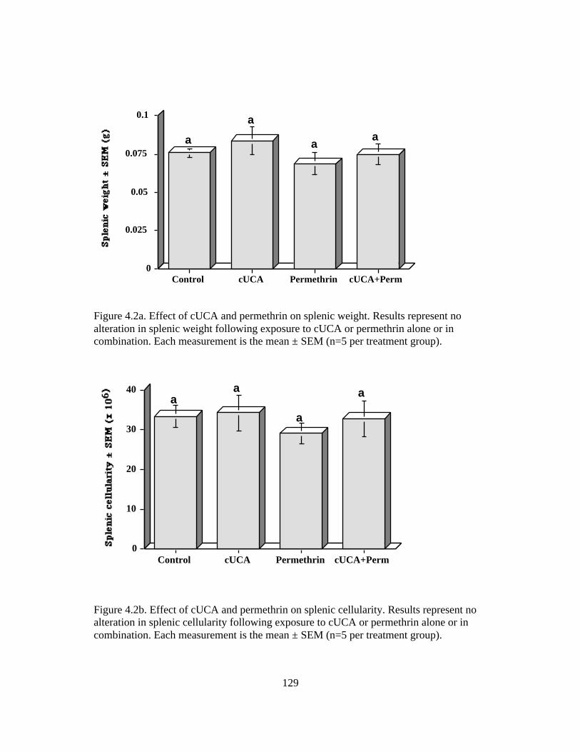
129
0
0.025
0.05
0.075
0.1
Control cUCA Permethrin cUCA+Perm
aa
a
a
Figure 4.2a. Effect of cUCA and permethrin on splenic weight. Results represent noalteration in splenic weight following exposure to cUCA or permethrin alone or incombination. Each measurement is the mean ± SEM (n=5 per treatment group).
0
10
20
30
40
Control cUCA Permethrin cUCA+Perm
a
a
aa
Figure 4.2b. Effect of cUCA and permethrin on splenic cellularity. Results represent noalteration in splenic cellularity following exposure to cUCA or permethrin alone or incombination. Each measurement is the mean ± SEM (n=5 per treatment group).

130
0
50
100
150
Control cUCA Permethrin cUCA + Perm
aa
bb
Figure 4.3a. Effect of cUCA and permethrin on splenocyte proliferation in femaleC57BL/6N mice. Results represent markedly diminished splenocyte proliferationfollowing exposure to permethrin alone (p=0.001) or in combination with cUCA(p=0.001). Each measurement is the mean ± SEM (n=5 per treatment group).
0
50
100
150
Control cUCA Permethrin cUCA + Perm
a
a
a a
Figure 4.3b. Effect of cUCA and permethrin on thymocyte proliferation in femaleC57BL/6N mice. Results represent no effect of permethrin or cUCA either alone or incombination. Each measurement is the mean ± SEM (n=5 per treatment group).

131
0
20
40
60
80
Control cUCA Permethrin cUCA + Perm
aa
bb
Figure 4.4. Effect of cUCA and permethrin on splenic macrophage production of activeoxygen species (hydrogen peroxide, chemiluminescence assay) in female C57BL/6Nmice. Results represent decreased ability of splenic macrophage H2O2 productionfollowing single exposure to permethrin (p=0.005), or permethrin + cUCA incombination (p=0.04). Each measurement is the mean ± SEM (n=5 per treatment group).

132
0
50
100
150
Control cUCA Permethrin cUCA+Perm
a
a
bb
Figure 4.5. Effect of cUCA and permethrin on splenic B lymphocyte antibody production(PFC assay). Results represent decreased splenocyte antibody production followingexposure to permethrin (p=0.0001) or permethrin and cUCA in combination (p=0.0001).Each measurement is the mean ± SEM (n=5 per treatment group).

133
0
5
10
15
20
25
1 2 3 4 5 6
1 = Control2 = 100 g cUCA x 5 days3 = 15 l permethrin4 = 15 l permethrin + 100 g cUCA x 5 days5 = 25 l permethrin6 = 25 l permethrin + 100 g cUCA x 5 days
a a
bb
b
c
Figure 4.6. Effect of cUCA and permethrin on neurotoxic deaths. Results representincreased incidence of neurotoxic deaths following exposure to 15 µL permethrin(p=0.0001), 25 µL permethrin (p=0.001), or cUCA + permethrin at 15 or 25 µL,respectively (p=0.0001, p=0.001). Each measurement is the mean ± SEM (n=5 pertreatment group).

134
CHAPTER 5: MOLECULAR MECHANISMS OF SUNLIGHT AND
PERMETHRIN-INDUCED ALTERATIONS IN CUTANEOUS IMMUNITY
MR Prater1, BL Blaylock2, RM Gogal Jr., 1 SD Holladay1
Corresponding author:
Dr. M. Renee Prater
Department of Biomedical Sciences and Pathobiology
Virginia-Maryland Regional College of Veterinary Medicine
Virginia Polytechnic Institute and State University
Blacksburg, VA 24061-0442, USA
Phone: (540) 231-2275
FAX: (540) 231-6033
Electronic mail: [email protected]
1Department of Biomedical Sciences and Pathobiology
Virginia-Maryland Regional College of Veterinary Medicine
Virginia Polytechnic Institute and State University
Blacksburg, VA 24061-0442, USA
2College of Pharmacy and Health Sciences
University of Louisiana at Monroe
Monroe, LA 71209, USA

135
Running title: Permethrin and cis-urocanic acid diminish contact hypersensitivity
Keywords: permethrin, cis-urocanic acid, pesticide, Langerhans cells, antigen
presentation, IFNγ, TNFα, CD1a, immunomodulation

136
5A. ABSTRACT
cUCA or UVB exposure has been shown to cause diminished cutaneous contact
hypersensitivity (CH) and induces systemic tolerance (increased suppressor T cells) in
mice. Permethrin is also a known CH inhibitor, but the molecular mechanisms are
currently poorly understood. CH was evaluated in an immunosensitive strain
(C57BL/6N), an immunoresistant strain (SvImJ), a strain developed from C57BL/6N
mice but genetically altered at both the TNFαp55R and p75R, a strain developed from
C57BL/6N but genetically deleted at the IFNγ locus, and a strain genetically equivalent
to SvImJ strain genetically deleted at the CD1a locus. C57BL/6N had diminished CH
following 5-day exposure to ID cUCA or single exposure to topical permethrin, and this
effect was exacerbated with concurrent exposure to both chemicals. CH in SvImJ was
both UVB- and permethrin-resistant relative to C57BL/6N mice, as 5-day cUCA or
single exposure to permethrin did not diminish CH, nor did concurrent exposure to cUCA
and permethrin. Mice deleted at both TNFαR loci displayed similar but somewhat
blunted diminished CH responses to cUCA or permethrin. This trend became significant
with combined chemical exposure, suggesting that TNFα may be involved in the
immunomodulatory effects of cUCA and permethrin. IFNγ knockout mice displayed
similar diminished CH responses to cUCA or permethrin alone. Unlike C57BL/6N mice,
the IFNγ knockout mice did not show further reduction in CH with combined chemical
exposure (Yamawaki et al. 1997). These results suggest that this cytokine may be
required for the more than additive depression of CH caused by cUCA + permethrin.
CD1a knockout mice were affected similarly to the SvImJ background strain, except that
permethrin caused a greater decrease in CH in the knockout mouse. These results
137
suggest that permethrin may affect antigen processing/presentation of events required for
CH.

138
5B. INTRODUCTION
Modulation of skin immunity, as may occur following exposure to sunlight or
topical insecticides, may result in a variety of persistent advantageous or deleterious
cutaneous effects such as eliciting or moderating cutaneous allergic disease, diminishing
allograft rejection or lessening graft-versus-host disease. Decreasing contact
hypersensitivity (CH) responses via chemical modulation of the immune system has been
utilized for improvement or treatment of systemic immune-mediated disorders such as
inflammatory bowel disease, where there is an imbalance between proinflammatory
(Th1) and antiinflammatory (Th2) cytokines. The process of oral tolerance induction of
orally-administered inflammatory bowel proteins results in increased transforming
growth factor –beta1 and 2 (TGFβ1 and 2), and decreased interferon-gamma (IFNγ)
serum levels by suppressor T lymphocytes, which results in diminished clinical signs
(Han et al. 2000).
Decreased skin immunity may more commonly have deleterious effects, such as
contributing to increased risk of infectious or neoplastic disease. Tumor-derived factors
that are produced by some skin cancers (such as human melanoma) actively suppress the
cutaneous immune environment by inhibiting differentiation and function of dendritic
cells (DC). As a result, transformed cells may escape immunosurveillance, promoting
development and spread of the tumor (Berthier-Vergnes et al. 2001). Several
immunosensitive tumors have been seen following exposure to UVB or cUCA exposure,
including melanoma, squamous cell carcinoma, basal cell carcinoma, and fibrosarcoma
(DeFabo and Kripke, 1980), which are likely due to decreased presentation of tumor-

139
associated antigens by blunted and immobilized Langerhans cells (LC) in the epidermis
(Geissert et al. 1997).
Chemical exposure is also thought to contribute to depressed CH responses and
lead to increased incidence of development of infectious or neoplastic skin disease. The
effects of the pyrethroid insecticide permethrin on systemic immunity include thymic
atrophy and hypocellularity, inhibition of natural killer cell activity, and inhibition of
lymphocyte proliferative responses to T cell mitogens (Santoni 1997, Diel et al. 1998,
Punareewattanna et al. 2000, 2001). However, though previous exposure to topical
permethrin has been shown to result in highly persistent reduction of the CH response,
the molecular mechanisms are currently poorly characterized.
There is considerable evidence suggesting that sunlight exposure also suppresses
cutaneous immunity, an effect initiated by isomerization of trans to cis urocanic acid in
the stratum corneum. Cutaneous exposure to UVB irradiation or cUCA has been shown
to depress various types of cellular immune responses, including delayed-type
hypersensitivity and antiviral and antitumor immunity (Araneo et al. 1989).
Administration of cUCA to mice 3-5 days prior to hapten sensitization may suppress LC
function and thus CH response to challenge. A cascade of secondary immune mediators
such as prostanoids, TNFα, IFNγ, and a shift from Th1 to Th2 cytokine profile are
thought to contribute to altered cell growth, natural killer cell and cytotoxic T lymphocyte
activation, and cutaneous immune competence (Jaksic et al. 1995, Phipps et al. 1991,
Streilein, 1993, Skov et al. 1998). Consequently cUCA-induced diminished CH is
thought to be an indirect rather than a direct effect, as in-vitro culture of normal human
epidermal keratinocytes with physiologic doses of cUCA (100 µg/ml) does not affect

140
cytokine production of TNFα, IL-1α or β, IL-6, IL-8, or TGFβ1 and does not affect
development of murine dendritic cells (Redondo et al. 1996, Lappin et al. 1997). Further
evidence suggests that there may be strain variations amongst laboratory rodents, as some
strains such as BALB/c have been shown to be relatively resistant to the deleterious
effects of UVB or cUCA. Other strains such as C3H-HeN and C57BL/6N have markedly
depressed CH following similar levels of UVB or cUCA exposure.
While some of the molecular mechanisms by which cUCA affects cutaneous
immunity have been determined, very little is currently known about potential
mechanisms of action by which permethrin inhibits skin immunity. The effects of
concurrent exposure to sunlight and topical permethrin have not been previously
considered. The present report examines mechanisms involved in CH suppression
following single exposure to permethrin or 5-day exposure to cUCA, or concurrent
exposure to both chemicals simultaneously.

141
5C. MATERIALS AND METHODS
1. MICE. Five-week-old female mice of the following strains were
purchased: C57BL/6N, SvImJ, B6.129S7-IFNγtm1Ts, C;129S-CD1tm1Gru and B6.129S-
TNFRsf1atm1MAKTNFRsf1ntm1MAK. C57BL/6N mice were obtained from Charles River
Laboratories (Portage, MI). All knockout mice and the SvImJ controls for CD1
knockouts were purchased from Jackson Laboratories (Bar Harbor, ME). Mice were
acclimated for one week and maintained in microisolator cages under controlled
conditions of temperature (22±1˚C), humidity (40-60%), and light (12/12-hour
light/dark cycle). Food (irradiated Teklad Global 18% protein rodent diet 2918,
Harlan Teklad, Madison, WI) and water were provided ad libitum. Mice in all
experiments were humanely treated, in accordance with the guidelines of the Virginia
Tech Institutional Animal Care and Use Committee (VT IACUC).
2. PERMETHRIN TREATMENT PROTOCOL. Permethrin was provided
by the US Army Center for Health Promotion and Preventive Medicine (Aberdeen
Proving Ground, MD) from stock purchased immediately before by the Army from
Coulston Industries (Coulston Products, Easton, PA). The permethrin was a 91.6%
pure mixture of 57.7% trans and 42.3% cis permethrin, respectively. Mice were
exposed to a single dose of 15 µL permethrin (approximately 660 mg/kg body
weight) on the shaved interscapular skin, to mimic the most common route of human
exposure (Snodgrass 1992). This dose was previously found to cause persistent
inhibition of CH in mice (unpublished data). Control groups received an identical
volume of vegetable oil (vehicle) in the interscapular space.

142
3. CIS-UROCANIC ACID. Cis-urocanic acid was isomerized under UVB
light from trans-urocanic acid (Sigma, St. Louis, MO), purified as previously
described (Takahashi et al. 1997), and then evaluated for purity by mass
spectroscopy prior to use. Administration of cUCA consisted of a daily
suberythremic intradermal dose of PBS vehicle or 100 µg cUCA dissolved in PBS
(equivalent to approximately 6.6 mg/kg cUCA) for five consecutive days in mice
arbitrarily assigned to treatment or control groups. This dosing schedule was
previously found to cause persistent inhibition of CH in mice (unpublished data).
4. CONTACT HYPERSENSITIVITY. The contact hypersensitivity
response was assayed by quantifying the ear swelling response to chemical
exposure using the method originally published by Asherson and Ptak (1968).
Briefly, mice in all treatment groups were sensitized by topical application in the
interscapular region of 25 µL 2% (w/v) 4-ethoxymethylene-2-phenyl–2-oxazolin-5-
one (oxazolone) dissolved in a 4:1 reagent-grade acetone/olive oil mixture 48 hours
following permethrin application or administration of the final dose of cUCA. Five
days after oxazolone sensitization, the mice were challenged with 10 µL of 0.5%
oxazolone to both the dorsal and ventral surfaces of the right ear. The acetone:olive
oil vehicle was applied to the left ear as a control for vehicle-induced irritancy.
Immunological response to permethrin or cis urocanic acid were assessed at 24
hours after challenge. The response was quantified as the difference in the thickness
of the challenged ear before and after challenge (Oditest 313 Series micrometer;
The Dyer Company, Lancaster, PA). The thickness of the left ear (vehicle-treated)

143
was also determined before and after treatment with the acetone:olive oil vehicle to
control for vehicle-induced irritation and swelling.
5. STATISTICAL ANALYSIS. Data were expressed as arithmetic mean ±
SEM. A one-way analysis of variance was completed. A randomized complete block
design was used for error control. Dunnett’s post-hoc t-test was used to establish
significant differences in treatment groups versus controls. Results described as
different in this paper indicate significantly different at p< 0.05.

144
5D. RESULTS
1. CONTACT HYPERSENSITIVITY IN C57BL/6N AND SvImJ MICE
EXPOSED TO cUCA AND PERMETHRIN. Results of this study supported
previous suggestions that C57BL/6N is a UVB immunologically sensitive strain of
mice. Following 5-day intradermal cUCA exposure, C57BL/N mice had a
significantly diminished ability to respond to oxazolone challenge in a CH assay
(n=5 per treatment group, p=0.0018, Fig. 5.1). A single administration of topical
permethrin caused a similar dramatic decrease in CH (n=5 per treatment group,
p=0.0006, Fig. 5.1), and this effect was significantly increased as compared to either
chemical alone when both chemicals were administered concurrently (n=5 per
treatment group, p=0.0023, Fig. 5.1).
Contact hypersensitivity responses in untreated mice from the SvImJ strain
were not different from C57BL/6N controls, however the two strains differed
dramatically in their CH response following exposure to cUCA or permethrin. SvImJ
mice exposed to 5-day ID cUCA showed no decrease in CH (n=5 per treatment
group, Fig. 5.1). Single dose permethrin (n=5 per treatment group, p=0.6784, Fig.
5.1), or concurrent administration of both chemicals (n=5 per treatment group,
p=0.7294, Fig. 5.1) likewise caused no alteration in CH, suggesting that SvImJ mice
may be relatively resistant to the cutaneous immunomodulatory effects of these
chemicals.
2. CONTACT HYPERSENSITIVITY IN C57BL/6N CONTROL AND
TNFαR KNOCKOUT MICE EXPOSED TO cUCA AND PERMETHRIN.

145
C57BL/6N mice that were genetically deleted at both the p55 and p75 TNFα receptor
loci demonstrated control CH responses that were similar to C57BL/6N controls. The
wild-type and knockout mice displayed approximately equal decreases in CH
following cUCA or permethrin exposure (n= 5 per treatment group, p=0.0025 and
p=0.003, respectively, Fig. 5.2). A non-significant trend toward reduced ear swelling
was present in both groups of knockout mice that received a single chemical
exposure. TNFαR knockout mice exposed to both cUCA and permethrin displayed
significantly less reduced ear swelling as compared to wild-type mice, suggesting a
role for TNFα in the severe inhibition of CH caused by the chemical mixture.
3. CONTACT HYPERSENSITIVITY IN C57BL/6N CONTROL AND IFNγ
KNOCKOUT MICE EXPOSED TO cUCA AND PERMETHIRN. Vehicle-exposed
C57BL/6N mice genetically deleted at the IFNγ locus demonstrated significantly
diminished CH responses as compared to C57BL/6N controls (n=5 per treatment
group, p=0.0001, Fig. 5.3). This observation indicates that IFNγ may be required to
mount a control-level CH response. The wild-type and IFNγ knockout mice showed
approximately equal depression of CH after treatment with either cUCA or
permethrin alone, suggesting that IFNγ was not required for depression of CH to the
level observed in these mice. When IFNγ knockout mice were co-exposed to cUCA
+ permethrin, no further depression in CH occurred than was observed after exposure
to either chemical alone. In contrast, wild-type C57BL/6N mice exposed to the
chemical mixture displayed considerable additional reduction in ear swelling which
appears to require IFNγ as a mediator. Collectively, these data suggest that
IFΝγ knockout mice: 1) cannot mount a control-level CH response; 2) are able to

146
show diminished CH after exposure to cUCA or permethrin alone that is apparently
IFNγ-independent, but 3) cannot achieve the maximal depression of CH that occurs in
mixture-exposed mice and thus appears to require IFNγ.
4. CONTACT HYPERSENSITIVITY IN C57BL/6N CONTROL AND
CD1a KNOCKOUT MICE EXPOSED TO cUCA AND PERMETHRIN. As
previously observed, the vehicle- or chemical-exposed SvImJ mouse did not display
reduced CH responses. The CD1a knockout mouse of otherwise similar genetic
background to this strain was also largely refractory to the effects of treatment on CH,
with the exception of mice exposed to permethrin only (n=5 per treatment group,
p=0.0001, Fig. 5.4). The reduced ear swelling that occurred in these mice suggests
that permethrin may affect antigen presentation to inhibit the ear swelling response.
For unknown reasons, mice exposed to both cUCA and permethrin did not show the
reduced ear swelling seen in permethrin-only mice.

147
5E. DISCUSSION
Cutaneous exposure to UVB irradiation or topical permethrin has been shown to
depress cellular immune responses, including delayed-type hypersensitivity (Araneo et al.
1989, Punareewattana et al. 2000). Elicitation of CH is biphasic and involves two sets of
antigen-specific Th1 cells: the first response is seen two hours following the antigenic
challenge and results from activation of initiator cells, and the second follows initiator
cell activation and is seen 24-48 hours later due to stimulation of effector cells. cUCA is
thought to prevent the function or recruitment of the initiating cells, which in turn inhibits
the effector phase (Norval et al. 1995).
It has been suggested that genetic variations among laboratory rodent strains may
cause varied susceptibilities to the innate and adaptive cell-mediated immune suppressive
effects of sunlight exposure. Immunosensitive strains include C57BL/6N and C3H-HeN,
whereas immunoresistant strains include BALB/c mice (Wakeham et al. 2000, Streilein
et al. 1993). Wakeham et al. observed that BALB/c developed an earlier response to
proinflammatory cytokines (IL-12, IFNγ, TNFα, and macrophage chemoattractive
protein-1) in response to bacterial infection and suggested that these cytokines were not
as dramatically affected following UVB exposure. Streilein et al. (1993) identified
genetic mutations at H-2 (TNFα) and LPS loci in the immunoresistant strains and
suggested that UVB-sensitivity is a dominantly inherited trait such that homozygous
mutations at these two loci are necessary to confer UVB resistance. Human studies have
demonstrated similar molecular links to UVB-altered cell-mediated immunity (Streilein
1993). It is likely that these two loci are more important than skin pigmentation in the
molecular mechanisms of cutaneous UVB sensitivity, and increased susceptibility to skin
148
cancer. However, additional study will be necessary to verify more specific mechanisms
to explain the high UVB-susceptible trait (35-45% of individuals) in human beings
(Streilein, 1993). A previous study demonstrated that C57BL/6N mice were highly
sensitive to systemic immune effects caused by cUCA (unpublished data). The present
report extends these findings to show that C57BL/6N mice are also highly sensitive to the
cutaneous immune effects of UVB, and further suggested that SvImJ are a UVB-resistant
strain.
Several molecular mediators have been implicated as possible contributors to the
multifactorial effects of chemically-induced CH suppression, including prostaglandins,
interleukin-10, and TNFα (Norval 1995). The prostanoid system is thought to be at least
partially involved in this effect, as cUCA synergized with histamine causes increased
prostaglandin E2 (PGE2) production by keratinocytes, which increases release of TNFα
and causes diminishment of CH responses, activation of suppressor T lymphocytes and
decreased Th1 production of IL-2 and IFNγ (Phipps et al. 1991, Jaksic et al. 1995). It is
thought that TGFβ2 also causes antigen presenting cells to upregulate TNFα production,
which biases the T cells in a direction that interferes with acquisition of antigen and
hapten transfer to the draining lymph node, thus suppressing induction of CH (Hecker et
al. 1999). This Th1 unresponsiveness is long-lasting (greater than 16 days) and not due to
cell death, but rather the result of an interference in the capacity of LC to deliver the
required activation costimulatory signal to the Th1 cells (Simon et al. 1991).
Consequently, the Th1/Th2 ratio shifts in favor of a Th2 response, which diminishes cell-
mediated immunity. Previous studies suggested that TNFα secretion by epidermal
keratinocytes reaches maximal levels just 6 hr after UVB irradiation, confers locally

149
depressed CH response, (Streilein, 1993, Skov et al. 1998) which results in
morphological changes in LC, migration of follicular dendritic cell precursors into
splenic follicles, interference with antigen processing and presentation to Th1 cells, and
altered activation of Th1 cell-driven inflammatory immune reactions in mice (Schmitt
and Ullrich 2000, Pasparakis et al. 2000, Amerio et al. 2001). However, recent reports
have suggested other molecular mediators besides TNFα may contribute to diminished
CH responses to oxazolone challenges, and that the dramatic cutaneous immune
suppression seen following 200 µg or less cUCA may be the result of multifactorial
chemical mediator effects (Norval et al. 1995).
The present study evaluating changes in CH responses in mice that were
genetically altered at both p55 and p75 TNFαR loci suggested that local
immunomodulatory effects caused by cUCA and permethrin may at least in part be
mediated by changes in TNFα production by epidermal cells. Both type I and type II
TNFα receptors (p55TNFαR and p75TNFαR) are required for full expression of the
delayed-type hypersensitivity reaction, as dendritic cells of mice lacking the p55TNFαR
had a defect in antigen uptake due to blunting of cytoplasmic processes and disruption of
vimentin microtubules in the cytoskeleton, but showed normal LC migration to local
lymph nodes (Goettsch et al. 1993, Bacci et al. 1996). Conversely, those mice with
dendritic cells demonstrating p75TNFαR deficiency had diminished LC migration to the
local lymph nodes after exposure to contact allergen, without hindrance of antigen uptake
itself (Becke et al. 2001).
IFNγ is thought to be a critical immunoprotective Th1 cytokine necessary for the
production of normal UVB-induced erythema, perhaps due to IFNγ’s effect of releasing

150
histamine from mast cells (Reeve et al. 1999). In-vivo UVB or cUCA suppresses IFNγ,
which diminishes CH by interfering with antigen processing and presentation (Bacci et
al. 1996, Laihia et al. 1996). IFNγ is thought to play a part in immunoprotection from the
adverse effects of UVB or cUCA on CH responses (Reeve et al. 1999) by enhancing IL-
12 production and diminishing IL-10 release by macrophages. IFNγ is an essential
regulatory protein secreted by activated CD4+ T cells that acts as a negative feedback
loop to eliminate by apoptosis excess activated CD4+ T cells in an attempt to maintain
CD4+ T cell homeostasis during an immune response. Therefore, activated CD4+ T cells
from IFNγ knockout mice that have clonally expanded following immune activation do
not undergo IL-2-mediated apoptosis, thus predisposing these animals to the development
of autoimmune disease (Dalton et al. 2000). The results of the present study seem to
support the theory that IFNγ is an important contributor to the development of CH in
murine skin, as the IFNγ knockout strain of mice demonstrated lower control (untreated)
CH levels as compared to the genetically similar C57BL/6N mice.
Reports of development of cutaneous nodules rich in LC (CD1a+, S100+, HLA-
DR+) following cutaneous treatment with permethrin for scabies (Hashimoto et al. 2000)
suggest that permethrin may inhibit the migration of LC to local lymph nodes. The
present study found that the CD1a knockout strain of mice had diminished CH following
exposure to permethrin. Following a single exposure of 4 minimal erythemal dose in
human beings, contact hypersensitivity was significantly reduced. While numbers of
CD1a+ LC were significantly diminished and remaining LC were rendered ineffective
functionally, numbers of CD1a- macrophages were increased in the skin (LeVee et al.
1997). Since the CD1a is involved in presentation of lipid and glycolipid antigens to a

151
variety of effector T cells (Sugita), deletion of this gene may contribute in small part to a
defective CH response following exposure to permethrin. TGFβ1 is thought to work
together with IL-4 in the development and differentiation of CD1+ LC from
hematopoietic progenitor cells in a normal inflammatory response (Caux et al. 1999).
Human epidermal CD1a+ Langerhans cells that have been previously exposed to UVB
irradiation activate regulatory T lymphocytes, which may help to dampen responses to
endogenous antigens that are pathogenic in autoimmune disorders. However, these same
mechanisms may also predispose an individual to increased risk of development of UV-
induced skin cancers (Baadsgaard et al. 1990). Exposure of experimental rodents to
UVB results in significantly diminished release of IL-2 and IFNγ, with concomitant
increased release of IL-4 (Araneo et al. 1989). Epidermal keratinocytes exposed to
inflammatory mediators promote induction of adhesion molecules such as intercellular
adhesion molecule-1 (ICAM-1), which results in the retention of T lymphocytes in
cutaneous immunohomeostasis (Mitra et al. 1993). However, exposure of epidermal (LC)
cells to UVB irradiation inhibits their ability to activate T cells through decreased
expression of ICAM-1 and resulting diminished T cell activation (De Luca et al. 1997).
Cis-urocanic acid has also been shown to inhibit fibroblast synthesis of cAMP, an
important second messenger involved in cutaneous immune responses such as IL-2
receptor expression (Johnson and Smith 1990, Bouscarel et al. 1998).
Repeated application of low-dose topical cUCA has also been shown to alter
murine contact hypersensitivity by decreased IL-1α release and increased IL-10
production by keratinocytes and mast cells in the epidermis (Rasanen et al. 1987,
Kurimoto et al. 2001). Increased IL-10 is thought to mediate systemic tolerance by

152
upregulation of suppressor T lymphocytes (Alard et al. 2001). Further support of IL-10-
mediated effects on cutaneous immunity was demonstrated with retroviral-mediated gene
transfer of viral IL-10, which mimics a switch from Th1 to Th2 cytokine profile and
results in prolonged allograft survival by altering antigen presenting cell function (Qin et
al. 2001). Murine recombinant IL-10 (200 ng) was injected intradermally on abdominal
skin of a UVB-sensitive strain (C3H-HeN) and a UVB-resistant strain (BALB/c) of mice.
This resulted in promotion of hapten-specific systemic tolerance to 2,4-
dinitrofluorobenzene (DNFB) within 30 minutes to 3 days following IL-10 injection in
both strains (Niizeki and Streilein 1997). Parallel experiments with UVB exposure in
these strains of mice resulted in suppression of the cutaneous CH response only in the
UVB-sensitive C3H-HeN strain (Niizeki and Streilein 1997). These studies suggest that
tolerance induction is a systemic upregulation of regulatory T lymphocytes, mediated in
part by IL-10, whereas the CH response is a local phenomenon, perhaps with divergent
molecular mechanisms. Further experiments evaluating gene expression of cytokines
potentially involved in the CH response in cultured murine keratinocytes may shed
additional light on the molecular mechanisms behind diminished contact hypersensitivity
following dermal cUCA or permethrin exposure. Additionally, further study of in-vivo
interventions with cytokines involved in LC differentiation and lymphocyte activation,
such as TCR/MHC II interaction and costimulatory signals involved in the CD28/CD80
or CD86 interactions would help to more clearly define the multifactorial effects of
chemical exposure on skin immunity.
5F. ACKNOWLEDGMENT. Supported by NIH RO1 ES09642-02

153
5G. REFERENCES
Abou-Donia MB, Wilmarth KR, Jensen KF, Oehme FW, Kurt TL. Neurotoxicity
resulting from coexposure to pyridostigmine bromide, DEET, and permethrin:
implications of Gulf War chemical exposures. J Toxicol Environ Health 48:35-56, 1996.
Alard P, Kurimoto I, Niizeki H, Doherty JM, Streilein JW. Hapten-specific tolerance
induced by acute, low-dose ultraviolet B radiation of skin requires mast cell
degranulation. Eur J Immunol 31:1736-1746, 2001.
Amerio P, Toto P, Feliciani C, Suzuki H, Shivji G, Wang B, Sauder DN. Rethinking the
role of tumour necrosis factor-alpha in ultraviolet (UV) B-induced immunosuppression:
altered immune response in UV-irradiated TNFR1R2 gene-targeted mutant mice. Br J
Dermatol 144:952-957, 2001.
Anadon A, Martinez-Larranaga MR, Diaz MJ, Bringas P. Toxicokinetics of permethrin
in the rat. Toxicol Appl Pharmacol 110:1-8, 1991.
Araneo BA, Dowell TG, Moon HB, Daynes RA. Regulation of murine lymphokine
production in vivo. Ultraviolet radiation exposure depresses IL-2 and enhances IL-4
production by T cells through and IL-1-dependent mechanism. J Immunol 143:1737-
1744, 1989.

154
Asherson G, Ptak W. Contact and delayed hypersensitivity in the mouse. I. Active
sensitization and passive transfer. Immunol 15:405-416, 1968.
Baadsgaard O, Salvo B, Mannie A. Dass B, Fox DA, Cooper KD. In vivo ultraviolet-
exposed human epidermal cells activate T suppressor cell pathways that involve
CD4+CD45RA+ suppressor-inducer T cells. J Immunol 145:2854-2861, 1990.
Bacci ST, Nakamura T, Streilein JW. Failed antigen presentation after UVB radiation
correlates with modifications of Langerhans cell cytoskeleton. J Invest Dermatol
107:838-843, 1996.
Bandyopadhyay R. Inhibition of acetylcoline esterase by permethrin and its reversion by
acetylthiocholine. Indian J Exp Biol 20:488-491, 1982.
Becke FM, Hehlgans T, Brockhoff G, Mannel DN. Development of allergic contact
dermatitis requires activation of both tumor necrosis factor-receptors. Eur Cytokine
Netw 12:45-50, 2001.
Beissert S, Mohammad T, Torri H, Lonati A, Yan Z, Morrison H, Granstein RD.
Regulation of tumor antigen presentation by urocanic acid. J Immunol 159:92-96, 1997.
Berthier-Vergnes O, Gaucherand M, Peguet-Navarro J, Plouet J, Pageaux JF, Schmitt D,
Staquet MJ. Human melanoma cells inhibit the earliest differentiation steps of human

155
Langerhans cell precursors but failed to affect the functional maturation of epidermal
Langerhans cells. Br J Cancer 85:1944-1951, 2001.
Bouscarel B, Noonan F, Ceryak S, Gettys TW, Phillips TM, DeFabo EC. Regulation of
stimulated cyclic AMP synthesis by urocanic acid. Photochem Photobiol 67:324-331,
1998.
Caux C, Massacrier C, Dubois B, Valladeau J, Dezutter-Dambuyant C, Durand I, Schmitt
D, Saeland S. Respective involvement of TGF-beta and IL-4 in the development of
Langerhans cells and non-Langerhans dendritic cells from CD34+ progenitors. J Leukoc
Biol 66:781-791, 1999.
Cavaliere MJ, Maeda MYS, Shih LWS, Puga FR. Changes in myelinated nerve fibers
and skeletal muscle of rats exposed to high doses of permethrin. Biomedical and
Environmental Sciences 3:139-145, 1990.
DeFabo EC, Kripke ML. Wavelength dependence and dose-rate independence of UV
radiation-induced immunologic unresponsiveness of mice to a UV-induced fibrosarcoma.
32:183-188, 1980.
DeLuca DJ, Trefzer U, Tubesing KA, Elmets CA. ICAM-1 mRNA levels and relative
transcription rates are decreased by UV irradiation of monocytes. Photochem Photobiol
65:609-615, 1997.

156
Diel F, Detscher M, Borck H, Schrimpf D, Diel E, Hoppe HW. Effects of permethrin on
human basophils and lymphocytes in vitro. Inflammation Research 47:S11-12, 1998.
Eells JT, Bandettini PA, Holman PA, Propp JM. Pyrethroid insecticide-induced
alterations in mammalian synaptic membrane potential. J Pharmacol Exp Ther 262:1173-
1181, 1992.
Gassner B, Wuthrich A, Scholtysik G, Solioz M. The pyrethroids permethrin and
cyhalothrin are potent inhibitors of the mitochondrial complex I. J Pharmacol Exp Ther
281:855-860, 1997.
Goettsch W, Garssen J, deGruijl FR, Loveren HV. UV-B and the immune system.
Thymus 21:93-114, 1993.
Han Y, Weksler-Zangen S, Ben-Horin S, Dimen J, Sauter B, Rabbani E, Engelhardt D,
Chowdhury NR, Chowdhury JR, Goldin E. Treatment of experimental colitis by oral
tolerance induction: a central role for suppressor lymphocytes. Am J Gastroenterol
95:966-973, 2000.
Hashimoto K, Fujiwara K, Punwaney J, DeGregorio F, Bostrom P, El-Hoshy K, Aronson PJ,
Schoenfeld RJ. Post-scabetic nodules: a lymphohistiocytic reaction rich in indeterminate cells.
J Dermatol 27:181-194, 2000.

157
Hecker KH, Niizeki H, Streilein JW. Distinct roles for transforming growth factor-beta-2 and
tumoour necrosis factor-alpha in immune deviation elicited by hapten-derivatized antigen-
presenting cells. Immunol 96:372-380, 1999.
Hudson PM, Tilson HA, Chen PH, Hong JS. Neurobehavioral effects of permethrin are
associated with alterations in regional levels of biogenic amine metabolites and amino acid
neurotransmitters. Neurotoxicol 7:143-154, 1986.
Jaksic A, Finlay-Jones JJ, Watson CJ, Spencer LK, Santucci I, Hart PH. Cis-urocanic acid
synergizes with histamine for increased PGE2 production by human keratinocytes: link to
indomethacin-inhibitable UVB-induced immunosuppression. Photochem Photobiol 61:303-309,
1995.
Johnson KW, Smith KA. cAMP regulation of IL-2 receptor expression. J Immunol 145:1144-
1151, 1990.
Kurimoto AP, Niizeki H, Doherty JM, Streilein JW. Hapten-specific tolerance induced
by acute, low-dose ultraviolet B radiation of skin requires mast cell degranulation. Eur J
Immunol 31:1736-1746, 2001.

158
Laihia JK, Uksila J, Luhtala M, Jansen CT. Expression of CD80 (B7/BB-1) and CD28 in
human white blood cells treated with urocanic acid. Arch Dermatol Res 288:570-574,
1996.
Lappin MB, Weiss JM, Schopf E, Norval M, Simon JC. Physiologic doses of urocanic
acid do not alter the allostimulatory function or the development of murine dendritic cells
in vitro. Photodermatol Photoimmunol Photomed 13:163-168, 1997.
Lees G. Effects of pyrethroid molecules on rat nerves in-vitro: potential to reverse
temperature-sensitive conduction block of demyelinated peripheral axons. Br J
Pharmacol 123:487-496, 1998.
LeVee GJ, Oberhelman L, Anderson T, Koren H, Cooper KD. UVAII exposure of
human skin results in decreased immunization capacity, increased induction of tolerance
and a unique pattern of epidermal antigen-presenting cell alteration. Photochem
Photobiol 65:622-629, 1997.
McDaniel KL, Moser VC. Utility of a neurobehavioral screening battery for
differentiating the effects of two pyrethroids, permethrin and cypermethrin. Neurotoxicol
Teratol 15:71-83, 1993.

159
Mitra JS, Shimizu Y, Nickoloff BJ. Histamine and cis-urocanic acid augment tumor
necrosis factor-alpha mediated induction of keratinocyte intercellular adhesion molecule-
1 expression. J Cell Physiol 156:348-357, 1993.
Niizeki H, Streilein JW. Hapten-specific tolerance induced by acute, low-dose ultraviolet
B radiation of skin is mediated via interleukin-10. J Invest Dermatol 109:25-30, 1997.
Norval M, Gibbs NK, Gilmour J. The role of urocanic acid in UV-induced
immunosuppression: recent advances (1992-1994). Photochem Photobiol 62:209-217,
1995.
Pasparakis M, Kousteni S, Peschon J, Kollias G. Tumor necrosis factor and the p55TNF
receptor are required for optimal development of the marginal sinus and for migration of
follicular dendritic cell precursors into splenic follicles. Cell Immunol 201:33-41, 2000.
Phipps RP, Stein SH, Roper RL. A new view of prostaglandin E regulation of the
immune response. Immunol Today 12:349-352, 1991.
Punareewattana K, Smith BJ, Blaylock BL, Robertson JR, Gogal RM Jr., Prater MR,
Longstreth J, Snodgrass HL, Holladay SD. Topical permethrin exposure causes
thymic atrophy and persistent inhibition of the contact hypersensitivity response in
C57Bl/6 mice. Int J Toxicol 19:383-389, 2000.

160
Punareewattana K, Smith BJ, Blaylock BL, Gogal RM Jr., Prater MR, Longstreth J,
Holladay SD. Topical permethrin exposure inhibits antibody production and macrophage
function in C57Bl/6N mice. Food Chem Toxicol 39:133-139, 2001.
Qin L, Ding Y, Tahara H, Bromberg JS. Viral IL-10-induced immunosuppression
requires Th2 cytokines and impairs APC function within the allograft. J Immunol
166:2385-2393, 2001.
Rasanen L, Jansen CT, Reunala T, Morrison H. Stereospecific inhibition of human
epidermal cell interleukin-1 secretion and HLA-DR expression by cis-urocanic acid.
Photodermatol 4:182-186, 1987.
Redondo P, Garcia-Foncillas J, Cuevillas F, Espana A, Quintanilla E. Effects of low
concentrations of cis- and trans-urocanic acid on cytokine elaboration by keratinocytes.
Photodermatol Photoimmunol Photomed 12:237-243, 1996.
Reeve VE, Bosnic M, Nisimura N. Interferon-γ is involved in photoimmunoprotection
by UVA (320-400 nm) radiation in mice. J Invest Dermatol 112:945-950, 1999.
Rose GP, Dewar AJ. Intoxication with four synthetic pyrethroids fails to show any
correlation between neuromuscular dysfunction and neurobiochemical abnormalities in
rats. Arch Toxicol 53:297-316, 1983.

161
Santoni G, Canalamessa F, Mazzucca L, Romagnoli S, Piccoli M. Prenatal exposure to
cypermethrin modulates rat NK cell cytotoxic functions. Toxicol 120:231-242, 1997.
Schmitt DA, Ullrich SE. Exposure to ultraviolet radiation causes dendritic
cells/macrophages to secrete immune-suppressive IL-12p40 homodimers. J Immunol
165:3162-3167, 2000.
Simon JC, Tigelaar RE, Bergstresser PR, Edelbaum D, Cruz PD Jr. Ultraviolet B
radiation converts Langerhans cells from immunogenic to tolerogenic antigen-
presenting cells. Induction of specific clonal anergy in CD4+ T helper 1 cells. J
Immunol 146:485-491, 1991.
Skov L, Hansen H. Allen M, Villadsen L, Norval M, Barker JNWN, Simon J,
Baadsgaard O. Contrasting effects of ultraviolet A1 and ultraviolet B excposure on
the induction of tumour necrosis factor-a in human skin. Br J Dermatol 138:216-220,
1998.
Streilein JW. Sunlight and skin-associated lymphoid tissues (SALT): if UVB is the
trigger and TNFα is the mediator, what is the message? J Invest Dermatol 100:47S-
52S, 1993.
Sugita M, van der Wel N, Rogers RA, Peters PJ, Brenner MB. CD1c molecules
broadly survey the endocytic system. Proc Natl Acad Sci USA. 97:8445-8450, 2000.

162
Takahashi M, Tezuka T. Quantitative analysis of histidine and cis and trans isomers
of urocanic acid by high-performance liquid chromatography: a new assay method
and its application. J Chromatography B 688:197-203, 1997.
Vaccari A, Ruiu S, Mocci I, Ssaba P. Selected pyrethroid insecticides stimulate
glutamate uptake in brain synaptic vesicles. NeuroReport 9:3519-3523, 1998.
Wakeham J, Wang J, Xing Z. Genetically determined disparate innate and adaptive cell-
mediated responses to pulmonary Mycobacterium bovis BCG infection in C57BL/6 and
BALB/c mice. Infection and Immunity 68:6946-6953, 2000.
Yamawaki M, Katiyar SK, Anderson CY, Tubesing KA, Mukhtar H, Elmets CA. Genetic
variation in low-dose UV-induced suppression of contact hypersensitivity and in the skin
photocarcinogenesis response. J Invest Dermatol 109:716-721, 1997.

163
0
50
100
150
200
250
Control cUCA Permethrin cUCA+Perm
SvImJC57BL/6N
aa a
a
a
b
b
c
Figure 5.1. Contact hypersensitivity response to five-day ID cUCA, single topicaladministration of permethrin, or cUCA + permethrin in C57BL/6N and SvImJ mousestrains. Results represent change in ear thickness following oxazolone challenge 48 hrafter final chemical exposure. Each measurement is the mean ± SEM (n=5 per treatmentgroup); groups that display different letter are statistically significant, p<0.05.

164
0
50
100
150
200
250
Control cUCA Permethrin cUCA+Perm
TNF RC57BL/6N
aa
b
b
a
b
b
c
Figure 5.2. Contact hypersensitivity response to five-day ID cUCA, single topicaladministration of permethrin, or cUCA + permethrin in C57BL/6N mice and C57BL/6Nmice that were genetically deleted at both the p55 and p75TNFα receptors. Resultsrepresent change in ear thickness following oxazolone challenge 48 hr after finalchemical exposure. Each measurement is the mean ± SEM (n=5 per treatment group);groups that display different letter are statistically significant, p<0.05.

165
0
50
100
150
200
250
Control cUCA Permethrin cUCA+Perm
IFNC57BL/6N
a
b
c c c
c
c
d
Figure 5.3. Contact hypersensitivity response to five-day ID cUCA, single topicaladministration of permethrin, or cUCA + permethrin in C57BL/6N mice and C57BL/6Nmice that were genetically deleted at the IFNγ locus. Results represent change in earthickness following oxazolone challenge 48 hr after final chemical exposure. Eachmeasurement is the mean ± SEM (n=5 per treatment group); groups that display differentletter are statistically significant, p<0.05. These results suggest that IFNγ release isneeded for a control-level CH response, however the diminished CH caused by eithercUCA or permethrin alone can be attained by IFNγ knockout mice, thus IFNγ is notrequired. The results of CH in combined-exposure mice suggest that these mice cannotrespond at a level beyond the effect seen with exposure to either cUCA or permethrinalone.

166
0
50
100
150
200
250
Control cUCA Permethrin cUCA+Perm
CD1aSvImJ
a
a a
b
aa
a
a
Figure 5.4. Contact hypersensitivity response to five-day ID cUCA, single topicaladministration of permethrin, or cUCA + permethrin in SvImJ mice and mice that weregenetically deleted at CD1a locus. Results represent change in ear thickness followingoxazolone challenge 48 hr after final chemical exposure. Each measurement is the mean± SEM (n=5 per treatment group); groups that display different letter are statisticallysignificant, p<0.05.

167
CHAPTER 6: CONCLUSIONS
Single dose exposure to topical permethrin caused a variety of immunological
changes in mice, with the thymus being more profoundly affected than the spleen.
Thymic atrophy and hypocellularity appeared to result, at least in part, from increased
thymocyte apoptosis and from antiproliferative chemical effects on thymocytes.
Antiproliferative effects in splenic T lymphocytes and reduced cell counts were also
seen following a single exposure to permethrin, as well as diminished antibody
production. Decreased ability to produce antibody has been demonstrated to have a
high concordance (predictive values) for immunosuppression in mice. Beyond
immunosuppression, recent data suggest possible increased incidence of allergic
disease associated with chronic inhalation exposure to permethrin, resulting from
increased histamine release by basophils and decreased IFNγ and IL-4 release by
peripheral lymphocytes caused by permethrin. These collective reports verify the
need for additional examination of the immunomodulatory effects of permethrin.
In contrast, a single exposure to intradermal cUCA produced few detectable
changes in the selected immune parameters or immune function assays aside from a
decrease in splenic macrophage phagocytosis. Thymic atrophy could not be produced
after single dose cUCA or after daily dosing for 5 consecutive days, but was dramatic
after four weeks dosing. This effect would not appear to be corticosteroid (stress) -
mediated, in that the most sensitive CD4+8+ thymocytes were least affected by cUCA.
Thymic atrophy was also more severe in C57Bl/6N mice than in C3H-HeN mice,
suggesting underlying differences in sensitivity of these mouse strains to UVB
irradiation. Finally, mice dosed daily for five days with cUCA did not show altered T

168
cell cytolytic ability or depressed antibody production, suggesting that these mice are
likely to respond at a control level to immunologic challenge.
However, when administered concurrently, cUCA and permethrin resulted in
alteration of several immune parameters, including ability of splenic B lymphocytes to
produce specific antibody and thymic weight, in which an additive effect of the chemicals
was evident. These results may suggest different mechanisms by which these compounds
affect precursor T cells (thymocytes) in the thymus, as compared to functionally mature T
cells or other immune cell targets present in the spleen. Cutaneous immunity was also
affected by 5-day exposure to cUCA, permethrin, or concurrent exposure to both
chemicals, and additive effects were seen in the immunosensitive C57BL/6N strain.
Several potential molecular mediators involved in the development of contact
hypersensitivity in mice were identified with the use of genetically altered strains of
mice. These included TNFα, IFNγ, and CD1a. The present studies using gene knockout
mice provided data that suggest TNFα is a required cytokine in the inhibition of contact
hypersensitivity caused by either cUCA or permethrin. IFNγ was also identified as an
important cytokine for mounting a control-level CH response, which appeared to be
required for the severe depression of CH caused by co-exposure to cUCA and
permethrin. Finally, permethrin is a well-characterized neurotoxicant that has been
widely used because of its combination of selective efficacy as an insecticide and
minimal described adverse effects in mammals. The observation that permethrin caused
neurotoxicity in some of the present mice is not novel; however the finding that a
chromophore induced by UV light (cUCA) increased the lethality of permethrin,

169
apparently by increasing its neurotoxic effects, is new and potentially important
information.
The present immunotoxocity and neurotoxicity data are clearly preliminary but
may suggest that human beings, and especially children, treated with relatively high
doses of topical permethrin for mites or lice, should avoid excessive sunlight exposure
during the immediate post-treatment period.

170
0
2.5
5
7.5
10
Control 5 uL Perm 10 uL Perm 15 uL Perm
Appendix I. Effect of a single topical exposure to permethrin on splenic macrophagephagocytosis in female C57BL/6N mice. Results are expressed as mean ± SEM, n=5 pertreatment group.

171
0
5
10
15
20
25
Control 5 uL Perm 10 uL Perm 15 uL Perm
Appendix II. Effect of a single topical exposure to permethrin on splenic macrophageability to produce active oxygen species (hydrogen peroxide, chemiluminescence assay)in female C57BL/6N mice. Results are expressed as mean ± SEM, n=5 per treatmentgroup.

172
0
10
20
30
Control 15 uL Perm
Appendix III. Effect of a single topical exposure to permethrin on splenic T lymphocytecytolytic activity (51Cr release assay) in female C57BL/6N mice. Results are expressedas mean ± SEM, n=5 per treatment group.

173
0
0.25
0.5
0.75
1
1.25
Control 15 uL Perm
Appendix IV. Effect of a single topical exposure to permethrin on thymic proliferativeresponse to Concanavalin A in female C57BL/6N mice. Results are expressed as mean ±SEM, n=5 per treatment group.

174
0
0.025
0.05
0.075
0.1
Control 5 ug cUCA 50 ug cUCA 100 ug cUCA 250 ug cUCA
Appendix V. Effect of a single exposure to cUCA on thymic weight in femaleC57BL/6N mice. Results are expressed as mean ± SEM, n=5 per treatment group.

175
0
50
100
150
Control 5 ug cUCA 50 ug cUCA 100 ug cUCA 250 ug cUCA
Appendix VI. Effect of a single topical exposure to cUCA on thymic cellularity infemale C57BL/6N mice. Results are expressed as mean ± SEM, n=5 per treatmentgroup.

176
0
0.02
0.04
0.06
0.08
Control 5 ug cUCA 50 ug cUCA 100 ug cUCA 250 ug cUCA
Appendix VII. Effect of a single exposure to cUCA on splenic weight in femaleC57BL/6N mice. Results are expressed as mean ± SEM, n=5 per treatment group.

177
0
10
20
30
40
Control 5 ug cUCA 50 ug cUCA 100 ug cUCA 250 ug cUCA
Appendix VIII. Effect of a single exposure to cUCA on splenic cellularity in femaleC57BL/6N mice. Results are expressed as mean ± SEM, n=5 per treatment group.
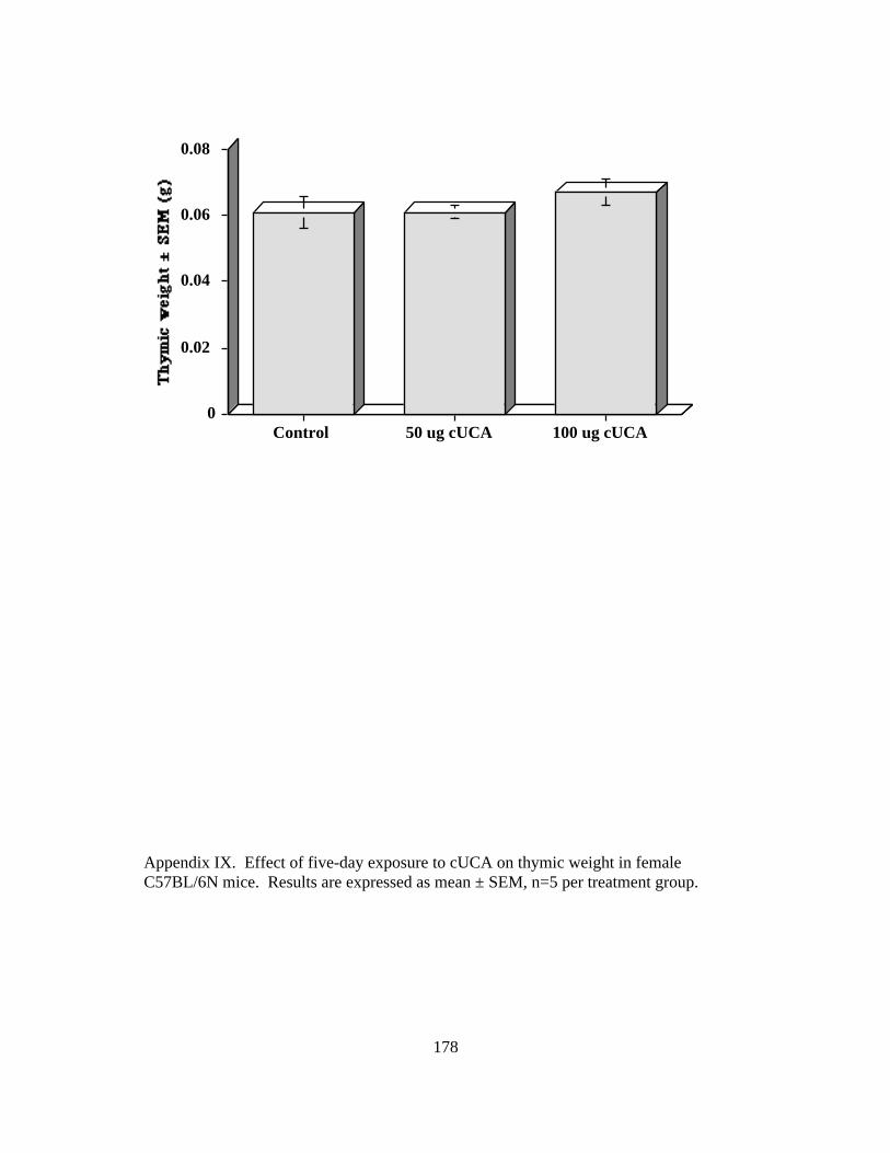
178
0
0.02
0.04
0.06
0.08
Control 50 ug cUCA 100 ug cUCA
Appendix IX. Effect of five-day exposure to cUCA on thymic weight in femaleC57BL/6N mice. Results are expressed as mean ± SEM, n=5 per treatment group.

179
0
10
20
30
40
Control 50 ug cUCA 100 ug cUCA
Appendix X. Effect five-day exposure to cUCA on thymic cellularity in femaleC57BL/6N mice. Results are expressed as mean ± SEM, n=5 per treatment group.

180
0
1
2
3
4
Control 100 ug cUCA
Appendix XI. Effect of five-day exposure to cUCA on splenic macrophage phagocytosisin female C57BL/6N mice. Results are expressed as mean ± SEM, n=5 per treatmentgroup.

181
0
10
20
30
40
50
Control 100 ug cUCA
Appendix XII. Effect of five-day exposure to cUCA on splenic lymphocyte antibodyproduction in female C57BL/6N mice. Results are expressed as mean ± SEM, n=5 pertreatment group.

182
0
20
40
60
80
Control 100 ug cUCA
Appendix XIII. Effect of five-day exposure to cUCA on splenic macrophage ability toproduce active oxygen species (hydrogen peroxide, chemiluminescence assay) in femaleC57BL/6N mice. Results are expressed as mean ± SEM, n=5 per treatment group.

183
0
10
20
30
40
Control 100 ug cUCA
Appendix XIV. Effect of five-day exposure to cUCA on splenic T lymphocyte cytolyticactivity (51Cr release assay) in female C57BL/6N mice. Results are expressed as mean ±SEM, n=5 per treatment group.
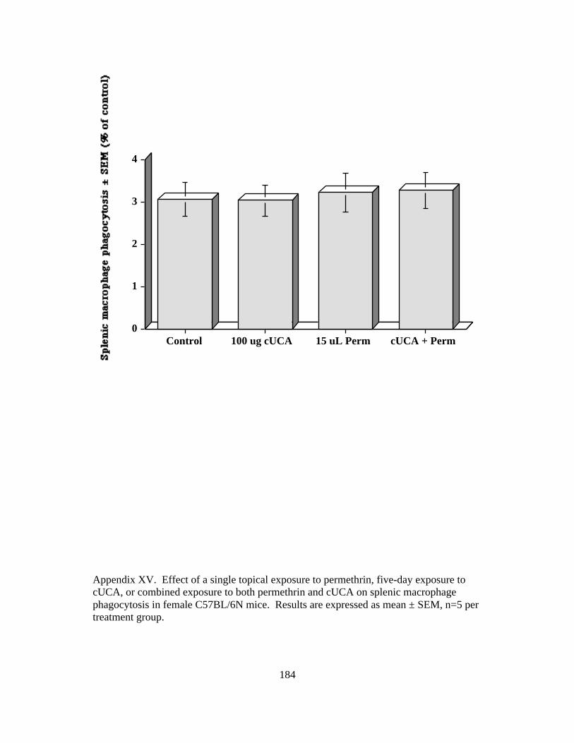
184
0
1
2
3
4
Control 100 ug cUCA 15 uL Perm cUCA + Perm
Appendix XV. Effect of a single topical exposure to permethrin, five-day exposure tocUCA, or combined exposure to both permethrin and cUCA on splenic macrophagephagocytosis in female C57BL/6N mice. Results are expressed as mean ± SEM, n=5 pertreatment group.

185
MARY RENEE PRATER CURRICULUM VITAE
EDUCATION Doctoral Candidate, Immunotoxicologic Pathology, 1999-2002VA-MD Regional College of Veterinary Medicine, Blacksburg, VA, GPA: 3.9/4.0
Clinical Pathology Resident, 1996-1999VA-MD Regional College of Veterinary Medicine, Blacksburg, VA, GPA: 3.9/4.0
Doctor of Veterinary Medicine with honors, 1992-1996VA-MD Regional College of Veterinary Medicine, Blacksburg, VA, GPA: 3.8/4.0
Master of Science, Physiology, 1985-1987University of Akron, Akron, OH, Magna cum laude GPA: 3.8/4.0
Bachelor of Science, Biology, 1981-1985University of Akron, Akron, OH, Magna cum laude GPA: 3.5/4.0
WORK EXPERIENCEVirginia-Maryland Regional College of Veterinary MedicineAssistant Editor, Schalm’s Veterinary Hematology Textbook 5th edition, 1998
Virginia-Maryland Regional College of Veterinary MedicineClinical Pathology Resident, 1996-1999- diagnostic cytology, hematology, histopathology, necropsy- teaching assistant: histology, applied laboratory medicine, clinical pathology, sheep week,clinical biochemistry, general pathology, immunology, and pathobiology journal club.
University of Virginia Department of Internal Medicine/CardiologyLab Specialist Advanced, 1989-92- pharmacology, molecular biology research of adenosine A1 and A2 bovine brain receptors- inventory management in support of laboratory research- supervision and training of research personnel, technical liaison for collaborating investigators- radiation safety training course
The Upjohn Company - Pharmaceutical Sales Representative, 1987-89- human diabetes, CNS, and cardiovascular pharmaceutical products- product information for private practitioners and regional hospitals- continuing education seminars for area physicians- inventory of controlled and noncontrolled physician samples
The University of Akron - Teaching Assistant, 1985-86- histology laboratory teaching assistant- natural science biology lecture discussion leader
Akron Children’s Hospital - Lab Technician, 1982-85- virology department-cell culture and virus isolation- vivarium-care of laboratory animals and in vivo virus isolation- pathology department-diener to assist pathologists in pediatric autopsiesRESEARCH
186
Immune Protection Against MNU-Induced Digital Defects. NIH-SERCA (K01) grantpending, priority score 167.
Virginia Tech, Center for Molecular Medicine and Infectious Disease (1998-2001)Immunotoxicity of Dermal Permethrin and Cis-urocanic Acid (Chemical Mixtures inEnvironmental Health).
Virginia Tech, Center for Molecular Medicine and Infectious Disease (1998-1999) MaternalImmune Protection Against Chemical-Induced Fetal Teratology.
Virginia Tech, Center for Molecular Medicine and Infectious Disease (1998-1999)Influence of Dietary Zinc Form and Level of Supplementation on Broiler Growth andImmune Function, Kemin Industries.
Virginia Tech, Center for Molecular Medicine and Infectious Disease (1998-1999)Utilization of the National Toxicology Program Approved Testing Battery for Chemical-induced Immunotoxicity.
Virginia Tech, College of Vet Medicine (1997) Graduate Research Development Project-Characterization of a metastatic intestinal adenocarcinoma with Paneth cell, enteroendocrinecell, and goblet cell differentiation in a mature Virginia opossum, with the use of electronmicroscopy, histochemistry, and immunohistochemistry to characterize the neoplasm.
Chemical Industry Institute of Toxicology (1995) Veterinary Medicine Clerkship-Physiologically based pharmacokinetic computer modeling, comparison of acetylcholinesteraseand carboxylesterase enzyme activity in the presence and absence of various organophosphorousesters in cell culture, tissue homogenates, and in vivo. This clerkship was in partial fulfillment ofthe Honor’s Program senior project at the Virginia-Maryland Regional College of VeterinaryMedicine.-fire safety training course.
Food and Drug Administration (1996) Veterinary Medicine Student Volunteer-Study of the utilization of physiologically based pharmacokinetic computer modeling in theapproval process of pharmaceuticals in the animal and human medical fields. This clerkship wasin partial fulfillment of the Honor’s Program senior project at the VMRCVM.
Virginia Tech, College of Vet Medicine (1995) Veterinary Medicine Clerkship-Acetylcholinesterase and carboxylesterase time-response and dose-response assays on rat andmouse brain and SH-SY5Y human neuroblastoma cells and NB41A3 murine neuroblastoma cellsin response to organophosphorous ester enzyme inhibition, in partial fulfillment of the seniorHonor’s Program project at the VMRCVM.-Development of pharmacokinetic computer models using Stella program.-Clinical pharmacology: appropriate drug therapy in large and small animal medicine.
The University of Virginia , Division of Cardiology (1989-1992) Lab Specialist Advanced-Adenosine receptor research in pharmacology and molecular biology: gel electrophoresis,protein purification via column chromatography, HPLC, radiolabelling, receptor-ligandcompetition and inhibition assays.
The University of Akron, Department of Biology (1985-1987) master’s degree candidateresearch

187
-dietary and drug effects on rat myocardial catecholamines and left ventricular function.-Langendorf isolated heart technique, spontaneously hypertensive rats as an animal model.
The University of Akron, Department of Biology (1984) undergraduate research student-neuroanatomy pilot research to isolate β-endorphin and G-6-PD activity in brain nuclei.
PRESENTATIONSAmerican College of Veterinary Pathologists, Amelia Island, FL 2000American Society for Veterinary Clinical Pathology, Amelia Island, FL 2000Society of Toxicology Poster Presentation, Philadelphia, PA 2000Virginia Tech 16th Annual Graduate Research Symposium, Blacksburg, VA 2000VMRCVM 12th Annual Research Symposium, Blacksburg, VA 2000Virginia Veterinary Medical Association, Hot Springs, VA, invited speaker, 2000American Society for Veterinary Clinical Pathology, Chicago IL 1999Eli Lilly and Company, Greenfield, IN, invited speaker 1999American College of Veterinary Pathologists, St. Louis, MO 1998American Society for Veterinary Clinical Pathology, Albuquerque, NM 1997American College of Veterinary Pathologists, Albuquerque, NM 1997Chemical Industry Institute of Toxicology, Research Triangle Park NC, invited speaker 1996Federation of American Societies for Experimental Biology, Washington DC 1991Federation of American Societies for Experimental Biology, Washington DC 1990Federation of American Societies for Experimental Biology, St. Louis, MO 1986
PUBLICATIONSPapersMR Prater, RM Gogal, Jr., J Longstreth, EC DeFabo, SD Holladay. Immunotoxic effectsof cis-urocanic acid exposure in C57BL/6N mice, in preparation.
MR Prater, BL Blaylock, SD Holladay. Molecular mechanisms of sunlight andpermethrin-induced alterations in cutaneous immunity, in preparation.
MR Prater, RM Gogal, Jr., BL Blaylock, J Longstreth, SD Holladay. Single-dose topicalexposure to the pyrethroid insecticide, permethrin in C57BL/6N mice: effects on thymusand spleen, Food and Chemical Toxicology, submitted 11/28/01.
MR Prater, BL Blaylock, RM Gogal, Jr., SD Holladay. Sunlight exposure, mimicked bycis-urocanic acid, increases both immunotoxicity and lethality of dermal permethrin inC57BL/6N mice, in preparation.
RM Gogal Jr., CT Larsen, MS Johnson, MR Prater, RB Duncan, SD Holladay. Influenceof orally-administered RDX on the Northern Bobwhite (Colinus virginianus). EnvironTox Chem submitted 4/2001.
KL Zimmerman, MR Prater, HS Bender, RB Duncan, GD Boon. A cervical mass from acat with weight loss. Vet Clin Pathol submitted 5//2001.
188
RM Gogal Jr., MS Johnson, CT Larsen, MR Prater, RB Duncan, DL Ward, SD Holladay.Influence of dietary 2,4,6-trinitrotoluene exposure in the Northern Bobwhite (Colinusvirginianus). Environ Tox Chem 21:81-86, 2002.
SD Holladay, LV Sharova, K Punareewattana, TC Hrubec, RM Gogal Jr., MR Prater, AASharov. Maternal immune stimulation in mice decreases fetal malformations caused byteratogens. International Immunopharmacology, 2:325-332, 2002.
MR Prater, KL Zimmerman, EG Besteman, GD Boon, GK Saunders. Splenic massaspirate from an aged pekapoo, in preparation.
MR Prater, RB Conolly, M Ehrich. Comparison of computer modeled kinetic data within-vitro data for inhibition of acetylcholinesterase activity induced by organophosphorouscompounds, in preparation.
MR Prater. Cerebrospinal fluid cytology, USDA Higher Education Challenge Grantsponsored digital textbook, in press.
RM Gogal Jr., MR Prater, BJ Smith, MS Johnson, SD Holladay. Bilateral dissectedspleens and thymus in rodents exhibit homogeneity in leukocyte markers. Toxicology157:217-223, 2001.
K Punareewattana, BJ Smith, BL Blaylock, RM Gogal Jr., MR Prater, J Longstreth, SDHolladay. Topical permethrin exposure inhibits antibody production and macrophagefunction in C57Bl/6N mice. Food and Chemical Toxicology 39:133-139, 2001.
K Punareewattana, BJ Smith, BL Blaylock, JR Robertson, RM Gogal Jr, MR Prater, JLongstreth, HL Snodgrass, and SD Holladay. Topical permethrin exposure causes thymicatrophy and persistent inhibition of the contact hypersensitivity response in C57Bl/6mice. International Journal of Toxicology 19:383-389, 2000.
MR Prater, B Flatland, S Newman, DP Sponenberg, J Chao. Diffuse annular fusiformadenocarcioma in a dog. JAAHA 36:169-173, 2000.
GK Saunders, DJ Blodgett, TA Hutchins, MR Prater, JL Robertson, PA Friday WKScarratt. Suspected citrus pulp toxicosis in dairy cattle. J Vet Diagn Invest 12:269-271,2000.
KL Zimmerman, HS Bender, GD Boon, MR Prater, CE Thorn, DA Prater, JL Robertson,GK Saunders, DP Sponenberg, KD Inzana, OI Lanz, E Wright. A comparison of thecytologic and histologic features of meningiomas in four dogs. Vet Clin Pathol 29:29-34,2000.
RB Duncan, BF Feldman, GK Saunders, MR Prater, G Troy. Mandibular salivary glandaspirate from a dog. Vet Clin Pathol 28:97-99, 1999.
189
MR Prater, RB Duncan, J Gaydos. Characterization of metastatic intestinaladenocarcinoma with differentiation into multiple morphologic cell types in a Virginiaopossum. Vet Pathol 36:463-468, 1999.
R Prater, R deGopegui, K Burdette, H Veit, and B Feldman. Cutaneous lesions with bonemarrow involvement in a cat. Vet Clin Pathol 28:52-58, 1999.
MR Prater, HS Bender. Splenomegaly in a four-year-old cat. Bloodline July 1998.
MR Prater, HS Bender Hepatosplenomegaly in an aged cat. Bloodline March, 1998.
R Prater, H Bender, P Sponenberg. Intraabdominal mass in a 16 year old dog. Vet ClinPathol 27:54, 65-66, 1998.
R Prater, H Bender, L Shell. Specialty Board Review: examples of cerebrospinal fluidcytology. Prog Vet Neuro 7:133-136, 159-160, 1996.
MR Prater, H Taylor, R Munshi, J Linden. Indirect effect of guanine nucleotides onantagonist binding to A1 adenosine receptors: occupation of cryptic binding sites byendogenous vesicular adenosine. Molecular Pharm 42:765-772, Nov. 1992.
J Linden, MR Prater, G Sullivan, R Johns, A Patel. Contamination of adenosinedeaminase by superoxide dismutase: stabilization of EDRF. Biochem J 41:273-279,1991.
AbstractsRM Gogal Jr., CT Larsen, MS Johnson, MR Prater, RB Duncan, SD Holladay. Influenceof oral exposure to RDX in Northern Bobwhite: Colinus virginianus. SETAC,November, 2001.
JD Longstreth, BL Blaylock, MR Prater, RM Gogal, SD Holladay. Immunotoxicity riskassessment for children with joint exposure to sunlight and permethrin. 29th AmericanSociety for Photobiology, Chicago, IL. 2001.
BL Blaylock , RM Gogal, Jr., MR Prater, SD Holladay. Combined dermal exposure topermethrin and cis-urocanic acid suppresses the contact hypersensitivity response inC57Bl/6 mice in an additive manner. 40th Annual Meeting of the Society of Toxicology,San Francisco, CA. March 26, 2001.
RM Gogal Jr., CT Larsen, MS Johnson, MR Prater, RB Duncan, SD Holladay. Influenceof acute oral exposure to RDX on northern bobwhite: Colinus virginianus. 40th AnnualMeeting of the Society of Toxicology, San Francisco, CA. March 26, 2001
J Chen, RM Gogal Jr., MR Prater, SD Holladay and BL Blaylock. Combined dermalexposure to permethrin and cis-urocanic acid suppresses the contact hypersensitivityresponse in C57Bl/6 mice in an additive manner. South Central Chapter, Society of

190
Toxicology Annual Meeting, University of Arkansas for Medical Sciences, Little Rock,AR. October 20, 2000.
MR Prater, RM Gogal, HS Bender, and SD Holladay. Permethrin-induced thymichypocellularity: are antiproliferative effects involved? American Society for VeterinaryClinical Pathologists, Amelia Island, FL. December 4, 2000.
MR Prater, RM Gogal, HS Bender, and SD Holladay . Evaluation of contacthypersensitivity response in mice exposed to permethrin and cis-urocanic acid. AmericanCollege of Veterinary Pathologists, Amelia Island, FL. December 4, 2000.
J Longstreth, MR Prater, SD Holladay. Risk assessment of joint exposure to twoimmunotoxic agents in children: step 1 - exposure assessments. 2000 Annual Meeting ofthe Society for Risk Analysis, Arlington, VA.
RM Gogal Jr., CT Larsen, MR Prater, RB Duncan, DL Ward, MS Johnson, and SDHolladay. Effects of dietary exposure to 2, 4, 6-trinitrotoluene (TNT) on NorthernBobwhite (Colinus virginianus) , in preparation.
RM Gogal Jr., LV Sharova, P Sura, MR Prater, SD Holladay. Maternal immunestimulation and prevention of teratogenesis. Toxicological Sciences 54:297, 2000.
MR Prater, RM Gogal Jr., SD Holladay. Subacute intradermal cis-urocanic acidsuppresses thymic cellularity in two strains of mice. Toxicological Sciences 54:1, 153,2000.
R Duncan, B Feldman, G Saunders, M Prater. Sialometaplasia in a dog. Vet Pathol 35:5,1998.
MR Prater, BB Lockee, EM Mills, HS Bender. Reaching out to veterinary practitionersthrough diagnostic pathology imaging: a program for clinical service and continuingeducation. Vet Pathol 35:431, 1998.
R Prater, R Duncan, M Nagarkatti, J Gaydos. Characterization of metastatic intestinaladenocarcinoma with Paneth cell differentiation in a Virginia opossum. Vet Pathol 34:492,1997.
MR Prater, R Munshi, J Linden. Guanine nucleotides modulate agonist but not antagonistbinding to purified A1 adenosine receptors. FASEB Journal 5:A1572, March 19, 1991.
J Linden, MR Prater, A Patel, G Sullivan, and R Johns. Adenosine deaminase elevatesEDRF due to contaminating SOD activity. FASEB Journal 2:A909, Feb. 28, 1990.
R Zwallen, S Inman, F Sadri, J Weigand, and D Ely. Preservation of heart function inbeating isolated rat heart. Fed Proc 45:766, 1986.

191
S Inman, Zwallen, F Sadri, D Ely. Catecholamine levels and left ventricular function innormotensive and hypertensive isolated rat hearts. Fed Proc 45:898, 1986.
Book ChaptersAcquired coagulopathy II: liver disease, Schalm's Veterinary Hematology, 5th edition,MR Prater, eds. B Feldman, J Zinkl, NC Jain, ©2000, Lippincott Williams and Wilkins,Philadelphia, PA.
Acquired coagulopathy I: avitaminosis K, Schalm's Veterinary Hematology, 5th edition,MR Prater, eds. B Feldman, J Zinkl, NC Jain, ©2000, Lippincott Williams and Wilkins,Philadelphia, PA.
Acquired coagulopathy II: liver disease, Manual of Veterinary Clinical Hematology, M.R. Prater, eds. B. Feldman, J. Zinkl, N. C. Jain, accepted 9/3/98.
Acquired coagulopathy I: avitaminosis K, Schalm's Manual of Veterinary ClinicalHematology, M. R. Prater, eds. B. Feldman, J. Zinkl, N. C. Jain, accepted 9/3/98.
HONORS AND AWARDS2002 Outstanding Graduate Student Award, VMRCVM2001 VMRCVM “hot team” interviewer2000 VMRCVM Accreditation CommitteeCL Davis Senior Pathology Resident AwardVa Tech Graduate Student Assembly Travel GrantsJackson Immunoresearch Company Travel GrantHill’s Pet Food Company Travel GrantVa Tech Graduate Research Development Project GrantPauline Willson-Gunn ScholarshipVanDresser Academic ScholarshipVaughn Academic ScholarshipWARDS Academic ScholarshipAssociation for Women Veterinarians ScholarshipThomas Moore Academic ScholarshipTop 500 Sales AwardOutstanding Young Women of AmericaInnovative Research of America GrantAmerican Heart Association Summer FellowshipPROFESSIONAL ASSOCIATIONSInternational Society for Animal Clinical BiochemistryAmerican Society for Veterinary Clinical PathologyPhi Zeta Honorary Veterinary Society, Chi ChapterAssociation for Women VeterinariansAmerican Veterinary Medical AssociationAmerican Animal Hospital AssociationAmerican Physiological SocietyPhi Sigma Alpha Honor Society