immunologia of joint diseases
TRANSCRIPT
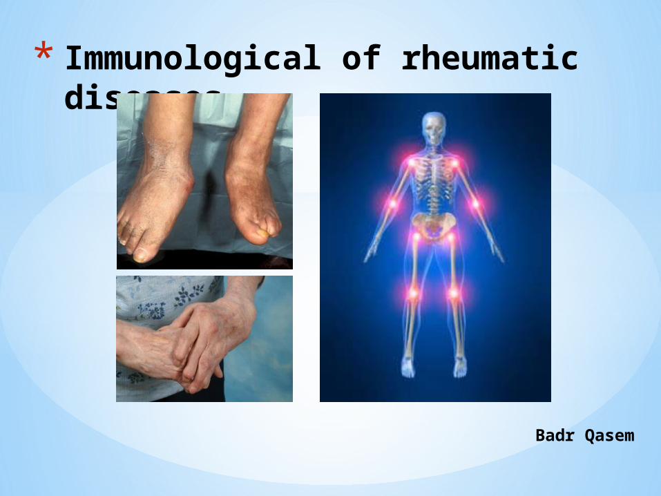
* Immunological of rheumatic diseases
Badr Qasem
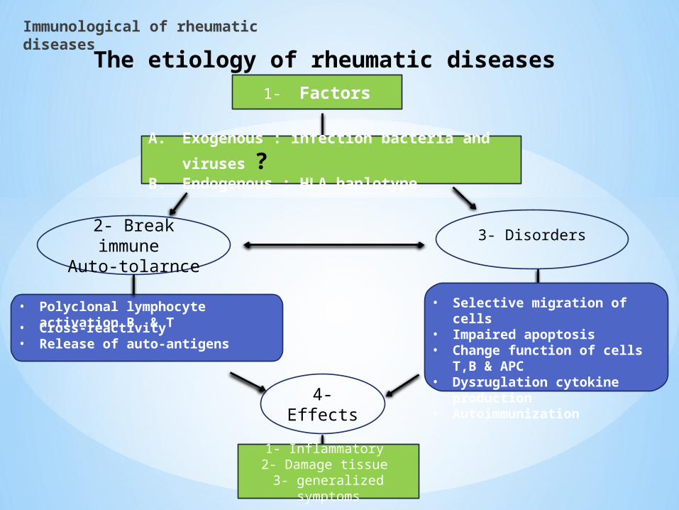
• Selective migration of cells • Impaired apoptosis• Change function of cells T,B & APC• Dysruglation cytokine production• Autoimmunization
The etiology of rheumatic diseases
Immunological of rheumatic diseases
1- Factors
4-Effects
A. Exogenous : infection bacteria and viruses ?B. Endogenous : HLA haplotype
2- Break immune Auto-tolarnce
3- Disorders
• Polyclonal lymphocyte activation B & T
• Cross-reactivity• Release of auto-antigens
1- Inflammatory 2- Damage tissue
3- generalized symptoms
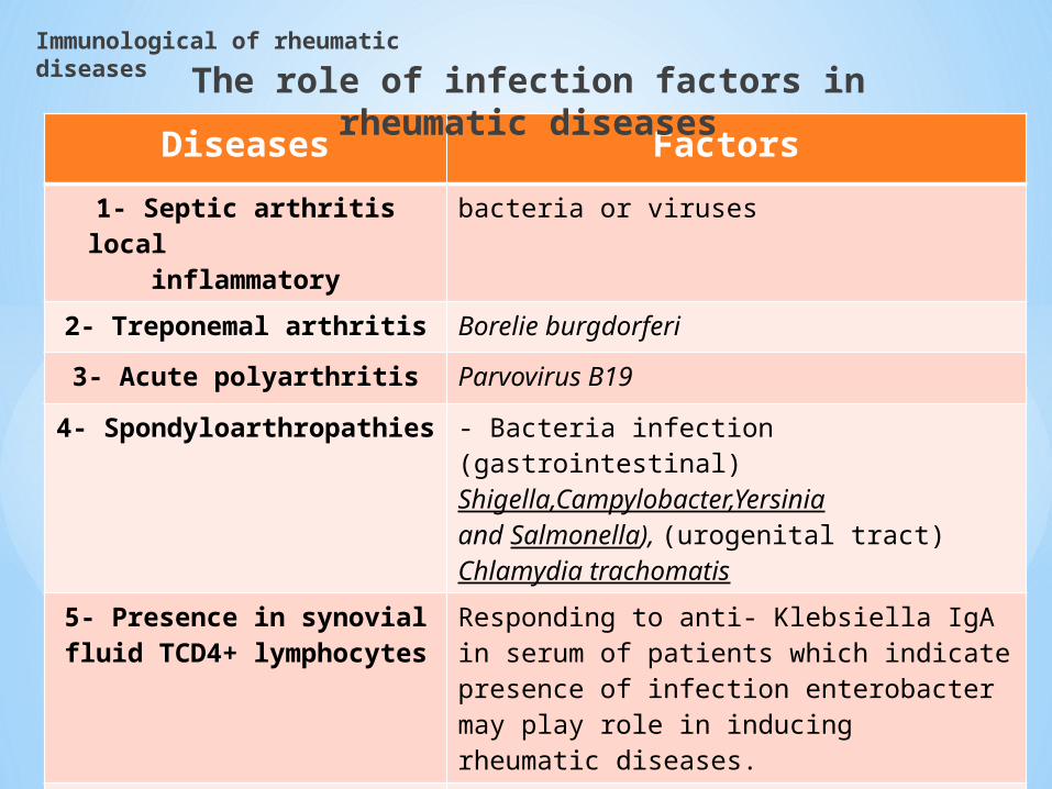
Diseases Factors
1- Septic arthritis local inflammatory
bacteria or viruses
2- Treponemal arthritis Borelie burgdorferi
3- Acute polyarthritis Parvovirus B19
4- Spondyloarthropathies - Bacteria infection (gastrointestinal)Shigella,Campylobacter,Yersiniaand Salmonella), (urogenital tract) Chlamydia trachomatis
5- Presence in synovial fluid TCD4+ lymphocytes
Responding to anti- Klebsiella IgA in serum of patients which indicate presence of infection enterobacter may play role in inducing rheumatic diseases.
6- Immune – complexes -In many of rheumatoid diseases(Rheumatic arthritis or Systemic Lupus Erythematous-SLE)
The role of infection factors in rheumatic diseases
Immunological of rheumatic diseases

The role of infection factors in rheumatic diseases
Immunological of rheumatic diseases
Why consequence of different infections in distant organs and tissues comes to joints
inflammatory ?

The role of infection factors in rheumatic diseases
Immunological of rheumatic diseases
Blood
Joints
Glycoprotein bone
Collagen type I
Adhesion protein Yad A
Staphylococcus aureus
YersiniaVirulence strains
- Phagocytic cells- Immune- complexes
- Notes : - In cells of synovial fluid reveal some RNA sequences derived from endogenous retroviruses in RA and JCA which suggested retroviral infection may play a role in the pathogenesis of autoimmune rheumatic disease

Diversity of Rheumatologic Diseases:
Common and Uncommon Diseases Involving Inflammatory and Immune
Responses
- Rheumatoid Arthritis*- Systemic Lupus Erythematosus*- Spondyloarthropathies*
Ankylosing spondylitisReactive Arthritis (Reiter’s
Syndrome)Psoriatic ArthritisSpondylitis associated with IBD
- Sjogren’s Syndrome- Polymositis/Dematomyositis- Lyme Disease- Rheumatic Fever- Behcet’s Syndrome- Systemic Sclerosis (Scleroderma)- Wegener’s Granulomatosis- Giant Cell Arteritis*
Immunologically-Mediated Diseases
(adaptive immunity)- Osteoarthritis*- Gout*- Pseudogout
Inflammatory Diseases (innate immunity)
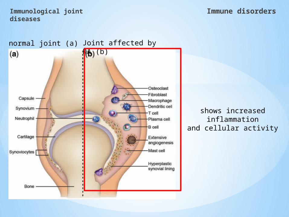
Immunological joint diseases
Immune disorders

Immunological joint diseases
Inflammatory cascade
NOXIOUS STIMULI(Injury or Infection)
INNATE(immunologically non-
specific)
ADAPTIVE(immunologically specific)
VASCULAR EVENTS CELLULAR EVENTSANTIBODY MEDIATED
RESPONSECELL MEDIATED
RESPONSE
MICROSCOPICLEVEL
PRODUCTION OF NUMBEROUS CHEMICAL MEDIATORSAUTOCOIDS (Greek: autos “self” and akos “medical agent or remedy”PARACRINE SECRETIONS OR LOCAL HORMONES
Neurotransmitters Hormones• REDDENED • HOT
• SWOLLEN • PAINFUL• INTERFERENCE
FIBRINOLYTIC CASCADE
COAGULATION CASCADE
KININ CASCADE
COMPLEMENT CASCADE
CELLS ALREADY PRESENT1. Vascular endothelial2. Mast3. Tissue macrophages
CELLS ENTERINGTHROUGH BLOOD1. Platelets2. Leukocytes
INDUCTION PHASE
EFFECTOR PHASE
MACROSCOPIC LEVEL
Immune disorders
Immunological joint diseases
Immune disorders
normal joint (a) Joint affected by RA (b)
shows increased inflammation
and cellular activity

Cytokines
Immunological joint diseases
Immune disorders

Immunological joint diseases
Immune disorders
Proinflammatory
TNF- IL-8
IL-1 IFN-
IL-2 LT
IL-6 GM-CSF
Anti-inflammatory
IL-4
IL-10 sIL-1R
IL-11IL-1Ra
TGF- IL-13
sTNFR
Cytokine Disequilibrium in the disease Process of Rheumatic diseases

Immunological joint diseases
Immune disorders
CytokinesPro-
inflammatory
Source function
Immunological Related with joint diseases
TNF- Monocytes/macrophages, synviocytes A >B
Supporting:- Proliferation of T & B
cells- Differentiation of B
cells - Cytotoxicity NK
Inducing:- - Production pro-inflammatory
cytokines - Expression of adhesion molecules- Production metalloproteinase and
PGE- Resorption & synthesis of
proteoglycan
IL- 1 Monocytes/macrophages, synviocytes A >B
Supporting:- Activation of T & B cells- Secretion cytokines
Th1- Cytotoxicity NK
Inducing:- - Production pro-inflammatory
cytokines - Expression of adhesion molecules- Production metalloproteinase and
PGE- synthesis of collagens- Osteoclasts activation- The main factor of pyretic
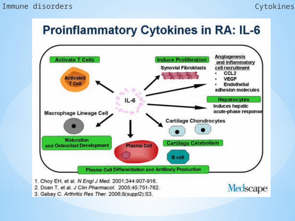
IL- 6 Monocytes/macrophages, synviocytes A >B
Increasing:- Production IL-2- Production antibodies
Supporting:- Production metalloproteinase and
PGE- Main factor induction phase acute
response - Inducing production sTNFR , IL-1Ra
metalloproteinase inhibitor.- Decreases production of IL-1 & TNF-
IL- 8 Monocytes/macrophages, Neutrophils ,synviocytes A &B
- Chemotactic factor For neutrophils & lymphocytes- Neutrophils activation
- Like immunological function- Main factor for inducing
angiogenesis
GM- CSF Monocytes/macrophages, synviocytes A >B
Neutrophils & macrophage activation
- Like immunological function

Immunological joint diseases
Immune disorders
Cytokines Pro-inflammatory
TNF-α
TNF-α : play as central role which inducing the production of IL-1 , and both cytokines following the production of pro-inflammatory cytokines

TNF-α & IL-1
Synoviocytes B & A
Prostaglandins E2 (PGE2)
a series of metalloproteinase
Synovial fluid and synovium
Degradation of cartilage
TNF-α , IL-6 and IL-1β
Acute phase response
TNF-a & IL-1
In liver
- Acute phase protein like complement components
- Protein coagulation proteases inhibitors
IL-6
- Cross-reactive protein (CRP)
- Serum amyloid A (SAA)
- Anti-trypsin or fibrinogen
Collagenase stromielizinesgelatinizes
TNF-α & IL-6
Synergistic manner
Destruction
of joint tissues
production
production
- Increase activation of MØ
- stimulation production IL-1β and TNF-α
Large amount presence in synovial fluid
Immune disorders Cytokines
- Production of collagenase by synoviocytes B
- Increase expression of adhesion molecules monocytes & neutrophils
Large amount presence in synovial fluid
Cytokines Pro-inflammatory

Immune disorders Cytokines
Proinflammatory
IL-6, IL-8, GM-CSFIL-1
TNF
Anti-inflammatory
IL-10, sTNFR, IL-1Ra,
Inflammation
Activates monocytes/macrophages
Bone resorption and erosions
Activates osteoclasts, suppresses osteoblasts
Cartilage breakdown
Activates chondrocytes,
releasing collagenases
TNF

Immune disorders Cytokines
Immune disorders Cytokines

CytokinesAnti-
inflammatory
Source function
Immunological Related with joint diseases
IL- 4 Th2 cells Stimulation :- Differentiation of T & B cells- Decreases production pro-
inflammatory cytokines
- Decreases as immunological function
- Production metalloproteinase
IL- 10 T lymphocytessynviocytes A
- Stimulation Th2 cells - Decreases Proliferation Th1- Activation macrophages &
production by not cytokines
- Like immunological function
IL- 13 - Stimulation differentiation B cells
- Decreases production pro-inflammatory cytokines
- Like immunological function
TGF-β Different cells - Decreases development and production cytokines by T cells
- Development and Differentiation of B cells
- Stop effect TNF-
- Like immunological function- Inducing production of tissues
inhibitors metalloproteinase- Supporting production collagen &
tissues repair processes
sTNF- R Different cells - Binding TNF- & neutralize it s effect
- Like immunological function
IL- 1Ra Different cells - Blocking binding IL-1 to receptor & stopping their biological effect
- Like immunological function
Immune disorders Cytokines

The major of cytokines anti-inflammatory
IL-10 TGF-B IL-4 IL-13
Lymphocytes /macrophage
Various types of cells
Th1 chemokine's
Th lymphocytes/m
ast cells /NK cells
Anti-inflammatory action for IL-6 & TNF-
α
The main action of anti-inflammatory cytokines
IL-4,IL-13,IL-10 & TGF-β
Ability to inhibition pro-inflammatory
cytokines
Increase production of IL-1Ra
Has a strong immunosuppression effect
Blocking proliferation and activation lymphocytes
Immune disorders Cytokines

Immune disorders Cytokines
In clinical course
Juvenile chronic arthritis (JCA)
High concentration of IL-6 in serum & synovial fluid
Anima
Impaired growth
Amyliodosis
osteoporosis
TNF-α Appear in vary important development of systemic symptoms ( fever , disorders of coagulation

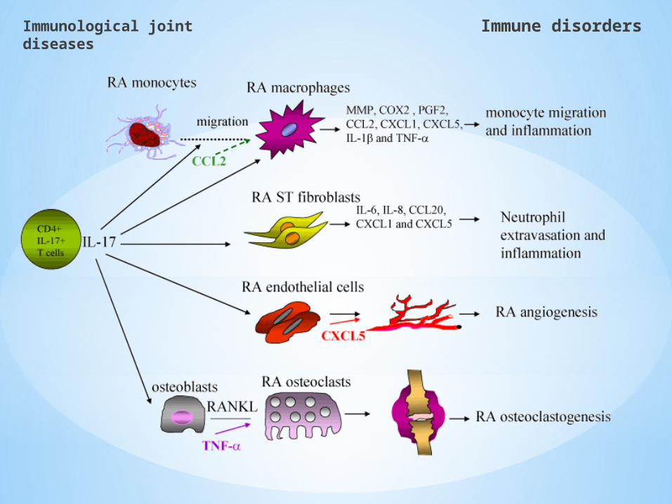
Selective migration of immunological cells to
inflamed joints
Immunological joint diseases
Immune disorders

normal joint (a) Joint affected by RA (b)
shows increased inflammation
and cellular activity
Immunological joint diseases
Immune disorders

Immunological joint diseases
Immune disorders
The multi-step paradigm of leukocyte migration: Step 1: Tethering & rolling
Cytokine (pro inflammatory cytokines )activated endothelial cells express adhesion molecules Leukocytes ‘marginate’ from the peripheral pool to the marginal pool
Tethering and rolling are mediated by SELECTINS and ADDRESSINS
Tethering4000 microns/sec
Rolling40 microns/sec

Immunological joint diseases
Immune disorders
Neutrophilis activated
bychemokine's
IL-8
Selecting is shedRolling
Cytokines from endothelial activate expression of Intracellular adhesion molecules (ICAMs)
INTEGRIN (adhesion molecule) has low affinity for ICAM
Cell activation changes integrin to high affinity
format
Steps 2 & 3: Activation & arrest
Presence both molecules has significant increase in RA
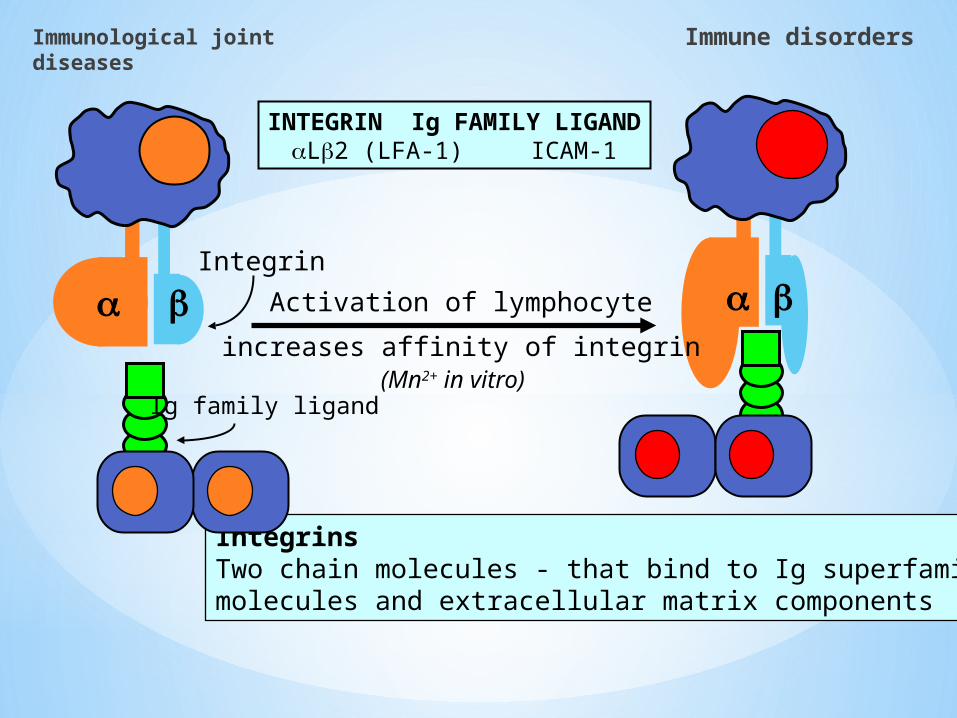
Integrin
IntegrinsTwo chain molecules - that bind to Ig superfamilymolecules and extracellular matrix components
INTEGRIN Ig FAMILY LIGANDaLb2 (LFA-1) ICAM-1
Ig family ligand
Activation of lymphocyte
increases affinity of integrin(Mn2+ in vitro)
Immunological joint diseases
Immune disorders

Step 4:Migration and diapedesis
Firm adhesion causes the leukocyte to flatten and migrate between the endothelial cells
Leukocyte migrates towards site of infection by detecting and following a gradient of chemokine.
Leukocytes migrate readily to the chemokine RANTES which synthesis by MØ , endothelial cells and synovial fibroblast
~10 Minutes
Immunological joint diseases
Immune disorders
Disorders of apoptosis
Immunological joint diseases
Immune disorders

apoptosis
Some studies that observed that apoptosis may contribute of development of autoimmune diseases including RA
Also with patients wit rheumatic diseases they shown us more complex picture of peripheral blood lymphocytes with SLE
By increasing spontaneous apoptotic death which suggested as a result that by enhance a apoptosis cells
Apoptosis of synoviocytes B may be responding for expansion in the synovium
Cytokines play role against apoptosis of synoviocytes
IL-1β
TGF- β
Inhibition apoptosis cells by inducing Fas/FasL
Reduce in these cells expression Fas and increase of expression Bcl-2 and proliferation cells
Immunological joint diseases
Immune disorders

Immunological joint diseases
Immune disorders
Antigen presenting cells
IL-6 & others enzymes degradation
Hyperplasia
IL-8 & othersInflammatory
mediators
Ab, AutoAb,Immune-complex
TNF,IL-1 & other enzymatic degradation
Inflammatory mediators
The influx and cell activation and cytokine production
Synoviocytes BneutrophilLymphocyte BSynoviocytes A
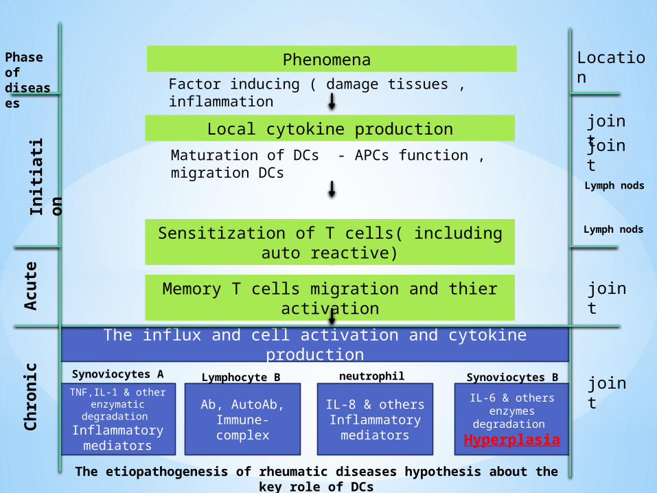
Factor inducing ( damage tissues , inflammation
Maturation of DCs - APCs function , migration DCs
joint
Chro
nic
Acu
teIn
itia
tion
joint
Sensitization of T cells( including auto reactive)
Local cytokine production
Phenomena
Memory T cells migration and thier activation
Location
Lymph nods
joint
joint
Lymph nods
Phase of diseases
The etiopathogenesis of rheumatic diseases hypothesis about the key role of DCs
Immunological joint diseases
Immune disorders
T Lymphocytes
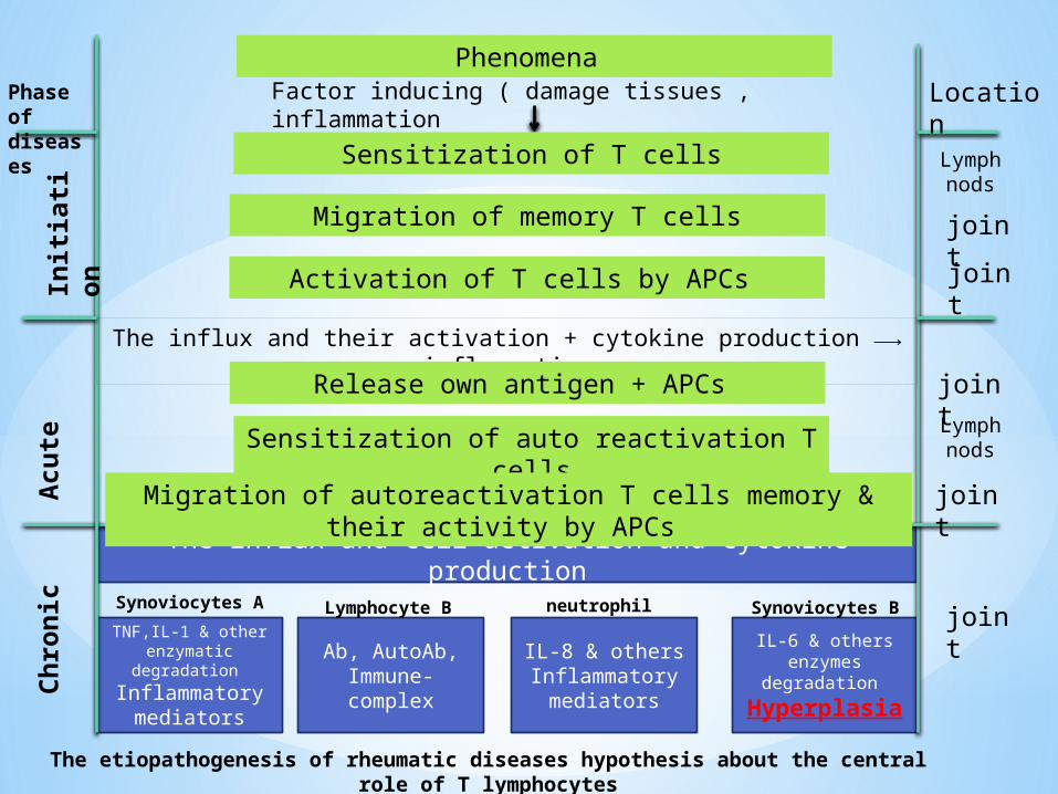
IL-6 & others enzymes degradation
Hyperplasia
IL-8 & othersInflammatory
mediators
Ab, AutoAb,Immune-complex
TNF,IL-1 & other enzymatic degradation
Inflammatory mediators
The influx and cell activation and cytokine production
Synoviocytes BneutrophilLymphocyte BSynoviocytes A
Factor inducing ( damage tissues , inflammation
The influx and their activation + cytokine production inflammation⟶
joint
Chro
nic
Acu
teIn
itia
tion
joint
Sensitization of auto reactivation T cells
Sensitization of T cells
Phenomena
Migration of autoreactivation T cells memory & their activity by APCs
Location
Lymph nods
joint
joint
Phase of diseases
The etiopathogenesis of rheumatic diseases hypothesis about the central role of T lymphocytes
Migration of memory T cells
Activation of T cells by APCs
Release own antigen + APCs
Lymph nods
joint
Immunological joint diseases
Immune disorders

Immunological joint diseases
Immune disorders
B Lymphocytes

B- Lymphocytes

binding to citrullinated proteins located in the cellular membrane
binding to FcγIIIa receptors
bound to other cellular receptors (such as TLR4)
The inflamed rheumatoid synovial membrane and fluid are a reservoir of activated immune cells and locally produced antibodies
a high proportion of the synovial IgG -expressing cells (memory and early plasma blasts)
Immunological joint diseases
Immune disorders

Neutrophil
Immunological joint diseases
Immune disorders

Immunological joint diseases
Immune disorders

Immunological joint diseases
Immune disorders

Synoviocytes type B
Immunological joint diseases
Immune disorders

This cells are fibroblasts and/or fibroblast-like cells that are found between the cartilaginous fibers in the synovial membrane of joints
secrete a variety of effector molecules that promote inflammation and joint destruction and, themselves are part of a complex network
of autocrine and paracrine acting factors
Doesn’t belong to immune cells but they have number of properties that make it play a vary important role in immunopathogensis of joint diseases
The best function in Rheumatic diseases they are fibroblast synthesis a protein constituting a connective tissues e.x. proteoglycan
In the presences of proinflammatory cytokines such as TNF-a and IL-1 this cells start produce numerous enzymes degrading the extracellular matrix protein (serine ,
protease , cathepsin and metalloproteinase)
In the case of patients with rheumatic diseases is predominant inhibitors of metalloproteinase(in degeneration joint diseases) which make this cells contribute to
destructive processes
This cells are also supporting the cytokine network with presence TNF-a & IL-1 start producing cytokines pro-inflammatory (IL-6,IL_8 and GM-CSF) & FGF
Immunological joint diseases
Immune disorders

Thank you for your attention