il-10 is up-regulated in multiple cell types during viremic hiv - blood
TRANSCRIPT

IMMUNOBIOLOGY
IL-10 is up-regulated in multiple cell types during viremic HIV infection andreversibly inhibits virus-specific T cells*Mark A. Brockman,1,2 *Douglas S. Kwon,1-3 Daniel P. Tighe,1 David F. Pavlik,1 Pamela C. Rosato,1 Jennifer Sela,1
Filippos Porichis,1,2 Sylvie Le Gall,1,2 Michael T. Waring,1,3 Kristin Moss,1 Heiko Jessen,4 Florencia Pereyra,1,2,5
Daniel G. Kavanagh,1,2 Bruce D. Walker,1-3 and Daniel E. Kaufmann1,2
1Ragon Institute of Massachusetts General Hospital, Massachusetts Institute of Technology and Harvard, Massachusetts General Hospital, Charlestown;2Harvard Medical School, Boston, MA; 3Howard Hughes Medical Institute, Chevy Chase, MD; 4Jessen Praxis, Berlin, Germany; and 5Brigham and Women’sHospital, Boston, MA
Murine models indicate that interleukin-10(IL-10) can suppress viral clearance, andinterventional blockade of IL-10 activityhas been proposed to enhance immunityin chronic viral infections. Increased IL-10levels have been observed during HIVinfection and IL-10 blockade has beenshown to enhance T-cell function in someHIV-infected subjects. However, the cat-egories of individuals in whom the IL-10pathway is up-regulated are poorly de-fined, and the cellular sources of IL-10 in
these subjects remain to be determined.Here we report that blockade of the IL-10pathway augmented in vitro proliferativecapacity of HIV-specific CD4 and CD8T cells in individuals with ongoing viralreplication. IL-10 blockade also increasedcytokine secretion by HIV-specific CD4T cells. Spontaneous IL-10 expression,measured as either plasma IL-10 proteinor IL-10 mRNA in peripheral blood mono-nuclear cells (PBMCs), correlated posi-tively with viral load and diminished after
successful antiretroviral therapy. IL-10mRNA levels were up-regulated in mul-tiple PBMC subsets in HIV-infected sub-jects compared with HIV-negative con-trols, particularly in T, B, and natural killer(NK) cells, whereas monocytes were amajor source of IL-10 mRNA in HIV-infected and -uninfected individuals.These data indicate that multiple cell typescontribute to IL-10–mediated immune sup-pression in the presence of uncontrolledHIV viremia. (Blood. 2009;114:346-356)
Introduction
Persistent viral infection often results in T-cell impairment dueto combined effects of changes in antigen-specific lymphocytesand antigen-presenting cell (APC) populations.1-4 Recent stud-ies have highlighted the role of inhibitory immunoregulatorypathways in T-cell exhaustion during chronic viral infection. Forexample, the cosignaling molecule PD-1 has been shown tomediate a reversible dysfunction of virus-specific cytotoxicT lymphocytes (CTLs) during chronic lymphocytic choriomen-ingitis virus (LCMV) infection in mice,5,6 as well as duringHIV,7-9 hepatitis B virus (HBV),10 and hepatitis C virus (HCV)11,12
infection in humans. Besides PD-1, other surface molecules canalso mediate reversible T-cell dysfunction in chronic HIVinfection, as shown by recent reports of the involvement ofcytotoxic T lymphocyte–associated antigen 4 (CTLA-4) inHIV-specific CD4 T-cell impairment13 and TIM-3 in HIV-specific CTL dysfunction.14 Studies in murine models suggestthat other cosignaling molecules expressed on the cell surfaceare also involved.6
Soluble factors, such as chemokines and cytokines, also regu-late T-cell function.15 Among these soluble factors, the cytokineinterleukin 10 (IL-10) has been shown to play a critical role in thebalance between immunopathology and protective responses ininfectious diseases (reviewed in Couper et al16 and Moore et al17).IL-10 is a pleiotropic cytokine that can be produced by multiplecell populations, with diverse effects on many hematopoietic cell
types. The ability of IL-10 to inhibit cytokine production by T cellsand natural killer (NK) cells is thought to be largely indirect, byalteration of monocyte/macrophage function.18 IL-10 decreasesmajor histocompatibility complex (MHC) class II and CD80/CD86expression on monocytes and macrophages and limits productionof proinflammatory cytokines. However, IL-10 can also stimulatethe proliferation of B cells and enhance their maturation intoplasma cells.19 Inflammatory bowel disease and other excessiveinflammatory responses occurring in IL-10�/� mice indicate thatIL-10 is critically involved in limiting inflammatory responses invivo.20,21 The impact of IL-10 on host immunity in the setting ofinfectious diseases is complex. IL-10 knockout or blockade canenhance resistance to infection with pathogens such as Listeriamonocytogenes,22 Mycobacterium avium,23 and Trypanozomacruzi.24 IL-10 plays a key role in facilitating establishment ofchronic Leishmania major infection.25 However, absence of IL-10signaling results in severe and often fatal immunopathology inToxoplasma gondii,26 malaria,27 and herpes simplex virus28 infec-tions. These studies indicate that a delicate balance betweenproinflammatory mechanisms and dampening of excessive im-mune activation through IL-10 is critical for successful clearance ofa pathogen without harm to the host.
Recent studies in the murine LCMV model29,30 have shed newlight on the role of IL-10 in the establishment of chronic viralinfection. Treating mice with an IL-10R� blocking antibody
Submitted December 1, 2008; accepted March 25, 2009. Prepublished online asBlood First Edition paper, April 13, 2009; DOI 10.1182/blood-2008-12-191296.
*M.A.B. and D.S.K. contributed equally to this work.
An Inside Blood analysis of this article appears at the front of this issue.
The online version of this article contains a data supplement.
The publication costs of this article were defrayed in part by page chargepayment. Therefore, and solely to indicate this fact, this article is herebymarked ‘‘advertisement’’ in accordance with 18 USC section 1734.
© 2009 by The American Society of Hematology
346 BLOOD, 9 JULY 2009 � VOLUME 114, NUMBER 2
For personal use only.on January 13, 2019. by guest www.bloodjournal.orgFrom

shortly after acute infection, or infecting IL-10�/� mice, resulted inenhancement of T-cell responses and rapid viral clearance of anLCMV strain that is usually associated with viral persistence.29,30
These findings in murine models suggest that manipulation ofthe IL-10 pathway might be used to treat chronic infections inhumans. Elevated IL-10 levels (IL-10 protein and/or mRNA) havebeen observed for several chronic infectious diseases in humans,including visceral leishmaniasis,31 leprosy,32 and tuberculosis33
(reviewed in Couper et al16 and Moore et al17). The observation thatthe herpesviruses Epstein-Barr virus (EBV)34 and cytomegalovirus(CMV)35 encode for IL-10 analogues suggest that IL-10 can indeedfacilitate viral persistence in humans. In chronic HIV infection, theconcentration of IL-10 protein in plasma increases over time.36
Research by Clerici et al37 first indicated that mitogen-stimulatedPBMCs of HIV-infected individuals produced more IL-10 than didPBMCs of HIV-negative controls. Furthermore, blockade of theIL-10 pathway with an IL-10 blocking antibody enhanced someproliferative responses to HIV Env peptides. Further studiesshowed that IL-10 blockade could restore Env-specific T-cellproliferative responses in subjects with relatively preserved CD4T-cell counts, but was lost in individuals with advanced HIVinfection38 and that increased IL-10 levels correlated with a moreadvanced disease stage and the presence of synctium-inducingvirus.39
Based upon the recent LCMV results,29,30 interventional studiesto block IL-10/IL-10R� have been proposed as a strategy toenhance underlying immunity in chronic viral infections, such asHIV, and as a potential adjuvant to vaccination regimens.40
However, prior to considering such trials in humans, a betterunderstanding of the role of IL-10 in HIV infection is essential. Wetherefore undertook a detailed study of IL-10 activity and IL-10expression in a cohort of 80 individuals. First, we assessed theability of antibody-mediated blockade of IL-10R� to restorevirus-specific CD4 and CD8 T-cell proliferative capacity in HIV-infected persons and to augment cytokine secretion by HIV-specificCD4 T cells. To define the categories of subjects who respond bestto IL-10 blockade, we associated differences with clinical markersof disease status. We also compared the impact of IL-10 blockadeon T-cell responses specific for HIV and for CMV, a virus thatcauses lifelong infection but is usually controlled by the immunesystem. Second, we quantified IL-10 protein in plasma andspontaneous mRNA expression in PBMCs from HIV-infectedindividuals at various stages of disease in cross-sectional andlongitudinal studies. Finally, we identified cell populations inPBMCs from uninfected and HIV-infected individuals that ex-pressed IL-10 mRNA. Based on the results of these studies, weconclude that IL-10 production correlates with HIV diseaseprogression, is up-regulated in multiple cell types, and acts as areversible mechanism to suppress both HIV-specific CD4 and CD8T-cell responses in individuals with uncontrolled viremia.
Methods
Study subjects
Chronically HIV-infected individuals were enrolled at the MassachusettsGeneral Hospital (MGH) or Lemuel Shattuck Hospital, in Boston, MA.Subjects identified during acute HIV infection were enrolled at the JessenPraxis Clinic, in Berlin, Germany. These studies were approved by therespective institutional review boards of each center. Untreated persons andindividuals on antiretroviral (ARV) therapy were enrolled in the study(range of viral loads: � 50 to 750 000 HIV RNA copies/mL plasma). “Elite
controllers” were defined as individuals with a plasma viremia below thelimit of detection of standard HIV-RNA assays (� 50 or 75 copies/mL) inthe absence of therapy. After written informed consent, in accordance withthe Declaration of Helsinki, whole blood was collected from each subject,plasma was collected, and peripheral blood mononuclear cells (PBMCs)were isolated by density centrifugation (Ficoll-Histopaque; Sigma-Aldrich)according to standard protocols. PBMCs were used as fresh or frozen cells,as indicated in the results.
HLA tissue typing
DNA was extracted from peripheral blood cells using Puregene DNAIsolation Kit for blood (Gentra Systems). High- and intermediate-resolutionHLA class I typing was performed by sequence-specific polymerase chainreaction (PCR) according to standard procedures.
Proliferation assays with IL-10R� blockade
For measurement of CD4 T-cell responses, 5- (and 6-)carboxyfluoresceindiacetate succinimidyl ester (CFSE)–based proliferation assays were per-formed as described previously.7,13 Freshly isolated PBMCs were depletedof CD8 T cells during Ficoll preparation using RosetteSep CD8 Depletionreagents (StemCell Technologies) to maximize sensitivity of the assays.13,41
Cells were labeled with 1.25 �M CFSE dye (Molecular Probes) for7 minutes, washed, and incubated with antigen in the presence of IL-10receptor alpha chain (IL-10R�) blocking antibody (clone 37607; R&DSystems) or IgG1 isotype control antibody at 10 �g/mL. Conditions ofstimulation tested were no antigen, phytohemagglutinin (PHA; 5 �g/mL;Sigma-Aldrich), recombinant HIV p24 protein (10 �g/mL; Protein Sci-ences), or CMV lysate (1 �g/mL; Advanced Biotechnologies). Cells wereincubated for 7 days in RPMI 1640 medium (Invitrogen) supplementedwith 10% human AB serum (Gemini Bioproducts) at 37°C, 5% CO2,stained with antibodies (CD3-phycoerythrin [PE]-Cy7, CD8–Alexa 700,and CD4-allophycocyanin [APC]; BD Biosciences), and analyzed by flowcytometry (LSRII flow cytometer; BD Biosciences).
CD8 T-cell proliferation was determined as described previously.7
PBMCs were left undepleted, and peptides corresponding to humanleukocyte antigen (HLA)–matched optimal CD8 T-cell epitopes were usedas HIV antigens (0.2 �g/mL; MGH Peptide Synthesis Core Facility). Noantigen and PHA were included as controls. At the end of the 7-dayincubation period, cells were stained with the relevant PE- or APC-labeledHLA class I tetramer (Beckman Coulter) or pentamer (ProImmune)refolded with the respective CD8 T-cell epitopes (listed in supplementalTable 1, available on the Blood website; see the Supplemental Materialslink at the top of the online article). After 30 minutes of incubation at 37°C,cells were washed and stained with surface antibodies (CD3-PE-Cy7,CD8-APC-H7, and CD4–Alexa 700; BD Biosciences) before acquisition(LSRII; BD Biosciences).
Cytokine secretion assays with IL-10R� blockade
For measurement of CD4 T-cell responses, freshly isolated PBMCs weredepleted of CD8 T cells during Ficoll preparation as for the proliferationassays. Cells (106) were then incubated with antigen in the presence ofIL-10R� blocking antibody (clone 37607; R&D Systems) or IgG1 isotypecontrol antibody at 10 �g/mL. After 48 hours of incubation at 37°C,5% CO2, supernatants were collected and IFN-� and IL-2 levels weredetected using a multiplex immunoassay (Milliplex beads; Millipore) on aBio-Plex 200 array system (Bio-Rad Laboratories).
Plasma IL-10 detection
Plasma IL-10 cytokine concentration was determined by luminex assayusing the Bio-Plex Precision Pro Human Cytokine 10-Plex Panel (Bio-RadLaboratories) according to the manufacturer’s instructions on a Bio-Plex200 array system (Bio-Rad Laboratories).
IL-10 mRNA detection
PBMCs were resuspended in RNA lysis solution (RLT buffer; QIAGEN)containing 1% �-mercaptoethanol (Sigma-Aldrich). Total RNA was ex-tracted using the RNeasy Mini kit (QIAGEN). Complementary DNA
IL-10 EXPRESSION AND BLOCKADE IN HIV INFECTION 347BLOOD, 9 JULY 2009 � VOLUME 114, NUMBER 2
For personal use only.on January 13, 2019. by guest www.bloodjournal.orgFrom
(cDNA) was generated from 0.5 �g total RNA using 0.5 �M oligo dTprimer and the MultiScribe reverse transcriptase enzyme and reagents(Applied Biosystems) for 60 minutes at 42°C. A 1:10 dilution of cDNA wasanalyzed by quantitative PCR using Brilliant II SYBR green enzyme mix(Stratagene) on an Mx3005P real-time PCR instrument (Stratagene; 40 cyclesat 95°C for 15 seconds, 60°C for 15 seconds, and 72°C for 15 seconds). Tominimize detection of genomic DNA, gene-specific primers were designedto recognize spliced transcripts for human IL-10 gene (IL10/NM_000572)(F, cgtggagcaggtgaagaatg; R, agagccccagatccgatttt; 195 nt) and the controlgene human hypoxanthine guanine phosphoribosyltransferase 1 (HPRT/NM_000194) (F, gaccccacgaagtgttggat; R, ggcgatgtcaataggactcca; 209 nt).Cycle threshold (Ct) values for each reaction were compared with astandard curve containing a known copy number of plasmid DNA encodingeach product. Results are shown as ratio of IL-10 copies per HPRT copiesfor each sample.
Isolation of PBMC subsets with cell sorter
Two panels of antibodies were used to isolate live cells subsets according tocombinations of surface markers. In the first panel, PBMCs were stainedwith antibodies CD3-PE-Cy7, CD4-PerCp-Cy5.5, CD8–fluorescein isothio-cyanate (FITC), CD14-PacBlue, CD19-APC-Cy7, and CD56-APC (BDBiosciences). A second panel was used to isolate dendritic cell (DC)subsets. PBMCs were stained with a lineage exclusion antibody cocktail:FITC, CD11c-PE, CD123-PE-Cy5, and HLA-DR-APC-Cy7 (BD Bio-sciences). T-cell subsets, B cells, NK cells, monocytes, and DC subsetswere sorted as outlined in supplemental Figure 3 on a FACS Aria (BDBiosciences) equipped for biohazardous material. The FACS Aria wasoperated at 70 pounds per square inch with a 70-�m nozzle. For all populations,20 000 cells were collected directly into RLT lysis buffer. IL-10 mRNAexpression was assessed by reverse-transcription (RT)–PCR as described forIL-10 mRNA detection in unseparated PBMCs.
Statistical analysis
Flow cytometry data were analyzed with FlowJo software version 8.7 forApple Macintosh (TreeStar). All statistical analyses were nonparametricand were performed using Prism 4.0 (GraphPad Software). Comparisons ofcellular proliferation responses between groups were made using theMann-Whitney U test. Pairwise comparisons were verified using theWilcoxon matched pairs test. Correlation coefficients were calculated usingthe Spearman rank sum test. Analysis of IL-10 protein and mRNAexpression among 3 or more groups was done using the Kruskall-Wallistest, followed by the Dunn posttest. IL-10 expression in cell subsets fromHIV-negative and HIV� patients was evaluated using the Mann-Whitney Utest. All tests were 2-tailed and P values less than .05 were consideredsignificant.
Results
Blockade of the IL-10/IL-10R� pathway restores proliferativeand effector CD4 T-cell functions in individuals withuncontrolled viremia
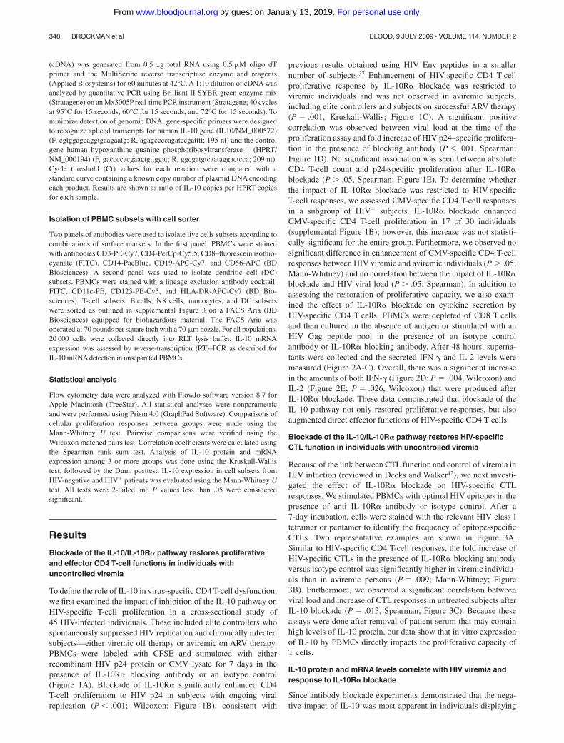
To define the role of IL-10 in virus-specific CD4 T-cell dysfunction,we first examined the impact of inhibition of the IL-10 pathway onHIV-specific T-cell proliferation in a cross-sectional study of45 HIV-infected individuals. These included elite controllers whospontaneously suppressed HIV replication and chronically infectedsubjects—either viremic off therapy or aviremic on ARV therapy.PBMCs were labeled with CFSE and stimulated with eitherrecombinant HIV p24 protein or CMV lysate for 7 days in thepresence of IL-10R� blocking antibody or an isotype control(Figure 1A). Blockade of IL-10R� significantly enhanced CD4T-cell proliferation to HIV p24 in subjects with ongoing viralreplication (P � .001; Wilcoxon; Figure 1B), consistent with
previous results obtained using HIV Env peptides in a smallernumber of subjects.37 Enhancement of HIV-specific CD4 T-cellproliferative response by IL-10R� blockade was restricted toviremic individuals and was not observed in aviremic subjects,including elite controllers and subjects on successful ARV therapy(P � .001, Kruskall-Wallis; Figure 1C). A significant positivecorrelation was observed between viral load at the time of theproliferation assay and fold increase of HIV p24–specific prolifera-tion in the presence of blocking antibody (P � .001, Spearman;Figure 1D). No significant association was seen between absoluteCD4 T-cell count and p24-specific proliferation after IL-10R�blockade (P .05, Spearman; Figure 1E). To determine whetherthe impact of IL-10R� blockade was restricted to HIV-specificT-cell responses, we assessed CMV-specific CD4 T-cell responsesin a subgroup of HIV� subjects. IL-10R� blockade enhancedCMV-specific CD4 T-cell proliferation in 17 of 30 individuals(supplemental Figure 1B); however, this increase was not statisti-cally significant for the entire group. Furthermore, we observed nosignificant difference in enhancement of CMV-specific CD4 T-cellresponses between HIV viremic and aviremic individuals (P .05;Mann-Whitney) and no correlation between the impact of IL-10R�blockade and HIV viral load (P .05; Spearman). In addition toassessing the restoration of proliferative capacity, we also exam-ined the effect of IL-10R� blockade on cytokine secretion byHIV-specific CD4 T cells. PBMCs were depleted of CD8 T cellsand then cultured in the absence of antigen or stimulated with anHIV Gag peptide pool in the presence of an isotype controlantibody or IL-10R� blocking antibody. After 48 hours, superna-tants were collected and the secreted IFN-� and IL-2 levels weremeasured (Figure 2A-C). Overall, there was a significant increasein the amounts of both IFN-� (Figure 2D; P � .004, Wilcoxon) andIL-2 (Figure 2E; P � .026, Wilcoxon) that were produced afterIL-10R� blockade. These data demonstrated that blockade of theIL-10 pathway not only restored proliferative responses, but alsoaugmented direct effector functions of HIV-specific CD4 T cells.
Blockade of the IL-10/IL-10R� pathway restores HIV-specificCTL function in individuals with uncontrolled viremia
Because of the link between CTL function and control of viremia inHIV infection (reviewed in Deeks and Walker42), we next investi-gated the effect of IL-10R� blockade on HIV-specific CTLresponses. We stimulated PBMCs with optimal HIV epitopes in thepresence of anti–IL-10R� antibody or isotype control. After a7-day incubation, cells were stained with the relevant HIV class Itetramer or pentamer to identify the frequency of epitope-specificCTLs. Two representative examples are shown in Figure 3A.Similar to HIV-specific CD4 T-cell responses, the fold increase ofHIV-specific CTLs in the presence of IL-10R� blocking antibodyversus isotype control was significantly higher in viremic individu-als than in aviremic persons (P � .009; Mann-Whitney; Figure3B). Furthermore, we observed a significant correlation betweenviral load and increase of CTL responses in untreated subjects afterIL-10 blockade (P � .013, Spearman; Figure 3C). Because theseassays were done after removal of patient serum that may containhigh levels of IL-10 protein, our data show that in vitro expressionof IL-10 by PBMCs directly impacts the proliferative capacity ofT cells.
IL-10 protein and mRNA levels correlate with HIV viremia andresponse to IL-10R� blockade
Since antibody blockade experiments demonstrated that the nega-tive impact of IL-10 was most apparent in individuals displaying
348 BROCKMAN et al BLOOD, 9 JULY 2009 � VOLUME 114, NUMBER 2
For personal use only.on January 13, 2019. by guest www.bloodjournal.orgFrom
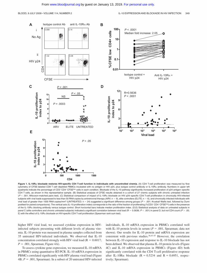
higher HIV viral load, we assessed cytokine expression in HIV-infected subjects presenting with different levels of plasma vire-mia. IL-10 protein was measured in plasma samples collected from35 untreated HIV-infected individuals. We observed that IL-10concentration correlated strongly with HIV viral load (R � 0.6017;P � .001, Spearman; Figure 4A).
To assess cytokine gene expression, we measured IL-10 mRNAin PBMCs using quantitative RT-PCR. IL-10 mRNA expression inPBMCs correlated significantly with HIV plasma viral load (Figure4B; P � .001, Spearman). In a subset of 20 untreated HIV-infected
individuals, IL-10 mRNA expression in PBMCs correlated wellwith IL-10 protein levels in serum (P � .001, Spearman; data notshown). Our results for IL-10 protein and mRNA expression areconsistent with previous studies.36,43-47 However, the correlationbetween IL-10 expression and response to IL-10 blockade has notbeen defined. We observed that plasma IL-10 protein levels (Figure4C) and IL-10 mRNA expression in PBMCs (Figure 4D) bothsignificantly correlated with the CD4 T-cell proliferative responseafter IL-10R� blockade (R � 0.5234 and R � 0.6951, respec-tively; Spearman).
A
0 102 103 104 105
0
103
104
105 0.14 99.9
000 102 103 104 105
0
103
104
105 0.099 99.9
00
0 102 103 104 105
0
103
104
105 3.08 97
000 102 103 104 105
0
103
104
105 1.32 98.6
00
Isotype control Ab anti IL-10Rα Ab
No Ag
HIV p24
B
D
E
0.01
0.1
1
10
100
%C
FS
E lo
w
CD
4+ c
ells
P < .0001Median fold increase: 2.05
Isotype control +HIV p24
Anti IL-10Rα +HIV p24
C
0
2
4
6
8
10
12NS
P < .05
P < .001
IL-1
0 p
rolif
erat
ion
ind
ex t
o p
24C
D4
CFSE
i
ii
101 102 103 104 105 1060
3
6
9
12
Viral load
IL-1
0 p
rolif
erat
ion
ind
e x t
o p
2 4
R=0.5636P < .0001
P = .0003
ARV ELITE UNTREATED
0 500 1000 15000
3
6
9
12
CD4
P > .05
IL-1
0 p
r olif
erat
ion
ind
e x t
o p
2 4
Figure 1. IL-10R� blockade restores HIV-specific CD4 T-cell function in individuals with uncontrolled viremia. (A) CD4 T-cell proliferation was measured by flowcytometry of CFSE-labeled CD8 T cell–depleted PBMCs incubated with no antigen or HIV p24, plus isotype control antibody or IL-10R� antibody. Numbers in upper leftquadrants indicate the percentage of CD3�CD4�CFSElow cells in each condition. Blockade of the IL-10 pathway significantly increased proliferation of p24 antigen–specificCD4 T cells, as shown in this representative sample. (B) Statistical analysis of CFSE results obtained in a cohort of 27 viremic subjects with chronic untreated infection(P � .001; Wilcoxon-matched pairs test). (C) Statistical comparison of impact of IL-10R� blockade on HIV p24–specific CD4 T-cell proliferation in chronically HIV-infectedsubjects with viral loads suppressed to less than 50 RNA copies by antiretroviral therapy (ARV; n � 9), elite controllers (ELITE; n � 6), and chronically infected individuals withviral load of greater than 1000 RNA copies/mm3 (UNTREATED; n � 24) suggested a significant difference among groups (P � .001; Kruskall Wallis test, followed by Dunnposttest for paired comparisons). The vertical axis (IL-10 proliferation index) corresponds to the ratio of the fraction of proliferating (%CD3�CD4�CFSElow) cells in the presenceof the IL-10R� blocking antibody versus isotype control. Short horizontal bars indicate median proliferation index. (D,E) Statistical analysis of data on untreated subjects inpanel C (elite controllers and chronic untreated subjects) indicated a significant correlation between viral load (R � 0.5636; P � .001) in panel D, but not CD4 count (P .05;E) with the effect of IL-10R� blockade on HIV-specific CD4 T-cell proliferation (Spearman rank sum test).
IL-10 EXPRESSION AND BLOCKADE IN HIV INFECTION 349BLOOD, 9 JULY 2009 � VOLUME 114, NUMBER 2
For personal use only.on January 13, 2019. by guest www.bloodjournal.orgFrom

Relationships between IL-10 mRNA expression and diseasestage
To further examine the impact of HIV infection and treatmentstatus on IL-10 expression, we measured IL-10 mRNA in PBMCscollected from 10 healthy HIV-negative subjects and 4 cohorts ofchronically HIV-infected individuals (Figure 5): 15 elite control-lers; 20 viremic, untreated subjects; 12 persons with uncontrolledviremia on ARV therapy; and 11 individuals with successful ARVtreatment. IL-10 mRNA levels differed among these groups(P � .002; Kruskal-Wallis), with a significant elevation of IL-10mRNA in individuals with ongoing viremia, either untreated or onARV, compared with elite controllers and uninfected controls(P � .01 for each, comparison Dunn posttest; Figure 5). Incontrast, IL-10 mRNA expression was similar in HIV controllersand uninfected individuals. Intermediate levels of IL-10 mRNAwere observed in subjects with viral loads lower than 50 copies/mLon ARV therapy, compared with the other 4 groups. Taken together,these results suggest that the reduced IL-10 expression observedupon successful ARV therapy is a consequence of decreased HIVviral load and does not result from a direct effect of ARV drugs onIL-10 expression.
Variations in IL-10 expression during periods of rapid viral loadchanges
Previous studies have indicated that plasma IL-10 protein levels areelevated during acute HIV infection.48 To better understand the linkbetween HIV viremia and IL-10 expression during non–steady-state disease, we first examined IL-10 protein concentration inlongitudinal samples collected from 3 individuals, beginning in
acute infection (Figure 6A-C). Substantial levels of plasma IL-10protein were detected at the earliest time point available; theydeclined rapidly during the first 6 months after infection in the2 untreated subjects (Figure 6A-B) as well as in the treatedindividual (Figure 6C), suggesting that resolution of acute infectionwas associated with a decline in plasma IL-10, even in the absenceof viral control. This analysis was extended to a total of 10individuals for whom acute/early (within 30 days after infection)and chronic (6-12 months after infection) samples were available,and we saw a significant decline in IL-10 protein concentration atthe later time point (P � .004, Wilcoxon; Figure 6D). Next, weanalyzed IL-10 mRNA and protein levels in longitudinal samplescollected from 2 individuals who discontinued ARV therapy(Figure 6E-F). In the absence of treatment, we observed a rapid risein HIV viremia, but only a gradual increase of plasma IL-10 proteinand mRNA expression in PBMCs for both individuals. Reinitiationof ARV treatment resulted in a marked reduction of both IL-10protein and mRNA, suggesting that the pathogenic process respon-sible for IL-10 induction is at least partially reversible.
IL-10 is up-regulated by multiple PBMC subsets in HIV-infectedindividuals
Monocytes have been reported to be major producers of IL-10 inHIV-infected individuals,37 but numerous cell types can expressthis cytokine (reviewed in Couper et al16 and Moore et al17). Tobetter identify IL-10–producing cell populations, we first usedantibody-conjugated magnetic beads to isolate cell subsets inPBMCs from HIV-infected subjects and healthy uninfected con-trols. We measured IL-10 mRNA expression using quantitativeRT-PCR and observed IL-10 mRNA up-regulation in all cellular
B CA
0
200
400
600
0
10
20
0
500
1000
1500
D E
0
2
4
6
8
0
2
4
6
8
0
10
20
30
Anti-IL-10Rα
HIV Gag pool
Anti-IL-10Rα
HIV Gag pool
P = .026410000
1000
100
10
1
P = .0035
Isotype control +HIV Gag pool
100
10
1
0.1
Isotype control +HIV Gag pool
Anti IL-10Rα +HIV Gag pool
Anti IL-10Rα +HIV Gag pool
IL-2
(pg
/mL)
IL-2
(pg
/mL)
IFN
-γ (
pg/m
L)
IFN
-γ (
pg/m
L)
IFN
-γ (
pg/m
L)IL
-2 (
pg/m
L)
IL-2
(pg
/mL)
IFN
-γ (
pg/m
L)
Figure 2. IL-10-R� blockade enhances cytokine secretion by HIV-specific CD4� T cells. (A-C) Cytokine secretion by HIV-specific CD4T-cell proliferation was measured by multiplex immunoassay in superna-tants of CD8 T cell–depleted PBMCs incubated with no antigen or an HIVGag peptide pool, plus isotype control antibody or IL-10R� antibody.Blockade of the IL-10 pathway increased IFN-� and IL-2 cytokine secretionby HIV-specific CD4 T cells, as shown in these 3 representative subjects.(D-E) Statistical analysis of data on 14 untreated subjects indicated asignificant increase of both IFN-� (P � .004; D) and IL-2 (P � .026; E) inthe presence of IL-10R� blockade (Wilcoxon matched pairs test).
350 BROCKMAN et al BLOOD, 9 JULY 2009 � VOLUME 114, NUMBER 2
For personal use only.on January 13, 2019. by guest www.bloodjournal.orgFrom
subsets isolated from HIV-infected subjects versus controls (supple-mental Figure 2), which were statistically significant for totalPBMCs and cellular populations isolated using antibodies recogniz-ing CD3, CD11b, CD11c, CD14, CD16, CD19, and CD56 (allP � .05; Mann-Whitney). These results suggest that in addition toCD14� monocytes, several other cell types contribute to theincreased IL-10 expression observed during chronic HIV infection.
To confirm findings obtained using bead-sorted cell subsets andto define the IL-10–producing populations with great accuracy, weused a live-cell fluorescence-activated cell sorting (FACS) sortingapproach to isolate CD123� plasmacytoid dendritic cells (pDCs),CD11c� myeloid dendritic cells (mDCs), CD14� monocytes,CD4� and CD8� T cells, CD19� B cells, and CD56� NK cellsfrom 10 HIV-infected individuals and 9 uninfected controls (for
Tet
ram
er
Isotype Control Ab
0 102 103 104 105
0102
103
104
105 1.36
0 102 103 104 105
0102
103
104
105 3.63
Anti IL-10Rα Ab
CD8
BA
0
2
4
6
8
10253035
P = .0089
AVIR
IL-1
0 p
rolif
erat
ion
ind
ex f
or
Tet
+ C
D8
cells
0 103 104 105
0
103
104
105 0 1.39
98.600 103 104 105
0
103
104
105 0 5.74
94.30
B*2
7-K
K10
A
*02-
SL9
VIR
101 102 103 104 105 1060
2
4
6
8
1030
40
Viral load
IL-1
0 p
rolif
erat
ion
ind
ex f
or
Tet
+ C
D8
cells
R=0.5445P = .0130C
Figure 3. IL-10R� blockade restores HIV-specific CTL function in individuals with uncontrolled viremia. (A) Flow cytometry was used to assess proliferation ofHIV-specific HLA class I tetramer–labeled CD8 T cells from 2 subjects incubated with the cognate HIV epitope, plus isotype control antibody or IL-10R� blocking antibody.Representative data from 2 subjects is shown. Numbers in upper right quadrants indicate the percentage of CD3�CD8�Tetramerhigh cells for each condition. (B) Statisticalcomparison of impact of IL-10R� blockade on HIV p24–specific Tet� CD8 T-cell proliferation in chronically HIV-infected subjects with undetectable viral loads (AVIR; n � 7),and chronically infected individuals with viral loads of greater than 1000 RNA copies/mm3 (VIR; n � 13) demonstrated a significant difference between these groups (P � .009;Mann-Whitney U test). The vertical axis (IL-10 proliferation index) corresponds to the ratio of the fraction of Tet� cells (%CD3�CD8�Tethigh) cells in the presence of the IL-10R�blocking antibody versus isotype control. Short horizontal bars indicate median proliferation index. (C) Statistical analysis of the data on subjects in panel B indicated asignificant correlation between untreated viral load and the effect of IL-10R� blockade on HIV-specific CD8 T-cell proliferation (R � 0.5445, P � .013; Spearman rank sumtest).
pla
sma
IL-1
0 (p
g/m
L)
A B
101 102 103 104 105 1060
10
20
30
40
50
60
10 2 10 3 10 4 10 5 10 60.0
2.5
5.0
7.5
10.0R=0.6754P = .0011
Viral Load (vRNA copies/mL)
R=0.6017P = .0001
IL-1
0 R
NA
(n
orm
.)
Viral Load (vRNA copies/mL)
D
0 10 20 30 40 50 600
2
4
610
12
plasma IL-10 (pg/mL)
IL-1
0 p
rolif
erat
ion
ind
ex t
o p
24
C
0.0 2.5 5.0 7.5 10.0 12.50
2
4
6
R= 0.6951P < .001
10
12
IL-10 RNA (norm.)
IL-1
0 p
r olif
erat
ion
ind
ex t
o p
24
R=0.5324P = .0338
Figure 4. IL-10 expression correlates with HIV viremia andresponse to IL-10R� blockade. The level of IL-10 protein inplasma and mRNA expression in bulk PBMCs were examined in35 untreated HIV� individuals. IL-10 protein concentration(A) and IL-10 mRNA expression values (B) correlated signifi-cantly with plasma viral load measurements (R � 0.6017 andR � 0.6754, respectively). Next, the impact of IL-10R� block-ade on HIV p24–specific CD4 T cells was correlated with IL-10protein or mRNA results. The IL-10 proliferation index, definedas the ratio of the fraction of proliferating (%CD3�CD4�CFSElow)cells in the presence of the IL-10R� blocking antibody versusisotype control, indicated a significant correlation between anindividual’s responsiveness to IL-10 pathway blockade and bothplasma IL-10 protein (R � 0.5324, P � .034) (C) or PBMCIL-10 mRNA (R � 0.6951, P � .001). (D) Correlation coeffi-cients suggest that IL-10 mRNA expression in PBMCs is abetter predictor of response to IL-10 blockade than plasmaIL-10 protein concentration. All statistical analyses used theSpearman rank sum test.
IL-10 EXPRESSION AND BLOCKADE IN HIV INFECTION 351BLOOD, 9 JULY 2009 � VOLUME 114, NUMBER 2
For personal use only.on January 13, 2019. by guest www.bloodjournal.orgFrom

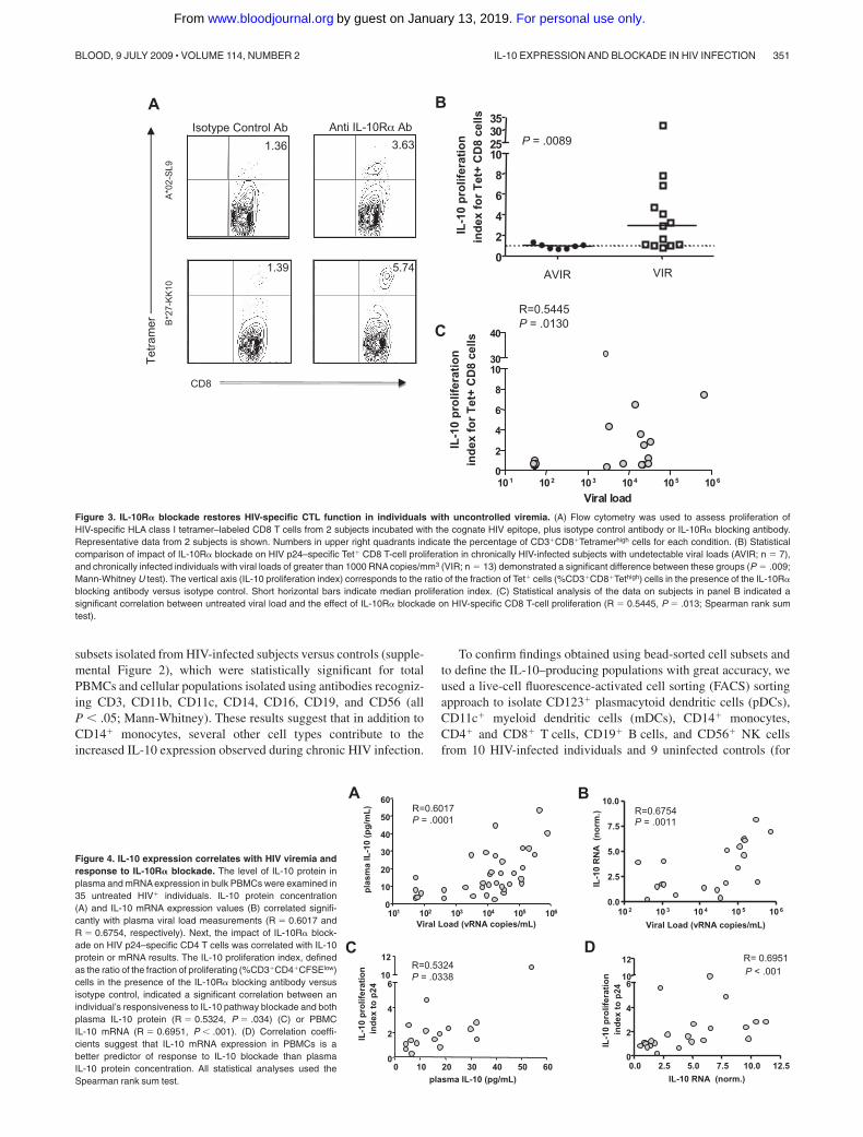
gating strategies, see supplemental Figure 3). Despite majordifferences in the frequencies of the cell populations sorted (range:� 0.1% for DC subsets to 10% for T-cell subsets), purities ofthese sorted populations were comparable and consistently higherthan 98%. IL-10 mRNA expression was analyzed in PBMCs and ineach purified subset using quantitative RT-PCR (Figure 7). IL-10mRNA levels in the total PBMCs from these HIV-infected individu-als were significantly elevated relative to healthy controls (P � .028,
Mann Whitney). In addition, we observed a significant elevation ofIL-10 in CD14� monocytes (P � .035), CD4� T cells (P � .001),CD8� T cells (P � .002), CD19� B cells (P � .001), and CD56�
NK cells (P � .004) in HIV� individuals (Figure 7D-H; allstatistics: Mann Whitney). In contrast, we did not see a significantdifference in IL-10 expression for the CD123� pDC subset(P � .161; Figure 7B) in HIV-infected individuals compared withHIV-negative controls, and observed a decline in IL-10 expressionin CD11c� mDCs (P � .028). The difference between IL-10mRNA levels observed in CD11c� isolated by beads and CD11c�
DCs sorted by FACS is likely due to the mixed populations inbead-sorted cells that contain a majority of CD11c-expressingmonocytes and a minority of CD11c� DCs. These data demonstratethat IL-10 expression is induced in multiple PBMC subsets inHIV-infected individuals and identify potential candidate suppres-sor cell populations.
Discussion
In this study, we examined IL-10 expression and the ability of thiscytokine to suppress HIV-specific T-cell function in HIV-infectedindividuals. Our results indicate that IL-10 mRNA expression isincreased in the setting of chronic uncontrolled HIV infection andthat IL-10 mRNA expression levels correlate with plasma viremiain infected persons. Increased IL-10 mRNA levels were identifiedin multiple different hematopoietic cells, and IL-10R� blockaderestored not only HIV-specific CD4 cell proliferation but alsoantigen-specific CD8 T-cell proliferation, although these effectswere seen only in viremic subjects, as well as cytokine secretion byHIV-specific CD4 T cells. Both IL-10 mRNA expression and IL-10
0 100 200 300 400 500101
102
103
104
105
106
107
IL-1
0 pr
otei
n(p
g/m
L) (
—) V
iral Load(R
NA
copies/mL)
( --
-)
Time of analysis (days)
Time following infection (days)25 50 75 100 125 150 175 200 225
0
25
50
75
100
0 100 200 300 400 500
0 500 1000 1500 2000 25000
1
2
101
102
103
104
105
106
5
15
IL-1
0 R
NA
(nor
m.)
(—
)
IL-1
0 pr
otei
n(p
g/m
L) (
—)
0 250 500 750 1000 1250 1500012345
101
102
103
104
105
106
10
20
30
P = .0039 Viral Load
(RN
A copies/m
L)(-
--)
IL-1
0 pr
otei
n(p
g/m
L) (
—)
Acute/Early Chronic0
50
100
150
200
A B C
D E F
Figure 6. Longitudinal analysis during acute infection and the impact of therapy on IL-10 suggest an indirect link with plasma viremia. Plasma IL-10 proteinconcentration was measured in 3 individuals identified during acute/early HIV infection. Longitudinal samples indicated that IL-10 protein levels (solid line) were initially high,and decreased over time in 2 untreated (A-B) as well as 1 treated subject (C), corresponding with a decline in HIV viral load (dashed line). A statistically significant decline inIL-10 protein (P � .004; Wilcoxon matched pairs test) in panel D was observed in a cross-sectional analysis of 10 HIV� individuals captured at acute/early (within the first30 days after infection) and chronic (6-12 months after infection) time points. IL-10 protein and mRNA expression were analyzed in 2 chronically infected individuals whointerrupted antiretroviral therapy. Initially, both subjects displayed undetectable plasma viral loads while on ARV, which increased substantially in the absence of treatment(E-F). Plasma IL-10 protein concentration (�) as well as IL-10 mRNA expression (�) increased slowly over time in both individuals while off of therapy, but declined afterreinitiation of suppressive antiretroviral treatment. Time points on ARV therapy are indicated by shaded regions in panels B-C and E-F.
0.0
2.5
5.0
7.5
10.0 P < .001
P < .01
P < .001
P < .001
P < .0001
HIV-n
eg
ELITE
UNTREATED
VIR A
RV
AVIR A
RV
IL-1
0 R
NA
(n
orm
.)
Figure 5. Relationships between IL-10 mRNA expression and disease stage.IL-10 mRNA levels in bulk PBMCs were measured in HIV-negative subjects (n � 10;HIV-neg), HIV-infected elite controllers (n � 15; ELITE), HIV� untreated patients(n � 20; UNTREATED), and antiretroviral drug-treated individuals, which includedsome subjects with suppressed viremia less than 50 copies/mL on HAART (n � 11;AVIR ARV) and others who remained viremic despite therapy (n � 12; VIR ARV).A significant difference was seen among these groups (P � .001; Kruskal Wallis test),reflecting a significant increase in IL-10 expression in HIV� untreated and viremic-treated individuals compared with HIV-negative and elite controller subjects (P � .01for all, Dunn posttest).
352 BROCKMAN et al BLOOD, 9 JULY 2009 � VOLUME 114, NUMBER 2
For personal use only.on January 13, 2019. by guest www.bloodjournal.orgFrom
plasma levels were reduced through successful ARV treatment,indicating a direct effect of viral antigen load on IL-10 production.These data show that IL-10 contributes to a reversible T-celldysfunction in HIV infected persons, and that viral antigen is amajor driver of the increased levels of IL-10 observed.
Blockade of the IL-10 pathway significantly increased HIV-specific CD4 and CD8 T-cell proliferation, and also augmentedeffector T-cell functions, as shown by the enhanced secretion of thecytokines IFN-� and IL-2 by antigen-specific CD4 T cells uponIL-10R� blockade. Although less consistent, IL-10R� blockadealso increased CMV-specific CD4 T proliferation in HIV-infectedindividuals. These data show that IL-10 contributes to a broadreversible T-cell dysfunction. Significant positive correlations wereobserved between the fold increase in HIV p24–specific prolifera-tion after blockade and plasma viral load or IL-10 mRNA. Thevariable enhancement of T-cell responses after IL-10R� blockadeobserved in individuals with similar HIV viremia is consistent witha significant heterogeneity in the degree of T-cell exhaustion andactivity of different inhibitory pathways among HIV-infectedsubjects. It is notable that we observed enhancement of HIV-specific T-cell proliferative responses upon IL-10R� blockade onlyin individuals with uncontrolled viral replication. Elite controllers
and individuals who suppressed viral replication while on success-ful ARV treatment failed to respond to IL-10R� blockade. Anassociation between IL-10 and viremia has been observed previ-ously,39 and this may help to explain inconsistent results of studieswhere individuals were not stratified on the basis of viral load.49
Consistent with results of IL-10R� blockade, we observed thatboth plasma IL-10 protein levels and IL-10 mRNA expression inPBMCs were elevated in chronically HIV-infected individuals. Asignificant positive correlation between IL-10 protein and IL-10mRNA suggests that IL-10 expression by PBMCs is representativeof IL-10 production in cellular compartments that contribute toplasma IL-10. IL-10 protein and mRNA levels were significantlyhigher in viremic individuals than in subjects with undetectableviral load, and directly correlated with plasma viremia. Weperformed longitudinal analyses to further address the relationshipbetween viral load and IL-10 up-regulation. Our data from10 individuals confirm previous work demonstrating elevatedplasma IL-10 levels at the time of acute infection,48 concurrent withvery high HIV viral load, which declined significantly afterresolution of primary infection, regardless of continued viremia.Conversely, our results from 2 additional patients illustrated thatinterruption of ARV therapy resulted in elevation of HIV viral loads
A B
C D
E F
G H
PBMC
HIV-neg (N=9) HIV + (N=10)0.001
0.01
0.1
1
10 P value .0279
CD123+ DCs
HIV-neg (N=8) HIV + (N=8)0.001
0.01
0.1
1
10 P value .1605
CD11c+ DCs
HIV-neg (N=8) HIV + (N=8)0.001
0.01
0.1
1
10 P value .0379
CD14+ monocytes
HIV-neg (N=9) HIV + (N=10)0.001
0.01
0.1
1
10 P value .0350
CD4+ T cells
HIV-neg (N=9) HIV + (N=10)0.001
0.01
0.1
1
10 P value .0006
CD8+ T cells
HIV-neg (N=9) HIV + (N=10)0.001
0.01
0.1
1
10 P value .0015
CD19+ B cells
HIV-neg (N=9) HIV + (N=10)0.001
0.01
0.1
1
10 P value P < .0001
CD56+ NK cells
HIV-neg (N=9) HIV + (N=10)0.001
0.01
0.1
1
10 P value .0041
Figure 7. Broad expression of IL-10 mRNA in FACS-sorted cellsubsets from HIV-infected subjects. CD11c� myeloid DCs,CD123� plasmacytoid DCs, CD14� monocytes, CD4� T cells,CD8� T cells, CD19� B cells, and CD56� NK cells were isolatedfrom total PBMCs using live cell FACS sorting from 9 HIV-negativeand 10 HIV-infected individuals, as described in supplementalFigure 3. IL-10 mRNA expression was determined in PBMCs andeach subset by quantitative RT-PCR, and values are presented asIL-10 copies relative to the HPRT housekeeping gene (A-H).Significant up-regulation of IL-10 mRNA was observed in HIV�
individuals compared with HIV-negative controls for total PBMCs(P � .028) in panel A, CD14� monocytes (P � .035) in panel D,CD4� T cells (P � .001) in panel E, CD8� T cells (P � .002) inpanel F, CD19� B cells, (P � .001) in panel G, and CD56� NK cells(P � .004) in panel H. A significant reduction in IL-10 expressionwas observed in CD11c� mDCs (P � .038) (C), whereas nochange was seen in CD123� pDCs (P � .161) in panel B. Allstatistics were calculated using the Mann-Whitney U test.
IL-10 EXPRESSION AND BLOCKADE IN HIV INFECTION 353BLOOD, 9 JULY 2009 � VOLUME 114, NUMBER 2
For personal use only.on January 13, 2019. by guest www.bloodjournal.orgFrom
and subsequent increases in IL-10 protein and mRNA levels.Reinstitution of ARV treatment led to a substantial decline in bothmeasures of IL-10. In each of these cases, changes in IL-10 weredelayed relative to changes in plasma viremia. Additional studieswill be necessary to define whether direct stimulation of the IL-10pathway in immune cells by viral products, indirect feedbackmechanisms due systemic immune hyperactivation, or both, lead toup-regulation of IL-10 in viremic individuals.
These data raise important questions about the cellular sourcesof IL-10 in viremic individuals. In the murine LCMV model, recentstudies by Brooks et al29 and Ejrnaes et al30 reach differentconclusions, either favoring the hypothesis that DCs from chroni-cally infected mice induce IL-10 up-regulation by CD4 T cells, orconcluding that regulatory T cells are the major source of IL-10during chronic LCMV infection, respectively. A similar contro-versy exists for HIV infection in humans as well. Early studiesassessing cytokines in culture supernatants of PBMCs stimulatedwith PHA suggested an up-regulation of IL-10 and a switch from aTh1 to a Th2 profile in T cells.50 This hypothesis was subsequentlychallenged by other reports.49 Trabattoni et al51 showed thatmonocytes, T cells, and B cells from HIV-infected individualsproduce more IL-10 than HIV-uninfected controls, but IL-10production by other subsets has been poorly characterized.
Our results provide a detailed assessment of IL-10 mRNAexpression by several PBMC subsets. Consistent with previousstudies,51 our assays suggest that CD14� monocytes are a majorsource of IL-10 in both HIV-infected and HIV-uninfected individu-als. However, analysis of highly purified cellular subsets obtainedby FACS indicated that CD14� monocytes showed a less dramaticup-regulation of IL-10 mRNA in HIV-infected subjects than someother subsets examined in our study, including CD19� B cells andT cells. In addition, CD11c� myeloid DCs, which are known toproduce significant amounts of IL-10 under certain physiologicalconditions, appeared to produce less IL-10 in chronic HIV-infectedindividuals compared with uninfected controls. This result is incontrast with data obtained at the time of acute viral infection in theLCMV model29 and suggests that the IL-10–producing cell popula-tions may differ among viral diseases, or perhaps vary in tissuelocation or change over time after acute infection. Such differencesin IL-10–producing subsets may contribute to the broad spectrumof outcomes observed upon blockade of the IL-10 pathway inseveral murine models of infectious diseases. Previous reports haveindicated that up-regulation of IL-10 by monocytes may be due to adirect effect of HIV Env52 and that the Env of different HIV strainscould induce various degrees of IL-10 secretion by monocyte-derived dendritic cells (MDDCs).53 However, we found no signifi-cant increase of IL-10 mRNA in pDCs and mDCs. The HIV Tatprotein has also been implicated in up-regulation of IL-10,54 andsoluble Tat may correlate with HIV viral load in untreatedinfection.55 Additional studies will be necessary to determinewhether IL-10–secreting cell populations differ in acute andchronic HIV infections and to define the cell types that are criticalfor IL-10–mediated T-cell inhibition. In particular, the role ofregulatory T cells remains to be better defined. Altogether, our datademonstrate that IL-10 expression by several diverse cell popula-tions, rather than induction of IL-10 by a single cell type, may be akey factor leading to the increase in IL-10 levels observed inHIV-infected subjects.
Interventional studies to block IL-10 activity have been pro-posed to enhance immunity in chronic human viral infections.Besides its impact on T-cell responses and viral load in established
LCMV infection,29,30 blockade of the IL-10 pathway also allowedan otherwise ineffective therapeutic DNA vaccine to stimulateantiviral immunity and enhance viral clearance.40 IL-10 blockademay therefore be a promising adjuvant therapy to boost preventiveor therapeutic vaccines. However, caution is needed with suchapproaches in HIV infection, since the relationship between IL-10and HIV disease progression is complex. First, IL-10 blockadedoes not enhance HIV-specific T-cell function in our assays forsubjects optimally treated with ARV therapy. If these findings invitro correlate with activity of the IL-10 pathway in vivo, this maysuggest that the potential impact of IL-10 blockade as a therapywould be limited in an era when optimal suppression of viralreplication is the goal of patient care. Second, whereas our study,and others, show that IL-10 levels increase with disease progres-sion and can mediate T-cell dysfunction, a genetic polymorphism inthe IL-10 promoter that leads to a decrease in IL-10 expression(�592C A) has been associated with more rapid disease progres-sion in late stages of HIV.56 A second IL-10 promoter polymor-phism (�1082A G) that may increase IL-10 expression has beenassociated with slower CD4 decline and longer survival in HIV�
individuals.57 Given the crucial role of immune activation in HIVdisease progression,58 it is possible that despite its inhibitory effecton T cells, IL-10 has a net beneficial impact during later stages ofHIV infection by limiting systemic immune hyperactivation andCD4 T-cell loss. Third, the direct effects of IL-10 on virusproduction are complicated, as IL-10 has been reported to inhibitHIV replication in monocytes,52,59 but also to induce expression ofCCR5 in CD4 T cells,60 which may hasten infection. A placebo-controlled trial investigating the tolerance and impact of recombi-nant IL-10 therapy on parameters of disease progression inHIV-infected individuals did not show significant changes in eitherviral load or CD4 T-cell counts during 4 weeks of treatment.61
Additional studies are required to better understand the overallimpact of the IL-10 pathway in viremic individuals, which likelycombines the detrimental effect of IL-10 on HIV-specific CD4 andCD8 T-cell immunity with a beneficial down-regulation of sys-temic immune activation. Our data showing IL-10 mRNA up-regulation in multiple cell types of HIV-infected subjects suggestthat, beyond T-cell responses, IL-10 could broadly alter innate andadaptive immune functions in HIV infection. It will therefore beimportant to develop new technical approaches selectively target-ing this pathway in specific cellular subsets. Such tools will provideadditional insight into HIV pathogenesis and may pave the pathtoward novel therapeutic interventions to enhance virus-specificimmunity.
Acknowledgments
We thank the clinical and laboratory staff at the MassachusettsGeneral Hospital and that of The International HIV ControllersStudy (http://www.hivcontrollers.org). We thank all study partici-pants for their invaluable role in this project.
This study was supported by the National Heart, Lung andBlood Institute of the National Institutes of Health (Bethesda, MD;R01-HL092565 [D.E.K.]), the Concerned Parents for AIDS Re-search Foundation (D.E.K., B.D.W.), the Howard Hughes MedicalInstitute (B.D.W.), the Bill and Melinda Gates Foundation (Seattle,WA), and a gift from the Mark and Lisa Schwartz Foundation.
354 BROCKMAN et al BLOOD, 9 JULY 2009 � VOLUME 114, NUMBER 2
For personal use only.on January 13, 2019. by guest www.bloodjournal.orgFrom

Authorship
Contribution: D.E.K. was responsible for the overall design andconduct and provided supervision; M.A.B., D.S.K., D.G.K., B.D.W.,and D.E.K provided intellectual input and contributed to theexperimental design; M.A.B., D.S.K., D.P.T., D.F.P., P.C.R., J.S.,F.P., S.L.G., and D.E.K. performed experiments; S.L.G., K.M.,
H.J., and F.P. provided clinical samples; M.T.W. provided technicalassistance for flow cytometry and cell sorting; and M.A.B., D.S.K.,and D.E.K wrote the paper.
Conflict-of-interest disclosure: The authors declare no compet-ing financial interests.
Correspondence: Daniel E. Kaufmann, Massachusetts GeneralHospital East, Partners AIDS Research Center, Rm 6618B, 14913th St, Charlestown, MA 02129; e-mail: [email protected].
References
1. Munier ML, Kelleher AD. Acutely dysregulated,chronically disabled by the enemy within: T-cellresponses to HIV-1 infection. Immunol Cell Biol.2007;85:6-15.
2. Shin H, Wherry EJ. CD8 T cell dysfunction duringchronic viral infection. Curr Opin Immunol. 2007;19:408-415.
3. Chougnet C, Gessani S. Role of gp120 in den-dritic cell dysfunction in HIV infection. J LeukocBiol. 2006;80:994-1000.
4. Piguet V, Steinman RM. The interaction of HIVwith dendritic cells: outcomes and pathways.Trends Immunol. 2007;28:503-510.
5. Barber DL, Wherry EJ, Masopust D, et al. Restor-ing function in exhausted CD8 T cells duringchronic viral infection. Nature. 2006;439:682-687.
6. Wherry EJ, Ha SJ, Kaech SM, et al. Molecularsignature of CD8� T cell exhaustion duringchronic viral infection. Immunity. 2007;27:670-684.
7. Day CL, Kaufmann DE, Kiepiela P, et al. PD-1expression on HIV-specific T cells is associatedwith T-cell exhaustion and disease progression.Nature. 2006;443:350-354.
8. Petrovas C, Casazza JP, Brenchley JM, et al.PD-1 is a regulator of virus-specific CD8� T cellsurvival in HIV infection. J Exp Med. 2006;203:2281-2292.
9. Trautmann L, Janbazian L, Chomont N, et al. Up-regulation of PD-1 expression on HIV-specificCD8� T cells leads to reversible immune dys-function. Nat Med. 2006;12:1198-1202.
10. Maier H, Isogawa M, Freeman GJ, Chisari FV.PD-1:PD-L1 interactions contribute to the func-tional suppression of virus-specific CD8� T lym-phocytes in the liver. J Immunol. 2007;178:2714-2720.
11. Golden-Mason L, Palmer B, Klarquist J,Mengshol JA, Castelblanco N, Rosen HR. Up-regulation of PD-1 expression on circulating andintrahepatic hepatitis C virus-specific CD8�T cells associated with reversible immune dys-function. J Virol. 2007;81:9249-9258.
12. Urbani S, Amadei B, Tola D, et al. Restoration ofHCV-specific T cell functions by PD-1/PD-L1blockade in HCV infection: effect of viremia levelsand antiviral treatment. J Hepatol. 2008;48:548-558.
13. Kaufmann DE, Kavanagh DG, Pereyra F, et al.Upregulation of CTLA-4 by HIV-specific CD4(�)T cells correlates with disease progression anddefines a reversible immune dysfunction. NatImmunol. 2007;8:1246-1254.
14. Jones RB, Ndhlovu LC, Barbour JD, et al. Tim-3expression defines a novel population of dysfunc-tional T cells with highly elevated frequencies inprogressive HIV-1 infection. J Exp Med. 2008;205:2763-2779.
15. Masopust D, Kaech SM, Wherry EJ, Ahmed R.The role of programming in memory T-cell devel-opment. Curr Opin Immunol. 2004;16:217-225.
16. Couper KN, Blount DG, Riley EM. IL-10: the mas-ter regulator of immunity to infection. J Immunol.2008;180:5771-5777.
17. Moore KW, de Waal Malefyt R, Coffman RL,O’Garra A. Interleukin-10 and the interleukin-10receptor. Annu Rev Immunol. 2001;19:683-765.
18. de Waal Malefyt R, Haanen J, Spits H, et al. Inter-leukin 10 (IL-10) and viral IL-10 strongly reduceantigen-specific human T cell proliferation by di-minishing the antigen-presenting capacity ofmonocytes via downregulation of class II majorhistocompatibility complex expression. J ExpMed. 1991;174:915-924.
19. Rousset F, Garcia E, Defrance T, et al. Interleukin10 is a potent growth and differentiation factor foractivated human B lymphocytes. Proc Natl AcadSci U S A. 1992;89:1890-1893.
20. Berg DJ, Davidson N, Kuhn R, et al. Enterocolitisand colon cancer in interleukin-10-deficient miceare associated with aberrant cytokine productionand CD4(�) TH1-like responses. J Clin Invest.1996;98:1010-1020.
21. Davidson NJ, Leach MW, Fort MM, et al. T helpercell 1-type CD4� T cells, but not B cells, mediatecolitis in interleukin 10-deficient mice. J Exp Med.1996;184:241-251.
22. Dai WJ, Kohler G, Brombacher F. Both innate andacquired immunity to Listeria monocytogenesinfection are increased in IL-10-deficient mice.J Immunol. 1997;158:2259-2267.
23. Denis M, Ghadirian E. IL-10 neutralization aug-ments mouse resistance to systemic Mycobacte-rium avium infections. J Immunol. 1993;151:5425-5430.
24. Reed SG, Brownell CE, Russo DM, Silva JS,Grabstein KH, Morrissey PJ. IL-10 mediatessusceptibility to Trypanosoma cruzi infection.J Immunol. 1994;153:3135-3140.
25. Belkaid Y, Hoffmann KF, Mendez S, et al. The roleof interleukin (IL)-10 in the persistence of Leish-mania major in the skin after healing and thetherapeutic potential of anti-IL-10 receptor anti-body for sterile cure. J Exp Med. 2001;194:1497-1506.
26. Gazzinelli RT, Wysocka M, Hieny S, et al. In theabsence of endogenous IL-10, mice acutely in-fected with Toxoplasma gondii succumb to a le-thal immune response dependent on CD4�T cells and accompanied by overproduction ofIL-12, IFN-gamma and TNF-alpha. J Immunol.1996;157:798-805.
27. Couper KN, Blount DG, Wilson MS, et al. IL-10from CD4CD25Foxp3CD127 adaptive regulatoryT cells modulates parasite clearance and pathol-ogy during malaria infection. PLoS Pathog. 2008;4:e1000004.
28. Sarangi PP, Sehrawat S, Suvas S, Rouse BT.IL-10 and natural regulatory T cells: two indepen-dent anti-inflammatory mechanisms in herpessimplex virus-induced ocular immunopathology.J Immunol. 2008;180:6297-6306.
29. Brooks DG, Trifilo MJ, Edelmann KH, Teyton L,McGavern DB, Oldstone MB. Interleukin-10 de-termines viral clearance or persistence in vivo.Nat Med. 2006;12:1301-1309.
30. Ejrnaes M, Filippi CM, Martinic MM, et al. Resolu-tion of a chronic viral infection after interleukin-10receptor blockade. J Exp Med. 2006;203:2461-2472.
31. Ghalib HW, Piuvezam MR, Skeiky YA, et al. Inter-leukin 10 production correlates with pathology inhuman Leishmania donovani infections. J ClinInvest. 1993;92:324-329.
32. Yamamura M, Uyemura K, Deans RJ, et al. De-fining protective responses to pathogens: cyto-kine profiles in leprosy lesions. Science. 1991;254:277-279.
33. Zhang M, Gong J, Iyer DV, Jones BE, Modlin RL,Barnes PF. T cell cytokine responses in personswith tuberculosis and human immunodeficiencyvirus infection. J Clin Invest. 1994;94:2435-2442.
34. Liu Y, de Waal Malefyt R, Briere F, et al. The EBVIL-10 homologue is a selective agonist with im-paired binding to the IL-10 receptor. J Immunol.1997;158:604-613.
35. Kotenko SV, Saccani S, Izotova LS,Mirochnitchenko OV, Pestka S. Human cytomeg-alovirus harbors its own unique IL-10 homolog(cmvIL-10). Proc Natl Acad Sci U S A. 2000;97:1695-1700.
36. Stylianou E, Aukrust P, Kvale D, Muller F, FrolandSS. IL-10 in HIV infection: increasing serum IL-10levels with disease progression–down-regulatoryeffect of potent anti-retroviral therapy. Clin ExpImmunol. 1999;116:115-120.
37. Clerici M, Wynn TA, Berzofsky JA, et al. Role ofinterleukin-10 in T helper cell dysfunction inasymptomatic individuals infected with the humanimmunodeficiency virus. J Clin Invest. 1994;93:768-775.
38. Landay AL, Clerici M, Hashemi F, Kessler H,Berzofsky JA, Shearer GM. In vitro restoration ofT cell immune function in human immunodefi-ciency virus-positive persons: effects of interleu-kin (IL)-12 and anti-IL-10. J Infect Dis. 1996;173:1085-1091.
39. Clerici M, Balotta C, Salvaggio A, et al. Humanimmunodeficiency virus (HIV) phenotype andinterleukin-2/interleukin-10 ratio are associatedmarkers of protection and progression in HIV in-fection. Blood. 1996;88:574-579.
40. Brooks DG, Lee AM, Elsaesser H, McGavern DB,Oldstone MB. IL-10 blockade facilitates DNAvaccine-induced T cell responses and enhancesclearance of persistent virus infection. J Exp Med.2008;205:533-541.
41. Boritz E, Palmer BE, Wilson CC. Human immuno-deficiency virus type 1 (HIV-1)-specific CD4�T cells that proliferate in vitro detected in samplesfrom most viremic subjects and inversely associ-ated with plasma HIV-1 levels. J Virol. 2004;78:12638-12646.
42. Deeks SG, Walker BD. Human immunodeficiencyvirus controllers: mechanisms of durable viruscontrol in the absence of antiretroviral therapy.Immunity. 2007;27:406-416.
43. Orsilles MA, Pieri E, Cooke P, Caula C. IL-2 andIL-10 serum levels in HIV-1-infected patients withor without active antiretroviral therapy. APMIS.2006;114:55-60.
44. Barcellini W, Rizzardi GP, Borghi MO, Fain C,Lazzarin A, Meroni PL. TH1 and TH2 cytokineproduction by peripheral blood mononuclear cellsfrom HIV-infected patients. AIDS. 1994;8:757-762.
45. Fakoya A, Matear PM, Filley E, et al. HIV infec-tion alters the production of both type 1 and 2 cy-tokines but does not induce a polarized type 1 or2 state. AIDS. 1997;11:1445-1452.
46. Imami N, Antonopoulos C, Hardy GA, Gazzard B,
IL-10 EXPRESSION AND BLOCKADE IN HIV INFECTION 355BLOOD, 9 JULY 2009 � VOLUME 114, NUMBER 2
For personal use only.on January 13, 2019. by guest www.bloodjournal.orgFrom
Gotch FM. Assessment of type 1 and type 2 cyto-kines in HIV type 1-infected individuals: impact ofhighly active antiretroviral therapy. AIDS ResHum Retroviruses. 1999;15:1499-1508.
47. Trabattoni D, Lo Caputo S, Biasin M, et al. Modu-lation of human immunodeficiency virus (HIV)-specific immune response by using efavirenz,nelfinavir, and stavudine in a rescue therapy regi-men for HIV-infected, drug-experienced patients.Clin Diagn Lab Immunol. 2002;9:1114-1118.
48. Norris PJ, Pappalardo BL, Custer B, Spotts G,Hecht FM, Busch MP. Elevations in IL-10, TNF-alpha, and IFN-gamma from the earliest point ofHIV Type 1 infection. AIDS Res Hum Retrovi-ruses. 2006;22:757-762.
49. Graziosi C, Pantaleo G, Gantt KR, et al. Lack ofevidence for the dichotomy of TH1 and TH2 pre-dominance in HIV-infected individuals. Science.1994;265:248-252.
50. Clerici M, Hakim FT, Venzon DJ, et al. Changesin interleukin-2 and interleukin-4 production inasymptomatic, human immunodeficiency virus-seropositive individuals. J Clin Invest. 1993;91:759-765.
51. Trabattoni D, Saresella M, Biasin M, et al. B7-H1
is up-regulated in HIV infection and is a novel sur-rogate marker of disease progression. Blood.2003;101:2514-2520.
52. Schols D, De Clercq E. Human immunodeficiencyvirus type 1 gp120 induces anergy in human periph-eral blood lymphocytes by inducing interleukin-10production. J Virol. 1996;70:4953-4960.
53. Shan M, Klasse PJ, Banerjee K, et al. HIV-1gp120 mannoses induce immunosuppressiveresponses from dendritic cells. PLoS Pathog.2007;3:e169.
54. Badou A, Bennasser Y, Moreau M, Leclerc C,Benkirane M, Bahraoui E. Tat protein of humanimmunodeficiency virus type 1 inducesinterleukin-10 in human peripheral blood mono-cytes: implication of protein kinase C-dependentpathway. J Virol. 2000;74:10551-10562.
55. Huigen MC, Kamp W, Nottet HS. Multiple effects ofHIV-1 trans-activator protein on the pathogenesis ofHIV-1 infection. Eur J Clin Invest. 2004;34:57-66.
56. Shin HD, Winkler C, Stephens JC, et al. Geneticrestriction of HIV-1 pathogenesis to AIDS by pro-moter alleles of IL10. Proc Natl Acad Sci U S A.2000;97:14467-14472.
57. Erikstrup C, Kallestrup P, Zinyama-Gutsire RB, et
al. Reduced mortality and CD4 cell loss amongcarriers of the interleukin-10-1082G allele in aZimbabwean cohort of HIV-1-infected adults.AIDS. 2007;21:2283-2291.
58. Silvestri G, Feinberg MB. Turnover of lympho-cytes and conceptual paradigms in HIV infection.J Clin Invest. 2003;112:821-824.
59. Kollmann TR, Pettoello-Mantovani M, KatopodisNF, et al. Inhibition of acute in vivo human immu-nodeficiency virus infection by human interleukin10 treatment of SCID mice implanted with humanfetal thymus and liver. Proc Natl Acad Sci U S A.1996;93:3126-3131.
60. Juffermans NP, Paxton WA, Dekkers PE, et al.Up-regulation of HIV coreceptors CXCR4 andCCR5 on CD4(�) T cells during human endotox-emia and after stimulation with (myco)bacterialantigens: the role of cytokines. Blood. 2000;96:2649-2654.
61. Angel JB, Jacobson MA, Skolnik PR, et al. A mul-ticenter, randomized, double-blind, placebo-controlled trial of recombinant human interleukin-10 in HIV-infected subjects. AIDS. 2000;14:2503-2508.
356 BROCKMAN et al BLOOD, 9 JULY 2009 � VOLUME 114, NUMBER 2
For personal use only.on January 13, 2019. by guest www.bloodjournal.orgFrom

online April 13, 2009 originally publisheddoi:10.1182/blood-2008-12-191296
2009 114: 346-356
Pereyra, Daniel G. Kavanagh, Bruce D. Walker and Daniel E. KaufmannSela, Filippos Porichis, Sylvie Le Gall, Michael T. Waring, Kristin Moss, Heiko Jessen, Florencia Mark A. Brockman, Douglas S. Kwon, Daniel P. Tighe, David F. Pavlik, Pamela C. Rosato, Jennifer and reversibly inhibits virus-specific T cellsIL-10 is up-regulated in multiple cell types during viremic HIV infection
http://www.bloodjournal.org/content/114/2/346.full.htmlUpdated information and services can be found at:
(5647 articles)Immunobiology and Immunotherapy Articles on similar topics can be found in the following Blood collections
http://www.bloodjournal.org/site/misc/rights.xhtml#repub_requestsInformation about reproducing this article in parts or in its entirety may be found online at:
http://www.bloodjournal.org/site/misc/rights.xhtml#reprintsInformation about ordering reprints may be found online at:
http://www.bloodjournal.org/site/subscriptions/index.xhtmlInformation about subscriptions and ASH membership may be found online at:
Copyright 2011 by The American Society of Hematology; all rights reserved.of Hematology, 2021 L St, NW, Suite 900, Washington DC 20036.Blood (print ISSN 0006-4971, online ISSN 1528-0020), is published weekly by the American Society
For personal use only.on January 13, 2019. by guest www.bloodjournal.orgFrom