ijcri-1046801201568-saavedra
DESCRIPTION
gTRANSCRIPT
case RePORT OPeN access
www.edoriumjournals.com
International Journal of Case Reports and Images (IJCRI)International Journal of Case Reports and Images (IJCRI) is an international, peer reviewed, monthly, open access, online journal, publishing high-quality, articles in all areas of basic medical sciences and clinical specialties.
Aim of IJCRI is to encourage the publication of new information by providing a platform for reporting of unique, unusual and rare cases which enhance understanding of disease process, its diagnosis, management and clinico-pathologic correlations.
IJCRI publishes Review Articles, Case Series, Case Reports, Case in Images, Clinical Images and Letters to Editor.
Website: www.ijcasereportsandimages.com
Systemic to pulmonary arteriovenous fistula in a patient with tuberculosis sequels: A sign of inflammatory neoangiogenesis
Maria Fernanda Saavedra, Maria Juliana Valenzuela, Monica Ocampo, Carlos Garavito, José Federico Saaibi, Mauricio Orozco-Levi
ABSTRACT
Introduction: The study of the causes of hemoptysis entails multiple diagnostic choices and tests. However, in 30% of the cases, a cause is not clearly identified (cryptogenic hemoptysis). Even though pulmonary arteriovenous malformations are uncommon, they must be taken into account as possible causes of hemoptysis. Particularly, acquired systemic-to-pulmonary vascular fistulas are atypical and represent both diagnostic and therapeutic challenges. Case Report: We describe a case of systemic to pulmonary arteriovenous malformation between the internal mammary and right subclavian arteries to the upper lobe pulmonary veins, in a 47-year-old male with a past history of pulmonary tuberculosis who presented with hemoptysis. The patient had received anti-tuberculous treatment in another institution after he was diagnosed with pulmonary tuberculosis made conclusive by positive smear tests and chest radiologic examination. He was subsequently treated on a second occasion in spite of the negative smears following the first treatment because of the persistent hemoptysis. Despite recurrent hemoptysis, the patient was brought to our institution six years after the beginning of the hemoptysis. Pulmonary arteriography was done revealing a right internal mammary and subclavian arteriovenous malformation in communication with the vessels of the right upper pulmonary lobe, which was successfully treated with endovascular embolization. Conclusion: We believe systemic to pulmonary fistulas can represent a sign of inflammatory pulmonary and extrapulmonary neoangiogenic process.
(This page in not part of the published article.)
International Journal of Case Reports and Images, Vol. 6 No. 1, January 2015. ISSN – [0976-3198]
Int J Case Rep Images 2015;6(1):34–38. www.ijcasereportsandimages.com
Saavedra et al. 34
CASE REPORT OPEN ACCESS
Systemic to pulmonary arteriovenous fistula in a patient with tuberculosis sequels: A sign of inflammatory
neoangiogenesis
Maria Fernanda Saavedra, Maria Juliana Valenzuela, Monica Ocampo, Carlos Garavito, José Federico Saaibi, Mauricio Orozco-Levi
AbstrAct
Introduction: the study of the causes of hemoptysis entails multiple diagnostic choices and tests. However, in 30% of the cases, a cause is not clearly identified (cryptogenic hemoptysis). Even though pulmonary arteriovenous malformations are uncommon, they must be taken into account as possible causes of hemoptysis. Particularly, acquired systemic-to-pulmonary vascular fistulas are atypical and represent both diagnostic and
Maria Fernanda Saavedra1, Maria Juliana Valenzuela2, Monica Ocampo3, Carlos Garavito4, José Federico Saaibi5, Mauricio Orozco-Levi6
Affiliations: 1MD, Respiratory Department Attending Physician, Respiratory Department, Fundación Cardiovascular de Colombia (FCV), Floridablanca, Santander, Colombia; 2MD, Hemodynamics Department Attending Physician, Hemodynamics Department, Fundación Cardiovascular de Colombia (FCV), Floridablanca, Santander, Colombia; 3MD, Radiology Department Chief, Radiology Department, Fundación Cardiovascular de Colombia (FCV), Floridablanca, Santander, Colombia; 4MD, Thorax Surgery Department Chief, Thorax surgery Department, Fundación Cardiovascular de Colombia (FCV), Floridablanca, Santander, Colombia; 5MD, Hemodynamics Department Chief, Hemodynamics Department, Fundación Cardiovascular de Colombia (FCV), Floridablanca, Santander, Colombia; 6MD, PhD, Respiratory Department Chief, Respiratory Department, Fundación Cardiovascular de Colombia (FCV), Floridablanca, Santander, Colombia, Universidad de Santander (UDES), Bucaramanga, Santander, Colombia, Universitat Pompeu Fabra, and CIBER of Respiratory Diseases, Spain.Corresponding Author: Maria Fernanda Saavedra, Calle 155a # 23-58 segundo piso Fundación Cardiovascular de Colombia, Floridablanca, Santander, Colombia; Ph: 577-6399292, Fax Number: 577-6392744; Email: [email protected]
Received: 24 September 2014Accepted: 17 October 2014Published: 01 January 2015
therapeutic challenges. case report: We describe a case of systemic to pulmonary arteriovenous malformation between the internal mammary and right subclavian arteries to the upper lobe pulmonary veins, in a 47-year-old male with a past history of pulmonary tuberculosis who presented with hemoptysis. the patient had received anti-tuberculous treatment in another institution after he was diagnosed with pulmonary tuberculosis made conclusive by positive smear tests and chest radiologic examination. He was subsequently treated on a second occasion in spite of the negative smears following the first treatment because of the persistent hemoptysis. Despite recurrent hemoptysis, the patient was brought to our institution six years after the beginning of the hemoptysis. Pulmonary arteriography was done revealing a right internal mammary and subclavian arteriovenous malformation in communication with the vessels of the right upper pulmonary lobe, which was successfully treated with endovascular embolization. conclusion: We believe systemic to pulmonary fistulas can represent a sign of inflammatory pulmonary and extrapulmonary neoangiogenic process.
Keywords: Acquired arteriovenous fistula, Hem-optysis, Inflammatory neoangiogenesis, Internal mammary artery, tuberculosis
How to cite this article
Saavedra MF, Valenzuela MJ, Ocampo M, Garavito C, Saaibi JF, Orozco-Levi M. Systemic to pulmonary arteriovenous fistula in a patient with tuberculosis sequels: A sign of inflammatory neoangiogenesis. Int J Case Rep Images 2015;6(1):34–38.
doi:10.5348/ijcri-201507-CR-10468
International Journal of Case Reports and Images, Vol. 6 No. 1, January 2015. ISSN – [0976-3198]
Int J Case Rep Images 2015;6(1):34–38. www.ijcasereportsandimages.com
Saavedra et al. 35
INtrODUctION
The expectoration of blood, or hemoptysis can vary from blood streaking of sputum to the presence of pure blood without associated sputum. It is not considered a disease but a sign of it. Otherwise massive hemoptysis is a life-threatening manifestation of a disease comprising the volume of 100–600 mL of blood in a 24-hour period [1]. This symptom remains as a daring field of multiple probable causal diseases. We report this case because of its interesting background in physiopathology that includes the neoangiogenic process to consider as a differential diagnosis.
cAsE rEPOrt
A 47-year-old male presented to our institution with massive hemoptysis in which approximately 150 cm3 of blood was expectorated at least twice a week in a period of 18 months. He had been treated in another institution for pulmonary tuberculosis six years ago. This was after the diagnosis with serial spontaneously sputum stains positive for acid-fast bacilli. He had to repeat this treatment in spite of the following negative smears because of the persistent hemoptysis, which had also increased in volume and frequency. Despite recurrent hemoptysis, the patient was brought to our institution four years after he finished treatment. On examination, he had low weight, and we found a continuous murmur at the level of the right superior thorax. We did not find vascular lesions in his mouth or on his skin. The rest of the physical examination was normal. The laboratory data including complete blood count and blood chemistries were also normal. Pulse oximetry at rest showed normal values. The patient denied any past history of liver disease. Computed tomography (CT) scan of chest revealed signs of volume loss and bronchiectasis involving the right superior lobe. We found an unusual image of vascular characteristic at the level of the right internal mammary artery. There were no caverns or suspicious images of pulmonary arteriovenous malformations (Figure 1). We decided to undertake an angiographic evaluation in which we observed a high flow systemic to pulmonary arteriovenous fistula from the internal mammary and right subclavian arteries, to the right upper lobar veins. We proceeded to perform embolization using two interlock coils and two amplatzer vascular plugs until the internal mammary artery was completely cut-off (Figure 2). The spirometric data showed a FEV1=2820 mL (82% ref), FVC=3050 mL (69% ref), and %FEV1/FVC=92. The patient was discharged after two days of hospital stay with resolution of the hemoptysis. The patient remains asymptomatic for dyspnea and hemoptysis.
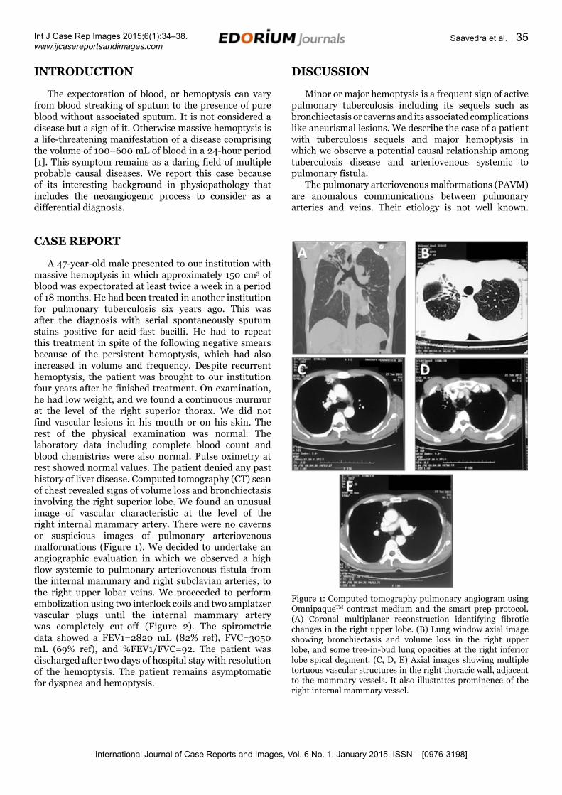
Figure 1: Computed tomography pulmonary angiogram using OmnipaqueTM contrast medium and the smart prep protocol. (A) Coronal multiplaner reconstruction identifying fibrotic changes in the right upper lobe. (B) Lung window axial image showing bronchiectasis and volume loss in the right upper lobe, and some tree-in-bud lung opacities at the right inferior lobe spical degment. (C, D, E) Axial images showing multiple tortuous vascular structures in the right thoracic wall, adjacent to the mammary vessels. It also illustrates prominence of the right internal mammary vessel.
DIscUssION
Minor or major hemoptysis is a frequent sign of active pulmonary tuberculosis including its sequels such as bronchiectasis or caverns and its associated complications like aneurismal lesions. We describe the case of a patient with tuberculosis sequels and major hemoptysis in which we observe a potential causal relationship among tuberculosis disease and arteriovenous systemic to pulmonary fistula.
The pulmonary arteriovenous malformations (PAVM) are anomalous communications between pulmonary arteries and veins. Their etiology is not well known.
International Journal of Case Reports and Images, Vol. 6 No. 1, January 2015. ISSN – [0976-3198]
Int J Case Rep Images 2015;6(1):34–38. www.ijcasereportsandimages.com
Saavedra et al. 36
Some authors have proposed theories explaining their origin from the fetal formation of the vessels. However, there are also many acquired medical conditions that can explain their formation. That is why PAVM can be congenital or acquired [2]. The congenital are related in 40% of the cases to the Osler-Weber-Rendu syndrome. The acquired PAVM are related to neoplasms, trauma, chronic inflammatory processes such as myocardial revascularization or sternotomy sequels, or post-infectious diseases (tuberculosis or fungal infections). The mechanism by which these medical conditions become correlated with PAVM is unknown. PAVM are supplied by pulmonary or systemic arteries, when PAVM are fed by systemic arteries, the presence of hereditary hemorrhagic telangiectasia or Osler Weber Rendu syndrome are ruled out since they are not commonly the cause. Hemodynamics in patients with PAVM represent
a right to left shunt that may not be manifested in the clinical aspect, nevertheless PAVM is one of the multiple causes of hemoptysis and can be associated with other symptoms like dyspnea and signs as a continuous murmur. Patients with this disease may even have normal pulmonary artery pressures.
In our literature research, only four case reports mentioning the coexistence between pulmonary tuberculosis and systemic to pulmonary fistulas were found [3–6]. In most of the cases, the authors describe a fistula involving the internal mammary artery and the pulmonary veins. In these cases, endovascular treatment was performed. Some authors explain the possibility of a “recruitment” of extrapulmonary local arteries and their connection to pulmonary vessels. Thomas et al. hypothesized that the inflammatory process surrounding a tuberculosis foci can help recruit local and systemic arteries to supply the inflammatory mass [4]. Pierce et al. postulated that the destruction of the pulmonary parenchyma secondary to tuberculosis disease might contribute to the parasitic arrival of the systemic artery supply to this area [6]. In this case, the primary hemoptysis cause cannot be defined, the diagnosis of a vascular malformation such as systemic PAVM is clearly exposed by the angiographic findings. One limitation of this case is that there are not previous or serial angiographic studies to objectively define the natural history of the identified systemic-pulmonary arteriovenous fistula. This implies that a causal relationship between tuberculosis and the genesis of the fistula in this patient cannot be established. Nevertheless, several possibilities can be mentioned in order to explain the association. A first possibility is the existence of congenital vascular malformations in this patient. If this is the case, tuberculosis disease may have been an epiphenomenon casually affecting a vascular area with a single preexistent fistula, in such case an additional fistula would be expected, but in both the invasive angiographic study and the CT scan a vascular malformation in the right upper lobe of the patient was the only finding. Moreover, the patient did not show other common clinical features of an Osler-Weber-Rendu syndrome. A second possibility to explain the systemic to pulmonary fistula is potential neoangeogenic process secondary to the chronic inflammatory events of tuberculosis and its sequel. The acute, subacute, and chronic inflammatory events during and/or after the disease could be chronic angiogenic triggers for vascular proliferation, erosion, and parietal transgression resulting in the formation of fistulous tracts between the internal mammary and subclavian arteries to the upper lobe pulmonary veins. This second alternative deserves special attention in our case. These findings allow us to emphasize that the growth of new vessels must be considered nonexclusive of the bronchial vascular area, but also can affect areas out of the pulmonary parenchyma in those patients with sequels of pulmonary chronic inflammatory diseases.
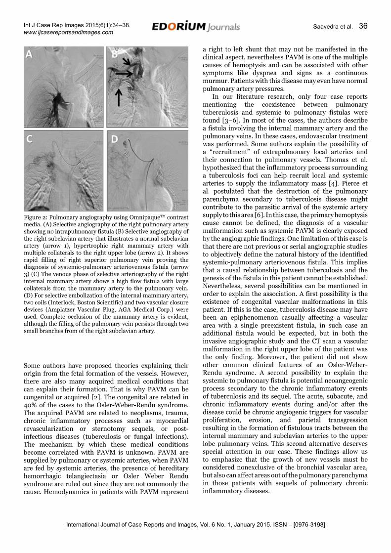
Figure 2: Pulmonary angiography using OmnipaqueTM contrast media. (A) Selective angiography of the right pulmonary artery showing no intrapulmonary fistula (B) Selective angiography of the right subclavian artery that illustrates a normal subclavian artery (arrow 1), hypertrophic right mammary artery with multiple collaterals to the right upper lobe (arrow 2). It shows rapid filling of right superior pulmonary vein proving the diagnosis of systemic-pulmonary arteriovenous fistula (arrow 3) (C) The venous phase of selective arteriography of the right internal mammary artery shows a high flow fistula with large collaterals from the mammary artery to the pulmonary vein. (D) For selective embolization of the internal mammary artery, two coils (Interlock, Boston Scientific) and two vascular closure devices (Amplatzer Vascular Plug, AGA Medical Corp.) were used. Complete occlusion of the mammary artery is evident, although the filling of the pulmonary vein persists through two small branches from of the right subclavian artery.
International Journal of Case Reports and Images, Vol. 6 No. 1, January 2015. ISSN – [0976-3198]
Int J Case Rep Images 2015;6(1):34–38. www.ijcasereportsandimages.com
Saavedra et al. 37
cONcLUsION
In conclusion, neoangiogenic stimuli and vascular remodeling resulting in acquired pulmonary arteriovenous malformations must be taken in mind when facing a patient with recurrent massive hemoptysis and a previous history of pulmonary tuberculosis.
*********
AcknowledgementsThis study was supported in part by “PLAN DE FORTALECIMIENTO INSTITUCIONAL 477-2012 Y 734-2013” and “Proyecto exención de impuestos por COLCIENCIAS ref. contrato 656624037813 2013”.
Author contributionsMaria Fernanda Saavedra – Substantial contributions to conception and design, Acquisition of data, Analysis and interpretation of data, Drafting the article, Revising it critically for important intellectual content, Final approval of the version to be publishedMaria Juliana Valenzuela – Substantial contributions to conception and design, Acquisition of data, Analysis and interpretation of data, Drafting the article, Revising it critically for important intellectual content, Final approval of the version to be publishedMonica Ocampo – Acquisition of data, Analysis and interpretation of data, Revising it critically for important intellectual content, Final approval of the version to be publishedCarlos Garavito – Acquisition of data, Analysis and interpretation of data, Revising it critically for important intellectual content, Final approval of the version to be publishedJosé Federico Saaibi – Substantial contributions to conception and design, Acquisition of data, Analysis and interpretation of data, Drafting the article, Revising it critically for important intellectual content, Final approval of the version to be published.Mauricio Orozco-Levi – Substantial contributions to conception and design, Acquisition of data, Analysis and interpretation of data, Drafting the article, Revising it critically for important intellectual content, Final approval of the version to be published.
GuarantorThe corresponding author is the guarantor of submission.
conflict of InterestAuthors declare no conflict of interest.
copyright© 2015 Maria Fernanda Saavedra et al. This article is distributed under the terms of Creative Commons Attribution License which permits unrestricted use, distribution and reproduction in any medium provided the original author(s) and original publisher are properly
credited. Please see the copyright policy on the journal website for more information.
rEFErENcEs
1. Jean-Baptiste E. Clinical assessment and management of massive hemoptysis. Crit Care Med 2000 May;28(5):1642–7.
2. Fernández FJ, Montes PM, Alcíbar J, Rodrigo D, Barrenetxea JI, Gotxi R. Percutaneous closure of a complex fistula between the internal mammary artery and a lobar branch of a pulmonary artery. Rev Esp Cardiol 2004 Jun;57(6):585–8. [Article in Spanish].
3. Cohen EM, Loew DE, Messer JV. Internal mammary arteriovenous malformation with communication to the pulmonary vessels. Am J Cardiol 1975 Jan;35(1):103–6.
4. Thomas R, Christopher DJ, Chacko J, Ponnaiya J. Pulmonary arteriovenous malformation in a patient with tuberculosis--an association? Eur J Cardiothorac Surg 2006 Aug;30(2):405–7.
5. Denlinger CE, Egan TM, Jones DR. Acquired systemic-to-pulmonary arteriovenous malformation secondary to Mycobacterium tuberculosis empyema. Ann Thorac Surg 2002 Oct;74(4):1229–31.
6. Pierce G, Ahuja C, Chadha M. Case report: Complex internal mammary to pulmonary artery fistula as a cause of hemoptysis in tuberculosis: Diagnosis and endovascular management using ethylene vinyl alcohol copolymer (Onyx). Indian J Radiol Imaging 2011 Jan;21(1):10–2.
sUGGEstED rEADING
• GossageJR.Pulmonaryarteriovenousmalformations:Epidemiology, etiology, pathology, and clinical features. In: UpToDate, Post TW (Ed), UpToDate, Waltham, MA. (Accessed on July 16, 2014).
• Ingbar,DH.Massivehemoptysis:Initialmanagemen.In: UpToDate, Post TW (Ed), UpToDate, Waltham, MA. (Accessed on July 16, 2014).

International Journal of Case Reports and Images, Vol. 6 No. 1, January 2015. ISSN – [0976-3198]
Int J Case Rep Images 2015;6(1):34–38. www.ijcasereportsandimages.com
Saavedra et al. 38
Access full text article onother devices
Access PDF of article onother devices

EDORIUM JOURNALS AN INTRODUCTION
Edorium Journals: On Web
About Edorium JournalsEdorium Journals is a publisher of high-quality, open ac-cess, international scholarly journals covering subjects in basic sciences and clinical specialties and subspecialties.
Edorium Journals www.edoriumjournals.com
Edorium Journals et al.
Edorium Journals: An introduction
Edorium Journals Team
But why should you publish with Edorium Journals?In less than 10 words - we give you what no one does.
Vision of being the bestWe have the vision of making our journals the best and the most authoritative journals in their respective special-ties. We are working towards this goal every day of every week of every month of every year.
Exceptional servicesWe care for you, your work and your time. Our efficient, personalized and courteous services are a testimony to this.
Editorial ReviewAll manuscripts submitted to Edorium Journals undergo pre-processing review, first editorial review, peer review, second editorial review and finally third editorial review.
Peer ReviewAll manuscripts submitted to Edorium Journals undergo anonymous, double-blind, external peer review.
Early View versionEarly View version of your manuscript will be published in the journal within 72 hours of final acceptance.
Manuscript statusFrom submission to publication of your article you will get regular updates (minimum six times) about status of your manuscripts directly in your email.
Our Commitment
Mentored Review Articles (MRA)Our academic program “Mentored Review Article” (MRA) gives you a unique opportunity to publish papers under mentorship of international faculty. These articles are published free of charges.
Favored Author programOne email is all it takes to become our favored author. You will not only get fee waivers but also get information and insights about scholarly publishing.
Institutional Membership programJoin our Institutional Memberships program and help scholars from your institute make their research accessi-ble to all and save thousands of dollars in fees make their research accessible to all.
Our presenceWe have some of the best designed publication formats. Our websites are very user friendly and enable you to do your work very easily with no hassle.
Something more...We request you to have a look at our website to know more about us and our services.
We welcome you to interact with us, share with us, join us and of course publish with us.
Browse Journals
CONNECT WITH US
Invitation for article submissionWe sincerely invite you to submit your valuable research for publication to Edorium Journals.
Six weeksYou will get first decision on your manuscript within six weeks (42 days) of submission. If we fail to honor this by even one day, we will publish your manuscript free of charge.
Four weeksAfter we receive page proofs, your manuscript will be published in the journal within four weeks (31 days). If we fail to honor this by even one day, we will pub-lish your manuscript free of charge and refund you the full article publication charges you paid for your manuscript.
This page is not a part of the published article. This page is an introduction to Edorium Journals and the publication services.