identification and characterization of compounds with
TRANSCRIPT

Identification and Characterization of
Compounds with Inhibitory Properties
towards Salmonella enterica Biofilms
Undergraduate Thesis
Presented in partial fulfillment of the requirements for graduation with
research distinction in Microbiology in the College of Arts and Sciences
at The Ohio State University.
Jasmine S. Moshiri
Bachelors of Science in Microbiology
College of Arts and Sciences
The Ohio State University
2016
Thesis Committee:
John Gunn, Ph.D., Advisor
Natividad Ruiz, Ph.D.

1
Table of Contents
Acknowledgments................................................................................................................2 Chapter 1. Introduction........................................................................................................3
1.1 Problem Statement.............................................................................................3 1.2 Hypothesis and Rationale..................................................................................4
Chapter 2. Materials and Methods.......................................................................................6 Chapter 3. Results................................................................................................................9 3.1 Identification of compounds with anti-biofilm properties.................................9 3.2 Characterization of the phenotypic effects of T315 and Cp315........................9 3.3 Evaluation of the mechanism of action of T315..............................................13 Chapter 4. Summary and Perspectives..............................................................................15 4.1 Aim 1...............................................................................................................15 4.2 Aim 2...............................................................................................................16 4.3 Aim 3...............................................................................................................20 4.4 Conclusions and Future Directions..................................................................21 Tables.................................................................................................................................23 Figures................................................................................................................................24 References..........................................................................................................................34

2
Acknowledgements
I would like to sincerely acknowledge the tireless support and guidance provided
by Dr. John Gunn, who I have had the privilege of working with throughout my
undergraduate career. Thank you, Dr. Gunn, for the time and energy you invest in
training undergraduate students; I feel extremely fortunate to have benefited from the
mentorship of a faculty member who prioritizes training young scientists as you do. I
would also like to acknowledge Jacob Koopman and Joanna Marshall, who served as
excellent mentors when I joined the lab and provided valuable training. Additionally, I
would like to thank past and present laboratory members including Ky Hoang, Juan
Gonzalez, Haley Adcox, Erin Vasicek, Dominique Olds, Olivia Harder, Darpan Kaur,
Michael Neiger, and Brad Eichar for their support. Thank you to our collaborators Chido
Hambira, Jim Fuchs, Santosh Salunke, and Ching-Shih Chen in the Ohio State University
Department of Medicinal Chemistry and Pharmacognosy. I would like to thank Dr.
Natacha Ruiz for her support as well as for her service on my thesis committee.
Lastly, I would like to acknowledge the funding sources for this work, which include the
National Institutes of Health, the SOLAR Research Fund, the Undergraduate Education
Summer Research Fellowship Program, OSU Biofilms in Human Medicine Group, OSU
Public Health Preparedness for Infectious Disease, and the OSU Dean's Discovery
Program.

3
Chapter 1
Introduction
1.1 Problem Statement
Salmonella enterica (S. enterica) is a facultative, intracellular, gram-negative
bacterium that causes serovar-specific disease in a range of hosts. Over 2,500 serovars of
S. enterica have been identified based on flagellar and lipopolysaccharide (LPS) antigen
specificity [1]. In the human host, S. enterica serovars may instigate typhoidal as well as
non-typhoidal illness, both with significant disease prevalence worldwide. Non-typhoidal
S. enterica is responsible for over 78 million cases of foodborne illnesses and over 59,000
deaths globally each year [2]. Salmonella enterica serovar Typhi (S. Typhi), the causative
agent of Typhoid Fever, infects and kills an estimated 20 million and 200,000 individuals
each year, respectively [3]. S. Typhi is of high clinical relevance in the regions of
Southern Africa, South-central Asia, and South-eastern Asia, where contamination of
both food and water sources in developing nations allows Salmonella to spread to new
hosts [3].
Upon ingestion of S. Typhi, successful bacteria will persist through the gastric
barrier of the stomach to reach the small intestine. At this location, S. Typhi crosses the
intestinal epithelium and is phagocytosed by macrophages to achieve systemic disease
[4]. Sites commonly infected during systemic salmonellosis include the ileum, liver,
spleen, bone marrow, and gallbladder [5].
After resolution of the acute infection, S. Typhi can persist in the gallbladder of 3-
5% of hosts through an asymptomatic chronic carrier state of disease [6]. During chronic
carriage, S. Typhi forms biofilms—communities of microorganisms that adhere to

4
surfaces and are recalcitrant to immune and antibiotic clearance—on the surface of
gallstones and the gallbladder epithelium [5, 7]. The process of biofilm formation is
characterized by sequential events that include initial attachment to a surface, secretion of
a protective extracellular polymeric substance (EPS), and dispersal of community
members to the environment [8] (Figure 1). As a human-restricted pathogen, the spread
of S. Typhi is dependent on this carrier state during which fecal shredding allows bacteria
to spread to new hosts through contamination of water sources. Thus, the study of S.
enterica biofilm processes is fundamental to impeding the spread of S. Typhi via chronic
carriage.
1.2 Hypothesis and Rationale
The central hypothesis for this thesis is that we can identify and characterize
compounds with the ability to disrupt the process of S. enterica biofilm formation.
Utilizing a model organism, Salmonella enterica subspecies enterica serovar
Typhimurium (S. Typhimurium), this hypothesis was examined with the three following
aims:
1. Identification of compounds with anti-biofilm properties.
2. Examination of the characteristics and activities of identified anti-biofilm
compounds.
3. Evaluation of the mechanism of action of anti-biofilm compound T315.
The discovery and characterization of compounds with the ability to specifically
inhibit early events in bacterial attachment will aid future studies of Salmonella biofilm

5
formation and is a promising step towards eradication of the Salmonella chronic carriage
state.

6
Chapter 2
Materials and Methods
2.1 Bacterial strains, growth conditions, and kinase inhibitors
The bacterial strains used in this study were S. Typhimurium 14028s (JSG210),
Acinetobacter baumannii (JSG3828) and Pseudomonas aeruginosa PAO1 (JSG3906).
Overnight cultures were grown in LB broth at 37°C with aeration. A 24h rapid
attachment or temporal addition assay for JSG210 included normalizing to OD600nm=0.8,
diluting 1:100 into 1:20 TSB, and incubating 24h post drug addition at 30°C in static
conditions. 24h rapid attachment assays for JSG3828 and JSG3906 included normalizing
to OD600nm=0.8, diluting 1:100 into LB, and incubating 24h post drug addition at 30°C in
static conditions. Kinase inhibitor stocks were stored in DMSO at 5 mM. Unless
otherwise indicated, drugs were administered via a 1:10 dilution of a 50 µM working
solution of 1% DMSO in PBS, yielding a final drug concentration of 5 µM.
2.2 Rapid Attachment and Temporal Addition Assays
Biofilm Rapid Attachment Assays were conducted using growth conditions and
drug concentrations described above with drug added prior to incubation in static
conditions. 24h post-drug addition, growth was read at OD600 and plates were washed 4
times in ddH20 to eliminate planktonic cells. Biofilms were heat fixed 1h at 60°C, stained
with 33% crystal violet solution (6.0 mL PBS, 3.3 mL crystal violet, 333 µL methanol,
333 µL isopropanol) for 5 min, and released using 33% acetic acid (3.3 mL glacial acetic
acid, 6.7 mL ddH20) to quantify the stain. Biofilm quantification was performed by
reading the released dye at OD570. Temporal Addition Assays were conducted using the

7
same conditions and crystal violet technique; however, drug addition was delayed to 1, 3,
and 6h post-initial incubation, with 15 min on orbital shaker after each addition to ensure
even drug dispersal.
2.3 Viability Assay
To determine bactericidal or bacteriostatic properties, S. Typhimurium and A.
baumannii were grown as previously described (37°C in LB, aerobic conditions) in the
presence or absence of drug at concentration of 5 or 10 µM. Samples were taken at 0, 2,
4, 6, 8, 16, and 24h after start of growth, serially diluted, and plated for enumeration.
2.4 EC50 Determination
The half maximal effective concentration (EC50) was calculated using
measurements of biofilm inhibition at concentrations of drug from 0.5 to 9 µM (24h at
30°C) via the Rapid Attachment Assay. Biofilms were quantified using the crystal violet
stain and dye release technique.
2.5 Generation of T315-Resistant Mutants
Following 24h exposure of S. Typhimurium to 5 µM T315 at 30°C, cells residing
within the biofilm were cultured in LB at 37°C and exposed to 5 µM T315 three
additional times before increasing drug exposure to 7.5 µM and then 10 µM. Additional
test well replicates were included from which biofilm-residing S. Typhimurium were not
obtained, and these wells were stained with crystal violet to observe development of
resistance to T315. Cultures at each stage were stored in 20% glycerol at -20°C. This

8
procedure was performed in triplicate. Resistance to T315 by an isolated mutant obtained
from biofilms after the 10 µM exposure will be confirmed via the Rapid Attachment
Assay with 10 µM T315 before submitting mutant gDNA for whole-genome sequencing.
2.6 Direct Pull-Down of T315 and Target
A polyethylene glycol (PEG) linker and biotin group will be added to T315 via
click-chemistry. After confirmation that PEGylation and biotinylation of T315 does not
inhibit anti-biofilm activity, biotinylated T315 will be exposed to a S. Typhimurium
cellular lysate (obtained via sonication in 50 mM Hepes pH 7.4, 120 mM NaCl, 20 mM
MgCl2, 0.1% Triton X-100, and 1x commercial complete protease inhibitor cocktail) and
T315-bound target will be isolated via streptavidin affinity chromatography using the
Pierce™ Biotinylated Protein Interaction Pull-Down Kit. Target identification will be
achieved via mass spectrometry.

9
Chapter 3
Results
3.1 Identification of kinase inhibitor compounds with anti-biofilm properties
An initial screen of 90 derivatives of kinase inhibitor compounds against S.
Typhimurium was performed using 5 µM of compound for 24h at 30°C. Biofilms were
assessed via crystal violet incorporation. Through this technique, two compounds were
identified that showed promising anti-biofilm activity: T315 and Cp315. The structure of
T315, as well as regions of T315 structurally identical to the closely related Cp315
molecule, are represented in Figure 2.
Rapid attachment assays performed in triplicate show that T315 and Cp315 at
concentrations of 5 µM inhibit S. Typhimurium biofilm formation by 59.4% and 58.4%,
respectively. Previous work with T315 indicate that it has anti-cancer properties, as it has
been demonstrated to disrupt the PI3K/AKT pathway, which contributes to cancer
progression [9]. Previously tested prostate and mammary epithelial cells did not show
cytotoxicity with T315 at concentrations within 1-5 µM [10], and the half-maximal lethal
dose of T315 against normal T cell and B cell lymphocytes was greater than 10 µM [9].
3.2 Characterization of the phenotypic effects of promising anti-biofilm agents T315
and Cp315
3.2 a. Evaluation of bacteriostatic or bactericidal effects
Viability assays were used to determine the effects of 5 µM T315 and Cp315 on
planktonic growth of S. Typhimurium. Over a 24h time-course at 37°C, no substantial
differences of S. Typhimurium growth was observed between T315 and Cp315 as

10
compared to a no drug control of 1% DMSO in PBS, as shown in Figure 3. These data
indicate that neither T315 or Cp315 cause a decrease in biofilm formation as a result of
bacteriostatic or bactericidal effects.
3.2 b. Characterization of temporal relationships of drug activity
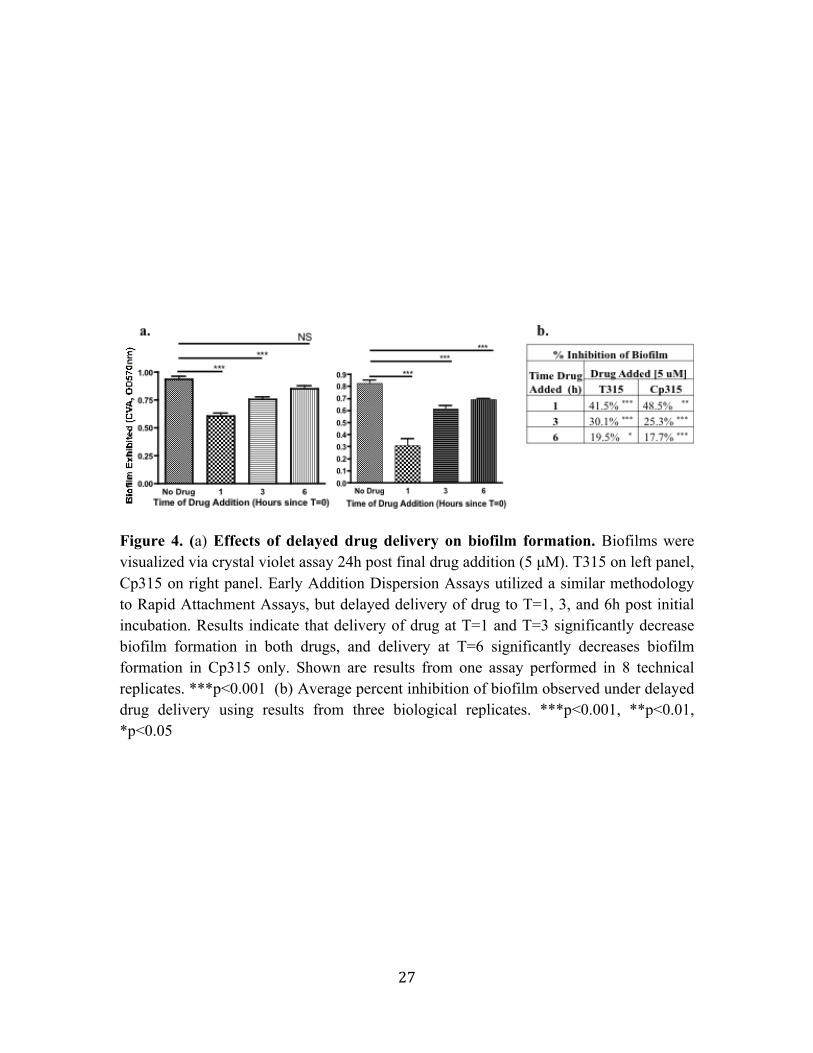
To better characterize the temporal relationship of drug activity with biofilm
formation, drug administration was delayed by 1, 3, and 6h post-initial incubation.
Delaying drug administration allows cells to begin the adherence process before exposure
to drug and differentiates between preventative and dispersal anti-biofilm effects. As
shown Figure 4, delayed administration of drugs results in a reduction in their anti-
biofilm activity. At drug administration 6h post-initial incubation (pii), 5 µM T315
exhibited only a 20% reduction in biofilm formation, as compared to 30% reduction at 3h
pii, and 42% at 1h pii. Similarly, 5 µM Cp315 exhibited 18% reduction in biofilm
formation at 6h pii, 25% reduction at 3h pii, and 49% at 1h pii. Thus, even a one-hour
allowance for Salmonella to begin the biofilm formation processes prior to drug exposure
reduced the anti-biofilm activity of both compounds as compared to activity quantified in
rapid attachment assays.
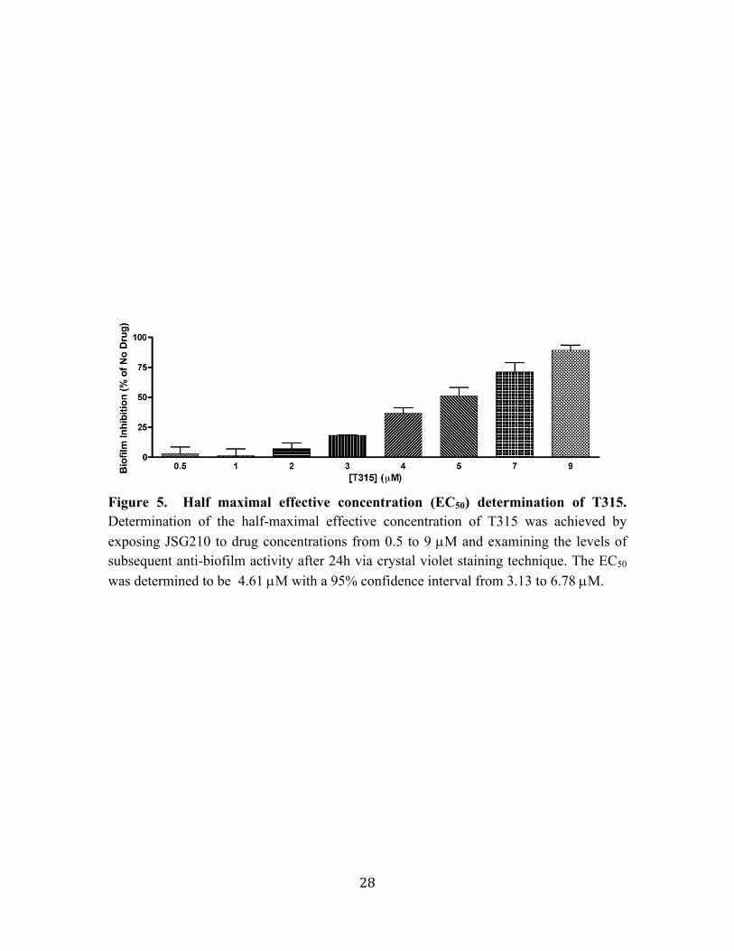
3.2 c. Determination of half-maximal effective concentration (EC50) of T315
In order to evaluate the efficiency of T315 activity, the half-maximal effective
concentration (EC50) was calculated by evaluating anti-biofilm activity at a range of
concentrations between 0.5 and 9µM. Figure 5 depicts the reduction in S. Typhimurium
biofilm observed by exposure to T315 at these varying concentrations. The EC50 was
determined to be equal to 4.61 µM with a 95% confidence interval from 3.13 to 6.78 µM.

11
3.2 d. Characterization of T315 activity against additional gram-negative biofilm-
forming pathogens A. baumannii and P. aeruginosa
The anti-biofilm activity of T315 towards gram-negative pathogenic biofilm
forming species A. baumannii and P. aeruginosa was evaluated (Figure 6a). 24h rapid
attachment assays at 30°C indicate that at a concentration of 10 µM, T315 significantly
reduces A. baumannii (JSG3828) biofilm formation by an average of 61.2% (p-value <
0.001). At the 5 µM concentration tested, T315 was not shown to significantly affect
biofilm formation of P. aeruginosa PAO1 (JSG3906). Enumeration of planktonic growth
A. baumannii at timepoints over 24h at 37°C indicates that 10 µM T315, while inhibiting
biofilm formation, does not work through bacteriostatic or bactericidal effects (Figure
6b).
3.2 e. Testing of T315 derivative In-T315 for anti-biofilm activity in S.
Typhimurium
A compound closely related to T315, termed In-T315, was obtained and evaluated
for anti-biofilm activity in S. Typhimurium. In-T315 relates to T315 in that the central
portion of the molecule has been modified to include an additional aromatic ring,
providing steric bulk to In-T315 while keeping side groups identical to those of T315.
Assessing biofilm formation with crystal violet staining technique, S. Typhimurium
biofilm formation 24h at 30°C, we observed that In-T315 exhibited a high level of anti-
biofilm activity at concentrations of 5 and 10 µM.
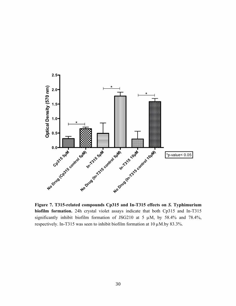
Figure 7 illustrates the average biofilm formation observed when JSG210 was
exposed to both Cp315 and In-T315 at a concentration of 5 µM as well as In-T315 at 10
µM. At both concentrations tested, Cp315 and InT315 significantly reduced biofilm
12
formation of JSG210. Cp315 exhibited an average of 58.4% inhibition at 5 µM. In-T315
exhibited an average of 74.8% biofilm inhibition at 5 µM and an average of 83.3%
biofilm inhibition at 10 µM against JSG210.
3.2 f. Characterization of T315 anti-biofilm activity against S. Typhimurium surface
mutants
T315 activity against S. Typhimurium surface appendage mutants was
characterized in hopes of providing insight to T315 functionality. Identification of S.
Typhimurium mutants used in these studies is included within Table 1 and includes
strains with reduced expression of curli fimbriae (JSG3736), motility (JSG1547), and
flagellar appendages FljB (JSG1178) and FliC (JSG1179) in a S. Typhimurium ATCC
14028s background. The ability of a S. Typhimurium SR11 quadruple fimbriae operon
mutant (JSG1174) to form biofilms in the presence of T315 was also characterized.
Strains were exposed to 5 and 10 µM T315 for 24h at 30°C, and biofilm-residing cells
were stained with crystal violet for quantification of biofilm formation. The level of
inhibition of biofilm formation observed due to the presence of T315 in the JSG3736,
JSG1547, JSG1178, and JSG1179 strains is depicted in Figure 8 and can be compared to
that observed in the JSG210 wild type S. Typhimurium 14028s strain. In these studies,
JSG1178 exposed to 10 µM T315 experienced an average of 58.9% inhibition of biofilm
formation while JSG210 displayed 78.4% inhibition at this concentration; this difference
is statistically significant. JSG1178 is deficient in expression of the S. Typhimurium FljB
flagellum, suggesting that this appendage may be a target of T315 compound, as the
compound exhibits reduced biological activity with the absence of this structure.
JSG1547, JSG3736, and JSG1179 bacterial strains also shown in Figure 8a did not

13
experience a statistically significant change in biofilm inhibition levels compared to the
JSG210 wild type background strain at any concentration of T315.
The anti-biofilm activity of T315 against a S. Typhimurium SR11 quadruple
fimbriae mutant (JSG1174) was studied at both 5 and 10 µM concentrations.
Quantification of biofilm formation was achieved after 24h exposure at 30°C through
crystal violet staining. JSG1174 is deficient in fim, lpfC, pefC, and agfB fimbriae
structures, and the response of this mutant to T315 was compared to SR11 wild type
strain JSG1169. No significant difference was observed of T315 anti-biofilm activity
towards JSG1174 as compared to the JSG1169 wild type background (Figure 8b),
suggesting that these fimbriae structures are not involved in the mechanism of action of
T315.
3.3 Evaluation of the mechanism of action of T315
3.3 a. Generation of a T315-resistant S. Typhimurium isolate
To understand the mechanism of action of T315, an attempt was made to generate
a S. Typhimurium JSG210 isolate that successfully formed biofilms in the presence of the
compound. Whole-genome sequencing would then be performed on this isolate to
identify loci altered from the wild-type and thus identify genes and gene products that
T315 interacts with. Following 24h exposure of S. Typhimurium to 5 µM T315 at 30°C,
cells residing within the biofilm were obtained and exposed to 5 µM T315 three
additional times before increasing drug exposure to 7.5 µM and then 10 µM. Additional
replicate wells were included which were not disturbed by the process of acquiring
biofilm-residing cells, and these cells were stained with crystal violet to monitor the

14
development of resistance to T315. We did not observe the development of any lasting
resistance in any of the three biological replicates performed. A transient resistance was
seen during repetition of the 5 µM drug exposure level, but this phenotype did not persist
upon an increase to 7.5 µM and subsequently 10 µM; this increase in drug concentration
from 5 µM was attempted twice.
3.3 b. Direct Pull Down of T315 and Target
Attachment of a polyethylene glycol (PEG) linker to T315 was achieved through
collaboration with the lab of Dr. Jim Fuchs at The Ohio State University via utilization of
click-chemistry. Synthesized PEG-T315-i included attachment of the PEG linker at the
site of the piperazine functional group. PEG-T315-i was evaluated for retention or
reduction of anti-biofilm activity. As depicted in Figure 9b, PEG-T315-i exhibited a loss
of biological activity. Given this information, we suspect that the (i) site is necessary for
the biological activity of T315. The polyethylene glycol linker is currently being attached
at the amide (ii) site of T315. If this location of PEG linkage retains biological activity, a
biotin marker will be attached, and this molecule will be used for direct pull down of
JSG210 target and mass spectrometry analysis.

15
Chapter 4
Discussion
The S. Typhi chronic carriage state includes the formation of biofilm communities
on the surface of gallstones in the gall bladder and on the gall bladder epithelium.
Recalcitrance of biofilms to antibiotic clearance results in difficulty resolving the chronic
infection. Targeting the biofilm formation process with anti-biofilm therapeutics could be
used in conjunction with antibiotic agents to eliminate the biofilm reservoir within
Salmonella carriers. In efforts to identify compounds that counter Salmonella biofilm
processes, we hypothesized that we can identify and characterize compounds with the
ability to disrupt the process of S. enterica biofilm formation. This hypothesis was
examined by (1) the identification of compounds with anti-biofilm properties, (2) the
examination of the characteristics and activities of identified anti-biofilm compounds and
(3) the evaluation of the mechanism of action of anti-biofilm compound T315.
4.1 Identification of compounds with anti-biofilm properties
Through a screen of 90 derivatives of kinase inhibitor compounds, we identified
two highly related compounds, T315 and Cp315, which significantly reduced the
formation of S. Typhimurium biofilms. At the 5 µM concentration used in the
preliminary screening, T315 and Cp315 inhibited biofilm formation by 59.4% and
58.4%, respectively. T315 has previously been characterized as an anti-cancer therapeutic
that targets the PI3K/AKT pathway. Studies indicate that it does not induce host cell
cytotoxicity in a number of human epithelial cell lines, including prostate and mammary
epithelial cells as well as T and B lymphocytes.

16
4.2 Examination of the characteristics and activities of identified anti-biofilm
compounds
Due to the promising anti-biofilm activity of T315 and Cp315 observed in the
preliminary screen, the functionality of these compounds were further characterized.
First, the bacteriostatic or bactericidal activity of the compounds were evaluated by
exposing S. Typhimurium to 5 µM of each compound and plating for enumeration at
several timepoints over 24h. Salmonella viable cell density during planktonic growth did
not appear to be affected by the presence of either T315 or Cp315 in these studies, which
suggests that the decrease in biofilm formation observed is not a resultant of
bacteriostatic or bactericidal activity of the compounds. Instead, the compounds interfere
with a bacterial process specific to the biofilm lifestyle.
To better understand the temporal relationship between drug addition and
effectiveness, drug administration was delayed 1, 3, and 6h to allow bacterial cells to
begin biofilm formation prior to drug exposure. 24h post-exposure, biofilms were
quantified by crystal violet staining, and we noted that delaying drug exposure impacted
the effectiveness of both T315 and Cp315 at 5 µM. Delaying T315 addition 1, 3, and 6h
post-addition of cells resulted in an average of 41.5%, 30.1%, and 19.5% biofilm
inhibition, respectively, compared to no drug. An identical experiment with Cp315
resulted in an average of 48.5%, 25.3%, and 17.7% biofilm inhibition, respectively,
compared to no drug. When added at the same time as initial incubation of Salmonella
the same concentrations of T315 and Cp315 inhibited biofilm formation by 59.4% and
58.4%, respectively. When drug administration was delayed to 24h post-initial incubation

17
of cells, when formation of a mature biofilm is underway, less than 3.5% difference
between biofilm formation with and without the presence of either compound was
observed (data not shown). These data indicate that both T315 and Cp315 are most
effective when administered early in the biofilm formation process and are ineffective at
dispersing a mature biofilm. This suggests that structures involved in the early attachment
stage of biofilm development, such as the expression of surface appendages, may be
affected by T315 and Cp315.
Due to the structural and functional similarities we observed between T315 and
Cp315, we chose to continue our characterization studies by focusing on T315. Previous
studies conducted on the anti-cancer properties of T315 described the cytotoxic effects of
the compound and the compound structure [9, 10]. We determined the half-maximal
effective concentration of T315 anti-biofilm properties towards S. Typhimurium to be
4.61 µM with a 95% confidence interval from 3.13 to 6.78 µM. This value is comparable
to the 5 µM drug concentration we have used throughout these studies. Issues with T315
solubility in 1x PBS, described in an upcoming section, prevented our EC50 studies from
exceeding 10 µM, which is a limitation of this work.
The effectiveness of T315 anti-biofilm activity towards gram-negative biofilm
forming pathogens A. baumannii and P. aeruginosa was characterized. 10 µM T315 is
effective at reducing A. baumannii biofilm formation by an average of 61.2%, and does
not appear to exhibit bactericidal or bacteriostatic effects on planktonic growth. P.
aeruginosa biofilms were unaffected by T315 in these studies, which indicates that T315
is a specific biofilm inhibitor. These data suggest that S. Typhimurium and A. baumannii
share one or more biofilm-formation processes which are not necessary for P. aeruginosa
18
biofilm formation, and that these processes are targeted by T315 to reduce biofilm
formation in a species-specific manner.
A compound closely related to T315, In-T315, was obtained from the lab of
Ching-Shih Chen for examination of potential anti-biofilm activity. In-T315 side groups
are identical to those of T315, but has a modified central structure that provides
additional steric bulk to the molecule. In these studies, exposure of S. Typhimurium to In-
T315 resulted in an average of 74.8% biofilm inhibition at 5 µM and an average of 83.3%
biofilm inhibition at 10 µM. These data suggest that In-T315 may be a more potent anti-
biofilm agent than T315; however, due to the inherent variability of these studies as a
result of compound instability in solution these results should be further replicated to
confirm this finding.
Due to observations that early addition of drug to bacterial static growth is
necessary for maximum anti-biofilm activity, we hypothesize that structures on the
cellular surface involved in early attachment phases of biofilm formation are affected by
the presence of T315. S. Typhimurium strains with mutations in curli fimbriae (ΔcsgA),
motility (ΔmotA), FliC flagellum (ΔfliC), FljB flagellum, and fimbriae operons (Δfim
ΔlpfC ΔpefC ΔagfB) were obtained and T315-induced inhibition of biofilm formation
was compared to that of appropriate background strains. A significant reduction in anti-
biofilm activity of T315 was observed in the JSG1178 strain, which is deficient in
expression of the FljB flagella appendage. When exposed to 10 µM T315 for 24h,
JSG1178 experienced an average of 58.9% inhibition of biofilm formation compared to
78.4% inhibition of biofilm formation that the background strain JSG210 displayed at
this concentration. This reduction in efficiency of drug activity suggests that the FljB

19
structure altered in the JSG1178 strain is involved in the functionality of T315. The role
of FljB in S. Typhimurium biofilm formation is inconclusive, which complicates this
analysis. FljB and FliC, S. Typhimurium flagellar filament structures, are controlled by
an expression switch. Studies conducted comparing S. Typhimurium attachment to
cholesterol-coated surfaces conclude that FliC, but not FljB, promote biofilm formation
[11], while studies on polystyrene at 37°C concluded that the absence of both FliC and
FljB is beneficial to biofilm formation [12]. In this work, the level of biofilm formation
between JSG210 and JSG1178 without drug is comparable, while the FliC- FljB+
JSG1179 strain was hindered in biofilm formation by an average of 45% compared to
background JSG210 (data not shown). Since the loss of FljB did not produce a biofilm-
deficient strain, and JSG1179 was attenuated in biofilm formation, one possible
hypothesis is that both FliC and FljB play a positive role in biofilm formation, but FliC to
a greater extent. The JSG1178 FljB- FliC+ strain maintains effective biofilm formation
because of its FliC expression. If T315 is binding directly to the FljB filament in S.
Typhimurium to hinder biofilm formation processes, this may be occurring
intracellularly, limiting FljB export and flagellar synthesis, at the cellular surface to coat
existing FljB flagella, or both. None of the remaining above-mentioned S. Typhimurium
surface appendage mutants experienced a significant difference in T315-induced anti-
biofilm activity compared to appropriate background strains. These studies may be
expanded by evaluating efficiency of T315 activity towards additional S. Typhimurium
mutants, such as those with altered lipopolysaccharide expression, and
polysaccharide/protein secretion profiles. It is also important to note that the compound

20
may be interfering with biofilm formation indirectly, such as by affecting signaling or
quorum sensing pathways necessary for the initiation of the biofilm lifestyle.
4.3 Evaluation of the mechanism of action of anti-biofilm compound T315
Gaining understanding of the mechanism of action of T315 has been attempted
via multiple approaches and is an ongoing effort. First, generation of a T315-resistant S.
Typhimurium isolate was attempted with the goal of performing whole-genome
sequencing on this isolate to identify genetic loci altered from the wild-type which allow
the bacterium to persist in biofilm formation in the presence of T315. Exposure of
biofilm-residing S. Typhimurium to repeated and stepwise increasing doses of T315
failed to generate an isolate with lasting resistance to the anti-biofilm agent. Upon
increasing T315 dosage from 5 to 7.5 µM, initially observed “resistant” mutants did not
retain this resistance. The idea of performing this process with a mutagenic S.
Typhimurium strain to increase probability of a desirable mutation was considered, but
ultimately not performed due to the challenges associated with evaluating individual
contributions of a high number of genetic mutations to the T315-resistant phenotype
within an obtained isolate.
Our ongoing approach to determine the mechanism of action of T315 is a direct
pull-down of biotinylated T315 with its S. Typhimurium cellular targets. A polyethylene
glycol (PEG) linker has been added to T315 at the site of the piperazine functional group
via click chemistry, but analysis of this molecule indicates that the PEG addition at this
site is detrimental to T315’s functionality. Our collaborators in the lab of Jim Fuchs are
currently working to synthesize T315 with a PEG addition at the site of the amide (T315-

21
PEG-ii). Once this molecule is synthesized, it will be evaluated to confirm that the PEG
addition does not interfere with anti-biofilm activity of T315. Following this
confirmation, a biotin marker will be added onto the PEG linker portion of T315-PEG-ii,
and biotinylated T315 will be exposed to S. Typhimurium cellular lysate before utilizing
biotin-streptavidin interactions to pull down T315 binding partners. Binding partners will
be identified via mass spectrometry.
4.4 Conclusions and Future Directions
As a human-restricted pathogen, the carrier state of S. Typhi infection is
fundamental to the spread of bacteria to new hosts and of consequential disease. The
biofilm lifestyle within the gall bladder of carriers is an important aspect of chronic
carriage that contributes to difficulties eradicating the spread of Salmonella. Used in
conjunction with antibiotic therapy, anti-biofilm agents could be a useful therapeutic tool
to limit chronic carriage and thus new S. Typhi infections.
The stability of T315 and Cp315 within solution has been a limitation of this
study. The compounds should be stored dry and not exposed to light for long-term
storage. Stock solutions should be prepared in dimethyl sulfoxide (DMSO) and stored at
-20°C, as the compounds are most soluble in DMSO. However, concentrations of DMSO
greater than 1% in solution is toxic to Salmonella, so these compounds must be diluted in
1x PBS directly before being administered. We believe this step of transfer into 1x PBS is
the source of solubility issues and high variation of compound effectiveness between
assays. Especially at concentrations over 10 µM, T315 is prone to precipitating out of
22
solution, which limited our EC50 studies to a narrow concentration range, as previously
described.
T315 is currently being derivatized to produce potential anti-biofilm compounds
with a lower EC50 and/or greater solubility in 1x PBS. A scheme for this derivitization is
depicted in Figure 10. Once synthesized, these compounds will be screened for biofilm
inhibition against S. Typhimurium 14028s. Studies towards the discovery of the
mechanism of action of T315 will be continued once the synthesis of T315-PEG-ii has
been achieved. Once synthesized, T315-PEG-ii will be evaluated for preservation of anti-
biofilm activity to ensure that drug interactions have not been compromised by the
addition of the polyethylene glycol linker. T315-PEG-ii will then be biotinylated, and this
biotinylated compound will be utilized for direct pull down of T315 targets, which will
be identified by mass spectrometry. Understanding the mechanism of action of T315 may
provide novel insight into the Salmonella biofilm formation processes as well as direct
future methods of synthesis of anti-biofilm agents to achieve diminution of the typhoid
carrier state.

23
Tables Strain Characteristics Reference
JSG210 S. Typhimurium ATCC 14028s (CDC6516-60); wild type
ATCC
JSG1547 JSG210 motA595::Tn10 Gift of T. Lino
JSG1169 S. Typhimurium SR11x4252 Gift of A. Baumler
JSG1174 JSG1169 Δ[fim-aph-11::Tn10]-391 lpfC::Kan pefC::Tet agfB::Cam
Gift of A. Baumler
JSG1178 JSG210 BC117 hin108::Tn10dCam (FljB off) Gift of B. Cookson
JSG1179 JSG210 BC119 fliC::Tn10 hin108::Tn10dCam (FljB on)
Gift of B. Cookson
JSG3736 JSG210 ΔcsgA via Wanner Adcox, et al. [13]
JSG3828 A. baumannii 19606 Gift of D. Wozniak
JSG3906 P. aeruginosa PAO1 Gift of D. Wozniak
Table 1. Bacterial strains and relevant characteristics.

24
Figures
Figure 1. Phases of biofilm development. "Biofilms: Multicellular Microbes?" [14]. The biofilm lifecycle is initiated via attachment of planktonic microorganisms to a surface in formation of a monolayer. The formation of microcolonies correlates with changes in gene expression to transition to biofilm lifecycle, such as down-regulation of motility genes, and precedes secretion of exopolysaccharide/extrapolymeric substance (EPS). Mature biofilms form characteristic biofilm towers encased in EPS, and are recalcitrant to immune and antimicrobial clearance. Return of biofilm members to planktonic state may be facilitated by passive detachment or active dispersal.

25
Figure 2. Chemical structure of T315. Regions of structural similarity between T315 and Cp315 are depicted in black while regions of dissimilarity between the compounds are depicted in red.

26
Figure 3. Viability assays of S. Typhimurium in presence of 5 µM T315 and Cp315. Results indicate that reduction of biofilm observed in Rapid Attachment Assays was not due to bacteriostatic or bactericidal effects of T315 or Cp315, as the growth curves observed in presence of each drug are not markedly different from those without drug. This experiment was performed once.
27
Figure 4. (a) Effects of delayed drug delivery on biofilm formation. Biofilms were visualized via crystal violet assay 24h post final drug addition (5 µM). T315 on left panel, Cp315 on right panel. Early Addition Dispersion Assays utilized a similar methodology to Rapid Attachment Assays, but delayed delivery of drug to T=1, 3, and 6h post initial incubation. Results indicate that delivery of drug at T=1 and T=3 significantly decrease biofilm formation in both drugs, and delivery at T=6 significantly decreases biofilm formation in Cp315 only. Shown are results from one assay performed in 8 technical replicates. ***p<0.001 (b) Average percent inhibition of biofilm observed under delayed drug delivery using results from three biological replicates. ***p<0.001, **p<0.01, *p<0.05
28
Figure 5. Half maximal effective concentration (EC50) determination of T315. Determination of the half-maximal effective concentration of T315 was achieved by exposing JSG210 to drug concentrations from 0.5 to 9 µM and examining the levels of subsequent anti-biofilm activity after 24h via crystal violet staining technique. The EC50 was determined to be 4.61 µM with a 95% confidence interval from 3.13 to 6.78 µM.

29
Figure 6. Evaluation of T315 anti-biofilm effects on P. aeruginosa (JSG3906) and A. baumannii (JSG3828). (a) After 24h exposure of 10 µM T315, JSG3828 biofilm formation was reduced by an average of 61.2%, quantified via crystal violet assay. Exposure of JSG3906 to 5 µM T315 did not significantly affect biofilm formation after 24h. ***p<0.001. (b) Enumeration of viable JSG3828 at timepoints over 24h exposure to 10 µM T315 suggests that anti-biofilm activity is not a resultant of bacteriostatic or bactericidal effects. This assay was performed once.
a.
30
Figure 7. T315-related compounds Cp315 and In-T315 effects on S. Typhimurium biofilm formation. 24h crystal violet assays indicate that both Cp315 and In-T315 significantly inhibit biofilm formation of JSG210 at 5 µM, by 58.4% and 78.4%, respectively. In-T315 was seen to inhibit biofilm formation at 10 µM.by 83.3%.

31
Figure 8. Evaluation of T315 anti-biofilm activity against S. Typhimurium flagella curli fimbriae, and motility mutants. Biofilm formation of each bacterial strain with and without the presence of 5 and 10 µM T315 was quantified after 24h by crystal violet staining. These data were used to compute percent inhibition of biofilm formation caused by the compound, shown above. (a) JSG1178 was seen to experience an average percent inhibition of biofilm formation significantly different than the wild type JSG210 background strain when exposed to 10 µM T315. In these studies, JSG1178 exposed to 10 µM T315 exhibited 58.9% inhibition of biofilm formation while JSG210 exhibited 78.4%. p<0.05. (b) No significant difference between T315-induced biofilm inhibition was observed between JSG1174 fimbriae mutant and JSG1169 SR11 wild type background.
a.

32
Figure 9. (a) Structure of T315-PEG-i. Sites of polyethylene glycol linker attachment for T315-PEG-i and T315-PEG-ii are indicated in blue. (i) pyrazole, (ii) amide site. (b) Loss of biological activity of T315-PEG-i. 24h drug exposure followed by crystal violet staining indicates that T315-PEG-i does not significantly reduce biofilm formation of S. Typhimurium at 5 or 10 µM concentrations.

33
Figure 10. Scheme for the derivatization of key functional moieties of T315 based on the established synthesis of T315 and related analogues for cancer screening.

34
References
1. LaRock, D.L., A. Chaudhary, and S.I. Miller, Salmonellae interactions with host
processes. Nat Rev Microbiol, 2015. 13(4): p. 191-205.
2. Havelaar, A.H., et al., World Health Organization Global Estimates and Regional
Comparisons of the Burden of Foodborne Disease in 2010. PLoS Med, 2015.
12(12): p. e1001923.
3. Crump, J., S. Luby, and E. Mintz, The global burden of typhoid fever. Bull World
Health Organ, 2004. 82(5): p. 346-353.
4. Parry, C.M., et al., Typhoid fever. N Engl J Med, 2002. 347(22): p. 1770-82.
5. Gonzalez-Escobedo, G., J.M. Marshall, and J.S. Gunn, Chronic and acute
infection of the gall bladder by Salmonella Typhi: understanding the carrier state.
Nature Reviews Microbiology, 2011. 9(1): p. 6.
6. Merselis, J.G., Jr., et al., Quantitative bacteriology of the typhoid carrier state.
Am J Trop Med Hyg, 1964. 13: p. 425-9.
7. Crawford, R.W., et al., Gallstones play a significant role in Salmonella spp.
gallbladder colonization and carriage Proceedings of the National Academy of
Sciences, 2010. 107(9): p. 4353-4358.
8. Costerton, J.W., et al., Microbial biofilms. Annu Rev Microbiol, 1995. 49: p. 711-
45.
9. Liu, T.M., et al., OSU-T315: a novel targeted therapeutic that antagonizes AKT
membrane localization and activation of chronic lymphocytic leukemia cells.
Blood, 2014.
35
10. Lee, S.L., et al., Identification and characterization of a novel integrin-linked
kinase inhibitor. J Med Chem, 2011. 54(18): p. 6364-74.
11. Crawford, R.W., K.E. Reeve, and J.S. Gunn, Flagellated but not hyperfimbriated
Salmonella enterica serovar Typhimurium attaches to and forms biofilms on
cholesterol-coated surfaces. J Bacteriol, 2010. 192(12): p. 2981-90.
12. Teplitski, M., A. Al-Agely, and B.M. Ahmer, Contribution of the SirA regulon to
biofilm formation in Salmonella enterica serovar Typhimurium. Microbiology,
2006. 152(Pt 11): p. 3411-24.
13. Adcox, H.E., et al., Salmonella extracellular matrix components influence biofilm
formation and gallbladder colonization. In preparation.
14. Slonczewski, J., Foster, JW., Gillan KM, Microbiology: An Evolving Science. 3rd
ed. 2014, London: W.W. Norton.