identification of organic colorants in fibers, paints, and ... · in cultural heritage objects is...
TRANSCRIPT
Identification of Organic Colorants in Fibers,Paints, and Glazes by Surface Enhanced Raman
SpectroscopyFRANCESCA CASADIO,† MARCO LEONA,*,‡
JOHN R. LOMBARDI,§ AND RICHARD VAN DUYNE⊥
†Conservation Department, The Art Institute of Chicago, 111 South MichiganAvenue, Chicago, Illinois 60603, ‡Department of Scientific Research, The
Metropolitan Museum of Art, 1000 Fifth Avenue, New York, New York 10028,§Department of Chemistry and Center for Analysis of Structures and Interfaces,The City University of New York, Convent Avenue at 138th Street, New York,
New York 10031, and ⊥Chemistry Department, Northwestern University,2145 Sheridan Road, Evanston, Illinois 60203
RECEIVED ON JANUARY 21, 2010
C O N S P E C T U S
Organic dyes extracted from plants, insects, and shellfish have been usedfor millennia in dyeing textiles and manufacturing colorants for painting.
The economic push for dyes with high tinting strength, directly related to highextinction coefficients in the visible range, historically led to the selection of sub-stances that could be used at low concentrations. But a desirable property for thecolorist is a major problem for the analytical chemist; the identification of dyesin cultural heritage objects is extremely difficult. Techniques routinely used in theidentification of inorganic pigments are generally not applicable to dyes: X-rayfluorescence because of the lack of an elemental signature, Raman spectros-copy because of the generally intense luminescence of dyes, and Fourier trans-form infrared spectroscopy because of the interference of binders and extenders.Traditionally, the identification of dyes has required relatively large samples(0.5-5 mm in diameter) for analysis by high-performance liquid chromatogra-phy. In this Account, we describe our efforts to develop practical approaches inidentifying dyes in works of art from samples as small as 25 µm in diameter withsurface-enhanced Raman scattering (SERS).
In SERS, the Raman scattering signal is greatly enhanced when organicmolecules with large delocalized electron systems are adsorbed on atomi-cally rough metallic substrates; fluorescence is concomitantly quenched. Recentnanotechnological advances in preparing and manipulating metallic parti-cles have afforded staggering enhancement factors of up to 1014. SERS is thusan ideal technique for the analysis of dyes. Indeed, rhodamine 6G and crys-tal violet, two organic compounds used to demonstrate the sensitivity of SERSat the single-molecule level, were first synthesized as textile dyes in the second half of the 19th century.
In this Account, we examine the practical application of SERS to cultural heritage studies, including the selection of appro-priate substrates, the development of analytical protocols, and the building of SERS spectral databases. We also considertheoretical studies on dyes of artistic interest.
Using SERS, we have successfully documented the earliest use of a madder lake pigment and the earliest occurrence oflac dye in European art. We have also found several examples of kermes and cochineal glazes, as well as madder, cochineal,methyl violet, and eosin lakes, from eras ranging from ancient Egypt to the 19th century. The ability to rapidly analyze verysmall samples with SERS makes it a particularly valuable tool in a museum context.
PHO
TOG
RAPH
PRO
VID
EDBY
DA
NZ.
JOH
NSO
N,C
UN
Y.
782 ACCOUNTS OF CHEMICAL RESEARCH 782-791 June 2010 Vol. 43, No. 6 Published on the Web 04/26/2010 www.pubs.acs.org/acr10.1021/ar100019q © 2010 American Chemical Society

IntroductionSurface-enhanced Raman spectroscopy (SERS) is currently at
the center of a powerful renaissance. Benefiting from the
exponential growth and interdisciplinary nature of scientific
investigations at the nanoscale level, hundreds of articles, jour-
nal special issues, and books are being published each year
on this topic.1,2
The first observations of surface Raman signals from pyri-
dine on electrochemically roughened Ag electrodes,3 followed
by the realization that they were enhanced by a factor of 106
generated considerable excitement.4,5 This phenomenon, now
known as SERS, promised ultrasensitive detection and identi-
fication of molecules. That promise has now been realized.
Ten years after the discovery of SERS, the first isolated iden-
tification of madder in a historical textile was reported, also
using Ag electrodes as the SERS active substrate.6 Today’s
developments in nanotechnology and nanofabrication of a
wide variety of robust plasmonic substrates have allowed the
achievement of enhancement factors up to 1014 allowing
detailed vibrational fingerprinting down to the single mole-
cule.7 It is currently accepted that such enhancements are
related to chemical and electromagnetic mechanisms,8
although their relative importance is the object of a lively
debate. The chemical mechanism implies resonance effects
within the target molecule and charge-transfer effects between
molecular orbitals and the conduction band of the noble metal
substrate. The electromagnetic mechanism, believed by most
to be the dominant contribution, is related to the collective
oscillation of the conduction electrons in noble metals that cre-
ates a localized surface plasmon resonance (LSPR) induced by
the incident laser light. The Raman signal of Raman active
molecules in the vicinity of such LSPR is exponentially
enhanced. All these phenomena endow SERS with extreme
sensitivity.
It is only in the past 6 years that the potential of SERS for
the ultrasensitive identification of molecules with otherwise
weak inelastic scattering probabilities, such as natural organic
materials used as artists’ colorants, has been fully realized and
exploited. In this Account, we describe our efforts to develop
SERS as a sensitive and selective tool for the identification of
dyes in works of art from samples as small as a single grain
of pigment or a fragment of dyed fiber a few micrometers in
length.
Dye Analysis in Cultural HeritageNatural dyes have been used since antiquity to color textiles
and to manufacture pigments; the discovery of mauveine and
the explosion of synthetic colorants chemistry9,10 (Figure 1) in
the second half of the 19th century further increased the
importance of dyes. Due to their high tinting strength, organic
dyes are usually present in very low concentrations in works
of art, offering a substantial analytical challenge.
Unambiguous, rapid, and ultrasensitive detection of natu-
ral and early synthetic dyestuffs is of paramount importance
to address questions on the conservation, context, and chro-
nology of works of art. Dye analysis is relevant to the long-
term preservation of artworks because many colorants are not
stable to prolonged light exposure.11 Many works by Vincent
Van Gogh (1853-1890) were painted with the bright pink
pigment eosine (the K or Na salt of 2,4,5,7-tetrabromofluo-
rescein, sold as an artist’s pigment under the name “geranium
lake”), which now appears completely faded.12 Similarly, famil-
iar masterpieces by artists such as Mary Cassatt (1844-1926)
have lost some of their vibrancy and on occasion part of
their meaning due to the fading of their cochineal carmine,
aniline, and redwood dyes.13 Even when the dyes are mac-
roscopically faded, the detection of trace amounts of colo-
rant, often preserved in interior painting layers or protected
by cellulosic fibers in works of art on paper (Figure 2), can
inform art historians and conservators on the original color
scheme of artwork and provide new insights into the art-
ist’s intention.14
The detection of specific colorants can also provide impor-
tant information about the historical context of the works of
FIGURE 1. Some natural and synthetic dyes of key artistic interest.
Identification of Organic Colorants by SERS Casadio et al.
Vol. 43, No. 6 June 2010 782-791 ACCOUNTS OF CHEMICAL RESEARCH 783

art or the technological ingenuity of the people who created
them or can assist in elucidating trade routes in antiquity. Dis-
tinguishing cochineal (from Dactylopius coccus Costa), kermes
(from Kermes vermilio Planchon), and lac dye (from Kerria laccaKerr) whose chromophores are carminic, kermesic, and laccaic
acids, respectively, is extremely relevant in art historical
research because kermes from Southern Europe and lac dye
from South East Asia were widely used until the 16th cen-
tury when cochineal, a dyestuff with much higher dye yields,
was imported from the Americas. Lastly, dye identification can
provide useful clues to the origin and dates ante quem or postquem an artifact was created, possibly leading to the uncov-
ering of forgeries.
Major constraints for the analysis of organic dyes in works
of art are the weak scattering and high fluorescence of the
biomolecules, invariably incorporated in matrices such as paint
layers or historic textiles that are themselves chemically com-
plex. Dyes are often incorporated into an inorganic substrate
(alumina, calcium carbonate or sulfate minerals, clays, etc.) to
make a lake that can be used as a pigment or fixed to textile
fibers by means of bridging inorganic ions (mordanting),
requiring extraction of the colorant prior to analysis. Finally,
limited (a few hundred micrometers or less) or no sampling
at all is typically allowed from works of art, and because the
dyestuff is present in very high dilution, noninvasive or
ultrasensitive techniques of analysis are preferred. UV-vis
absorbance spectroscopy,15 fluorimetry,16,17 FTIR,18 NIR,19
and Raman spectroscopy have all been investigated for dye
analysis. However, electronic spectroscopy methods tend to
have poor specificity, and data interpretation is challeng-
ing for all the above-mentioned spectroscopic methods.
Normal Raman spectroscopy has been successfully used to
characterize mineral pigments and some 19th to 20th cen-
tury synthetic colorants.20-22 In general however, the flu-
orescence of the majority of dyes, or of the matrices in
which they are found, is a major obstacle to normal Raman
detection.
To date, high-performance liquid chromatography (HPLC)
has demonstrated the most consistent results for dye
analysis.23,24 While chromatographic methods are highly
selective, sensitive, and specific, they require the removal of
approximately 1 mg of sample or 0.5-5 mm of dyed fiber,
which is a very large amount for rare and priceless works of
art.
SERS significantly reduces the amount of material needed
for analysis compared with HPLC, so when the genus of the
plant or species of insects used to derive the colorant is not an
important question to address or when the sample size
required for HPLC is not available, SERS provides a very pow-
erful analytical alternative for art applications.
SERS Research in Cultural HeritageAlthough SERS has been around for almost four decades, it
is only recently that sustained efforts have been devoted to
its application to cultural heritage objects: these are the sub-
ject of recent reviews.7,25 Most work to date has been car-
ried out on reference materials, leading to the publication
of high-quality, detailed spectra of anthraquinones, fla-
vonoids, and indigoid dyes.26-30 Fewer are the studies on
alkaloids, curcumin, redwoods, orchil dyes, and melanin
sepia,31-33 but researchers are constantly expanding the
range of dyes probed with SERS, so this gap will probably
be filled soon. In addition to providing useful reference
spectra, these studies have also investigated aspects such
as complexation geometry, influence of pH, and orienta-
tion of the analyzed molecules with respect to the noble
metal plasmonic substrate.
FIGURE 2. (A) Winslow Homer “For to Be a Farmer’s Boy” 1887(Gift of Mrs. George T. Langhorne in memory of Edward CarsonWaller, AIC 1963.760). This image had long puzzled scholars due tothe seemingly unfinished and flat sky in a highly finished work. (B)Optical stereomicrograph showing an area at the upper left cornerof the watercolor, displaying a few colored particles trapped underthe paper fibers. (C) Photomicrograph of pigment grains taken frompanel B. SER spectra identified the lake pigments as Indian purple (acochineal carmine lake precipitated with copper salts) and purplemadder.
Identification of Organic Colorants by SERS Casadio et al.
784 ACCOUNTS OF CHEMICAL RESEARCH 782-791 June 2010 Vol. 43, No. 6
Because the orientation of the molecule with respect to the
SERS-active surface results in selective enhancement of cer-
tain peaks, interpretation of the spectra can be very difficult.
While ab initio computational methods can be used to assign
normal modes and interpret SERS data,27,29,31,32 spectral data-
bases of reference colorants and an enhanced understand-
ing of the interactions of the dyestuffs of interest with various
SERS-active substrates are necessary. Going from microscope
slide to museum case introduces further challenges and brings
about the ambitious goal of obtaining high-quality spectra on
single pigment grains or minuscule clippings of textile frag-
ments from actual art objects, which are extremely complex
systems.
“One Size Does Not Fit All”: Tailored SERSSubstrates for Art AnalysisThe practical application of SERS to cultural heritage studies
has required extensive design and testing of optimized plas-
monic nanostructured surfaces and colloids for analysis of var-
ious classes of art materials. The ultimate goal is to achieve
dramatic fluorescence quenching and significant enhancement
of the weak Raman scattering effect for the target analytes,
while minimizing the amount of sample material required and
its handling. In our work, we have developed parallel
approaches that have led to the successful detection of micro-
scopic amounts of biomolecules in extremely aged and com-
plex matrices like archeological objects, faded pastels, and
glaze layers in paintings.
Solid State Substrates: Silver Island Films (AgIFs) andSilver Films over Nanospheres (AgFONs). The use of sil-
ver island films (AgIFs) as SERS substrates to identify and
characterize several reference red dyes has been
demonstrated.28,34 Moving from model systems to real
world applications, however, requires optimization of the
methodology. The nonuniformity of AgIFs substrates hin-
ders the acquisition of consistent spectra, thus rendering
collection of high-quality data a time-consuming operation.
A significant improvement is offered by the use of silver
films over nanospheres (AgFONs), which offer high repro-
ducibility and extreme tunability, as demonstrated also for
the quantitative detection of analytes such as biowarfare
agents (anthrax) and glucose.35-37 AgFON fabrication
involves drop-coating polystyrene or SiO2 nanospheres onto
a clean glass substrate and then depositing ∼200 nm of Ag
over the nanospheres. AgFONs not only are easily fabri-
cated and economical but are composed of a highly
ordered, uniform surface, which provides highly consistent
SER spectra. The LSPR of a AgFON can be tuned simply by
changing the size of the nanospheres, ensuring that they
can be excited with various laser wavelengths and easily
matched with the wavelength of visible absorption maxi-
mum of the studied dyes, giving rise to surface-enhanced
resonance Raman effects (SERRS).
We have used these substrates for the detection of sub-
nanogram quantities of the red dyes alizarin, carminic acid,
and laccaic acid individually and in mixtures. Pairing a red
laser excitation line (λ0 ) 632.8 nm) with AgFONs fabricated
with 390 nm diameter SiO2 spheres and a green laser line (λ0
) 532.15 nm) with 300 nm diameter SiO2 spheres, we
achieved preresonance and resonance conditions, respec-
tively, for these red dyes, leading to a 2 orders of magnitude
enhancement of the Raman signal, compared with nonreso-
nant measurements performed with a 785 nm laser excita-
tion line coupled with 550 nm diameter SiO2 spheres.38 We
also used AgFONs to analyze crocin, alizarin, purpurin, and
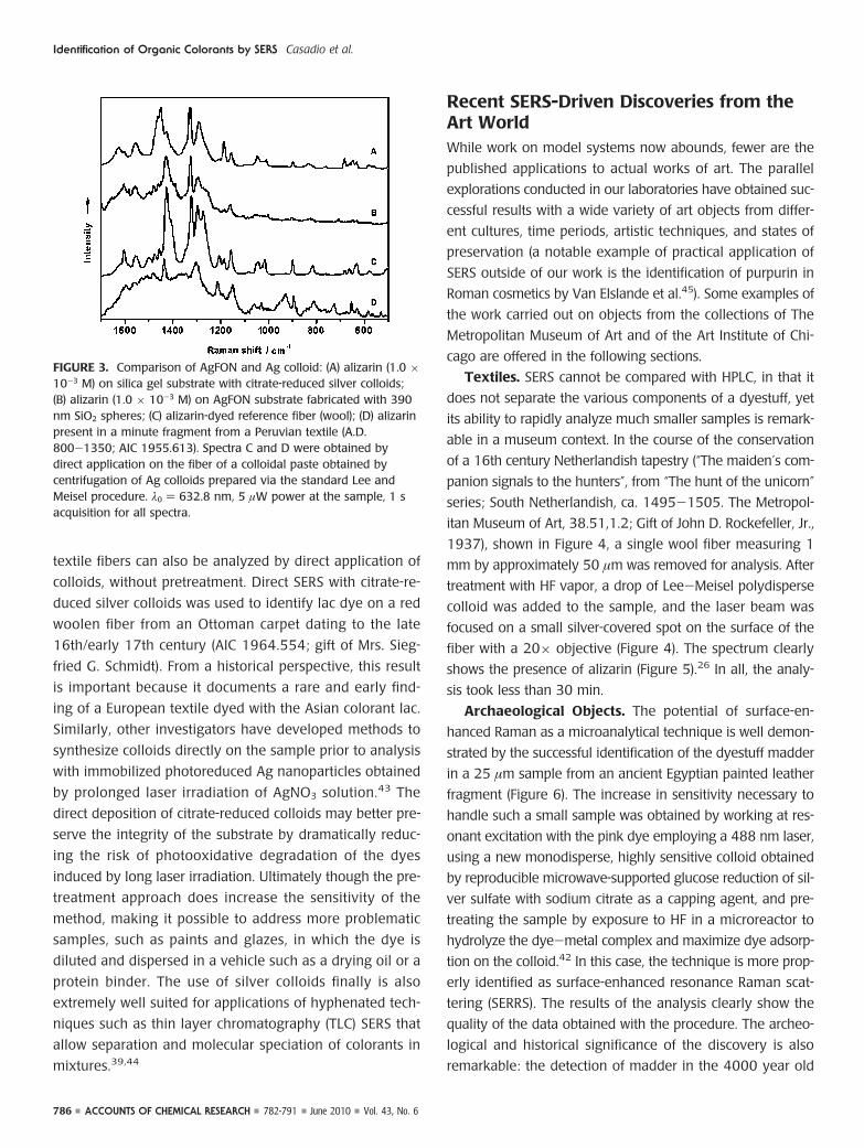
carminic acid reference dyestuffs;39 however when compared
with the spectra obtained from the same dyes with silver col-
loids deposited on silica gels, the AgFON spectra displayed
larger fluorescence backgrounds and inferior band resolution.
The tunability advantage of the AgFONs in fact is often off-
set by substrate contamination with carbon arising during the
silver deposition phase. It can be hypothesized that the
organic dyes have lower binding affinity than the carbon con-
tamination, which competes with the target molecules to
occupy surface active sites (Figure 3). Contamination issues
notwithstanding, the use of AgFONs is worth further explora-
tion, given the extreme tunability of the substrates: promis-
ing strategies for cleaning substrates have recently been
reviewed.40
Silver Colloids. Silver colloids have been by far the most
popular substrate for the identification of dyes in cultural her-
itage objects with SERS. By use of mostly the standard
Lee-Meisel citrate-reduced Ag colloids41 or modifications
thereof,42 versatile SERS substrates can be easily prepared and
used by any museum conservation departments with access
to a Raman microscope.
Silver colloids have been used at the Metropolitan
Museum of Art to investigate samples from textiles, works
of art on paper, polychrome objects, and paintings, rang-
ing in age from antiquity to the 19th century. While most
of the work carried out at the Metropolitan Museum on
mordant dyes and lake pigment has been based on a pre-
liminary sample treatment step (a gas-solid nonextractive
hydrolysis of the dye-metal complex carried out by expos-
ing the sample to a HF saturated atmosphere), recent work
at the Art Institute has successfully demonstrated that the
Identification of Organic Colorants by SERS Casadio et al.
Vol. 43, No. 6 June 2010 782-791 ACCOUNTS OF CHEMICAL RESEARCH 785
textile fibers can also be analyzed by direct application of
colloids, without pretreatment. Direct SERS with citrate-re-
duced silver colloids was used to identify lac dye on a red
woolen fiber from an Ottoman carpet dating to the late
16th/early 17th century (AIC 1964.554; gift of Mrs. Sieg-
fried G. Schmidt). From a historical perspective, this result
is important because it documents a rare and early find-
ing of a European textile dyed with the Asian colorant lac.
Similarly, other investigators have developed methods to
synthesize colloids directly on the sample prior to analysis
with immobilized photoreduced Ag nanoparticles obtained
by prolonged laser irradiation of AgNO3 solution.43 The
direct deposition of citrate-reduced colloids may better pre-
serve the integrity of the substrate by dramatically reduc-
ing the risk of photooxidative degradation of the dyes
induced by long laser irradiation. Ultimately though the pre-
treatment approach does increase the sensitivity of the
method, making it possible to address more problematic
samples, such as paints and glazes, in which the dye is
diluted and dispersed in a vehicle such as a drying oil or a
protein binder. The use of silver colloids finally is also
extremely well suited for applications of hyphenated tech-
niques such as thin layer chromatography (TLC) SERS that
allow separation and molecular speciation of colorants in
mixtures.39,44
Recent SERS-Driven Discoveries from theArt WorldWhile work on model systems now abounds, fewer are the
published applications to actual works of art. The parallel
explorations conducted in our laboratories have obtained suc-
cessful results with a wide variety of art objects from differ-
ent cultures, time periods, artistic techniques, and states of
preservation (a notable example of practical application of
SERS outside of our work is the identification of purpurin in
Roman cosmetics by Van Elslande et al.45). Some examples of
the work carried out on objects from the collections of The
Metropolitan Museum of Art and of the Art Institute of Chi-
cago are offered in the following sections.
Textiles. SERS cannot be compared with HPLC, in that it
does not separate the various components of a dyestuff, yet
its ability to rapidly analyze much smaller samples is remark-
able in a museum context. In the course of the conservation
of a 16th century Netherlandish tapestry (“The maiden’s com-
panion signals to the hunters”, from “The hunt of the unicorn”
series; South Netherlandish, ca. 1495-1505. The Metropol-
itan Museum of Art, 38.51,1.2; Gift of John D. Rockefeller, Jr.,
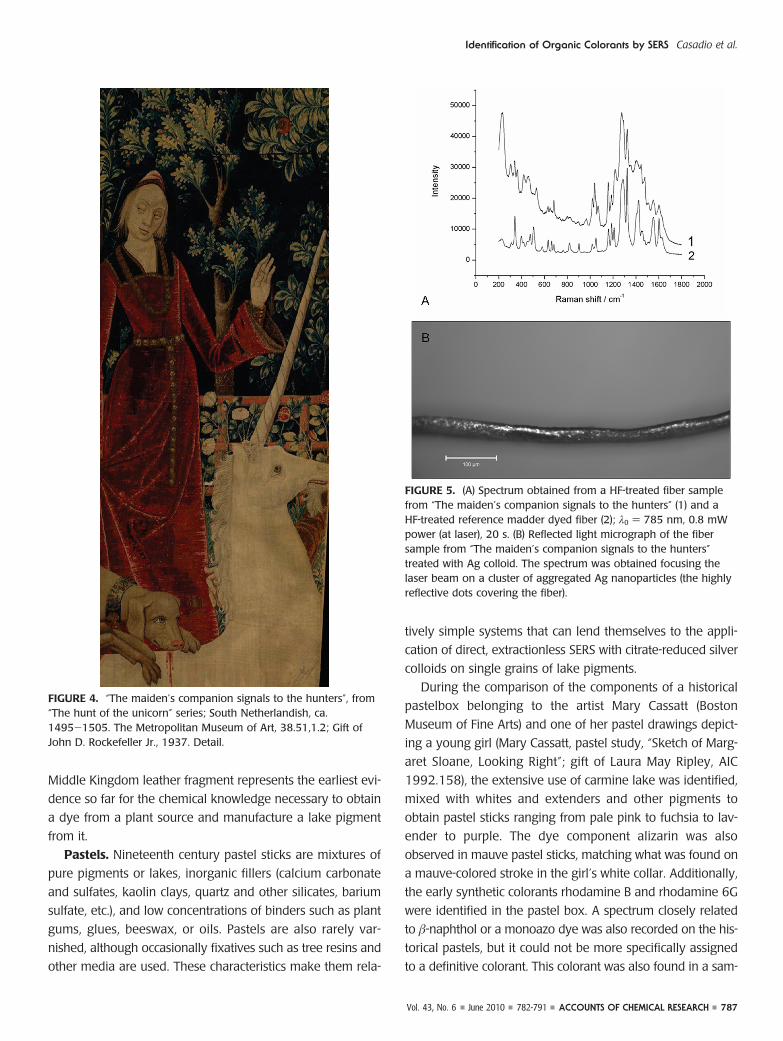
1937), shown in Figure 4, a single wool fiber measuring 1
mm by approximately 50 µm was removed for analysis. After
treatment with HF vapor, a drop of Lee-Meisel polydisperse
colloid was added to the sample, and the laser beam was
focused on a small silver-covered spot on the surface of the
fiber with a 20× objective (Figure 4). The spectrum clearly
shows the presence of alizarin (Figure 5).26 In all, the analy-
sis took less than 30 min.
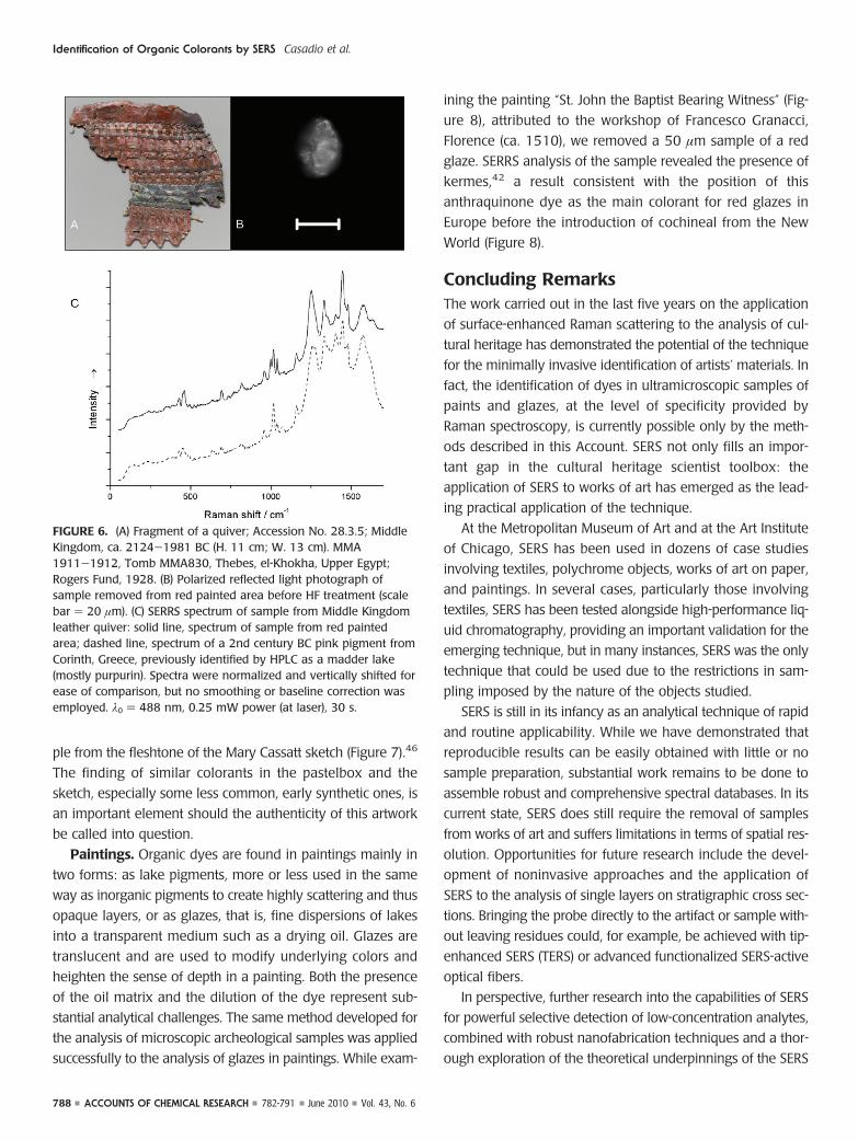
Archaeological Objects. The potential of surface-en-
hanced Raman as a microanalytical technique is well demon-
strated by the successful identification of the dyestuff madder
in a 25 µm sample from an ancient Egyptian painted leather
fragment (Figure 6). The increase in sensitivity necessary to
handle such a small sample was obtained by working at res-
onant excitation with the pink dye employing a 488 nm laser,
using a new monodisperse, highly sensitive colloid obtained
by reproducible microwave-supported glucose reduction of sil-
ver sulfate with sodium citrate as a capping agent, and pre-
treating the sample by exposure to HF in a microreactor to
hydrolyze the dye-metal complex and maximize dye adsorp-
tion on the colloid.42 In this case, the technique is more prop-
erly identified as surface-enhanced resonance Raman scat-
tering (SERRS). The results of the analysis clearly show the
quality of the data obtained with the procedure. The archeo-
logical and historical significance of the discovery is also
remarkable: the detection of madder in the 4000 year old
FIGURE 3. Comparison of AgFON and Ag colloid: (A) alizarin (1.0 ×10⁻³ M) on silica gel substrate with citrate-reduced silver colloids;(B) alizarin (1.0 × 10⁻³ M) on AgFON substrate fabricated with 390nm SiO2 spheres; (C) alizarin-dyed reference fiber (wool); (D) alizarinpresent in a minute fragment from a Peruvian textile (A.D.800-1350; AIC 1955.613). Spectra C and D were obtained bydirect application on the fiber of a colloidal paste obtained bycentrifugation of Ag colloids prepared via the standard Lee andMeisel procedure. λ0 ) 632.8 nm, 5 µW power at the sample, 1 sacquisition for all spectra.
Identification of Organic Colorants by SERS Casadio et al.
786 ACCOUNTS OF CHEMICAL RESEARCH 782-791 June 2010 Vol. 43, No. 6
Middle Kingdom leather fragment represents the earliest evi-
dence so far for the chemical knowledge necessary to obtain
a dye from a plant source and manufacture a lake pigment
from it.
Pastels. Nineteenth century pastel sticks are mixtures of
pure pigments or lakes, inorganic fillers (calcium carbonate
and sulfates, kaolin clays, quartz and other silicates, barium
sulfate, etc.), and low concentrations of binders such as plant
gums, glues, beeswax, or oils. Pastels are also rarely var-
nished, although occasionally fixatives such as tree resins and
other media are used. These characteristics make them rela-
tively simple systems that can lend themselves to the appli-
cation of direct, extractionless SERS with citrate-reduced silver
colloids on single grains of lake pigments.
During the comparison of the components of a historical
pastelbox belonging to the artist Mary Cassatt (Boston
Museum of Fine Arts) and one of her pastel drawings depict-
ing a young girl (Mary Cassatt, pastel study, “Sketch of Marg-
aret Sloane, Looking Right”; gift of Laura May Ripley, AIC
1992.158), the extensive use of carmine lake was identified,
mixed with whites and extenders and other pigments to
obtain pastel sticks ranging from pale pink to fuchsia to lav-
ender to purple. The dye component alizarin was also
observed in mauve pastel sticks, matching what was found on
a mauve-colored stroke in the girl’s white collar. Additionally,
the early synthetic colorants rhodamine B and rhodamine 6G
were identified in the pastel box. A spectrum closely related
to �-naphthol or a monoazo dye was also recorded on the his-
torical pastels, but it could not be more specifically assigned
to a definitive colorant. This colorant was also found in a sam-
FIGURE 4. “The maiden’s companion signals to the hunters”, from“The hunt of the unicorn” series; South Netherlandish, ca.1495-1505. The Metropolitan Museum of Art, 38.51,1.2; Gift ofJohn D. Rockefeller Jr., 1937. Detail.
FIGURE 5. (A) Spectrum obtained from a HF-treated fiber samplefrom “The maiden’s companion signals to the hunters” (1) and aHF-treated reference madder dyed fiber (2); λ0 ) 785 nm, 0.8 mWpower (at laser), 20 s. (B) Reflected light micrograph of the fibersample from “The maiden’s companion signals to the hunters”treated with Ag colloid. The spectrum was obtained focusing thelaser beam on a cluster of aggregated Ag nanoparticles (the highlyreflective dots covering the fiber).
Identification of Organic Colorants by SERS Casadio et al.
Vol. 43, No. 6 June 2010 782-791 ACCOUNTS OF CHEMICAL RESEARCH 787
ple from the fleshtone of the Mary Cassatt sketch (Figure 7).46
The finding of similar colorants in the pastelbox and the
sketch, especially some less common, early synthetic ones, is
an important element should the authenticity of this artwork
be called into question.
Paintings. Organic dyes are found in paintings mainly in
two forms: as lake pigments, more or less used in the same
way as inorganic pigments to create highly scattering and thus
opaque layers, or as glazes, that is, fine dispersions of lakes
into a transparent medium such as a drying oil. Glazes are
translucent and are used to modify underlying colors and
heighten the sense of depth in a painting. Both the presence
of the oil matrix and the dilution of the dye represent sub-
stantial analytical challenges. The same method developed for
the analysis of microscopic archeological samples was applied
successfully to the analysis of glazes in paintings. While exam-
ining the painting “St. John the Baptist Bearing Witness” (Fig-
ure 8), attributed to the workshop of Francesco Granacci,
Florence (ca. 1510), we removed a 50 µm sample of a red
glaze. SERRS analysis of the sample revealed the presence of
kermes,42 a result consistent with the position of this
anthraquinone dye as the main colorant for red glazes in
Europe before the introduction of cochineal from the New
World (Figure 8).
Concluding RemarksThe work carried out in the last five years on the application
of surface-enhanced Raman scattering to the analysis of cul-
tural heritage has demonstrated the potential of the technique
for the minimally invasive identification of artists’ materials. In
fact, the identification of dyes in ultramicroscopic samples of
paints and glazes, at the level of specificity provided by
Raman spectroscopy, is currently possible only by the meth-
ods described in this Account. SERS not only fills an impor-
tant gap in the cultural heritage scientist toolbox: the
application of SERS to works of art has emerged as the lead-
ing practical application of the technique.
At the Metropolitan Museum of Art and at the Art Institute
of Chicago, SERS has been used in dozens of case studies
involving textiles, polychrome objects, works of art on paper,
and paintings. In several cases, particularly those involving
textiles, SERS has been tested alongside high-performance liq-
uid chromatography, providing an important validation for the
emerging technique, but in many instances, SERS was the only
technique that could be used due to the restrictions in sam-
pling imposed by the nature of the objects studied.
SERS is still in its infancy as an analytical technique of rapid
and routine applicability. While we have demonstrated that
reproducible results can be easily obtained with little or no
sample preparation, substantial work remains to be done to
assemble robust and comprehensive spectral databases. In its
current state, SERS does still require the removal of samples
from works of art and suffers limitations in terms of spatial res-
olution. Opportunities for future research include the devel-
opment of noninvasive approaches and the application of
SERS to the analysis of single layers on stratigraphic cross sec-
tions. Bringing the probe directly to the artifact or sample with-
out leaving residues could, for example, be achieved with tip-
enhanced SERS (TERS) or advanced functionalized SERS-active
optical fibers.
In perspective, further research into the capabilities of SERS
for powerful selective detection of low-concentration analytes,
combined with robust nanofabrication techniques and a thor-
ough exploration of the theoretical underpinnings of the SERS
FIGURE 6. (A) Fragment of a quiver; Accession No. 28.3.5; MiddleKingdom, ca. 2124-1981 BC (H. 11 cm; W. 13 cm). MMA1911-1912, Tomb MMA830, Thebes, el-Khokha, Upper Egypt;Rogers Fund, 1928. (B) Polarized reflected light photograph ofsample removed from red painted area before HF treatment (scalebar ) 20 µm). (C) SERRS spectrum of sample from Middle Kingdomleather quiver: solid line, spectrum of sample from red paintedarea; dashed line, spectrum of a 2nd century BC pink pigment fromCorinth, Greece, previously identified by HPLC as a madder lake(mostly purpurin). Spectra were normalized and vertically shifted forease of comparison, but no smoothing or baseline correction wasemployed. λ0 ) 488 nm, 0.25 mW power (at laser), 30 s.
Identification of Organic Colorants by SERS Casadio et al.
788 ACCOUNTS OF CHEMICAL RESEARCH 782-791 June 2010 Vol. 43, No. 6

effects for organic dyes may hopefully soon reach critical mass
so that the technique can become not only an established part
of the cultural heritage scientist’s toolbox but an analytical
technique of general applicability.
The authors thank the Andrew W. Mellon Foundation (M.L. and
F.C.), the National Science Foundation (R.V.D. and F.C., Grants
CHE-0414554, CHE-0911145, and DMR-0520513), the David
H. Koch Foundation (M.L.), and the National Institute of Jus-
tice (J.R.L., Department of Justice Award No. 2006-DN-BX-
K034). We also acknowledge the contributions of Christa L.
Brosseau, Maria Vega Canamares, and Ron L. Birke.
BIOGRAPHICAL INFORMATION
Francesca Casadio is A.W. Mellon Senior Conservation Scien-tist at the Art Institute of Chicago where she has founded anddirects the scientific research laboratory. She received her Ph.D.in Chemistry from the University of Milan, Italy. Her research inter-ests focus on the vibrational characterization of materials of cul-tural heritage and applications of synchrotron radiation to studiesof museum objects. In 2006, she received the L’Oreal Art and Sci-ence of Color Silver Prize for her collaborative research on SERSof artistic colorants with Richard Van Duyne.
Marco Leona is the David H. Koch Scientist in Charge of theDepartment of Scientific Research at the Metropolitan Museum ofArt. He received his Ph.D. in Mineralogy and Crystallography fromthe University of Pavia, Italy. His research interests include the
FIGURE 7. (A) Mary Cassatt, “Sketch of Margaret Sloane, looking right” (pastel on tan wove paper, Gift of Laura May Ripley, AIC 1992.158).(B) SERS spectra of (upper curve) pastel stick #7 and (lower curve) a pink sample of fleshtone in face from the Mary Cassatt’s pastel. Peaksthat are characteristic for a yet unidentified �-naphtol or azo red red pigment, which also appears in pastel stick #7, are labeled. (C)Photomicrograph of a small sample from pastel stick #7. (D) Photomicrograph of a sample removed from a mauve stroke in the sitter’sbroad collar. (E) SERS spectra of (upper curve) madder root (Rubia tinctorum L.); and (lower curve) the sample illustrated in panel D. Longdashed lines indicate peaks that are consistent with madder root dye, and dashed lines with peak position noted indicate bands that areconsistent with pastel stick #14 containing rhodamine B and rhodamine 6G. (F) Mary Cassatt’s pastel box, courtesy of the Boston Museum ofFine Arts, Conservation Department. Citrate bands are indicated on panels B and D with an asterisk.
FIGURE 8. (A) St. John the Baptist Bearing Witness (detail). St. Johnthe Baptist Bearing Witness. Accession no. 1970.134.2; workshopof Francesco Granacci, Florence (ca. 1510). 75.6 × 209.6 cm.Purchase, Gwynne Andrews, Harris Brisbane Dick, Dodge, Fletcher,and Rogers Funds, funds from various donors, Ella Morris dePeyster Gift, Mrs. Donald Oenslager Gift, and Gifts in memory ofRobert Lehman, 1970. (B) SERRS spectrum of red glaze sample fromSt. John the Baptist Bearing Witness: solid line, spectrum of samplefrom red glaze; dashed line, spectrum of a reference sample ofkermesic acid. Spectra were normalized and vertically shifted forease of comparison, but no smoothing or baseline correction wasemployed. λ0 ) 488 nm, 0.25 mW power (at laser), 30 s.
Identification of Organic Colorants by SERS Casadio et al.
Vol. 43, No. 6 June 2010 782-791 ACCOUNTS OF CHEMICAL RESEARCH 789

development of new techniques for the noninvasive analysis ofworks of art, the study of East and South East Asian painting tech-niques and materials, and the application of surface-enhancedRaman scattering and UV resonance Raman spectroscopy to theidentification of natural and synthetic dyes.
John R. Lombardi was born in 1941 in Yonkers, New York. Heattended Cornell University as an undergraduate and received hisPh.D. from Harvard University in 1967. He was assistant profes-sor at the University of Illinois and is currently a Professor ofChemistry at The City College of New York. Other lines of inter-est include work on surface-enhanced Raman scattering on semi-conductor quantum dots.
Richard P. Van Duyne is Charles E. and Emma H. Morrison Pro-fessor of Chemistry at Northwestern University. He received hisPh.D. from the University of North Carolina at Chapel Hill. Hisresearch interests include surface-enhanced Raman spectroscopy,nanosphere lithography, localized surface plasmon resonancespectroscopy, molecular plasmonics, spectroscopic methods forchemical and biological sensing, structure and function of bio-molecules on surfaces, tip-enhanced Raman spectroscopy (TERS),ultrahigh vacuum scanning tunneling microscopy, ultrahigh vac-uum surface science, Raman spectroscopy of mass-selected clus-ters, and application of SERS to the study of works of art.
FOOTNOTES
*To whom correspondence should be addressed. E-mail: [email protected].
REFERENCES1 Aroca, R. Surface-Enhanced Vibrational Spectroscopy; John Wiley & Sons:
Chichester, U.K., 2006.2 Kneipp, K., Moskovits, M., Kneipp, H., Eds. Surface-Enhanced Raman Scattering:
Physics and Applications; Topics in Applied Physics; Springer: New York, 2006.3 Fleischmann, M. P.; Hendra, J.; McQuillan, A. J. Raman Spectra of Pyridine
Adsorbed at a Silver Electrode. Chem. Phys. Lett. 1974, 26, 163–166.4 Jeanmaire, D. L. L.; Van Duyne, R. P. Surface Raman Spectroelectrochemistry. J.
Electroanal. Chem. Interfacial Electrochem. 1977, 84, 1–20.5 Albrecht, M. G.; Creighton, J. A. Intense Raman Spectra of Pyridine at a Silver
Electrode. J. Am. Chem. Soc. 1977, 99, 5215–5217.6 Guineau, B.; Guichard, V. Identification des colorants organiques naturels par
microspectrometrie Raman de resonance et par effet Raman exalte de surface(SERS), in, ICOM Committee for Conservation: 8th triennial meeting, Sydney,Australia, 6-11 September, 1987 Preprints; The Getty Conservation Institute:Marina del Rey, CA, 1987; Vol. II, pp 659-666.
7 Wustholz, K. L.; Brosseau, C. L.; Casadio, F.; Van Duyne, R. P. Surface-EnhancedRaman Spectroscopy of Dyes: From Single Molecules to the Artists’ Canvas. Phys.Chem. Chem. Phys. 2009, 11, 7350–7359.
8 Lombardi, J. R.; Birke, R. L. A Unified View of Surface Enhanced Raman Scattering.Acc. Chem. Res. 2009, 42, 734–742.
9 Cardon, D. Natural Dyes: Sources, Tradition, Technology and Science; ArchetypePublications Ltd.: London, 2007.
10 Venkataraman, K. The Chemistry of Synthetic Dyes; Academic Press: New York,1952.
11 Saunders, D.; Kirby, J. Light-Induced Colour Changes in Red and Yellow LakePigments. Natl. Gallery Tech. Bull. 1994, 15, 79–97.
12 Burnstock, A.; Lanfear, I.; van den Berg, K. J.; Carlyle, L.; Clarke, M.; Hendriks, E.;Kirby, J. Comparison of the Fading and Surface Deterioration of Red Lake Pigmentsin Six Paintings by Vincent Van Gogh with Artificially Aged Paint Reconstructions. InPreprints of the 14th triennial meeting of the ICOM Committee for Conservation,James & James: London, 2005; Vol. I, pp 459-466.
13 Stratis, H. K. Innovation and Tradition in Mary Cassatt’s Pastels: A Study of HerMethods and Materials. In Mary Cassatt: Modern Woman; The Art Institute ofChicago Harry N. Abrams Inc.: New York, 1998; pp 212-226.
14 Brosseau, C. L.; Casadio, F.; Van Duyne, R. P. Revealing the Invisible - UsingSurface-Enhanced Raman Spectroscopy to Identify Minute Remnants of Color inWinslow Homer’s Colorless Skies. J. Raman Spectrosc., in press.
15 Karapanagiotis, L.; Valianou, L.; Daniilia, S.; Chryssoulakis, Y. Organic Dyes inByzantine and Post-Byzantine Icons from Chalkidiki (Greece). J. Cult. Herit. 2007, 8,294–298.
16 Claro, A.; Melo, M. J.; Schafer, S.; de Melo, J. S.; Pina, F.; van den Berg, K. J.;Burnstock, A. The Use of Microspectrofluorimetry for the Characterization of LakePigments. Talanta. 2008, 74, 922–929.
17 Clementi, C.; Miliani, C.; Romani, A.; Santamaria, U.; Morresi, F.; Mlynarska, K.;Favaro, G. In-Situ Fluorimetry: A Powerful Non-invasive Diagnostic Techniquefor Natural Dyes Used in Artefacts: Part II Identification of Orcein andIndigo in Renaissance Tapestries. Spectrochim. Acta, Part A. 2009, 71, 2057–2062.
18 Gillard, R. D.; Hardman, S. M.; Thomas, R. G.; Watkinson, D. E. The Detection ofDyes by FTIR Microscopy. Stud. Conserv. 1994, 39, 187–192.
19 Bruni, S.; Caglio, S.; Guglielmi, V.; Poldi, G. The Joined Use of n.i. SpectroscopicAnalyses - FTIR, Raman, Visible Reflectance Spectrometry and EDXRF - To StudyDrawings and Illuminated Manuscripts. Appl. Phys. A: Mater. Sci. Process. 2008,92, 103–108.
20 Bell, I. M.; Clark, R. J. H.; Gibbs, P. J. Raman spectroscopic library of natural andsynthetic pigments (pre- ≈ 1850 AD). Spectrosc. Acta, Part A 1997, 53, 2159–2179.
21 Vandenabeele, P.; Moens, L.; Edwards, H. G. M.; Dams, R. Raman SpectroscopicDatabase of Azo Pigments and Application to Modern Art Studies. J. RamanSpectrosc. 2000, 31, 509–517.
22 Scherrer, N. C.; Zumbuehl, S.; Delavy, F.; Fritsch, A.; Kuehnen, R. Synthetic OrganicPigments of the 20th and 21st Century Relevant to Artist’s Paints: Raman SpectraReference Collection. Spectrochim. Acta, Part A 2009, 73, 505–524.
23 Wouters, J. High Performance Liquid Chromatography of Anthraquinones:Analysis of Plant and Insect Extracts and Dyed Textiles. Stud. Conserv. 1985,30, 119–128.
24 Wouters, J.; Verhecken, A. The Coccid Insect Dyes: HPLC and Computerized Diode-Array Analysis of Dyed Yarns. Stud. Conserv. 1989, 34, 189.
25 Chen, K.; Leona, M.; Vo-Dinh, T. Surface-Enhanced Raman Scattering for theIdentification of Organic Pigments and Dyes in Works of Art and Cultural HeritageMaterial. Sens. Rev. 2007, 27, 109–120.
26 Leona, M.; Steger, J.; Ferloni, E. Application of Surface-Enhanced Raman ScatteringTechniques to the Ultra-sensitive Identification of Natural Dyes in Works of Art. J.Raman Spectrosc. 2006, 37, 981–992.
27 Whitney, A. V.; Van Duyne, R. P.; Casadio, F. An Innovative Surface-EnhancedRaman Spectroscopy (SERS) Method for the Identification of Six Traditional RedLakes and Dyestuffs. J. Raman Spectrosc. 2006, 37, 993–1002.
28 Whitnall, R., Shadi, I. T.; Chowdry, B. Z. Case Study: The Analysis of Dyes bySERRS. In Raman Spectroscopy in Archaeology and Art History; Edwards,H. G. M., Chalmers, J. M., Eds.; Royal Society of Chemistry: London, 2005; pp152-165.
29 Canamares, M. V.; Garcia-Ramos, J. V.; Domingo, C.; Sanchez-Cortes, S. Surface-Enhanced Raman Scattering Study of the Adsorption of the Anthraquinone PigmentAlizarin on Ag Nanoparticles. J. Raman Spectrosc. 2004, 35, 921–927.
30 Shadi, I. T.; Chowdhry, B. Z.; Snowden, M. J.; Withnall, R. Semi-quantitativeAnalysis of Alizarin and Purpurin by Surface-Enhanced Resonance RamanSpectroscopy (SERRS) Using Silver Colloids. J. Raman Spectrosc. 2004, 35, 800–807.
31 Leona, M.; Lombardi, J. R. Identification of Berberine in Archaeological Textilesby Surface Enhanced Raman Spectroscopy. J. Raman Spectrosc. 2007, 38,853–85.
32 Canamares, M. V.; Lombardi, J. R.; Leona, M. Surface-Enhanced Raman Scatteringof Protoberberine Alkaloids. J. Raman Spectrosc. 2008, 39, 1907–1914.
33 Centeno, S. A.; Shamir, J. Surface Enhanced Raman Scattering (SERS) and FTIRCharacterization of the Sepia Melanin Pigment Used in Works of Art. J. Mol. Struct.2008, 873, 149–159.
34 Whitney, A. V.; Van Duyne, R. P.; Casadio, F. Silver Island Films as Substrate forSurface-Enhanced Raman Spectroscopy (SERS): A Methodological Study on TheirApplication to Artists’ Red Dyestuffs. Proc. SPIE 2005, 117–126.
35 Stuart, D. A.; Yuen, J. M.; Shah, N. C.; Lyandres, O.; Yonzon, C. R.; Glucksberg,M. R.; Walsh, J. T.; Van Duyne, R. P. In Vivo Glucose Measurement by Surface-Enhanced Raman Spectroscopy. Anal. Chem. 2006, 78, 7211–7215.
36 Zhang, X.; Young, M. A.; Lyandres, O.; Van Duyne, R. P. Rapid Detection of anAnthrax Biomarker by Surface-Enhanced Raman Spectroscopy. J. Am. Chem. Soc.2005, 127, 4484–4489.
37 Zhang, X.; Zhao, J.; Whitney, A. V.; Elam, J. W.; Van Duyne, R. P. UltrastableSubstrates for Surface-Enhanced Raman Spectroscopy: Al2O3 Overlayers Fabricated
Identification of Organic Colorants by SERS Casadio et al.
790 ACCOUNTS OF CHEMICAL RESEARCH 782-791 June 2010 Vol. 43, No. 6

by Atomic Layer Deposition Yield Improved Anthrax Biomarker Detection. J. Am.Chem. Soc. 2006, 128, 10304–10309.
38 Whitney, A. V.; Casadio, F.; Van Duyne, R. P. Identification and Characterization ofArtists’ Red Dyes and Their Mixtures by Surface-Enhanced Raman Spectroscopy.Appl. Spectrosc. 2007, 61, 994–1000.
39 Brosseau, C. L.; Gambardella, A.; Casadio, F.; Van Duyne, R. P.; Grzywacz, C.;Wouters, J. Ad-Hoc SERS Methodologies for the Detection of Artist Dyestuffs:Thin Layer Chromatography-Surface Enhanced Raman Spectroscopy (TLC-SERS) and In Situ On the Fiber Analysis. Anal. Chem. 2009, 81, 3056-3062.
40 Lin, X. M.; Cui, Y.; Xu, Y. H.; Ren, B.; Tian, Z. Q. Surface-Enhanced RamanSpectroscopy: Substrate-Related Issues. Anal. Bioanal. Chem. 2009, 394, 1729–1745.
41 Lee, P. C.; Meisel, D. Adsorption and Surface-Enhanced Raman of Dyes on Silverand Gold Sols. J. Phys. Chem. 1982, 86, 3391–3395.
42 Leona, M. Microanalysis of Organic Pigments and Glazes in Polychrome Works ofArt by Surface-Enhanced Resonance Raman Scattering. Proc. Natl. Acad. Sci. U.S.A.2009, 106, 14757–14762.
43 Jurasekova, Z.; Domingo, C.; Garcia-Ramos, J. V.; Sanchez-Cortes, S. In SituDetection of Flavonoids in Weld-Dyed Wool and Silk Textiles by Surface-EnhancedRaman Scattering. J. Raman Spectrosc. 2008, 39, 1309–1312.
44 Geiman, I.; Leona, M.; Lombardi, J. R. Application of Raman Spectroscopy andSERS to the Analysis of Synthetic Dyes Found in Ballpoint inks. J. Forensic Sci.2009, 54, 947–952.
45 Van Elslande, E.; Lecomte, S.; Le Ho, A. S. Micro-Raman Spectroscopy (MRS) andSurface-Enhanced Raman Scattering (SERS) on Organic Colorants in ArchaeologicalPigments. J. Raman Spectrosc. 2008, 39, 1001–1006.
46 Brosseau, C. L.; Rayner, K.; Casadio, F.; Van Duyne, R. P.; Grzywacz, C. M. Surface-Enhanced Raman Spectroscopy: An In-Situ Method To Identify Colorants in VariousArtist Media. Anal. Chem. 2009, 81, 7443–7447.
Identification of Organic Colorants by SERS Casadio et al.
Vol. 43, No. 6 June 2010 782-791 ACCOUNTS OF CHEMICAL RESEARCH 791