human primary hepatocytes – product overview...and metabolize xenobiotics somewhere during the...
TRANSCRIPT
LifeNetHealth.org 1
HEPATOCYTE BRIEF
General Background
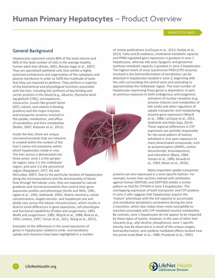
Hepatocytes represent nearly 80% of the total volume and 60% of the total number of cells in the average healthy human adult liver (Kmiec, 2001, Bioulac-Sage et al., 2007). They are specialized epithelial cells that exhibit a highly polarized architecture and organization of the cytoplasm and plasma membrane in order to fulfill the multitude of tasks that they are required to perform. They perform a majority of the biochemical and physiological functions associated with the liver, including the synthesis of key binding and carrier proteins in the blood (e.g., albumin, thyroxine-bind-ing globulin [TBG], ceruloplasmin, transcortin, insulin-like growth factor [IGF], retinol, and vitamin D binding proteins) and the major enzymes and transporter proteins involved in the uptake, metabolism, and efflux of xenobiotics and their metabolites (Rodés, 2007; Klaassen et al., 2013).
Inside the liver, there are unique microenvironments that are inherent-ly created within the context of the liver’s native microanatomy within which hepatocytes reside in vivo. The liver acinus is demarcated into three zones: zone 1 is the peripor-tal region; zone 2 is the midlobular region; and zone 3 is the pericentral region (Rappaport, 1977, Ito and McCuskey, 2007). Due to the particular location of hepatocytes along the microvasculature and the directionality of blood flow through the lobular units, they are exposed to natural gradients and microenvironments that control their gene expression profiles and phenotype (Smith and Wills, 1981, Ugele et al., 1991, Gebhardt, 1992). Matrix chemistry, solute concentrations, oxygen tension, and hepatocyte size and ploidy vary across the lobular microstructure, which results in distinct zonal differences in gene expression, cell phenotypes and functional capabilities (Probst and Jungermann, 1983, Wolfe and Jungermann, 1985, Wojcik et al., 1988, Reid et al., 1992, Lindros, 1997, Turner et al., 2011, Wang et al., 2011).
Examples of the differences in the zonal expression of genes in hepatocytes related to endo- and xenobiotic uptake and clearance have been highlighted in a number
of review publications (LeCluyse et al., 2012; Godoy et al., 2013). Fatty acid β-oxidation, cholesteral metabolic capacity and PPAR-regulated gene expression is greatest in zone 1 hepatocytes, whereas bile acid, lipogenic and glutamine synthase metabolic capacity is greatest in zone 3 hepatocytes. The highest levels of most cytochrome P450 (CYP) enzymes involved in the biotransformation of xenobiotics can be detected in hepatocytes located in zone 3, beginning with the cells surrounding the central veins and extending to approximately the midlobular region. The total number of hepatocytes expressing these genes is dependent, in part, on previous exposure to both endogenous and exogenous
activators of nuclear receptors (e.g. enzyme inducers and metabolites of bile acids) and other regulators of uptake transporter and metabolizing enzyme gene expression (Wojcik et al., 1988, LeCluyse et al., 2012, Gebhardt and Matz-Soja, 2014). These regional differences in CYP expression are partially responsible for the zonal pattern of toxicity exhibited in vivo upon exposure to many bioactivated compounds, such as acetaminophen (APAP), carbon tetrachloride, bromobenzene, and chloroform (Black, 1984; Tomasi et al., 1985; Anundi et al., 1993; Moon et al., 2010).
Many important uptake transporter proteins are also expressed in a zone-specific fashion. For example, human liver sections stained with antibodies against human OATP1B1 and OATP1B3 exhibit a similar pattern as that for CYP3A4 in zone 3 hepatocytes. The overlapping expression of both transporter and CYP proteins in zone 3 cells suggests that hepatocytes acquire the ‘mature’ phenotype with the full capacity to accumulate and metabolize xenobiotics somewhere during the zone 2 transition, which also makes them more susceptible to toxicities associated with CYP-mediated reactive metabolites. By contrast, zone 1 hepatocytes do not appear to be impacted by these types of events. However, in the case of other liver toxicants (e.g., allyl alcohol, phosphorus), zone 1 specific toxicity may be observed as a result of the unique oxygen, biotransformation, and cytokine-mediated effects located near the portal triad (Badr et al., 1986; Przybocki et al., 1992).
Human Primary Hepatocytes – Product Overview

HEPATOCYTE BRIEF
LifeNetHealth.org 2
Human Primary Hepatocytes – Product Overview
Utility of primary human hepatocytes for ADMET applications
In the pharmaceutical industry in vitro hepatocyte culture strategies have proven useful during drug development for studying drug metabolism and drug-drug interactions before in vivo proof-of-concept studies are initiated (Hughes et al., 2011; Astashkina et al., 2012). As generally accepted, primary hepatocyte-based in vitro models are considered the gold standard for assessing the metabolism and enzyme induction potential of a new chemical entity. For example, in vitro testing strategies using primary human hepatocytes in suspension culture have been applied successfully to predict the in vivo pharmacokinetics and clearance of compounds for years, or using them in a sandwich culture (SC) model system for assessing the potential of compounds to be involved in significant adverse interactions through the induction or inhibition of liver enzymes (Lin, 2006; Hewitt et al., 2007; Obach, 2009; Obach et al., 2008). These sandwich cultures also have proven useful for the study of drug uptake and efflux transporters, and predicting the biliary secretion of compounds and their metabolites (Swift et al., 2010).
In the event that mechanisms of hepatotoxicity are being investigated, in vitro model systems using primary human hepatocytes allows for the rapid testing of larger sets of drug candidates – such as assessing the formation of reactive metabolites which can complex with cellular proteins (protein adducts), perturbation of mitochondrial function, or inhibition of key transport efflux proteins – among the many factors that have been shown to be important contributing factors to DILI (Evans et al., 2004, Pedersen et al., 2013). Other studies have confirmed that much greater fidelity with clinical outcomes is achieved using hepatocyte-based assays, especially where species differences, metabolites or other major pathways of uptake or efflux are involved in the disposition of drugs and bile acids (Oorts et al., 2015; Yang et al., 2015; Yang et al., 2016).
More recently, new hepatocyte culture systems which are longer lasting and more metabolically stable have proven valuable to study both drug metabolism and transport as well as DILI in vitro (Ramsden et al., 2014; Ballard et al., 2016; Khetani et al., 2013). In addition, these more stable, long-term primary hepatocyte culture models are particularly useful to addressing long term clearance of drugs with low turnover rates (Lin et al., 2016). Improvements towards a more physiologically-relevant culture system has been accomplished by culturing hepatocytes in three-dimensional
(3D) spheroid structures, which also allow long-term culture with greater metabolic stability. Recent publications have shown that such in vitro models have the potential to detect in vivo liver toxicities not observed with 2D monolayers (Messner et al., 2013). Both the SC and spheroid 3D culture approaches have significantly improved stability and longevity of liver-like in vitro properties as well as the predictivity when used as a model for DILI (Sistare et al., 2016).
Current isolation practices and the impact on hepatocyte profiles
Most protocols utilized today for the isolation of primary human hepatocytes are based on the original 2-step methods developed originally by Berry and Friend (1969) and Seglen et al. (1976) and rely on the use of crude, ill-defined preparations of collagenase and non-specific proteases. In addition further enrichment using Percoll density gradient centrifugation is universally used almost without exception (Kreamer et al., 1986; LeCluyse et al., 1996). These methods when adapted for the isolation of primary human hepatocytes (LeCluyse et al., 2005; LeCluyse and Alexandre, 2010) produce batches of cells that vary in their composition and phenotype as they represent a mixture of zonal hepatocyte subpopulations with varying amounts of ‘contaminating’ NPCs.
LNH hepatocytes are procured under the most state-of-the-art conditions which utilize enhanced tissue flushing and preservation methods, which minimize warm and cold ischemia times, including the time from recovery to laboratory. The overall impact of these additional measures, which are unique to this industry, on the overall success rates of tissue processing and the quality of the hepatocyte preparations is immense, and represents a new standard for quality and performance. Moreover, the care and quality control measures that have been established for the production and characterization of our clinical products, which have been developed over the 35+ year history of LNH, are also brought to bear in our preclinical tissue and cell products. This new level of recovery and testing procedures under higher QC standards represents a new era for hepatocyte quality and performance that has been missing from this industry for the past decade.
Each batch of cryopreserved hepatocytes is carefully characterized to determine the post-thaw viability and yields per vial, as well as the morphological integrity and suitability for cell culture applications. In addition, our certificate of
HEPATOCYTE BRIEF
LifeNetHealth.org 3
Human Primary Hepatocytes – Product Overview
analysis (CoAs) for each batch of cryopreserved human hepatocytes contains all the relevant donor demographics, blood chemistries, BMI, pertinent drug history and serological test results. If needed, we can provide histological data (H&E, trichrome staining) and pathological reports for each donor tissue upon request. All CoAs also contain complete post-thaw reports that include the average yield and viability per vial, plus the recommended seeding density for obtaining optimal confluency for plateable lots (based on a 24-well plate format). The CoAs also contain specific activity data for the major P450 enzymes (reported as pmol/min/M viable cells) and phase II enzyme activity using 7-EC and 7-HC as probe substrates. In addition, our CoA’s contain the corresponding induction data (i.e. fold change for CYP1A2, CYP2B6 and CYP3A4 mRNA and specific activities) for all plateable batches of cryopreserved hepatocyte after treatment with omeprazole, phenobarbital and rifampin, respectively.
Subcategories of cryopreserved hepatocyte lots
After completing the post-thaw assessment, each batch of cryopreserved hepatocytes is categorized according to several criteria for general or specific applications. The following are the major classes of cryopreserved hepatocytes that currently are provided for your convenience. Please inquire for additional options or categories that are not listed below:
a) Adult Suspension/Metabolism Qualified:
Primary adult human hepatocytes are considered the ‘gold standard’ for determining the metabolic stability and metabolism of new compounds in development. Metabolic stability refers to the susceptibility of compounds to biotransformation and provides measures of intrinsic clearance from which secondary pharmacokinetic parameters, such as bioavailability and half-life, can be calculated when other data on volume of distribution and fraction absorbed are available (Obach et al., 2008; Obach, 2009). In the early phases of drug discovery, new chemical entities cannot be administered to humans; hence, predictions of these properties using in vitro cellular/subcellular fractions are the method of choice. Our suspension batches of cryopreserved hepatocytes have been characterized for CYP enzyme activity using prototype selective substrates for CYP1A2, CYP2B6, CYP2C9, CYP2C8, CYP2C19, and CYP3A4 (testosterone/midazolam), as well as for combined phase 1 and 2 metabolism of 7-ethoxycoumarin
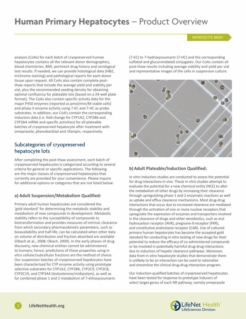
(7-EC) to 7-hydroxycoumarin (7-HC) and the corresponding sulfated and glucuronidated conjugates. Our CoAs contain all post-thaw results including average viability and yield per vial and representative images of the cells in suspension culture.
b) Adult Plateable/Induction Qualified:
In vitro induction studies are conducted to assess the potential for drug interactions in vivo. These in vitro studies attempt to evaluate the potential for a new chemical entity (NCE) to alter the metabolism of other drugs by increasing their clearance through upregulating phase 1 and 2 enzymatic reactions as well as uptake and efflux clearance mechanisms. Most drug-drug interactions that occur due to increased clearance are mediated through the activation of one or more nuclear receptors that upregulate the expression of enzymes and transporters involved in the clearance of drugs and other xenobiotics, such as aryl hydrocarbon receptor (AhR), pregnane-X-receptor (PXR), and constitutive androstane receptor (CAR). Use of cultured primary human hepatocytes has become the accepted gold standard for conducting in vitro testing of new drugs for their potential to reduce the efficacy of co-administered compounds or be involved in potentially harmful drug-drug interactions due to induction of hepatic clearance pathways. Moreover, data from in vitro hepatocyte studies that demonstrate there is unlikely to be an interaction can be used to rationalize and streamline the clinical drug-drug interaction program.
Our induction-qualified batches of cryopreserved hepatocytes have been tested for response to prototype inducers of select target genes of each NR pathway, namely omeprazole

HEPATOCYTE BRIEF
LifeNetHealth.org 4
Human Primary Hepatocytes – Product Overview
(CYP1A2), phenobarbital (CYP2B6), and rifampicin (CYP3A4), after 48 and 72 hour treatment and measurement of the fold change in both mRNA and enzymatic activity, respectively. Our cells also have been characterized for CYP enzyme activity using prototype selective substrates for CYP1A2, CYP2B6, CYP2C9, CYP2C8, CYP2C19, and CYP3A4 (testosterone/midazolam), as well as for combined phase 1 and 2 metabolism of 7-ethoxycoumarin (7-EC) to 7-hydroxycoumarin (7-HC) and the corresponding sulfated and glucuronidated conjugates. Our CoAs contain all donor information, post-thaw results including average viability and yield per vial, recommended seeding density for optimal monolayer formation, and representative images of the cell monolayers after 5 days in culture. All of our prequalified lots are guaranteed to produce stable confluent monolayers for a minimum of 1 week and meet or surpass our induction specifications when used in conjunction with our recommended culture conditions.
c) Adult High BMI/NAFLD/NASH:
Non-alcoholic fatty liver disease (NAFLD) is the most common chronic liver disease in Western countries with a wide disease spectrum. It ranges from the hepatic accumulation of lipids known as steatosis, to non-alcoholic steatohepatitis (NASH) wherein steatosis is accompanied by inflammation and fibrosis, or it can further progress to cirrhosis and hepatocellular carcinoma. NAFLD is defined by the presence of steatosis in more than 5% of hepatocytes in livers from patients with little or no alcohol consumption and it is frequently associated with obesity,
lipotoxicity, insulin resistance, hyperglycemia, hypertension, and dyslipidemia (Anderson and Borlak, 2008; Lewis and Mohanty, 2010; Cohen et al., 2011). The progression of NAFLD to non-alcoholic steatohepatitis and cirrhosis, and ultimately to carcinomas, is governed by interplay of pro-inflammatory pathways, oxidative stress, as well as fibrogenic and apoptotic cues (Lickteig et al., 2007).
As the liver is the major organ of biotransformation, perturbations in hepatic signaling pathways due to NAFLD and other related diseases have effects on both xenobiotic and endobiotic metabolism (Fisher et al., 2009; Wree et al., 2011; Naik et al., 2013). Several major nuclear receptors involved in the transcription and regulation of phase I and II drug metabolizing enzymes and transporters also have endobiotic ligands including several lipids (Lee et al., 2008; Masson et al., 2008). Studies have shown that the presence of steatosis, oxidative stress and inflammatory mediators like TNF-α and IL-6 lead to alterations in nuclear receptor signaling, such as CAR, PXR, PPARα (Bhalla et al., 2004; Zhou et al., 2006; di Masi et al., 2009; Dong et al., 2009; Gao et al., 2009). These factors result in altered expression and activity of drug metabolizing enzymes (DMEs) or transporters (Naik et al., 2013; Cobbina and Akhlaghi, 2017). Existing evidence suggests that NAFLD has effects on CYP3A4, CYP2E1 and MRP3, whereby CYP3A4 activity is down-regulated in NASH and the activity of CYP2E1 and the efflux transporter MRP3 is up-regulated (Weltman et al., 1998; Donato et al., 2006, 2007; Varela et al., 2008; Aubert et al., 2011). As such, this growing body of evidence has been made it clear that the alterations associated with NAFLD could be a potential source of drug variability in patients and could have serious implications for the safety and efficacy of xenobiotics (Barshop et al., 2011). As such, primary hepatocytes from patients with clinical manifestations of varying stages of NAFLD and NASH serve as an important research tool for the discovery of new drug targets and a better understanding of the clearance and toxicity of drugs in this growing patient population (Browning and Horton, 2004).
Because of our unique network of partner institutions, LNH has access to a broader range of tissues representing both healthy and disease individuals. Accordingly, we are able to provide primary hepatocyte lots from donors with various stages of NAFLD, including steatosis, NASH and HCC. A histopathological evaluation of formalin-fixed, paraffin-embedded tissue sections after H&E and trichrome staining is performed by a certified liver pathologist. Primary hepatocytes that have been isolated from NAFLD livers are produced under our proprietary procurement and isolation

HEPATOCYTE BRIEF
LifeNetHealth.org 5
Human Primary Hepatocytes – Product Overview
conditions using best practices and leading-edge methods for recovery, isolation and preservation. All cryopreserved batches of cells are carefully characterized for quality, plateability and functionality. We supply both suspension and plateable batches of hepatocytes from both healthy and diseased organs, and provide CYP activity data for CYP1A2, CYP2B6, CYP2C9, CYP2C8, CYP2C19, and CYP3A4 (testosterone/midazolam), as well as data for the combined phase 1 and 2 metabolism of 7-ethoxycoumarin (7-EC) to 7-hydroxycoumarin (7-HC) and the corresponding sulfated and glucuronidated conjugates. Our CoAs contain all donor information, post-thaw results including average viability and yield per vial, recommended seeding density for optimal monolayer formation where applicable, and representative images of the cell suspensions or monolayers after 5 days in culture. For diseased tissues, representative histological sections after H&E and trichrome staining and a pathology report can be provided with each CoA upon request.
d) Neonatal/Pediatric/Juvenile:
Developmental changes in the expression of drug metabolism and other clearance mechanisms determine the pharmaco- and toxicokinetics of chemicals at different life stages. These differences are critical in regulating the clearance and accumulation of drugs, and thus influence the pharmaco- and toxico-dynamic responses in newborns and children. Early studies have shown that the ontogeny of drug metabolizing enzymes and uptake/efflux transport proteins as the underlying mechanisms for altered drug
disposition during liver development (de Wildt et al., 1999; Hines, 2006, 2007; Richard et al., 2001; Strassburg et al., 2002; Peng et al., 2012; Cui et al., 2012). With the evolving knowledge on drug transporters in liver and other essential drug-processing organs, it is important to have access to human hepatocytes to study the age-related differences in metabolism and transport function, as well as to investigate the impact these may have on the potential of hepatotoxic events (Hines, 2006; Klaassen and Aleksunes, 2010).
Because of our unique network of partner institutions and access to a broader range of patient populations, we are able to source liver tissue from different life stages, including neonatal (<2 mo.), pediatric (1-12 y.o.), and juvenile (13-18 y.o.). Our hepatocyte lots have been produced under the most stringent conditions using best handling practices and leading-edge methods for procurement, isolation and preservation. All cryopreserved batches of cells are carefully characterized for quality and functionality. We supply both suspension and plateable batches of hepatocytes from these age groups, and provide CYP activity data for CYP1A2, CYP2B6, CYP2C9, CYP2C8, CYP2C19, and CYP3A4 (testosterone/midazolam), as well as data for the combined phase 1 and 2 metabolism of 7-ethoxycoumarin (7-EC) to 7-hydroxycoumarin (7-HC) and the corresponding sulfated and glucuronidated conjugates. Our CoAs contain all donor information, post-thaw results including average viability and yield per vial, recommended seeding density for optimal monolayer formation where warranted, and representative images of the cell suspensions or monolayers after 5 days in culture.

HEPATOCYTE BRIEF
LifeNetHealth.org 6
Human Primary Hepatocytes – Product Overview
References1. Anderson, N., and Borlak, J. Molecular mechanisms and therapeutic targets in
steatosis and steatohepatitis. Pharmacol.Rev. 60:311–357, 2008.
2. Anundi, I., Lahteenmaki, T., Rungren, M., Moldeus, P. and Lindros, K. O. Zonation of acetaminophen metabolism and cytochrome P450 2E1-mediated toxicity studied in isolated periportal and perivenous hepatocytes. Biochem Pharmacol, 45: 1251-9, 1993.
3. Astashkina A, Mann B, and Grainger DW. A critical evaluation of in vitro cell culture models for high-throughput drug screening and toxicity. Pharmacol Ther. 134(1):82-106, 2012.
4. Aubert, J., Begriche, K., Knockaert, L., Robin, M.A., and Fromenty, B. Increased expression of cytochrome P450 2E1 in nonalcoholic fatty liver disease: mechanisms and pathophysiological role. Clin. Res. Hepatol. Gastroenterol. 35:630–637, 2011.
5. Badr, M. Z., Belinsky, S. A., Kauffman, F. C. and Thurman, R. G. Mechanism of hepatotoxicity to periportal regions of the liver lobule due to allyl alcohol: role of oxygen and lipid peroxidation. J Pharmacol Exp Ther, 238: 1138-42, 1986.
6. Ballard, T. E., Wang, S., Cox, L. M., Moen, M. A., Krzyzewski, S., Ukairo, O. and Obach, R. S. Application of a Micropatterned Cocultured Hepatocyte System To Predict Preclinical and Human-Specific Drug Metabolism. Drug Metab Dispos, 44: 172-9, 2016.
7. Barshop, N.J., Capparelli, E.V., Sirlin, C. B., Schwimmer, J.B., and Lavine, J. E. Acetaminophen pharmacokinetics in children with nonalcoholic fatty liver disease. J. Pediatr. Gastroenterol. Nutr. 52:198–202, 2011.
8. Berry, M.N., and Friend, D.S. High yield preparation of isolated rat liver parenchymal cells. J. Cell Biol. 43:506-520, 1969.
9. Bhalla, S., Ozalp, C., Fang, S., Xiang, L., and Kemper, J.K. Ligand-activated pregnane X receptor interferes with HNF-4 signaling by targeting a common coactivator PGC-1alpha. Functional implications in hepatic cholesterol and glucose metabolism. J. Biol. Chem. 279:45139–45147, 2004.
10. Bioulac-Sage, P., LeBail, B. and Balabaud, C. Liver and biliary tract histology, In: Rodes, J., Benhamou, J.-P., Blei, A. T., Reichen, J. & Rizzetto, M. (eds.) Textbook of Hepatology: From Basic Science to Clinical Practice, 3rd Edition. Malden: Wiley-Blackwell Publishing, 2007.
11. Black, M. Acetaminophen hepatotoxicity. Annu Rev Med, 35: 577-93, 1984.
12. Browning, J.D., and Horton, J.D. Molecular mediators of hepatic steatosis and liver injury. J. Clin. Invest. 114:147–152, 2004.
13. Cobbina E, and Akhlaghi F. Non-alcoholic fatty liver disease (NAFLD) - pathogenesis, classification, and effect on drug metabolizing enzymes and transporters. Drug Metab Rev. Mar 17:1-15, 2017. [Epub ahead of print]
14. Cohen, J.C., Horton, J.D., and Hobbs, H. H. Human fatty liver disease: old questions and new insights. Science 332:1519–1523, 2011.
15. Cui J.Y., Gunewardena S.S., Yoo B., Liu J., Renaud H.J., Lu H., Zhong X.B., and Klaassen C.D. RNA-Seq reveals different mRNA abundance of transporters and their alternative transcript isoforms during liver development. Toxicol Sci. 127(2):592-608, 2012.
16. Cui J.Y., Renaud H.J., and Klaassen C.D. Ontogeny of novel cytochrome P450 gene isoforms during postnatal liver maturation in mice. Drug Metab Dispos. 40(6):1226-37, 2012.
17. de Wildt, S. N., Kearns, G. L., Leeder, J. S., and van den Anker, J. N. Cytochrome P450 3A: Ontogeny and drug disposition. Clin. Pharmacokinet. 37:485–505, 1999.
18. di Masi, A., DeMarinis, E., Ascenzi, P., and Marino, M. Nuclear receptors CAR and PXR: molecular, functional, and biomedical aspects. Mol. Aspects Med. 30:297–343, 2009.
19. Donato, M.T., Jimenez, N., Serralta, A., Mir, J., Castell, J.V., and Gomez-Lechon, M.J. Effects of steatosis on drug-metabolizing capability of primary human hepatocytes. Toxicol. In Vitro 21:271–276, 2007.
20. Donato, M.T., Lahoz, A., Jimenez, N., Perez, G., Serralta, A., Mir, J., Castell, J.V., and Gómez-Lechón, M.J. Potential impact of steatosis on cytochrome P450 enzymes of human hepatocytes isolated from fatty liver grafts. Drug Metab. Dispos. 34:1556–1562, 2006.
21. Dong, B., Saha, P.K., Huang, W.D., Chen, W.L., Abu-Elheiga, L.A., Wakil, S.J., Stevens R.D., Ilkayeva O., Newgard C.B., Chan L., and Moore D.D. Activation of nuclear receptor CAR ameliorates diabetes and fatty liver disease. Proc. Natl. Acad. Sci. U.S.A. 106:18831–18836, 2009.
22. Evans, D. C., Watt, A. P., Nicoll-Griffith, D. A. and Baillie, T. A. Drug-protein adducts: an industry perspective on minimizing the potential for drug bioactivation in drug discovery and development. Chem Res Toxicol, 17: 3-16, 2004.
23. Fisher, C.D., Lickteig, A.J., Augustine, L. M., Ranger-Moore, J., Jackson, J. P., Ferguson, S.S., and Cherrington N.J. Hepatic cytochrome P450 enzyme alterations in humans with progressive stages of nonalcoholic fatty liver disease. Drug Metab. Dispos. 37:2087–2094, 2009.
24. Fu ZD, Csanaky IL, and Klaassen CD. Effects of aging on mRNA profiles for drug-metabolizing enzymes and transporters in livers of male and female mice. Drug Metab Dispos. 40(6):1216-25, 2012.
25. Gao, J., He, J.H., Zhai, Y.G., Wada, T.R.,and Xie, W. The constitutive androstane receptor is an anti-obesity nuclear receptor that improves insulin sensitivity. J. Biol. Chem. 284:25984–25992, 2009.
26. Gebhardt, R. and Matz-Soja, M. Liver zonation: Novel aspects of its regulation and its impact on homeostasis. World J Gastroenterol, 20: 8491-504, 2017.
27. Gebhardt, R. Metabolic zonation of the liver: regulation and implications for liver function. Pharmacol Ther, 53: 275-354, 1992.
28. Godoy P., Hewitt NJ, Albrecht U, Andersen ME, Ansari N, Bhattacharya S, Bode JG, Bolleyn J, Borner C, Böttger J, Braeuning A, Budinsky RA, Burkhardt B, Cameron NR, Camussi G, Cho CS, Choi YJ, Craig Rowlands J, Dahmen U, Damm G, Dirsch O, Donato MT, Dong J, Dooley S, Drasdo D, Eakins R, Ferreira KS, Fonsato V, Fraczek J, Gebhardt R, Gibson A, Glanemann M, Goldring CE, Gómez-Lechón MJ, Groothuis GM, Gustavsson L, Guyot C, Hallifax D, Hammad S, Hayward A, Häussinger D, Hellerbrand C, Hewitt P, Hoehme S, Holzhütter HG, Houston JB, Hrach J, Ito K, Jaeschke H, Keitel V, Kelm JM, Kevin Park B, Kordes C, Kullak-Ublick GA, LeCluyse EL, Lu P, Luebke-Wheeler J, Lutz A, Maltman DJ, Matz-Soja M, McMullen P, Merfort I, Messner S, Meyer C, Mwinyi J, Naisbitt DJ, Nussler AK, Olinga P, Pampaloni F, Pi J, Pluta L, Przyborski SA, Ramachandran A, Rogiers V, Rowe C, Schelcher C, Schmich K, Schwarz M, Singh B, Stelzer EH, Stieger B, Stöber R, Sugiyama Y, Tetta C, Thasler WE, Vanhaecke T, Vinken M, Weiss TS, Widera A, Woods CG, Xu JJ, Yarborough KM, and Hengstler JG. Recent advances in 2D and 3D in vitro systems using primary hepatocytes, alternative hepatocyte sources and non-parenchymal liver cells and their use in investigating mechanisms of hepatotoxicity, cell signaling and ADME. Arch Toxicol, 87: 1315-530, 2013.

HEPATOCYTE BRIEF
LifeNetHealth.org 7
Human Primary Hepatocytes – Product Overview
29. Hewitt, N. J., De Kanter, R. and LeCluyse, E. Induction of drug metabolizing enzymes: a survey of in vitro methodologies and interpretations used in the pharmaceutical industry--do they comply with FDA recommendations? Chem Biol Interact, 168: 51-65, 2007.
30. Hines, R. N. Developmental and tissue-specific expression of human flavin-containing monooxygenases 1 and 3. Expert Opin. Drug Metab. Toxicol. 2:41–49, 2006.
31. Hines, R. N. Ontogeny of human hepatic cytochromes P450. J. Biochem. Mol. Toxicol. 21:169–175, 2007.
32. Hughes, J. P., Rees, S., Kalindjian, S. B. and Philpott, K. L. Principles of early drug discovery. Br J Pharmacol, 162: 1239-49, 2011.
33. Ito, Y. and McCuskey, R. Hepatic microcirculation In: Rodes, J., Benhamou, J., Blei, A., Reichen, J. and Rizzetto, M. (eds.) Textbook of Hepatology: From Basic Sciences to Clinical Practice. 3rd Ed. Malden Wiley-Blackwell Publishing, 2007.
34. Khetani, S. R., Kanchagar, C., Ukairo, O., Kryzewski, S., Moore, A., Shi, J., Aoyama, S., Aleo, M. and Will, Y. Use of micropatterned cocultures to detect compounds that cause drug-induced liver injury in humans. Toxicol Sci, 132: 107-17, 2013.
35. Klaassen, C. D., Casarett, L. J. and Doull, J. Casarett and Doull’s toxicology the basic science of poisons. McGraw-Hill Medical Pub, 2013.
36. Kmiec, Z. Cooperation of liver cells in health and disease. Adv Anat Embryol Cell Biol, 161, Iii-xiii, 1-151, 2001.
37. Kreamer, B.L., Staecker, J.L., Sawada, N., Sattler, G.L., Hsia, M.T.S., and Pitot, H.C. Use of a low-speed, iso-density percoll centrifugation method to increase the viability of isolated rat hepatocyte preparations. In Vitro 22(4):201-211, 1986.
38. LeCluyse E.L., Alexandre E., Hamilton G.A., Viollon-Abadie C., Coon D.J., Jolley S., and Richert L. Isolation and culture of primary human hepatocytes. Methods Mol. Biol. 290:207-230, 2005.
39. LeCluyse, E. L., Witek, R. P., Andersen, M. E. and Powers, M. J. Organotypic liver culture models: meeting current challenges in toxicity testing. Crit Rev Toxicol, 42: 501-48, 2012.
40. LeCluyse, E.L., and Alexandre, E. Isolation and culture of primary hepatocytes from resected human liver tissue. Methods Mol. Biol. 640:57-82, 2010.
41. LeCluyse, E.L., Bullock, P., Parkinson, A. and Hochman, J.H.: Cultured rat hepatocytes. Pharm. Biotechnol. 8:121-59, 1996.
42. Lee, J.H., Zhou, J., and Xie, W. PXR and LXR in hepatic steatosis: a new dog and an old dog with new tricks. Mol. Pharm. 5:60–66, 2008.
43. Lewis, J.R., and Mohanty, S.R. Non-alcoholic fatty liver disease: a review and update. Dig. Dis. Sci. 55:560–578, 2010.
44. Lickteig, A.J., Fisher, C.D., Augustine, L. M., and Cherrington, N.J. Genes of the antioxidant response undergo upregulation in a rodent model of nonalcoholic steatohepatitis. J. Biochem. Mol. Toxicol. 21: 216–220, 2007.
45. Lin, C., Shi, J., Moore, A. and Khetani, S. R. Prediction of drug clearance and drug-drug interactions in microscale cultures of human hepatocytes. Drug Metab Dispos, 44: 127-36, 2016.
46. Lin, J. H. CYP induction-mediated drug interactions: in vitro assessment and clinical implications. Pharm Res, 23: 1089-116, 2006.
47. Lindros, K. O. Zonation of cytochrome P450 expression, drug metabolism and toxicity in liver. Gen Pharmacol, 28: 191-6, 1997.
48. Masson, D., Qatanani, M., Sberna, A.L., Xiao, R., DeBarros, J.P.P., Grober, J., Deckert V., Athias A., Gambert P., Lagrost L., Moore D.D., and Assem M. Activation of the constitutive androstane receptor decreases HDL in wild-type and human apoA-I transgenic mice. J. Lipid Res. 49:1682–1691, 2008.
49. Messner, S., Agarkova, I., Moritz, W. & Kelm, J. M. Multi-cell type human liver microtissues for hepatotoxicity testing. Arch Toxicol, 87: 209-13, 2013.
50. Moon, K. H., Lee, Y. M. and Song, B. J. Inhibition of hepatic mitochondrial aldehyde dehydrogenase by carbon tetrachloride through JNK-mediated phosphorylation. Free Radic Biol Med, 48: 391-8, 2010.
51. Naik A, Belič A, Zanger UM, and Rozman D. Molecular interactions between NAFLD and xenobiotic metabolism. Front Genet. 4(2):1-14, 2013.
52. Obach, R. S. Predicting drug-drug interactions from in vitro drug metabolism data: challenges and recent advances. Curr Opin Drug Discov Devel, 12: 81-9, 2009.
53. Obach, R. S., Kalgutkar, A. S., Soglia, J. R. and Zhao, S. X. Can in vitro metabolism-dependent covalent binding data in liver microsomes distinguish hepatotoxic from nonhepatotoxic drugs? An analysis of 18 drugs with consideration of intrinsic clearance and daily dose. Chem Res Toxicol, 21: 1814-22, 2008.
54. Oorts, M., Richert, L. and Annaert, P. Drug-induced cholestasis detection in cryopreserved rat hepatocytes in sandwich culture. J Pharmacol Toxicol Methods, 73: 63-71, 2015.
55. Pedersen, J. M., Matsson, P., Bergstrom, C. A., Hoogstraate, J., Noren, A., LeCluyse, E. L. and Artursson, P. Early identification of clinically relevant drug interactions with the human bile salt export pump (BSEP/ABCB11). Toxicol Sci, 136: 328-43, 2013.
56. Peng L., Yoo B., Gunewardena S.S., Lu H., Klaassen C.D., and Zhong X.B. RNA sequencing reveals dynamic changes of mRNA abundance of cytochromes P450 and their alternative transcripts during mouse liver development. Drug Metab. Dispos. 40(6):1198-209, 2012.
57. Probst, I. and Jungermann, K. The glucagon-insulin antagonism and glucagon-dexamethasone synergism in the induction of phosphoenolpyruvate carboxykinase in cultured rat hepatocytes. Hoppe Seylers Z Physiol Chem, 364: 1739-46, 1983.
58. Przybocki JM, Reuhl KR, Thurman RG, Kauffman FC. Involvement of nonparenchymal cells in oxygen-dependent hepatic injury by allyl alcohol. Toxicol Appl Pharmacol. 115(1):57-63, 1992.
59. Ramsden, D., Tweedie, D. J., Chan, T. S., Taub, M. E. and Li, Y. Bridging in vitro and in vivo metabolism and transport of faldaprevir in human using a novel cocultured human hepatocyte system, HepatoPac. Drug Metab Dispos, 42: 394-406, 2014.
60. Rappaport, A. M. Microcirculatory units in the mammalian liver. Their arterial and portal components. Bibl Anat, 116-20, 1977.
61. Reid, L. M., Fiorino, A. S., Sigal, S. H., Brill, S. and Holst, P. A. Extracellular matrix gradients in the space of Disse: relevance to liver biology. Hepatology, 15: 1198-203, 1992.
62. Richard, K., Hume, R., Kaptein, E., Stanley, E. L., Visser, T. J., and Coughtrie, M. W. Sulfation of thyroid hormone and dopamine during human development: Ontogeny of phenol sulfotransferases and arylsulfatase in liver, lung, and brain. J. Clin. Endocrinol. Metab. 86:2734–2742, 2001.

HEPATOCYTE BRIEF
Human Primary Hepatocytes – Product Overview
LifeNet Health helps to save lives, restore health and give hope to thousands of patients each year. We are the world’s most trusted provider of transplant solutions, from organ procurement to new innovations in bio-implant technologies and cellular therapies—a leader in the field of regenerative medicine, while always honoring the donors and healthcare professionals that allow the healing process.
LifeNet Health LifeSciences Division 1864 Concert Drive, Virginia Beach, VA 23454
LifeNetHealth.org
The LifeNet Health logo is a registered trademark of LifeNet Health. ©2017 LifeNet Health. All rights reserved.
68-20-178 .00
63. Rodes, J. Textbook of hepatology : from basic science to clinical practice, 3rd Edition. Malden, Mass., Blackwell, 2007.
64. Seglen, P.O. Preparation of isolated rat liver cells, in: Methods in Cell Biology, Volume 19 (D.M. Prescott, ed.), Academic, New York, pp. 29-83, 1976.
65. Smith, M. T. and Wills, E. D. Effects of dietary lipid and phenobarbitone on the distribution and concentration of cytochrome P-450 in the liver studied by quantitative cytochemistry. FEBS Lett, 127: 33-6, 1981.
66. Strassburg, C. P., Strassburg, A., Kneip, S., Barut, A., Tukey, R. H., Rodeck, B., and Manns, M. P. Developmental aspects of human hepatic drug glucuronidation in young children and adults. Gut 50:259–265, 2002.
67. Swift, B., Pfeifer, N. D. and Brouwer, K. L. Sandwich-cultured hepatocytes: an in vitro model to evaluate hepatobiliary transporter-based drug interactions and hepatotoxicity. Drug Metab Rev, 42: 446-71, 2010.
68. Tomasi, A., Albano, E., Biasi, F., Slater, T. F., Vannini, V. and Dianzani, M. U. Activation of chloroform and related trihalomethanes to free radical intermediates in isolated hepatocytes and in the rat in vivo as detected by the ESR-spin trapping technique. Chem Biol Interact, 55: 303-16, 1985.
69. Turner, R., Lozoya, O., Wang, Y., Cardinale, V., Gaudio, E., Alpini, G., Mendel, G., Wauthier, E., Barbier, C., Alvaro, D. and Reid, L. M. Human hepatic stem cell and maturational liver lineage biology. Hepatology, 53: 1035-45, 2011.
70. Ugele, B., Kempen, H. J., Kempen, J. M., Gebhardt, R., Meijer, P., Burger, H. J. and Princen, H. M. 1991. Heterogeneity of rat liver parenchyma in cholesterol 7 alpha-hydroxylase and bile acid synthesis. Biochem J, 276 ( Pt 1): 73-7, 1991.
71. Varela, N.M., Quinones, L.A., Orellana, M., Poniachik, J., Csendes, A., Smok, G., Rodrigo R., Cáceres D.D., and Videla, L.A. Study of cytochrome P450 2E1 and its allele variants in liver injury of non- diabetic, nonalcoholic steatohepatitis obese women. Biol. Res. 41:81–92, 2008.
72. Wang, Y., Cui, C. B., Yamauchi, M., Miguez, P., Roach, M., Malavarca, R., Costello, M. J., Cardinale, V., Wauthier, E., Barbier, C., Gerber, D. A., Alvaro, D. and Reid, L. M. Lineage restriction of human hepatic stem cells to mature fates is made efficient by tissue-specific biomatrix scaffolds. Hepatology, 53: 293-305, 2011.
73. Weltman, M.D., Farrell, G.C., Hall, P., Ingelman-Sundberg, M., and Liddle, C. Hepatic cytochrome P450 2E1 is increased in patients with nonalcoholic steatohepatitis. Hepatology 27:128–133, 1998.
74. Wojcik, E., Dvorak, C., Chianale, J., Traber, P. G., Keren, D. and Gumucio, J. J. Demonstration by in situ hybridization of the zonal modulation of rat liver cytochrome P-450b and P-450e gene expression after phenobarbital. J Clin Invest, 82: 658-66, 1988.
75. Wolfe, D. and Jungermann, K. Long-term effects of physiological oxygen concentrations on glycoysis and gluconeogenesis in hepatocyte culture. Eur J Biochem, 151: 299-303, 1985.
76. Wree, A., Kahraman, A., Gerken, G., and Canbay, A. Obesity affects the liver–the link between adipocytes and hepatocytes. Digestion 83:124–133, 2011.
77. Yang, K., Guo, C., Woodhead, J. L., St Claire, R. L., 3rd, Watkins, P. B., Siler, S. Q., Howell, B. A. and Brouwer, K. L. Sandwich-Cultured Hepatocytes as a Tool to Study Drug Disposition and Drug-Induced Liver Injury. J Pharm Sci, 105: 443-59, 2016.
78. Yang, K., Pfeifer, N. D., Kock, K. and Brouwer, K. L. Species differences in hepatobiliary disposition of taurocholic acid in human and rat sandwich-cultured hepatocytes: implications for drug-induced liver injury. J Pharmacol Exp Ther, 353: 415-23, 2015.
79. Zhou, C., Tabb, M.M., Nelson, E. L., Grun, F., Verma, S., Sadatrafiei, A., Lin M., Mallick S., Forman B.M., Thummel K.E., and Blumberg B. Mutual repression between steroid and xenobiotic receptor and NF-kappaB signaling pathways links xenobiotic metabolism and inflammation. J. Clin. Invest. 116:2280–2289, 2006.
1-888-847-7831 (US & Canada) 1-757-464-4761 ext. 2000 (OUS) LifeNetHealth.org
For more information: [email protected]