increased tolerance to organic xenobiotics following
TRANSCRIPT

1
Title: Increased tolerance to organic xenobiotics following recent allopolyploidy in Spartina
(Poaceae)
Authors: Armand Cavé-Radet1, Armel Salmon1, Oscar Lima1, Malika L. Ainouche1 and
Abdelhak El Amrani1†
1Université de Rennes 1, OSUR/CNRS-UMR 6553, Ecosystèmes-Biodiversité-Evolution, Campus
de Beaulieu, Bâtiment 14A, 35042 Rennes cedex, France.
† Corresponding author: Abdelhak El Amrani (Email: [email protected])
Université de Rennes 1
Centre National de la Recherche Scientifique
UMR 6553 Ecosystems-Biodiversity-Evolution
Campus de Beaulieu, Bâtiment 14A
263 avenue du Général Leclerc
F-35042 Rennes Cedex, France
Highlights:
First PAH tolerance comparative analyses in Spartina.
Increased tolerance highlighted in the allopolyploid S. anglica compared to its parents.
Expression analysis of candidate GST genes (Tau family related) in Spartina.
Allopolyploidization and phenanthrene induced stress reprogrammed GST gene expression
patterns.
The neoallopolyploid S. anglica is a suitable candidate for phytoremediation.
Abstract
Genome doubling or polyploidy is a widespread phenomenon in plants where it has important
evolutionary consequences affecting the species distribution and ecology. PAHs are ubiquitous
organic pollutants, which represent a major environmental concern. Recent data showed that
tolerance to organic xenobiotics involve specific signaling pathways, and detoxifying gene sets
referred as ‘the xenome’. However, no data are available about how polyploidy impacts tolerance
to organic xenobiotics. In the present paper, we investigated PAH tolerance following
allopolyploidization in Spartina alterniflora, S. maritima and their derived allopolyploid species S.
anglica. We performed comparative analyses of cellular compartmentalization, photosynthetic
indices, and oxidative stress markers under phenanthrene-induced stress, and found that S. anglica
ACCEPTED MANUSCRIP
T

2
exhibit increased tolerance compared to its parents. Based on 52 genes potentially involved in
phenanthrene detoxification previously identified in A. thaliana, we investigated the Spartina
xenome using genomic and transcriptomic available resources. Subsequently, we focused on
GSTs, a ubiquitous enzymes class involved in organic xenobiotic detoxification. We examined
expression profiles of selected genes by RT-qPCR, and revealed various patterns of parental
expression alteration in the allopolyploid. The impacts of allopolyploidization on phenanthrene-
induced stress and their potential ecological implications are discussed. The neo-allopolyploid S.
anglica appears as a potential candidate for phytoremediation in PAH-polluted marshes.
Key words: Genome doubling, PAHs, abiotic stress, GSTs, Spartina
1. INTRODUCTION
Whole genome duplication (WGD, or polyploidization) is an important mechanism in plant
evolutionary history [1]. Polyploidy is accompanied by significant structural and functional
genome reorganizations, which may affect the adaptive dynamics of newly formed species (e.g.
larger habitats, increased fitness) [2–5]. In allopolyploids, both genome merger (“genomic shock”
resulting from interspecific hybridization) and genome redundancy (resulting from genome
doubling per se) result in novel genetic and epigenetic interactions, affecting the subsequent
evolution and adaptation of species [6].
In this paper, we explore the hypothesis of increased stress tolerance to organic xenobiotics
following allopolyploidy in Spartina species. Spartina is a monophyletic lineage that was recently
included in the former paraphyletic genus Sporobolus [7]. This lineage is affected by recurrent
hybridization and genome duplication, and includes 13 to 15 perennial species colonizing
estuaries, coastlines and continental wetlands worldwide. Three species are of particular
ecological and evolutionary interests: the European native S. maritima (Curtis) Fernald (2n = 6x =
60), the recently introduced to Europe from eastern America S. alterniflora Loisel. (2n = 6x = 62),
and the allopolyploid S. anglica C. E. Hubbard (2n = 12x = 120, 122 or 124). Spartina alterniflora
(maternal parent) hybridized with S. maritima (paternal parent) which resulted in the formation of
a sterile F1 hybrid S. x townsendii, discovered in 1870 (H. Groves & J. Groves; 2n = 6x = 62). A
few decades later (around the 1890’s), chromosome doubling of this hybrid led to the formation of
a new fertile and vigorous allododecaploid species S. anglica (see Supplemental Fig. S1), now
being listed among the 100 worst invasive alien species (IUCN list [8]). Spartina anglica
represents a classic example of recent allopolyploid speciation in natural environments, and an
excellent system to explore the immediate consequences of allopolyploidy in a well-established
historical and phylogenetic framework [9–11]. Hence, when comparing S. anglica to its parents,
several studies [12–15] highlighted gene expression evolution, and epigenetic modifications such
ACCEPTED MANUSCRIP
T

3
as DNA methylation alterations. Spartina species are regularly confronted with fluctuating
environments: they are able to tolerate several hours of submersion in seawater, thanks to a
developed aerenchyma [16] and efficient ‘anaerobic respiration’ capabilities [17]. They are also
strongly tolerant to flooding [16,17], salt stress [18] and chemical pollution (e.g. PAHs, crude oil,
heavy metals; see [19–24]).
Spartina species are recurrently exposed to oil spills [20,24]. In this context, we argued that
allopolyploidy might contribute to Spartina tolerance to abiotic stress, such as tolerance to PAHs
(Polycyclic Aromatic Hydrocarbons). PAHs are ubiquitous organic pollutants which represent a
major concern in terms of ecotoxicology and public health [25–27]. PAH plants detoxification
mechanisms are still poorly understood, but studies reported that plants are able to absorb,
metabolize and/or degrade PAHs. Most organic xenobiotics are taken up by plant roots from soil,
and translocated through the xylem to the shoot system for compartmentation and degradation
[28,29]. Indeed, the uptake of PAHs by plant roots occurs through simple diffusion [29] or active
transport [30].
In response to PAHs, plants are experiencing xenobiotics toxicity and associated oxidative stress
damages. Xenobiotics affect the plant physiology by reducing major metabolic functions such as
photosynthesis and respiration [31–33]. Moreover, PAHs are described to induce oxidative stress,
through ROS (Reactive Oxygen Species) accumulation in plant cells, which in turn reduce plant
development and growth, chlorophyll levels, and lead to trichome deformation and necrosis
[31,32,34]. Oxidative stress damages induced by ROS under PAH stress are balanced by
antioxidant enzymes, that are most likely limiting xenobiotic tolerance abilities [32]. More
specifically, Dumas et al. [35] found that PAH accumulation in leaves inhibits electron transfer
and photosynthesis within a few minutes, disrupting the energy transformation. Additionally, a
microarray-based transcriptional profiling has established transcriptional reprogramming in
Arabidopsis exposed to PAH [35,36]. These investigations under PAH-induced stress highlighted
the importance of plant hormones such as ethylene [36] and other secondary metabolites [37]
involved in defense pathways.
Introduced by Edwards et al. [38], the concept of ‘xenome’ was developed to characterize genes
involved in tolerance mechanisms and metabolization of xenobiotics. In Arabidopsis,
transcriptomic analysis under PAH-induced stress (plantlets exposed to phenanthrene) led to the
identification of a gene set potentially involved in PAH sensing and detoxification [35]. PAH
transformation involves oxidoreductases and hydrolases (including cytochromes P450: CYPs,
peroxidases, dioxygenases and carboxylesterases) for xenobiotic solubilization. PAHs are then
conjugated to endogenous molecules by the intervention of transferases, including glutathione-S-
ACCEPTED MANUSCRIP
T

4
transferases (GSTs), glycosyltransferases (GTs) and malonyltransferases (MTs). These
transformed molecules are then either neutralized by partnering with cell wall polymers such as
lignin, stored in the vacuole through ATP-binding cassette (ABC) transporters, or exported out of
the cell [39,38]. Plant response to xenobiotics occurs rapidly, and can be divided into three phases
as (1) perception and signaling, (2) detoxification, and (3) degradation [40,35]. Most of our
knowledge of the molecular and physiological mechanisms involved in xenobiotic tolerance relies
on model systems such as Arabidopsis. Recently, Alvarez et al. [41] performed transcriptome
profiling of resilient S. alterniflora natural populations exposed to crude oil, that provided some
candidate genes involved in xenobiotic detoxification mechanisms.
In the present paper, we explored the Spartina model in order to examine the impact of
interspecific hybridization and whole genome duplication (allopolyploidization) in the
establishment of tolerance mechanisms to phenanthrene. Hence, we conducted comparative
analyses of physiological changes in the parental species and the allopolyploid S. anglica under
phenanthrene-induced stress, quantification and histochemical localization in leaf tissues. In
parallel, we assessed changes of oxidative stress, and photosynthetic capacity (chlorophyll
fluorescence) under phenanthrene-induced stress, and analyzed transcriptional changes of
candidate genes involved in PAH detoxification pathways. The impact of allopolyploidy is
subsequently discussed.
2. MATERIALS AND METHODS
2.1. Plant materials
We collected plants in various locations along the Brittany coastline (France). We sampled S.
alterniflora in Hopital-Camfrout (Finistère, France), whereas S. maritima and S. anglica were
collected at Le Hezo (Morbihan, France). We maintained plants transplanted in pots containing a
mixture of soil and sand (70 % and 30%, respectively) in the greenhouse, under fluorescent
light/dark regime of 16/8h, and an average ambient temperature of 20°C.
2.2. In vitro experimental design: culture conditions and phenanthrene treatments
As germination of Spartina seeds is problematic (most particularly in S. maritima), we designed
an in vitro protocol, using square leaf portions (0.5-1 cm large) placed in Petri dishes (15×15 cm).
We used phenanthrene (phe: C14H10), which is commonly reported as a PAH representative
molecule. The molecular structure of phenanthrene is composed of three fused benzene rings,
ACCEPTED MANUSCRIP
T

5
resulting in a low molecular weight, and efficient plant uptake. In crude oil, phenanthrene may
represent up to 17% of total PAHs [42].
First, we incubated leaf tissues for 5 min in sodium hypochlorite (1.25% active chlorine), and
rinsed successively three times during 5 min in sterile water. We prepared Petri dishes with a solid
growth medium (half-strength Murashige and Skoog: 0.5 MS; 0.8% agar; pH= 5.6). These media
were supplemented or not with phenanthrene from a stock solution dissolved in absolute ethanol at
200 mM, reaching concentrations of 0, 100 and 400 µM phe. Such treatments allowed us to
compare stress tolerance levels between species (control and phenanthrene-induced stress
conditions), or to assess more precise dose-response effects. We added identical volumes of
ethanol alone to controls. This simplified procedure provides an optimal control of experimental
conditions, phenanthrene being assimilated by foliar uptake, as explained by Smith and Jones [43].
We cultivated foliar tissues in Petri dishes in a phytotronic chamber with light/dark regime of
16/8h, temperature 22/18°C. We conducted all experiments in three biological and technical
replicates.
2.3. Phenanthrene histolocalisation and quantification in foliar tissues
2.3.1. Phenanthrene histolocalisation
We performed microscopic observations at the Cell Imaging platform (INRA Toulouse, France)
on semi-thin sections of leaves treated with 0 and 400 µM phe. Per treatment, we observed
sections from nine leaf fragments by species (three biological replicates from distinct plants and
technical triplicates). Phenanthrene fluorescence was detected using epifluorescence microscopy
(Axiozoom V16, Zeiss). We also analyzed samples by confocal microscopy (LEICA SP2), by
scanning along different wavelengths in the visible spectrum to identify specific emissions for
phenanthrene and cellular components. Exited at 253 nm, chlorophylls are observed in red, and
phenanthrene in blue (emitting around 430 and 455 nm). Phenanthrene fluorescence is
distinguished from lignin autofluorescence which specifically emit at 472 nm (see Supplemental
Fig. S2). Then, we used three-dimensional fluorescent reconstructions of leaf tissues to identify
and specifically locate the xenobiotic into the cellular matrix.
2.3.2. Phenanthrene quantification
We estimated phenanthrene contents in Spartina tissues on leaf portions treated under 100 µM
phe. We first rinsed the harvested plant material (around 200 mg of leaf tissues from at least three
fragments from biological triplicates) in absolute ethanol and in distilled water. Samples were
lyophilized for 72h, before being ground and weighted. We extracted organic matter samples
ACCEPTED MANUSCRIP
T

6
using an accelerated solvent extractor (ASE 200, Dionex) with dichloromethane at 100°C under
pressure. After evaporation, we diluted extracts with dichloromethane, and added quantification
standards. Thereafter, we first performed fractionation by liquid chromatography, then we
proceeded to identification and quantification of phenanthrene by gas chromatography and mass
spectrometry (GC-MS).
2.4. Physiological and biochemical analyses
2.4.1. Chlorophyll fluorescence
We quantified chlorophyll fluorescence using a pulse amplitude-modulated fluorometer (Junior-
PAM, Walz GmbH; Effeltrich, Germany). The device delivers a saturating pulse through a light
guide; and collects different parameters related to the plant photochemistry. Photon absorption by
chlorophyll molecules occurred at the excited state. By returning to their basal state, the resulting
energy dissipated by the chlorophyll is either used for photosynthesis, dissipated as heat or re-
emitted as fluorescence [44]. These three processes are closely related to each other. Thus,
determining a change in chlorophyll fluorescence is equivalent to identify a change in the yield of
photochemical photosystem. Here we focus on photosystem II (PS II) and most particularly on
two parameters as indices of photosynthetic capacity: Fo (basic fluorescence yield) and Fm
(maximal fluorescence yield). These indices represent minimum and maximum fluorescence
values where reaction centers of PS II are respectively activated and inactivated, and Fv is defined
as the difference between the two indices as the maximum variable fluorescence (Fv = Fm-Fo).
Fv/Fm normalized ratio is used to approximate the maximum photochemical quantum yield of PS
II, to assess photosynthetic apparatus status in response to stress.
We performed measurements on leaf fractions (nine foliar fragments from biological and technical
triplicates) cultivated 10 days under phenanthrene treatments (0, 100 and 400 µM), and acclimated
for 30 min in the dark before measurements, preventing PS II activity.
2.4.2. Oxidative stress response: detection of superoxide radicals
We used nitroblue tetrazolium (NBT; N6876, Sigma-Aldrich) to detect superoxide radicals O2-
presence (ROS), according to the method described by Rao and Davis [45]. Leaf fragments grown
2 days on phenanthrene concentrations of 0 and 400 µM (and even up to 800 µM phe) were
immersed and infiltrated under vacuum with 3.5 mg.ml-1 NBT staining solution in a potassium
phosphate buffer (10 mM). Per treatment, we observed at least nine foliar portions from biological
and technical triplicates. Then, samples were bathed in an acetic acid-glycerol-ethanol (in
respective volumes 1:1:3) solution at 100°C for 5 min, before being bleached in 80% acetone.
ACCEPTED MANUSCRIP
T

7
NBT allowed the observation of O2- compounds which precipitate in blue color on plant tissues
[34,46]. ROS production detected in leaf tissues reflects oxidative stress damages related to
phenanthrene exposure. Leaves were then stored in a glycerol-ethanol solution (vol. 1:4) and
photographed.
2.5. In silico and gene expression analysis
2.5.1. In silico search for candidate genes involved in phenanthrene stress response
Recently, xenomic candidates involved in PAH tolerance mechanisms were identified in A.
thaliana grown in controlled conditions [35], and compared with functionally annotated crude oil
responsive genes in Spartina [41]. Here, we investigated homologous genes in Spartina using the
reference transcriptomes assembled from leaves and roots of S. maritima and S. alterniflora
[47,48], in order to compare their expression profile in the parents and the allopolyploid under
phenanthrene-induced stress in comparison to controlled conditions. Reference transcriptomes are
composed of EST contigs resulting from assemblies of both short (Illumina) and long (Roche-454)
reads from multiple cDNA libraries for each species (as described in [47,48]). Reads were
assembled with 90% identity to build consensus sequences of homoeologs used as references that
were functionally annotated. These transcriptomes resulted in 44,158 contigs in S. alterniflora and
60,644 contigs in S. maritima. To optimize the investigations, we also used genomic contigs
assembled from short reads WGS (Whole Genome Shotgun) of S. maritima DNA libraries (A.
Salmon & M. L. Ainouche, unpublished).
Based on the model described by Edwards et al. [38], we identified 99 differentially expressed
(DE) candidate xenome genes (Supplemental Fig. S3, [35]). Because xenobiotic signaling and
sensing mechanisms are still poorly understood, we focused on genes specifically involved in
xenobiotic detoxification, conjugation, putative transformation and metabolization processes.
These genes are classified within GSTs, GTs, CYPs, MTs, and ABC transporters. They represent
80 out of the total 99 DE genes reported in Arabidopsis under phenanthrene exposure (see [35]).
We chose to select among them a set of overexpressed genes that might have a significant impact
on the physiological plant tolerance to xenobiotics. Thus, we recovered 52 A. thaliana candidate
genes (16 GSTs, 15 GTs, 15 CYPs, 1 MTs and 5 ABC transporters) for identifying their homologs
in Spartina genomic and transcriptomic datasets.
In the present work, we aimed at identifying in Spartina the homologous xenome genes in
Arabidopsis using full-length CDS sequences recovered from the TAIR database (TAIR10,
http://www.arabidopsis.org). To improve our investigations, we also considered the rice genome
(Oryza sativa 204, v7.0) available on Phytozome (http://phytozome.jgi.doe.gov). The pipeline
ACCEPTED MANUSCRIP
T

8
depicting step by step the investigation process of candidate genes in Spartina is summarized in
Supplemental Fig. S4. We first identified some candidates already functionally annotated in the
Spartina transcriptomes [47,49]. O. sativa homologs to Arabidopsis candidates were also
identified using rice genome annotations. In addition, we performed BLAST alignments (BLASTn
and tBLASTx) of A. thaliana gene sequences and their O. sativa homologs on S. alterniflora and
S. maritima transcriptomes, and S. maritima genomic contigs. Gene hits with an e.value lower
than 10-6 and homologies higher than 80% over at least 70 bases (BLASTn) or 50 bases
(tBLASTx) were retained for A. thaliana and Monocot species alignments, while homologies
higher than 80% over at least 100 bases (BLASTn) or 80 bases (tBLASTx) were retained for O.
sativa - Spartina alignments.
2.5.2. GSTs phylogeny and gene expression analysis
Glutathione-S-transferases (GSTs) are a family of enzymes involved in many detoxification
processes, including xenobiotic metabolism and limiting oxidative stress [50–53]. In the present
work, the expression of GSTs genes were used as an example to highlight how the xenome
components are transcriptionally regulated in the context of allopolyploidy under phenanthrene-
induced stress.
- GSTs phylogeny and homology assessment:
We used GST homologs identified in Spartina, Oryza, and Arabidopsis as queries through
BLASTp [54] requests on sorghum (Sorghum bicolor 313, v3.1), maize (Zea mays 284, v5b) and
Oropecium thomaeum (386, v1.0) full length protein databases (from Phytozome) to identify
putative orthologous sequences. O. thomaeum like Spartina, is member of the Chloridoideae
subfamily, and currently represents the most related sequenced genome published to date. We
retained homologies presenting an e.value cutoff lower than 10-6, alignment lengths greater than
100 bases, and sequence identities of more than 60%.
To confirm candidate GSTs identified in Spartina, we validated the presence of functional protein
domains by HMMsearch (Hidden Markov Models, e.value threshold 10-3) against Pfam-A
(version 30.0, [55]). We excised GST domains recovered in Spartina contigs, and aligned
sequences using the MAFFT software (version 7 with basic parameters; [56]) on GST CDS
sequences from A. thaliana, O. sativa, S. bicolor, Z. mays and O. thomaeum. We subjected the
resulting alignment to Gblocks [57] to parse poorly aligned positions. Then, we performed
phylogenetic analysis using the IQ-TREE web server [58]. We used the Maximum Likelihood
method for tree construction with the TVM+G substitution model, and performed ultrafast
ACCEPTED MANUSCRIP
T

9
bootstrap analysis [59] on 10,000 replicates. We displayed phylogenetic tree using FigTree v1.4.3
(http://tree.bio.ed.ac.uk).
- GSTs gene expression under phenanthrene-induced stress:
We performed GST genes expression analysis on foliar fragments treated 10 days with
phenanthrene concentrations of 0 and 400 µM. After sampling, we immediately froze leaves in
liquid nitrogen. We proceeded to RNA extraction with Trizol reagent (Sigma), according to a
protocol previously employed in Spartina [15,47]. This method consists in two successive cycles
of precipitation, using isopropanol and absolute ethanol with sodium acetate (3M, pH= 5.3). We
quantified each RNA sample using Nanodrop Spectrophotometer ND 1000 (Nanodrop
Technologies, ThermoFisher Scientific Inc.) and purified extracts with a Turbo DNA-free kit
(Ambion, Life technologies) to remove DNA contaminations, before storage at -80°C. We used
about 700 ng of total RNA per sample for first-strand cDNA synthesis (kit ThermoScript RT-
PCR, Invitrogen, Life technologies), and normalized cDNA concentrations to 5 ng.µl-1 before RT-
qPCR analysis.
Each amplified sample contained 0.5 ng of cDNA, 5 µl of PowerUp SYBR Green Master Mix
(ThermoFisher), 1 µl of specific Reverse and Forward primers (5 µM), and sterile water for a final
volume of 10 µl. We designed twenty base primers using Primer3 software (http://primer3.ut.ee/),
with an expected amplicon size ranging between 100 and 200 bp, an average GC content of 55%
and a Tm close to 60°C. We first tested primers on Spartina genomic DNA, and selected only
single band amplifications for the quantification of gene expression.
In total, we analyzed the expression of 18 cDNA samples (3 species and 3 biological replicates
from control and phe treated leaves) for each selected GST homologs (3 technical replicates were
performed). We performed negative controls containing only sterile water, and 5 points dilution
ranges with a pool of total cDNA (4 fold diluted steps from 0.5 to 2.10-3 ng.µl-1) for each target
gene. We conducted quantitative PCR on the LifeCycler 480 II (Roche), using the following
program: pre-incubation at 95°C for 10 min, followed by 45 cycles of 10 sec incubation at 95°C,
10 sec annealing at 58°C, and 10 sec elongation at 68°C followed by optical reading. We
performed melting curves (fluorescence acquisition every 0.5°C from 65 to 95°C) to check
specificity of amplified fragments, and normalized levels of gene expression compared to the
housekeeping gene glyceraldehyde-3-phosphate dehydrogenase (GA3PDH), which remains stable
under these conditions [47,60]. This gene was amplified with the primer sequences
AGAGTGCCTCGTCAAGGAGA (F) and CTCCCAAGCAATCCTCATGT (R).
ACCEPTED MANUSCRIP
T

10
2.6. Statistical analysis
We performed statistics with the RStudio software [61]. We analyzed phenanthrene quantification
in foliar tissues between species, using one-way analysis of variance (ANOVA; model parameters
checked by Shapiro and Bartlett tests), and pairwise comparisons using the Duncan procedure (at
95% confidence interval). We compared chlorophyll fluorescence data within species by Kruskal-
Wallis multiple comparison’s rank test with Bonferroni correction. We tested gene expression
levels between species and treatments with two-way ANOVAs (model parameters checked by
Shapiro and Bartlett tests), and conducted pairwise comparisons using post hoc Tukey HSD. We
performed alternative analysis when model parameters were invalid, using Kruskal and Wallis and
pairwise Wilcoxon-Mann Whitney tests. We also conducted comparisons between allopolyploid’s
expression levels and MPV (Mid-Parent Value, expected under additive parental gene expression
in the allopolyploid) between treatments using the same procedure.
3. RESULTS
3.1. Phenanthrene absorption and histolocalisation
We observed histological sections of semi-thin treated (400 µM phe) and untreated leaves
cultivated in vitro for one week by epifluorescence microscopy (Fig. 1). We did not detect
phenanthrene bright spots (blue fluorescent patches) in control leaves (Fig. 1A). In treated leaves,
we observed phenanthrene by specific blue fluorescence in specific tissues depending on the
species considered, as indicated by orange arrows in Fig. 1B. We confirmed that phenanthrene
presence in cells of the leaf margins and the xylem in the three Spartina species, but we also found
xenobiotic in sclerenchyma cells in S. alterniflora and S. anglica. It was present in the mesophilic
achlorophyllous parenchyma in the allopolyploid, and in the sub-epidermal parenchyma in S.
alterniflora. If distinguishing phenanthrene fluorescence from lignin autofluorescence is
challenging, because they emit at partially overlapping wavelengths (between 450 and 500 nm for
lignin and 420-480 nm for phenanthrene on samples exited at 253 nm), phenanthrene specific
emission spectra was detected at 420-440 nm which allowed to specifically identify the xenobiotic
into the cellular matrix. In addition, more detailed analyses by confocal microscopy indicated that
the presence of the pollutant is actually intracellular (see Fig. 1C), either cytosolic or vacuolar.
These observations suggest that compartmentalization strategies could impact tolerance to
xenobiotics of these three species.
ACCEPTED MANUSCRIP
T

11
We used GC-MS for phenanthrene quantification in order to estimate the free phenanthrene
content absorbed by leaf tissues (Fig. 2). After 10 days treatment (100 µM phe), we found 213.8 ±
43.7 µg.gDW-1 phe in S. anglica, against 265.6 ± 65.1 in S. alterniflora and 468.8 ± 23.0 in S.
maritima. These results revealed significantly higher phenanthrene content in S. maritima as
compared to other species (p.value < 0.05). However, these data considered only phenanthrene in
its toxic (and not chemically modified) form, which was not detected if subjected to any metabolic
process (such as hydroxylation or glutathione conjugation).
3.2. Photosynthetic activity under phenanthrene-induced stress
After ten days of phenanthrene treatments (0, 100 and 400 µM phe), the average Fv/Fm ratios
estimated on control leaves (0 μM phe) were about 0.80 ± 0.01 for S. anglica and 0.72 ± 0.03 for
S. alterniflora and S. maritima (Fig. 3). In S. anglica, we did not observe any significant treatment
impact, even under high phenanthrene concentrations (whether on 100 or 400 µM phe). However,
the photosynthetic capacity of leaves was significantly reduced under 400 µM phe in S.
alterniflora (Fv/Fm = 0.47 ± 0.06, p.value = 0.0113) and from 100 µM phe in S. maritima (Fv/Fm
= 0.52 ± 0.08, p.value < 0.05).
3.3. Production of superoxide radicals under phenanthrene treatments
Under control conditions, NBT detection shows slight blue coloration whatever the considered
species (Fig. 4). These colorations are highly localized, suggesting that superoxide radical
production (ROS) occurred on specific locations, probably because of leaf handling. In both S.
alterniflora and S. anglica, phenanthrene treatment up to 800 µM did not impact the production of
superoxide radicals. In contrast, phenanthrene induced high oxidative stress on treated S. maritima
leaves. This assumes that for such phenanthrene concentrations (whether 400 or 800 µM phe),
oxidative stress was limited in S. alterniflora and S. anglica whereas S. maritima seems more
sensitive showing a pronounced reaction. Interestingly, we found that long time exposure to
phenanthrene treatment (30 days under 100 µM phe) did not induce any stress markers such as
superoxide radical production and reduction of chlorophyll content in S. anglica. In contrast, the
parental species S. alterniflora and S. maritima lost all chlorophyll contents and present severe
senescent phenotype indicating cell death and tissue necrosis (results not shown).
3.4. Candidate genes involved in phenanthrene tolerance
In total, we validated 35 homologs in S. maritima and 31 in S. alterniflora using alignments to A.
thaliana xenomic candidates. In complement, we added 11 homologies in S. maritima and 4 in S.
ACCEPTED MANUSCRIP
T

12
alterniflora using alignments to O. sativa candidates. Among this set of putative homologs, we
used 12 candidate genes only characterized within S. maritima genomic contigs for a final BLAST
request (BLASTn and tBLASTx) on the transcriptome of S. alterniflora, which revealed four
additional homologies on this species. Finally, we identified sequence homologies for 46 and 39
xenome candidate genes in S. maritima and S. alterniflora respectively (see Supplemental Table
S1), related to 47 A. thaliana locus (5 ABC transporters, 15 CYPs, 12 GSTs and 15 GTs). Among
these genes, 38 present homologous sequences in both parental Spartina species.
In the present work, GSTs expression was used as an example to highlight how the xenome
components are transcriptionally regulated in the context of the polyploidy under phenanthrene-
induced stress. In total, we found 14 homologous sequences in the two parental Spartina species
relative to 12 A. thaliana-GSTs. Phylogenetic reconstruction by Maximum Likelihood based on A.
thaliana, rice, maize, sorghum and O. thomaeum GST CDS sequence alignments is displayed on
Fig. 5 (TVM substitution model; [58]). We used rice and Arabidopsis GSTs from the Omega
family as outgroup to root the tree. A total of 10 CDS sequences in A. thaliana, 14 in O. sativa, 19
in S. bicolor, 15 in Z. mays, 6 in O. thomaeum and 14 in Spartina were considered in the analysis.
From this phylogenetic reconstruction, GST candidates retained in Spartina belong to the Omega
GST gene family (Salt_2_contig34019, Salt_2_contig5351 and Smar_2_contig39931), and the
plant specific Tau family. Spartina GST candidates were all similar to already known GSTs, and
grouped within Tau or Omega GST families (ultrafast bootstrap value of 93). The tree topology
distinguishes consistent specific clusters of Poaceae GSTs (highlighted in grey on the tree) as
described by Jain et al. [62]. Among the 14 Spartina GST candidate sequences, we retained 6
contigs from S. maritima genomic sequencing after primer design (see Table 1, genes in bold on
Supplemental Table S1) and followed their expressions patterns by RT-qPCR. However, we
detected primer dimers on contig GA_02873 and removed the sequence from the analysis. Some
of the technical replicates were invalidated and excluded from our analyses, but the limited extent
of Cp (Crossing point) value distributions between them remain consistent. We checked PCR
amplification efficiencies (E= 10(-1/slope)) by standard curves, and relative gene expression values
were calculated with the 2-ΔCp method [63].
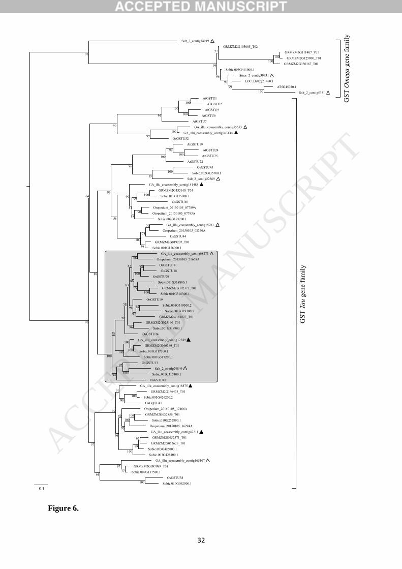
The relative expression ratios are presented in Fig. 6. Spartina GST homologous contigs identified
present various expression patterns, depending on the species and the treatment applied. First, we
compared expression profiles of GST contigs associated to S. alterniflora, S. maritima, and S.
anglica within species between phenanthrene treatments (0 and 400 µM). In the parental species,
the GST contigs selected did not display significant differential expressions considering the effect
of ten days treatments on 400 µM phe. In the neo-allopolyploid, similar patterns were observed for
ACCEPTED MANUSCRIP
T

13
contigs GA_151485, GA_12349, GA_15763 and GA_263144. However, a significant down-
regulation is detected in response to phenanthrene in S. anglica for the GA_47211 contig, where
relative ratios (±SE) decreased from 1.93. 10-2 ± 3.53. 10-3 to 7.39. 10-3 ± 2.53. 10-3 (Fig. 6A,
p.value < 0.01). Across species, additive expression patterns were observed in S. anglica for
contigs GA_151485, GA_12349 and GA_15763, as MPV relative ratios estimated under control
(0 µM phe) and treated conditions (400 µM phe) were respectively equivalent to those of the
allopolyploid (p.value > 0.05). Conversely, we observed non-additive expressions in the
allopolyploid for the contig GA_47211 with significant transgressive up-regulation (Fig. 6A,
p.value < 0.001). We also observed that contig GA_263144 was expressed similarly to S.
maritima in the allopolyploid under control condition (p.value = 0.50933). However, under stress
condition its expression appeared slightly induced as compared to the same parent (Fig. 6E,
p.value = 0.02809).
4. DISCUSSION
Understanding how plants adapt to challenging environment is a crucial question in evolutionary
ecology. A growing body of evidence revealed the prominent role of whole genome duplication
(polyploidy) in the plant diversification and adaptation [64]. In the present work, we explored the
impact of allopolyploidization on plant xenobiotic tolerance in the saltmarsh lineage Spartina by
comparing the recently formed allododecaploid species S. anglica to its parental species. This
work represents the first comparative analyses performed to date between related Spartina species.
The present analyses shed light on the detoxification processes involved in the three Spartina
species exposed to phenanthrene in the context of natural allopolyploidization event, at the
histochemical, biochemical and molecular levels.
4.1. Tolerance to phenanthrene-induced stress in Spartina
Phenanthrene histochemical investigations in Spartina revealed localized xenobiotic bright spots
near the leaf margins, suggesting a potential establishment of evacuation processes by
volatilization. Phenanthrene visualization in cells adjacent to the xylem tissues suggests that
phenanthrene may be transported through the leaf by vascular elements as shown by Paterson et
al. [65]. Furthermore, we confirmed by confocal microscopy that phenanthrene crossed the cell
wall components and the plasmalemma, and was compartmented inside the cells (see Fig. 1C),
either cytosolic or vacuolar. Intracellular presence of phenanthrene suggest that beyond being
incorporated between cell walls, detoxification mechanisms of xenobiotics via metabolic pathways
such as described in Edwards et al. [66] are ongoing in plant cells. However, differences observed
ACCEPTED MANUSCRIP
T

14
between Spartina species are introducing distinct cellular specific compartmentalization strategies,
which may affect detoxifying properties. In S. anglica and S. alterniflora, we also detected
phenanthrene in sclerenchyma and parenchyma, in contrast with S. maritima where it is only
reported near the cells of the xylem. Highly localized phenanthrene bright spots suggest that
xenobiotics in S. alterniflora and S. anglica were transferred into supportive and storage tissues,
maybe for degradation and accumulation.
Concerning ROS production analyses, the absence of superoxide radicals observed in S. anglica
and S. alterniflora even under high phenanthrene contents (800 µM phe) revealed high abilities of
oxidative stress management. In contrast, Shiri et al. [34] recently analyzed the PAH sensitive
species Arabidopsis thaliana and showed that ROS induction was detected at much lower
phenanthrene concentrations (25 µM phe), supporting that phenanthrene stress levels used in our
study highlights high tolerance abilities to PAHs in Spartina species.
4.2. Identification of homologous xenome genes in Spartina
Xenobiotic detoxification systems are still poorly understood in plants and have been explored in a
few model systems [67,66]. Recent studies have explored the functional dynamics of the xenome
in Arabidopsis under phenanthrene-induced stress [35,36,38], and comparisons with xenome
responses to crude oil in natural S. alterniflora populations were performed [41]. Here, in silico
detection of homologous xenome genes (as described in [35]) allowed an accurate identification of
candidate genes involved in phenanthrene tolerance in Spartina. We retained a total of 38 genes
from the A. thaliana xenome by homology searches in Spartina. Candidate xenome genes we
identified represents putative phenanthrene detoxifying components in non-model Monocots such
as Spartina which may be addressed in further analyses to study molecular mechanisms related to
PAH detoxification.
We focused on the GST gene family which concentrate ubiquitous enzymes responsive to
numerous stresses [51,52] to investigate the impact of allopolyploidization on xenome gene
expression patterns under phenanthrene-induced stress. Several studies have demonstrated their
role in the oxidative stress [68] by promoting antioxidant regeneration [69], or in xenobiotic
detoxification by glutathione compounds conjugation [70]. Using available transcriptomic and
genomic resources in the studied Spartina species [47,49] (A. Salmon & M. L. Ainouche,
unpublished), we identified 14 Spartina GST contigs. Three of them (Salt_2_contig34019,
Salt_2_contig5351 and Smar_2_contig39931) are grouped among the Omega GST family. Others
belong to the plant specific GST Tau family, a class already described to have a major role in
several detoxification processes [71].
ACCEPTED MANUSCRIP
T

15
Expression patterns of five GSTs from the Tau family were analyzed (filled triangles in the tree
Fig. 5). Among four selected GST candidate genes, expression profiles did not reveal significant
changes under phenanthrene-induced stress in the three Spartina species. However, significant
down-regulation in response to phenanthrene found in S. anglica (contig GA_47211; Fig. 6A)
illustrates divergent regulation pathways opportunities under genome duplication event. While
most of GST candidate genes analyzed did not present significant responses to phenanthrene, it is
not clear how these genes may be involved in detoxification response to phenanthrene in Spartina,
as suggested by Alvarez et al. [41]. However, Spartina and Arabidopsis belong to divergent
lineages in Monocots and Eudicots respectively, which separated 170-220 MYA [72]. Thus,
tolerance to organic xenobiotics may then involve distinct metabolic pathways and detoxification
mechanisms, maybe through different transformation processes (Supplemental Fig. S3) recruiting
other GSTs, GTs and malonyltransferases [36,66]. Interestingly, our phylogenetic analysis
revealed Poaceae specific GST clades, as described by Jain et al. [62]. Hence, such genes may
have acquired novel functions, as illustrated by the GST Spartina contigs which appeared quite
distant from other GSTs in the phylogenetic analysis (see Fig. 5). Here, we only focused on GSTs
as an example, but larger xenome profiling between species in controlled conditions are needed to
clarify molecular mechanisms involved in phenanthrene detoxification in Spartina.
4.3. Does allopolyploidy impact tolerance to xenobiotics?
The recent formation of S. anglica during the end of the 19th century offers a unique opportunity to
explore the early evolutionary changes associated with the formation of a new allopolyploid
species in natural populations, comparing the neo-allopolyploid to its parental species [12].
Morphological and ecological traits of the rapidly expanding neo-allopolyploid compared to its
parental species have been thoroughly described [73,74].
Our comparative physiological analysis highlights that S. maritima is the most sensitive species to
PAH-induced stress. The significant production of superoxide radicals in S. maritima leaves
indicates that oxidative stress through superoxide production was pronounced in this species,
contrasting with the tolerance exhibited by S. alterniflora and S. anglica, which developed stress
tolerance abilities (Figure 2 and 4). Spartina maritima has reduced antioxidant scavenging
capacities, thus increasing phenanthrene toxicity. Our findings also agree with the reduction in
photosynthetic activity observed in S. maritima, as ROS are known to negatively impact
photosynthetic rates [75]. Moreover, phenanthrene uptake assays performed in leaf tissues
revealed contrasted free phenanthrene contents among species, the lowest amount being quantified
in the neo-allopolyploid S. anglica and the highest in S. maritima. In complement, additional
ACCEPTED MANUSCRIP
T

16
histolocalisation of xenobiotics into S. alterniflora and S. anglica plant tissues (sclerenchyma and
parenchyma) may be related with enhanced detoxification mechanisms.
Hence, we provide experimental data supporting that enhanced xenobiotic tolerance are found in
S. anglica and S. alterniflora in contrast with pronounced sensitivity in S. maritima, consistently
with Fv/Fm ratio measured under 100 µM phe, significantly reduced in the paternal parent in
contrast to other species. Altogether, these findings may be related to ecological traits of the
compared species. S. anglica and S. alterniflora are described as vigorous invasive species [76–
81] whereas S. maritima is restricted to its native area along European and African coast where its
seems declining in some sites (e.g. England; [82]). The three species usually colonize low mash
habitats and mudflats with lower salinity ranges [83–85] but S. anglica and S. alterniflora are also
present in areas more frequently flooded or high marsh zones, and may be exposed to stronger salt
and drought levels [77,86–88].
The photosynthetic system is a key indicator to estimate plant tolerance to biotic and abiotic stress.
Previous studies showed that S. alterniflora photosynthetic rates were severely affected when
growing in PAH contaminated soils [89,90]. Our study reveals that phenanthrene stress tolerance
increased with ploidy level in Spartina, since S. anglica maintained high chlorophyll fluorescence
levels under phenanthrene-induced stress in contrast to its parental species. Under phenanthrene
treatment, we found that photosynthetic activity was significantly reduced in the parental species.
Thus, it seems that such phenanthrene concentrations (400 µM phe) did not affect S. anglica
photosynthetic apparatus, as opposed to the parental species. Moreover, by comparing S. anglica
to S. alterniflora we observed different patterns of phenanthrene localization, mainly accumulated
in the sub-epidermal parenchyma in S. alterniflora, in contrast to the mesophilic parenchyma in S.
anglica.
Differences in tolerance levels between species were detected through enhanced tolerance reported
in S. anglica, compared to its parental species (Figure 1 and 3), consistently with long term
tolerance experiments (30 days under 100 µM phe) which highlight senescent phenotypes in the
parental species while S. anglica did not exhibit any phenotypic stress markers. This suggests that
the allopolyploid S. anglica cope with chronic organic xenobiotic exposure through efficient
metabolic detoxification pathways. As the recent origin and the low inter-individual genetic
diversity related to clonal propagation reported in S. anglica are limiting the effects of selection
acting on different genotypes [9–11], it is reasonable to speculate that tolerance to PAHs results
from immediate evolutionary effects of WGD. To our knowledge, the present work demonstrates
ACCEPTED MANUSCRIP
T

17
for the first time that allopolyploidy may enhance tolerance to organic pollutants. Nevertheless,
one can keep in mind that other mechanisms resulting from the species history might interact.
Allopolyploidy highly impacts gene expression (i.e. deviation from expected parental additivity),
and may have critical adaptive impact on newly formed polyploid species [91]. Previous
transcriptomic investigations in polyploid Spartina were performed in both controlled [15] and
natural [92] conditions and revealed various expression patterns in S. anglica compared to its
parental species. In the present study, expression of the GST genes involved in A. thaliana
xenobiotic tolerance was explored. Transgressive up-regulation was recorded for contig
GA_47211 with respect to MPV and parental expression patterns. Non-additive expression was
also exhibited by the contig GA_263144 in control condition, while expression in S. anglica was
statistically equivalent to its expression in S. maritima, but differed from the MPV and from its
expression in S. alterniflora (expression dominance mimicking the paternal expression pattern).
Interestingly, paternal dominance appeared to be lost under stress conditions, as expression in S.
anglica was higher and additive with respect to parental expression patterns (statistically
equivalent to the MPV).
Allopolyploidy is a combination of two different evolutionary events, i.e. the merger of divergent
genomes (resulting from hybridization) and whole genome duplication (resulting from polyploidy)
that shape the newly formed allopolyploid genome [13,15]. Thus, changes detected in S. anglica
may reflect one or both evolutionary events. Additional comparative analyses of the xenome in the
F1 hybrid (S. x townsendii) will help deciphering the relative contributions of hybridization and/or
genome doubling per se. Expression plasticity exploiting the union of divergent expression and
regulatory networks inherited from S. maritima and S. alterniflora which diverged since 2-4 MYA
[93], combined with important epigenetic alterations [13,14], most likely represent a key
component in S. anglica enhanced tolerance to xenobiotics. As halophytes represent an emerging
trend in phytoremediation [94], our results are supporting using S. anglica for such purpose on
organic pollutants.
Author contributions
A.E, A.S and M.A. designed the experiments. A.E., O.L. and A.C.R. performed the experiments.
A.E., A.S., M.A. and A.C.R. analyzed data. A.E., A.S., M.A. and A.C.R. wrote the article.
Acknowledgements
This work was supported by the Ministère de l’Enseignement Supérieur et de la Recherche, by the
CNRS, and the Observatoire des Sciences et de l’Univers de Rennes (OSUR). The authors would
ACCEPTED MANUSCRIP
T

18
like to knowledge Olivier Catrice with his assistance for the achievement of confocal microscopy
analysis. We also thank two anonymous reviewers for their constructive comments.
Conflicts of interest: ‘none'
ACCEPTED MANUSCRIP
T

19
REFERENCES
[1] D.E. Soltis, V.A. Albert, J. Leebens-Mack, C.D. Bell, A.H. Paterson, C. Zheng, D.
Sankoff, C.W. de Pamphilis, P.K. Wall, P.S. Soltis, Polyploidy and angiosperm diversification,
Am. J. Bot. 96 (2009) 336–348.
[2] L. Comai, The advantages and disadvantages of being polyploid, Nat. Rev. Genet. 6 (2005)
836–846.
[3] A. Madlung, J.F. Wendel, Genetic and epigenetic aspects of polyploid evolution in plants,
Cytogenet. Genome Res. 140 (2013) 270–285.
[4] P.S. Soltis, D.B. Marchant, Y. Van de Peer, D.E. Soltis, Polyploidy and genome evolution
in plants, Curr. Opin. Genet. Dev. 35 (2015) 119–125.
[5] K. Alix, P.R. Gérard, T. Schwarzacher, J.S. (Pat) Heslop-Harrison, Polyploidy and
interspecific hybridization: partners for adaptation, speciation and evolution in plants, Ann. Bot.
120 (2017) 183–194.
[6] J.J. Doyle, L.E. Flagel, A.H. Paterson, R.A. Rapp, D.E. Soltis, P.S. Soltis, J.F. Wendel,
Evolutionary genetics of genome merger and doubling in plants, Annu. Rev. Genet. 42 (2008)
443–461.
[7] P.M. Peterson, K. Romaschenko, Y.H. Arrieta, J.M. Saarela, A molecular phylogeny and
new subgeneric classification of Sporobolus; (Poaceae: Chloridoideae: Sporobolinae), Taxon. 63
(2014) 1212–1243.
[8] S. Lowe, M. Browne, S. Boudjelas, M. De Poorter, 100 of the world’s worst invasive alien
species: A selection from the Global invasive species database., Invasive Species Spec. Group
ISSG Spec. Group Species Surviv. Comm. SSC World Conserv. Union IUCN. First published as
special lift-out in Aliens 12 (2000) 12pp.
[9] M.L. Ainouche, A. Baumel, A. Salmon, G. Yannic, Hybridization, polyploidy and
speciation in Spartina (Poaceae), New Phytol. 161 (2003) 165–172.
[10] M. Ainouche, H. Chelaifa, J. Ferreira, S. Bellot, A. Ainouche, A. Salmon, Polyploid
evolution in Spartina: Dealing with highly redundant hybrid genomes, in: P.S. Soltis, D.E. Soltis
(Eds.), Polyploidy Genome Evol., Springer Berlin Heidelberg, Berlin, Heidelberg, 2012: pp. 225–
243.
[11] A. Baumel, M.L. Ainouche, J.E. Levasseur, Molecular investigations in populations of
Spartina anglica C.E. Hubbard (Poaceae) invading coastal Brittany (France), Mol. Ecol. 10 (2001)
1689–1701.
[12] M.L. Ainouche, A. Baumel, A. Salmon, Spartina anglica C. E. Hubbard: a natural model
system for analysing early evolutionary changes that affect allopolyploid genomes: evolution of
the Spartina anglica, Biol. J. Linn. Soc. 82 (2004) 475–484.
ACCEPTED MANUSCRIP
T

20
[13] A. Salmon, M.L. Ainouche, J.F. Wendel, Genetic and epigenetic consequences of recent
hybridization and polyploidy in Spartina (Poaceae), Mol. Ecol. 14 (2005) 1163–1175.
[14] C. Parisod, A. Salmon, T. Zerjal, M. Tenaillon, M.-A. Grandbastien, M. Ainouche, Rapid
structural and epigenetic reorganization near transposable elements in hybrid and allopolyploid
genomes in Spartina, New Phytol. 184 (2009) 1003–1015.
[15] H. Chelaifa, A. Monnier, M. Ainouche, Transcriptomic changes following recent natural
hybridization and allopolyploidy in the salt marsh species Spartina × townsendii and Spartina
anglica (Poaceae), New Phytol. 186 (2010) 161–174.
[16] B.R. Maricle, R.W. Lee, Aerenchyma development and oxygen transport in the estuarine
cordgrasses Spartina alterniflora and S. anglica, Aquat. Bot. 74 (2002) 109–120.
[17] B.R. Maricle, J.J. Crosier, B.C. Bussiere, R.W. Lee, Respiratory enzyme activities
correlate with anoxia tolerance in salt marsh grasses, J. Exp. Mar. Biol. Ecol. 337 (2006) 30–37.
[18] J.B. Adams, G.C. Bate, Ecological implications of tolerance of salinity and inundation by
Spartina maritima, Aquat. Bot. 52 (1995) 183–191.
[19] A.W. Watts, T.P. Ballestero, K.H. Gardner, Uptake of polycyclic aromatic hydrocarbons
(PAHs) in salt marsh plants Spartina alterniflora grown in contaminated sediments,
Chemosphere. 62 (2006) 1253–1260.
[20] I.A. Mendelssohn, G.L. Andersen, L.P. Rozas, Oil impacts on coastal wetlands:
Implications for the Mississippi river delta ecosystem after the deepwater horizon oil spill,
BioScience. 62 (2012) 562–574.
[21] S. Redondo-Gomez, Bioaccumulation of heavy metals in Spartina, Funct. Plant Biol.
(2013).
[22] G. Curado, A.E. Rubio-Casal, E. Figueroa, J.M. Castillo, Potential of Spartina maritima in
restored salt marshes for phytoremediation of metals in a highly polluted estuary, Int. J.
Phytoremediation. 16 (2014) 1209–1220.
[23] P. Biber, W. Wu, M. Peterson, Z. Liu, L. Pham, Oil contamination in Mississippi salt
marsh habitats and the impacts to Spartina alterniflora photosynthesis, in: J. Alford, M. Peterson,
C. Green (Eds.), Impacts Oil Spill Disasters Mar. Habitats Fish. N. Am., CRC Press, 2014: pp.
133–172.
[24] A. Bergen, C. Alderson, R. Bergfors, C. Aquila, M.A. Matsil, Restoration of a Spartina
alterniflorasalt marsh following a fuel oil spill, New York City, NY, Wetl. Ecol. Manag. 8 (2000)
185–195. doi:10.1023/A:1008496519697.
[25] C.H. Chaîneau, J.L. Morel, J. Oudot, Phytotoxicity and plant uptake of fuel oil
hydrocarbons, J. Environ. Qual. 26 (1997) 1478.
ACCEPTED MANUSCRIP
T

21
[26] P.J. Harvey, B.F. Campanella, P.M.L. Castro, H. Harms, E. Lichtfouse, A.R. Schäffner, S.
Smrcek, D. Werck-Reichhart, Phytoremediation of polyaromatic hydrocarbons, anilines and
phenols, Environ. Sci. Pollut. Res. 9 (2002) 29–47.
[27] B.M. Greenberg, PAH Interactions with plants: Uptake, toxicity and phytoremediation, in:
P.E.T. Douben (Ed.), PAHs Ecotoxicological Perspect., John Wiley & Sons, Ltd, 2003: pp. 263–
273.
[28] Y. Gao, L. Zhu, Plant uptake, accumulation and translocation of phenanthrene and pyrene
in soils, Chemosphere. 55 (2004) 1169–1178.
[29] E. Pilon-Smits, Phytoremediation, Annu. Rev. Plant Biol. 56 (2005) 15–39.
[30] X. Zhan, X. Zhang, X. Yin, H. Ma, J. Liang, L. Zhou, T. Jiang, G. Xu, H+/phenanthrene
symporter and aquaglyceroporin are implicated in phenanthrene uptake by wheat (L.) roots, J.
Environ. Qual. 41 (2012) 188.
[31] M. Alkio, T.M. Tabuchi, X. Wang, A. Colon-Carmona, Stress responses to polycyclic
aromatic hydrocarbons in Arabidopsis include growth inhibition and hypersensitive response-like
symptoms, J. Exp. Bot. 56 (2005) 2983–2994.
[32] H. Liu, D. Weisman, Y. Ye, B. Cui, Y. Huang, A. Colón-Carmona, Z. Wang, An oxidative
stress response to polycyclic aromatic hydrocarbon exposure is rapid and complex in Arabidopsis
thaliana, Plant Sci. 176 (2009) 375–382.
[33] C. Sulmon, G. Gouesbet, F. Ramel, F. Cabello-Hurtado, C. Penno, N. Bechtold, I. Couée,
A.E. Amrani, Carbon dynamics, development and stress responses in Arabidopsis: Involvement of
the APL4 subunit of ADP-Glucose pyrophosphorylase (starch synthesis), PLoS ONE. 6 (2011).
[34] M. Shiri, M. Rabhi, C. Abdelly, A.E. Amrani, The halophytic model plant Thellungiella
salsuginea exhibited increased tolerance to phenanthrene-induced stress in comparison with the
glycophitic one Arabidopsis thaliana: Application for phytoremediation, Ecol. Eng. 74 (2015)
125–134.
[35] A.-S. Dumas, L. Taconnat, E. Barbas, G. Rigaill, O. Catrice, D. Bernard, A. Benamar, D.
Macherel, A. El Amrani, R. Berthomé, Unraveling the early molecular and physiological
mechanisms involved in response to phenanthrene exposure, BMC Genomics. 17 (2016).
[36] D. Weisman, M. Alkio, A. Colón-Carmona, Transcriptional responses to polycyclic
aromatic hydrocarbon-induced stress in Arabidopsis thaliana reveal the involvement of hormone
and defense signaling pathways, BMC Plant Biol. 10 (2010) 59.
[37] A.C. Singer, D.E. Crowley, I.P. Thompson, Secondary plant metabolites in
phytoremediation and biotransformation, Trends Biotechnol. 21 (2003) 123–130.
[38] R. Edwards, D.P. Dixon, I. Cummins, M. Brazier-Hicks, M. Skipsey, New perspectives on
the metabolism and detoxification of synthetic compounds in plants, in: P. Schröder, C.D. Collins
(Eds.), Org. Xenobiotics Plants, Springer Netherlands, Dordrecht, 2011: pp. 125–148.
ACCEPTED MANUSCRIP
T

22
[39] G. Taguchi, T. Ubukata, H. Nozue, Y. Kobayashi, M. Takahi, H. Yamamoto, N.
Hayashida, Malonylation is a key reaction in the metabolism of xenobiotic phenolic glucosides in
Arabidopsis and tobacco: Phenolic-xenobiotics metabolism in Arabidopsis, Plant J. 63 (2010)
1031–1041.
[40] A. El Amrani, A.-S. Dumas, L.Y. Wick, E. Yergeau, R. Berthomé, “Omics” Insights into
PAH Degradation Toward Improved Green Remediation Biotechnologies, Environ. Sci. Technol.
49 (2015) 11281–11291.
[41] M. Alvarez, J. Ferreira de Carvalho, A. Salmon, M.L. Ainouche, A. Cavé-Radet, A. El
Amrani, T.E. Foster, S. Moyer, C.L. Richards, Transcriptome response to the Deepwater Horizon
oil spill identifies novel candidate genes for oil tolerance in natural populations of the foundation
plant Spartina alterniflora, Mol. Ecol. (2018).
[42] Z. Liu, J. Liu, Q. Zhu, W. Wu, The weathering of oil after the Deepwater Horizon oil spill:
insights from the chemical composition of the oil from the sea surface, salt marshes and
sediments, Environ. Res. Lett. 7 (2012) 035302.
[43] K.E.C. Smith, K.C. Jones, Particles and vegetation: implications for the transfer of
particle-bound organic contaminants to vegetation, Sci. Total Environ. 246 (2000) 207–236.
[44] A. Narayan, M. Misra, R. Singh, Chlorophyll fluorescence in plant biology, in: P.D.A.N.
Misra (Ed.), Biophysics, InTech, 2012.
[45] M.V. Rao, K.R. Davis, Ozone-induced cell death occurs via two distinct mechanisms in
Arabidopsis: the role of salicylic acid, Plant J. 17 (1999) 603–614.
[46] F. Ramel, C. Sulmon, F. Cabello-Hurtado, L. Taconnat, M.-L. Martin-Magniette, J.-P.
Renou, A. El Amrani, I. Couée, G. Gouesbet, Genome-wide interacting effects of sucrose and
herbicide-mediated stress in Arabidopsis thaliana: novel insights into atrazine toxicity and
sucrose-induced tolerance, BMC Genomics. 8 (2007) 450.
[47] J. Ferreira de Carvalho, J. Poulain, C. Da Silva, P. Wincker, S. Michon-Coudouel, A.
Dheilly, D. Naquin, J. Boutte, A. Salmon, M. Ainouche, Transcriptome de novo assembly from
next-generation sequencing and comparative analyses in the hexaploid salt marsh species Spartina
maritima and Spartina alterniflora (Poaceae), Heredity. 110 (2013) 181–193.
[48] J. Boutte, B. Aliaga, O. Lima, J. Ferreira de Carvalho, A. Ainouche, J. Macas, M.
Rousseau-Gueutin, O. Coriton, M. Ainouche, A. Salmon, Haplotype detection from Next
Generation Sequencing in high ploidy-level species: 45S rDNA gene copies in the hexaploid
Spartina maritima, G3 GenesGenomesGenetics. (2015).
[49] J. Boutte, C.J. Ferreira de, M. Rousseau-Gueutin, J. Poulain, C. Da Silva, P. Wincker, M.
Ainouche, A. Salmon, Reference transcriptomes and detection of duplicated copies in hexaploid
and allododecaploid Spartina species (Poaceae), Genome Biol. Evol. (2016) evw209.
ACCEPTED MANUSCRIP
T

23
[50] A.S. Milligan, A. Daly, M.A.J. Parry, P. Lazzeri, I. Jepson, The expression of a maize
glutathione S-transferase gene in transgenic wheat confers herbicide tolerance, both in planta and
in vitro, Mol. Breed. 7 (2001) 301–315.
[51] N.E. Labrou, A.C. Papageorgiou, O. Pavli, E. Flemetakis, Plant GSTome: structure and
functional role in xenome network and plant stress response, Curr. Opin. Biotechnol. 32 (2015)
186–194.
[52] K. Nahar, M. Hasanuzzaman, M. Fujita, Physiological roles of glutathione in conferring
abiotic stress tolerance to plants, in: N. Tuteja, S.S. Gill (Eds.), Abiotic Stress Response Plants,
Wiley-VCH Verlag GmbH & Co. KGaA, Weinheim, Germany, 2016: pp. 155–184.
[53] B.P. DeRidder, Induction of Glutathione S-Transferases in Arabidopsis by herbicide
safeners, PLANT Physiol. 130 (2002) 1497–1505.
[54] S.F. Altschul, W. Gish, W. Miller, E.W. Myers, D.J. Lipman, Basic local alignment search
tool, J. Mol. Biol. 215 (1990) 403–410.
[55] R.D. Finn, P. Coggill, R.Y. Eberhardt, S.R. Eddy, J. Mistry, A.L. Mitchell, S.C. Potter, M.
Punta, M. Qureshi, A. Sangrador-Vegas, G.A. Salazar, J. Tate, A. Bateman, The Pfam protein
families database: towards a more sustainable future, Nucleic Acids Res. 44 (2016) D279–D285.
[56] K. Katoh, D.M. Standley, MAFFT Multiple sequence alignment software version 7:
Improvements in performance and usability, Mol. Biol. Evol. 30 (2013) 772–780.
[57] G. Talavera, J. Castresana, K. Kjer, R. Page, J. Sullivan, Improvement of phylogenies after
removing divergent and ambiguously aligned blocks from protein sequence alignments, Syst. Biol.
56 (2007) 564–577.
[58] L.-T. Nguyen, H.A. Schmidt, A. von Haeseler, B.Q. Minh, IQ-TREE: A Fast and Effective
Stochastic Algorithm for Estimating Maximum-Likelihood Phylogenies, Mol. Biol. Evol. 32
(2015) 268–274.
[59] B.Q. Minh, M.A.T. Nguyen, A. von Haeseler, Ultrafast Approximation for Phylogenetic
Bootstrap, Mol. Biol. Evol. 30 (2013) 1188–1195.
[60] N. Baisakh, P.K. Subudhi, N.P. Parami, cDNA-AFLP analysis reveals differential gene
expression in response to salt stress in a halophyte Spartina alterniflora Loisel, Plant Sci. 170
(2006) 1141–1149.
[61] R Core Team, R: a language and environement for statistical computing, R foundation for
statistical computing, Vienne (Autriche), 2015. http://www.R-project.org/.
[62] M. Jain, C. Ghanashyam, A. Bhattacharjee, Comprehensive expression analysis suggests
overlapping and specific roles of rice glutathione S-transferase genes during development and
stress responses, BMC Genomics. 11 (2010) 73.
[63] M.W. Pfaffl, A new mathematical model for relative quantification in real-time RT-PCR,
Nucleic Acids Res. 29 (2001) 45.
ACCEPTED MANUSCRIP
T

24
[64] Y. Van de Peer, E. Mizrachi, K. Marchal, The evolutionary significance of polyploidy,
Nat. Rev. Genet. 18 (2017) 411–424.
[65] S. Paterson, D. Mackay, C. McFarlane, A model of organic chemical uptake by plants from
soil and the atmosphere, Environ. Sci. Technol. 28 (1994) 2259–2266.
[66] R. Edwards, M. Brazier-Hicks, D.P. Dixon, I. Cummins, Chemical manipulation of
antioxidant defences in plants, in: Adv. Bot. Res., Elsevier, 2005: pp. 1–32.
[67] H. Sandermann, Plant metabolism of xenobiotics, Trends Biochem. Sci. 17 (1992) 82–84.
[68] P.G. Sappl, A.J. Carroll, R. Clifton, R. Lister, J. Whelan, A. Harvey Millar, K.B. Singh,
The Arabidopsis glutathione transferase gene family displays complex stress regulation and co-
silencing multiple genes results in altered metabolic sensitivity to oxidative stress: Genomic and
reverse genetic analysis of plant GSTs, Plant J. 58 (2009) 53–68.
[69] P.-A. Lallement, B. Brouwer, O. Keech, A. Hecker, N. Rouhier, The still mysterious roles
of cysteine-containing glutathione transferases in plants, Front. Pharmacol. 5 (2014).
[70] D.P. Dixon, R. Edwards, Glutathione Transferases, Arab. Book. 8 (2010).
[71] D.P. Dixon, B.G. Davis, R. Edwards, Functional Divergence in the Glutathione transferase
superfamily in plants. Identification of two classes with putative functions in redox homeostasis in
Arabidopsis thaliana, J. Biol. Chem. 277 (2002) 30859–30869.
[72] Y.-W. Yang, K.-N. Lai, P.-Y. Tai, W.-H. Li, Rates of nucleotide substitution in
angiosperm mitochondrial DNA sequences and dates of divergence between Brassica and other
angiosperm lineages, J. Mol. Evol. 48 (1999) 597–604.
[73] C.J. Marchant, Evolution in Spartina (Gramineae): I. The history and morphology of the
genus in Britain, J. Linn. Soc. Lond. Bot. 60 (1967) 1–24.
[74] A.J. Gray, D.F. Marshall, A.F. Raybould, A Century of Evolution in Spartina anglica, in:
Adv. Ecol. Res., Elsevier, 1991: pp. 1–62.
[75] A.R. Reddy, A.S. Raghavendra, Photooxidative stress, in: K.. Madhava Rao, A.S.
Raghavendra, K. Janardhan Reddy (Eds.), Physiol. Mol. Biol. Stress Toler. Plants, Kluwer
Academic Publishers, Dordrecht, 2006: pp. 157–186.
[76] Q. Wang, S.-Q. An, Z.-J. Ma, J.-K. Chen, B. Li, Invasive Spartina alterniflora: biology,
ecology and management, Acta Phytotaxon. Sin. 44 (2006) 559.
[77] C.M. Anderson, M. Treshow, A Review of Environmental and Genetic Factors That Affect
Height in Spartina alterniflora Loisel. (Salt Marsh Cord Grass), Estuaries. 3 (1980) 168.
[78] J.D. Thompson, The biology of an invasive plant, 41 (1991) 393–401.
[79] J. Adams, E. van Wyk, T. Riddin, First record of Spartina alterniflora in southern Africa
indicates adaptive potential of this saline grass, Biol. Invasions. 18 (2016) 2153–2158.
ACCEPTED MANUSCRIP
T

25
[80] M. Ainouche, A. Gray, Invasive Spartina: lessons and challenges, Biol. Invasions. 18
(2016) 2119–2122.
[81] W.H. Elmer, R.E. Marra, H. Li, B. Li, Incidence of Fusarium spp. on the invasive Spartina
alterniflora on Chongming Island, Shanghai, China, Biol. Invasions. 18 (2016) 2221–2227.
[82] A.F. Raybould, A.J. Gray, M.J. Lawrence, D.F. Marshall, The evolution of Spartina
anglica C. E. Hubbard (Gramineae): genetic variation and status of the parental species in Britain,
Biol. J. Linn. Soc. 44 (1991) 369–380.
[83] M.D. Bertness, Zonation of Spartina patens and Spartina alterniflora in New England salt
marsh, Ecology. 72 (1991) 138.
[84] J.M. Castillo, L. Fernandez-Baco, E.M. Castellanos, C.J. Luque, M.E. FigUeroa, A.J.
Davy, Lower limits of Spartina densiflora and S. maritima in a Mediterranean salt marsh
determined by different ecophysiological tolerances, J. Ecol. 88 (2000) 801–812.
[85] S.D. Hacker, D. Heimer, M.N. Dethier, A marine plant (Spartina anglica) invades widely
varying habitats: Potential mechanisms of invasion and control, Biol. Invasions. 3 (2001) 211–
217.
[86] C.L. Richards, S.C. Pennings, L.A. Donovan, Habitat range and phenotypic variation in
salt marsh plants, Plant Ecol. 176 (2005) 263–273.
[87] C.L. Richards, J.L. Hamrick, L.A. Donovan, R. Mauricio, Unexpectedly high clonal
diversity of two salt marsh perennials across a severe environmental gradient, Ecol. Lett. 7 (2004)
1155–1162.
[88] J. Rozema, H. Gude, G. Pollak, An ecophysiological study of the salt secretion of four
halophytes, New Phytol. 89 (1981) 201–217.
[89] S.R. Pezeshki, R.D. De Laune, Effect of crude oil on gas exchange functions of Juncus
roemerianus and Spartina alterniflora, Water. Air. Soil Pollut. 68 (1993) 461–468.
[90] Q. Lin, I.A. Mendelssohn, Evaluation of tolerance limits for restauration anf
phytoremediation with Spartina alterniflora in crude oil contaminated coastal salt marshes, Int.
Oil Spill Conf. Proc. 2008 (2008) 869–873.
[91] M.L. Ainouche, J.F. Wendel, Polyploid speciation and genome evolution: Lessons from
recent allopolyploids, in: P. Pontarotti (Ed.), Evol. Biol. Genome Evol. Speciat. Coevol. Orig.
Life, Springer International Publishing, Cham, 2014: pp. 87–113.
[92] J. Ferreira de Carvalho, J. Boutte, P. Bourdaud, H. Chelaifa, K. Ainouche, A. Salmon, M.
Ainouche, Gene expression variation in natural populations of hexaploid and allododecaploid
Spartina species (Poaceae), Plant Syst. Evol. (2017).
[93] M. Rousseau-Gueutin, S. Bellot, G.E. Martin, J. Boutte, H. Chelaifa, O. Lima, S. Michon-
Coudouel, D. Naquin, A. Salmon, K. Ainouche, M. Ainouche, The chloroplast genome of the
ACCEPTED MANUSCRIP
T

26
hexaploid Spartina maritima (Poaceae, Chloridoideae): Comparative analyses and molecular
dating, Mol. Phylogenet. Evol. 93 (2015) 5–16.
[94] E. Manousaki, N. Kalogerakis, Halophytes—An Emerging Trend in Phytoremediation, Int.
J. Phytoremediation. 13 (2011) 959–969.
[95] W.S. Alves, E.A. Manoel, N.S. Santos, R.O. Nunes, G.C. Domiciano, M.R. Soares,
Detection of polycyclic aromatic hydrocarbons (PAHs) in Medicago sativa L. by fluorescence
microscopy, Micron. 95 (2017) 23–30.
FIGURES AND TABLES
Figure 1. Histological observation of cross sections of Spartina leaves by confocal and
epifluorescence microscopy. A: Phe accumulated in the cells was detected by specific blue
fluorescent bright spots. In control leaves, no phe specific fluorescence was observed (scale
bars= 50 µm). B: Phe locations are different between species treated with 400 µM phe (scale
bars= 100 µm). In the three species, phe was detected in the neighboring cells of the xylem.
Phe was also specifically found in the sclerenchyma cells close to leaf margins, and in the
mesophilic achlorophyllous parenchyma in S. anglica. In S. alterniflora, it is also present in
the parenchyma cells and the sub-epidermal sclerenchyma. C: Phenanthrene colocalization
into Spartina leaf plant tissues by confocal microscopy (scale bar = 25 µm.). Detection of the
phenanthrene specific emission spectrum allowed us to identify the pollutant by assigning it a
violet coloration. Confocal microscopy pics clearly distinguish phenanthrene (1) intracellular
presence inside the cell walls (2), and chlorophyll fluorescence in red (3).
Figure 2. Phe concentrations in Spartina leaves cultivated 10 days on medium containing 100
µM phe. Means are calculated from three biological replicates; bars correspond to standard
errors. The different letters are corresponding to significant concentration differences
according to Duncan’s test (p.value < 0.05).
Figure 3. Fv/Fm ratio under phenanthrene treatment (0, 100 and 400 µM), an index of the
maximum quantum yield of PS II for each of the three species of Spartina. Measurements
were carried out after 10 days of phe treatment. Means are calculated from three biological
replicates; bars correspond to the standard errors. Values annotated with different letters by
species are significantly different according to Kruskal-Wallis multiple comparison’s test
(with Bonferroni correction, p.value < 0.05).
ACCEPTED MANUSCRIP
T

27
Figure 4. Visualization of superoxide radical production upon leaves NBT infiltrations.
Spartina leaves were cultivated 2 days in medium containing 0, 400 and up to 800 µM phe.
Superoxide radicals were detected by the presence of blue spots. Light blue spots were visible
on S. anglica and S. alterniflora leaves, whatever the treatment provided. In contrast, S.
maritima leaves present a severe coloration from 400 µM phe, reflecting a strong oxidative
stress. Scale bars = 1.5 mm.
Figure 5. GSTs phylogenetic reconstruction by Maximum Likelihood method based on the
TVM (transversion) substitution model with a discrete Gamma distribution (4 categories). In
total, 78 CDS sequences were considered among A. thaliana, O. sativa, S. bicolor, Z. mays, O.
thomaeum and Spartina. Tree is rooted by CDS sequences from the Omega GST gene family,
and numbers at the nodes represent ultrafast bootstrap values from 10,000 replicates. Spartina
contigs are displayed with black triangles, those we evaluate their expression by RT-qPCR are
stained and belong to the Tau family. Corresponding sequences IDs: GRMZM─ for Z. mays;
Sobic─ for S. bicolor; Oropecium─ for O. thomaeum. Scale bar indicates 0.1 residue
substitution per site.
Figure 6. Relative expression levels of five candidates contigs (GST homologs) estimated by
RT-qPCR on three biological and technical replicates for each Spartina species in control
conditions and under phenanthrene induced stress (0 and 400 µM). Values were calculated
with the 2-ΔCp method; bars correspond to standard errors. **: p.value < 0.01.
-
ACCEPTED MANUSCRIP
T

28
Figure 1.
ACCEPTED MANUSCRIP
T

29
Figure 2.
ACCEPTED MANUSCRIP
T

30
Figure 3.
Figure 4.
ACCEPTED MANUSCRIP
T

31
Figure 5.
ACCEPTED MANUSCRIP
T
32
Figure 6.
ACCEPTED MANUSCRIP
T

33
ACCEPTED MANUSCRIP
T

34
Table 1. Primers designed and validated for the six GSTs homologs in Spartina used for the RT-qPCR. Related A. thaliana genes and
supplemental comments are provided.
Table 1.
N° Spartina contigs A. thaliana related homologs Forward primer (5’ 3’) Reverse primer (5’ 3’) Supplemental comments
1 GA_47211 AtGSTU6, AtGSTU7, AtGSTU8 GATTCTGGGCCCACTTCTTC TCGTCTCTTCCGTGAACTCC -
2 GA_151485 AtGSTU25, AtGSTU22, AtGSTU19 GACGTAGTCTGCCCAGAAGC AGAAGATCCCAGTGCTGCTC -
3 GA_12349 AtGSTU14 GAGGTCCTGCTCGACGTACT GAGGCAACTGCTAGAGCCTG -
4 GA_02873 AtGSTU5 GAGTTTGGTGGCGCTAAATG AGACTTGCCCTCAGCTTCAA Deleted because of primer dimers
5 GA_15763 AtGSTU24 GACGAGGGACTCGCACAC GTGGCGTACGAGGAGAAATC -
6 GA_263144 AtGSTU1 AGCTGCGGCATCTGGT TGTTCTTGAGATCCTCCTCGAT -
ACCEPTED MANUSCRIP
T