human gamma delta t regulatory cells in cancer: …keywords: gamma delta t cells, cancer...
TRANSCRIPT

MINI REVIEW ARTICLEpublished: 20 November 2014
doi: 10.3389/fimmu.2014.00598
Human gamma deltaT regulatory cells in cancer: fact orfiction?Daniela Wesch1, Christian Peters1 and Gabrielle Melanie Siegers2*1 Institute of Immunology, Christian-Albrechts University of Kiel, Kiel, Germany2 Department of Oncology, University of Alberta, Edmonton, AB, Canada
Edited by:Julie Dechanet-Merville, CentreNational de la Recherche Scientifique(CNRS), France
Reviewed by:David L. Wiest, Fox Chase CancerCenter, USABruno Martin, Centre National de laRecherche Scientifique (CNRS),France
*Correspondence:Gabrielle Melanie Siegers,Department of Oncology, Universityof Alberta, 5-142E Katz GroupBuilding, 114th Street and 87thAvenue, Edmonton, AB T6G 2E1,Canadae-mail: [email protected]
While gamma delta T cell (γδTc) anticancer immunotherapies are being developed, recentreports suggest a regulatory role for γδTc tumor-infiltrating lymphocytes. This mini-reviewsurveys available evidence, determines strengths and weaknesses thereof and suggestdirections for further exploration.We focus on human γδTc, as mouse and human γδTc reper-toires differ. Regulatory γδTc are defined and compared to conventionalTregs and their rolesin health and disease (focusing in on cancer) are discussed. We contrast the suggestedregulatory roles for γδTc in breast and colorectal cancer with their cytotoxic capabilitiesin other malignancies, emphasizing the context dependence of γδTc functional plasticity.Since γδTc can be induced to exhibit regulatory properties (in some cases reversible), wecarefully scrutinize experimental procedures in published reports. As γδTc garner increas-ing interest for their therapeutic potential, it is critical that we appreciate the full extentof their role(s) and interactions with other cell types in both the circulation and the tumormicroenvironment. A comprehensive understanding will enable manipulation of γδTc toimprove anti-tumor efficacy and patient outcomes.
Keywords: gamma delta T cells, cancer immunotherapy, regulatory T cells, human cancer, gamma delta T cellfunctional plasticity
INTRODUCTIONWhile those of us in the immunotherapy world tend to focuson the anti-infection and anti-tumor properties of γδTc, we arenow beginning to appreciate that, under certain conditions, theseremarkable cells can inhibit or suppress the maturation and/oractivation of immune cells around them, leading to beneficial orpotentially pathological consequences.
A suppressor function of human γδTc was first described in1989 by Patel and colleagues; upon in vitro stimulation with poke-weed mitogen, most γδTc clones could suppress the generationof Immunoglobulin(Ig)-secreting B cells by CD4+ T helper cellstreated with mitomycin C (Figure 1A) (1). Since then, regula-tory roles for γδTc have been described in several contexts. BothVδ1 and Vδ2 T cell subsets (Vδ1Tc and Vδ2Tc, respectively) mayexhibit regulatory properties, albeit in different settings.
Human peripheral blood-derived γδTc displaying regulatoryproperties are phenotypically different from conventional reg-ulatory CD4+ αβ T cells (Treg). In contrast to Treg, freshlyisolated γδTc express only low levels of CD25 and cytotoxic Tlymphocyte-associated antigen (CTLA)-4, and do not express thetranscription factor forkhead box P3 (FoxP3) (2–4). Similar toconventional αβ T cells (αβTc), CD25 is up-regulated on γδTcafter initial phytohemagglutinin (PHA) or anti-γδ TCR mono-clonal antibody (mAb) stimulation (5). Additionally, CD25 is alsoup-regulated on Vδ2Tc after stimulation with pyrophosphates(phosphorylated antigens), which are intermediates of the iso-prenoid pathway and induce selective expansion of Vδ2Tc withinperipheral blood mononuclear cells (PBMC) 7–10 days after ini-tial stimulation (6, 7). Furthermore, FoxP3 expression can be
detected with PCH101 mAb but not with the more Treg-specific259D mAb, in γδTc as well as in Treg-depleted αβTc after acti-vation (4). FoxP3 expression as identified by PCH101 mAb doesnot correlate with suppressive function (8, 9). In addition, thetranscription factor Helios, which is highly expressed by Treg, isconstitutively expressed in roughly one-third of freshly isolatedγδTc (4). While Helios seems to be involved in the differentiationof (regulatory) γδTc, it is not a specific marker for suppressiveγδTc (4, 10). Thus, while freshly isolated γδTc do not expresscharacteristic Treg markers, the literature provides evidence thatγδTc may nevertheless exhibit regulatory activity, which will befurther described below.
REGULATORY ROLES FOR γδTC IN NON-CANCER CONTEXTSBefore focusing in on the potential regulatory role of γδTc in can-cer, it is worthwhile to consider some other contexts in which thesecells have displayed suppressive properties. For a more compre-hensive description of regulatory roles of γδTc outside of cancer,we recommend a recent review (10).
Immunosuppression via γδTc plays a protective role in severalcontexts. For example, in pregnancy, decidual γδTc contribute toan immunosuppressive milieu enabling successful implantationand protecting the growing fetus from attack by the mother’simmune system (11–14). In celiac disease, patients on a gluten-free diet have enhanced suppressor intestinal intraepithelial γδTcthat protect the small intestine from attack by CD8+ TCRαβ+
intraepithelial lymphocytes (IEL) via secretion of transforminggrowth factor-beta one (TGF-β1); patients with active diseasehave lower frequencies of these suppressor γδTc IEL (15). Lower
www.frontiersin.org November 2014 | Volume 5 | Article 598 | 1

Wesch et al. Human gamma delta T regulatory cells in cancer
FIGURE 1 | γδTc exhibiting regulatory properties may be generatedin vitro by various means. Details are given in the text and the indicatedreferences. The γδTc are depicted in red, αβTc in green, B cells in blue,dendritic cells in yellow, and senescent cells in gray. Ag, antigen; APC,antigen-presenting cell; BrHPP, bromohydrin pyrophosphate; fresh, freshly
isolated; Ig, immunoglobulin; IPP, isopentenyl pyrophosphate; Mito C,mitomycin C; PBMC, peripheral blood mononuclear cell; PWM, pokeweedmitogen; SE, Staphylococcus aureus enterotoxins. (A) Patel et al., (B) Kuhlet al., Peters et al., (C) Casetti et al., (D) Traxlmayr et al., (E) Li et al., (F) Peterset al., and (G) Hua et al.
Frontiers in Immunology | T Cell Biology November 2014 | Volume 5 | Article 598 | 2

Wesch et al. Human gamma delta T regulatory cells in cancer
peripheral blood γδTc numbers, more specifically a decreasedproportion of central memory γδTc, are correlated with systemiclupus erythematosus pathogenesis, suggesting a protective rolefor regulatory γδTc in this autoimmune disease as well (16). Ofnote, Vδ1Tc/Vδ2Tc subset ratios are inverted in patients com-pared to healthy controls (i.e., Vδ1Tc predominate in blood)(16). Similarly, a higher Vδ1Tc/Vδ2Tc ratio may contribute to theachievement of operational tolerance in pediatric liver transplantrecipients (17).
HOW TO MAKE REGULATORY γδTCSo far, it is unknown whether specific subsets, e.g., CD27+ Helios-expressing γδTc, are innately suppressive or whether their broadrange of functional plasticity enables suppressive activity undercertain stimulatory conditions (Figure 1). An observation com-mon to all studies on suppressive Vδ2Tc is that they realize theirimmunosuppressive potential only in the presence of antigen-presenting cells (APC) or after co-stimulation with anti-CD28mAb (Figures 1B,C) (2–4). CD28 and CTLA-4 are critical reg-ulators of immunosuppressive T cells, whereby CD28 plays a dualrole in both the generation and the termination of an immuneresponse (18).
Freshly isolated isopentenyl pyrophosphate (IPP)-stimulatedVδ2Tc can inhibit the proliferation of CD4+ and CD8+ αβTcin response to strong recall antigens such as Tetanus toxoid,superantigens such as Staphylococcus aureus enterotoxins (SE) oralloantigens in the presence of APCs (Figure 1D) (19). How-ever, the authors could not completely rule out low frequencyactivation of αβTc by antigen-specific (e.g., Tetanus toxoid) stim-ulation. Nevertheless, peripheral blood Vδ2Tc also suppress pro-liferation of co-cultured CD4+ αβTc after polyclonal stimulationby anti-CD3/CD28 mAb, which simultaneously activates αβTc(Figure 1B) (3, 4). All in all, the presence and strength of a co-stimulatory APC-signal seem to play an important role in theinduction of Vδ2Tc suppressive capacity (4).
While TGF-β1 alone does not induce the generation of reg-ulatory Vδ2Tc, this switch can occur in the presence of addi-tional cytokines (Figures 1B,C,E,F) (2, 4, 9, 16). Up to 30%of Vδ2Tc within IPP-stimulated PBMC cultivated in the pres-ence of TGF-β1 and interleukin (IL)-15 expressed FoxP3 (clone259D); after subsequent cell sorting, these FoxP3+ enrichedVδ2Tc suppressed the proliferation of anti-CD3/CD28 mAb-simulated PBMC (Figure 1C) (2). Peters and colleagues have sincedemonstrated that the observed FoxP3 expression was transient,with a steady increase in FoxP3 over 8 days of cell culture fol-lowed by a decrease to nearly undetectable protein levels after16 days (4).
In contrast to the work of Casetti and colleagues, in the studyof Peters et al. TGF-β1 and IL-15 did not induce regulatory func-tions in bromohydrin pyrophosphate (BrHPP)-expanded γδTc.Only anti-CD3/CD28 mAb-stimulated γδTc expanded in thepresence of TGF-β1 and IL-15 were able to suppress the pro-liferation of αβTc induced by a mixture of SE (Figure 1F) (4).The observed suppressive activity was not dependent on FoxP3expression but was rather dependent on the presence of initialCD28-co-stimulation. The discrepancy between these two stud-ies might be explained by differences in γδTc expansion as well
as stimulatory conditions in the suppression assays. Casetti et al.used IPP-stimulated PBMC from which Vδ2Tc were sorted afterexpansion, whereas Peters et al. expanded magnetically isolated,highly purified γδTc (20). In their suppression assay, Casettiet al. analyzed the Vδ2Tc suppression of PBMC stimulated byanti-CD3/CD28 mAb, which could potentially activate other sup-pressive T cell subsets within the PBMC. In contrast, Petersand colleagues used CD25-depleted CD4+ T cells as respondercells, which were stimulated by a mixture of SE and BrHPP-restimulation for the co-cultured γδTc. Common to both studies isa correlation between CD28-co-stimulation (although at differenttime points) and the suppressive effect. This suggests that CD28signaling in γδTc-mediated suppression should be examined inmore detail.
While FoxP3 and γδTc regulatory activity are not strictly con-nected, it is worthwhile to note that FoxP3 expression can beinduced in both Vδ1Tc and Vδ2Tc subsets. Similar to Vδ2Tc,FoxP3 was prominently induced in Vδ1Tc in the presence ofTGF-β1 and additional cytokines such as IL-2 after stimulatingPBMC with anti-γδTCR for 10 days (16). Additionally, there wasan increased expression of both TGF-β1 and its receptor (CD105)on Vδ1Tc compared to Vδ2Tc; upon activation, TGF-β1 decreasedand CD105 increased on Vδ1Tc. The authors assumed a regulatoryrole for the Vδ1 CD45−CD27+ γδTc subset due to its increasedFoxP3 expression. While they demonstrated inhibition of CD4+
T cell proliferation by CD27+ Vδ1Tc, the authors unfortunatelydid not directly compare the suppressive activity of CD27+ versusCD27− Vδ1Tc (Figure 1E) (16). In this context, the analysis ofFoxP3 expression in purified Vδ2Tc versus Vδ1Tc under differentculture conditions would be interesting.
Finally, Hua and colleagues induced regulatory Vδ1Tc in vitro,upon stimulation of PBMC with plate-bound anti-TCRVδ1 mAb,that expressed FoxP3 (identified by mAb clone 259D/C7) and sup-pressed CD4+ T cell proliferation (Figure 1G) (21). The authorssuggested that Vδ1Tc FoxP3 expression was sustained by a positivefeedback loop instigated by Vδ1Tc producing TGF-β1; in addition,Vδ1Tc secreted IL-10 (21).
In summary, it is difficult to compare these studies,as their inherent differences in experimental design (cellsource/subset/milieu/stimuli) are further confounded by the lackof a defined regulatory γδTc marker. However, it is clear that γδTccan be induced to exhibit regulatory properties.
HOW DO γδTC SUPPRESS OTHER CELLS?There are, however, some controversial data regarding mecha-nism(s) of suppression by γδTc. Kühl and colleagues assumedmediation by the immunosuppressive cytokines TGF-β1 andIL-10, which were secreted by γδTc after anti-CD3/CD28 mAbstimulation (Figure 1B). After 48 h stimulation, γδTc secreted sig-nificantly more TGF-β1 than conventional CD4+CD25+ Tregs(3). Unfortunately, their ELISAs did not distinguish betweenTGF-β1 secretion by Vδ1Tc and Vδ2Tc; however, higher TGF-β1mRNA levels after 3 day Concanavalin A treatment would sug-gest that Vδ1Tc have a greater suppressive capacity than Vδ2Tc orαβTc (3).
Peters and co-workers demonstrated that co-culture withresponder cells (CD25-depleted CD4+ αβTc) induced the
www.frontiersin.org November 2014 | Volume 5 | Article 598 | 3
Wesch et al. Human gamma delta T regulatory cells in cancer
upregulation of CD80 and CD86 as well as programed death-ligand (PDL)-1 on stimulated Vδ2Tc, which could then inter-act with CTLA-4 or PD-1 on responder cells, leading to theirsuppression (4). Furthermore, transwell experiments suggestedcell-contact-dependence, as this process was inhibited by mAbdisrupting CD86:CTLA-4 or PDL-1:PD-1 interactions betweenanti-CD3/CD28 mAb-activated Vδ2Tc and activated αβTc (4).Interestingly, the immunosuppressive capacity of Vδ2Tc was abro-gated by Toll-like-receptor (TLR) 2 ligands as well as by activatingαβTc with a mixture of five SE (in contrast to the publication ofTraxlmayr where only one SE was applied), which both inducea strong Th1-response [(4, 19); Peters and Wesch, unpublisheddata]. Abrogated suppression correlated with increased phos-phorylation of Akt and NFκB in αβTc and down-regulation ofinhibitory molecules such as PD-1 and CTLA-4 (4). Similarly,Peng and colleagues found that the regulatory γδTc phenotypecould be reversed through administration of TLR8 ligand Poly-G(Figure 2A) (22, 23). Only ligands to TLR8 (and not TLRs 2, 3,4, 5, 7, or 9) blocked induction of senescence observed in T cellsresponding to suppression via regulatory γδTc (Figure 2A) (23).These observations exemplify the functional plasticity of γδTc thatare influenced by the nature of a stimulus and the surroundingcytokine milieu.
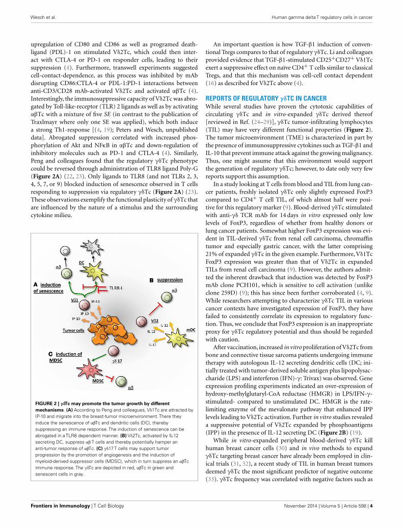
FIGURE 2 | γδTc may promote the tumor growth by differentmechanisms. (A) According to Peng and colleagues, Vδ1Tc are attracted byIP-10 and migrate into the breast-tumor microenvironment. There theyinduce the senescence of αβTc and dendritic cells (DC), therebysuppressing an immune response. The induction of senescence can beabrogated in a TLR8 dependent manner. (B) Vδ2Tc, activated by IL-12secreting DC, suppress αβ T cells and thereby potentially hamper ananti-tumor response of αβTc. (C) γδ17 T cells may support tumorprogression by the promotion of angiogenesis and the induction ofmyeloid-derived suppressor cells (MDSC), which in turn suppress an αβTcimmune response. The γδTc are depicted in red, αβTc in green andsenescent cells in gray.
An important question is how TGF-β1 induction of conven-tional Tregs compares to that of regulatory γδTc. Li and colleaguesprovided evidence that TGF-β1-stimulated CD25+CD27+ Vδ1Tcexert a suppressive effect on naïve CD4+ T cells similar to classicalTregs, and that this mechanism was cell-cell contact dependent(16) as described for Vδ2Tc above (4).
REPORTS OF REGULATORY γδTC IN CANCERWhile several studies have proven the cytotoxic capabilities ofcirculating γδTc and in vitro-expanded γδTc derived thereof[reviewed in Ref. (24–29)], γδTc tumor-infiltrating lymphocytes(TIL) may have very different functional properties (Figure 2).The tumor microenvironment (TME) is characterized in part bythe presence of immunosuppressive cytokines such as TGF-β1 andIL-10 that prevent immune attack against the growing malignancy.Thus, one might assume that this environment would supportthe generation of regulatory γδTc; however, to date only very fewreports support this assumption.
In a study looking at T cells from blood and TIL from lung can-cer patients, freshly isolated γδTc only slightly expressed FoxP3compared to CD4+ T cell TIL, of which almost half were posi-tive for this regulatory marker (9). Blood-derived γδTc stimulatedwith anti-γδ TCR mAb for 14 days in vitro expressed only lowlevels of FoxP3, regardless of whether from healthy donors orlung cancer patients. Somewhat higher FoxP3 expression was evi-dent in TIL-derived γδTc from renal cell carcinoma, chromaffintumor and especially gastric cancer, with the latter comprising21% of expanded γδTc in the given example. Furthermore, Vδ1TcFoxP3 expression was greater than that of Vδ2Tc in expandedTILs from renal cell carcinoma (9). However, the authors admit-ted the inherent drawback that induction was detected by FoxP3mAb clone PCH101, which is sensitive to cell activation (unlikeclone 259D) (9); this has since been further corroborated (4, 9).While researchers attempting to characterize γδTc TIL in variouscancer contexts have investigated expression of FoxP3, they havefailed to consistently correlate its expression to regulatory func-tion. Thus, we conclude that FoxP3 expression is an inappropriateproxy for γδTc regulatory potential and thus should be regardedwith caution.
After vaccination, increased in vitro proliferation of Vδ2Tc frombone and connective tissue sarcoma patients undergoing immunetherapy with autologous IL-12 secreting dendritic cells (DC; ini-tially treated with tumor-derived soluble antigen plus lipopolysac-charide (LPS) and interferon (IFN)-γ: Trivax) was observed. Geneexpression profiling experiments indicated an over-expression ofhydroxy-methylglutaryl-CoA reductase (HMGR) in LPS/IFN-γ-stimulated- compared to unstimulated DC. HMGR is the rate-limiting enzyme of the mevalonate pathway that enhanced IPPlevels leading to Vδ2Tc activation. Further in vitro studies revealeda suppressive potential of Vδ2Tc expanded by phosphoantigens(IPP) in the presence of IL-12 secreting DC (Figure 2B) (19).
While in vitro-expanded peripheral blood-derived γδTc killhuman breast cancer cells (30) and in vivo methods to expandγδTc targeting breast cancer have already been employed in clin-ical trials (31, 32), a recent study of TIL in human breast tumorsdeemed γδTc the most significant predictor of negative outcome(33). γδTc frequency was correlated with negative factors such as
Frontiers in Immunology | T Cell Biology November 2014 | Volume 5 | Article 598 | 4

Wesch et al. Human gamma delta T regulatory cells in cancer
advanced tumor stage, positive lymph node status, and human epi-dermal growth factor receptor 2 (HER2) expression. Exhaustivestatistical analysis correlated γδTc with FoxP3+ cells (identifiedwith clone 236A/E7) and inversely with CD8+ cytotoxic Tc, sug-gesting a negative role for γδTc (33). However, double staining ofγδTc and FoxP3 was not done, leaving the identity of FoxP3+ cellsambiguous, and there was no indication as to whether staining wasperformed on serial sections. Furthermore, γδTc subsets were notspecified, likely due to a dearth of subset-specific antibodies suit-able for their detection via immunohistochemistry (33). Finally,while γδTc frequency in breast tumors may prove to be a valu-able prognostic marker, their role in disease pathogenesis was notdetermined.
This same group, however, had previously suggested regulatoryproperties for Vδ1Tc TIL in breast tumors (22). γδTc TIL wereextracted from a digested human breast tumor, expanded in vitrofor 1 week in 1000 IU/ml IL-2, after which bulk TILs were main-tained at 50 IU/ml IL-2. Tumor-reactive clones were then gener-ated and both the bulk population and selected clones derivedthereof suppressed naïve T cell proliferation, IL-2 secretion, andDC maturation (22). This may not reflect the case in situ. Whilethis study proves that Vδ1Tc can assume a regulatory phenotype,several caveats demand attention:
Firstly, the subset prevalence of γδTc in the original tumor wasnot reported and thus (regulatory) Vδ2Tc may have comprisedthe majority of tumor-derived cell suspensions at the outset butmay have been subsequently eliminated by high levels of IL-2 inthe culturing process, since Vδ2Tc are known to be susceptible toactivation-induced cell death (34–36). Broad ranges of Vδ1Tc lev-els were only determined after culturing, while Vδ2Tc percentageswere not reported (22). In a follow-up paper, recruitment of γδTcwith a regulatory phenotype was linked to high levels of IFN-γinducible protein 10 (IP-10) in the TME (Figure 2A); however,Vδ1Tc and Vδ2Tc were unfortunately not distinguished (37). Sec-ondly, the high level of IL-2 used to culture TILs may in itself havesupported expansion of a regulatory phenotype not truly reflectiveof the original functional orientation of these cells. Thirdly, mostexperiments were carried out with one cell line and clones derivedfrom a single tumor, thus cannot represent a universal truth. It isalso not clear whether the same Vδ1Tc lines were used in subse-quent publications. While valuable insight into the plasticity andregulatory potential of Vδ1Tc can be gleaned from these studies,further investigation of γδTc TIL in situ are required to substan-tiate claims of regulatory function contributing to poor patientprognosis.
While breast-tumor TIL-derived Vδ1Tc can exhibit regula-tory properties in vitro, Vδ1Tc TIL from other cancers have beenreported to be cytotoxic (38, 39). Polyclonal γδTc TIL lines killmelanoma cell lines, and secrete tumor necrosis factor alpha(TNFα) and IFN-γ (38). This functional diversity could well becontext-dependent or perhaps, as Donia and colleagues suggest,clones with various Vγ pairings are differentially activated (39). Itis also possible that these cytotoxic γδTc TIL are simultaneouslycapable of as-of-yet unnoticed regulatory functions.
Finally, an indirect regulatory role for γδTc has been reportedin colorectal cancer (CRC), whereby IL-17 secreting γδTc (γδ17)
in the TME may attract and help support immunosuppressivemyeloid-derived suppressor cells (MDSC) (Figure 2C). In vitroexperiments showed that activated inflammatory DC secrete IL-23facilitating the generation of γδ17. DC activation is thought to becaused by release of bacterial products through the compromisedepithelial barrier characterizing CRC. Of note, γδ17 isolated fromCRC tumors were predominantly Vδ1Tc, secreted higher levels ofIL-17 compared to normal tissue controls and did not secrete IL-4,IL-22 or immunosuppressive IL-10 (40).
AVENUES TO EXPLOREIf γδTc TIL are indeed regulatory, it is crucial to determine whetherthey are inherently so or whether factors in the TME induce thisfunction. If the former is true, then presumably infusion of largenumbers of cytotoxic γδTc into patients should cause no safetyconcern (with respect to the further promotion of tumor growth).However, if the latter is true, we need to find a way to target theTME to prevent a potentially detrimental shift to a regulatory phe-notype. Better models mimicking the human TME could help usaddress this question.
Since γδTc can be induced to realize regulatory potential in var-ious ways, including those involving cytokines typically present inthe TME, some degree of regulatory function is plausible. How-ever, so far the evidence is scant, limited to in vitro experimentswith ex vivo expanded γδTc. Admittedly, there is an inherent dif-ficulty in assessing the regulatory capacity of γδTc TIL in situ,as they are only present in relatively low abundance. Ye and col-leagues attempted to address this by performing experiments withfreshly purified γδTc from tumor tissues; however, depending onthe nature of the antibodies used for purification, γδTc functionmay already have been altered (23). Finally, as discussed above,assessment using markers such as FoxP3 should be consideredcarefully because not every mAb clone detecting FoxP3 expressiondenotes regulatory function.
CONCLUDING REMARKSClearly, a more reliable panel of markers or epigenetic signaturecorrelated to the regulatory phenotype of γδTc will be requiredfor us to assess their true function(s) in situ. Furthermore, aclear distinction should be made between Vδ1Tc and Vδ2Tc,which may differ dramatically in terms of plasticity and func-tion depending on their localization and exposure to variousstimuli/cytokine milieus. γδTc can be both cytotoxic and/or reg-ulatory; therein lies their incredible therapeutic potential in thecontexts of autoimmune diseases and cancer. A fuller under-standing of these processes should enable us to manipulate γδTcplasticity to ensure optimal efficacy and ultimately improve patientoutcomes.
ACKNOWLEDGMENTSThis work was supported by the Federal Ministry of Eco-nomics and Technology (KF2784501AJO) and the Else Kröner-Fresenius Stiftung (2013_A276). Gabrielle Melanie Siegers grate-fully acknowledges support from the London Regional CancerProgram, London, ON (Translational Breast Cancer Postdoctoralaward) and the Cancer Research Society (CRSOG2013).
www.frontiersin.org November 2014 | Volume 5 | Article 598 | 5

Wesch et al. Human gamma delta T regulatory cells in cancer
REFERENCES1. Patel SS, Wacholtz MC, Duby AD, Thiele DL, Lipsky PE. Analysis of the func-
tional capabilities of CD3+CD4-CD8- and CD3+CD4+CD8+ human T cellclones. J Immunol (1989) 143:1108–17.
2. Casetti R, Agrati C, Wallace M, Sacchi A, Martini F, Martino A, et al. Cut-ting edge: TGF-beta1 and IL-15 Induce FOXP3+ gammadelta regulatory Tcells in the presence of antigen stimulation. J Immunol (2009) 183:3574–7.doi:10.4049/jimmunol.0901334
3. Kuhl AA, Pawlowski NN, Grollich K, Blessenohl M, Westermann J, Zeitz M, et al.Human peripheral gammadelta T cells possess regulatory potential. Immunology(2009) 128:580–8. doi:10.1111/j.1365-2567.2009.03162.x
4. Peters C,Oberg HH,Kabelitz D,Wesch D. Phenotype and regulation of immuno-suppressive Vdelta2-expressing gammadelta T cells. Cell Mol Life Sci (2014)71:1943–60. doi:10.1007/s00018-013-1467-1
5. Pechhold K, Wesch D, Schondelmaier S, Kabelitz D. Primary activation ofV gamma 9-expressing gamma delta T cells by Mycobacterium tuberculosis.Requirement for Th1-type CD4 T cell help and inhibition by IL-10. J Immunol(1994) 152:4984–92.
6. Poccia F, Boullier S, Lecoeur H, Cochet M, Poquet Y, Colizzi V, et al. PeripheralV gamma 9/V delta 2 T cell deletion and anergy to nonpeptidic mycobacter-ial antigens in asymptomatic HIV-1-infected persons. J Immunol (1996) 157:449–61.
7. Wesch D, Marx S, Kabelitz D. Comparative analysis of alpha beta and gammadelta T cell activation by Mycobacterium tuberculosis and isopentenyl pyrophos-phate. Eur J Immunol (1997) 27:952–6. doi:10.1002/eji.1830270422
8. Tran DQ, Ramsey H, Shevach EM. Induction of FOXP3 expression in naivehuman CD4+FOXP3 T cells by T-cell receptor stimulation is transforminggrowth factor-beta dependent but does not confer a regulatory phenotype. Blood(2007) 110:2983–90. doi:10.1182/blood-2007-06-094656
9. Kang N, Tang L, Li X, Wu D, Li W, Chen X, et al. Identification and character-ization of Foxp3(+) gammadelta T cells in mouse and human. Immunol Lett(2009) 125:105–13. doi:10.1016/j.imlet.2009.06.005
10. Kabelitz D, Peters C,Wesch D, Oberg HH. Regulatory functions of gammadelta Tcells. Int Immunopharmacol (2013) 16:382–7. doi:10.1016/j.intimp.2013.01.022
11. Mincheva-Nilsson L, Hammarstrom S, Hammarstrom ML. Human decid-ual leukocytes from early pregnancy contain high numbers of gamma delta+cells and show selective down-regulation of alloreactivity. J Immunol (1992)149:2203–11.
12. Nagaeva O, Jonsson L, Mincheva-Nilsson L. Dominant IL-10 and TGF-betamRNA expression in gammadelta T cells of human early pregnancy deciduasuggests immunoregulatory potential. Am J Reprod Immunol (2002) 48:9–17.doi:10.1034/j.1600-0897.2002.01131.x
13. Exley MA, Boyson JE. Protective role of regulatory decidual gammadelta T cellsin pregnancy. Clin Immunol (2011) 141:236–9. doi:10.1016/j.clim.2011.09.004
14. Fan DX, Duan J, Li MQ, Xu B, Li DJ, Jin LP. The decidual gamma-delta T cellsup-regulate the biological functions of trophoblasts via IL-10 secretion in earlyhuman pregnancy. Clin Immunol (2011) 141:284–92. doi:10.1016/j.clim.2011.07.008
15. Bhagat G, Naiyer AJ, Shah JG, Harper J, Jabri B, Wang TC, et al. Small intestinalCD8+TCRgammadelta+NKG2A+ intraepithelial lymphocytes have attributesof regulatory cells in patients with celiac disease. J Clin Invest (2008) 118:281–93.doi:10.1172/JCI30989
16. Li X, Kang N, Zhang X, Dong X, Wei W, Cui L, et al. Generation of humanregulatory gammadelta T cells by TCRgammadelta stimulation in the presenceof TGF-beta and their involvement in the pathogenesis of systemic lupus ery-thematosus. J Immunol (2011) 186:6693–700. doi:10.4049/jimmunol.1002776
17. Schulz-Juergensen S, Marischen L, Wesch D, Oberg HH, Fandrich F, KabelitzD, et al. Markers of operational immune tolerance after pediatric liver trans-plantation in patients under immunosuppression. Pediatr Transplant (2013)17:348–54. doi:10.1111/petr.12079
18. Bour-Jordan H, Bluestone JA. Regulating the regulators: costimulatory signalscontrol the homeostasis and function of regulatory T cells. Immunol Rev (2009)229:41–66. doi:10.1111/j.1600-065X.2009.00775.x
19. Traxlmayr MW, Wesch D, Dohnal AM, Funovics P, Fischer MB, Kabelitz D, et al.Immune suppression by gammadelta T-cells as a potential regulatory mecha-nism after cancer vaccination with IL-12 secreting dendritic cells. J Immunother(2010) 33:40–52. doi:10.1097/CJI.0b013e3181b51447
20. Marischen L, Oberg H-H, Peters C, Ussat S, Ly H, Kabelitz D, et al. Highlypurified peripheral blood γ/δ T cells isolated by MACS® Technology respond
to NOD2 ligand. In: Schaloske R, editor. MACS&more (Vol. 15-1), BergischGladbach: Miltenyi Biotec GmbH (2013). p. 20–3.
21. Hua F, Kang N, Gao YA, Cui LX, Ba DN, He W. Potential regulatory role ofin vitro-expanded Vdelta1 T cells from human peripheral blood. Immunol Res(2013) 56:172–80. doi:10.1007/s12026-013-8390-2
22. Peng G, Wang HY, Peng W, Kiniwa Y, Seo KH, Wang RF. Tumor-infiltrating gam-madelta T cells suppress T and dendritic cell function via mechanisms controlledby a unique toll-like receptor signaling pathway. Immunity (2007) 27:334–48.doi:10.1016/j.immuni.2007.05.020
23. Ye J, Ma C, Hsueh EC, Eickhoff CS, Zhang Y, Varvares MA, et al. Tumor-derived gammadelta regulatory T cells suppress innate and adaptive immunitythrough the induction of immunosenescence. J Immunol (2013) 190:2403–14.doi:10.4049/jimmunol.1202369
24. Bonneville M, Scotet E. Human Vgamma9Vdelta2 T cells: promising new leadsfor immunotherapy of infections and tumors. Curr Opin Immunol (2006)18:539–46. doi:10.1016/j.coi.2006.07.002
25. Kabelitz D, Wesch D, He W. Perspectives of gammadelta T cells in tumorimmunology. Cancer Res (2007) 67:5–8. doi:10.1158/0008-5472.CAN-06-3069
26. Caccamo N, Meraviglia S, Scarpa F, La Mendola C, Santini D, Bonanno CT,et al. Aminobisphosphonate-activated gammadelta T cells in immunother-apy of cancer: doubts no more. Expert Opin Biol Ther (2008) 8:875–83.doi:10.1517/14712598.8.7.875
27. Gomes AQ, Martins DS, Silva-Santos B. Targeting gammadelta T lymphocytesfor cancer immunotherapy: from novel mechanistic insight to clinical applica-tion. Cancer Res (2010) 70:10024–7. doi:10.1158/0008-5472.CAN-10-3236
28. Hannani D, Ma Y, Yamazaki T, Dechanet-Merville J, Kroemer G, Zitvogel L.Harnessing gammadelta T cells in anticancer immunotherapy. Trends Immunol(2012) 33:199–206. doi:10.1016/j.it.2012.01.006
29. Siegers GM, Lamb LS Jr. Cytotoxic and regulatory properties of circulatingVdelta1+ gammadelta T cells: a new player on the cell therapy field? Mol Ther(2014) 22:1416–22. doi:10.1038/mt.2014.104
30. Guo BL, Liu Z, Aldrich WA, Lopez RD. Innate anti-breast cancer immunity ofapoptosis-resistant human gammadelta-T cells. Breast Cancer Res Treat (2005)93:169–75. doi:10.1007/s10549-005-4792-8
31. Meraviglia S, Eberl M, Vermijlen D, Todaro M, Buccheri S, Cicero G, et al.In vivo manipulation of Vgamma9Vdelta2 T cells with zoledronate and low-dose interleukin-2 for immunotherapy of advanced breast cancer patients. ClinExp Immunol (2010) 161:290–7. doi:10.1111/j.1365-2249.2010.04167.x
32. Noguchi A, Kaneko T, Kamigaki T, Fujimoto K, Ozawa M, Saito M, et al.Zoledronate-activated Vgamma9gammadelta T cell-based immunotherapy isfeasible and restores the impairment of gammadelta T cells in patients withsolid tumors. Cytotherapy (2011) 13:92–7. doi:10.3109/14653249.2010.515581
33. Ma C, Zhang Q, Ye J, Wang F, Zhang Y, Wevers E, et al. Tumor-Infiltrating gam-madelta T Lymphocytes Predict Clinical Outcome in Human Breast Cancer.J Immunol (2012) 189(10):5029–36. doi:10.4049/jimmunol.1201892
34. Janssen O, Wesselborg S, Heckl-Ostreicher B, Pechhold K, Bender A, Schon-delmaier S, et al. T cell receptor/CD3-signaling induces death by apoptosis inhuman T cell receptor gamma delta + T cells. J Immunol (1991) 146:35–9.
35. Schilbach K, Frommer K, Meier S, Handgretinger R, Eyrich M. Immune responseof human propagated gammadelta-T-cells to neuroblastoma recommend theVdelta1+ subset for gammadelta-T-cell-based immunotherapy. J Immunother(2008) 31:896–905. doi:10.1097/CJI.0b013e31818955ad
36. Siegers GM, Dhamko H, Wang XH, Mathieson AM, Kosaka Y, Felizardo TC, et al.Human Vdelta1 gammadelta T cells expanded from peripheral blood exhibitspecific cytotoxicity against B-cell chronic lymphocytic leukemia-derived cells.Cytotherapy (2011) 13:753–64. doi:10.3109/14653249.2011.553595
37. Ye J, Ma C, Wang F, Hsueh EC, Toth K, Huang Y, et al. Specific recruitmentof gammadelta regulatory T cells in human breast cancer. Cancer Res (2013)73:6137–48. doi:10.1158/0008-5472.CAN-13-0348
38. Cordova A, Toia F, La Mendola C, Orlando V, Meraviglia S, Rinaldi G, et al. Char-acterization of human gammadelta T lymphocytes infiltrating primary malig-nant melanomas. PLoS One (2012) 7:e49878. doi:10.1371/journal.pone.0049878
39. Donia M, Ellebaek E, Andersen MH, Straten PT, Svane IM. Analysis of Vdelta1T cells in clinical grade melanoma-infiltrating lymphocytes. Oncoimmunology(2012) 1:1297–304. doi:10.4161/onci.21659
40. Wu P, Wu D, Ni C, Ye J, Chen W, Hu G, et al. gammadeltaT17 cells pro-mote the accumulation and expansion of myeloid-derived suppressor cells inhuman colorectal cancer. Immunity (2014) 40:785–800. doi:10.1016/j.immuni.2014.03.013
Frontiers in Immunology | T Cell Biology November 2014 | Volume 5 | Article 598 | 6

Wesch et al. Human gamma delta T regulatory cells in cancer
Conflict of Interest Statement: The authors declare that the research was conductedin the absence of any commercial or financial relationships that could be construedas a potential conflict of interest.
Received: 30 September 2014; paper pending published: 23 October 2014; accepted: 07November 2014; published online: 20 November 2014.Citation: Wesch D, Peters C and Siegers GM (2014) Human gamma delta T regulatorycells in cancer: fact or fiction? Front. Immunol. 5:598. doi: 10.3389/fimmu.2014.00598
This article was submitted to T Cell Biology, a section of the journal Frontiers inImmunology.Copyright © 2014 Wesch, Peters and Siegers. This is an open-access article distributedunder the terms of the Creative Commons Attribution License (CC BY). The use, dis-tribution or reproduction in other forums is permitted, provided the original author(s)or licensor are credited and that the original publication in this journal is cited, inaccordance with accepted academic practice. No use, distribution or reproduction ispermitted which does not comply with these terms.
www.frontiersin.org November 2014 | Volume 5 | Article 598 | 7