human anatomy, 3rd edition prentice hall, © 2001 endocrine & reproductive systems chapter 19...
TRANSCRIPT

Human Anatomy, 3rd editionPrentice Hall, © 2001
Endocrine & Reproductive Systems
Chapter 19 & 27

Human Anatomy, 3rd editionPrentice Hall, © 2001
Introduction– The endocrine system consists of cells, tissues,
& organs that secrete hormones into the blood– Hormone – an organic substance secreted by a
cell that has an effect on the metabolic activity of another cell or tissue
– Target cells – cells that are affected by the hormone
• Have specific receptors for the hormones
– Types of hormones• Steroid• Amino acid derivative• Peptide

Human Anatomy, 3rd editionPrentice Hall, © 2001
How Hormones Work– Activation of 2nd messengers
• Hormone (first messenger) binds to a receptor on the plasma membrane
• Receptor/hormone complex activates another substance in the cell (2nd messenger) which then triggers the cell’s response
– Activation of genes• Hormone crosses the cell membrane
• Hormone binds to a receptor in the cytoplasm or the nucleus
• Receptor/hormone complex binds to the DNA and alters gene activity

Human Anatomy, 3rd editionPrentice Hall, © 2001
How Hormones Work

Human Anatomy, 3rd editionPrentice Hall, © 2001
The Hypothalamus– Three methods of endocrine regulation
• Sympathetic neurons control the adrenal medulla
• Releases hormones– Antidiuretic hormone (ADH)
– Oxytocin
• Secretes regulatory hormones (regulatory factors) that control the anterior pituitary gland
– Releasing factors
– Inhibiting factors

Human Anatomy, 3rd editionPrentice Hall, © 2001
Hypothalamic Regulation

Human Anatomy, 3rd editionPrentice Hall, © 2001
Overview of the Endocrine System

Human Anatomy, 3rd editionPrentice Hall, © 2001
The Pituitary Gland– aka hypophysis = “growing below”
• Located in sella turcica
• Connected to the hypothalamus by the infundibulum
– Master gland of the endocrine system– 2 parts
• Posterior pituitary = Neurohypophysis– “Nervous part”
• Anterior pituitary = adenohypophysis– Hypophyseal portal veins supply the anterior pituitary
from the hypothalamus

Human Anatomy, 3rd editionPrentice Hall, © 2001
Hypophyseal Portal System– Carries RFs and IFs from the hypothalamus to
the anterior pituitary– 2 capillary beds connected by portal veins– Median eminence – neurons secrete regulatory
factors• Diffuse into fenestrated capillaries
– Portal veins connect to second capillary network in anterior pituitary
• Releasing factors diffuse out, stimulate surrounding endocrine cells

Human Anatomy, 3rd editionPrentice Hall, © 2001
The Pituitary Gland

Human Anatomy, 3rd editionPrentice Hall, © 2001
Posterior Pituitary Hormones– Neurohypophysis is nervous tissue
• Cell bodies are in the hypothalamus– Make hormones
• Axons run down the infundibulum– Carry hormones to axon terminals
• Axon terminals are in the posterior pituitary– Store hormones
– Produces 2 hormones• Antidiuretic hormone (ADH)
– Target – kidneys– Effect – reabsorption of water
• Oxytocin – Targets – reproductive organs– Effects – contractions of smooth muscles (labor contractions,
milk ejection; ductus deferens, prostate gland – ejaculations)

Human Anatomy, 3rd editionPrentice Hall, © 2001
Anterior Pituitary Hormones– Gonadotropins stimulate growth &
development of gonads• Follicle stimulating hormone (FSH) stimulates
gametes– Targets – follicle cells (females), testes (males)
– Effects – follicle development & estrogen secretion (females), sperm maturation (males)
• Luteinizing hormone (LH)– Targets – follicle cells (females), interstitial cells of testes
(males)
– Effects – ovulation, formation of corpus luteum, secretion of progesterone (females), testosterone secretion (males)

Human Anatomy, 3rd editionPrentice Hall, © 2001
Anterior Pituitary Hormones– Thyroid stimulating hormone (TSH)
• Target – thyroid gland
• Effect – triggers the release of thyroid hormones
– Adrenocorticotropic hormone (ACTH) • Target – adrenal cortex
• Effect – cells that produce steroid hormones called glucocorticoids

Human Anatomy, 3rd editionPrentice Hall, © 2001
Anterior Pituitary Hormones– Prolactin
• Target - breast
• Effect - stimulates milk production
– Growth hormone• Target – all cells
• Effect - stimulates growth in general and the skeletal system in particular
– Melanocyte stimulating hormone (MSH)• Target - melanocytes
• Effect – increases melanin production and distribution

Human Anatomy, 3rd editionPrentice Hall, © 2001
Pituitary Hormones

Human Anatomy, 3rd editionPrentice Hall, © 2001
The Thyroid Gland– Location – inferior to thyroid cartilage– Thyroid follicles
• Follicle cells make thyroglobulin (contains tyrosine) & absorb iodine from the interstitial fluid
• Tyrosine + iodine makes thyroxine (T4) or triiodothyronine (T3)
• Target cells – most cells• Effect of thyroid hormones – increase energy utilization,
oxygen consumption, growth, development• Thyroid hormone release is controlled by TSH from the
anterior pituitary
– Structure also includes C cells• Produce calcitonin (CT)• Targets – bone, kidneys• Effect of calcitonin – lowers blood calcium levels

Human Anatomy, 3rd editionPrentice Hall, © 2001
The Thyroid Gland

Human Anatomy, 3rd editionPrentice Hall, © 2001
Thyroid Follicles and C Cells

Human Anatomy, 3rd editionPrentice Hall, © 2001
Control of Thyroid Hormones

Human Anatomy, 3rd editionPrentice Hall, © 2001
The Parathyroid Glands– Location – posterior surfaces of the thyroid
gland– Principal cells (chief cells) produce
parathyroid hormone (PTH)– Target cells – bone, kidneys, intestines – Effect of PTH – increases blood calcium levels

Human Anatomy, 3rd editionPrentice Hall, © 2001
The Parathyroid Glands

Human Anatomy, 3rd editionPrentice Hall, © 2001
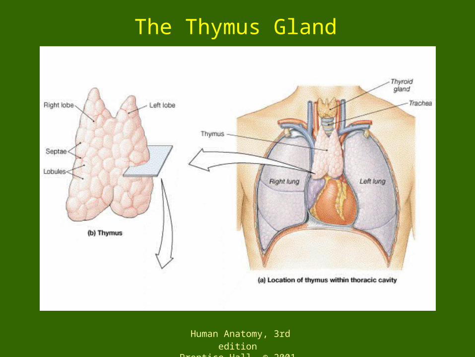
The Thymus– Location – posterior to the sternum– Produces thymosins which enhance
lymphycyte production– Development
• Childhood – large
• Puberty – largest
• Adulthood – decreases in size
Human Anatomy, 3rd editionPrentice Hall, © 2001
The Thymus Gland

Human Anatomy, 3rd editionPrentice Hall, © 2001
The Adrenal Gland– Location – on top of the kidney– Structure – outer cortex and inner medulla– Medulla
• Secretes epinephrine & norepinephrine
• Target – most cells
• Effect– Epinephrine – increase cardiac activity, blood pressure,
blood glucose; constricts blood vessels in skin, dilates blood vessels in skeletal & cardiac muscle
– Norepinephrine – increases cardiac activity, constricts most blood vessels

Human Anatomy, 3rd editionPrentice Hall, © 2001
The Adrenal Glands– The adrenal cortex is composed of 3 layers
• Zona glomerulosa secretes mineralocorticoids, mostly aldosterone
– Target – kidneys– Effect – increases blood sodium levels, decreases blood
potassium levels
• Zona fasciculata – secretes glucocorticoids (cortisol, corticosterone)
– Target – most cells– Effect – conserve blood glucose, anti-inflammatory
effects– Controlled by ACTH
• Zona reticulares – secretes androgens– Effects are uncertain

Human Anatomy, 3rd editionPrentice Hall, © 2001
The Adrenal Glands

Human Anatomy, 3rd editionPrentice Hall, © 2001
The Adrenal Glands

Human Anatomy, 3rd editionPrentice Hall, © 2001
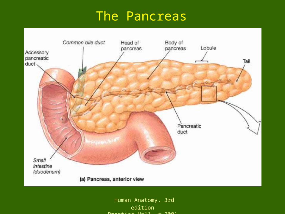
The Pancreas– Location – between the spleen and the duodenum
– Its functions are both exocrine and endocrine
– The endocrine cells are in the Islets of Langerhans• Alpha cells secrete glucagon
– Targets – liver, adipose tissues
– Effect - increase blood sugar levels
• Beta cells secrete insulin– Most cells
– Effect - decrease blood sugar levels
• Delta cells secrete somatostatin– Targets – alpha & beta cells, digestive epithelium
– Effect - antagonistic to growth hormone
– Diabetes mellitis
Human Anatomy, 3rd editionPrentice Hall, © 2001
The Pancreas

Human Anatomy, 3rd editionPrentice Hall, © 2001
Islet of Langerhans

Human Anatomy, 3rd editionPrentice Hall, © 2001
The Male Reproductive System
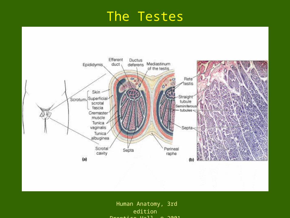
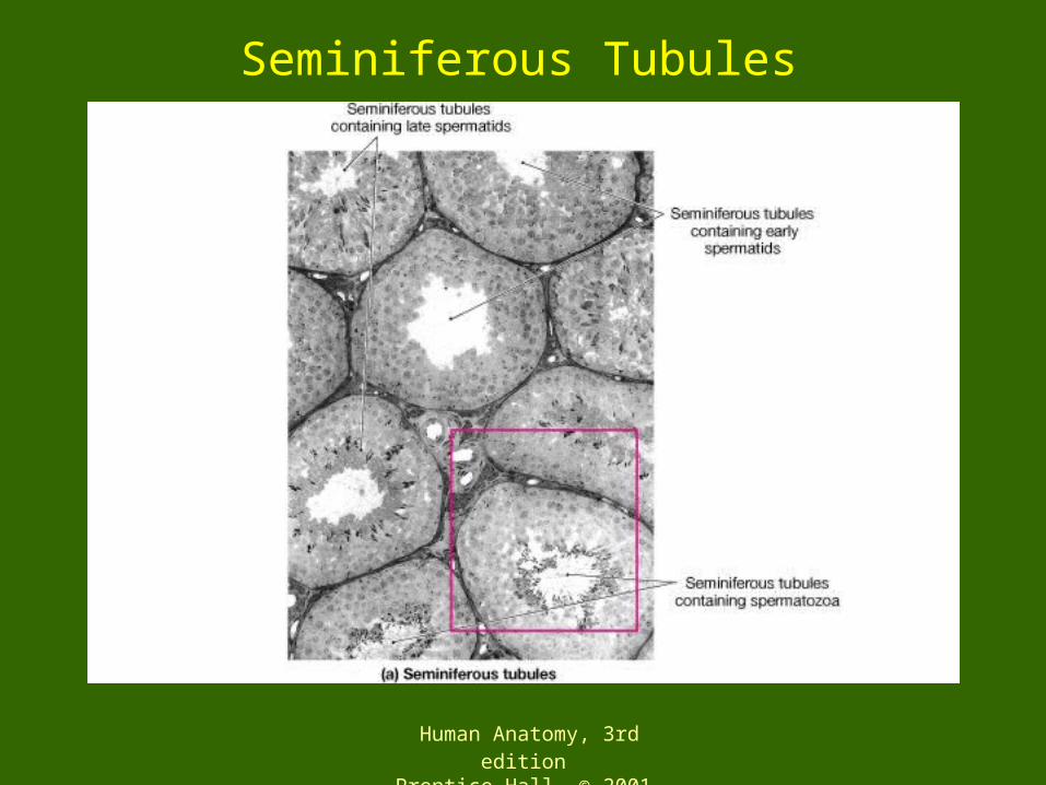
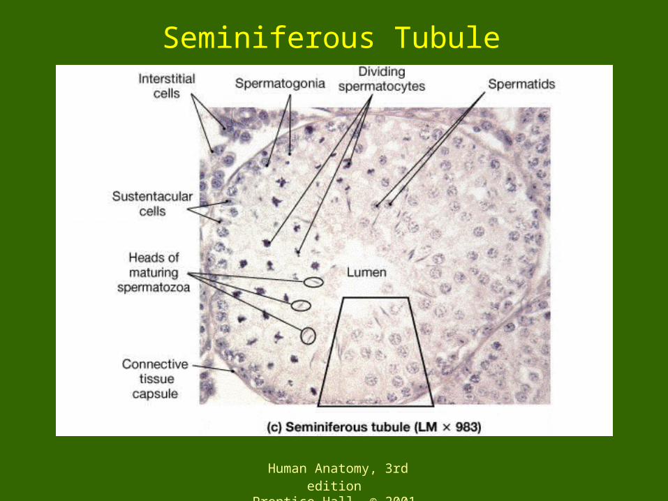
– Sperm are produced in the seminiferous tubules of the testes (spermatogenesis)
– Sperm are stored in the epidymus, then transported through the vas deferens, and leave the body through the urethra
– Seminal fluids (semen) are added to the sperm by the seminal vesicles, prostate gland, and bulbourethral gland

Human Anatomy, 3rd editionPrentice Hall, © 2001
The Male Reproductive System

Human Anatomy, 3rd editionPrentice Hall, © 2001
Testes– Interstitial cells secrete androgens (male sex
hormones); testosterone is the most important• Target – most cells
• Effects – maturation of sperm; protein synthesis in skeletal muscle; male secondary sex characteristics & behaviors
– Some interstitial cells secrete inhibin• Target – anterior pituitary
• Effect – inhibits the secretion of FSH
Human Anatomy, 3rd editionPrentice Hall, © 2001
The Testes
Human Anatomy, 3rd editionPrentice Hall, © 2001
Seminiferous Tubules
Human Anatomy, 3rd editionPrentice Hall, © 2001
Seminiferous Tubule
Human Anatomy, 3rd editionPrentice Hall, © 2001
The Female Reproductive System– Ova (oocytes) are produced in the ovaries
(oogenesis)– Approximately every 28 days one ovum is
expelled from the ovary (ovulation)– An ovum transported through the fallopian
tube to the uterus where it becomes embedded in the uterine lining
• Fertilization occurs in the fallopian tube
– If the egg is not fertilized, the uterine lining detaches and is shed in menstruation
– If the egg is fertilized, the uterine lining is held in place and supported by progesterone

Human Anatomy, 3rd editionPrentice Hall, © 2001
The Female Reproductive System
Human Anatomy, 3rd editionPrentice Hall, © 2001
Ovaries– Follicle cells secrete estrogen
• Targets – most cells• Effects – follicle maturation; female secondary sex
characteristics and behaviors
– Follicle cells also secrete inhibin• Target – anterior pituitary• Effect – inhibits secretion of FSH
– Corpus luteum secretes progesterone and relaxin• Progesterone
– Targets – uterus, mammary glands– Effects – prepare uterus for implantation and mammary glands
for secretion
• Relaxin– Targets – pubic symphysis, uterus, mammary glands– Effects – loosens pubic symphysis, relaxes cervical muscles,
stimulates mammary gland development

Human Anatomy, 3rd editionPrentice Hall, © 2001
Hormonal Regulation of the Female Reproductive System

Human Anatomy, 3rd editionPrentice Hall, © 2001
The Pineal Gland– Location = epithalamus– Pinealocytes secrete melatonin
• Derived from the neurotransmitter seratonin
• Light inhibits production– Regulates circadian rythms
• Target – hypothalamus
• Effects – Inhibits the releasing factors that control FSH & LH secretion
– Slows maturation of sperm, oocytes, & reproductive organs

Human Anatomy, 3rd editionPrentice Hall, © 2001
Endocrine Disorders