hsa circ 0123190 acts as a competitive endogenous rna to
TRANSCRIPT
RESEARCH ARTICLE Open Access
Hsa_circ_0123190 acts as a competitiveendogenous RNA to regulate APLNRexpression by sponging hsa-miR-483-3p inlupus nephritisChunyi Zhang, Congcong Gao, Xueqi Di, Siwan Cui, Wenfang Liang, Wenbo Sun, Menghui Yao, Shengyun Liu andZhaohui Zheng*
Abstract
Background: Lupus nephritis (LN) is one of the most severe complications of systemic lupus erythematosus (SLE).Circular RNAs (circRNAs) can act as competitive endogenous RNAs (ceRNAs) to regulate gene transcription, which isinvolved in mechanism of many diseases. However, the role of circRNA in lupus nephritis has been rarely reported.In this study, we aim to investigate the clinical value of circRNAs and explore the mechanism of circRNAinvolvement in the pathogenesis of LN.
Methods: Renal tissues from three untreated LN patients and three normal controls (NCs) were used toidentify differently expressed circRNAs by next-generation sequencing (NGS). Validated assays were used byquantitative reverse transcription polymerase chain reaction (qRT-PCR). The interactions between circRNAand miRNA, or miRNA and mRNA were further determined by luciferase reporter assay. The extent of renalfibrosis between the two groups was assessed by Masson-trichome staining and immunohistochemistry(IHC) staining.
Results: 159 circRNAs were significantly dysregulated in LN patients compared with NCs. The expression ofhsa_circ_0123190 was significantly decreased in the renal tissues of patients with LN (P = 0.014). Bio-informatics analysis and luciferase reporter assay illustrated that hsa_circ_0123190 can act as a sponge forhsa-miR-483-3p, which was also validated to interact with APLNR. APLNR mRNA expression was related withchronicity index (CI) of LN (P = 0.033, R2 = 0.452). Moreover, the fibrotic-related protein, transforming growthfactor-β1 (TGF-β1), which was regulated by APLNR, was more pronounced in the LN group (P = 0.018).
Conclusion: Hsa_circ_0123190 may function as a ceRNA to regulate APLNR expression by sponging hsa-miR-483-3p in LN.
Keywords: circRNAs, MicroRNAs, Competitive endogenous RNAs, Lupus nephritis, Systemic lupuserythematosus
© The Author(s). 2021 Open Access This article is licensed under a Creative Commons Attribution 4.0 International License,which permits use, sharing, adaptation, distribution and reproduction in any medium or format, as long as you giveappropriate credit to the original author(s) and the source, provide a link to the Creative Commons licence, and indicate ifchanges were made. The images or other third party material in this article are included in the article's Creative Commonslicence, unless indicated otherwise in a credit line to the material. If material is not included in the article's Creative Commonslicence and your intended use is not permitted by statutory regulation or exceeds the permitted use, you will need to obtainpermission directly from the copyright holder. To view a copy of this licence, visit http://creativecommons.org/licenses/by/4.0/.The Creative Commons Public Domain Dedication waiver (http://creativecommons.org/publicdomain/zero/1.0/) applies to thedata made available in this article, unless otherwise stated in a credit line to the data.
* Correspondence: [email protected] of Rheumatology, The First Affiliated Hospital of ZhengzhouUniversity, 1 Eastern Jianshe Road, Zhengzhou 450052, Henan, China
Zhang et al. Arthritis Research & Therapy (2021) 23:24 https://doi.org/10.1186/s13075-020-02404-8

BackgroundLupus nephritis (LN) is one of the most common andsevere complications of systemic lupus erythematosus(SLE) and seriously affects the quality of life andprognosis of SLE patients. About 80% of children and60% of adults are susceptible, and up to 30% of pa-tients could progress to end-stage renal disease(ESRD) [1]. Previous studies investigated 491 cases ofLN patients in central plains of China with 5-year,10-year, 15-year, and 20-year survival rates of 88%,77%, 53%, and 46%, respectively, and found that renalfailure was the main cause of death [2]. The loss oflabor in patients with LN who have advanced to end-stage renal disease places an enormous economic bur-den on society and families. Therefore, exploring thepathogenesis of LN is important and urgent.Circular RNA (circRNA) is a new type of non-
coding RNA, which produced from precursor mRNAback-splicing by covalently closed, single-strandedRNA circles at the junction site of 3’5’-phosphodiesterbond [3]. CircRNA expression is more stable to bedetected because of its closed circular structure. Dueto the cell type-specific or tissue-specific manner [4,5], circRNAs are involved in the pathogenesis of vari-ous human diseases. CircRNAs are rich in microRNAbinding sites, acting as miRNA sponges and regulat-ing gene transcription, which is called competitive en-dogenous RNA (ceRNA) molecules [6, 7]. In thestudies on renal diseases, Wang et al. reported thatandrogen receptor enhanced migration and invasionof renal transparent cell carcinoma by inhibiting theexpression of circHTAT1 regulating miR-195-5p/29a-3p/29c-3p [8]. In addition, circRNA ZNF609 wasfound to regulate fork head box P4 (FOXP4) expres-sion by targeting miR-138-5p in renal carcinoma [9].However, the role and mechanism of circRNAs in LNhas been rarely reported.Renal fibrosis is a common pathological feature of pro-
gressive LN, which is closely related to ESRD [10, 11].Apelin and its receptor (apelin receptor, APLNR) can bewidely distributed in the heart, lung, pancreas, kidney,and other tissues [12]. Numerous evidences indicate thatapelin and APLNR play a key role in various kidney dis-eases, such as renal fibrosis, renal ischemia/reperfusioninjury, polycystic kidney disease, and diabetic nephropa-thy [13]. Particularly, apelin and APLNR could inhibitthe deposition of extracellular matrix (ECM) and attenu-ate renal fibrosis by acting on TGF-β [14]. In the presentstudy, we firstly established the circRNA expression pro-file in the kidney tissues of patients with LN. And then,we further explored that hsa_circ_0123190 was a novelbiomarker of peripheral blood for LN and could act as asponge for hsa-miR-483-3p to regulate APLNR expres-sion involved in renal fibrosis in LN.
MethodsSubjects and samplesA total of 10 LN patients with renal biopsy were en-rolled in this study between May 2018 and December2018 from the Department of Rheumatology and Im-munology of the First Affiliated Hospital of ZhengzhouUniversity. Five patients with renal tumor were from theUrology Department at the same hospital. Ten periph-eral blood samples were collected from the volunteers ashealthy controls (HCs). All patients and volunteers werefemale, between 18 and 60 years old. The following wereexclusion criteria: (1) patients with serious infectionwithin 1 month before admission, (2) patients with ma-lignant tumors, (3) patients with other autoimmune dis-eases, (4) patients with pregnancy, and (5) patients witheGFR lower than 30 mL/min/1.73 m2.Kidney tissues from LN patients were obtained from
renal biopsies before the treatment with steroid and/orimmunosuppressant. Renal normal controls (NCs) werekidney tissues at least 5 cm from the edge of tumor frompatients with renal cancer, and then confirmed to benormal histological morphology under microscopy. Allfresh tissues were stored in RNAlater® Solution (ThermoFisher Scientific, CA, USA) and then frozen in − 80 °Cuntil RNA extraction. All peripheral blood samples (2mL) were drawn from the median cubital vein with aPAXgene Blood RNA Tube (Qiagen, Hilden, Germany).
NGS profiling analysisTotal RNA was extracted from the frozen renal tissueswith using Trizol LS reagent (Invitrogen, CA, USA).Total RNA from fresh peripheral blood samples is iso-lated by PAXgene Blood RNA Kit (Qiagen, Hilden,Germany). Total RNA was quantified and qualified byan Agilent 2100 Bioanalyzer (Agilent Technologies, CA,USA), NanoDrop™ 2000 spectrophotometer (ThermoFisher Scientific, CA, USA) and 1% agarose gel. The cri-teria of total RNA was used for subsequent library prep-aration: (1) the value of OD260/280 was between1.8 ~ 2.2and OD260/230 was above 2.0, and (2) the value of RINwas above seven.NGS library preparation was performed by the Gen-
esky Biotechnologies Inc. (Shanghai, China). The rRNAwas depleted from total RNA by Ribo-ZeroTM rRNA re-moval kit (Human/Mouse/Rat) (Illumina, CA, USA) be-fore building the RNA-seq library. After purification,divalent cations at higher temperatures were applied formaking small pieces of fragments of the residual RNAfractions. The reverse transcription of all the cleavedRNA fragments was used to construct the complemen-tary DNA (cDNA) library with TruSeq Stranded TotalRNA Library Prep Kit (Illumina, CA, USA) according tothe manufacturer’s instructions. The library quality wasevaluated with Agilent 2100 Bioanalyzer (Agilent
Zhang et al. Arthritis Research & Therapy (2021) 23:24 Page 2 of 12

Technologies, CA, USA). RNA libraries were denaturedas single-stranded DNA molecules. Finally, sequencingwas carried out using a 2 × 150 base paired-end configur-ation with Illumina Hiseq 2500 (Illumina, CA, USA).
GO and KEGG pathway analysisThe predicted functions of the differentially expressedcircRNAs between LN and NC were conducted by geneontology (GO, http://geneontology.org) and KyotoEncyclopedia of Genes and Genomes (KEGG, http://www.kegg.jp) analysis. Hierarchical clustering of the dif-ferentially expressed circRNA according to three cat-egories, the biological process (BP), cellular component(CC), and molecular function (MF) was used by GO ana-lysis. Pathway analysis of circRNA was performed byKEGG database.
QRT-PCR validationThe candidate circRNAs were selected according to thefollowing: Firstly, the top 20 differently expressed cir-cRNAs between the LN group and the control groupwere selected. Secondly, according to the results of GOand KEGG enrichment analysis and literature, the cir-cRNAs with most likelihood to be related to the patho-genesis of LN was analyzed. Thirdly, the primers ofcircRNAs for qRT-PCR could be designed. Finally, everyof the candidate circRNAs had only one circBase ID.QRT-PCR with SYBR green analysis was used to validatethe expression of the selected circRNAs from circRNAprofiles. Total RNA was extracted by using Trizol re-agent (Invitrogen, Carlsbad, CA, USA). The cDNA wassynthesized by Reverse Transcriptase M-MLV (Takara,Tokyo, Japan). In addition, qRT-PCR was performedwith SYBR® Premix Ex Taq™ II (Takara, Tokyo, Japan).GAPDH was used as endogenous control for cir-cRNA qRT-PCR and U6 for miRNA. One microgramof RNA was mixed with 1 μL of RNase R (20 U/μL)at 37 °C for 20 min and then washed with RNeasycleaning agent. All of the primers are listed inTable 1. The relative circRNA expression was calcu-lated using 2−ΔΔCt, with ΔCt = Ct target – Ct β-actin,−ΔΔCt = − (ΔCt sample − ΔCt control).
Dual-luciferase reporter assayLuciferase reporter assays were used to detect the directbinding between selected RNAs. The hsa_circ_0123190wild-type (WT) 3′-UTR, APLNR WT 3′-UTR and mu-tant sequence hsa_circ_0123190 (MUT) 3′-UTR, APLNR MUT 3′-UTR were constructed. Subsequently, we in-oculated HEK293T cells into 96-well plates and co-transfected 5 pmol hsa-miR-483-3p mimics or negativecontrol with the 0.16 μg hsa_circ_0123190 and APLNRwild-type or mutant type plasmids. We tested the fluor-escence intensity using the dual-luciferase reporter geneassay system (Promega, Madison, USA) after 48 h oftransfection.
Histology and immunostainingParaffin-embedded kidney tissues from the human werecut at 2-μm thickness, deparaffinized, and rehydrated.Then, for histological examination, kidney sections werestained with Masson-trichrome reagent to explore thedegree of fibrosis. Moreover, for immunohistochemistry(IHC) staining, kidney sections were incubated in citratebuffer for 20 min at 95 °C to retrieve antigen. Non-specific binding was blocked with 10% normal goatserum for 30min at RT. The slides were incubated withantibodies of TGF-β1, α-smooth muscle antibody (α-SMA) overnight at 4 °C, followed by incubation withbiotin-conjugated goat anti-mouse/rabbit immunoglobu-lin IgG for 30 min at RT, and then reacted withstreptavidin-conjugated peroxidase for 30 min at RT. OnIHC staining, we semi-quantified the stained slides usinga specific immunohistochemical histological score tech-nique, H-score [15]. The H-score was obtained by multi-plying the staining intensity by a constant to adjust themean to the strongest staining, to produce a score in therange of 0–300. Specifically, H-score = scale × percent-age of strong staining (0–100%). H-score = 1.0 indicateda weak percentage, 2.0 indicated a moderate percentage,and 3.0 indicated a strong percentage.
Western blotRIPA buffer was used for tissues lysis, according to themanufacturer’s instructions, and the protein concentra-tion was determined using a Bicinchoninic Acid (BCA)Protein Assay Kit. Proteins were separated by SDS-
Table 1 The sequences of primers
Gene Forward primer sequence Reverse primer sequence
hsa_circ_0123190 TGAGGATGGAGAACCCACCAA CCCCCATCACATGAGCACAA
hsa_circ_0000660 TGCTTCCAGTGGGAATCCACAT TCAGAGAGCCGTAGGTTGCGTAT
hsa_circ_0007379 TCTCTTTCTCCAAGGAGCTCCACA TGCTGATGAAGCTGAGCAGGGA
hsa_circ_0003302 TGGATGTTCCACAGGAAGAAGTGC GGGCCACGGCGATAAGGAAAAT
APLNR AGGCAGCAGGGCTGATGAATGG TGCAGACACCCCTCCATCCTCT
hsa-miR-483-3p TCACTCCTCTCCTCCCGTCTT
Zhang et al. Arthritis Research & Therapy (2021) 23:24 Page 3 of 12

PAGE using Tris-Glycine 10% polyacrylamide gels inSDS page running buffer and transferred to PVDF mem-brane (Servicebio, Wuhan, China). The membranes wereimmunoblotted with antibodies against APLNR (1:500,Proteintech, 20341-1-AP) after blocking in 5% milk. Fol-lowing primary antibody incubation, the membraneswere probed with HRP-conjugated mouse anti-rabbitsecondary antibody (1:5000, Santa Cruz, sc-374015) andimaged using the Chemidoc system (BioRad).
Statistical analysisStatistical software SPSS 25.0 (IBM Corporation, USA) wasused for statistical analysis. Scatter diagrams were drawn byGraphPad Prism version 6.0 (GraphPad Software, Inc., CA,USA). Differences between two groups were analyzed forstatistical significance by t test. The correlation between cir-cRNAs level and clinical parameters of LN were analyzed byPearson’s linear correlation. ROC curves were performed,and the specificity and sensitivity of predictive power wereassessed by area under curve (AUC). A P value < 0.05 wasconsidered as statistically significant.
ResultsProfiling and characteristics of circRNAs in the renaltissues of LN patientsFirstly, we analyzed the circRNAs profiling in the renaltissues of three LN patients and three NCs by next-generation sequencing. The clinical information for the3 patients and NCs for circRNA profiling were shown inSupplement Tables 1 and 2. Results showed that total159 circRNAs were identified to be abnormallyexpressed in renal tissues of LN patients compared withNCs (log2|fold change| ≥ 1, P < 0.05), of which 73 cir-cRNAs were significantly upregulated and 86 circRNAswere remarkably downregulated. The top 10 upregulatedand downregulated circRNAs were summarized inTable 2. Hierarchical clustering heatmap and volcanoplots were used to show the different expression levelsof circRNAs between LN and NC (Additional file 1: Sup-plement Figure 1).
GO and KEGG analysis in LNGene Ontology (GO) analysis predicted the functions ofdifferentially expressed circRNAs through the host genesbased on three common aspects, including biologicalprocesses (BP), cellular components (CC), and molecularfunctions (MF). We selected the top 10 GO terms wherecircRNAs were significantly enriched in each of threebiological functions. In the MF and CC category, wefound that lysophosphatidic acid acyltransferase activityand apical plasma membrane were significantly regulatedby the differentially expressed circRNAs in the LN kid-neys. Moreover, in the BP category, enriched terms in-cluded biological regulation, cellular process, and
metabolic process. Positive regulation of autophagy,which plays an important role in the pathogenesis ofLN, was significantly regulated by these circRNAs in theBP category (Additional file 1: Supplement Figure 2).Correspondingly, the Kyoto Encyclopedia of Genes
and Genomes (KEGG) analysis can define the mo-lecular function pathway of target host genes of the159 dysregulated circRNAs. Target genes of differen-tially expressed circRNAs were enriched in pathways,such as apelin signaling pathway and tumor necrosisfactor (TNF) signaling pathway. The molecular func-tion of the associated apelin signaling pathway in-volves in regulating the expression of beclin1 andLC3, which are both important factors in autophagy,one of well-known pathogenesis in LN [16]. TNF-αcontributes to the development of T cells, B cells,and dendritic cells in pathogenesis of SLE [17]. Bothpathways were associated with activation of PI3K,which is an important factor in the pathogenesis ofLN (Additional file 1: Supplement Figure 2).
Validation of the selected circRNAs in the renal tissuesSince females are predominantly susceptible to lupusthan males, ten female patients with LN were enrolled inthis study, except for males. The characteristics of all 10LN patients are shown in Table 3. With age and gender
Table 2 The top 10 abnormally expressed circRNAs
circBase ID circRNA ID P value Type
NA chr2:153431650-153437563:+ 0.038640885 Up
hsa_circ_0008683 chr14:102486230-102489217:+ 0.0035304 Up
hsa_circ_0002980 chr7:141336760-141349133:+ 0.004624716 Up
hsa_circ_0002153 chrX:107083900-107097934:+ 0.005655858 Up
hsa_circ_0000734 chr17:1746097-1756483:+ 0.040425419 Up
hsa_circ_0003757 chr1:24140680-24147083:- 0.039814259 Up
hsa_circ_0004958 chr17:58711214-58725443:+ 0.039891741 Up
hsa_circ_0002319 chr3:197592983-197598333:+ 0.028417612 Up
NA chr1:246890194-246903603:+ 0.032809392 Up
hsa_circ_0003302 chr11:120916383-120930794:+ 0.031378282 Up
NA chr2:40655613-40673788:- 0.014846209 Down
NA chr4:122411146-122446439:+ 0.009160292 Down
hsa_circ_0005806 chr16:30675536-30677862:+ 0.004353445 Down
NA chr4:166960491-166999182:+ 0.015788499 Down
hsa_circ_0004284 chr1:71304482-71320883:+ 0.002193657 Down
hsa_circ_0003602 chr3:47702784-47719801:- 0.005016822 Down
hsa_circ_0000994 chr2:40655613-40657444:- 0.027239613 Down
hsa_circ_0000660 chr15:94899366-94945248:+ 0.019867591 Down
hsa_circ_0007379 chr14:35020920-35024118:- 0.005379896 Down
hsa_circ_0123190 chr3:195415404-195435712:+ 6.1567E-06 Down
NA not applicable. *P value < 0.05 was considered as statistically significant
Zhang et al. Arthritis Research & Therapy (2021) 23:24 Page 4 of 12
match, five females with renal tumor were used as thecontrol group of the normal kidneys. In LN patients,urinary analysis showed 24-h urinary total protein excre-tion and numbers of red blood cell, white blood cell, andserology displayed levels of complements, erythrocytesedimentation rate, serum creatine, albumin and routineblood indexes, the increased score of systemic lupus ery-thematous activity of disease indices (SLEDAI), and dif-ferent severity of renal dysfunction. Ten LN patientspresented various pathological classifications (class III,n = 2; class IV, n = 5; class III + V, n = 1; class IV + V,n = 2), when activity index (AI) and chronicity index(CI) were both assessed.We selected three downregulated circRNAs (hsa_circ_
0000660, hsa_circ_0007379, and hsa_circ_0123190) andone upregulated circRNAs (hsa_circ_0003302) for thefurther validation in renal samples of 10 LN patients and5 NC subjects by qRT-PCR. All four circRNAs showedsimilar expression patterns between the qRT-PCR andthe circRNA sequencing. However, only hsa_circ_
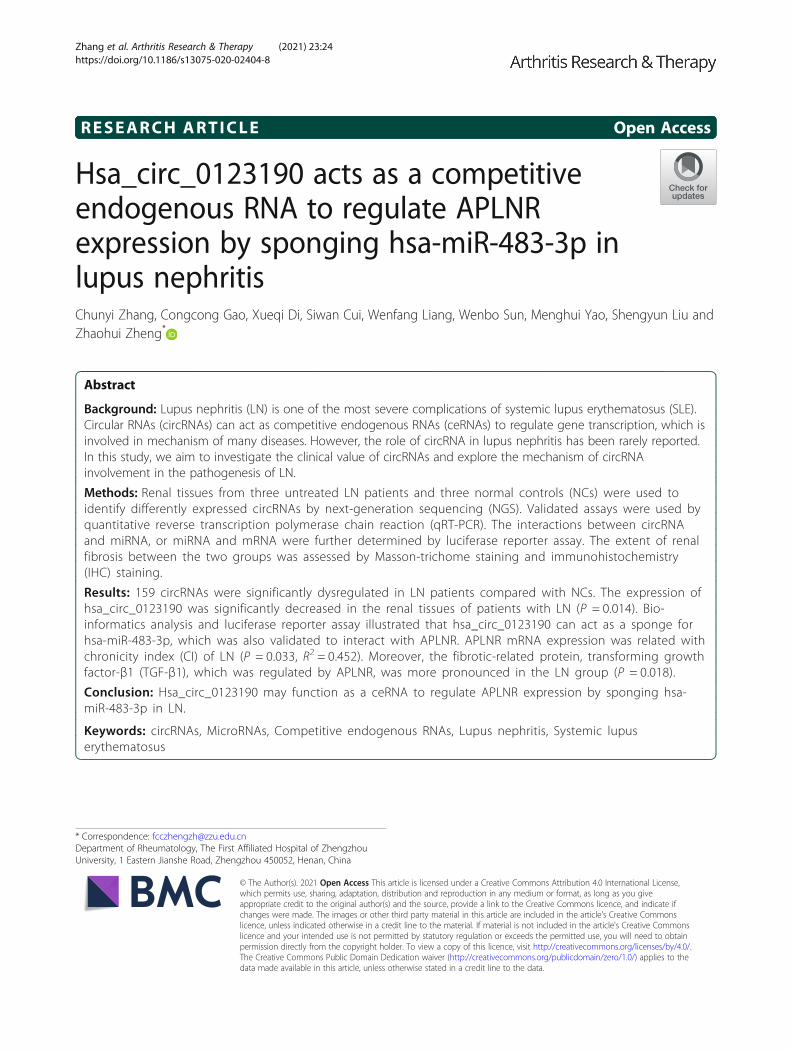
0123190 was significantly dysregulated between the renaltissues of patients with LN and NCs (P = 0.014) (Fig. 1a).Therefore, hsa_circ_0123190 was considered as a candi-date for further analysis. Moreover, hsa_circ_0123190was resistant to RNase R digestion (P > 0.05), suggest-ing that hsa_circ_0123190 is stable (Fig. 1b). The clin-ical significance of hsa_circ_0123190 in LN patientswas investigated. There was no association of hsa_circ_0123190 with clinical parameters which we collected(all P > 0.05).
Hsa_circ_012319 serves as a sponge for hsa-miR-483-3pIn view of the fact that circRNA can act as a miRNAsponge, the potential targets miRNAs of hsa_circ_0123190 were predicted using bioinformatics (CircIn-teractome, and miRanda). As a result, we found thathsa-miR-483-3p was the most likely complementarymiRNA, which had a perfect match sequence to bindhsa_circ_0123190. We examined the expression ofhsa-miR-483-3p in renal tissues using qRT-PCR. The
Table 3 Clinical characteristics of LN patients
Serum
Pt Age (years) Disease duration(months)
C3 (g/L) C4 (g/L) ESR(mm/h)
CRP(mg/L)
WBC(*10^9/L)
RBC(*10^12/L)
Hb (g/L)
PLT(*10^9/L)
1 39 144 0.43 0.06 37 3.06 2.80 3.09 89.0 104
2 38 48 0.65 0.11 27 3.13 3.64 4.15 116.8 209
3 34 19 0.59 0.10 60 54.83 6.40 2.69 81.0 96
4 20 36 0.19 0.07 3 3.23 2.32 3.90 115.9 128
5 23 6 1.03 0.15 12 3.13 5.56 5.46 117.8 221
6 35 36 0.25 0.07 38 3.13 2.13 3.30 103.3 106
7 19 60 0.56 0.11 10 3.13 4.60 2.37 74.0 110
8 48 156 0.34 0.08 14 1.00 5.40 2.84 85.0 80
9 36 1 0.33 0.05 11 9.00 2.10 3.45 105.0 136
10 46 120 0.62 0.05 74 32.52 3.50 3.14 98.0 227
Serum Urine Renal biopsy SLEDAI(score)
Pt Creatinine(umol/L)
Urea (mmol/L) eGFR (mL/min/1.73m2)
UTP (g/24 h)
RBC (/uL) WBC(/uL)
Classification AI (score) CI(score)
1 59 5.03 111.223 8.30 95 56 IV-S(A) + V 9 0 9
2 63 4.50 107.899 2.52 26 4 III-(A) 8 1 8
3 130 12.44 46.224 6.03 252 94 IV-G(A) + V 19 3 11
4 51 3.85 133.345 2.07 50 0 III-(A/C) 6 1 12
5 58 7.11 109.314 2.03 50 0 III-(A) + V 0 0 6
6 135 12.40 38.299 6.25 92 22 IV-G(A/C) 10 2 4
7 77 4.90 96.472 2.82 350 12 IV-S(A) 9 0 8
8 56 10.36 98.682 6.39 45 22 IV-G(A) 15 0 5
9 69 4.31 98.028 2.35 184 50 IV-S(A) 9 0 4
10 59 6.91 93.006 1.40 37 28 IV-S(A) 7 0 4
Pt patient, WBC white blood cells, RBC red blood cells, Hb hemoglobin, PLT Platelet, C3 complement 3, C4 complement 4, AI activity index, CI chronicity index,eGFR estimated glomerular filtration rate based on CKD-EPI formula, SLEDAI systemic lupus erythematosus disease activity index, UTP urinary total proteinuriaper day
Zhang et al. Arthritis Research & Therapy (2021) 23:24 Page 5 of 12
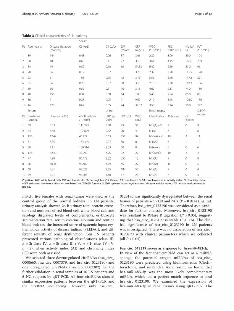
results revealed that the expression of hsa-miR-483-3p was significantly increased in LN compared withNCs (P = 0.0498) (Fig. 2a). To further verify the hy-pothesis that hsa-miR-483-3p directly targets hsa_circ_0123190, we performed the dual-luciferase re-porter assay with HEK293T cells, and the resultsshowed that the relative luciferase activity was signifi-cantly reduced in cells co-transfected with hsa_circ_0123190 WT and hsa-miR-483-3p mimics comparedwith control. The relative luciferase activity was un-changed in co-transfected cell with hsa_circ_0123190MUT and hsa-miR-483-3p mimics compared withcontrol group (Fig. 2b). In a word, all results indicatethat hsa_circ_0123190 targets hsa-miR-483-3p.
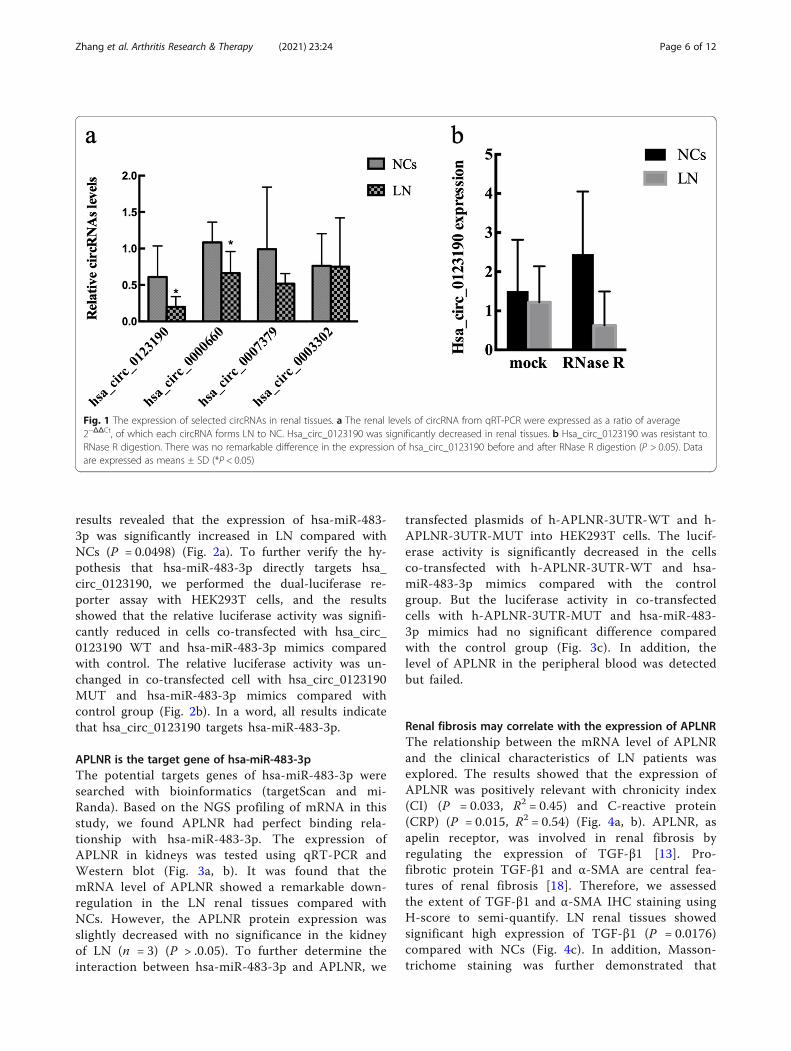
APLNR is the target gene of hsa-miR-483-3pThe potential targets genes of hsa-miR-483-3p weresearched with bioinformatics (targetScan and mi-Randa). Based on the NGS profiling of mRNA in thisstudy, we found APLNR had perfect binding rela-tionship with hsa-miR-483-3p. The expression ofAPLNR in kidneys was tested using qRT-PCR andWestern blot (Fig. 3a, b). It was found that themRNA level of APLNR showed a remarkable down-regulation in the LN renal tissues compared withNCs. However, the APLNR protein expression wasslightly decreased with no significance in the kidneyof LN (n = 3) (P > .0.05). To further determine theinteraction between hsa-miR-483-3p and APLNR, we
transfected plasmids of h-APLNR-3UTR-WT and h-APLNR-3UTR-MUT into HEK293T cells. The lucif-erase activity is significantly decreased in the cellsco-transfected with h-APLNR-3UTR-WT and hsa-miR-483-3p mimics compared with the controlgroup. But the luciferase activity in co-transfectedcells with h-APLNR-3UTR-MUT and hsa-miR-483-3p mimics had no significant difference comparedwith the control group (Fig. 3c). In addition, thelevel of APLNR in the peripheral blood was detectedbut failed.
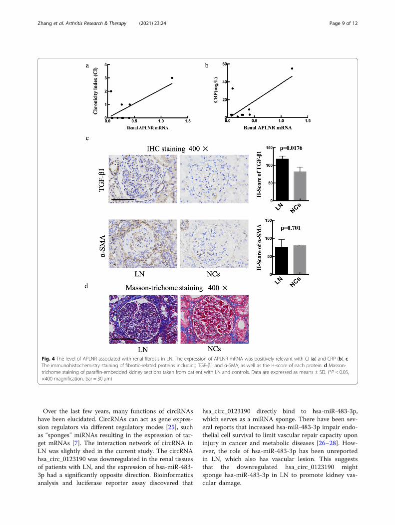
Renal fibrosis may correlate with the expression of APLNRThe relationship between the mRNA level of APLNRand the clinical characteristics of LN patients wasexplored. The results showed that the expression ofAPLNR was positively relevant with chronicity index(CI) (P = 0.033, R2 = 0.45) and C-reactive protein(CRP) (P = 0.015, R2 = 0.54) (Fig. 4a, b). APLNR, asapelin receptor, was involved in renal fibrosis byregulating the expression of TGF-β1 [13]. Pro-fibrotic protein TGF-β1 and α-SMA are central fea-tures of renal fibrosis [18]. Therefore, we assessedthe extent of TGF-β1 and α-SMA IHC staining usingH-score to semi-quantify. LN renal tissues showedsignificant high expression of TGF-β1 (P = 0.0176)compared with NCs (Fig. 4c). In addition, Masson-trichome staining was further demonstrated that
Fig. 1 The expression of selected circRNAs in renal tissues. a The renal levels of circRNA from qRT-PCR were expressed as a ratio of average2−ΔΔCt, of which each circRNA forms LN to NC. Hsa_circ_0123190 was significantly decreased in renal tissues. b Hsa_circ_0123190 was resistant toRNase R digestion. There was no remarkable difference in the expression of hsa_circ_0123190 before and after RNase R digestion (P > 0.05). Dataare expressed as means ± SD (*P < 0.05)
Zhang et al. Arthritis Research & Therapy (2021) 23:24 Page 6 of 12

fibrosis was significantly promoted in the LN group(Fig. 4d).
Validation of hsa_circ_0123190 expression in peripheralblood of LN patientsThe recent studies revealed that circRNA had tissue-specific manner, we identified hsa_circ_0123190 fromthe renal tissues. However, renal biopsy is an invasiveand expensive operation, and hsa_circ_0123190 was sup-posed to be validated by qRT-PCR in peripheral blood often LN patients and ten healthy controls. As a result, theexpression of hsa_circ_0123190 was remarkably decreasedin LN patients (P = 0.0005) (Fig. 5a). In addition, weassessed the correlation between circRNA levels and clin-ical characteristics in LN patients. The expression of hsa_circ_0123190 was negatively correlated with leukocytelevels (P = 0.0123, R2 = 0.5638) and complement 4 (C4)
levels (P = 0.0099, R2 = 0.5855). Moreover, hsa_circ_0123190 expression was significantly associated withserum creatine levels (P = 0.044, R2 = 0.4151) (Fig. 5b).The important utility of hsa_circ_0123190 was fur-
ther explored by ROC curve analysis. The area underthe curve (AUC) for hsa_circ_0123190 in the periph-eral blood when distinguishing LN patients fromNCs was 0.900 (95% CI 0.7659–1.034, P = 0.0025).The maximum Youden’s J index (sensitivity and spe-cificity) was 70% (90% and 80%) for hsa_circ_0123190 to differentiate LN patients from controls,and the corresponding optimal cutoff values was0.6773 (Fig. 5c).
DiscussionThe poor prognosis of LN is still a seriously clinical andeconomic problem, as the mechanisms of LN remain
Fig. 2 The interaction of renal hsa_circ_0123190 and hsa-miR-483-3p in LN. a The expression of hsa-miR-483-3p in the renal tissues wassignificantly increased in LN patients compared with controls. b Binding sequence prediction of hsa_circ_0123190 WT and hsa-miR-483-3p andsequence construction of hsa_circ_0123190 MUT. Dual-luciferase reporter assay showed the binding relationship between hsa_circ_0123190 andhsa-miR-483-3p in HEK293T cells
Zhang et al. Arthritis Research & Therapy (2021) 23:24 Page 7 of 12

indistinct [19]. Thus, the identification of novel treat-ment targets for LN would be very desirable. CircRNA isa special class of endogenous RNAs with multiple func-tions [20]. Many studies have revealed the abnormalcircRNAs were associated with several renal diseases,such as AKI [21] and carcinoma [8, 22]. However, therole of circRNA in LN has been rarely reported. Inthe present study, we conducted integrative analysisusing circRNA sequencing in renal tissues and identi-fied 159 circRNAs with significantly differential ex-pression, of which 73 were upregulated and 86downregulated. Hsa_circ_0123190 maybe act as asponge for hsa-miR-483-3p, which regulates APLNRexpression in LN. In addition, peripheral blood hsa_circ_0123190 would be a biomarker for patients withLN.Special attention must be paid to the type of speci-
men since circRNAs were highly expressed in a celltype-specific or tissue-specific manner [4, 5]. Ouyanget al. discovered that upregulated plasma circRNA_002453 level in LN patients was associated with the
severity of renal involvement and served as a novelbiomarker for LN patient diagnosis [23]. Luan et al.showed that circHLA-C played an important role inthe pathogenesis by sponging miR-150 in LN [24].These data are not consistent across researches dueto different samples and methods. For instance, Luanet al. reported a circRNA profiling in single class IVof LN and found seven differentially expressed cir-cRNAs. The reasons why the results were differentfrom our study may be as follows: different samplesources, different sample sizes, and different renalpathological types. Compared with study by Luanet al., the present study covered the common prolifer-ative classification of LN to explore the role of cir-cRNA and validated differentially expressed circRNAsboth in the peripheral blood and kidney tissues. Inaddition, we performed ROC curve analysis to ensurethe clinical value of the circRNAs as diagnostic bio-markers for LN. The profiling of circRNAs from thisstudy may provide a novel database and new view tostudy mechanisms of LN.
Fig. 3 The expression of APLNR and the interaction of hsa-miR-483-3p and APLNR in LN. a The level of APLNR mRNA in the renal tissues wasdecreased in LN patients. b Western blot results showed the protein level of APLNR. c Binding sequence prediction of APLNR WT and hsa-miR-483-3p and sequence construction of APLNR MUT. Dual-luciferase reporter assay to validate the binding relationship between APLNR and hsa-miR-483-3p in HEK293T cells
Zhang et al. Arthritis Research & Therapy (2021) 23:24 Page 8 of 12
Over the last few years, many functions of circRNAshave been elucidated. CircRNAs can act as gene expres-sion regulators via different regulatory modes [25], suchas “sponges” miRNAs resulting in the expression of tar-get mRNAs [7]. The interaction network of circRNA inLN was slightly shed in the current study. The circRNAhsa_circ_0123190 was downregulated in the renal tissuesof patients with LN, and the expression of hsa-miR-483-3p had a significantly opposite direction. Bioinformaticsanalysis and luciferase reporter assay discovered that
hsa_circ_0123190 directly bind to hsa-miR-483-3p,which serves as a miRNA sponge. There have been sev-eral reports that increased hsa-miR-483-3p impair endo-thelial cell survival to limit vascular repair capacity uponinjury in cancer and metabolic diseases [26–28]. How-ever, the role of hsa-miR-483-3p has been unreportedin LN, which also has vascular lesion. This suggeststhat the downregulated hsa_circ_0123190 mightsponge hsa-miR-483-3p in LN to promote kidney vas-cular damage.
Fig. 4 The level of APLNR associated with renal fibrosis in LN. The expression of APLNR mRNA was positively relevant with CI (a) and CRP (b). cThe immunohistochemistry staining of fibrotic-related proteins including TGF-β1 and α-SMA, as well as the H-score of each protein. d Masson-trichome staining of paraffin-embedded kidney sections taken from patient with LN and controls. Data are expressed as means ± SD. (*P < 0.05,×400 magnification, bar = 30 μm)
Zhang et al. Arthritis Research & Therapy (2021) 23:24 Page 9 of 12

In addition, hsa-miR-483-3p was only interacted withAPLNR in this study, which was decreased in the renaltissues of LN. APLNR is the orphan G protein-coupledapelin receptor, which could be expressed in variousorgan and tissues [13]. Hus-Citharel et al. discoveredthat the APLNR mRNA was widely expressed in therat kidney, and the level of APLNR in all nephronsegments was lower than the glomeruli [29]. Thereare multiple functions of APLNR and apelin, such asregulation of blood pressure, immune response, andanti-inflammatory effect [30]. They also play an im-portant role in organ fibrosis [14]. Renal fibrosis is amajor feature of chronic kidney disease including LN.Collagen, α-SMA, and TGF-β are considered to beimportant fibrosis-related proteins. In the presentstudy, Masson-trichome staining laterally demon-strated that renal fibrosis is a typical pathologicalmanifestation of the LN kidney. A large amount ofstudies on kidney diseases indicates that APLNR and
apelin can improve renal interstitial fibrosis byrestraining the expression of TGF-β1 [14]. TGF-βparticipates in chronic renal inflammation and renalfibrosis through the Smad signaling pathway, proteinkinase C pathway, and mitogen-activated protein kin-ase pathway. A research on mice with complete uni-lateral ureteral obstruction (UUO) illustrated thatapelin treatment could significantly reduce the expres-sion of α-SMA, TGF-β1, and its receptor [31]. In thisstudy, we found that the expression of TGF-β1 wassignificantly increased in LN, which was opposite toAPLNR. Therefore, we tentatively hypothesized APLNR may be involved in renal fibrosis of LN by regulat-ing TGF-β1.The expression of peripheral blood hsa_circ_
0123190 was downregulated and negatively associatedwith serum creatine level in LN, which indicated thatit might be involved in renal injury in patients withLN. Moreover, we ensured the clinical value of this
Fig. 5 The expression and clinical relevance of hsa_circ_0123190 in peripheral blood of LN compared with NC. The levels of hsa_circ_0123190from qRT-PCR were expressed as a ratio of average 2−ΔΔCt. Data are expressed as means ± SD (*P < 0.05). a Hsa_circ_0123190 was significantlydecreased in peripheral blood samples. b Hsa_circ_0123190 was negatively associated with serum creatine levels. c The receiver operatingcharacteristic (ROC) curve analysis of hsa_circ_0123190
Zhang et al. Arthritis Research & Therapy (2021) 23:24 Page 10 of 12
circRNA as a diagnostic biomarker for LN throughperforming ROC curve analysis. These results indi-cated that hsa_circ_0123190 in the peripheral bloodcould be a novel promising diagnostic and non-invasive biomarker of LN patients.There are some limitations in our study. First of
all, our sample sizes were comparatively small. Theexamination of circRNAs in the kidneys and bloodsamples in larger cohorts of LN patients may defineits clinical value as a diagnostic biomarker. Secondly,the mechanisms of hsa_circ_0123190 in the develop-ment and pathogenesis of LN has not yet been com-pletely studied. Thus, further experiments in vitroand in vivo are needed. Thirdly, the expression ofhsa_circ_0123190 in T cells, B cells, and other im-mune cells will be detected and compared in thenext step.
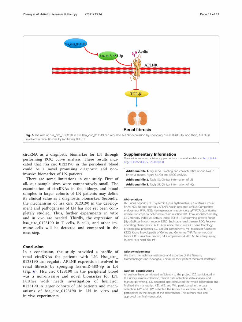
ConclusionIn a conclusion, the study provided a profile ofrenal circRNAs for patients with LN. Hsa_circ_0123190 can regulate APLNR expression involved inrenal fibrosis by sponging hsa-miR-483-3p in LN(Fig. 6). Hsa_circ_0123190 in the peripheral bloodwas a non-invasive and novel biomarker for LN.Further work needs investigation of hsa_circ_0123190 in larger cohorts of LN patients and mech-anisms of hsa_circ_0123190 in LN in vitro andin vivo experiments.
Supplementary InformationThe online version contains supplementary material available at https://doi.org/10.1186/s13075-020-02404-8.
Additional file 1. Figure S1. Profiling and characteristics of circRNAs inLN renal tissues. Figure S2. Go and KEGG analysis.
Additional file 2. Table S2. Clinical information of LN
Additional file 3. Table S1. Clinical information of NCs
AbbreviationsLN: Lupus nephritis; SLE: Systemic lupus erythematosus; CircRNAs: CircularRNAs; NCs: Normal controls; APLNR: Apelin receptor; ceRNA: Competitiveendogenous RNA; NGS: Next-generation sequencing; qRT-PCR: Quantitativereverse transcription polymerase chain reaction; IHC: Immunohistochemistry;CI: Chronicity index; AI: Activity index; TGF-β1: Transforming growth factor-β1; α-SMA: α-Smooth muscle; ESRD: End-stage renal disease; ROC: Receiveroperating characteristic; AUC: Area under the curve; GO: Gene Ontology;BP: Biological processes; CC: Cellular components; MF: Molecular functions;KEGG: Kyoto Encyclopedia of Genes and Genomes; TNF: Tumor necrosisfactor; CRP: C-reactive protein; C4: Complement 4; AKI: Acute kidney injury;FOXP4: Fork head box P4
AcknowledgementsWe thank the technical assistance and expertise of the GeneskyBiotechnologies Inc. (Shanghai, China) for their perfect technical assistance.
Authors’ contributionsAll authors have contributed sufficiently to the project. C.Z. participated inthe kidney sample collection, clinical data collection, data analysis, andmanuscript writing. Z.Z. designed and conducted the whole experiment andfinalized the manuscript. X.D., W.S. and W.L. participated in the datacollection. M.Y. and Q.W. collected the kidney tissues from patients. C.G.participated in the design of the experiments. The authors read andapproved the final manuscript.
Fig. 6 The role of hsa_circ_0123190 in LN. Hsa_circ_012319 can regulate APLNR expression by sponging hsa-miR-483-3p, and then, APLNR isinvolved in renal fibrosis by inhibiting TGF-β1
Zhang et al. Arthritis Research & Therapy (2021) 23:24 Page 11 of 12
FundingThis work was supported by the Medical Science and Technology ResearchProject of Henan in China [No. LHGJ20190260] and [No.SBGJ202002098].
Availability of data and materialsThe datasets analyzed during the current study are available from thecorresponding author on reasonable request.
Ethics approval and consent to participateThe study was approved by the Ethics Committee of the First AffiliatedHospital of Zhengzhou University (2018-KY-22). All patients provided writteninformed consent.
Consent for publicationNot applicable.
Competing interestsThe authors declare no competing interests.
Received: 2 May 2020 Accepted: 22 December 2020
References1. Aljaberi N, Bennett M, Brunner HI, Devarajan P. Proteomic profiling of urine:
implications for lupus nephritis. Expert Rev Proteomics. 2019;16(4):303–13.2. Zheng ZH, Zhang LJ, Liu WX, Lei YS, Xing GL, Zhang JJ, et al. Predictors of
survival in Chinese patients with lupus nephritis. Lupus. 2012;21(10):1049–56.
3. Chen LL. The biogenesis and emerging roles of circular RNAs. Nat Rev MolCell Biol. 2016;17(4):205–11.
4. Liang D, Wilusz JE. Short intronic repeat sequences facilitate circular RNAproduction. Genes Dev. 2014;28(20):2233–47.
5. Starke S, Jost I, Rossbach O, Schneider T, Schreiner S, Hung LH, et al. Exoncircularization requires canonical splice signals. Cell Rep. 2015;10(1):103–11.
6. Jeck WR, Sorrentino JA, Wang K, Slevin MK, Burd CE, Liu J, et al. CircularRNAs are abundant, conserved, and associated with ALU repeats. RNA (NewYork). 2013;19(2):141–57.
7. Mahmoudi E, Cairns MJ. Circular RNAs are temporospatially regulatedthroughout development and ageing in the rat. Sci Rep. 2019;9(1):2564.
8. Wang K, Sun Y, Tao W, Fei X, Chang C. Androgen receptor (AR) promotesclear cell renal cell carcinoma (ccRCC) migration and invasion via alteringthe circHIAT1/miR-195-5p/29a-3p/29c-3p/CDC42 signals. Cancer Lett. 2017;394:1–12.
9. Xiong Y, Zhang J, Song C. CircRNA ZNF609 functions as a competitiveendogenous RNA to regulate FOXP4 expression by sponging miR-138-5p inrenal carcinoma. J Cell Physiol. 2019;234(7):10646–54.
10. Austin HA, Muenz LR, Joyce KM, Antonovych TT, Balow JE. Diffuseproliferative lupus nephritis: identification of specific pathologic featuresaffecting renal outcome. Kidney Int. 1984;25(4):689–95.
11. Zhou D, Liu Y. Renal fibrosis in 2015: Understanding the mechanisms ofkidney fibrosis. Nat Rev Nephrol. 2016;12(2):68–70.
12. Antushevich H, Wójcik M. Review: apelin in disease. Clin Chim Acta. 2018;483:241–8.
13. Huang Z, Wu L, Chen L. Apelin/APJ system: a novel potential therapy targetfor kidney disease. J Cell Physiol. 2018;233(5):3892–900.
14. Huang S, Chen L, Lu L, Li L. The apelin-APJ axis: a novel potentialtherapeutic target for organ fibrosis. Clin Chim Acta. 2016;456:81–8.
15. Batu ED, Erden A, Seyhoğlu E, Kilic L, Büyükasık Y, Karadag O, et al.Assessment of the HScore for reactive haemophagocytic syndrome inpatients with rheumatic diseases. Scand J Rheumatol. 2017;46(1):44–8.
16. Wang L, Law HK. The role of autophagy in lupus nephritis. Int J Mol Sci.2015;16(10):25154–67.
17. Postal M, Appenzeller S. The role of tumor necrosis factor-alpha (TNF-α) inthe pathogenesis of systemic lupus erythematosus. Cytokine. 2011;56(3):537–43.
18. Djudjaj S, Boor P. Cellular and molecular mechanisms of kidney fibrosis. MolAspects Med. 2019;65:16–36.
19. Anders HJ, Saxena R, Zhao MH, Parodis I, Salmon JE, Mohan C. Lupusnephritis. Nat Rev Dis Primers. 2020;6(1):7.
20. Kristensen LS, Andersen MS, Stagsted LVW, Ebbesen KK, Hansen TB, Kjems J.The biogenesis, biology and characterization of circular RNAs. Nat RevGenet. 2019;20(11):675–91.
21. Kölling M, Seeger H, Haddad G, Kistler A, Nowak A, Faulhaber-Walter R, et al.The circular RNA predicts survival in critically ill patients with acute kidneyinjury. Kidney Int Rep. 2018;3(5):1144–52.
22. Jin C, Shi L, Li Z, Liu W, Zhao B, Qiu Y, et al. Circ_0039569 promotes renalcell carcinoma growth and metastasis by regulating miR-34a-5p/CCL22. AmJ Transl Res. 2019;11(8):4935–45.
23. Ouyang Q, Huang Q, Jiang Z, Zhao J, Shi GP, Yang M. Using plasmacircRNA_002453 as a novel biomarker in the diagnosis of lupus nephritis.Mol Immunol. 2018;101:531–8.
24. Luan J, Jiao C, Kong W, Fu J, Qu W, Chen Y, et al. circHLA-C plays animportant role in lupus nephritis by sponging miR-150. Mol Ther NucleicAcids. 2018;10:245–53.
25. Han B, Chao J, Yao H. Circular RNA and its mechanisms in disease: from thebench to the clinic. Pharmacol Ther. 2018;187:31–44.
26. Kuschnerus K, Straessler ET, Müller MF, Lüscher TF, Landmesser U, Kränkel N.Increased expression of miR-483-3p impairs the vascular response to injuryin type 2 diabetes. Diabetes. 2019;68(2):349–60.
27. Abue M, Yokoyama M, Shibuya R, Tamai K, Yamaguchi K, Sato I, et al.Circulating miR-483-3p and miR-21 is highly expressed in plasma ofpancreatic cancer. Int J Oncol. 2015;46(2):539–47.
28. Pepe F, Visone R, Veronese A. The glucose-regulated influences keysignaling pathways in cancer. Cancers. 2018;10(6):181.
29. Hus-Citharel A, Bouby N, Frugière A, Bodineau L, Gasc JM, Llorens-Cortes C.Effect of apelin on glomerular hemodynamic function in the rat kidney.Kidney Int. 2008;74(4):486–94.
30. Wu L, Chen L, Li L. Apelin/APJ system: a novel promising therapy target forpathological angiogenesis. Clin Chim Acta. 2017;466:78–84.
31. Wang LY, Diao ZL, Zhang DL, Zheng JF, Zhang QD, Ding JX, et al. Theregulatory peptide apelin: a novel inhibitor of renal interstitial fibrosis.Amino Acids. 2014;46(12):2693–704.
Publisher’s NoteSpringer Nature remains neutral with regard to jurisdictional claims inpublished maps and institutional affiliations.
Zhang et al. Arthritis Research & Therapy (2021) 23:24 Page 12 of 12