histopathology of selected parasitic … of selected parasitic salmonid diseases: a color atlas...
TRANSCRIPT

HISTOPATHOLOGY OF SELECTED PARASITIC SALMONID DISEASES: A COLOR ATLAS
William T. Yasutake Senior Scientist Emeritus
U.S. Geological Survey Western Fisheries Research Center
Seattle, Washington

HISTOPATHOLOGY OF SELECTED PARASITIC SALMONID DISEASES: A COLOR ATLAS
Table of Contents Plate 1 Figure 1. Ichthyobodo necator Figure 2. I. necator Figure 3. Thecamoeba hoffmani Plate 2
Figure 4. T. hoffmani Figure 5. T. hoffmani Figure 6. Ichyophthirius multifiliis
Plate 3
Figure 7. I. multifiliis Figure 8. Trichodina sp. Figure 9. Trichodina sp.
Plate 4
Figure 10. Trichodina sp. Figure 11. Trichophrya sp. Figure 12. Trichophrya sp.
Plate 5
Figure 13. Trypanoplasma (Cryptobia) salmositica Figure 14. T. salmositica Figure 15. T. salmositica
Plate 6
Figure 16. Spironucleus (Hexamita) salmonis Figure 17. Ceratomyxa shasta Figure 18. C. shasta
Plate 7
Figure 19. C. shasta Figure 20. C. shasta Figure 21. C. shasta
Plate 8
Figure 22. C. shasta Figure 23. Myxobolus (Myxosoma) cerebralis Figure 24. M. cerebralis

Plate 9 Figure 25. M. cerebralis Figure 26. M. cerebralis Figure 27. M. cerebralis
Plate 10
Figure 28. M. cerebralis Figure 29. Myxobolus squamalis Figure 30. M. squamalis
Plate 11
Figure 31. Myxobolus sp. Figure 32. Myxobolus sp. Figure 33. Myxobolus sp.
Plate 12
Figure 34. Myxobolus kisutchi Figure 35. M. kisutchi Figure 36. Parvicapsula sp.
Plate 13
Figure 37. Parvicapsula sp. Figure 38. Parvicapsula sp. Figure 39. Parvicapsula sp.
Plate 14
Figure 40. Proliferative kidney disease (PKD) (Caused by Tetracapsuloides Figure 41. T. bryosalmonae bryosalmonae) Figure 42. T. bryosalmonae
Plate 15
Figure 43. T. bryosalmomae Figure 44. T. bryosalmonae Figure 45. T. bryosalmonae
Plate 16
Figure 46. Chloromyxum majori Figure 47. C. majori Figure 48. C. majori
Plate 17
Figure 49. Myxdium sp. Figure 50. Myxdium minteri Figure 51. Myxdium sp.
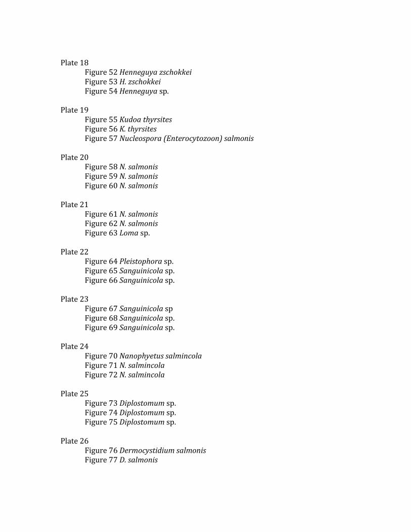
Plate 18
Figure 52 Henneguya zschokkei Figure 53 H. zschokkei Figure 54 Henneguya sp.
Plate 19
Figure 55 Kudoa thyrsites Figure 56 K. thyrsites Figure 57 Nucleospora (Enterocytozoon) salmonis
Plate 20
Figure 58 N. salmonis Figure 59 N. salmonis Figure 60 N. salmonis
Plate 21
Figure 61 N. salmonis Figure 62 N. salmonis Figure 63 Loma sp.
Plate 22
Figure 64 Pleistophora sp. Figure 65 Sanguinicola sp. Figure 66 Sanguinicola sp.
Plate 23
Figure 67 Sanguinicola sp Figure 68 Sanguinicola sp. Figure 69 Sanguinicola sp.
Plate 24
Figure 70 Nanophyetus salmincola Figure 71 N. salmincola Figure 72 N. salmincola
Plate 25
Figure 73 Diplostomum sp. Figure 74 Diplostomum sp. Figure 75 Diplostomum sp.
Plate 26
Figure 76 Dermocystidium salmonis Figure 77 D. salmonis

Acknowledgements
Thanks to the following individuals for their contribution of photomicrographs for this project: John Moldin, California State Fish and Game; the late Dr. Clearance Becker, Battelle Northwest; the late James Wood, Washington State Fish and Game; the late Harold Wolf, California Fish and Game; Charlie Smith, U. S. Fish and Wildlife Service; Robert Troth, California Fish and Game; Beth MacConnell, U. S. Fish and Wildlife Service; John Morrison, U. S. Fish and Wildlife Service; Bruce M. Bortz, Washington Cooperative Fishery Research Unit; Dr. Robert Olsen, Hatfield Marine and Science Museum, Newport, OR. A special thanks to Drs. James Winton, Diane Elliott and Gary Wedemeyer, U.S. Geological Survey, Western Fisheries Research Center, Seattle, Washington, for reviewing the final draft of the manuscript. I am also indebted to the late Dr. A. C. Fox, former Director and Dr. J. Rolland, the present Director of the Western Fisheries Research Center.

Plate 1
Figure 1
Ichthyobodo necator (Costia) are
attached to the gill surface (arrow) of
a yearling sockeye salmon,
Oncorhynchus nerka. Note
hypertrophy and hyperplasia of the
lamellae epithelium.
May-Grünwald Giemsa (M-G G)
stain. X800.
Figure 2
I. necator (arrow) are attached to
several loose skin epidermal cells of a
fingerling rainbow trout, O. mykiss.
Iron Hematoxylin (FeH) stain.
X2000.
(Courtesy of H. Wolf)
Figure 3
Thecamoeba hoffmani (arrow), an
amoeba, in wet mount made from gill
scraping of a fingerling Chinook
salmon, O. tchawytscha.
Phase contrast. X700.
(Courtesy of J. Moldin)

Plate 2
Figure 5
T. hoffmani (arrow). Higher
magnification of Fig. 4. Note
hypertrophy and hyperplasia of the
lamellar epithelium.
M-G G stain. X500.
Figure 6
Icthyophthirius multifiliis in the gill
of a fingerling rainbow trout. Note
the characteristic horseshoe-shaped
mucleus (arrow).
H&E stain. X250.
Figure 4
T. hoffmani in gill section of a
fingerling Chinook salmon. Note
hyperplastic lamellar epithelium
(arrow).
M-G G stain. X200.

Plate 3
Figure 7
I. multifilitiis (arrow) in the
epidermis of a spawning
coho salmon O. kisutch.
M-G G stain. X700.
Figure 8
Trichodina sp. from a
juvenile coho salmon gill
scraping fixed in
Schaudinn’s fluid. Note the
denticulate ring (arrow).
FeH stain. X1000.
Figure 9
Trichodina sp. (arrows)
between the lamellae of a
juvenile coho salmon. Note
the minimal tissue reaction.
M-G G stain. X1000.

Plate 4
Figure 10
Cross section of Trichodina sp.
between gill lamellae of a fingerling
coho salmon, O. kisutch. Note
minimal tissue reaction.
M-G G srain. X1000.
Figure 11
Trichophrya sp., a suctoran ciliate.
Note numerous rod-like tentacles
(arrows) on the gill lamellae of
sockeye salmon.
Phase contrast. X500.
Figure 12
Trichophrya sp. (arrows) on the gill
section of a fingerling sockeye
salmon.
M-G G stain. X700.

Plate 5
Figure 13
Trypanoplasma (Cryptobia)
salmositica, a hemoflagellate, in
peripheral blood smear of a young
rainbow trout. Note prominent
basophilic kinetoplast.
Leishman-Giemsa stain. X4000.
(Courtesy of C. D. Becker)
Figure 14
T. salmositica in the gill blood
vessels of an infected steelhead, O.
mykiss (arrows).
M-G G stain. X800.
Figure 15
T. salmositica with prominent
kinetoplasts (arrows) in the central
vein of the liver of a fingerling
Chinook salmon.
M-G G stain. X800.

Plate 6
Figure 16
Spironucleus (Hexamita) salmonis
(arrow), a diplomonad flagellate, in a
smear made from mucoid material
from the intestine of a fingerling
Chinook salmon.
FeH stain. X800.
Figure 17
Ceratomyxa shasta. Perforated
intestine (arrows) of a spawning fall
Chinook salmon infected with
Ceratomya shasta, a myxosporean.
(Courtesy of J. W. Wood)
Figure 18
C. shasta in wet mount with
pansporoblasts.
Phase contrast. X4500.
(Courtesy of H. Wolf)

Plate 7
Figure 19
C. shasta trophozoites in the lamina
propria of a spawning coho salmon
pyloric cecum. Note the extensisve
inflammatory response to the
trophozoites (arrow).
M-M G stain. X200.
Figure 20
C. shasta trophozoites. High
magnification of Fig. 19. Note the
various stages of the pansporoblasts
(arrow) and the extensive tissue
response.
M-G G. stain. X550.
Figure 21
C. shasta trophozoites and spores in
the lamina propria of spawning coho
salmon pyloric cecum. Note the
extensive inflammatory response
(arrow).
M-G G stain. X200.

Plate 8
Figure 22
C. shasta trophozoites and spores.
Note the spores (arrows), some of
which are destained enough to exhibit
the paired polar capsules.
M-G G stain. X640.
Figure 23
Myxobolus (Myxosoma) cerebralis
infection in yearling cutthroat trout,
O. clarki, showing scoliosis and
lordosis. Top two fish are normal.
Figure 24
M. cerebralis spores (arrow) in wet
mount made from infected fingerling
steelhead trout, O. mykiss.
Differential interference contrast
(DIC). X1800.
(Courtesy of H. Wolf)
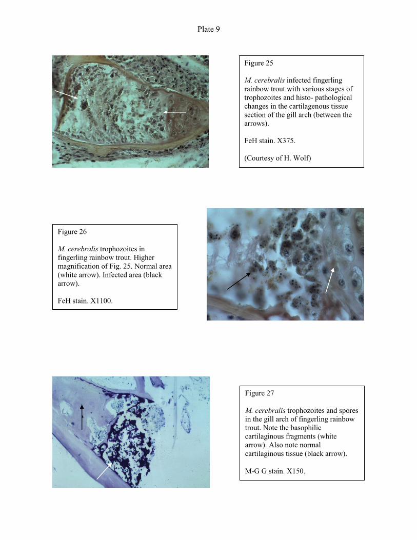
Plate 9
Figure 25
M. cerebralis infected fingerling
rainbow trout with various stages of
trophozoites and histo- pathological
changes in the cartilagenous tissue
section of the gill arch (between the
arrows).
FeH stain. X375.
(Courtesy of H. Wolf)
Figure 26
M. cerebralis trophozoites in
fingerling rainbow trout. Higher
magnification of Fig. 25. Normal area
(white arrow). Infected area (black
arrow).
FeH stain. X1100.
Figure 27
M. cerebralis trophozoites and spores
in the gill arch of fingerling rainbow
trout. Note the basophilic
cartilaginous fragments (white
arrow). Also note normal
cartilaginous tissue (black arrow).
M-G G stain. X150.

Plate 10
Figure 28
M. cerebralis spores in yearling
rainbow trout. Note the 2 polar
capsules (black arrow) and
sporoplasm ( white arrow).
M-G G stain. X3500.
(Courtesy of H. Wolf)
Figure 29
Myxobolus squamalis spores in the
scale of a fingerling rainbow trout
(arrow).
M-G G stain. X200.
Figure 30
M. squamalis spores in the scale of a
fingerling rainbow trout. Note the 2
polar capsules and sporoplasm in the
spores (arrows).
High magnification of Fig. 29.
M-G G stain. X2000.

Plate 11
Figure 31 Myxobolus sp. spore in wet mount showing iodinophilous vacuole in the sporoplasm (arrow). Specimen taken from body muscle of a yearling coho salmon. Stained with Lugol’s iodine solution. X3700.
Figure 32 Myxobolus sp. spores (arrows) in the dorsal striated muscle of a yearling coho salmon. M-G G stain. X140.
Figure 33 Myxobolus sp. spores (arrow). Higher magnification of Fig. 32. Note the two pyriform polar capsules in the spore. M-G G stain. X2000.

Plate 12
Figure 34
Myxobolus kisutchi spores (arrows) in
the spinal cord of a fingerling coho
salmon. Note the lack of
inflammatory response in the
peripheral area of the spores.
M-G G stain. X1700.
(Courtesy of H. Wolf)
Figure 35
M. kisutchi spores in the spinal cord,
just posterior to the medulla
oblongata of the fingerling coho
salmon.
M-G G stain. X 3500.
Figure 36
Parvicapsula sp., myxosporean
spores (arrows) in wet mount of pen-
reared yearling coho salmon kidney.
Note two spherical eye-like polar
capsules.
DIC. X1600.
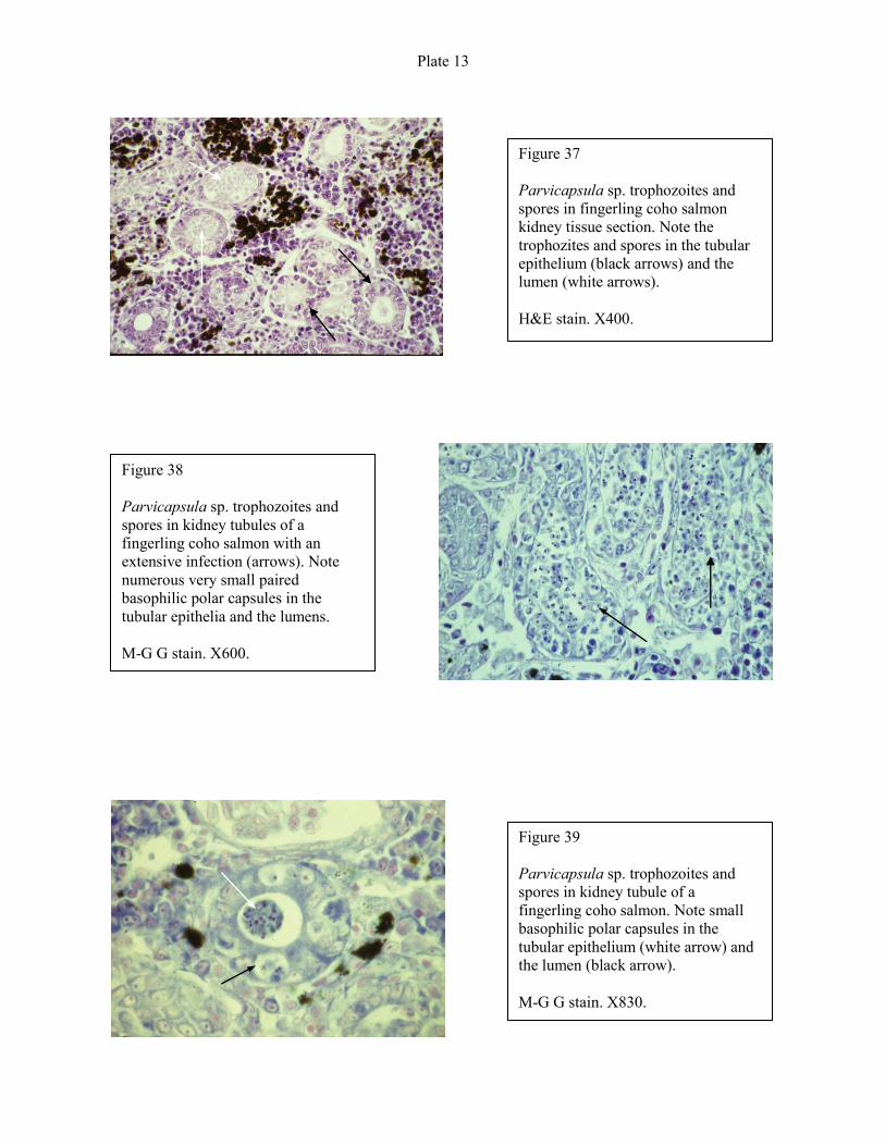
Plate 13
Figure 37
Parvicapsula sp. trophozoites and
spores in fingerling coho salmon
kidney tissue section. Note the
trophozites and spores in the tubular
epithelium (black arrows) and the
lumen (white arrows).
H&E stain. X400.
Figure 38
Parvicapsula sp. trophozoites and
spores in kidney tubules of a
fingerling coho salmon with an
extensive infection (arrows). Note
numerous very small paired
basophilic polar capsules in the
tubular epithelia and the lumens.
M-G G stain. X600.
Figure 39
Parvicapsula sp. trophozoites and
spores in kidney tubule of a
fingerling coho salmon. Note small
basophilic polar capsules in the
tubular epithelium (white arrow) and
the lumen (black arrow).
M-G G stain. X830.
Plate 14
Figure 40
Proliferative kidney disease (PKD).
6-month-old rainbow trout infected
with PKD showing extensively
swollen kidneys (white arrows)
exhibiting enlarged spleen (black
arrow).
(Courtesy of C. E. Smith)
Figure 41
PKD organism (arrow) in kidney
imprint from a 6-month-old rainbow
trout. The caustive agent is
Tetracapsuloides bryosalmonae, a
malacosporean.
Leishman-Giema (L-G) stain. X1100.
(Courtesy of C. E. Smith)
Figure 42
PKD infected 7-month-old rainbow
trout kidney showing proliferative
tissue response (arrow).
H&E stain. X230.
(Courtesy of C. E. Smith)

Plate 15
Figure 43
PKD infected 5-month-old rainbow
trout kidney showing several T.
bryosalmonae. Note PKD organism
stained light magenta when stained
with PAS (periodic acid-Schiff) stain
(arrow). X700.
(Courtesy of C. E. Smith)
Figure 44
PKD organism, T. bryosalmonae
(arrow), in the gill lamella of an
infected rainbow trout.
H&E stain. X1600.
(Courtesy of C. E. Smith)
Figure 45
T. bryosalmonae (black arrow) in the
dorsal muscle of an infected rainbow
trout. Note the inflammatory response
(white arrow).
H&E stain. X1600.

Plate 16
Figure 46
Chloromyxum majori spores (arrow)
in wet mount made from an adult
rainbow trout kidney.
Phase contrast. X3500.
Figure 47
C. majori trophozoites and spores in
kidney glomeruli of a 6-month-old
Chinook salmon. Note basophilic
spores and trophozoites occupying
much of the glomeruli, leaving very
little of the capillary tufts (arrows).
M-G G stain. X1500.
Figure 48
C. majori trophozoites and spores.
Higher magnification of Fig. 47. Note
the four polar capsules of a spore
clustered together (arrow).
M-G G stain. X1500.

Plate 17
Figure 49
Myxidium sp. infection in the liver
of a 4-year-old rainbow trout.
Note mottled areas (arrows) where the cysts of the
myxosporiea occur.
(Courtesy of R. J. Troth)
Figure 49
Myxidium sp. infection in the liver of
a 4-year-old rainbow trout. Note
mottled areas (arrows) where the
cysts of the myxosporiea occur.
(Courtesy of R. J. Troth)
Figure 50
Myxidium minteri trophozoites and
spores in the lumen of a fingerling
steelhead trout kidney tubules. Note
the spore with polar capsules
(arrows).
M-G G stain. X900.
Figure 51
Myxidium sp. cyst in a 2-year-old
rainbow trout liver. One pyriform or
spherical polar capsule occurs on
each end of the spore (arrows).
M-G G stain. X1500.

Plate 18
Figure 52
Henneguya zschokkei. White cysts
(arrows) in muscle of an adult
Chinook salmon.
(Courtesy of H. Wolf)
Figure 53
H. zschokkei spores in a sectioned
cyst from a yearling rainbow trout
body muscle. Note paired polar
capsules (arrows).
M-G G stain. X640.
Figure 54
Henneguya sp. spores from a cyst
in muscle of a rainbow trout. Note
the 2 polar capsules at the anterior
end and the long paired hair-like
processes extending from the
posterior end of each spore
(arrows).
Wet mount. DIC. X1100.

Plate 19
Figure 55
Kudoa thyrsites (arrow) in body
muscle of a yearling Atlantic salmon.
M-G G stain. X600.
Figure 56
K. thyrsites spores (arrow) in the
striated body musculature of a
yearling Atlantic salmon.
M-G G stain. X1000.
Figure 57
Nucleospora (Enterocytozoon)
salmonis infected fingerling steelhead
kidney imprint. Note parasitized
lymphoblasts containing spherical,
light colored, nondescript, nuclear
inclusions (arrows).
L-G stain. X1000.
(Courtesy of B. MacConnell)

Plate 20
Figure 58
N. salmonis. Section of a fingerling
steelhead posterior intestine showing
extensive lymphocytic infiltration of
N. salmonis (arrow) in the lamina
propria.
H&E stain. X160.
(Courtesy of B. MacConnell)
Figure 59
N. salmonis. Parsitized lymphoid
cells in lamina propria (arrow) of a
fingerling steelhead.
H&E stain. X1000.
(Courtesy of B. MacConnell)
Figure 60
N. salmonis infected fingerling
steelhead with perivascular cuffing of
affected hepatic cells (arrow).
H&E stain. X160.
(Courtesy of B. MacConnell)

Plate 21
Figure 61
N. salmonis. Kidney imprint of
fingerling Chinook salmon
infected with N. salmonis. Note
lymphocyte nucleus (arrow) with
prespore stage of N. salmonis.
.
Electron photomicrograph.
(Courtesy of John Morrison)
Figure 62
N. salmonis. Developing
sporoblast (arrows) within the
host cell nucleus of a fingerling
Chinook salmon.
Electron photomicrograph.
(Courtesy of John Morrison *)
Figure 63
Loma sp. a microsporidian, in a
luamellar cyst of a 2-year-old
steelhead trout.
M-G G stain. X160.
*Morrison, J. K., E. MacConnell, P. F. Chapman, R. L. Westgard. 1990. A microsporidium-induced lymphoblastosis in
Chinook salmon Oncorhynchus tshawytscha in freshwater. Dis. Aquat. Org. 8: 99-104
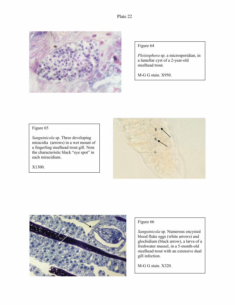
Plate 22
Figure 64
Pleistophora sp. a microsporidian, in
a lamellar cyst of a 2-year-old
steelhead trout.
M-G G stain. X950.
Figure 65
Sanguinicola sp. Three developing
miracidia (arrows) in a wet mount of
a fingerling steelhead trout gill. Note
the characteristic black “eye spot” in
each miracidium.
X1300.
Figure 66
Sanguinicola sp. Numerous encysted
blood fluke eggs (white arrows) and
glochidium (black arrow), a larva of a
freshwater mussel, in a 5-month-old
steelhead trout with an extensive dual
gill infection.
M-G G stain. X320.
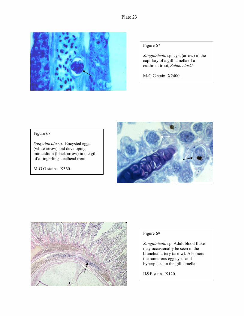
Plate 23
Figure 67
Sanguinicola sp. cyst (arrow) in the
capillary of a gill lamella of a
cutthroat trout, Salmo clarki.
M-G G stain. X2400.
Figure 68
Sanguinicola sp. Encysted eggs
(white arrow) and developing
miracidium (black arrow) in the gill
of a fingerling steelhead trout.
M-G G stain. X360.
Figure 69
Sanguinicola sp. Adult blood fluke
may occasionally be seen in the
branchial artery (arrow). Also note
the numerous egg cysts and
hyperplasia in the gill lamella.
H&E stain. X120.

Plate 24
Figure 70
Nanophyetus salmincola (commonly
known as the salmon poisoning fluke)
metacercariae (arrows) in the kidney
of an adult coho salmon. Note
minimal tissue reaction or
inflammation around the parasites.
M-G G stain. X150.
Figure 71
N. salmincola metacercaria in the
heart ventricle of a spawning coho
salmon. Note again the minimal
tissue reaction around the organism.
M-G G stain. X150.
Figure 72
N. salmincola metacercaria (a) in
thyroid tissue (b) area of a yearling
coho salmon. Note the fibrosis and
granulomatous reactions (c) adjacent
to the metacercaria.
H&E stain. X150.
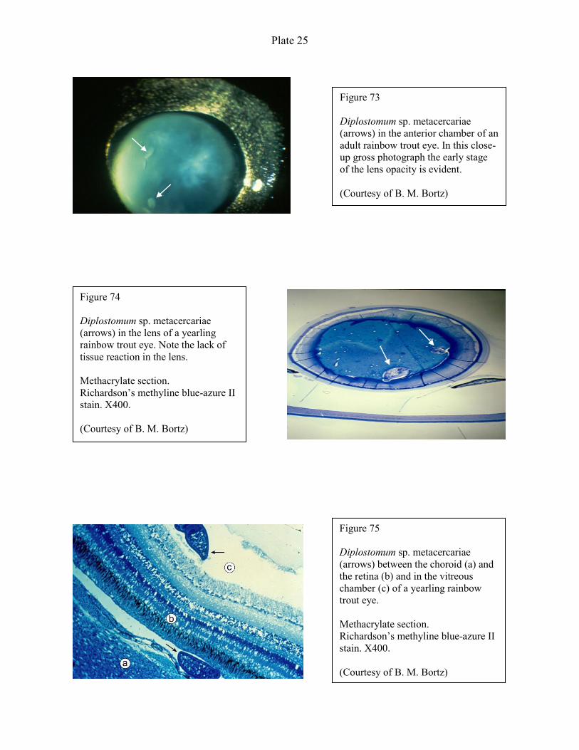
Plate 25
Figure 73
Diplostomum sp. metacercariae
(arrows) in the anterior chamber of an
adult rainbow trout eye. In this close-
up gross photograph the early stage
of the lens opacity is evident.
(Courtesy of B. M. Bortz)
Figure 74
Diplostomum sp. metacercariae
(arrows) in the lens of a yearling
rainbow trout eye. Note the lack of
tissue reaction in the lens.
Methacrylate section.
Richardson’s methyline blue-azure II
stain. X400.
(Courtesy of B. M. Bortz)
Figure 75
Diplostomum sp. metacercariae
(arrows) between the choroid (a) and
the retina (b) and in the vitreous
chamber (c) of a yearling rainbow
trout eye.
Methacrylate section.
Richardson’s methyline blue-azure II
stain. X400.
(Courtesy of B. M. Bortz)
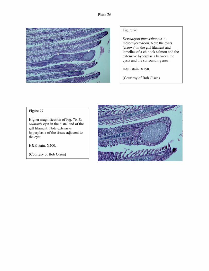
Plate 26
Figure 76
Dermocystidium salmonis, a
mesomycetozoon. Note the cysts
(arrows) in the gill filament and
lamellae of a chinook salmon and the
extensive hyperplasia between the
cysts and the surrounding area.
H&E stain. X150.
(Courtesy of Bob Olsen)
Figure 77
Higher magnification of Fig. 76. D.
salmonis cyst in the distal end of the
gill filament. Note extensive
hyperplasia of the tissue adjacent to
the cyst.
H&E stain. X200.
(Courtesy of Bob Olsen)