hiperinsulinismo en el recien nacido. katherine lord, mda ... nn_2018.pdf · prompt evaluation to...
TRANSCRIPT
Hiperinsulinismo en el Recien Nacido. Katherine Lord, MDa,b, Diva D. De León, MD, MSCEa,b,* Clin Perinatol 45 (2018) 61–74 Introducción. El Hiperinsulinismo (HI), es la causa más común de hipoglucemia persistente en bebés, conlleva un alto riesgo de morbilidad a largo plazo. En HI, los resultados de secreción de insulina desregulada en la hipoglucemia grave, la supresión de la respuesta contrarreguladora a la hipoglucemia, y, más significativamente, la supresión de la producción de cuerpos cetónicos, que son combustibles alternativos cruciales para el cerebro. Por lo tanto, la hipoglucemia hipoketótica grave y recurrente resultante de HI, si no se trata, se asocia con daño cerebral irreversible. Los la frecuencia de retrasos del neurodesarrollo en la HI es tan alta como 30% a 50% y, lo que es más importante, esto afecta no solo a los niños con formas congénitas y permanentes de HI sino también a los niños con reconocimiento transitorio HI. La prontitud en el reconocimiento y administración apropiada son fundamental para disminuir el riesgo de estos malos resultados. La dilucidación de la genética molecular de HI y avances en pruebas de diagnóstico, específicamente el uso de 18-fluoro-L-3,4- La PET con dihidroxifenilalanina (F-DOPA) para localizar las lesiones focales ha resultado en una enfoque personalizado del manejo y disminución de la morbilidad. Le invitamos a leer, la publicación completa de este interesante tema.

Hyperinsulinism in theNeonate
Katherine Lord, MDa,b, Diva D. De León, MD, MSCEa,b,*
INTRODUCTION
Hyperinsulinism (HI), the most common cause of persistent hypoglycemia in infants,carries a high risk of long-term morbidity. In HI, dysregulated insulin secretion resultsin severe hypoglycemia, suppression of the counterregulatory response to hypoglyce-mia, and, more significantly, suppression of ketone bodies production, which arecrucial alternative fuels for the brain. Thus, severe and recurrent hypoketotic hypogly-cemia resulting from HI, if untreated, is associated with irreversible brain damage. Thefrequency of neurodevelopmental delays in HI is as high as 30% to 50% and, impor-tantly, this affects not only children with congenital and permanent forms of HI but alsochildren with transient HI.1–3 Prompt recognition and appropriate management arecritical to decrease the risk of these poor outcomes. Elucidation of the molecular ge-netics of HI and advances in diagnostic testing, specifically the use of 18-fluoro-L-3,4-dihydroxyphenylalanine (18F-DOPA) PET to localize focal lesions, has resulted in apersonalized approach to management and decreased morbidity.
Disclosure Statement: The authors have nothing to disclose.a The Division of Endocrinology and Diabetes, The Children’s Hospital of Philadelphia, 3401Civic Center Boulevard, Philadelphia, PA 19104, USA; b Department of Pediatrics, The PerelmanSchool of Medicine, University of Pennsylvania, 3401 Civic Center Boulevard, Philadelphia, PA19104, USA* Corresponding author. The Children’s Hospital of Philadelphia, Abramson Research Center,Room 802A, 3615 Civic Center Boulevard, Philadelphia, PA.E-mail address: [email protected]
KEYWORDS
� Hypoglycemia � Neonate � Hyperinsulinism � Insulin � Pancreas � Pancreatectomy
KEY POINTS
� Hyperinsulinism (HI) is the most common cause of persistent hypoglycemia and is asso-ciated with high rates of neurodevelopmental deficits.
� Prompt evaluation to establish the diagnosis is important to start appropriate treatment.
� As soon as a diagnosis of HI is established, an initial trial of diazoxide is necessary to iden-tify those who are likely to benefit from specialized evaluation.
� Infants with diazoxide-unresponsive HI require expedited genetic testing for ABCC8 andKCNJ11 to determine the likelihood of focal disease.
� The goal of medical therapy is to allow infants to maintain normal feeding patterns and tosustain plasma glucose greater than 70 mg/dL.
Clin Perinatol 45 (2018) 61–74https://doi.org/10.1016/j.clp.2017.10.007 perinatology.theclinics.com0095-5108/18/ª 2017 Elsevier Inc. All rights reserved.
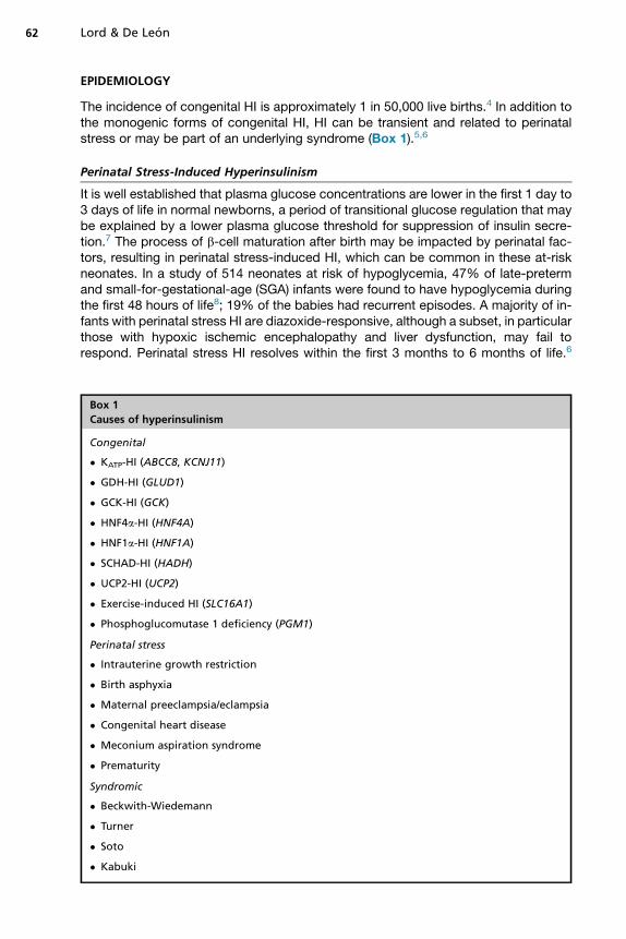
EPIDEMIOLOGY
The incidence of congenital HI is approximately 1 in 50,000 live births.4 In addition tothe monogenic forms of congenital HI, HI can be transient and related to perinatalstress or may be part of an underlying syndrome (Box 1).5,6
Perinatal Stress-Induced Hyperinsulinism
It is well established that plasma glucose concentrations are lower in the first 1 day to3 days of life in normal newborns, a period of transitional glucose regulation that maybe explained by a lower plasma glucose threshold for suppression of insulin secre-tion.7 The process of b-cell maturation after birth may be impacted by perinatal fac-tors, resulting in perinatal stress-induced HI, which can be common in these at-riskneonates. In a study of 514 neonates at risk of hypoglycemia, 47% of late-pretermand small-for-gestational-age (SGA) infants were found to have hypoglycemia duringthe first 48 hours of life8; 19% of the babies had recurrent episodes. A majority of in-fants with perinatal stress HI are diazoxide-responsive, although a subset, in particularthose with hypoxic ischemic encephalopathy and liver dysfunction, may fail torespond. Perinatal stress HI resolves within the first 3 months to 6 months of life.6
Box 1
Causes of hyperinsulinism
Congenital
� KATP-HI (ABCC8, KCNJ11)
� GDH-HI (GLUD1)
� GCK-HI (GCK)
� HNF4a-HI (HNF4A)
� HNF1a-HI (HNF1A)
� SCHAD-HI (HADH)
� UCP2-HI (UCP2)
� Exercise-induced HI (SLC16A1)
� Phosphoglucomutase 1 deficiency (PGM1)
Perinatal stress
� Intrauterine growth restriction
� Birth asphyxia
� Maternal preeclampsia/eclampsia
� Congenital heart disease
� Meconium aspiration syndrome
� Prematurity
Syndromic
� Beckwith-Wiedemann
� Turner
� Soto
� Kabuki
Lord & De Leon62
These neonates may be at risk for long-term neurologic deficits, however, as demon-strated by the study of a large cohort of neonates at risk followed-up to 4.5 years andfound to have a dose-dependent increased risk of poor executive function and visualmotor function.9 Thus, identification and appropriate treatment of these neonates areimportant to prevent long-term neurologic deficits.
GENETICS
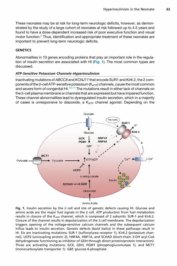
Abnormalities in 10 genes encoding proteins that play an important role in the regula-tion of insulin secretion are associated with HI (Fig. 1). The most common types arediscussed.
ATP-Sensitive Potassium Channels–Hyperinsulinism
Inactivatingmutations ofABCC8 andKCNJ11 that encodeSUR1 andKir6.2, the 2 com-ponentsof theb-cell ATP-sensitivepotassium (KATP) channels, cause themost commonand severe form of congenital HI.10,11 The mutations result in either lack of channels ontheb-cell plasmamembraneor channels that are expressedbut have impaired function.These channel abnormalities lead to dysregulated insulin secretion, which in a majorityof cases is unresponsive to diazoxide, a KATP channel agonist. Depending on the
Fig. 1. Insulin secretion by the b cell and site of genetic defects causing HI. Glucose andamino acids are the major fuel signals in the b cell. ATP production from fuel metabolismresults in closure of the KATP channel, which is composed of 2 subunits: SUR-1 and Kir6.2.Closure of the channel results in depolarization of the b cell membrane. The depolarizationtriggers opening of the voltage-sensitive calcium channels and the subsequent calciuminflux leads to insulin secretion. Genetic defects (bold italics) in these pathways result inHI. Six are inactivating mutations: SUR-1 (sulfonylurea receptor 1), Kir6.2 (potassium chan-nel), UCP2 (uncoupling protein 2), HNF4A, HNF1A, and SCHAD (short-chain 3-OH acyl-CoAdehydrogenase functioning as inhibitor of GDH through direct protein/protein interaction).Three are activating mutations: GCK, GDH, PGM1 (phosphoglucomutase 1), and MCT1(monocarboxylate transporter 1). G6P, glucose-6-phosphate.
Hyperinsulinism in the Neonate 63

mutation and its impact on the channel expression and function, KATP-HI can be reces-sive diazoxide-unresponsive, dominant diazoxide-unresponsive, or dominant diazo-xide-responsive.12 Infants with diazoxide-unresponsive KATP-HI present with severehypoglycemia, large-for-gestational-age (LGA) birth weight, and high glucose infusionrate (GIR) requirements. Infants with the diazoxide-responsive form typically presentwith milder disease.13
KATP-HI has 2 distinct histologic subtypes: diffuse and focal (Fig. 2). In diffuse KATP-HI, b-cells throughout the pancreas show signs of hyperactivity.14 In contrast, adiscrete area of b-cell proliferation or adenomatosis characterizes the focal form.Diffuse KATP-HI results from biallelic recessive mutations in the KATP genes; lesscommonly, dominant mutations are found. Focal KATP-HI is the result of a 2-hit mech-anism: (1) a paternally inherited recessive mutation in ABCC8 or KCNJ11 and (2) so-matic loss of the maternally inherited 11p15 chromosomal region, compensated bypaternal uniparental disomy.15,16 The histologic differences in the 2 forms lead todivergent treatment options and outcomes. Patients with focal KATP-HI are curedwith resection of the focal lesion, whereas pancreatectomy for patients with the diffuseform is palliative.Given these different outcomes, distinguishing between the focal and diffuse forms
of KATP-HI is crucial. Neonates with the diffuse form are more likely to present at birth,whereas those with focal form may fail detection in the neonatal period and present atseveral weeks to months of life with hypoglycemia seizures.17 Due to significant over-lap in clinical presentation, however, clinical features alone cannot be used to distin-guish between the 2 forms. Genetic testing offers the best means of identifying infantswith focal KATP-HI: a single recessive paternally inherited mutation in ABCC8 orKCNJ11 has a positive predictive value of 94% for focal HI.18
Glutamate Dehydrogenase–Hyperinsulinism
Activating mutations in GLUD1, which encodes glutamate dehydrogenase (GDH),cause the second most common form of HI, known as HI/hyperammonemia
Fig. 2. (A) Section of pancreas from a diffuse case showing a pancreatic islet demonstratingb-cell nucleomegaly (white arrow), histologic hallmark of diffuse disease (H&E, originalmagnification � 400). (B) Adenomatous lesion from a case of focal HI (H&E, original magni-fication � 200). Normal pancreas tissue is seen on the right side of the image.
Lord & De Leon64

(HI/HA) syndrome.19 GDH is involved in amino acid–stimulated insulin secretion.Mutations in GLUD1 most commonly occur de novo (70%), with the remainderinherited in an autosomal dominant manner. Infants with HI/HA syndrome presentwith fasting and protein-induced hypoglycemia and ammonia levels 3 times to 5times above the normal range. This form of HI is associated with increased ratesof seizures and learning disabilities that may not be directly the result of the hy-poglycemia or the HA.20 Individuals with HI/HA syndrome are responsive todiazoxide.
Glucokinase-Hyperinsulinism
Glucokinase (GCK) is the key enzyme regulating the glucose threshold for insulinsecretion.21 Activating mutations of GCK, encoded by GCK, cause an autosomaldominant form of HI, which has variable degrees of severity and diazoxide responsive-ness. Althoughmost infants may bemanagedmedically, the severe cases may requirenear-total pancreatectomy.22
Hepatic Nuclear Factor 4 Alpha—Hyperinsulinism
Mutations in the pancreatic transcription factor, hepatic nuclear factor 4 alpha(HNF4a), cause diazoxide-responsive HI.23 Infants with HNF4A-HI are typically macro-somic and can be treated with low dose diazoxide. The hypoglycemia resolves withinthe first several years of life. Progressive b-cell failure occurs, however, and results in amonogenic form of early onset diabetes (maturity-onset diabetes of the young[MODY1]).24 A similar clinical progression (from HI to diabetes) has been describedin individuals with mutations in another pancreatic transcription factor, hepatic nuclearfactor 1nalpha (HNF1a), which results in MODY3.25
DIAGNOSISClinical Presentation
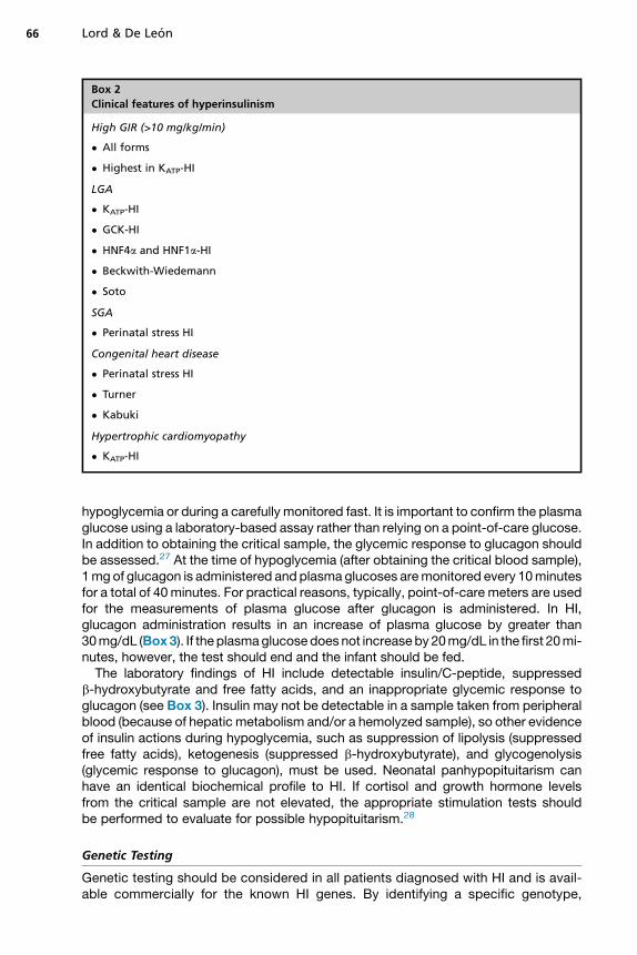
The classic presentation of HI is an LGA infant with a high GIR (>10 mg/kg/min), as iscommonly seen in KATP-HI. The clinical phenotype, however, is a spectrum and somepatients present with normal birth weight and minimally elevated glucose require-ments. Clinical features can suggest a specific phenotype and guide genetic testing(Box 2).
Indications for a Hypoglycemia Evaluation
An expert committee of pediatric endocrinologists and neonatologists recently pub-lished guidelines for the evaluation and management of hypoglycemia in neonates, in-fants, and children.26 The committee recommended evaluation for a persistenthypoglycemia disorder in neonates unable to consistently maintain plasma glucoseconcentrations greater than 60 mg/dL by the third day of life, those with severe hypo-glycemia (symptomatic hypoglycemia or requiring intravenous dextrose), and thosewho are at high risk of having a persistent hypoglycemic disorder (LGA, SGA, perinatalstress, maternal diabetes, congenital syndromes, or family history of genetic hypogly-cemia disorders).
Laboratory Evaluation
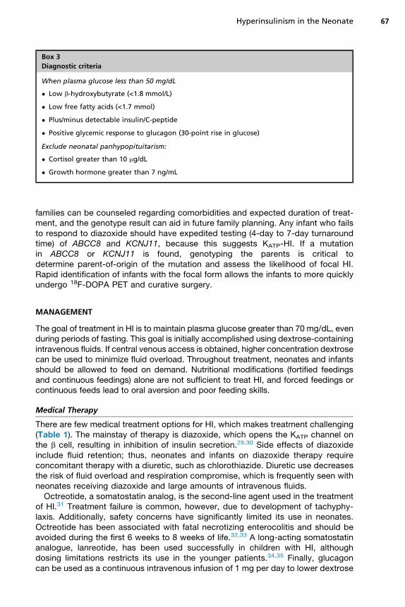
A diagnosis of HI is based on a critical blood sample obtained at the time of hypogly-cemia. The critical sample is used to measure plasma concentrations of the hormonesand alternative fuels involved in the physiologic response to fasting. To minimize false-positive results, the plasma glucose threshold for obtaining a critical sample is lessthan 50 mg/dL. This sample may be obtained during a spontaneous episode of
Hyperinsulinism in the Neonate 65
hypoglycemia or during a carefully monitored fast. It is important to confirm the plasmaglucose using a laboratory-based assay rather than relying on a point-of-care glucose.In addition to obtaining the critical sample, the glycemic response to glucagon shouldbe assessed.27 At the time of hypoglycemia (after obtaining the critical blood sample),1mg of glucagon is administered and plasma glucoses aremonitored every 10minutesfor a total of 40 minutes. For practical reasons, typically, point-of-care meters are usedfor the measurements of plasma glucose after glucagon is administered. In HI,glucagon administration results in an increase of plasma glucose by greater than30mg/dL (Box3). If the plasmaglucosedoes not increaseby 20mg/dL in the first 20mi-nutes, however, the test should end and the infant should be fed.The laboratory findings of HI include detectable insulin/C-peptide, suppressed
b-hydroxybutyrate and free fatty acids, and an inappropriate glycemic response toglucagon (see Box 3). Insulin may not be detectable in a sample taken from peripheralblood (because of hepatic metabolism and/or a hemolyzed sample), so other evidenceof insulin actions during hypoglycemia, such as suppression of lipolysis (suppressedfree fatty acids), ketogenesis (suppressed b-hydroxybutyrate), and glycogenolysis(glycemic response to glucagon), must be used. Neonatal panhypopituitarism canhave an identical biochemical profile to HI. If cortisol and growth hormone levelsfrom the critical sample are not elevated, the appropriate stimulation tests shouldbe performed to evaluate for possible hypopituitarism.28
Genetic Testing
Genetic testing should be considered in all patients diagnosed with HI and is avail-able commercially for the known HI genes. By identifying a specific genotype,
Box 2
Clinical features of hyperinsulinism
High GIR (>10 mg/kg/min)
� All forms
� Highest in KATP-HI
LGA
� KATP-HI
� GCK-HI
� HNF4a and HNF1a-HI
� Beckwith-Wiedemann
� Soto
SGA
� Perinatal stress HI
Congenital heart disease
� Perinatal stress HI
� Turner
� Kabuki
Hypertrophic cardiomyopathy
� KATP-HI
Lord & De Leon66
families can be counseled regarding comorbidities and expected duration of treat-ment, and the genotype result can aid in future family planning. Any infant who failsto respond to diazoxide should have expedited testing (4-day to 7-day turnaroundtime) of ABCC8 and KCNJ11, because this suggests KATP-HI. If a mutationin ABCC8 or KCNJ11 is found, genotyping the parents is critical todetermine parent-of-origin of the mutation and assess the likelihood of focal HI.Rapid identification of infants with the focal form allows the infants to more quicklyundergo 18F-DOPA PET and curative surgery.
MANAGEMENT
The goal of treatment in HI is to maintain plasma glucose greater than 70 mg/dL, evenduring periods of fasting. This goal is initially accomplished using dextrose-containingintravenous fluids. If central venous access is obtained, higher concentration dextrosecan be used to minimize fluid overload. Throughout treatment, neonates and infantsshould be allowed to feed on demand. Nutritional modifications (fortified feedingsand continuous feedings) alone are not sufficient to treat HI, and forced feedings orcontinuous feeds lead to oral aversion and poor feeding skills.
Medical Therapy
There are few medical treatment options for HI, which makes treatment challenging(Table 1). The mainstay of therapy is diazoxide, which opens the KATP channel onthe b cell, resulting in inhibition of insulin secretion.29,30 Side effects of diazoxideinclude fluid retention; thus, neonates and infants on diazoxide therapy requireconcomitant therapy with a diuretic, such as chlorothiazide. Diuretic use decreasesthe risk of fluid overload and respiration compromise, which is frequently seen withneonates receiving diazoxide and large amounts of intravenous fluids.Octreotide, a somatostatin analog, is the second-line agent used in the treatment
of HI.31 Treatment failure is common, however, due to development of tachyphy-laxis. Additionally, safety concerns have significantly limited its use in neonates.Octreotide has been associated with fatal necrotizing enterocolitis and should beavoided during the first 6 weeks to 8 weeks of life.32,33 A long-acting somatostatinanalogue, lanreotide, has been used successfully in children with HI, althoughdosing limitations restricts its use in the younger patients.34,35 Finally, glucagoncan be used as a continuous intravenous infusion of 1 mg per day to lower dextrose
Box 3
Diagnostic criteria
When plasma glucose less than 50 mg/dL
� Low b-hydroxybutyrate (<1.8 mmol/L)
� Low free fatty acids (<1.7 mmol)
� Plus/minus detectable insulin/C-peptide
� Positive glycemic response to glucagon (30-point rise in glucose)
Exclude neonatal panhypopituitarism:
� Cortisol greater than 10 mg/dL
� Growth hormone greater than 7 ng/mL
Hyperinsulinism in the Neonate 67
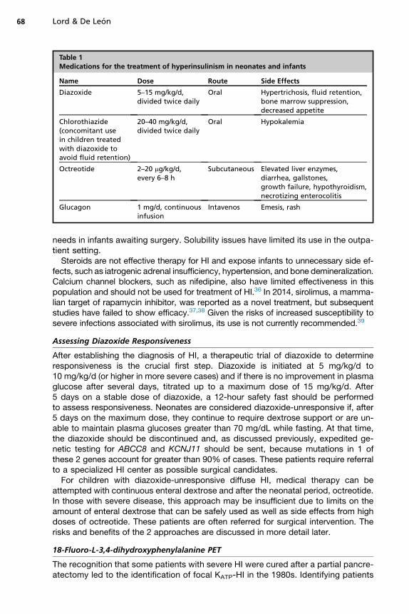
needs in infants awaiting surgery. Solubility issues have limited its use in the outpa-tient setting.Steroids are not effective therapy for HI and expose infants to unnecessary side ef-
fects, such as iatrogenic adrenal insufficiency, hypertension, and bone demineralization.Calcium channel blockers, such as nifedipine, also have limited effectiveness in thispopulation and should not be used for treatment of HI.36 In 2014, sirolimus, a mamma-lian target of rapamycin inhibitor, was reported as a novel treatment, but subsequentstudies have failed to show efficacy.37,38 Given the risks of increased susceptibility tosevere infections associated with sirolimus, its use is not currently recommended.39
Assessing Diazoxide Responsiveness
After establishing the diagnosis of HI, a therapeutic trial of diazoxide to determineresponsiveness is the crucial first step. Diazoxide is initiated at 5 mg/kg/d to10 mg/kg/d (or higher in more severe cases) and if there is no improvement in plasmaglucose after several days, titrated up to a maximum dose of 15 mg/kg/d. After5 days on a stable dose of diazoxide, a 12-hour safety fast should be performedto assess responsiveness. Neonates are considered diazoxide-unresponsive if, after5 days on the maximum dose, they continue to require dextrose support or are un-able to maintain plasma glucoses greater than 70 mg/dL while fasting. At that time,the diazoxide should be discontinued and, as discussed previously, expedited ge-netic testing for ABCC8 and KCNJ11 should be sent, because mutations in 1 ofthese 2 genes account for greater than 90% of cases. These patients require referralto a specialized HI center as possible surgical candidates.For children with diazoxide-unresponsive diffuse HI, medical therapy can be
attempted with continuous enteral dextrose and after the neonatal period, octreotide.In those with severe disease, this approach may be insufficient due to limits on theamount of enteral dextrose that can be safely used as well as side effects from highdoses of octreotide. These patients are often referred for surgical intervention. Therisks and benefits of the 2 approaches are discussed in more detail later.
18-Fluoro-L-3,4-dihydroxyphenylalanine PET
The recognition that some patients with severe HI were cured after a partial pancre-atectomy led to the identification of focal KATP-HI in the 1980s. Identifying patients
Table 1Medications for the treatment of hyperinsulinism in neonates and infants
Name Dose Route Side Effects
Diazoxide 5–15 mg/kg/d,divided twice daily
Oral Hypertrichosis, fluid retention,bone marrow suppression,decreased appetite
Chlorothiazide(concomitant usein children treatedwith diazoxide toavoid fluid retention)
20–40 mg/kg/d,divided twice daily
Oral Hypokalemia
Octreotide 2–20 mg/kg/d,every 6–8 h
Subcutaneous Elevated liver enzymes,diarrhea, gallstones,growth failure, hypothyroidism,necrotizing enterocolitis
Glucagon 1 mg/d, continuousinfusion
Intavenos Emesis, rash
Lord & De Leon68

with focal HI and accurately localizing the lesion in the pancreas became 2 of thebiggest management challenges for the disease. Understanding the genetic mech-anisms of HI led to the recognition that patients with focal HI carry paternallyinherited ABCC8 or KCNJ11 mutations. Localization of the focal lesion, however,remained a challenge. Conventional imaging, such as ultrasound, CT, and MRI,cannot identify focal lesions, and interventional radiology techniques, such as arte-rial stimulation venous sampling, were invasive and had poor accuracy at localizinglesions.40
The introduction of the 18F-DOPA PET scan was one of the most significant ad-vances in the care of children with HI. Introduced in 2003, it is used to differentiatefocal from diffuse disease and to localize focal lesions in the pancreas.41,42 The tracer,18F-DOPA, is taken up by neuroendocrine tissue. In focal disease, there is an area ofincreased tracer uptake in a specific region of the pancreas, corresponding to the areaof b-cell adenomatosis (Fig. 3). In patients with diffuse HI, the uptake of tracer is uni-form throughout the pancreas. In the largest published series to date, 105 infants withHI underwent 18F-DOPA scans, followed by surgery.43 The sensitivity and specificityfor diagnosing focal disease were 85% and 96%, respectively; 100% of lesionswere correctly localized in the pancreas.All infants who have genetics consistent with focal HI require an 18F-DOPA PET scan
prior to surgery. Additionally, patients with diazoxide-unresponsive, genetic-negativeHI should also undergo an 18F-DOPA PET scan, because they still have the possibilityof a focal lesion. Patients who are diazoxide-responsive or those with genetic resultsknown to cause diffuse disease, such as biallelic recessive mutations in ABCC8 orKCNJ11 or a GCK mutation, do not benefit from imaging.
Surgical Intervention
Surgery is indicated for infants who have a focal lesion or those with diffuse diseasewho fail medical therapy. Patients with focal lesions should undergo surgery at
Fig. 3. (A) Frontal view of a MIP 18F-DOPA PET image showing a uniform pattern ofuptake throughout the pancreas, consistent with diffuse disease. (B) Frontal view of aMIP 18F-DOPA PET image demonstrating increased uptake in the tail of the pancreas, consis-tent with a focal lesion (black arrow). Normal liver (L), kidney (K), pancreas (P), and bladder(B) uptake is seen in both images. MIP, maximum intensity projection.
Hyperinsulinism in the Neonate 69

specialized HI centers, which have the multidisciplinary expertise to ensure com-plete incision of the lesion while minimizing the amount of pancreas resected.44
At the time of surgery, intraoperative ultrasound can be used to confirm the locationof the focal lesion. Frozen section evaluation of biopsies by experienced patholo-gists allows for confirmation of the focal lesion and guides the extent of pancreaticresection.Infants with diffuse HI requiring surgical intervention undergo near-total pancreatec-
tomy with gastrostomy tube placement. The gastrostomy tube is necessary for post-operative management because a majority of patients continue to have hypoglycemia,although less severe.17 For children with diffuse disease, the decision to proceed withsurgery is complex and requires careful consideration of the risks and benefits. Sur-gery decreases the severity of the hypoglycemia and makes it easier to manage medi-cally. This must be weighed, however, against the risk of a surgical procedure as wellas the long-term complications of diabetes and pancreatic insufficiency. Medical ther-apy avoids these complications but carries its own risks with more exposure to hypo-glycemia as well as side effects from the high doses of somatostatin analogs that arerequired.
PROGNOSISSurgical Outcomes
A review of 223 surgical cases from the Children’s Hospital of Philadelphia foundthat 94% of infants with focal HI were cured and the majority required less thana 50% pancreatectomy.17 In contrast, only a quarter of patients with diffuse dis-ease were euglycemic after pancreatectomy. More than 40% of patients requiredcontinued treatment of hypoglycemia, and the remainder were treated forhyperglycemia.Individuals who undergo near-total pancreatectomy in infancy have a high risk
of developing diabetes.2,45 More than 90% of these patients develop insulin-dependent diabetes mellitus during the first 2 decades of life; the median age at diag-nosis of diabetes is 8 years.
Neurodevelopmental Outcomes
Despite advances in the field, children with HI continue to have a high risk of neuro-cognitive abnormalities. Studies have shown that 26% to 48% of children and adultswith HI have developmental delays and neurologic issues and 13% to 25% have sei-zures.1,46 Poor developmental outcomes are not only limited to children with persis-tent forms of HI but also involve children with transient forms of HI.3 Furthermore,the rates of developmental delay and seizures for children treated in the 2000s remainsimilar to those individuals treated in the decades before.2 These findings suggest theneed for improved screening protocols to identify infants with HI as early as possible toavoid these neurologic sequela.
SUMMARY
With limited medical therapies and a high risk of neurologic damage, HI remainsa challenging disorder to treat. Advances in management, such as the18F-DOPA PET scan, however, have resulted in improved outcomes for the subsetof infants with focal HI. Ongoing research into the genetic and molecularbasis of HI will hopefully result in novel treatments that benefit those infants withdiffuse HI.
Lord & De Leon70

ACKNOWLEDGMENTS
The authors thank Dr Trisha Bhatti for providing the histology images and Dr LisaStates for providing the 18F-DOPA PET scan images.
REFERENCES
1. Meissner T, Wendel U, Burgard P, et al. Long-term follow-up of 114 patients withcongenital hyperinsulinism. Eur J Endocrinol 2003;149(1):43–51.
2. Lord K, Radcliffe J, Gallagher PR, et al. High risk of diabetes and neurobehavioraldeficits in individuals with surgically treated hyperinsulinism. J Clin EndocrinolMetab 2015;100(11):4133–9.
3. Avatapalle HB, Banerjee I, Shah S, et al. Abnormal neurodevelopmental out-comes are common in children with transient congenital hyperinsulinism. FrontEndocrinol 2013;4:60.
4. James C, Kapoor RR, Ismail D, et al. The genetic basis of congenital hyperinsu-linism. J Med Genet 2009;46(5):289–99.
5. Kalish JM, Boodhansingh KE, Bhatti TR, et al. Congenital hyperinsulinism in chil-dren with paternal 11p uniparental isodisomy and Beckwith-Wiedemann syn-drome. J Med Genet 2016;53(1):53–61.
6. Hoe FM, Thornton PS, Wanner LA, et al. Clinical features and insulin regulation ininfants with a syndrome of prolonged neonatal hyperinsulinism. J Pediatr 2006;148(2):207–12.
7. Stanley CA, Rozance PJ, Thornton PS, et al. Re-evaluating “transitional neonatalhypoglycemia”: mechanism and implications for management. J Pediatr 2015;166(6):1520–5.e1.
8. Harris DL, Weston PJ, Harding JE. Incidence of neonatal hypoglycemia in babiesidentified as at risk. J Pediatr 2012;161(5):787–91.
Best Practices
What is the current best practice?
� Prompt evaluation of infants with suspected HI
� Early identification of infants with focal KATP-HI and referral to specialized HI center for18F-DOPA PET
What changes in current practice are likely to improve outcomes?
� Support plasma glucose with intravenous fluids and allow infants to feed orally on demand
� Use of diuretics in all infants on diazoxide
Major recommendations
� Diagnostic evaluation of neonates who are at high risk of having a persistent hypoglycemicdisorder
� Initiate trial of diazoxide as soon as diagnosis of HI is made
� Infants who are diazoxide-unresponsive require expedited genetic testing for ABCC8 andKCNJ11
� The goal of medical therapy is to allow infants with HI to maintain plasma glucose greaterthan 70 mg/dL while fasting
Summary statement
Early recognition and appropriate treatment of infants with HI are crucial to avoid long-termcomplications, such as developmental delays and diabetes
Hyperinsulinism in the Neonate 71
9. McKinlay CJD, Alsweiler JM, Anstice NS, et al. Association of neonatal glycemiawith neurodevelopmental outcomes at 4.5 years. JAMA Pediatr 2017;171(10):972–83.
10. Thomas PM, Cote GJ, Wohllk N, et al. Mutations in the sulfonylurea receptor genein familial persistent hyperinsulinemic hypoglycemia of infancy. Science 1995;268(5209):426–9.
11. Thomas P, Ye Y, Lightner E. Mutation of the pancreatic islet inward rectifier Kir6.2also leads to familial persistent hyperinsulinemic hypoglycemia of infancy. HumMol Genet 1996;5(11):1809–12.
12. De Leon D, Stanley CA. Pathophysiology of diffuse ATP-sensitive potassiumchannel hyperinsulinism. In: De Leon D, Stanley CA, editors. Monogenic hyperin-sulinemic hypoglycemia disorders, vol. 21, 1st edition. Basel (Switzerland):Karger; 2012. p. 18–29.
13. Pinney SE, MacMullen C, Becker S, et al. Clinical characteristics and biochemicalmechanisms of congenital hyperinsulinism associated with dominant KATP chan-nel mutations. J Clin Invest 2008;118(8):2877–86.
14. Rahier J, Falt K, Muntefering H, et al. The basic structural lesion of persistentneonatal hypoglycaemia with hyperinsulinism: deficiency of pancreatic D cellsor hyperactivity of B cells? Diabetologia 1984;26(4):282–9.
15. De Lonlay P, Fournet JC, Rahier J, et al. Somatic deletion of the imprinted 11p15region in sporadic persistent hyperinsulinemic hypoglycemia of infancy is spe-cific of focal adenomatous hyperplasia and endorses partial pancreatectomy.J Clin Invest 1997;100(4):802–7.
16. Verkarre V, Fournet JC, De Lonlay P, et al. Paternal mutation of the sulfonylureareceptor (SUR1) gene and maternal loss of 11p15 imprinted genes lead to persis-tent hyperinsulinism in focal adenomatous hyperplasia. J Clin Invest 1998;102(7):1286–91.
17. Lord K, Dzata E, Snider KE, et al. Clinical presentation and management of chil-dren with diffuse and focal hyperinsulinism: a review of 223 cases. J Clin Endo-crinol Metab 2013;98(11):E1786–9.
18. Snider KE, Becker S, Boyajian L, et al. Genotype and phenotype correlations in417 children with congenital hyperinsulinism. J Clin Endocrinol Metab 2013;98(2):E355–63.
19. Stanley CA, Lieu YK, Hsu BY, et al. Hyperinsulinism and hyperammonemia in in-fants with regulatory mutations of the glutamate dehydrogenase gene. N Engl JMed 1998;338(19):1352–7.
20. Bahi-Buisson N, Roze E, Dionisi C, et al. Neurological aspects of hyperinsulinism-hyperammonaemia syndrome. Dev Med Child Neurol 2008;50(12):945–9.
21. Glaser B, Kesavan P, Heyman M, et al. Familial hyperinsulinism caused by anactivating glucokinase mutation. N Engl J Med 1998;338(4):226–30.
22. Sayed S, Langdon DR, Odili S, et al. Extremes of clinical and enzymatic pheno-types in children with hyperinsulinism caused by glucokinase activating muta-tions. Diabetes 2009;58(6):1419–27.
23. Pearson ER, Boj SF, Steele AM, et al. Macrosomia and hyperinsulinaemic hypo-glycaemia in patients with heterozygous mutations in the HNF4A gene. PLoSMed 2007;4(4):e118.
24. Kapoor RR, Locke J, Colclough K, et al. Persistent hyperinsulinemic hypoglyce-mia and maturity-onset diabetes of the young due to heterozygous HNF4A muta-tions. Diabetes 2008;57(6):1659–63.
Lord & De Leon72
25. Stanescu DE, Hughes N, Kaplan B, et al. Novel presentations of congenital hy-perinsulinism due to mutations in the MODY genes: HNF1A and HNF4A. J ClinEndocrinol Metab 2012;97(10):E2026–30.
26. Thornton PS, Stanley CA, De Leon DD, et al. Recommendations from the pediat-ric endocrine society for evaluation and management of persistent hypoglycemiain neonates, infants, and children. J Pediatr 2015;167(2):238–45.
27. Finegold DN, Stanley CA, Baker L. Glycemic response to glucagon during fastinghypoglycemia: an aid in the diagnosis of hyperinsulinism. J Pediatr 1980;96(2):257–9.
28. Kelly A, Tang R, Becker S, et al. Poor specificity of low growth hormone andcortisol levels during fasting hypoglycemia for the diagnoses of growth hormonedeficiency and adrenal insufficiency. Pediatrics 2008;122(3):e522–8.
29. Drash A, Wolff F. Drug therapy in leucine-sensitive hypoglycemia. Metab Clin Exp1964;13:487–92.
30. Dayton PG, Pruitt AW, Faraj BA, et al. Metabolism and disposition of diazoxide. Amini-review. Drug Metab Dispos 1975;3(3):226–9.
31. Hirsch HJ, Loo S, Evans N, et al. Hypoglycemia of infancy and nesidioblastosis.Studies with somatostatin. N Engl J Med 1977;296(23):1323–6.
32. Laje P, Halaby L, Adzick NS, et al. Necrotizing enterocolitis in neonates receivingoctreotide for the management of congenital hyperinsulinism. Pediatr Diabetes2010;11(2):142–7.
33. Hawkes CP, Adzick NS, Palladino AA, et al. Late presentation of fulminant necro-tizing enterocolitis in a child with hyperinsulinism on octreotide therapy. Horm ResPaediatr 2016;86(2):131–6.
34. Modan-Moses D, Koren I, Mazor-Aronovitch K, et al. Treatment of congenital hy-perinsulinism with lanreotide acetate (Somatuline Autogel). J Clin EndocrinolMetab 2011;96(8):2312–7.
35. Kuhnen P, Marquard J, Ernert A, et al. Long-term lanreotide treatment in six pa-tients with congenital hyperinsulinism. Horm Res Paediatr 2012;78(2):106–12.
36. Guemes M, Shah P, Silvera S, et al. Assessment of nifedipine therapy in hyperin-sulinemic hypoglycemia due to mutations in the ABCC8 gene. J Clin EndocrinolMetab 2017;102(3):822–30.
37. Senniappan S, Alexandrescu S, Tatevian N, et al. Sirolimus therapy in infants withsevere hyperinsulinemic hypoglycemia. N Engl J Med 2014;370(12):1131–7.
38. Szymanowski M, Estebanez MS, Padidela R, et al. mTOR inhibitors for the treat-ment of severe congenital hyperinsulinism: perspectives on limited therapeuticsuccess. J Clin Endocrinol Metab 2016;101(12):4719–29.
39. Banerjee I, De Leon D, Dunne MJ. Extreme caution on the use of sirolimus for thecongenital hyperinsulinism in infancy patient. Orphanet J Rare Dis 2017;12(1):70.
40. Stanley CA, Thornton PS, Ganguly A, et al. Preoperative evaluation of infants withfocal or diffuse congenital hyperinsulinism by intravenous acute insulin responsetests and selective pancreatic arterial calcium stimulation. J Clin EndocrinolMetab 2004;89(1):288–96.
41. Hardy OT, Hernandez-Pampaloni M, Saffer JR, et al. Accuracy of [18F]Fluoro-dopa positron emission tomography for diagnosing and localizing focal congen-ital hyperinsulinism. J Clin Endocrinol Metab 2007;92(12):4706–11.
42. Otonkoski T, Nanto-Salonen K, Seppanen M, et al. Noninvasive diagnosis of focalhyperinsulinism of infancy with [18F]-DOPA positron emission tomography. Dia-betes 2006;55(1):13–8.
43. Laje P, States LJ, Zhuang H, et al. Accuracy of PET/CT Scan in the diagnosis ofthe focal form of congenital hyperinsulinism. J Pediatr Surg 2013;48(2):388–93.
Hyperinsulinism in the Neonate 73
44. Adzick NS, Thornton PS, Stanley CA, et al. A multidisciplinary approach to thefocal form of congenital hyperinsulinism leads to successful treatment by partialpancreatectomy. J Pediatr Surg 2004;39(3):270–5.
45. Beltrand J, Caquard M, Arnoux JB, et al. Glucose metabolism in 105 children andadolescents after pancreatectomy for congenital hyperinsulinism. Diabetes Care2012;35(2):198–203.
46. Menni F, de Lonlay P, Sevin C, et al. Neurologic outcomes of 90 neonates and in-fants with persistent hyperinsulinemic hypoglycemia. Pediatrics 2001;107(3):476–9.
Lord & De Leon74