hemodilution and fluid management in neurosurgery · hemodilution and fluid management in...
TRANSCRIPT

CHAPTER 26
Hemodilution and Fluid Management in Neurosurgery
Ramachandra P. Tummala, M.D., Rishi N. Sheth, M.D., and Roberto C. Heros, M.D., F.A.C.S.
Early in his career, the senior author (RCH) became inter-ested in the experimental study of cerebral vasospasm,
which continues to be one of the most important causes ofmorbidity from subarachnoid hemorrhage (SAH).21–23,45 Theinitial approach was to find a single pharmacological agent,the so-called “silver bullet,” to prevent and reverse vaso-spasm. Arterial smooth muscle relaxants were the first classof tempting agents that were studied. These agents turned outto be “fool’s gold” and were generally ineffective in treatingvasospasm.19,20,55,58 While treatments were being developedin experimental models, clinical observations were reportedthat hypertension may improve neurological deficits resultingfrom cerebrovascular insufficiency.9 This led to the demon-stration that vasospasm-induced cerebral ischemia could betreated successfully with iatrogenic hypertension.13,34 Thebenefits of intravascular volume expansion combined withhypertension were reported soon afterwards.27 These founda-tions, along with the additional strides made in the under-standing of rheology, cerebral oxygen transport, and cerebralblood flow (CBF) augmentation, led to the formalization ofthe concept of hyperdynamic therapy. Gaining broad accep-tance by the late 1980s, the combination of hypervolemia,hypertension, and hemodilution, colloquially known as “tri-ple-H therapy,” is now considered essential in the treatmentfor cerebral vasospasm.
We have observed that the hypervolemic and hyperten-sive arms of the hyperdynamic therapy receive the mostattention from clinicians. It seems that the hemodilutionaspect of the treatment is often overlooked or taken forgranted because some degree of hemodilution occurs with anincrease in intravascular volume. However, the benefits ofhemodilution are well grounded in the laboratory, beginningwith the pioneering work of Wood et al.59–61 The seniorauthor concentrated on the effects of hemodilution on cere-bral ischemia for longer than 15 years in his laboratory. Inthis report, we shall discuss the evolution of this work and itstranslation into clinical practice. We shall also review thecurrent status of hemodilution in clinical practice and de-scribe the implications of this work for fluid therapy ingeneral for neurosurgical patients.
RATIONALE FOR HEMODILUTION INCEREBRAL ISCHEMIA
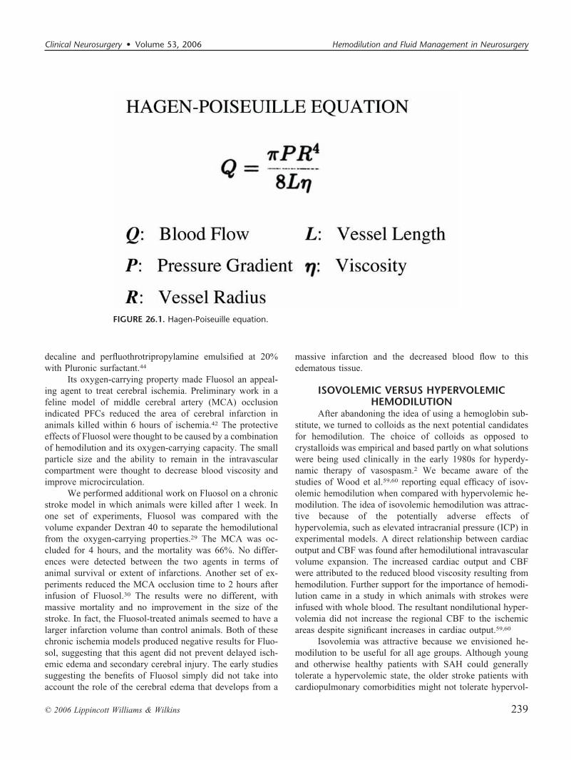
The Hagen-Poiseuille equation indicates that flow isinversely proportional to viscosity (Fig. 26.1). Blood viscos-ity is a complex variable determined by several factors,including erythrocyte aggregation and flexibility, platelet ag-gregation, plasma viscosity, and hematocrit. Of these factors,hematocrit is by far the most important determinant of bloodviscosity.15,52 In ischemic brain, the regional blood vesselsare dilated maximally, and blood viscosity becomes a majordeterminant of blood flow. The low blood flow inherent to theischemic region results in a dramatic rise in viscosity, favor-ing microaggregation and thrombus formation. Thus, thehematocrit becomes an even more important factor in theselow-flow states.15
It follows that hemodilution is an effective way toincrease perfusion to the ischemic brain, and a large amountof evidence supports this hypothesis. Although hemodilutionincreases perfusion in the ischemic brain, it also reduces theoxygen-carrying capacity of blood. In ischemic brain, theautoregulation of blood flow is lost, and rheological factors,such as viscosity, hence, hematocrit, become a most impor-tant determinant of regional blood flow.
HEMODILUTION WITH A HEMOGLOBINSUBSTITUTE
Our initial work focused on the hypothesis that theoptimal hemodilutional agent would achieve the rheologicaladvantages of reduced viscosity and at the same time enhanceor at least maintain the oxygen-carrying capacity of blood.Perfluorocarbons (PFCs) are inert organic compounds de-rived from the substitution of fluorine for hydrogen. Thesecompounds have a high affinity for oxygen and carbondioxide, and interest grew in their potential for the gastransport function of red blood cells.12 PFCs are frequentlyreferred to as substitutes for blood, a technically incorrectconcept. They are intended to supplement the oxygen-carry-ing capacity of red blood cells, hence, are considered morecorrectly as oxygen carriers or red blood cell substitutes.These compounds must be prepared in a microemulsion toprevent liquid embolism during intravenous administration.The first commercial PFC tested and approved for red cellsupplementation was Fluosol-DA, a mixture of perfluoro-
Copyright © 2006 by Lippincott Williams & Wilkins0148-703/06/5301-0238
Clinical Neurosurgery • Volume 53, 2006238
decaline and perfluothrotripropylamine emulsified at 20%with Pluronic surfactant.44
Its oxygen-carrying property made Fluosol an appeal-ing agent to treat cerebral ischemia. Preliminary work in afeline model of middle cerebral artery (MCA) occlusionindicated PFCs reduced the area of cerebral infarction inanimals killed within 6 hours of ischemia.42 The protectiveeffects of Fluosol were thought to be caused by a combinationof hemodilution and its oxygen-carrying capacity. The smallparticle size and the ability to remain in the intravascularcompartment were thought to decrease blood viscosity andimprove microcirculation.
We performed additional work on Fluosol on a chronicstroke model in which animals were killed after 1 week. Inone set of experiments, Fluosol was compared with thevolume expander Dextran 40 to separate the hemodilutionalfrom the oxygen-carrying properties.29 The MCA was oc-cluded for 4 hours, and the mortality was 66%. No differ-ences were detected between the two agents in terms ofanimal survival or extent of infarctions. Another set of ex-periments reduced the MCA occlusion time to 2 hours afterinfusion of Fluosol.30 The results were no different, withmassive mortality and no improvement in the size of thestroke. In fact, the Fluosol-treated animals seemed to have alarger infarction volume than control animals. Both of thesechronic ischemia models produced negative results for Fluo-sol, suggesting that this agent did not prevent delayed isch-emic edema and secondary cerebral injury. The early studiessuggesting the benefits of Fluosol simply did not take intoaccount the role of the cerebral edema that develops from a
massive infarction and the decreased blood flow to thisedematous tissue.
ISOVOLEMIC VERSUS HYPERVOLEMICHEMODILUTION
After abandoning the idea of using a hemoglobin sub-stitute, we turned to colloids as the next potential candidatesfor hemodilution. The choice of colloids as opposed tocrystalloids was empirical and based partly on what solutionswere being used clinically in the early 1980s for hyperdy-namic therapy of vasospasm.2 We became aware of thestudies of Wood et al.59,60 reporting equal efficacy of isov-olemic hemodilution when compared with hypervolemic he-modilution. The idea of isovolemic hemodilution was attrac-tive because of the potentially adverse effects ofhypervolemia, such as elevated intracranial pressure (ICP) inexperimental models. A direct relationship between cardiacoutput and CBF was found after hemodilutional intravascularvolume expansion. The increased cardiac output and CBFwere attributed to the reduced blood viscosity resulting fromhemodilution. Further support for the importance of hemodi-lution came in a study in which animals with strokes wereinfused with whole blood. The resultant nondilutional hyper-volemia did not increase the regional CBF to the ischemicareas despite significant increases in cardiac output.59,60
Isovolemia was attractive because we envisioned he-modilution to be useful for all age groups. Although youngand otherwise healthy patients with SAH could generallytolerate a hypervolemic state, the older stroke patients withcardiopulmonary comorbidities might not tolerate hypervol-
FIGURE 26.1. Hagen-Poiseuille equation.
Clinical Neurosurgery • Volume 53, 2006 Hemodilution and Fluid Management in Neurosurgery
© 2006 Lippincott Williams & Wilkins 239

emia. Furthermore, the maintenance of a hypervolemic stateis more labor intensive and requires frequent intravascularinfusions.
CHOICE OF AN ISCHEMIC MODELAlthough our primary clinical interest was in cerebral
aneurysms and, therefore, subarachnoid hemorrhage and va-sospasm, we thought that experimental models of vasospasmwere too unpredictable and unreliable to properly study anisolated form of therapy. This is particularly true because,although angiographic vasospasm could be predictablyachieved with several of the models we had at the time,hemodilution was not directed at angiographic vasospasm perse, but, rather, at the ischemic consequences, which couldonly be studied by looking at the effect of the therapy in theseverity of brain infarction and in neurological outcome.Existing models of vasospasm rarely resulted in a predictablefocal brain infarction and, unless the animals died, it was onlyrare and then only with primate models that a focal deficitcould be achieved by inducing experimental subarachnoidhemorrhage.8,10,26 Therefore, we settled on a simple model ofischemia by MCA occlusion.24,36,53,54 It became quickly ap-parent that we had to use a large animal to be able to performall of the intended physiological measurements, to measureCBF, and to keep the animal alive for eventual histologicalstudy of infarction and measurement of clinical outcome.Within these parameters, a canine model seemed ideal forthese experiments. This model was used widely and providedadequate opportunity to study histologically the effects ofhemodilution on ischemic brain.5,6,40 As we began our pilotstudies, we encountered tremendous variability in blood vol-ume measurements from animal to animal and even withinone animal at different times. Unfortunately, we had to killmore dogs than we want to remember before we found anobscure reference in the literature to the effect that the caninespleen had a tremendous capacity to accumulate large vol-umes of blood that were released into the circulation at timesof stress.3 This made all of our previous volume measure-ments in normal dogs worthless; we overcame this obstacleby performing splenectomies in the dogs 1 week before theexperiments. Once this modification was made, we obtainedrelatively reliable volume measurements.
The effects of isovolemic hemodilution on regionalCBF and the size of infarction were studied after inducingfocal ischemia. Seventy-six mongrel dogs were used for thisstudy. Seven animals were excluded from the study becauseof technical errors or trauma to the brain during craniotomy.The remaining 69 animals were randomized into two groupsof hemodilution (treatment groups) and a control group. Inthe hemodilution group, 28 animals were subjected to 6 hoursof arterial occlusion (proximal MCA and distal internal ca-rotid artery) and 7 animals had a sham operation (arterialmanipulation without occlusion). In the control group, 26
animals received 6 hours of arterial occlusion and 8 animalswere sham-operated. Thirty minutes into occlusion or shamsurgery, isovolemic hemodilution was performed by repeatedwithdrawal of 50 ml of blood and infusion of 35 ml of 10%low molecular weight dextran until the hematocrit reached 30to 32%. Dextran was used for volume replacement because itis readily available, inexpensive, safe, has a similar molecularweight to physiological albumin, it can effectively expand theblood volume 1.5 times the amount infused, and it canmaintain an isovolemic state in a splenectomized dogs for atleast 1 week. In deciding the value for hematocrit, 30% wasthought to be ideal based on physiological experiments thatindicated that, indeed, this was the optimal range to deliveroxygen to tissues.50 Obviously, with increasing hematocrit,there is higher oxygen content, but the increased viscositymay compromise flow at the level of capillaries. On the otherhand, lowering the hematocrit may decrease the oxygen-carrying capacity of blood, which may override the advantageof increased flow to the tissue; a hematocrit of approximately30% achieves an ideal balance between these two conflictingeffects.35,39
The hemodilution groups were further subdivided intoacute and chronic groups. The acute group underwent bloodflow measurements using the radiolabeled microsphere tech-nique. Microspheres are approximately 15 �m in diameterand carry a radioactive label to the brain. They are injecteddirectly into the left atrium and they do not pass beyond thecapillaries of the brain. The amount of radioactive micro-spheres that gets “fixed” in the brain reflects the amount ofblood flow at the time of injection. Four different radionu-clides were used for measuring the blood flow at four differ-ent times. The CBF was measured at baseline (microsphereslabeled with tin-113), 30 minutes after occlusion of intracra-nial arteries (using ruthenium-103), 2 hours after hemodilu-tion (niobium-95), and lastly 30 minutes after reperfusion(scandium-46). After 8 hours, animals were killed, the radio-activity was measured using a gamma counter in the braintissue samples, and the local blood flow was calculated usinga standard method. We used tetrazolium salts (TTC), ahistochemical indicator of mitochondrial respiratory enzymeviability, to estimate the size of infarction in the acute group.In our earlier studies, we found TTC to be a reliable markerof cerebral infarction under the conditions studied currently.37
TTC is taken up by functioning mitochondria, and it isthought that the tissue that does not take up this dye willbecome infarcted (Fig. 26.2). Although this method providedan indirect estimation of infarct size, we had dedicated thesecond half of our experiment to use the “gold standard,”histological preparations, to confirm and compare the infarctsize between the groups. After four blood flow measurementswith microspheres, animals go into pulmonary hypertensionand, as a result, cardiac failure. These animals do not survivelong enough for final histological studies, for which we
Tummala et al. Clinical Neurosurgery • Volume 53, 2006
© 2006 Lippincott Williams & Wilkins240

wanted to keep the dogs alive for at least 1 week. In addition,these animals were considered to be radioactively contami-nated and could not be housed thereafter. For these reasons,we had to divide the experiment into an acute group withmeasurements of CBF and a chronic group for histologicalstudies.
At the end of 1 week, the chronic group was injectedwith fluorescein and killed. The areas stained with fluoresceinthat corresponded to the areas of infarct, which were con-firmed by histopathological examination, were measured andquantified (Fig. 26.3).
In the first part, we studied the effects of isovolemichemodilution on hemodynamics, hemorheology, and ICP.53
The hemorheological parameters studied were viscosity, he-matocrit, and plasma fibrinogen, and all three had a statisti-cally significantly reduction after hemodilution. The meanviscosity and the hematocrit fell to 61% and 69% of itsoriginal baseline value, respectively, at 2 hours after hemodi-lution. An average hematocrit value of 32.9% was stillmaintained at the end of 1 week in the chronic group. The
mean plasma fibrinogen level dropped from 0.26 � 0.03gm% to 0.18 � 0.02 gm% with hemodilution. There was alsoexcellent correlation between hematocrit and viscosity (r �0.81) (Fig. 26.4).
Even though the mean arterial pressure dropped slightlyfrom baseline after hemodilution, there was no significantchange in central venous pressure, wedge pressure, or pul-monary pressure. Cardiac index decreased slightly in bothhemodiluted and control groups, which may be attributed toanesthesia. ICP increased significantly after arterial occlusionin both hemodiluted and control groups but there was a trendtoward lower ICP in the hemodiluted group.
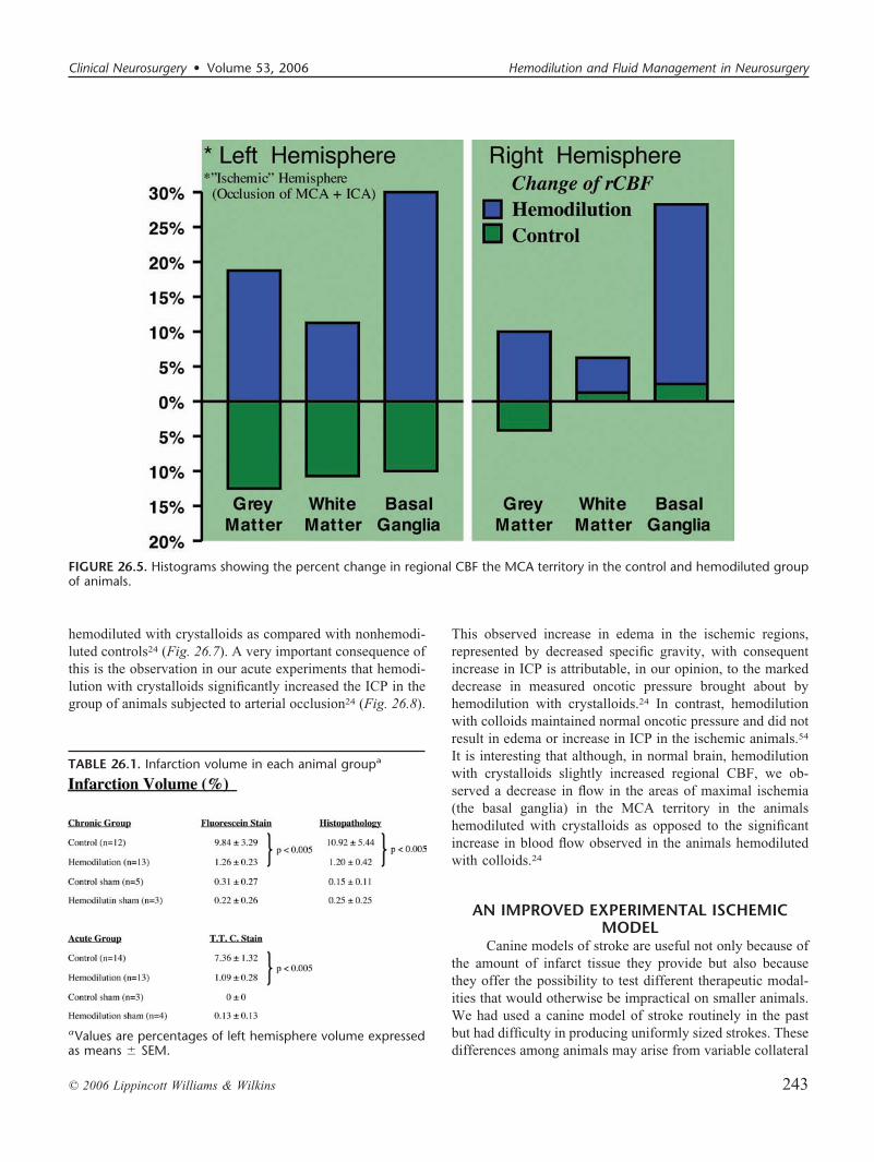
The CBF was measured at baseline, 30 minutes afterarterial occlusion, 2 hours after hemodilution, and 30 minutesafter reperfusion. In the hemodiluted and the control groups,the CBF decreased, especially in the MCA territory. Afterhemodilution, the decrease in the CBF was substantiallyimproved, in contrast to the control group; however, the CBFworsened as time progressed (Fig. 26.5). Reperfusion tendedto restore the CBF toward baseline in the hemodiluted ani-mals, whereas this was only partially restored in the controlanimals. The mean infarct volume (lack of staining withTTC) was significantly less in the hemodiluted groups com-pared with control groups (P �0.005) when examined at 8hours after arterial occlusion (Table 26.1).
Animals in the chronic group treated with hemodilutionfared better neurologically than those in the control groups.These animals had a significant improvement in their neuro-logical exam and returned to normal after 3 days, whereas thecontrol animals never returned to their baseline. Both his-topathological examination and fluorescein stain showed asignificantly higher volume of infarcted tissue in the controlgroup than the hemodiluted group (P �0.005) (Table 26.1).Finally, there was a significant correlation between the infarctvolume (P �0.001) and hematocrit, and between infarctvolume and viscosity (P �0.001).
COLLOIDS VERSUS CRYSTALLOIDS FORHEMODILUTION
When resuscitation of all critically ill patients is con-sidered, it seems that the choice of fluid used for resuscitationhas no effect on mortality.43 Crystalloids extravasate into theinterstitial space, resulting in edema formation. For example,1 L of infused lactated Ringer’s solution expands the plasmavolume by only 250 ml. Therefore, it would seem that theinfusion of large amounts of crystalloid would result in anedematous patient with a depleted intravascular space. Otherconcerns regarding crystalloids include impaired gas ex-change from pulmonary edema and metabolic acidosis fromexcessive normal saline administration. Nevertheless, itseems that, in patients with systemic trauma, shock, andburns, crystalloids are as effective as colloids.4,43,56 It shouldbe kept in mind, however, that, generally, these patients have
FIGURE 26.2. Cross-section of brain showing infarct area (lackof TTC staining) in the distribution of left MCA
FIGURE 26.3. Canine brain stained with fluorescein. Areas that“light up” after staining correspond to infarct as demonstratedhistologically.
Clinical Neurosurgery • Volume 53, 2006 Hemodilution and Fluid Management in Neurosurgery
© 2006 Lippincott Williams & Wilkins 241

normal brains; the situation may be different, as we haveshown, in patients with injured or ischemic brain.
In contrast, to crystalloids, colloids are much moreefficient plasma volume expanders. Typically, smaller vol-umes of colloid are required to produce similar resuscitationend points as crystalloids. There are four types of colloid—albumin, dextrans, gelatins, and starches. The model colloidis albumin, which is responsible for the vast majority ofplasma oncotic pressure. Its main advantages are that it doesnot cause coagulopathies and has a half-life of 16 hours,resulting in sustained intravascular volume repletion. Its dis-advantages include its short supply, its significant expense,and the rare possibility of anaphylaxis. One myth surroundingalbumin is the risk of viral transmission. There have been noreports of viral transmission through albumin infusion sinceits inception longer than 60 years ago. Albumin is essentiallya byproduct of plasma electrophoresis. Between the improvedscreening process for donors and its pasteurization, the risk ofdisease transmission with albumin infusion seems to betheoretical. Hypersensitivity reactions also are rare with anincidence between 0.011% and 1.5%.11,38
Dextrans and gelatins are rarely used for resuscitationbecause of their limited half-life and their anticoagulanteffects. Starches, such as hetastarch, increase plasma volumefor a prolonged period with small effects on coagulation.
They are inexpensive and have a long shelf life. Afterconsidering all of the above, we decided to use dextran as thecolloid agent in our hemodilution studies.
We compared the hemodilutional properties of crystal-loids with colloids in a canine model of cerebral ischemia.Isovolemic hemodilution with a hematocrit of 30% to 32%was reached by either the infusion of lactated Ringer’ssolution or dextran in two respective groups of animals thatunderwent temporary occlusion of the internal carotid andmiddle cerebral arteries. Although systemic parameters, suchas mean arterial, central venous, pulmonary arterial, andpulmonary wedge pressures remained the same in bothgroups, the neurological status of the crystalloid group wasconsistently worse than the colloid group. Furthermore, themedian infarct volume of the crystalloid group was almostseven times greater than that of the colloid group54 (Fig.26.6). These results demonstrated that hemodilution withcrystalloids was detrimental in the setting of temporary focalcerebral ischemia, whereas hemodilution with colloids wasbeneficial. The likely explanation for these results was thedecrease in oncotic pressure from crystalloid administration,leading to the formation of brain edema in the area ofischemic injury. In fact, this was confirmed by specificgravity measurements that demonstrated a marked increase inwater content in the region of ischemia in the animals
FIGURE 26.4. Graph showing positive correlation(r � 0.81) between viscosity and hematocrit ofblood.
Tummala et al. Clinical Neurosurgery • Volume 53, 2006
© 2006 Lippincott Williams & Wilkins242
hemodiluted with crystalloids as compared with nonhemodi-luted controls24 (Fig. 26.7). A very important consequence ofthis is the observation in our acute experiments that hemodi-lution with crystalloids significantly increased the ICP in thegroup of animals subjected to arterial occlusion24 (Fig. 26.8).
This observed increase in edema in the ischemic regions,represented by decreased specific gravity, with consequentincrease in ICP is attributable, in our opinion, to the markeddecrease in measured oncotic pressure brought about byhemodilution with crystalloids.24 In contrast, hemodilutionwith colloids maintained normal oncotic pressure and did notresult in edema or increase in ICP in the ischemic animals.54
It is interesting that although, in normal brain, hemodilutionwith crystalloids slightly increased regional CBF, we ob-served a decrease in flow in the areas of maximal ischemia(the basal ganglia) in the MCA territory in the animalshemodiluted with crystalloids as opposed to the significantincrease in blood flow observed in the animals hemodilutedwith colloids.24
AN IMPROVED EXPERIMENTAL ISCHEMICMODEL
Canine models of stroke are useful not only because ofthe amount of infarct tissue they provide but also becausethey offer the possibility to test different therapeutic modal-ities that would otherwise be impractical on smaller animals.We had used a canine model of stroke routinely in the pastbut had difficulty in producing uniformly sized strokes. Thesedifferences among animals may arise from variable collateral
TABLE 26.1. Infarction volume in each animal groupa
aValues are percentages of left hemisphere volume expressedas means � SEM.
FIGURE 26.5. Histograms showing the percent change in regional CBF the MCA territory in the control and hemodiluted groupof animals.
Clinical Neurosurgery • Volume 53, 2006 Hemodilution and Fluid Management in Neurosurgery
© 2006 Lippincott Williams & Wilkins 243
circulation, thus, leading to difficulty with statistical compar-isons between groups. We had to use a large number ofanimals in our studies to overcome this variability in the sizeof infarct. Our aim was to develop a reliable canine modelthat would produce a moderate size infarct because smallinfarcts in dogs have no consequence and large infarcts areincompatible with life. We postulated and later showed thatsomatosensory evoked potential (SSEP) amplitude just afterocclusion of arteries could be used to predict which animalswould have a moderate sized infarct. We found that animalsthat showed a significant deterioration of SSEPs after tem-porary occlusion of the MCA would predictably develophuge infarcts; therefore, when they occurred, the clip wasremoved and the animals were excluded and used for otherexperiments. Animals that retained good SSEPs after MCAocclusion went on to have occlusion of the azygous anteriorcerebral artery (ACA); if the SSEPs remained intact, wefound that these animals would develop either no infarct or avery small one; therefore, these animals were also rejected.
This left us with the animals that had minimal or no changeof SSEPs on MCA occlusion and then developed a significantchange with additional occlusion of the azygous ACA (Fig.26.9). These were the animals used for further studies ofischemia because they predictably developed a moderatelysized infarct compatible with survival.41
OPTIMAL DEGREE OF HEMODILUTIONNow that it was proven that hemodilution improves
CBF in ischemic brain, the question arises of what is theoptimal degree of hemodilution. As stated before, the mostimportant determinant of blood viscosity is the hematocrit. Infact, there is an inverse relation between the hematocrit andCBF.16 With hemodilution, the CBF increases, but loweringthe hematocrit further could compromise the oxygen-carryingcapacity of the blood and, thus, be detrimental. The balancebetween these two factors becomes critical, especially whenautoregulation is lost in infarcted tissue. Here, the vessels aremaximally dilated and the rheological factors of blood mayplay a major role in determining the oxygen delivery to thebrain.
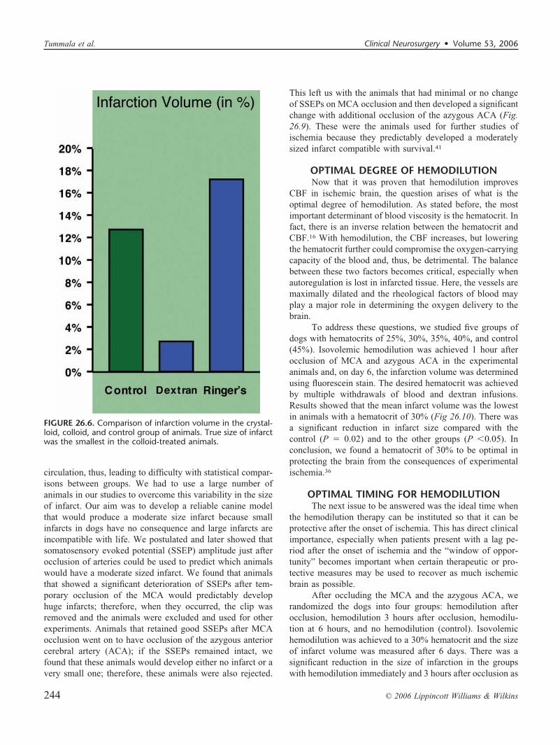
To address these questions, we studied five groups ofdogs with hematocrits of 25%, 30%, 35%, 40%, and control(45%). Isovolemic hemodilution was achieved 1 hour afterocclusion of MCA and azygous ACA in the experimentalanimals and, on day 6, the infarction volume was determinedusing fluorescein stain. The desired hematocrit was achievedby multiple withdrawals of blood and dextran infusions.Results showed that the mean infarct volume was the lowestin animals with a hematocrit of 30% (Fig 26.10). There wasa significant reduction in infarct size compared with thecontrol (P � 0.02) and to the other groups (P �0.05). Inconclusion, we found a hematocrit of 30% to be optimal inprotecting the brain from the consequences of experimentalischemia.36
OPTIMAL TIMING FOR HEMODILUTIONThe next issue to be answered was the ideal time when
the hemodilution therapy can be instituted so that it can beprotective after the onset of ischemia. This has direct clinicalimportance, especially when patients present with a lag pe-riod after the onset of ischemia and the “window of oppor-tunity” becomes important when certain therapeutic or pro-tective measures may be used to recover as much ischemicbrain as possible.
After occluding the MCA and the azygous ACA, werandomized the dogs into four groups: hemodilution afterocclusion, hemodilution 3 hours after occlusion, hemodilu-tion at 6 hours, and no hemodilution (control). Isovolemichemodilution was achieved to a 30% hematocrit and the sizeof infarct volume was measured after 6 days. There was asignificant reduction in the size of infarction in the groupswith hemodilution immediately and 3 hours after occlusion as
FIGURE 26.6. Comparison of infarction volume in the crystal-loid, colloid, and control group of animals. True size of infarctwas the smallest in the colloid-treated animals.
Tummala et al. Clinical Neurosurgery • Volume 53, 2006
© 2006 Lippincott Williams & Wilkins244

compared with control (P �0.0001) (Fig. 26.11). The neu-rological function was significantly better in the animalstreated immediately and 3 hours after the occlusion comparedwith the other two groups. Animals treated after 6 hours hada trend toward a larger infarct size and had a significantlyhigher incidence of hemorrhagic infarction.
This study suggests that hemodilution can be protectivewhen instituted as late as 3 hours after arterial occlusion;however, after 6 hours, hemodilution is not helpful and maybe unsafe.62 Not surprisingly, this “window of opportunity”for hemodilution is similar to that generally found in exper-imental and clinical studies of reperfusion.
MECHANISM OF THE EFFECT OFHEMODILUTION
Preliminary work in hemodilution clearly showed an in-crease in CBF.18 This naturally raised the question of howhemodilution caused an augmentation of CBF. It was unclearwhether the increased blood flow was a direct rheological effectrelated to decreased blood viscosity or whether it was an indirect
vasodilatory effect of reduced oxygen-carrying capacity. Itseemed that the former hypothesis was more logical because ofthe maximal vasodilatation already present in the ischemic brainfrom inherently increased oxygen demands in this territory.32,33
Therefore, an additional decrease in oxygen transport fromhemodilution should not cause further vasodilatation.
The hypothesis was tested in a rabbit model of cerebralischemia. Hypoxia was first induced in normal animals byeither hemodilution (anemic hypoxia) or reduction of theinspired oxygen concentration (hypoxic hypoxia). The CBFresponse was exactly the same between the anemic hypoxiagroup and the hypoxic hypoxia group. Both types of hypoxia,in these animals with normal brain, caused a linear increase inCBF as the arterial oxygen content decreased.
The experiment was then repeated in animals withcerebral ischemia. The results clearly showed that there wasa significant increase in CBF with graded hemodilution (ane-mic hypoxia). In contrast, graded hypoxic hypoxia did notincrease CBF in the ischemic brain, although it did so innormal brain. We concluded that the improvement in CBF in
FIGURE 26.7. Histogram comparing the specific gravity of the brain tissue in the MCA territory at different sites in hemodilutedand control (nonhemodiluted) group of animals.
Clinical Neurosurgery • Volume 53, 2006 Hemodilution and Fluid Management in Neurosurgery
© 2006 Lippincott Williams & Wilkins 245

the ischemic brain was mainly caused by a rheologicalresponse to decreased blood viscosity as opposed to a vaso-dilatory response to decrease arterial oxygen content.31 Thisconfirmed that in hypoxic brain, despite maximal reactivevasodilation, improvement in CBF can still be achieved byrheological manipulations, such as a decrease in viscosity.These findings also implied that in ischemic brain, viscosityin likely to be a major determinant of regional blood flow.
CLINICAL EXPERIENCE WITH HEMODILUTIONSupported by the large body of experimental evidence
that hemodilution improves blood flow to ischemic brain,several trials were designed in the 1980s to establish clinicalbenefit. The first randomized, controlled trial was based at asingle center in Sweden and measured the benefit of isov-olemic hemodilution during the first 48 hours after an isch-emic stroke. The treatment arm of the study involved vene-section and infusion of dextran to reduce the mean hematocritfrom 43 to 37%. In this study, the treated patients hadimproved early neurological recovery and less functionalimpairment compared with the control group. However, he-modilution did not affect mortality.48
The positive results from this study led to a larger, mul-ticenter trial in which the Scandanavian Stroke Study Groupfollowed the initial protocol described above.46,47 However, thisstudy failed to demonstrate any clinical benefit in patients treatedwith hemodilution. Similar negative results were obtained froma multicenter Italian trial, which followed a similar protocol ofphlebotomy and infusion of dextran 40.25
A trial from North America investigated the benefits ofhypervolemic hemodilution based on the experimental evidencethat increased cardiac output enhances blood flow to ischemicbrain. This study, which used pentastarch as a volume expander,found a modest benefit in patients treated within 12 hours ofstroke onset, patients with 15% reduction in hematocrit, andthose with a 10% increase in cardiac output. However, 4 deathsoccurred in the treatment group of 45 patients. These deathswere attributed to severe cerebral edema from massive strokes.51
The issue of hemodilution resurfaced in a double-blindedstudy from Austria in the late 1990s. In this study, patients wererandomized within 6 hours of stroke onset and treated patientsreceived hydroxyethyl starch to achieve mild hypervolemichemodilution. Although treatment was initiated within 6 hours,a maximum reduction of hematocrit of 10% was not achieved
FIGURE 26.8. ICP after hemodilutionwith crystalloids in ischemic animals.
Tummala et al. Clinical Neurosurgery • Volume 53, 2006
© 2006 Lippincott Williams & Wilkins246

until the fifth day of treatment. This study failed to show anysignificant benefit for the treated patients and added to theclinical weight against hemodilution as a treatment for ischemicstroke.1
The results of these clinical trials raise the question of whyhemodilution, a concept well grounded in theory and in thelaboratory, has failed to show clinical efficacy. Several factorsmay be responsible for the discordant clinical findings. Hemodi-lution therapy was initiated relatively late in these studies. TheScandanavian and Italian trials used entry times of 48 and 12hours, respectively. The proportion of patients in who underwenthemodilution within 6 hours of neurological symptoms was only6% in the Scandanavian trial and 55% in the Italian study. TheAustrian study attempted to balance experimental and practicalconsiderations by randomizing all patients within 6 hours. How-ever, based on the experimental evidence, even this time win-dow may be too big for hemodilution to be effective. Further-more, the gradual hemodilution regimen over several days in the
Austrian trial may have been too mild to achieve a positiveresult.
The protocol of phlebotomy before adequate volume re-placement in the Scandanavian and Italian studies could have ledto an initial period of potentially dangerous hypovolemia. There-fore, the intended benefit of improved rheology from hemodi-lution may have been prevented by an initial phase of negativerheological effects. Finally, the degree of hemodilution achievedin these studies may have been inadequate. The mean hematocritafter treatment in both studies was 37%, far from the optimaldegree of hemodilution that we demonstrated in our experimen-tal work.
IMPLICATIONS FOR FLUID MANAGEMENT INCLINICAL NEUROSURGERY
Our experimental work has established that hemodilu-tion is beneficial in protecting ischemic brain when institutedvery early (less than 6 hours) after the insult. We also
FIGURE 26.9. Stroke paradigm used to select animals with uniform, moderately sized strokes. Animals were subjected to MCAocclusion. If the SSEP amplitude remains above 25%, the animals were subjected to further occlusion of azygous ACA. If the SSEPamplitude drops to less than 15% of baseline, the animals were included in the stroke group.
Clinical Neurosurgery • Volume 53, 2006 Hemodilution and Fluid Management in Neurosurgery
© 2006 Lippincott Williams & Wilkins 247

established that hemodilution increases regional CBF in isch-emic brain through a decrease in viscosity rather than throughthe decrease in oxygen capacity that results from hemodilu-tion. We have observed that a hematocrit of 30% is the limitof effective hemodilution, below which, augmentation ofCBF no longer compensates for the reduction in oxygen-carrying capacity. We, along with others, have investigatedvarious agents used to achieve hemodilution. Despite all ofthese experimental advancements, there is little clinical evi-dence to support the use of hemodilution in the setting ofcerebral ischemia.14,28 The number of variables in the clinicalsetting is enormous and cannot be controlled as strictly as inthe laboratory. The use of hyperdynamic therapy (hypervo-lemic hemodilution in conjunction with hypertension) is sup-ported in the specific setting of vasospasm,7 but the effect ofhemodilution itself has not been isolated in clinical practice.
Perhaps, most importantly, our work lays an experi-mental foundation for fluid therapy in patients with cerebral
ischemia. Although hypervolemic hemodilution is well-toler-ated by the normal brain, it is potentially injurious to isch-emic brain and can result in cardiac overload and pulmonaryedema in patients with compromised cardiovascular function.Colloids such as albumin exert their inherent oncotic pres-sure, and tend to stay in the intravascular space until theblood-brain barrier is completely disrupted (which, of course,is the case with profound ischemia and infarction). In con-trast, crystalloids tend to “leak” into ischemic brain, even inearly stages where the blood-brain barrier is not yet com-pletely disrupted. We confirmed this important presumptionwith specific gravity measurements that demonstrated amarked increase in water content in ischemic brain of animalshemodiluted with crystalloids compared with that of animalsinfused with colloids. This increase in water content inischemic brain (edema) resulted in a significant increase inICP in the animals hemodiluted with crystalloids as opposedto control animals and animals hemodiluted with colloids.
FIGURE 26.10. Graph showing the volume of infarct size at various degrees of hemodilution. Hematocrit of 30% was optimalhemodilution at which the size of infarct was significantly smaller than in the control animals (ischemic but nonhemodiluted).HCT, hematocrit.
Tummala et al. Clinical Neurosurgery • Volume 53, 2006
© 2006 Lippincott Williams & Wilkins248

The likely explanation for this difference probably lies in ourobservation that the animals hemodiluted with colloids main-tained normal oncotic pressure, whereas a marked decrease inoncotic pressure was observed in the animals hemodilutedwith crystalloids.
The concepts of “healthy brain” and “injured brain” areextremely important with regard to fluid management ofpatients with neurosurgical problems. Although it seems clearthat colloids are superior to crystalloids with respect to brainedema, there is still controversy because of inappropriateextrapolations from fluid management of patients with sys-temic problems, such as shock and trauma, but usually withuninjured brains. Failure to separate these concepts of“healthy” and “injured” brain has potentially severe conse-quences for patient management.
Fluid administration is one of the most basic principlesin resuscitation and is a routine part of most hospitalizedpatients. Although the trauma literature includes many poorstudies, meta-analyses suggest that the choice of resuscitationfluid has little effect on mortality in the setting of hemor-
rhagic shock.43 Colloids have also been associated withhigher mortality in trauma patients.56 Despite analyses thatare more recent that favor albumin, the most optimisticevaluation of these studies is that there is a trend towarddecreased mortality when crystalloids are used to resuscitatesurgical and trauma patients.4,57 Therefore, crystalloids re-main the fluid of choice for the resuscitation of most traumapatients. However, none of the major trauma studies catego-rize patients with head injury. There is an assumption that thepatients in these studies have healthy, uninjured brains. If so,then crystalloids should carry no neurological morbidity be-cause the blood-brain barrier is completely intact. However,we have observed a trend of increased crystalloid use in themanagement of the “injured brain” (i.e., neurotrauma,stroke). We think that this detrimental practice is becomingmore prevalent because crystalloids are used widely in fluidresuscitation and because fluid resuscitation is a routine partof hospital practice. The results of experimental and clinicalwork on cerebral ischemia seem to have been overshadowedby the more prevalent trauma work.
FIGURE 26.11. Graph shows the size of in-farct volume at different times when hemodi-lution was instituted. There was a significantreduction (P �0.0001) in the size of infarct inthe 0 hour and 3 hour group compared withthe control and 6-hour group.
Clinical Neurosurgery • Volume 53, 2006 Hemodilution and Fluid Management in Neurosurgery
© 2006 Lippincott Williams & Wilkins 249

The practical considerations of all this debate lie withthe cost and availability of colloid, particularly albumin.Because of inappropriate extrapolation from the trauma lit-erature, we have suffered pressures from hospital committeesto restrict or discontinue the use of albumin as part of thehyperdynamic therapy for vasospasm.17,49 These types ofrestrictions are not based on scientific evidence, and even thefiscal justifications are not well grounded. Although the initialexpense of colloid use may be high, the reduced hospital stayand improved neurological outcomes result in far greaterlong-term reduction of medical expenses.49
CONCLUSIONSOur work has shown that isovolemic hemodilution with
a colloid, namely low molecular weight dextran, markedlyreduces the size of an infarct that results from a 6-hour periodof MCA occlusion in dogs. Importantly, it does so without aresultant increase in ICP or in cardiac output, both of whichare well-known side effects of hypervolemic hemodilution.Hemodilution with crystalloids, on the other hand, is detri-mental in the setting of ischemia and results in larger infarc-tions than in animals hemodiluted with colloids or in controlanimals. The detrimental effects of crystalloids seem to berelated to worsening of brain edema and consequent increasein ICP. The optimal hematocrit to protect ischemic brain isbetween 30% and 32%. To be effective, hemodilution mustbe completed within 6 hours of ischemia onset and may bedetrimental if initiated beyond this small window of oppor-tunity. The mechanism whereby hemodilution increases CBFin ischemic brain seems to be related to its rheological effectof decreasing blood viscosity rather than to a compensatoryeffect against reduced oxygen-carrying capacity.
There are clear implications of this work to the generalfluid management of neurosurgical patients. Neurosurgicalpatients with nonischemic brains (i.e., spine, epilepsy, func-tional, and peripheral nerve patients) should tolerate largeinfusions of crystalloids if they have good cardiovascularfunction. However, in the setting of brain injury, cerebralischemia, or increased ICP, large infusions of crystalloids aredangerous because of their tendency to increase cerebraledema, raise ICP, and consequently exacerbate cerebral isch-emia. Colloid infusions seem to be not only safe in thissetting but actually beneficial, particularly in ischemic con-ditions such as vasospasm, acute stroke, and iatrogenic orplanned arterial occlusion.
ACKNOWLEDGMENTSThe following fellows and residents in Dr. Heros lab-
oratory were primarily responsible for the performance of theexperiments described in this chapter: S. Kolluri, C. Ogilvy,Y.K. Tu, K. Korosue, S.H. Lee, J.C. Mullan, G. Candia, D.Karacostas, K. Yanaka, P. Camarata,.
The work discussed here was generously supported bythe following National Institutes of Health (NIH) grantsT32-NS-07361–01; IK08-NS-01745–01; 5R01-NS-23682;2R01-HL-22573–08; 1R01-NS-HL-23682–01-A1; and R01-HL-28152–3.
REFERENCES1. Aichner FT, Fazekas F, Brainin M, et al.: Hypervolemic hemodilution in
acute stroke trial: The Multicenter Austrian Hemodilution Stroke Trial(MAHST). Stroke 29:743–749, 1998.
2. Babikian VL, Ackerman RH, Correia JA, et al.: Effects of hemodilutionon intravascular and cerebral physiology in acute stroke disease. Neu-rology 26–140, 1986.
3. Carneiro JJ, Donald DE: Blood reservoir function of dog spleen, liver,and intestine. Am J Physiol 232:67–72, 1977.
4. Choi PT, Yip G, Quinonez LG, Cook DJ: Crystalloids vs. colloids in fluidresuscitation: A systematic review. Crit Care Med 27:200–210, 1999.
5. Crowell RM, Olsson Y: Effect of extracranial-intracranial vascular bypassgraft on experimental acute stroke in dogs. J Neurosurg 38:26–31, 1973.
6. Diaz F, Mastri A, Ausman JI, Chou SN: Acute cerebral revascularizationafter regional cerebral ischemia in the dog: Part 2—Clinicopathologicalcorrelations. J Neurosurg 51:644–653, 1979.
7. Egge A, Waterloo K, Sjoholm H, et al.: Systematic review of theprevention of delayed ischemic neurological deficits with hypertension,hypervolemia, and hemodilution therapy following subarachnoid hem-orrhage. J Neurosurg 98:978–984, 2003.
8. Espinosa F, Weir B, Boisvert D, Overton T, Castor W: Chronic cerebralvasospasm after large subarachnoid hemorrhage in monkeys. J Neuro-surg 57:224–232, 1982.
9. Farhat SM, Schneider RC: Observations on the effect of systemic bloodpressure on intracranial circulation in patients with cerebrovascularinsufficiency. J Neurosurg 27:441–445, 1967.
10. Frazee JG: A primate model of chronic cerebral vasospasm. Stroke13(5):612–614, 1982.
11. Gales BJ, Erstad BL: Adverse reactions to human serum albumin. AnnPharmacother 27:87–94, 1993.
12. Geyer RP: Oxygen transport in vivo by means of perfluorochemicalpreparations. N Eng J Med 307:304–305, 1982.
13. Giannotta SL, McGillicuddy JE, Kindt GW: Diagnosis and treatment ofpostoperative cerebral vasospasm. Surg Neurol 8:286–290, 1977.
14. Goslinga H, Eijzenbach V, Heuvelmans JH, et al.: Custom-tailoredhemodilution with albumin and crystalloids in acute ischemic stroke.Stroke 23:181–188, 1992.
15. Grotta JC, Ackerman R, Correia J, et al.: Whole blood viscosityparameters and cerebral blood flow. Stroke 13:296–301, 1982.
16. Henriksen L, Paulson OB, Smith RJ: Cerebral blood flow followingnormovolemic hemodilution in patients with high hematocrit. AnnNeurol 9:454–457, 1981.
17. Heros RC: Fluid management, editorial. J Neurosurg 100:581–582,2004.
18. Heros RC, Korosue K: Hemodilution for cerebral ischemia. Stroke20:423–427, 1989.
19. Heros RC, Lavyne MH, Zervas NT: Limitations of diazoxide reversal ofvasospasm. Stroke 7:118–120, 1976.
20. Heros RC, Zervas NT, Lavyne MH, et al.: Reversal of experimentalcerebral vasospasm by intravenous nitroprusside therapy. Surg Neurol6:227–229, 1976.
21. Heros RC, Zervas NT, Negoro M: Cerebral vasospasm. Surg Neurol5:354–362, 1976.
22. Heros RC, Zervas NT, Varsos V: Cerebral vasospasm after subarachnoidhemorrhage: An update. Ann Neurol 14:599–608, 1983.
23. Hijdra A, Braakman R, van Gijn J, et al.: Aneurysmal subarachnoidhemorrhage. Complications and outcome in a hospital population.Stroke 18:1061–1067, 1987.
24. Hyodo A, Heros RC, Tu Y-K, Ogilvy C, Graichen R, Lagree K, KorosueK: Acute effects of isovolemic hemodilution with crystalloids in acanine model of focal cerebral ischemia. Stroke 20:534–540, 1988.
Tummala et al. Clinical Neurosurgery • Volume 53, 2006
© 2006 Lippincott Williams & Wilkins250

25. Italian Acute Stroke Study Group: Haemodilution in acute stroke:Results of the Italian haemodilution trial. Lancet 1:318–320, 1988.
26. Kamiya K, Kuyama H, Symon L: An experimental study of the acutestage of subarachnoid hemorrhage. J Neurosurg 59:917–924, 1983.
27. Kassell NF, Peerless SJ, Durward QJ, et al.: Treatment of ischemicdeficits from vasospasm with intravascular volume expansion and in-duced arterial hypertension. Neurosurgery 11:337–343, 1982.
28. Koller M, Haenny P, Hess K, et al.: Adjusted hemodilution in acuteischemic stroke. Stroke 21:1429–1434, 1990.
29. Kolluri S, Heros RC, Hedley-White ET, et al.: Comparison of the effectof Fluosol DA and Dextran 40 on regional cerebral blood flow, infarc-tion size, and mortality in cats with temporary occlusion of the middlecerebral artery. Surg Neurol 26:3–8, 1986.
30. Kolluri S, Heros RC, Hedley-White ET, et al.: Effect of Fluosol onoxygen availability, regional cerebral blood flow, and infarct size in amodel of temporary focal cerebral ischemia. Stroke 17:976–980, 1986.
31. Korosue K, Heros RC: Mechanism of cerebral blood flow augmentationby hemodilution in rabbits. Stroke 23:1487–1493, 1992.
32. Korosue K, Heros RC, Ogilvy CS, et al.: Comparison of crystalloids andcolloids for hemodilution in a model of focal cerebral ischemia. J Neu-rosurg 73:576–584, 1990.
33. Korosue K, Ishida K, Matsuoka H, et al.: Clinical, hemodynamic, andhemorheological effects of isovolemic hemodilution in acute cerebralischemia. Neurosurgery 23:148–153, 1988.
34. Kosnik EJ, Hunt WE: Postoperative hypertension in the management ofpatients with intracranial arterial aneurysms. J Neurosurg 45:148–154,1976.
35. Laks H, O’Connor NE, Pilon RN, et al.: Acute normovolemic hemodi-lution: Effects on hemodynamics, oxygen transport, and lung water inanesthetized man. Surg Forum 24:201–203, 1973.
36. Lee SH, Heros RC, Mullan JC, et al.: Optimum degree of hemodilutionfor brain protection in a canine model of focal cerebral ischemia.J Neurosurg 80:469–475, 1994.
37. Liszczak TM, Hedley-Whyte JF, Adams DH, Kolluri VS, Vacanti FX,Heros RC, Zervas NT: Limitations of tetrazolium salts in delineatinginfarcted brain. Acta Neuropathol (Berl) 65:150–157, 1984.
38. McClelland DB: Safety of human albumin as a constituent of biologicaltherapeutic products. Transfusion 38:690–699, 1998.
39. Messmer K, Lewis DH, Sunder-Plassmann L, et al: Acute normovolemichemodilution. Changes of cerebral hemodynamics and microcirculatoryflow in skeletal muscle. Eur Surg Res 4:55–70, 1972.
40. Molinari GF, Laurent JP: A classification of experimental models ofbrain ischemia. Stroke 7:14–17, 1976.
41. Mullan JC, Korouse K, Heros RC: The use of somatosensory evokedpotential monitoring to produce a canine model of uniform, moderatelysevere stroke with permanent arterial occlusion. Neurosurgery 32:967–973, 1993.
42. Peerless SJ, Ishikawa R, Hunter IG, et al.: Protective effect of Fluo-sol-DA in acute cerebral ischemia. Stroke 12:558–563, 1981.
43. Rizoli SB: Crystalloids and colloids in trauma resuscitation: A briefoverview of the current debate. J Trauma 54:82–88, 2003.
44. Sarteschi LM, Sagripanti A, Carpi A, et al.: Rationale for the develop-ment of red-cell substitutes and status of the research. Int Med 9:36–44,2001.
45. Saveland H, Hillman J, Brandt L, et al.: Overall outcome in aneurysmal
subarachnoid hemorrhage. A prospective study from neurosurgical unitsin Sweden during a 1-year period. J Neurosurg 76:729–734, 1992.
46. Scandinavian Stroke Study Group: Multicenter trial of hemodilution inischemic stroke, I: Results in the total patient population. Stroke18:691–699, 1987.
47. Scandinavian Stroke Study Group: Multicenter trial of hemodilution inischemic stroke, II: Subgroup analyses. Stroke 19:464–471, 1988.
48. Strand T, Asplund K, Eriksson S, et al.: A randomized controlled trial ofhemodilution therapy in acute ischemic stroke. Stroke 15:980–989,1984.
49. Suarez JI, Shannon L, Zaidat OO, et al.: Effect of human albuminadministration on clinical outcome and hospital cost in patients withsubarachnoid hemorrhage. J Neurosurg 100:585–590, 2004.
50. Sunder-Plassmann L, Klovekorn WP, Messmer K: Hemodynamic andrheological changes induced by hemodilution with colloids, in MessmerK, Schmid-Schonbein H (eds): Hemodilution. Theoretical Basis andClinical Application. Basel, Karger, 1972, pp 184–202.
51. The Hemodilution in Stroke Study Group: Hypervolemic hemodilutionin acute stroke: Results of a randomized multicenter trial using pen-tastarch. Stroke 20:317–323, 1989.
52. Thomas DJ: Whole blood viscosity and cerebral blood flow. Stroke13:285–287, 1982.
53. Tu YK, Heros RC, Candia G, Hyodo A, Lagree K, Callahan R, ZervasNT, Karacostas D: Isovolemic hemodilution in experimental focal ce-rebral ischemia: Part I—Effects on hemodynamics, hemorheology, andintracranial pressure. J Neurosurg 69:82–91, 1988.
54. Tu Y-K, Heros RC, Karacostas D, Liszczak T, Hyodo A, Candia G,Zervas NT, Lagree K: Isovolemic hemodilution in experimental focalcerebral ischemia: Part II—Effects on regional blood flow and size ofinfarction. J Neurosurg 69:82–91, 1988.
55. Varsos VG, Liszczak TM, Han DH, et al.: Delayed cerebral vasospasmis not reversible by aminophylline, nifedipine, or paparverine in a“two-hemorrhage” canine model. J Neurosurg 58:11–17, 1983.
56. Velanovich V: Crystalloid versus colloid fluid resuscitation: A meta-analysis of mortality. Surgery 105:65–71, 1989.
57. Wilkes MM, Navickis RJ: Patient survival after human albumin admin-istration: A meta-analysis of randomized, controlled trials. Ann InternMed 135:149–164, 2001.
58. Wilkins RH: Attempts at treatment of intracranial arterial spasm inanimals and human beings. Surg Neurol 1:148–159, 1973.
59. Wood JH, Simeone FA, Fink EA, et al.: Hypervolemic hemodilution inexperimental focal cerebral ischemia. Elevation of cardiac output, re-gional cortical blood flow, and ICP after intravascular volume expansionwith low molecular weight dextran. J Neurosurg 59:500–509, 1983.
60. Wood JH, Simeone FA, Kron RE, et al.: Experimental hypervolemichemodilution: Physiological correlations of cortical blood flow, cardiacoutput, and intracranial pressure with fresh blood viscosity and plasmavolume. Neurosurgery 14:709–722, 1984.
61. Wood JH, Snyder LL, Simeone FA: Failure of intravascular volumeexpansion without hemodilution to elevate cortical blood flow in regionof experimental focal ischemia. J Neurosurg 56:80–91, 1982.
62. Yanaka K, Camarata PJ, Spellman SR, et al.: Optimal timing of hemodi-lution for brain protection in a canine model of focal cerebral ischemia.Stroke 27:906–912, 1996.
Clinical Neurosurgery • Volume 53, 2006 Hemodilution and Fluid Management in Neurosurgery
© 2006 Lippincott Williams & Wilkins 251