hedgehog signaling is a novel ... - cancer...
TRANSCRIPT

Therapeutics, Targets, and Chemical Biology
Hedgehog Signaling Is a Novel Therapeutic Target inTamoxifen-Resistant Breast Cancer Aberrantly Activatedby PI3K/AKT Pathway
Bhuvaneswari Ramaswamy1,5, Yuanzhi Lu2, Kun-yu Teng2, Gerard Nuovo3, Xiaobai Li4,Charles L. Shapiro1,5, and Sarmila Majumder2,5
AbstractEndocrine resistance is amajor challenge in themanagement of estrogen receptor (ER)-positive breast cancers.
Althoughmultiplemechanisms leading to endocrine resistance have been proposed, the poor outcomeof patientsdeveloping resistance to endocrine therapy warrants additional studies. Here we show that noncanonicalHedgehog (Hh) signaling is an alternative growth promoting mechanism that is activated in tamoxifen-resistanttumors. Importantly, phosphoinositide 3-kinase inhibitor/protein kinase B (PI3K/AKT) pathway plays a key rolein regulating Hh signaling by protecting key components of this pathway from proteasomal degradation. Thelevels of Hh-signaling molecules SMO and GLI1 and the targets were significantly elevated in tamoxifen-resistantMCF-7 cells and T47D cells. Serial passage of the resistant cells in mice resulted in aggressive tumors thatmetastasized to distant organs with concurrent increases in Hh marker expression and epithelial mesenchymaltransition. RNAi-mediated depletion of SMO or GLI1 in the resistant cells resulted in reduced proliferation,clonogenic survival and delayed G1–S transition. Notably, treatment of resistant cells with PI3K inhibitorsdecreased SMO and GLI1 protein levels and activity that was rescued upon blocking GSK3b and proteasomaldegradation. Furthermore, treatment of tamoxifen-resistant xenografts with anti-Hh compound GDC-0449blocked tumor growth in mice. Importantly, high GLI1 expression correlated inversely with disease-free andoverall survival in a cohort of 315 patients with breast cancer. In summary, our results describe a signaling eventlinking PI3K/AKT pathwaywithHh signaling that promotes tamoxifen resistance. Targeting Hh pathway alone orin combination with PI3K/AKT pathway could therefore be a novel therapeutic option in treating endocrine-resistant breast cancer. Cancer Res; 72(19); 5048–59. �2012 AACR.
IntroductionBreast cancer is the most common cause of cancer-related
death in women globally. Death rates from breast cancer havebeen steadily decreasing since 1990, which is attributed largelyto better screening methods and improved treatment options.Perhaps, the major breakthrough in the treatment of breastcancer was the development of targeted therapies with drugssuch as tamoxifen, a selective estrogen receptor (ER) modula-tor that blocks estrogen signaling. This therapeutic approachhas been successfully used to treat approximately two-thirds of
ER-positive breast cancers resulting in 50% improvement indisease-free survival (1). A recurring problem is, however, thedevelopment of acquired resistance to ER-targeted therapiesin about 30% to 40% of the woman treated with tamoxifen for 5years. Several signaling pathways are implicated in tamoxifenresistance including PI3K/mTOR/Akt, HER2/ERB, and insulin-like growth factor receptor (IGF-R) pathways (2–6). A fewagents targeting these pathways in hormone-refractory breastcancers are in clinical trials (5, 7). To date, however, there is noapproved targeted therapy to improve outcomes in hormone-refractory breast cancers without resorting to chemotherapy.
The hedgehog (Hh) signaling pathway is highly conservedand plays a crucial role in vertebrate embryogenesis (8). TheHh ligands (SHH, IHH, and DHH) bind to the cell surfacereceptor Patched (PTCH), which otherwise inhibits the activityof the transmembrane receptor like protein Smoothened(SMO). Release of SMO from PTCH-mediated repressionresults in posttranslational processing of the GLI (glioma-associated oncogene homolog)–zinc-finger transcription fac-tors. Three mammalian GLI proteins are known to exist out ofwhich GLI1 and GLI2 usually act as transcriptional activatorsand GLI3 acts as a transcriptional repressor (9). Aberrantactivation of the Hh pathway has been reported in severalcancers including basal cell carcinomas, medulloblastomas,
Authors' Affiliations: 1Division of Medical Oncology, 2Department ofMolecular and Cellular Biochemistry, 3Department of Pathology, 4Centerfor Biostatistics, and 5Comprehensive Cancer Center, The Ohio StateUniversity, Columbus, Ohio
Note: Supplementary data for this article are available at Cancer ResearchOnline (http://cancerres.aacrjournals.org/).
B. Ramaswamy and Y. Lu contributed equally to this work.
Corresponding Author: Sarmila Majumder, 511 Biomedical ResearchTower, 450 West 12th Avenue, Columbus, OH 43210. Phone: 614-292-0103; Fax: 614-292-6356; E-mail: [email protected]
doi: 10.1158/0008-5472.CAN-12-1248
�2012 American Association for Cancer Research.
CancerResearch
Cancer Res; 72(19) October 1, 20125048
on May 3, 2018. © 2012 American Association for Cancer Research. cancerres.aacrjournals.org Downloaded from
Published OnlineFirst August 8, 2012; DOI: 10.1158/0008-5472.CAN-12-1248

pancreatic adenocarcinomas, and glioblastomas (10–12). Sev-eral lines of evidence point toward involvement of Hh signalingin breast carcinogenesis, and hence provide an attractive,rational therapeutic target in treating this cancer (13).Mice with heterozygous disruption of Ptch1 showed marked
abnormalities inmammary glands resembling ductal dysplasiasand hyperplasias (14). Further, expression of activated humanSMO (SmoM2) in mouse mammary epithelium led to increasedproliferation, altered differentiation, and ductal dysplasiasdistinct from those caused by Ptch1 heterozygosity (15). Hhsignaling is also activated in humanmammary stem/progenitorcells and is downregulated upon cell differentiation (16). It mayalso play a part in breast cancer progression through its role incommunication between epithelial and stromal compartments(17, 18). Furthermore, higher expressionof SHH inbreast tumorswas significantly associated with increased risk of metastasisand breast cancer-specific death (19).Here we present in vitro and in vivo data showing the
dependence of endocrine-resistant breast cancer cells onactivated Hh signaling for growth and the mechanism for thisactivation. In addition, we also present data supporting theclinical use of Hh inhibitors in endocrine-resistant tumors.
Materials and MethodsReagentsInhibitors of PI3K (LY294002 and Wortmannin) and glyco-
gen synthase kinase-3 (GSK-3; LiCl) were from Sigma. AKTinhibitor [1L-6-hydroxymethyl-chiro-inositol 2-(R)-2-O-meth-yl-3-O-octadecylcarbonate] was from EMD Biosciences. GDC-0449 was synthesized at the Pharmacology core facility of TheOhio State University, following published protocol (20). MIS-SION siRNA universal negative Control and siRNA to SMO andGLI1 were from Sigma.
Cell culture and tissue procurementTamoxifen-sensitive MCF7 cells and OHTR100 cells (resis-
tant to 100 nmol/L tamoxifen) were obtained fromDr. KennethP. Nephew (Indiana University, Bloomington, Indiana) andmaintained as described (21). OHTR100 cells were furthercultured in 500 nmol/L 4-hydroxy tamoxifen (4-OHT) for 1month to obtain the OHTR500 cells, followed by exposure to1,000 nmol/L tamoxifen to obtain the OHTR1000 cells. T47Dcells were obtained from American Type Culture Collectionand maintained as instructed. Cell lines were not passagedlonger than 6 months after receipt. Primary human breastsamples were obtained from the Stephanie Spielman TissueBank (protocol 2003C0036).
RT-PCR analysiscDNA was synthesized from DNase-treated total RNA and
gene expression (SHH, SMO, GLI1, GLI2, and GLI3) was mea-sured in quadruplicate using Taqman assays (Applied Biosys-tems). Expression of SNAIL, MYC, and BMI1 was measuredusing Sybr Green chemistry.
Western blot analysisWhole cell extracts were prepared in the cell lysis buffer,
followed by immunoblotting as described (22). Protein expres-
sion was quantified by the ImageJ Software (http://rsbweb.nih.gov/ij/).
Plasmids, transfections, and luciferase assayDr. Hiroshi Sasaki gifted a 8 � 30Gli BS-d50-Luc plasmid (9).
pLKO.1–shGLI1 plasmid was from Thermo Scientific. Cells(1.6 � 105/well) seeded in a 24-well plate were transfected for6 hours with 8� 30Gli-BS-d50-Luc plasmid and pRLTK (internalcontrol, Promega) using Lipofectamine 2000 (Invitrogen).Luciferase activity was measured using the Dual-LuciferaseAssay System (Promega) after 48 hours of transfection.OHTR2nd cells were routinely used to study the mechanismof Hh pathway activation as this pathway is highly active inthese cells.
Cell proliferation assayCells (4,000/well) seeded in 96-well plateswere serumstarved
overnight and treated with 5 mmol/L tamoxifen for 72 hours.Cell proliferation was monitored using a MTT assay kit (RocheMolecular Biochemicals), following company's protocol.
Colony formation assayThe assay was conducted as described previously (23).
Briefly, exponentially growing cells (500/well) in 6-well plateswere treated with indicated concentrations of tamoxifen for2 weeks. When colonies were readily visible (>50 cells/colony)they were fixed in methanol and stained with 1% crystal violetsolution. The colony number was quantified using the Alpha-View Software 3.1 (FluorChem Q).
Flow cytometric analysisTrypsinized cells were fixed in ice-cold 70% ethanol and
stored at �20�C. Before analysis, samples were washed inphosphate-buffered saline (PBS) and stained with 5.0 mg/mLpropidium iodide in PBS containing 0.2 mg/mL RNaseA. Thesingle-cell suspensions were analyzed by a FACSCalibur (BDBiosciences) using the Cell Quest Software, and cell-cycledistribution was assessed using the ModFit program (VeritySoftware House).
Mouse mammary tumor induction and drug treatmentCells (5 � 106) were grafted in mammary fat pads of ovari-
ectomized Balb/c nude mice (Charles River Laboratory) withsubcutaneous (s.c) supplementation of 0.72 mg 17-b-estradiolpellet (Innovative Research of America). When the tumorreached approximately 100 mm3, the mice received 5 mg, 60-day sustained release pellet of tamoxifen citrate (InnovativeResearch of America) subcutaneously. Freshly formulated sus-pension of GDC-0449, in 0.5% methylcellulaose, 0.1% Tween-80(24) was administered orally at 100 mg/kg body weight (25),twice daily for 10 days. Length (L) and width (W) of the tumorsweremeasured twice a week, and tumor volume was calculatedusing the formulapLW2/6, as previously described (26).At least 5animals per group were used in each experiment.
Immunohistochemical analysisThe expression of Hh markers in primary tumors was
determined by immunohistochemistry using Benchmark LT
Hedgehog Signaling in Tamoxifen-Resistant Breast Cancer
www.aacrjournals.org Cancer Res; 72(19) October 1, 2012 5049
on May 3, 2018. © 2012 American Association for Cancer Research. cancerres.aacrjournals.org Downloaded from
Published OnlineFirst August 8, 2012; DOI: 10.1158/0008-5472.CAN-12-1248

automated system (Ventana Medical Systems; ref. 27).The positive and negative controls are expressing and non-expressing tissues, respectively, with and without primaryantibody. Expressions of the markers were scored on thebasis of percentage-positive cells and intensity of the stainingby two independent pathologists. In case of discrepancy, anindependent reader was consulted and consensus wasobtained. A score of 10% or more was considered positive. Allclinical follow-up was derived from the James Tumor Registry,OSU with a median follow-up of 96 months (0.93, 139.1months).
Statistical analysisThe standard 2-tailed Student t-test was used for 2 data sets.
ANOVA followed by Bonferroni/Dunn posttests were used fordata sets with multiple groups using Prism 5.0 (GraphPadSoftware Inc.). Two-sided P values of equal or less than 0.05were deemed to be statistically significant. Representative datafrom 2 to 3 reproducible experiments are presented, whereerror bars represent standard deviation (SD) or standard errorof the mean (SEM) in animal experiments. For patient data,summary statistics were calculated to show patient character-istics. Fisher's exact test was used to evaluate the associationbetween 2 categorical variables, such as 2 Hh markers expres-sion levels (high vs. low for both). Staining score of 10% ormorewas considered high asmentioned in themethodology section.Survival analysis was conducted to assess the impact of Hhmarker expression and other known clinical prognostic factors(age at diagnosis, tumor grade, breast cancer subtypes, andnodal status) on disease-free and overall survival. Survivalcurves were estimated by Kaplan–Meier plots for a dichoto-mized variable and compared by the log-rank test. Variableswith a P value less than 0.05 in the univariate Cox regressionanalysis were further examined in multivariate Cox regressionmodels. Proportional hazard assumption was checked duringthe modeling process. Upon significant violation of theassumption, stratified Cox models were utilized by includingthe predictor as a stratification factor. The covariate adjustedhazard ratio (HR) and the 95% confidence interval (CI) for theHh marker of interest was reported on the basis of the finalmodel. The log-rank test was used to test theHhmarker's effecton clinical outcome in patients with node positive and ERpositive breast cancer. All patient data analyses were con-ducted using SAS 9.1 statistical software (SAS institute Inc).
ResultsHedgehog signaling pathway is aberrantly activated intamoxifen-resistant cell lines
As a first step to determine if Hh signaling pathway isactivated in tamoxifen-resistant breast cancer cells, the expres-sion of genes encoding Hh pathway components and theirprotein levels were analyzed in tamoxifen-sensitive and tamox-ifen-resistant breast cancer cell lines. Themodel cell lines usedfor this study were tamoxifen-sensitive MCF7 cells and a panelof MCF7 cells that are resistant to increasing concentration of4-OHT (OHTR100, OHTR500, and OHTR1000; SupplementaryFig. S1A). About 20% decrease in cell growth was observedwhen the resistant cells were exposed to 2.5 and 5 mmol/L
tamoxifen (Supplementary Fig. S1B), showing the resistance.Real-time RT-PCR revealed a consistent increase in expressionof SHH, SMO, and GLI1 in the OHTR cell panel that correlatedwith the level of tamoxifen resistance (Fig 1A), whereas GLI2and GLI3 expression were not significantly changed (data notshown). There was no marked change in the amount of PTCHRNA in OHTR cells (Fig. 1A). Western analysis of the Hhsignaling molecules confirmed a marked increase in the pro-tein levels of SMO and GLI1 in the resistant cells, whereas SHHwas not detected (Fig. 1B). The protein levels of PTCH (Fig. 1B),GLI2, and GLI3 remain unaltered (Supplementary Fig. S1C).
It is known that T47D cells are relatively more resistant totamoxifen compared with other ER-positive cell lines (28).Consistent with this finding, the level of SMO and GLI1proteins were significantly higher in the T47D cells comparedwith tamoxifen-sensitive cell linesMCF7 andBT474 (Fig.1C). Incontrast, the expression of PTCH was comparable in all celllines.
Because GLI1 is a transcription factor, we tested the func-tional significance of increased expression of this gene in thetamoxifen-sensitive and tamoxifen-resistant cells, using a GLIresponsive luciferase reporter vector (9). Analysis of luciferaseexpression revealed a 6- to 7-fold increase in promoter activityin the OHTR cells compared with the MCF7 cells (P < 0.001;Fig. 1D), showing that transcriptional activity of GLI1 iscomparatively higher in the tamoxifen-resistant cells. Further-more, depletion of GLI1 using GLI1-specific shRNA led toabout 65% (P < 0.01) decrease in GLI-driven promoter activity(Fig. 1E).
We next analyzed the expression of the Hh target genes intheMCF-7 andOHTR cell lines. The expression of SNAIL, BMI1,andMYCmRNA in tamoxifen-resistant cells were significantlyincreased (2.5- to 4-fold; Fig. 1F). MYC and BMI1 protein levelswere also found to be markedly higher (�5-fold and >17-fold,respectively) in the tamoxifen-resistant cells, and directlycorrelated with higher levels of tamoxifen resistance (Fig. 1G).
Tamoxifen-resistant xenografts in mouse mammarygland undergo micro-metastasis with concurrentincrease in Hedgehog signaling
To study further the relationship between activated Hhsignaling and tamoxifen resistance of breast tumors in vivo,tumor xenografts were established in Balb/c nude mice usingOHTR100 cells. The tumors were serially passaged in micetreated with tamoxifen and a consistent increase in the growthrate of the tumors was observed after each passage (Fig. 2A).Microscopic analysis of hematoxylin and eosin stain (H&E)sections from the liver and lung of mice after the third passagerevealed metastatic lesions in both tissues, indicating that thetamoxifen resistant cells acquired a more aggressive pheno-type after in vivo passage (Fig. 2B). After each in vivo passage,cells were isolated from the macro-dissected tumors andcultured in vitro (OHTR1st, 2nd, and 3rd). Comparison of themorphology of these cells revealed the acquisition of a moremesenchymal phenotype in the OHTR3rd cells (Fig. 2C). Con-sistent with this finding, we observed increased expression ofvimentin, decreased expression of E-cadherin (Fig. 2D) andincreased mammosphere formation (Supplementary Fig. S2A
Ramaswamy et al.
Cancer Res; 72(19) October 1, 2012 Cancer Research5050
on May 3, 2018. © 2012 American Association for Cancer Research. cancerres.aacrjournals.org Downloaded from
Published OnlineFirst August 8, 2012; DOI: 10.1158/0008-5472.CAN-12-1248

and B) in in vivo passaged cells. The expression of SMO andGLI1 increased after successive passages andwere significantlyhigher (�3-fold) in the cells derived after the second and thirdpassage in the mice (Fig. 2E). Similar increases in expression ofthe GLI1 target genesMYC were also observed (Fig.2E). Immu-nocytochemical analysis revealed a marked increase in SMO,GLI1, and BMI1 in tamoxifen-resistant cells, with distinctnuclear localization of GLI1 (Fig. 2F). These data showed astrong relationship between acquisition of the aggressivephenotype of tamoxifen-resistant cells in vivo and the activa-tion of Hh signaling in the resistant tumors.
Knockdown of Hh pathway components inhibits growthof tamoxifen-resistant cells
To determine the functional significance of Hh pathway intamoxifen resistance, we depleted SMO and GLI1 fromOHTR2nd cells using the respective siRNA pools (Fig. 3A). A60% decrease in SMO and 30% decrease in GLI1 protein levelwas routinely observed in the siRNA-transfected cells (Fig 3A).Comparison of cell proliferation to the scrambled siRNA-transfected control cells revealed a 66% (P < 0.001) and 25%(P< 0.001) decrease in growth of SMO- andGLI1-depleted cells,respectively. In the presence of 1.0 mmol/L tamoxifen, the
Figure 1. Hh pathway is activated in tamoxifen-resistant breast cancer cells. A and B, real-time RT-PCR (A) andWestern blot analysis of SHH, SMO, GLI1, andPTCH inMCF7 andOHTRcells (T100:OHTR100, T500:OHTR500, T1000:OHTR1000; B) quantified in the bar diagram. C,Western analysis andquantificationofHhmarkers in ER-positive cell lines. D andE,GLI-driven luciferase expression inMCF7 andOHTRcells (D) and after depletion ofGLI1 inOHTR1000 cells (E).F and G, real-time RT-PCR (F) and Western analysis (G) of Hh target genes in MCF7 and OHTR cells quantified in the bar diagram.
Hedgehog Signaling in Tamoxifen-Resistant Breast Cancer
www.aacrjournals.org Cancer Res; 72(19) October 1, 2012 5051
on May 3, 2018. © 2012 American Association for Cancer Research. cancerres.aacrjournals.org Downloaded from
Published OnlineFirst August 8, 2012; DOI: 10.1158/0008-5472.CAN-12-1248

growth of SMO-depleted cells was further reduced by 25%(P < 0.001) and that of GLI1-depleted cells by an additional 12%(P < 0.05), whereas the growth of the control cells remainedunaffected (Fig. 3B). When assessed for clonogenic growth, weobserved a 30% (P < 0.001) decrease in colony formation upondepletion of both SMO and GLI1. In the presence of 1.0 and 2.0mmol/L tamoxifen, the colony formation was reduced byapproximately 50% (P < 0.001) and 85% (P < 0.01), respectively,when SMO or GLI1 was depleted compared with the tamox-ifen-treated control cells (Fig. 3C). Cell cycle analysis revealedsignificant retention of GLI1-depleted cells in the G1 phaseboth in the absence (P < 0.001) and presence of tamoxifen(P < 0.001) at 6 and 12 hours compared with the control cells(Fig. 3D). There was no increase in the sub-G0 population orapoptosis (Supplementary Fig. S3A and B). Similar growthsuppression and sensitization to tamoxifen were also observedin 2 independently derived GLI1 shRNA knockdown T47Dclones (Supplementary Fig. S3C–E). These clones showeddecreased growth in the presence of tamoxifen and, decreasedcolony formation.
Analysis of target gene expression in SMO-depletedOHTR2nd cells and GLI1-depleted T47D cells revealed amarked reduction in the MYC protein (45%, Fig. 3E) and RNAlevel in both the cell lines (OHTR: 60%, T47D: 63—75%, Fig. 3Eand Supplementary Fig. S3F). Further, the expression of BMI1and SNAIL were also significantly reduced in these cells (�50%and >75% respectively, Fig. 3E, Supplementary Fig. S3F) andGLI1-depleted OHTR cells (data not shown).
The PI3K/AKT pathway activates Hh signaling intamoxifen-resistant breast cancer
To elucidate the mechanism underlying aberrant activationof Hh pathway in tamoxifen-resistant breast cancer, we firstused the GLI–luciferase reporter to determine whether a SHHautocrine loop could account for pathway activation in resis-tant cell. While the GLI reporter was strongly activated inOHTR2nd cells, addition of recombinant SHH ligand had noeffect on luciferase activity in OHTR2nd or MCF7 cells (Fig. 4A).The data suggested that a noncanonical pathway might func-tion to activate Hh signaling components and contribute to
Figure 2. Serial passage of tamoxifen-resistant xenografts inmice resulted inmetastasis. A, growth curve of OHTR100 cells grafted in Balb/c nudemice (n¼ 5)at initial transplant (OHTR100), 2nd (OHTR2nd), and 3rd passage (OHTR3rd). B, H&E stain of lung and liver sections ofmice-bearingOHTR3rd primary tumors. C,morphological changes acquired by OHTR100 cells upon serial passage in mice. D and E, Western analysis of E-cadherin and vimentin (D), Hh signaling andtarget proteins (E) in OHTR cells quantified in the bar diagram. F, immunocytochemical analysis of SMO, GLI, and BMI1, with insets at higher magnification.
Ramaswamy et al.
Cancer Res; 72(19) October 1, 2012 Cancer Research5052
on May 3, 2018. © 2012 American Association for Cancer Research. cancerres.aacrjournals.org Downloaded from
Published OnlineFirst August 8, 2012; DOI: 10.1158/0008-5472.CAN-12-1248

tamoxifen resistance of these cells. To test this hypothesis,OHTR2nd cells were routinely used to study the mechanism ofHh pathway activation as this pathway is highly active in thesecells.We hypothesized that the PI3K/AKT pathway, known to be
associated with poor patient outcome in endocrine-resistantbreast tumors (29, 30) is involved in Hh pathway activation.Consistent with this hypothesis, treatment with 20 mmol/LLY294002, a specific inhibitor of PI3K, resulted in a 50%
decrease inGLI-dependent luciferase activity inOHTR2nd cells,but not in MCF7 cells (Fig. 4B). Similar inhibition of the GLI-reporter was also observed when cells were treated with 500nmol/L ofWortmanin (45%, P < 0.05) or 20 mmol/L of a specificAKT inhibitor [1L-6-hydroxymethyl-chiro-inositol-2-(R)-2-O-methyl-3-O-octadecylcarbonate; 46%, P < 0.05; SupplementaryFig. S4A]. The inhibition by LY294002 was rescued when theconstitutively active PI3K subunit P110 or an activated form ofAKT was ectopically expressed in these cells (Fig. 4C–D).
Figure 3. SMO and GLI1 depletioninhibits tamoxifen-resistant cellgrowth. A, Western analysis of SMOand GLI1 knocked down OHTR2nd
cells quantified in the bar diagram.B and C, cell proliferation assay (B)and clonogenic survival of SMO andGLI1 depleted cells in the absence(T0) and presence of 2 mmol/Ltamoxifen (T2) quantified in the bardiagram (C). D, cell-cycle analysis ofGLI1depletedOHTR2nd cellswithout(T0) and with (T5) 5 mmol/L tamoxifentreatment. E and F, Western analysisof MYC (E) and real-time RT-PCRof MYC, SNAIL, and BMI1 (F) inSMO-depleted OHTR2nd cells.
Hedgehog Signaling in Tamoxifen-Resistant Breast Cancer
www.aacrjournals.org Cancer Res; 72(19) October 1, 2012 5053
on May 3, 2018. © 2012 American Association for Cancer Research. cancerres.aacrjournals.org Downloaded from
Published OnlineFirst August 8, 2012; DOI: 10.1158/0008-5472.CAN-12-1248

Furthermore, when vectors expressing dominant negativeversions of P110 or AKT were transfected in the OHTR2nd
cells, the luciferase activity decreased by more than 90% and
25%, respectively (Fig. 4E). Conversely, coexpression of aconstitutively active form of P110 in MCF7 cells resultedin nearly 2-fold (P < 0.01) increase in GLI-reporter activity
Figure 4. PI3K/AKT pathway activates Hh signaling in tamoxifen-resistant cells. A, GLI- driven luciferase activity in OHTR2nd and MCF-7 cells treated withSHH.B, luciferase activity inOHTR2nd andMCF-7 cells treatedwithDMSO (Con) and20mmol/L LY294002 for 3 hours. CandD, luciferase activity inOHTR2nd
cells overexpressing constitutively active P110 subunit of PI3K (P110�; C), and constitutively active AKT (AKT�) or the corresponding empty vectors(D), treated with 20 mmol/L LY294002 (LY). E and F, luciferase activity in OHTR2nd cells overexpressing dominant negative mutants of PI3K [P110(KR)] orAKT[AKT(KR); E] and inMCF-7 cells overexpressing P110� or AKT� (F). Ectopic expression of P110 and AKTmutants are shown in the inset. Western analysisof SMOandGLI1 inOHTR2nd cells treatedwith 20mmol/L LY294002 for 3 hours (G), 50mmol/L LiCl for 6 hours alone or in combination of 50mmol/LMG132 forthe last 1 hour (H), and of SMO in OHTR2nd cells treated with 20 mmol/L LY294002 (LY) and/or 50 mmol/L LiCl for 6 hours, or 50 mmol/L MG132 for the last1 hour (I), as indicated. Bar diagrams present quantified data.
Ramaswamy et al.
Cancer Res; 72(19) October 1, 2012 Cancer Research5054
on May 3, 2018. © 2012 American Association for Cancer Research. cancerres.aacrjournals.org Downloaded from
Published OnlineFirst August 8, 2012; DOI: 10.1158/0008-5472.CAN-12-1248

(Fig. 4F). A small but significant increase in reporter activitywas also observed in MCF-7 cells forced to express constitu-tively active AKT (0.25-fold, P ¼ 0.045; Fig. 4F).To explore a potential mechanism underlying the PI3K
pathway-activated Hh signaling, SMO and GLI1 protein levelsweremeasured in LY294002-treated OHTR2nd cells. These cellsshowed approximately 50% decrease in SMO protein levelscompared with vehicle dimethyl sulfoxide (DMSO)-treatedcontrols (Fig. 4G, left). A similar reduction in GLI1 level wasalso observed after treatment with LY294002 (Fig. 4G, right).GSK3b is a serine-threonine kinase that is negatively regu-
lated by PI3K/AKT-mediated phosphorylation. We hypothe-sized that GSK3b-mediated phosphorylation of SMO and GLI1facilitate ubiquitination and subsequent proteasomal degra-dation of these proteins in breast tumor cells. Activation ofAKT in tamoxifen-resistant cells could thus rescue proteaso-mal degradation of these proteins by inhibiting GSK3b. Indeed,bioinformatics analysis using GPS software (31) revealed thepresence of several potential phosphorylation motifs in SMO(Supplementary Table S1) and in GLI1 (Supplementary TableS2). To test this hypothesis, OHTR2nd cells were treated withLiCl, a potent and specific inhibitor of GSK3b, alone or withMG132 that inhibits proteasomal degradation. Western blotanalysis showed approximately a 2-fold increase in SMO andGLI1 protein in the presence of LiCl or MG132 alone or incombination (Fig. 4H). Furthermore, stabilization of SMO andGLI1 by LiCl and MG132 was blocked when the cells were
treated simultaneously with the PI3K inhibitor (Fig. 4I andSupplementary Fig. S4B).
These data showed that GSK3b-mediated phosphorylationcan indeed lead to proteasomal degradation of these twoproteins and that the PI3K/AKT pathway plays a key role instabilizing these signaling molecules thereby promoting acti-vation of the Hh pathway in the resistant cells.
GDC-0449, a small molecule inhibitor of SMO inhibitstamoxifen-resistant cell growth both in vitro and in vivo
GDC-0449, a small molecule inhibitor of SMO is in clinicaltrials for several cancers with activated Hh pathway (http.clinicaltrials.gov), including one in breast cancer. We used thiscompound to determine if the growth of tamoxifen-resistantcells could be suppressed with this inhibitor alone or incombination with tamoxifen using both in vivo and in vitroassays (Fig. 5A–D). For in vivo assays, xenografts were estab-lished in athymic nude mice with OHTR2nd cells. Afterimplanted tumors reached approximately 100 mm3 in size,the mice were randomized in 4 groups: vehicle, tamoxifenalone, GDC-0449 alone, and both tamoxifen and GDC-0449.The mice were treated for 10 consecutive days and the tumorgrowth was monitored for an additional 2 weeks (Fig. 5A).While the control group tumor reached 500 mm3 in volume atthe endpoint of the assay, the GDC-0449-treated tumors failedto grow. In fact, the tumors shrank by approximately 50% fromthe day of treatment by day 24. Treatment with tamoxifen
Figure 5. GDC-0449 inhibits growthof OHTR cells in vitro and in vivo. A,timeline of tumor induction andGDC-0449/tamoxifen treatment in mice. B,comparison of growth curve ofOHTR2nd cell-induced xenografts inmice treated with GDC-0449 alone orin combinationwith tamoxifen (n¼8).Average tumor volume � SEM isplotted against time (in days). C,representative images of tumorbearing mice. D and E, growth ofOHTR2nd (D) and T47D (E) cells inthe presence of GDC-0449(50 mmol/L, G50) alone or incombination with tamoxifen(0.5 mmol/L, T0.5).
Hedgehog Signaling in Tamoxifen-Resistant Breast Cancer
www.aacrjournals.org Cancer Res; 72(19) October 1, 2012 5055
on May 3, 2018. © 2012 American Association for Cancer Research. cancerres.aacrjournals.org Downloaded from
Published OnlineFirst August 8, 2012; DOI: 10.1158/0008-5472.CAN-12-1248
alone caused a 2.5-fold reduction in tumor size compared withthe control group, whereas the combination of both tamoxifenand GDC-0449 had no additional effect compared with GDC-0449 alone (Fig. 5B and C).
In in vitro studies, GDC -0449 alone inhibited the growth ofOHTR2nd and T47D cells by about 50% (P < 0.05). Importantly,the combination of GDC-0449 with tamoxifen suppressedgrowth of OHTR2nd and T47D cells (P < 0.001, Fig. 5D andE) almost completely. Similar observations were also recordedwith all other OHTR cell lines (data not shown). Growth ofparental MCF7 cells was inhibited by approximately 15% whentreated with GDC-0449 (data not shown). These data suggestthat GDC-0449 alone could be a targeted therapy for patientswith ERþ breast cancer who failed to respond to tamoxifen asthe first line of therapy.
GLI1 expression in primary human breast tumorscorrelates inversely with disease-free survival andoverall survival
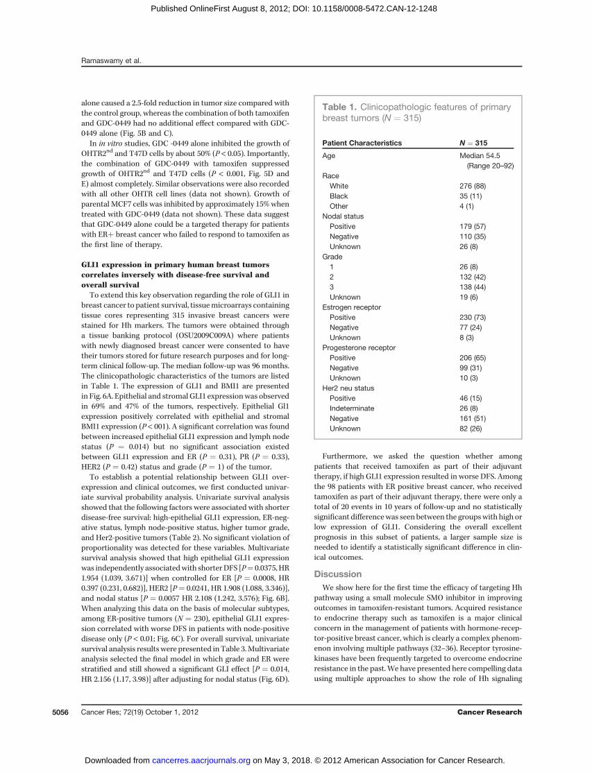
To extend this key observation regarding the role of GLI1 inbreast cancer to patient survival, tissuemicroarrays containingtissue cores representing 315 invasive breast cancers werestained for Hh markers. The tumors were obtained througha tissue banking protocol (OSU2009C009A) where patientswith newly diagnosed breast cancer were consented to havetheir tumors stored for future research purposes and for long-term clinical follow-up. The median follow-up was 96 months.The clinicopathologic characteristics of the tumors are listedin Table 1. The expression of GLI1 and BMI1 are presentedin Fig. 6A. Epithelial and stromal GLI1 expressionwas observedin 69% and 47% of the tumors, respectively. Epithelial Gl1expression positively correlated with epithelial and stromalBMI1 expression (P < 001). A significant correlation was foundbetween increased epithelial GLI1 expression and lymph nodestatus (P ¼ 0.014) but no significant association existedbetween GLI1 expression and ER (P ¼ 0.31), PR (P ¼ 0.33),HER2 (P ¼ 0.42) status and grade (P ¼ 1) of the tumor.
To establish a potential relationship between GLI1 over-expression and clinical outcomes, we first conducted univar-iate survival probability analysis. Univariate survival analysisshowed that the following factors were associated with shorterdisease-free survival: high-epithelial GLI1 expression, ER-neg-ative status, lymph node-positive status, higher tumor grade,and Her2-positive tumors (Table 2). No significant violation ofproportionality was detected for these variables. Multivariatesurvival analysis showed that high epithelial GLI1 expressionwas independently associatedwith shorterDFS [P¼ 0.0375, HR1.954 (1.039, 3.671)] when controlled for ER [P ¼ 0.0008, HR0.397 (0.231, 0.682)], HER2 [P¼ 0.0241, HR 1.908 (1.088, 3.346)],and nodal status [P ¼ 0.0057 HR 2.108 (1.242, 3.576); Fig. 6B].When analyzing this data on the basis of molecular subtypes,among ER-positive tumors (N ¼ 230), epithelial GLI1 expres-sion correlated with worse DFS in patients with node-positivedisease only (P < 0.01; Fig. 6C). For overall survival, univariatesurvival analysis results were presented in Table 3.Multivariateanalysis selected the final model in which grade and ER werestratified and still showed a significant GLI effect [P ¼ 0.014,HR 2.156 (1.17, 3.98)] after adjusting for nodal status (Fig. 6D).
Furthermore, we asked the question whether amongpatients that received tamoxifen as part of their adjuvanttherapy, if high GLI1 expression resulted in worse DFS. Amongthe 98 patients with ER positive breast cancer, who receivedtamoxifen as part of their adjuvant therapy, there were only atotal of 20 events in 10 years of follow-up and no statisticallysignificant differencewas seen between the groupswith high orlow expression of GLI1. Considering the overall excellentprognosis in this subset of patients, a larger sample size isneeded to identify a statistically significant difference in clin-ical outcomes.
DiscussionWe show here for the first time the efficacy of targeting Hh
pathway using a small molecule SMO inhibitor in improvingoutcomes in tamoxifen-resistant tumors. Acquired resistanceto endocrine therapy such as tamoxifen is a major clinicalconcern in the management of patients with hormone-recep-tor-positive breast cancer, which is clearly a complex phenom-enon involving multiple pathways (32–36). Receptor tyrosine-kinases have been frequently targeted to overcome endocrineresistance in the past.We have presented here compelling datausing multiple approaches to show the role of Hh signaling
Table 1. Clinicopathologic features of primarybreast tumors (N ¼ 315)
Patient Characteristics N ¼ 315
Age Median 54.5(Range 20–92)
RaceWhite 276 (88)Black 35 (11)Other 4 (1)
Nodal statusPositive 179 (57)Negative 110 (35)Unknown 26 (8)
Grade1 26 (8)2 132 (42)3 138 (44)Unknown 19 (6)
Estrogen receptorPositive 230 (73)Negative 77 (24)Unknown 8 (3)
Progesterone receptorPositive 206 (65)Negative 99 (31)Unknown 10 (3)
Her2 neu statusPositive 46 (15)Indeterminate 26 (8)Negative 161 (51)Unknown 82 (26)
Ramaswamy et al.
Cancer Res; 72(19) October 1, 2012 Cancer Research5056
on May 3, 2018. © 2012 American Association for Cancer Research. cancerres.aacrjournals.org Downloaded from
Published OnlineFirst August 8, 2012; DOI: 10.1158/0008-5472.CAN-12-1248

pathway in inducing tamoxifen-resistance, and themechanismof its activation.Furthermore, we have showed a significant correlation
between activation of Hh signaling in primary tumors (n ¼315), and disease outcomes, confirming the negative prognos-tic effect of high GLI1 expression on DFS and OS. AlthoughGLI1 expression did not correlate with a particular molecularsubtype, among ER positive, node positive tumors DFS wasworse in tumors with high GLI1expression. Hence the overallassociation of Hh activation and poor outcome in breastcancer signifies that activation of this pathway facilitatestumor growth and progression, supporting recently reportedassociation of increased Hh ligand expression with increasedrisk of metastasis and breast cancer-specific death (19).
Our in vivo study revealed that blocking SMOwithGDC-0449alone could effectively suppress the tamoxifen-resistant xeno-grafts inmice. Unlike our in vitro studies, addition of tamoxifento GDC-0449 had no further benefit, which could be because ofthe high dosage of GDC-0449 used in the in vivo studies, or thetamoxifen concentrations achieved in the tumor may be lowerdepending on its conversion to active metabolite in vivo. Usingmonoclonal antibody 5E1 to Hh ligand (Shh) O'Toole andcolleagues showed metastases reduction in animal models ofbreast cancer (19). Our data is unique in showing that targetingthe noncanonical Hh pathway specifically in ER-positivebreast cancers that are tamoxifen resistant using a clinicallyavailable Hh inhibitor (GDC-0449) improves outcomes.GDC-0449 (vismodegib) is a well tolerated, orally bioavailable
Figure 6. GLI1 expression inverselycorrelates with DFS and OS inprimary human breast tumors. A,representative pictures of breasttumors (H&E), GLI1, and BMI1(immunohistochemistry). B,comparison of DFS amongst breastcancer patients with ER-positivetumors expressing high versus lowGLI1.C, comparisonofDFSamongstbreast cancer patients with node-positive, ER-positive tumorsexpressing high versus low GLI1.D, comparison of overall survivalamongst patients with breast cancertumors expressing high versus lowGLI1.
Table 2. Univariate analysis of factors impacting disease-free survival
Variable Categorization N No. of events P HR (95%CI)
ER Positive 230 73 <0.0001 0.391 (0.268–0.570)Negative 77 44
PR Positive 206 66 <0.0001 0.513 (0.354–0.743)Negative 99 50
HER2 Positive 46 28 <0.0001 2.682 (1.701–4.230)Negative 187 58
Nodal Status Positive 110 55 <0.0001 2.654 (1.777–3.965)Negative 179 44
Grade G3 138 60 0.0036 1.744 (1.193–2.548)G1 and G2 158 49
Gli1 (epithelial) Positive 200 83 0.0188 1.714 (1.088–2.701)Negative 89 24
Hedgehog Signaling in Tamoxifen-Resistant Breast Cancer
www.aacrjournals.org Cancer Res; 72(19) October 1, 2012 5057
on May 3, 2018. © 2012 American Association for Cancer Research. cancerres.aacrjournals.org Downloaded from
Published OnlineFirst August 8, 2012; DOI: 10.1158/0008-5472.CAN-12-1248

compound that is approved for the management of metastaticor locally advanced basal cell carcinomas and being explored inother cancers (http.clinicaltrials.gov). Our work indicates thatpatients who developed resistance to tamoxifen, GDC-0049alone will be effective in treating recurrent tumors and there-fore designing appropriate clinical trials is warranted. It will beinteresting to determine if resistance to other endocrinetherapies such as aromatase inhibitors results in Hh pathwayactivation, which we plan to pursue in the future.
One of the major challenges in treating a drug-resistanttumor is the complex networking pathways that are involved inthe cross-talk between the distinct signaling pathways. ThePI3K/AKT pathway is one ofmany pathways that contribute toendocrine resistance (37). Activation of the PI3K/AKT and thep42/44 mitogen-activated protein kinase pathways by recep-tors such as EGFR and IGF1-R suppresses the expression of ERand PR (29, 38–42) and therefore, reduce estrogen dependence.Our work clearly shows cross-talk between PI3K/AKT and Hhpathway, and provides an additional mechanism for PI3K/AKT-mediated endocrine resistance. Importantly, we showedthat increased activity of PI3K/AKT activity could stabilizeSMO and GLI1 protein by suppressing GSK3b–mediated phos-phorylation and proteasomal degradation, leading to nonca-nonical Hh pathway activation. Although a previous studyreported cross-talk between PI3K/AKT and Hh pathway, it isin NIH3T3 cells through activation of Hh ligands (43). Our datasuggests a rationale for combined targeting of PI3K/AKT andHh pathway to effectively overcome endocrine resistance.
In summary, there is still an urgency in designing targetedtherapies to overcome endocrine resistance. Our studypresents a potential therapeutic option to overcome suchresistance and a clinical trial targeting Hh pathway inadvanced hormone-refractory breast cancer is warranted and
is currently being pursued. Further, our finding underscoresthe importance of PI3K pathway in activating Hh pathway andprovides the rationale for combinatorial targeted therapy inbreast cancer.
Disclosure of Potential Conflicts of InterestNo potential conflicts of interest were disclosed.
Authors' ContributionsConception and design: B. Ramaswamy, S. MajumderDevelopment of methodology: Y. Lu, K. Teng, G. J. Nuovo, S. MajumderAcquisition of data (provided animals, acquired and managed patients,provided facilities, etc.): B. Ramaswamy, Y. Lu, K. Teng, G. J. Nuovo, C. L.ShapiroAnalysis and interpretation of data (e.g., statistical analysis, biostatistics,computational analysis): B. Ramaswamy, Y. Lu, K. Teng, G. J. Nuovo, X. Li, S.MajumderWriting, review, and/or revision of the manuscript: B. Ramaswamy, Y. Lu,X. Li, C. L. Shapiro, S. MajumderAdministrative, technical, or material support (i.e., reporting or orga-nizing data, constructing databases): K. Teng, S. MajumderStudy supervision: B. Ramaswamy, S. Majumder
AcknowledgmentsWe thank Dr. Michael Ostrowski for critical reading of the manuscript and
editorial suggestions and Drs. Samson T. Jacob, Miguel A. Villalona, GustavoLeone, Guido Marcucci, and Cynthia Timmers for critically reading the manu-script.We thankDr.Hiroshi Sasaki forkindlyproviding theGliBS-d50-LucplasmidandDr.KristianHelin for providing thePI3KandAKTexpression vectors.Wealsothank Satavisha Roy and Thomas Kaffenberger for technical help.
Grant SupportThis work was supported by National Institutes of Health Grant CA137567 to
S. Majumder, CA133250-01 to B. Ramaswamy, Pelotonia Idea grant to S.Majumder and B. Ramaswamy, and Experimental Therapeutics OSU internalgrant to S. Majumder and B. Ramaswamy.
The costs of publication of this article were defrayed in part by the payment ofpage charges. This article must therefore be hereby marked advertisement inaccordance with 18 U.S.C. Section 1734 solely to indicate this fact.
Received April 2, 2012; revised May 30, 2012; accepted July 16, 2012;published OnlineFirst August 8, 2012.
References1. Tamoxifen for early breast cancer: an overview of the randomised
trials. Early Breast Cancer Trialists' Collaborative Group. Lancet1998;351:1451–67.
2. Arpino G, Green SJ, Allred DC, Lew D, Martino S, Osborne CK,et al. HER-2 amplification, HER-1 expression, and tamoxifen
response in estrogen receptor-positive metastatic breast cancer:a southwest oncology group study. Clin Cancer Res 2004;10:5670–6.
3. Campbell RA, Bhat-Nakshatri P, Patel NM, Constantinidou D, Ali S,Nakshatri H. Phosphatidylinositol 3-kinase/AKT-mediated activation
Table 3. Univariate survival analysis results for overall survival
Variable Categorization N No. of events P HR (95%CI)
ER Positive 230 65 <0.0001 0.428 (0.286–0.640)Negative 77 38
PR Positive 206 58 0.0013 0.526 (0.355–0.778)Negative 99 45
HER2 Positive 46 26 0.0001 2.564 (1.592–4.129)Negative 187 51
Nodal Status Positive 110 50 <0.0001 2.983 (1.928–4.616)Negative 179 35
Grade G3 138 40 0.002 1.907 (1.266–2.872)G1 and G2 158 55
Gli1 (epithelial) Positive 200 74 0.0055 2.076 (1.240–3.476)Negative 89 18
Ramaswamy et al.
Cancer Res; 72(19) October 1, 2012 Cancer Research5058
on May 3, 2018. © 2012 American Association for Cancer Research. cancerres.aacrjournals.org Downloaded from
Published OnlineFirst August 8, 2012; DOI: 10.1158/0008-5472.CAN-12-1248

of estrogen receptor alpha: a new model for anti-estrogen resistance.J Biol Chem 2001;276:9817–24.
4. Ellis MJ, Tao Y, Young O, White S, Proia AD, Murray J, et al. Estrogen-independent proliferation is present in estrogen-receptor HER2-pos-itive primary breast cancer after neoadjuvant letrozole. J Clin Oncol2006;24:3019–25.
5. Miller TW, Perez-Torres M, Narasanna A, Guix M, Sta�l O, P�erez-
Tenorio G, et al. Loss of Phosphatase and Tensin homologue deletedon chromosome 10 engages ErbB3 and insulin-like growth factor-Ireceptor signaling to promote antiestrogen resistance in breast can-cer. Cancer Res 2009;69:4192–201.
6. Shou J, Massarweh S, Osborne CK, Wakeling AE, Ali S, Weiss H, et al.Mechanisms of tamoxifen resistance: increased estrogen receptor-HER2/neu cross-talk in ER/HER2-positive breast cancer. J Natl Can-cer Inst 2004;96:926–35.
7. Baselga J, CamponeM, Piccart M, Burris HA III, RugoHS, Sahmoud T,et al. Everolimus in postmenopausal hormone-receptor-positiveadvanced breast cancer. N Engl J Med 2011;366:520–9.
8. Varjosalo M, Taipale J. Hedgehog: functions and mechanisms. GenesDev 2008;22:2454–72.
9. SasakiH,Nishizaki Y,HuiC,NakafukuM,KondohH.RegulationofGli2andGli3 activitiesbyanamino-terminal repressiondomain: implicationof Gli2 and Gli3 as primary mediators of Shh signaling. Development1999;126:3915–24.
10. Hahn H, Wicking C, Zaphiropoulous PG, Gailani MR, Shanley S,Chidambaram A, et al. Mutations of the human homolog of Drosophilapatched in the nevoid basal cell carcinoma syndrome. Cell 1996;85:841–51.
11. Johnson RL, Rothman AL, Xie J, Goodrich LV, Bare JW, Bonifas JM,et al. Human homolog of patched, a candidate gene for the basal cellnevus syndrome. Science 1996;272:1668–71.
12. Reifenberger J,WolterM,WeberRG,MegahedM,RuzickaT, Lichter P,et al. Missense mutations in SMOH in sporadic basal cell carcinomasof the skin and primitive neuroectodermal tumors of the centralnervous system. Cancer Res 1998;58:1798–803.
13. Hatsell S, Frost AR. Hedgehog signaling in mammary gland develop-ment and breast cancer. J Mammary Gland Biol Neoplasia 2007;12:163–73.
14. LewisMT,RossS,StricklandPA,SugnetCW,JimenezE,ScottMP,etal.Defects in mouse mammary gland development caused by conditionalhaploinsufficiency of Patched-1. Development 1999;126:5181–93.
15. Moraes RC, Zhang X, Harrington N, Fung JY, Wu MF, Hilsenbeck SG,et al. Constitutive activation of smoothened (SMO) inmammary glandsof transgenic mice leads to increased proliferation, altered differenti-ation and ductal dysplasia. Development 2007;134:1231–42.
16. Liu S, Dontu G, Mantle ID, Patel S, Ahn NS, Jackson KW, et al.Hedgehog signaling and Bmi-1 regulate self-renewal of normal andmalignant human mammary stem cells. Cancer Res 2006;66:6063–71.
17. LammML, CatbaganWS, Laciak RJ, Barnett DH, Hebner CM, GaffieldW, et al. Sonic hedgehog activates mesenchymal Gli1 expressionduring prostate ductal bud formation. Dev Biol 2002;249:349–66.
18. Fiaschi M, Rozell B, Bergstrom A, Toftgard R, Kleman MI. TargetedexpressionofGLI1 in themammary gland disrupts pregnancy-inducedmaturation and causes lactation failure. J Biol Chem 2007;282:36090–101.
19. O'Toole SA, Machalek DA, Shearer RF, Millar EK, Nair R, Schofield P,et al. Hedgehogoverexpression is associatedwith stromal interactionsand predicts for poor outcome in breast cancer. Cancer Res2011;71:4002–14.
20. Gunzer JL, Sutherlijn DP, Stanley MS, Bao L, Castendeo G , LalondeRL, et al. Pyridyl inhibitors of hedgehog signaling. WO/2009/126863,PCT/US2009/040165. 2009.
21. FanM,YanPS,Hartman-FreyC,Chen L, PaikH,Oyer SL, et al. Diversegene expression and DNA methylation profiles correlate with differ-ential adaptation of breast cancer cells to the antiestrogens tamoxifenand fulvestrant. Cancer Res 2006;66:11954–66.
22. Datta J, Kutay H, Nasser MW, Nuovo GJ, Wang B, Majumder S, et al.Methylation mediated silencing of MicroRNA-1 gene and its role inhepatocellular carcinogenesis. Cancer Res 2008;68:5049–58.
23. Franken NA, Rodermond HM, Stap J, Haveman J, van Bree C.Clonogenic assay of cells in vitro. Nat Protoc 2006;1:2315–9.
24. Dijkgraaf GJ, Alicke B, Weinmann L, Januario T, West K, Modrusan Z,et al. Small molecule inhibition of GDC-0449 refractory smoothenedmutants and downstreammechanisms of drug resistance. Cancer Res2011;71:435–44.
25. Romer JT, Kimura H, Magdaleno S, Sasai K, Fuller C, Baines H, et al.Suppression of the Shh pathway using a small molecule inhibitoreliminates medulloblastoma in Ptc1(þ/�)p53(�/�) mice. Cancer Cell2004;6:229–40.
26. Gupta GP, Nguyen DX, Chiang AC, Bos PD, Kim JY, Nadal C, et al.Mediators of vascular remodelling co-opted for sequential steps inlung metastasis. Nature 2007;446:765–70.
27. Nasser MW, Datta J, Nuovo G, Kutay H, Motiwala T, Majumder S, et al.Down-regulation ofmicro-RNA-1 (miR-1) in lung cancer. Suppression oftumorigenic property of lung cancer cells and their sensitization todoxorubicin-induced apoptosis by miR-1. J Biol Chem 2008;283:33394–405.
28. Horwitz KB, Mockus MB, Lessey BA. Variant T47D human breastcancer cells with high progesterone-receptor levels despite estrogenand antiestrogen resistance. Cell 1982;28:633–42.
29. CreightonCJ, FuX,HennessyBT,CasaAJ, ZhangY,Gonzalez-AnguloAM, et al. Proteomic and transcriptomic profiling reveals a link betweenthe PI3K pathway and lower estrogen-receptor (ER) levels and activityin ERþ breast cancer. Breast Cancer Res 2010;12:R40.
30. Kirkegaard T, Witton CJ, McGlynn LM, Tovey SM, Dunne B, Lyon A,et al. AKT activation predicts outcome in breast cancer patients treatedwith tamoxifen. J Pathol 2005;207:139–46.
31. Xue Y, Zhou F, Zhu M, Ahmed K, Chen G, Yao X. GPS: a comprehen-sive www server for phosphorylation sites prediction. Nucleic AcidsRes 2005;33:W184–7.
32. Lu Y, Roy S, Nuovo G, Ramaswamy B, Miller T, Shapiro C, et al. Anti-microRNA-222 (anti-miR-222) and -181B suppress growth of tamox-ifen-resistant xenografts in mouse by targeting TIMP3 protein andmodulating mitogenic signal. J Biol Chem 2011;286:42292–302.
33. Majumder S, Jacob ST. Emerging role of microRNAs in drug-resistantbreast cancer. Gene Expr 2011;15:141–51.
34. Musgrove EA, Sutherland RL. Biological determinants of endocrineresistance in breast cancer. Nat Rev Cancer 2009;9:631–43.
35. Osborne CK, Schiff R. Mechanisms of endocrine resistance in breastcancer. Annu Rev Med 2011;62:233–47.
36. Miller TE, Ghoshal K, Ramaswamy B, Roy S, Datta J, Shapiro CL, et al.MicroRNA-221/222 confers tamoxifen resistance in breast cancer bytargeting p27Kip1. J Biol Chem 2008;283:29897–903.
37. Clark AS, West K, Streicher S, Dennis PA. Constitutive and inducibleAkt activity promotes resistance to chemotherapy, trastuzumab, ortamoxifen in breast cancer cells. Mol Cancer Ther 2002;1:707–17.
38. Bayliss J, Hilger A, Vishnu P, Diehl K, El-Ashry D. Reversal of theestrogen receptor negative phenotype in breast cancer and res-toration of antiestrogen response. Clin Cancer Res 2007;13:7029–36.
39. Cui X, ZhangP, DengW,Oesterreich S, LuY,Mills GB, et al. Insulin-likegrowth factor-I inhibits progesterone receptor expression in breastcancer cells via the phosphatidylinositol 3-kinase/Akt/mammaliantarget of rapamycin pathway: progesterone receptor as a potentialindicator of growth factor activity in breast cancer. Mol Endocrinol2003;17:575–88.
40. Lopez-Tarruella S, Schiff R. The dynamics of estrogen receptor statusin breast cancer: re-shaping the paradigm. Clin Cancer Res 2007;13:6921–5.
41. Cui X,Schiff R, ArpinoG,OsborneCK, LeeAV.Biology of progesteronereceptor loss in breast cancer and its implications for endocrinetherapy. J Clin Oncol 2005;23:7721–35.
42. Guo S, Sonenshein GE. Forkhead box transcription factor FOXO3aregulates estrogen receptor alpha expression and is repressed by theHer-2/neu/phosphatidylinositol 3-kinase/Akt signaling pathway. MolCell Biol 2004;24:8681–90.
43. Riobo NA, Lu K, Ai X, Haines GM, Emerson CP Jr. Phosphoinositide 3-kinase and Akt are essential for Sonic Hedgehog signaling. Proc NatlAcad Sci U S A 2006;103:4505–10.
Hedgehog Signaling in Tamoxifen-Resistant Breast Cancer
www.aacrjournals.org Cancer Res; 72(19) October 1, 2012 5059
on May 3, 2018. © 2012 American Association for Cancer Research. cancerres.aacrjournals.org Downloaded from
Published OnlineFirst August 8, 2012; DOI: 10.1158/0008-5472.CAN-12-1248
2012;72:5048-5059. Published OnlineFirst August 8, 2012.Cancer Res Bhuvaneswari Ramaswamy, Yuanzhi Lu, Kun-yu Teng, et al. PI3K/AKT PathwayTamoxifen-Resistant Breast Cancer Aberrantly Activated by Hedgehog Signaling Is a Novel Therapeutic Target in
Updated version
10.1158/0008-5472.CAN-12-1248doi:
Access the most recent version of this article at:
Material
Supplementary
http://cancerres.aacrjournals.org/content/suppl/2012/08/09/0008-5472.CAN-12-1248.DC1
Access the most recent supplemental material at:
Cited articles
http://cancerres.aacrjournals.org/content/72/19/5048.full#ref-list-1
This article cites 42 articles, 25 of which you can access for free at:
Citing articles
http://cancerres.aacrjournals.org/content/72/19/5048.full#related-urls
This article has been cited by 2 HighWire-hosted articles. Access the articles at:
E-mail alerts related to this article or journal.Sign up to receive free email-alerts
Subscriptions
Reprints and
To order reprints of this article or to subscribe to the journal, contact the AACR Publications Department at
Permissions
Rightslink site. Click on "Request Permissions" which will take you to the Copyright Clearance Center's (CCC)
.http://cancerres.aacrjournals.org/content/72/19/5048To request permission to re-use all or part of this article, use this link
on May 3, 2018. © 2012 American Association for Cancer Research. cancerres.aacrjournals.org Downloaded from
Published OnlineFirst August 8, 2012; DOI: 10.1158/0008-5472.CAN-12-1248