head and neck embryology
TRANSCRIPT

8/8/2019 Head and Neck Embryology
http://slidepdf.com/reader/full/head-and-neck-embryology 1/56
Head and Neck Head and Neck EmbryologyEmbryology

8/8/2019 Head and Neck Embryology
http://slidepdf.com/reader/full/head-and-neck-embryology 2/56
Head & Neck EmbryologyHead & Neck Embryology
BranchialBranchial Apparatus Apparatus Thyroid GlandThyroid Gland
TongueTongue Development of the faceDevelopment of the face
– – NoseNose
– – PalatePalate
EarEar

8/8/2019 Head and Neck Embryology
http://slidepdf.com/reader/full/head-and-neck-embryology 3/56
BranchialBranchial Apparatus Apparatus
4 arches are well developed by 44 arches are well developed by 4thth week of gestationweek of gestation
55thth and 6and 6thth arches are still rudimentaryarches are still rudimentary
Development takes place over weeks 4 to 7Development takes place over weeks 4 to 7
Contribute mostly to neck development but the firstContribute mostly to neck development but the first
arch contributes to facial developmentarch contributes to facial development

8/8/2019 Head and Neck Embryology
http://slidepdf.com/reader/full/head-and-neck-embryology 4/56
BranchialBranchial Apparatus Apparatus

8/8/2019 Head and Neck Embryology
http://slidepdf.com/reader/full/head-and-neck-embryology 5/56
BranchialBranchial Apparatus Apparatus
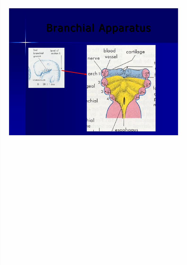
Arches Arches – – mesenchymalmesenchymal tissue surrounded by ectodermtissue surrounded by ectoderm
and endodermand endoderm
Clefts (or grooves)Clefts (or grooves) – – Separate adjacent arches alongSeparate adjacent arches along ectodermalectodermal
surfacesurface
PouchesPouches – – OutpouchingOutpouching of endoderm from foregutof endoderm from foregut
– – Penetrate adjacentPenetrate adjacent mesenchymemesenchyme
8/8/2019 Head and Neck Embryology
http://slidepdf.com/reader/full/head-and-neck-embryology 6/56
BranchialBranchial Apparatus Apparatus

8/8/2019 Head and Neck Embryology
http://slidepdf.com/reader/full/head-and-neck-embryology 7/56
BranchialBranchial Apparatus Apparatus

8/8/2019 Head and Neck Embryology
http://slidepdf.com/reader/full/head-and-neck-embryology 8/56
BranchialBranchial Arches Arches
Each arch contains:Each arch contains:
A A cartilagenouscartilagenous componentcomponent
A muscular component A muscular component
An aortic arch (artery) An aortic arch (artery)
A nerve A nerve
8/8/2019 Head and Neck Embryology
http://slidepdf.com/reader/full/head-and-neck-embryology 9/56
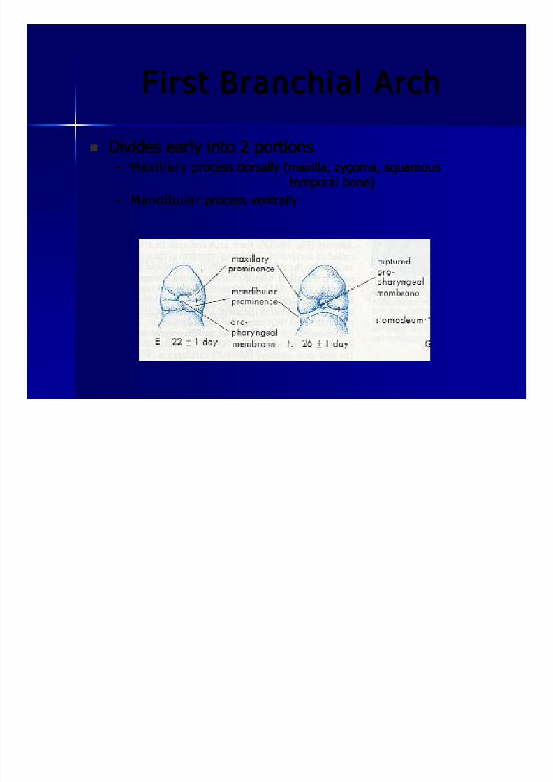
FirstFirst BranchialBranchial Arch Arch
Divides early into 2 portionsDivides early into 2 portions – – MaxillaryMaxil lary process dorsally (maxilla,process dorsally (maxilla, zygomazygoma,, squamoussquamous
temporal bone)temporal bone)
– – MandibularMandibular process ventrallyprocess ventrally

8/8/2019 Head and Neck Embryology
http://slidepdf.com/reader/full/head-and-neck-embryology 10/56
FirstFirst BranchialBranchial Arch Arch
Cartilage (Cartilage (MeckelMeckel’ ’ ss Cartilage)Cartilage) – – Dorsal end becomes theDorsal end becomes the malleusmalleus andand incusincus
– – Intermediate portion regresses, but theIntermediate portion regresses, but the perichondriumperichondrium
forms:forms:
Anterior ligament of the Anterior ligament of the malleusmalleus SphenomandibularSphenomandibular ligamentligament
– – Ventral portion forms the mandible Ventral portion forms the mandible

8/8/2019 Head and Neck Embryology
http://slidepdf.com/reader/full/head-and-neck-embryology 11/56
FirstFirst BranchialBranchial Arch Arch
Muscular componentMuscular component
Muscles of mastication (Muscles of mastication (temporalistemporalis,, massetermasseter, med & lat, med & lat
pterygoidspterygoids))
Accessorymuscles Accessorymuscles of mastication (of mastication (mylohyoidmylohyoid, ant belly of , ant belly of digastricdigastric))
Tensor tympaniTensor tympani
TensorTensor veliveli palatinipalatini

8/8/2019 Head and Neck Embryology
http://slidepdf.com/reader/full/head-and-neck-embryology 12/56
FirstFirst BranchialBranchial Arch Arch
Aortic arch Aortic arch Maxillary arteryMaxillary artery
NerveNerve Trigeminal nerve (CN V)Trigeminal nerve (CN V)

8/8/2019 Head and Neck Embryology
http://slidepdf.com/reader/full/head-and-neck-embryology 13/56
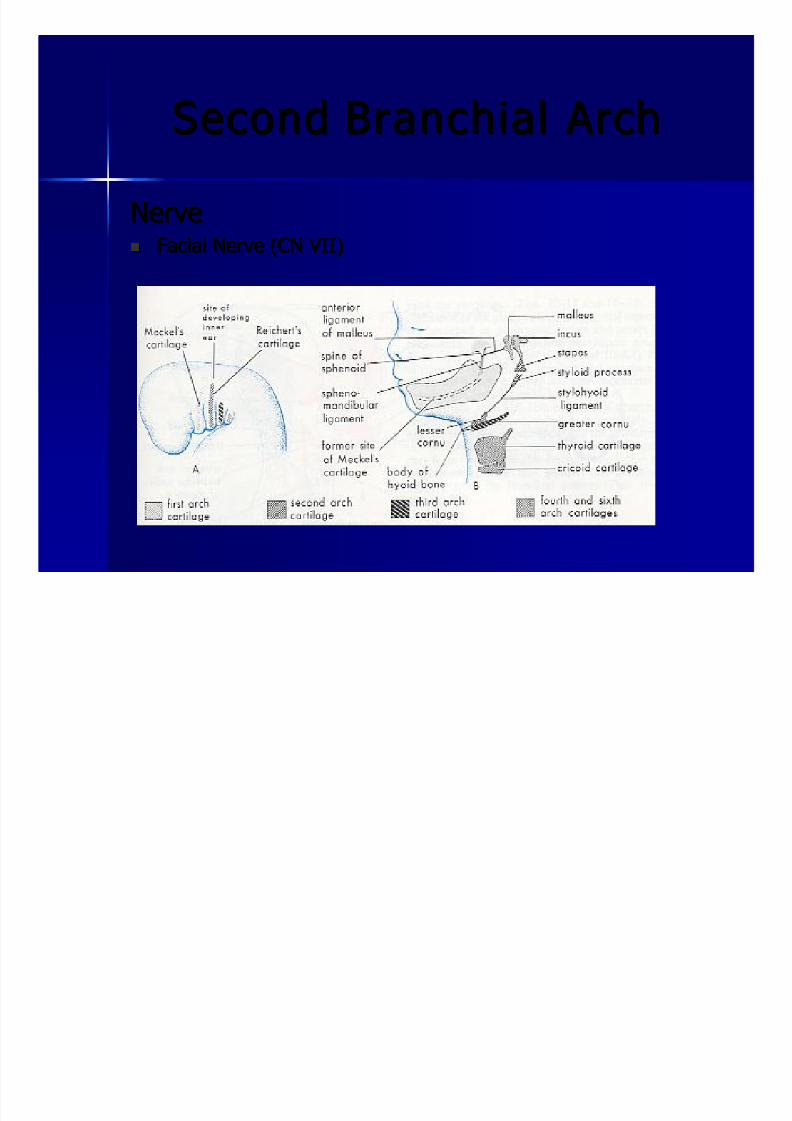
SecondSecond BranchialBranchial Arch Arch
Cartilage (ReichertCartilage (Reichert’ ’ s Cartilage)s Cartilage) – – Dorsal end becomes stapes (except footplate) andDorsal end becomes stapes (except footplate) and styloidstyloid
processprocess
– – Intermediate portion regresses andIntermediate portion regresses and perichondriumperichondrium formsforms
thethe stylohyoidstylohyoid ligamentligament – – Ventral end forms the lesser Ventral end forms the lesser cornucornu of the hyoid and theof the hyoid and the
upper half of the hyoid boneupper half of the hyoid bone

8/8/2019 Head and Neck Embryology
http://slidepdf.com/reader/full/head-and-neck-embryology 14/56
SecondSecond BranchialBranchial Arch Arch
Muscular ComponentMuscular Component – – Migrates over superficial face to form the muscles of facialMigrates over superficial face to form the muscles of facial
expressionexpression
– – StapediusStapedius musclemuscle
– – StylohyoidStylohyoid musclemuscle – – Posterior belly of Posterior belly of digastricdigastric
Aortic Arch Aortic Arch – – Hyoid arteryHyoid artery
– – StapedialStapedial arteryartery
8/8/2019 Head and Neck Embryology
http://slidepdf.com/reader/full/head-and-neck-embryology 15/56
SecondSecond BranchialBranchial Arch Arch
NerveNerve Facial Nerve (CN VII)Facial Nerve (CN VII)

8/8/2019 Head and Neck Embryology
http://slidepdf.com/reader/full/head-and-neck-embryology 16/56
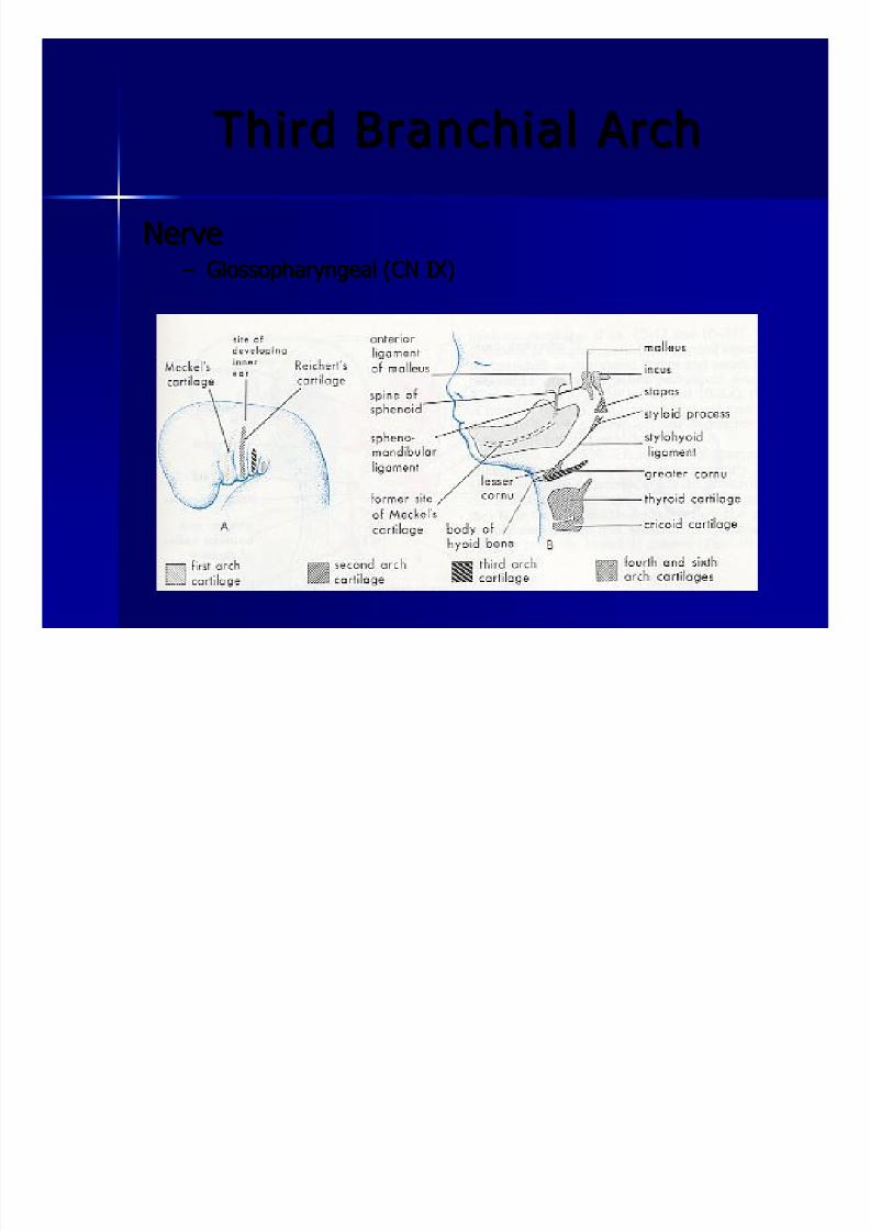
ThirdThird BranchialBranchial Arch Arch
CartilageCartilage – – Located ventrally and forms the lower half of the body of Located ventrally and forms the lower half of the body of the hyoid and the greaterthe hyoid and the greater cornucornu
Muscular ComponentMuscular Component – – Only one muscle:Only one muscle: stylopharyngeusstylopharyngeus
Aortic Arch Aortic Arch – – Common carotid, external carotid, proximal internalCommon carotid, external carotid, proximal internalcarotidcarotid
8/8/2019 Head and Neck Embryology
http://slidepdf.com/reader/full/head-and-neck-embryology 17/56
ThirdThird BranchialBranchial Arch Arch
NerveNerve – – GlossopharyngealGlossopharyngeal (CN IX)(CN IX)

8/8/2019 Head and Neck Embryology
http://slidepdf.com/reader/full/head-and-neck-embryology 18/56
FourthFourth BranchialBranchial Arch Arch
CartilageCartilage – – Thyroid cartilageThyroid cartilage
Muscular ComponentMuscular Component
– – The 3 pharyngeal constrictorsThe 3 pharyngeal constrictors – – CricothyroidCricothyroid musclemuscle
Aortic Arch Aortic Arch – – Left: Aortic archLeft: Aortic arch
– – Right: RightRight: Right subclaviansubclavian
NerveNerve – – Superior laryngeal branch of Superior laryngeal branch of vagusvagus (CN X)(CN X)
8/8/2019 Head and Neck Embryology
http://slidepdf.com/reader/full/head-and-neck-embryology 19/56
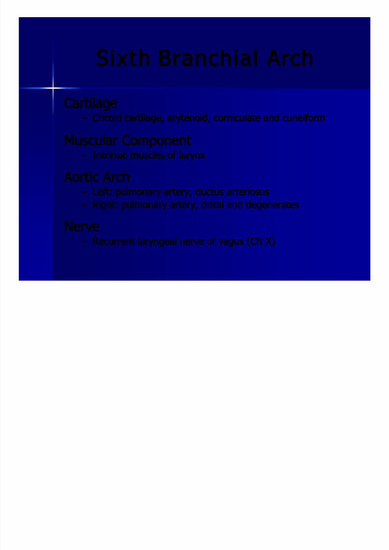
SixthSixth BranchialBranchial Arch Arch
CartilageCartilage – – CricoidCricoid cartilage,cartilage, arytenoidarytenoid,, corniculatecorniculate and cuneiformand cuneiform
Muscular ComponentMuscular Component
– – Intrinsic muscles of larynxIntrinsic muscles of larynx
Aortic Arch Aortic Arch – – Left: pulmonary artery,Left: pulmonary artery, ductusductus arteriosusarteriosus
– – Right: pulmonary artery, distal end degeneratesRight: pulmonary artery, distal end degenerates
NerveNerve – – Recurrent laryngeal nerve of Recurrent laryngeal nerve of vagusvagus (CN X)(CN X)
8/8/2019 Head and Neck Embryology
http://slidepdf.com/reader/full/head-and-neck-embryology 20/56
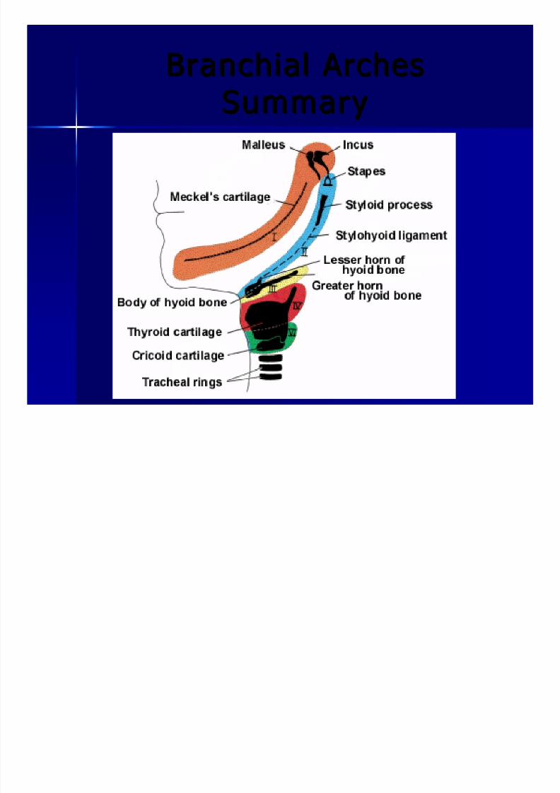
BranchialBranchial Arches ArchesSummarySummary

8/8/2019 Head and Neck Embryology
http://slidepdf.com/reader/full/head-and-neck-embryology 21/56
8/8/2019 Head and Neck Embryology
http://slidepdf.com/reader/full/head-and-neck-embryology 22/56
BranchialBranchial CleftsClefts(or grooves)(or grooves)
4 clefts4 clefts
The 2The 2ndnd to 4to 4thth clefts become buried by theclefts become buried by the
overgrowth of the 2overgrowth of the 2ndnd arch to form the cervicalarch to form the cervical
sinussinus
Cervical sinus hasCervical sinus has dissapeareddissapeared by week 7by week 7
The first cleft persists and invades theThe first cleft persists and invades the mesenchymemesenchyme
opposite the first pouchopposite the first pouch
This becomes the EAC and ectoderm of the TMThis becomes the EAC and ectoderm of the TM

8/8/2019 Head and Neck Embryology
http://slidepdf.com/reader/full/head-and-neck-embryology 23/56
BranchialBranchial CleftsClefts

8/8/2019 Head and Neck Embryology
http://slidepdf.com/reader/full/head-and-neck-embryology 24/56
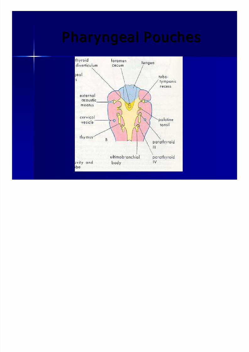
Pharyngeal PouchesPharyngeal Pouches

8/8/2019 Head and Neck Embryology
http://slidepdf.com/reader/full/head-and-neck-embryology 25/56
11 stst Pharyngeal PouchPharyngeal Pouch
Elongates into theElongates into the tubotympanictubotympanic recessrecess
The distal part contacts the 1The distal part contacts the 1stst pharyngeal cleft andpharyngeal cleft and
forms the inner lining of the TMforms the inner lining of the TM
TheThe tubotympanictubotympanic recess becomes the tympanicrecess becomes the tympanic
cavity and mastoidcavity and mastoid antrumantrum
Connection of the recess with the pharynx becomesConnection of the recess with the pharynx becomesthethe eustachianeustachian tubetube

8/8/2019 Head and Neck Embryology
http://slidepdf.com/reader/full/head-and-neck-embryology 26/56
22 ndnd Pharyngeal PouchPharyngeal Pouch
Forms theForms the tonsillartonsillar fossafossa
Endoderm forms the surface epithelium and liningEndoderm forms the surface epithelium and lining
of of tonsillartonsillar cryptscrypts
At 20 weeks lymphoid tissue invades the endoderm At 20 weeks lymphoid tissue invades the endoderm
and forms the palatine tonsilsand forms the palatine tonsils

8/8/2019 Head and Neck Embryology
http://slidepdf.com/reader/full/head-and-neck-embryology 27/56
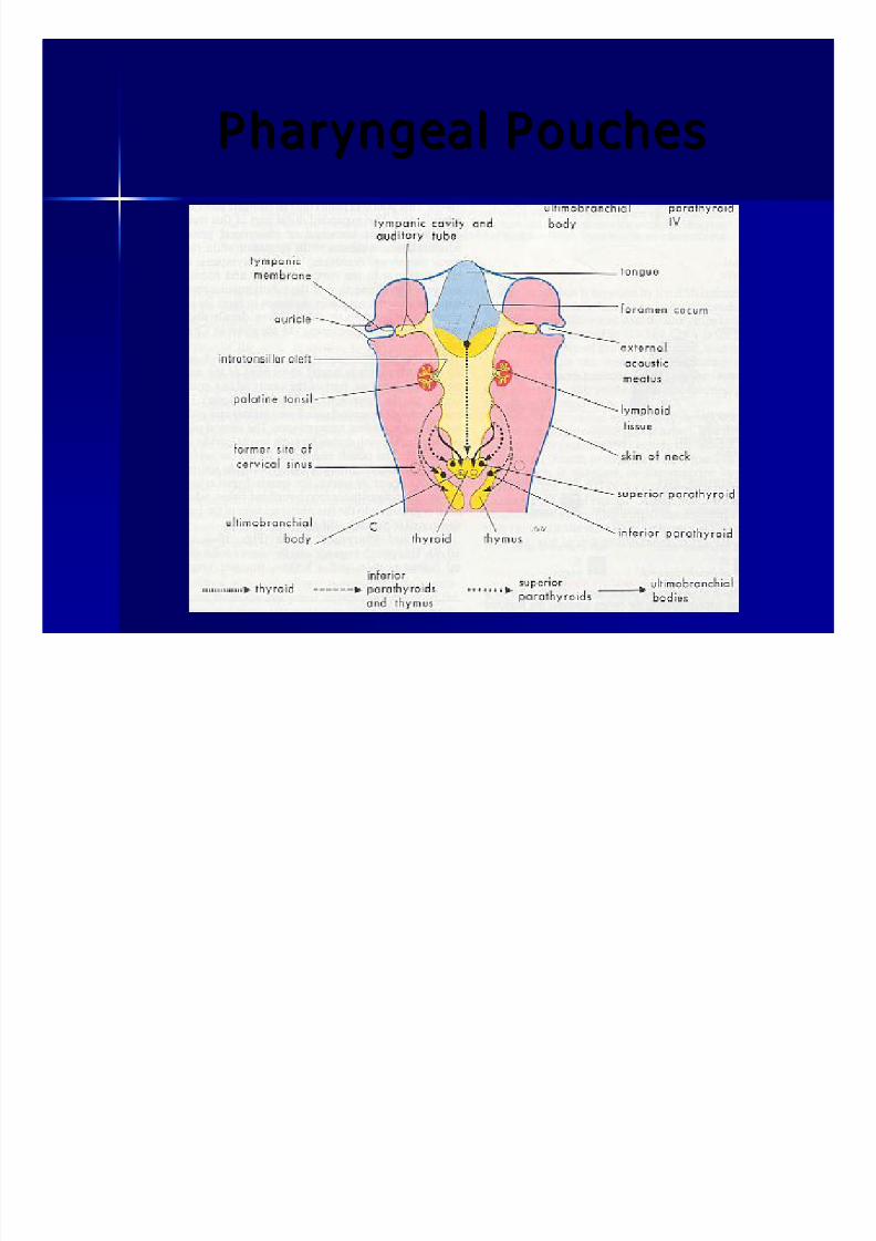
33 rdrd Pharyngeal PouchPharyngeal Pouch
Forms 2Forms 2 diverticuladiverticula: dorsal and ventral: dorsal and ventral Endoderm of Endoderm of dorsaldorsal diverticuladiverticula:: inf inf parathyroidparathyroid
Endoderm of Endoderm of ventraventra ll diverticuladiverticula: lobule of thymus: lobule of thymus
TheseThese diverticuladiverticula become detached from the wallbecome detached from the wall
and migrate caudally.and migrate caudally.
Thymus comes to lie in the superiorThymus comes to lie in the superior mediastinummediastinum
8/8/2019 Head and Neck Embryology
http://slidepdf.com/reader/full/head-and-neck-embryology 28/56
Pharyngeal PouchesPharyngeal Pouches
8/8/2019 Head and Neck Embryology
http://slidepdf.com/reader/full/head-and-neck-embryology 29/56
Pharyngeal PouchesPharyngeal Pouches
8/8/2019 Head and Neck Embryology
http://slidepdf.com/reader/full/head-and-neck-embryology 30/56
44 thth Pharyngeal PouchPharyngeal Pouch
Also develops dorsal and ventral Also develops dorsal and ventral diverticuladiverticula TheThe dorsaldorsal bud becomes the sup. parathyroidbud becomes the sup. parathyroid
TheThe ventralventral bud becomes thebud becomes the UltimobranchialUltimobranchial
bodybody
TheThe ultimobranchialultimobranchial body fuses with the thyroidbody fuses with the thyroid
gland and disseminates within it to give thegland and disseminates within it to give the
parafollicularparafollicular C cells which produceC cells which produce calcitonincalcitonin

8/8/2019 Head and Neck Embryology
http://slidepdf.com/reader/full/head-and-neck-embryology 31/56
Pharyngeal PouchesPharyngeal Pouches

8/8/2019 Head and Neck Embryology
http://slidepdf.com/reader/full/head-and-neck-embryology 32/56
Pharyngeal PouchesPharyngeal Pouches
55thth
pouch never developspouch never develops Controversy re:Controversy re: originof originof ultimobranchialultimobranchial
bodybody

8/8/2019 Head and Neck Embryology
http://slidepdf.com/reader/full/head-and-neck-embryology 33/56
ThyroidThyroid
In 4In 4thth week begins asweek begins as endodermalendodermal thickening in floor of thickening in floor of
primitive pharynxprimitive pharynx
The thickening becomes anThe thickening becomes an outpouchingoutpouching: thyroid: thyroid diverticulumdiverticulum
Thyroid descends anterior to hyoid and thyroid cartilageThyroid descends anterior to hyoid and thyroid cartilage
Connected to tongue byConnected to tongue by thyroglossalthyroglossal ductduct
Week 7: Thyroid reaches final positionWeek 7: Thyroid reaches final position
ThyroglossalThyroglossal duct has degeneratedduct has degenerated
Pyramidal lobe: Persistence of distal end of Pyramidal lobe: Persistence of distal end of thyroglossalthyroglossal ductductPresent in 50% of peoplePresent in 50% of people

8/8/2019 Head and Neck Embryology
http://slidepdf.com/reader/full/head-and-neck-embryology 34/56
ThyroidThyroid

8/8/2019 Head and Neck Embryology
http://slidepdf.com/reader/full/head-and-neck-embryology 35/56
TongueTongue
44thth week: elevation on floor of week: elevation on floor of
pharynx, justpharynx, just rostralrostral to foramento foramen
cecumcecum:: Median Tongue BudMedian Tongue Bud
((TuberculumTuberculum imparimpar))
Distal Tongue BudsDistal Tongue Buds developdevelop just lateral to median tongue bud just lateral to median tongue bud
Both of the above originate inBoth of the above originate in
mesenchymemesenchyme of firstof first branchialbranchialarcharch

8/8/2019 Head and Neck Embryology
http://slidepdf.com/reader/full/head-and-neck-embryology 36/56
TongueTongue
Distal tongue buds overgrow theDistal tongue buds overgrow the
median tongue bud and merge withmedian tongue bud and merge with
each othereach other
These form the ant 2/3 of the tongueThese form the ant 2/3 of the tongue
Median tongue bud forms no adultMedian tongue bud forms no adult
structurestructure

8/8/2019 Head and Neck Embryology
http://slidepdf.com/reader/full/head-and-neck-embryology 37/56
TongueTongue
At same time 2 elevations develop caudal At same time 2 elevations develop caudal
to foramento foramen cecumcecum::
1. Copula: from 21. Copula: from 2ndnd archarch
2.2. HypobranchialHypobranchial emminenceemminence::
from 3from 3rdrd & 4& 4thth archesarches
TheThe hypobranchialhypobranchial emminenceemminenceovergrows the copula which disappearsovergrows the copula which disappears
The post 1/3 of the tongue is formed byThe post 1/3 of the tongue is formed by
thethe rostralrostral part of thepart of the hypobranchialhypobranchial
emminenceemminence (Arch 3)(Arch 3)
Caudal part of Caudal part of hypobranchialhypobranchial emminenceemminence(Arch 4) forms the epiglottis(Arch 4) forms the epiglottis
8/8/2019 Head and Neck Embryology
http://slidepdf.com/reader/full/head-and-neck-embryology 38/56
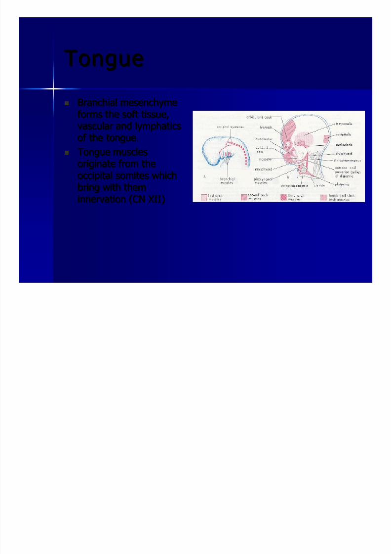
TongueTongue
BranchialBranchial mesenchymemesenchyme
forms the soft tissue,forms the soft tissue,vascular andvascular and lymphaticslymphatics
of the tongue.of the tongue.
Tongue musclesTongue muscles
originate from theoriginate from theoccipitaloccipital somitessomites whichwhich
bring with thembring with them
innervationinnervation (CN XII)(CN XII)

8/8/2019 Head and Neck Embryology
http://slidepdf.com/reader/full/head-and-neck-embryology 39/56
TongueTongue
InnervationInnervation
to tongue:to tongue:
Ant 2/3: CN V Ant 2/3: CN V
Post 1/3: CN IXPost 1/3: CN IX

8/8/2019 Head and Neck Embryology
http://slidepdf.com/reader/full/head-and-neck-embryology 40/56
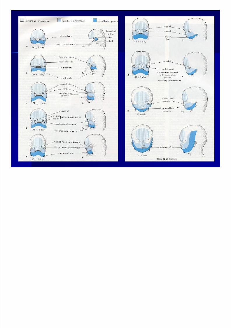
Development of the FaceDevelopment of the Face
Five facialFive facial primordiaprimordia contribute to development of the face:contribute to development of the face:
– – TheThe frontonasalfrontonasal prominenceprominence
– – Paired Maxillary prominencesPaired Maxillary prominences
– – PairedPaired MandibularMandibular prominencesprominences
8/8/2019 Head and Neck Embryology
http://slidepdf.com/reader/full/head-and-neck-embryology 41/56

8/8/2019 Head and Neck Embryology
http://slidepdf.com/reader/full/head-and-neck-embryology 42/56
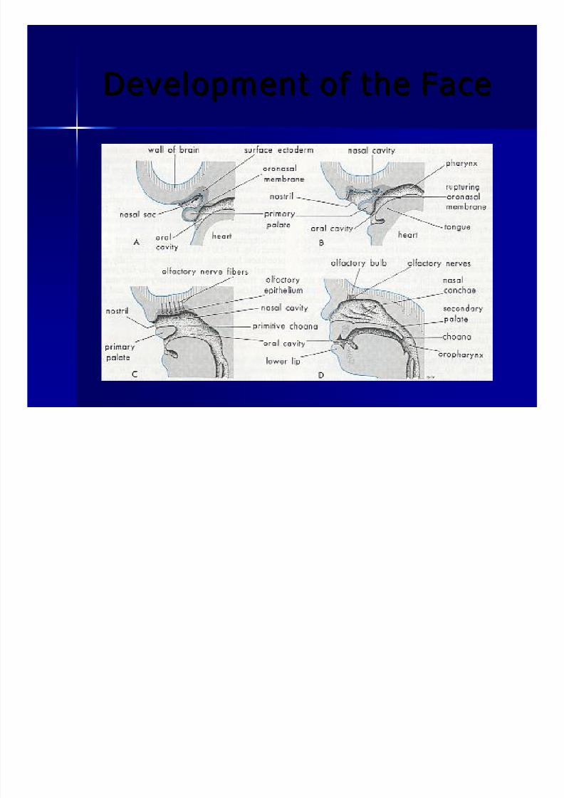
Development of the FaceDevelopment of the Face
44thth week: thickening of ectoderm in theweek: thickening of ectoderm in the ventrolateralventrolateral parts of parts of
the FNP:the FNP: NasalNasal PlacodesPlacodes
MesenchymeMesenchyme on the edges of theon the edges of the placodesplacodes proliferates toproliferates to
form:form: medial and lateral nasal prominencesmedial and lateral nasal prominences
As a result the nasal As a result the nasal placodesplacodes now lie in a depression callednow lie in a depression called
nasal pitsnasal pits which enlarge dorsally to form the nasal cavities.which enlarge dorsally to form the nasal cavities.
These nasal cavities are separated from the oral cavity by theThese nasal cavities are separated from the oral cavity by the
oronasaloronasal membranesmembranes which rupture to form the primitivewhich rupture to form the primitive
choanachoana
8/8/2019 Head and Neck Embryology
http://slidepdf.com/reader/full/head-and-neck-embryology 43/56
Development of the FaceDevelopment of the Face
8/8/2019 Head and Neck Embryology
http://slidepdf.com/reader/full/head-and-neck-embryology 44/56
Development of the FaceDevelopment of the Face
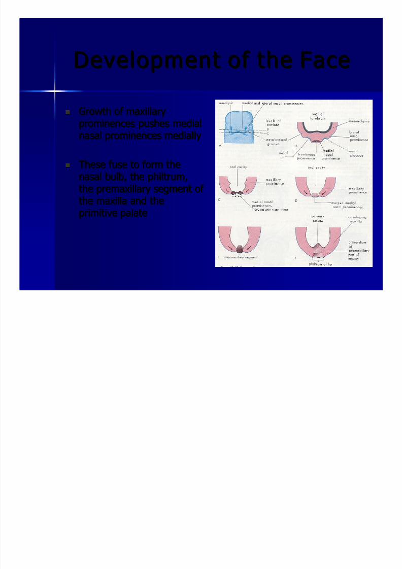
Growth of maxillaryGrowth of maxillaryprominences pushes medialprominences pushes medial
nasal prominences mediallynasal prominences medially
These fuse to form theThese fuse to form thenasal bulb, thenasal bulb, the philtrumphiltrum,,
thethe premaxillarypremaxillary segment of segment of
the maxilla and thethe maxilla and the
primitive palateprimitive palate

8/8/2019 Head and Neck Embryology
http://slidepdf.com/reader/full/head-and-neck-embryology 45/56
Development of the FaceDevelopment of the Face
The lateral nasal prominences, which become the ala areThe lateral nasal prominences, which become the ala are
separated from the maxillary prominences by theseparated from the maxillary prominences by thennasolacrimalasolacrimal groovesgrooves which become thewhich become the nasolacrimalnasolacrimal
ducts.ducts.
The sinuses form asThe sinuses form as outpouchingsoutpouchings of the ectoderm of lateralof the ectoderm of lateralnasal wallsnasal walls
The olfactory epithelium develops from ectodermThe olfactory epithelium develops from ectoderm

8/8/2019 Head and Neck Embryology
http://slidepdf.com/reader/full/head-and-neck-embryology 46/56

8/8/2019 Head and Neck Embryology
http://slidepdf.com/reader/full/head-and-neck-embryology 47/56
PalatePalate
Primary and Secondary PalatePrimary and Secondary Palate
Primary PalatePrimary Palate Develops from the fusion of the medial nasal prominencesDevelops from the fusion of the medial nasal prominences
between the maxillary prominencesbetween the maxillary prominences
Forms the adult portion of the palate which is anterior to theForms the adult portion of the palate which is anterior to the
incisive foramenincisive foramen

8/8/2019 Head and Neck Embryology
http://slidepdf.com/reader/full/head-and-neck-embryology 48/56
PalatePalate
Secondary PalateSecondary Palate Origin of the hard and soft palateOrigin of the hard and soft palate
Develops from internal projections of the maxillaryDevelops from internal projections of the maxillary
prominences called theprominences called the lateral palatine processeslateral palatine processes
As mandible develops, the tongue drops and the palatine As mandible develops, the tongue drops and the palatineprocesses grow medially and fuse in the midline.processes grow medially and fuse in the midline.
They also fuse with the nasal septum and the primary palate.They also fuse with the nasal septum and the primary palate.
Ossification occurs in anOssification occurs in an anteroantero--posterior directionposterior direction

8/8/2019 Head and Neck Embryology
http://slidepdf.com/reader/full/head-and-neck-embryology 49/56

8/8/2019 Head and Neck Embryology
http://slidepdf.com/reader/full/head-and-neck-embryology 50/56
The EarThe Ear
Inner EarInner Ear 44thth week: thickening in surface ectoderm calledweek: thickening in surface ectoderm called oticotic placodeplacode
InvaginatesInvaginates into underlyinginto underlying mesenchymemesenchyme and detaches fromand detaches from
ectoderm: now calledectoderm: now called oticotic vesiclevesicle
OticOtic vesicle divides into 2 regions:vesicle divides into 2 regions: utricularutricular portion andportion andsaccularsaccular portionportion
UtricularUtricular portionportion
Utricle, semicircular canals andUtricle, semicircular canals and endolymphaticendolymphatic ductduct
SaccularSaccular portionportion
SacculeSaccule and cochlear duct (becomes cochlea)and cochlear duct (becomes cochlea)

8/8/2019 Head and Neck Embryology
http://slidepdf.com/reader/full/head-and-neck-embryology 51/56
Inner EarInner Ear

8/8/2019 Head and Neck Embryology
http://slidepdf.com/reader/full/head-and-neck-embryology 52/56
Middle EarM iddle Ear

8/8/2019 Head and Neck Embryology
http://slidepdf.com/reader/full/head-and-neck-embryology 53/56
External EarExternal Ear
Develops from 6Develops from 6 mesenchymalmesenchymal swellings, calledswellings, called auricularauricular
hillockshillocks , which develop around the first, which develop around the first branchialbranchial cleftcleft
TheThe mesenchymemesenchyme is derived from mesoderm in the first andis derived from mesoderm in the first and
22ndnd branchialbranchial arches. As the ear grows,arches. As the ear grows, contrbutionscontrbutions of theof the
firstfirst branchialbranchial arch become reduced.arch become reduced.
TheThe pinnapinna initially develops in the neck. As the mandibleinitially develops in the neck. As the mandiblegrows, it moves up to the level of the eyes.grows, it moves up to the level of the eyes.
Part of the auricle originating from the firstPart of the auricle originating from the first branchialbranchial arch isarch is
innervated by CN Vinnervated by CN V
The part originating from the 2The part originating from the 2ndnd arch is innervated by thearch is innervated by thecervical plexus (namely the lesser occipital and greatercervical plexus (namely the lesser occipital and greater
auricular nerves)auricular nerves)

8/8/2019 Head and Neck Embryology
http://slidepdf.com/reader/full/head-and-neck-embryology 54/56
External EarExternal Ear

8/8/2019 Head and Neck Embryology
http://slidepdf.com/reader/full/head-and-neck-embryology 55/56
External EarExternal Ear

8/8/2019 Head and Neck Embryology
http://slidepdf.com/reader/full/head-and-neck-embryology 56/56
Thank You Thank You