embryology of head face &oral cavity
DESCRIPTION
embryologyTRANSCRIPT
Embryology of the Head,Embryology of the Head, Face and Oral CavityFace and Oral Cavity
Raj Gopalakrishnan B.D.S., Ph.D.Raj Gopalakrishnan B.D.S., Ph.D.Oral and Maxillofacial PathologyOral and Maxillofacial Pathology
Dept. of Diagnostic and Biological SciencesDept. of Diagnostic and Biological SciencesUniversity of Minnesota School of DentistryUniversity of Minnesota School of Dentistry

Prenatal Development
Figure from Ten Cate’s Oral Histology, Ed., Antonio Nanci, 6th edition
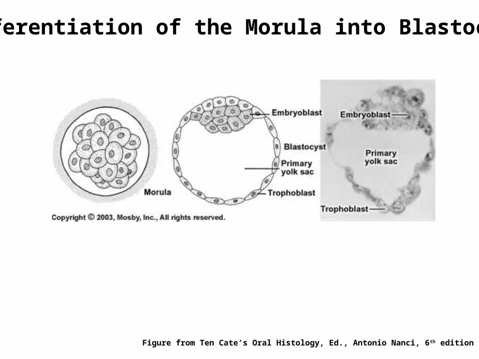
Differentiation of the Morula into Blastocyst
Figure from Ten Cate’s Oral Histology, Ed., Antonio Nanci, 6th edition
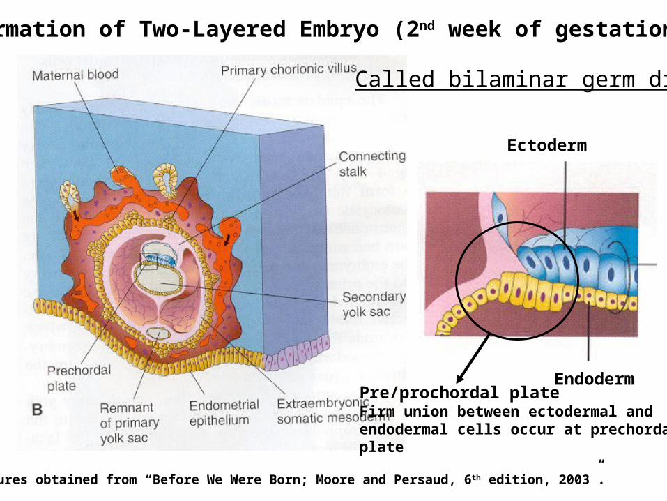
Formation of Two-Layered Embryo (2nd week of gestation)
Figures obtained from “Before We Were Born; Moore and Persaud, 6 th edition, 2003”.
Called bilaminar germ disk
Ectoderm
EndodermPre/prochordal plateFirm union between ectodermal andendodermal cells occur at prechordalplate
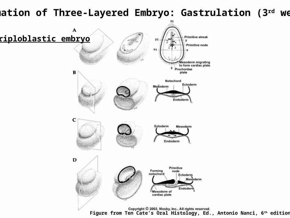
Formation of Three-Layered Embryo: Gastrulation (3rd week)
Figure from Ten Cate’s Oral Histology, Ed., Antonio Nanci, 6th edition
Triploblastic embryo
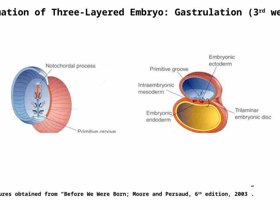
Formation of Three-Layered Embryo: Gastrulation (3rd week)
Figures obtained from “Before We Were Born; Moore and Persaud, 6 th edition, 2003”.
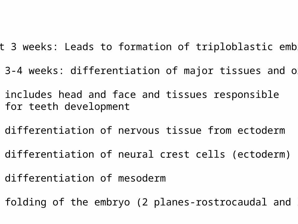
First 3 weeks: Leads to formation of triploblastic embryo
Next 3-4 weeks: differentiation of major tissues and organs
includes head and face and tissues responsiblefor teeth development
differentiation of nervous tissue from ectoderm
differentiation of neural crest cells (ectoderm)
differentiation of mesoderm
folding of the embryo (2 planes-rostrocaudal and lateral)
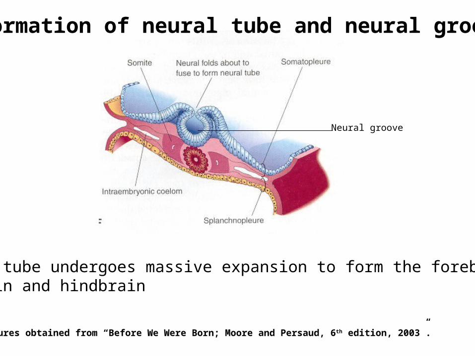
Neural tube undergoes massive expansion to form the forebrain,midbrain and hindbrain
Formation of neural tube and neural groove
Figures obtained from “Before We Were Born; Moore and Persaud, 6 th edition, 2003”.
Neural groove
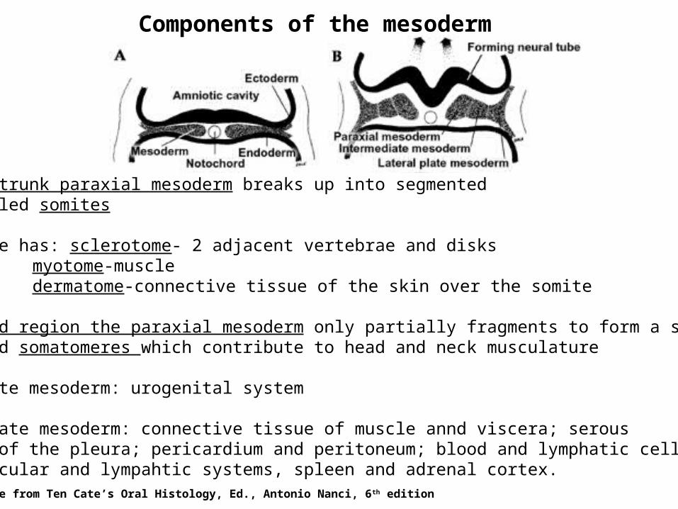
Components of the mesoderm
Along the trunk paraxial mesoderm breaks up into segmentedblocks called somites
Each somite has: sclerotome- 2 adjacent vertebrae and disks myotome-muscledermatome-connective tissue of the skin over the somite
In the head region the paraxial mesoderm only partially fragments to form a seriesof numbered somatomeres which contribute to head and neck musculature
Intermediate mesoderm: urogenital system
Lateral plate mesoderm: connective tissue of muscle annd viscera; serousmembranes of the pleura; pericardium and peritoneum; blood and lymphatic cells; cardiovascular and lympahtic systems, spleen and adrenal cortex.
Figure from Ten Cate’s Oral Histology, Ed., Antonio Nanci, 6th edition
In the head, the neural tube undergoes massive expansion to formthe forebrain, midbrain and hindbrain
The hindbrain segments into series of eight bulges calledrhombomeres which play an important role in development of the head
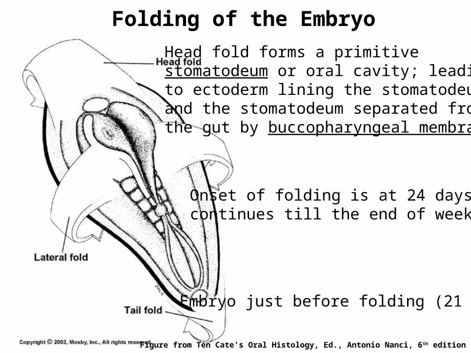
Folding of the Embryo
Head fold forms a primitivestomatodeum or oral cavity; leadingto ectoderm lining the stomatodeumand the stomatodeum separated fromthe gut by buccopharyngeal membrane
Onset of folding is at 24 days andcontinues till the end of week 4
Embryo just before folding (21 days)
Figure from Ten Cate’s Oral Histology, Ed., Antonio Nanci, 6th edition
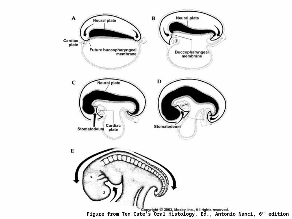
Figure from Ten Cate’s Oral Histology, Ed., Antonio Nanci, 6th edition
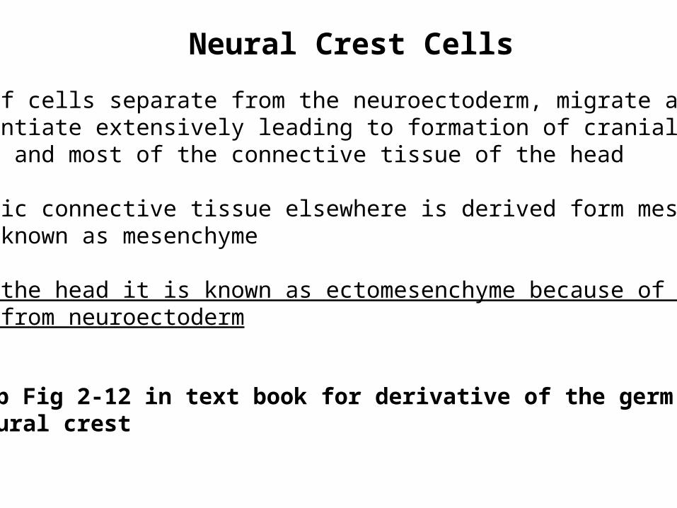
Neural Crest Cells
Group of cells separate from the neuroectoderm, migrate anddifferentiate extensively leading to formation of cranial sensoryganglia and most of the connective tissue of the head
Embryonic connective tissue elsewhere is derived form mesodermand is known as mesenchyme
But in the head it is known as ectomesenchyme because of itsorigin from neuroectoderm
Look up Fig 2-12 in text book for derivative of the germ layersand neural crest
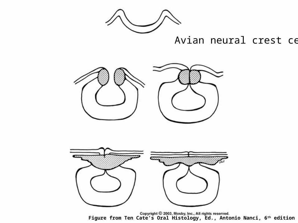
Figure from Ten Cate’s Oral Histology, Ed., Antonio Nanci, 6th edition
Avian neural crest cells
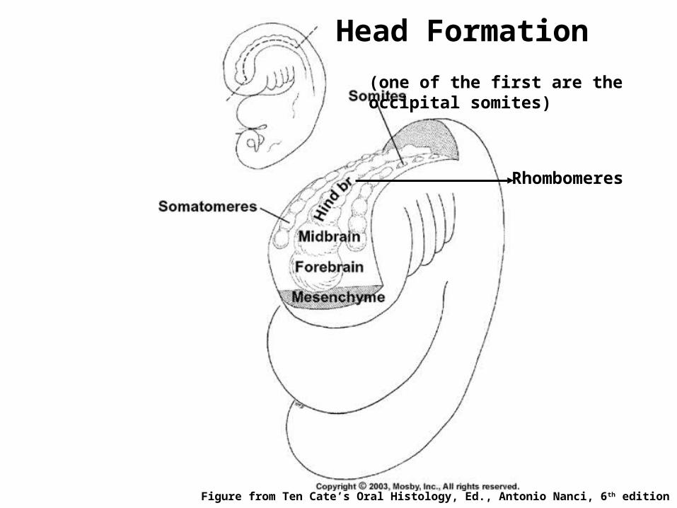
Head Formation
Figure from Ten Cate’s Oral Histology, Ed., Antonio Nanci, 6th edition
Rhombomeres
(one of the first are theoccipital somites)
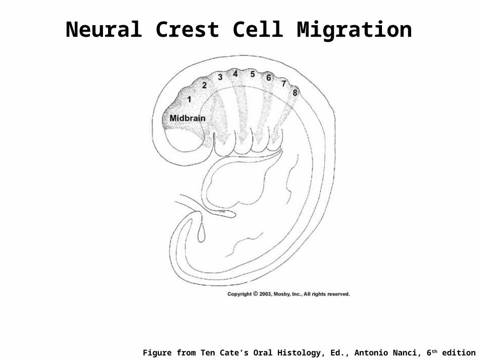
Neural Crest Cell Migration
Figure from Ten Cate’s Oral Histology, Ed., Antonio Nanci, 6th edition
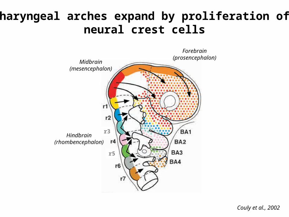
Pharyngeal arches expand by proliferation of neural crest cells
Couly et al., 2002
Forebrain(prosencephalon)
Midbrain(mesencephalon)
Hindbrain(rhombencephalon)
r3
r5
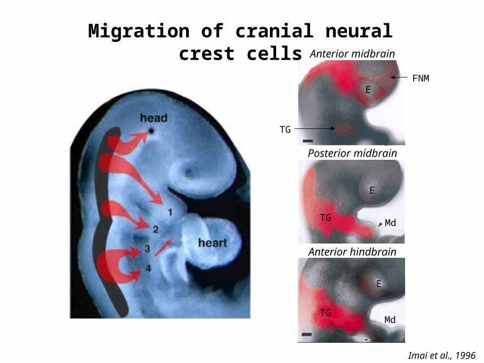
Migration of cranial neural crest cellsAnterior midbrain
Posterior midbrain
Anterior hindbrain
Imai et al., 1996
E
E
E
FNM
TG
TG
TG
Md
Md
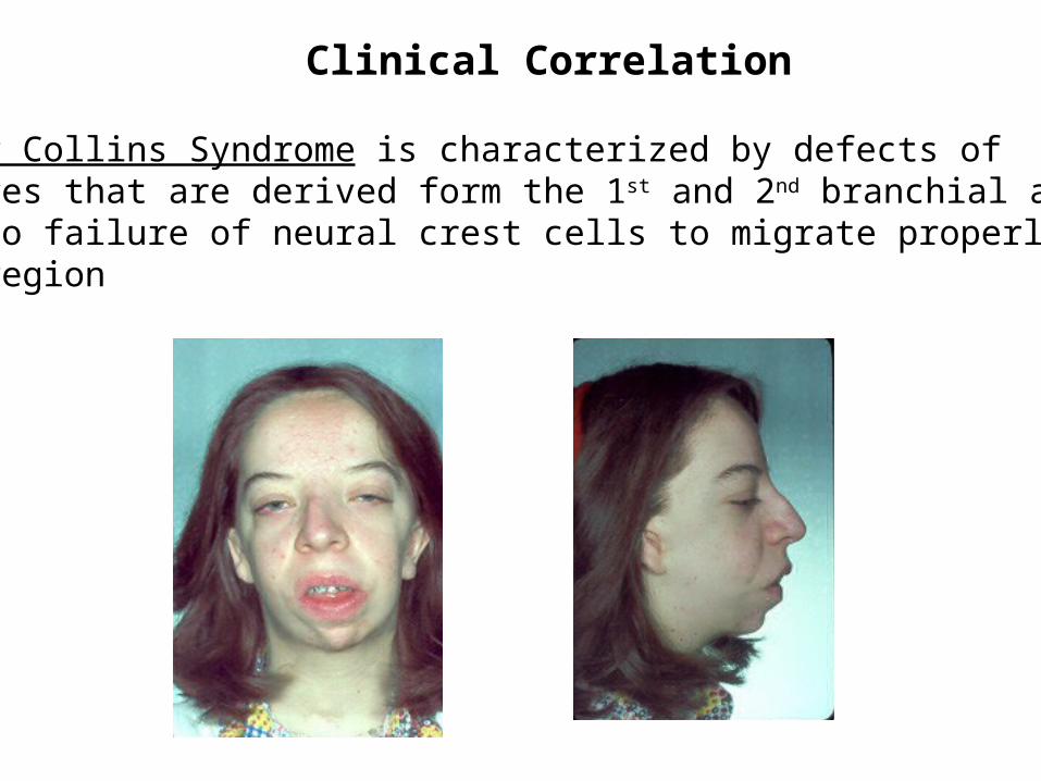
Clinical Correlation
Treacher Collins Syndrome is characterized by defects ofstructures that are derived form the 1st and 2nd branchial arches andis due to failure of neural crest cells to migrate properly to thefacial region
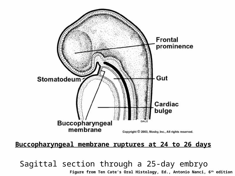
Sagittal section through a 25-day embryoFigure from Ten Cate’s Oral Histology, Ed., Antonio Nanci, 6th edition
Buccopharyngeal membrane ruptures at 24 to 26 days

Internal View of the Oral Pit at 3.5 weeks
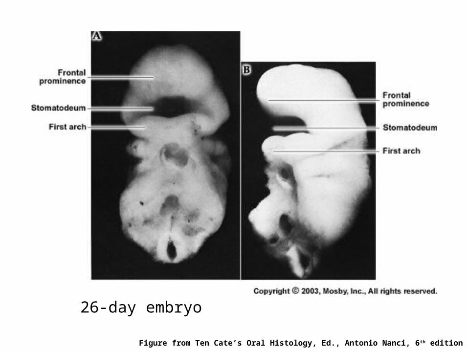
26-day embryo
Figure from Ten Cate’s Oral Histology, Ed., Antonio Nanci, 6th edition
The Developing Human by Moore & Persaud
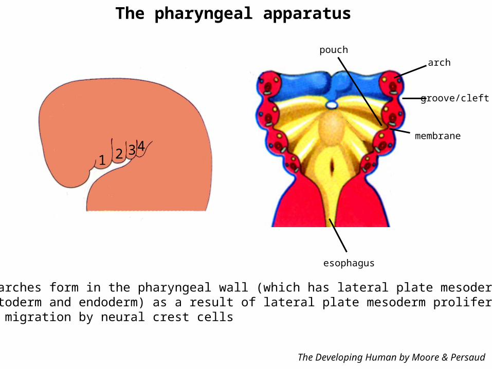
groove/cleft
poucharch
membrane
esophagus
The pharyngeal apparatus
1 2 34
Branchial arches form in the pharyngeal wall (which has lateral plate mesoderm sandwichedbetween ectoderm and endoderm) as a result of lateral plate mesoderm proliferation andsubsequent migration by neural crest cells
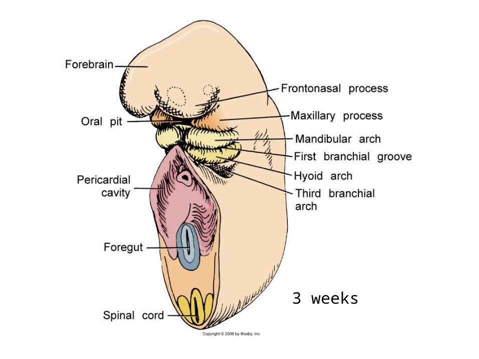
3 weeks
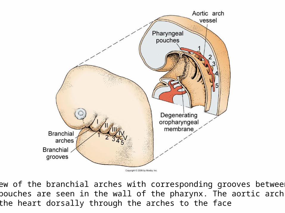
Sagittal view of the branchial arches with corresponding grooves between each arch.Pharyngeal pouches are seen in the wall of the pharynx. The aortic arch vasculatureleads from the heart dorsally through the arches to the face
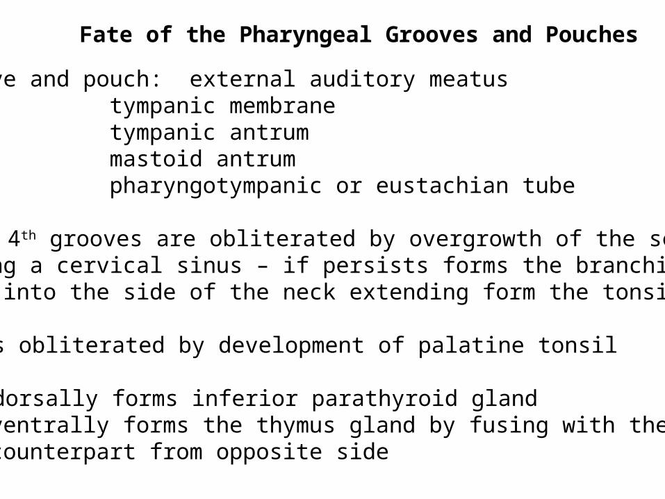
Fate of the Pharyngeal Grooves and Pouches
First groove and pouch: external auditory meatus tympanic membrane tympanic antrum mastoid antrum pharyngotympanic or eustachian tube
2nd, 3rd and 4th grooves are obliterated by overgrowth of the secondarch forming a cervical sinus – if persists forms the branchial fistulathat opens into the side of the neck extending form the tonsillar sinus
2nd pouch is obliterated by development of palatine tonsil
3rd pouch: dorsally forms inferior parathyroid gland ventrally forms the thymus gland by fusing with the counterpart from opposite side
4th pouch: dorsal gives rise to the superior parathyroid gland ventral gives rise to the ultimobranchial body (which gives rise to the parafollicular cells of the thyroid gland)
5th pouch in humans is incorporated with the 4th pouch
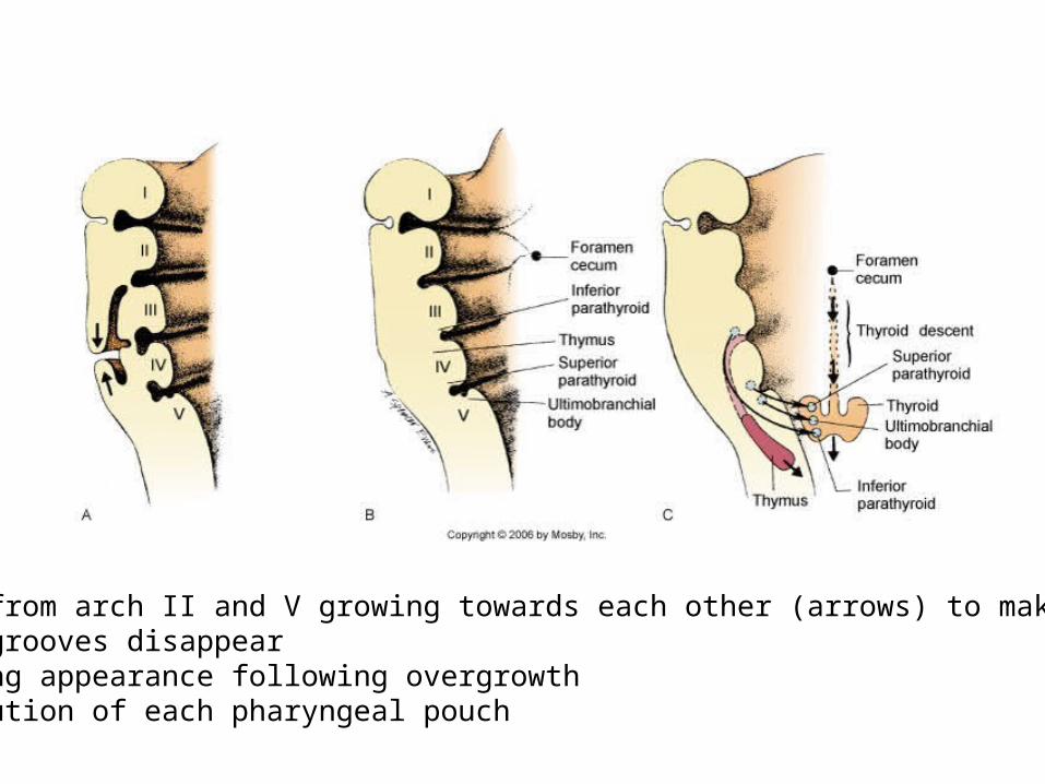
(A) Tissue from arch II and V growing towards each other (arrows) to make branchialarches and grooves disappear(B) Resulting appearance following overgrowth(C) Contribution of each pharyngeal pouch
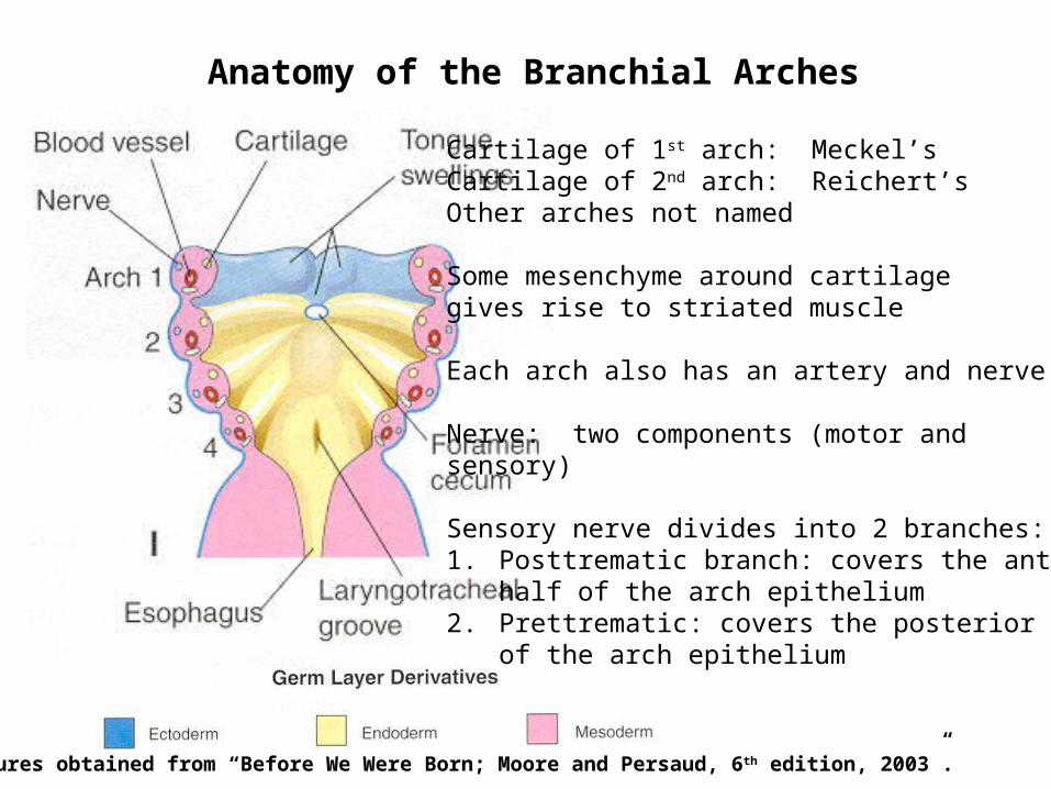
Anatomy of the Branchial Arches
Cartilage of 1st arch: Meckel’s Cartilage of 2nd arch: Reichert’s Other arches not named
Some mesenchyme around cartilagegives rise to striated muscle
Each arch also has an artery and nerve
Nerve: two components (motor andsensory)
Sensory nerve divides into 2 branches:1. Posttrematic branch: covers the anterior
half of the arch epithelium2. Prettrematic: covers the posterior half
of the arch epithelium
Figures obtained from “Before We Were Born; Moore and Persaud, 6 th edition, 2003”.
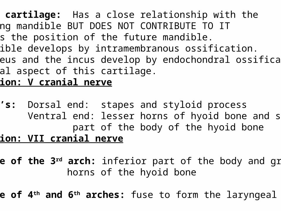
Meckel’s cartilage: Has a close relationship with thedeveloping mandible BUT DOES NOT CONTRIBUTE TO ITIndicates the position of the future mandible.The mandible develops by intramembranous ossification.The malleus and the incus develop by endochondral ossification ofthe dorsal aspect of this cartilage. Innervation: V cranial nerve
Reichert’s: Dorsal end: stapes and styloid process Ventral end: lesser horns of hyoid bone and superior
part of the body of the hyoid boneInnervation: VII cranial nerve
Cartilage of the 3rd arch: inferior part of the body and greater horns of the hyoid bone
Cartilage of 4th and 6th arches: fuse to form the laryngeal cartilage
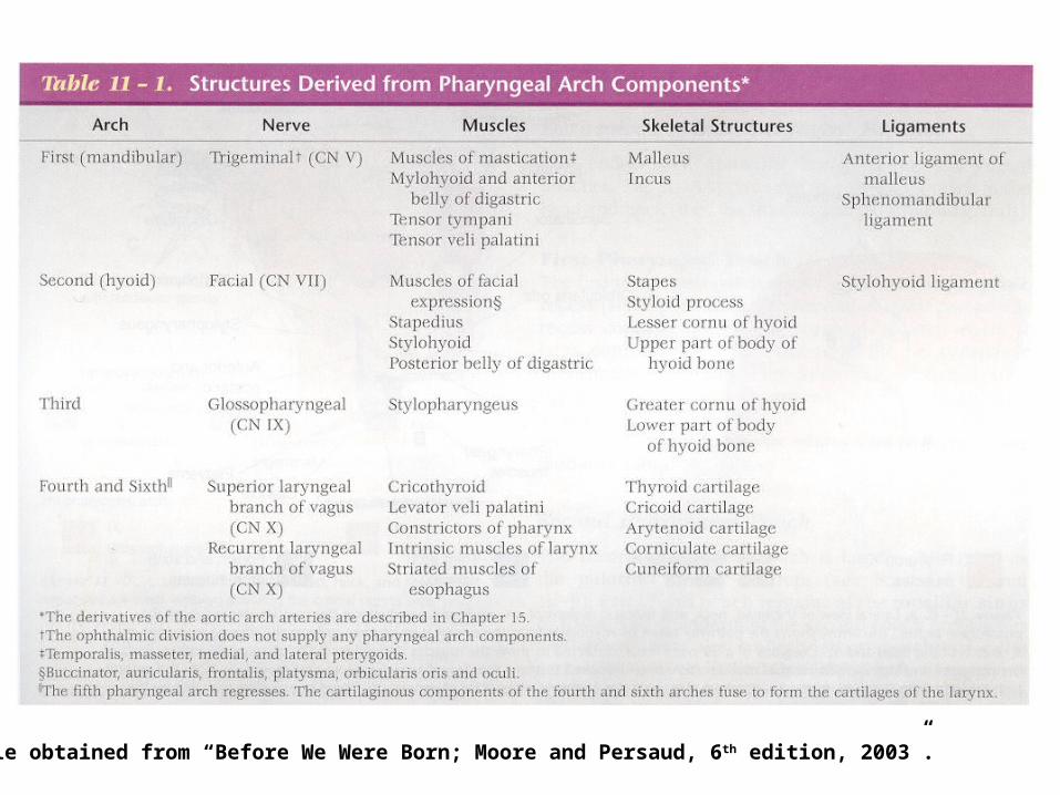
Table obtained from “Before We Were Born; Moore and Persaud, 6th edition, 2003”.

Aortic Vasculature Development
(A) At 4 weeks the anterior vessels have passed through each branchial arch tissueand have disappeared. The pouches project laterally between each arch.
(B) At 5 weeks the 3rd branchial arch vessel becomes the common carotid, whichsupplies the face by means of the internal carotid and stapedial arteries.
Face, Neck and Brain are supplied by the common carotid through internal carotid.But by 7 weeks the circulation of face and neck shifts from the internal carotid toexternal carotid. The internal carotid continues to supply the brain.
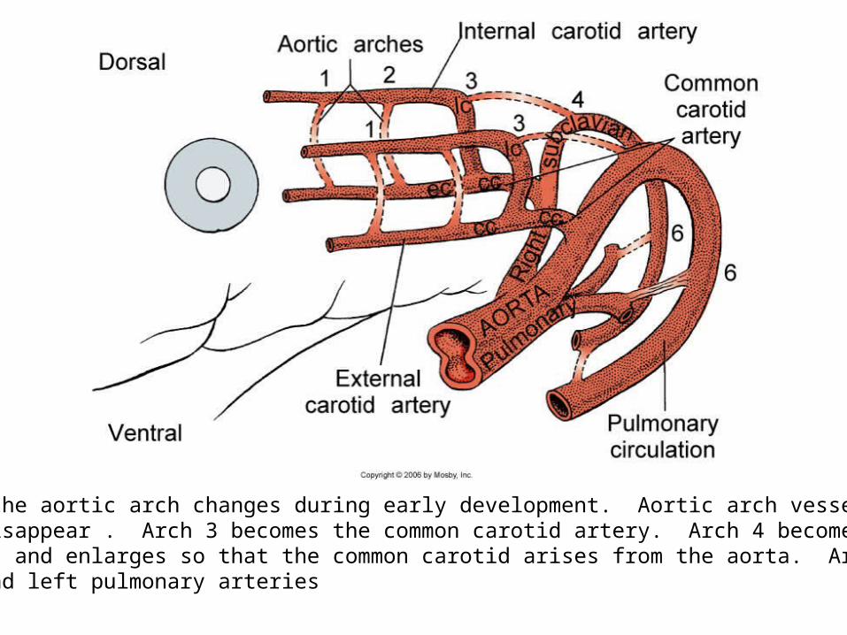
Details of the aortic arch changes during early development. Aortic arch vessels numbers1,2 and 5 disappear . Arch 3 becomes the common carotid artery. Arch 4 becomes thedorsal aorta and enlarges so that the common carotid arises from the aorta. Arch 6 becomesthe right and left pulmonary arteries

Shift in the vascular supply to the face
(A)Face and brain are supplied first by the internal carotid artery
(B) Facial vessels detach from the internal carotid and attach to theexternal carotid

Muscle cells in the first arch become apparentduring the 5th week and begin to spread withinthe mandibular arch into each muscle site’sorigin in the 6th and 7th week. These form themuscles of mastication – masseter, medialpterygoid, lateral pterygoid and temporalismuscle. They all relate to the developing mandible
By 7 weeks the muscles of 2nd arch growupward to form the muscles of face.As these muscles grow and expand theyforms sheet over the face and forms themuscles of facial expression
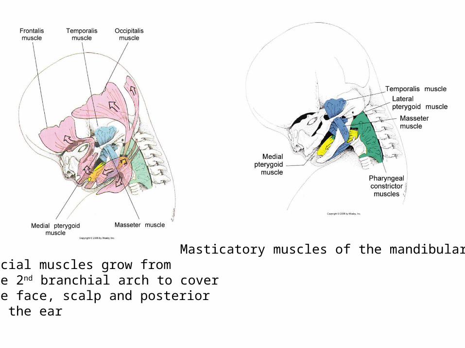
Facial muscles grow fromthe 2nd branchial arch to coverthe face, scalp and posteriorto the ear
Masticatory muscles of the mandibular arch

Cranial Nerves growing into Branchial Arches
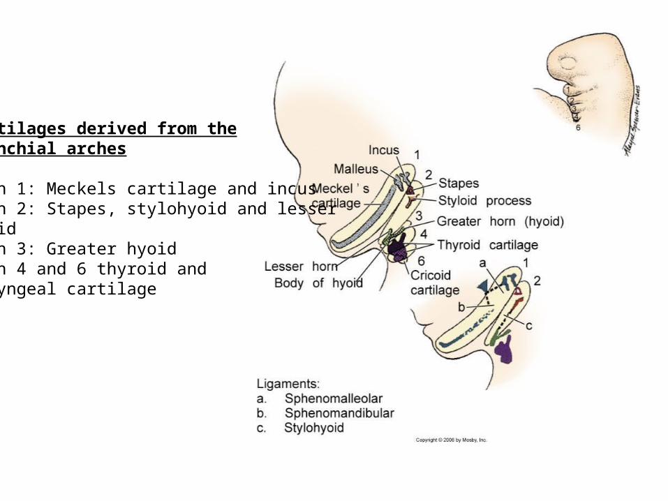
Cartilages derived from thebranchial arches
Arch 1: Meckels cartilage and incusArch 2: Stapes, stylohyoid and lesserhyoidArch 3: Greater hyoidArch 4 and 6 thyroid andlaryngeal cartilage
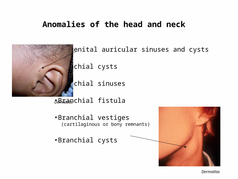
•Congenital auricular sinuses and cysts
•Branchial cysts
•Branchial sinuses
•Branchial fistula
•Branchial vestiges (cartilaginous or bony remnants)
•Branchial cysts
Anomalies of the head and neck
Dermatlas
Dermatlas
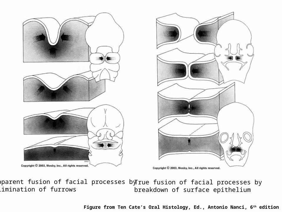
Apparent fusion of facial processes byelimination of furrows
True fusion of facial processes bybreakdown of surface epithelium
Figure from Ten Cate’s Oral Histology, Ed., Antonio Nanci, 6th edition
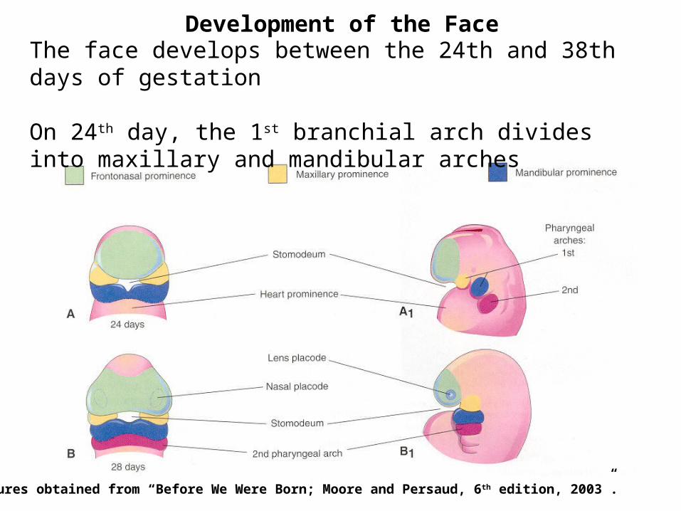
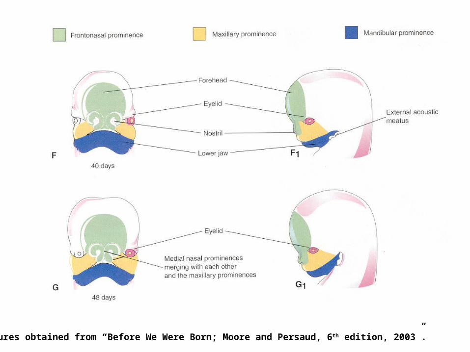
Development of the FaceThe face develops between the 24th and 38th days of gestation
On 24th day, the 1st branchial arch divides into maxillary and mandibular arches
Figures obtained from “Before We Were Born; Moore and Persaud, 6 th edition, 2003”.
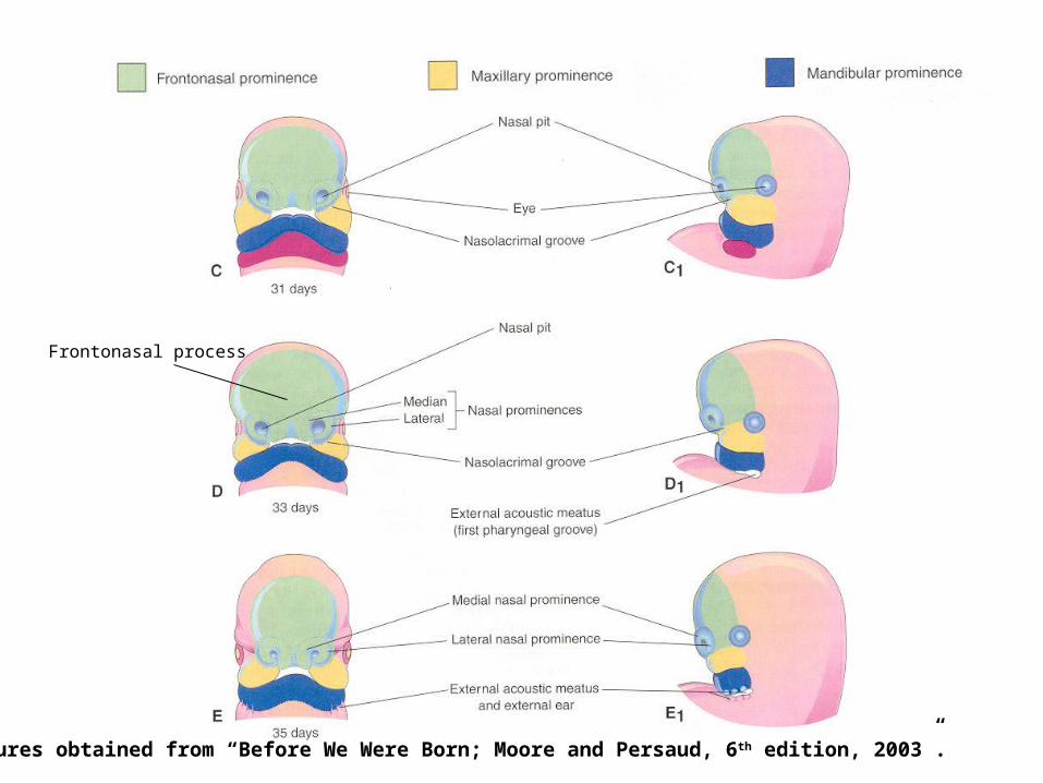
Frontonasal process
Figures obtained from “Before We Were Born; Moore and Persaud, 6 th edition, 2003”.
Figures obtained from “Before We Were Born; Moore and Persaud, 6 th edition, 2003”.
Figures obtained from “Before We Were Born; Moore and Persaud, 6 th edition, 2003”.
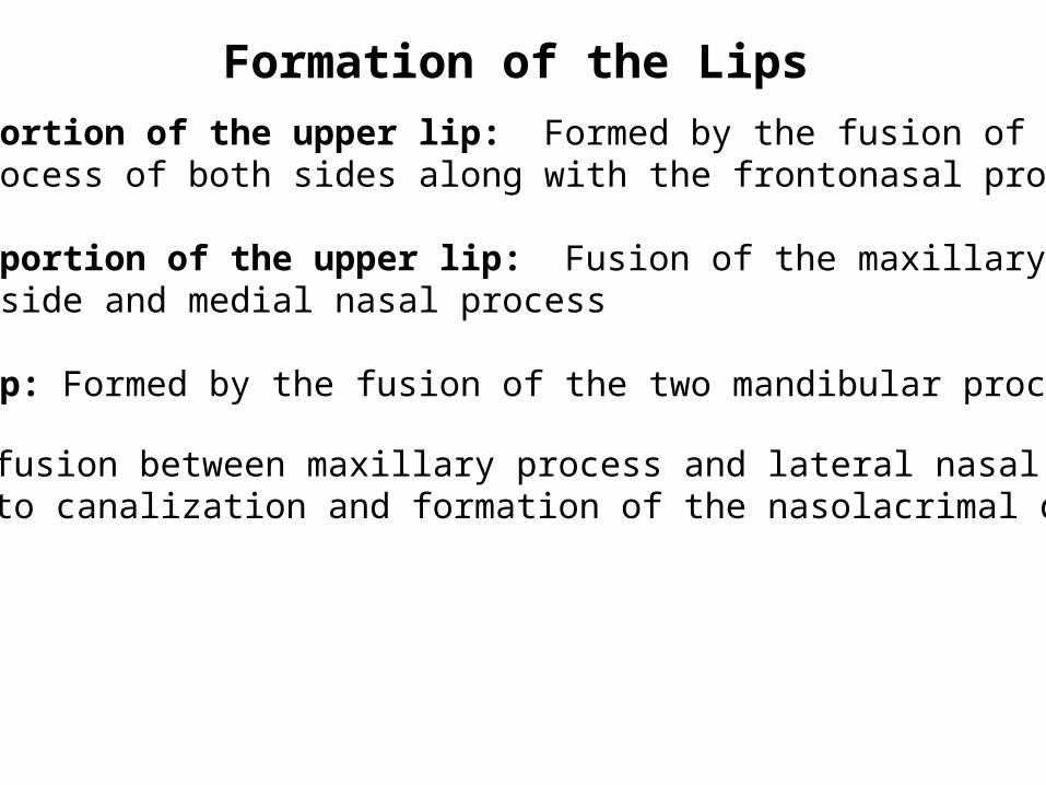
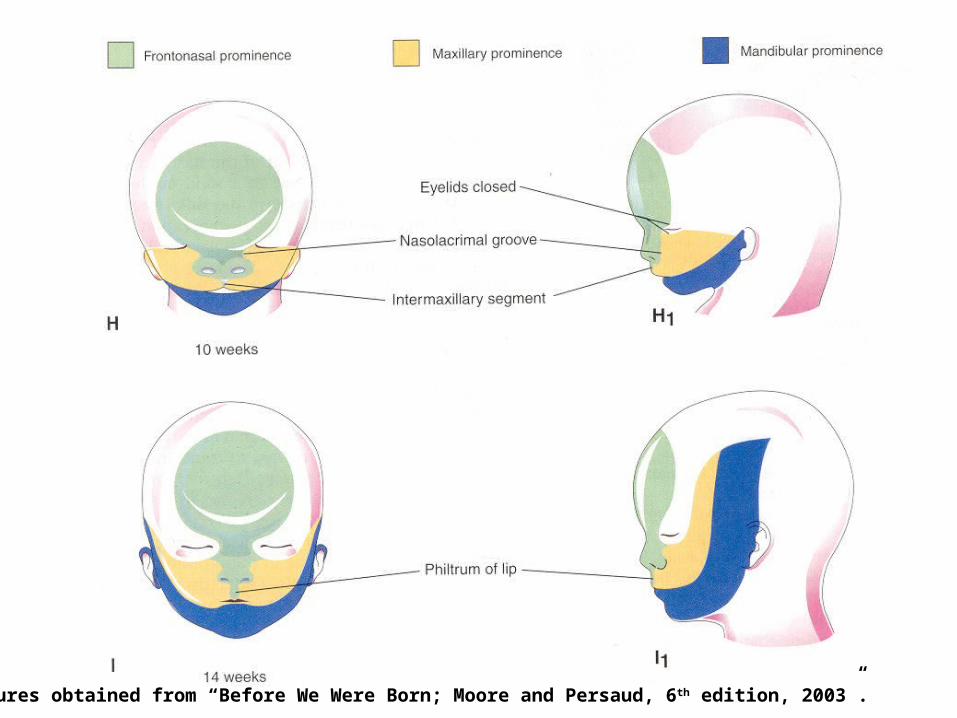
Middle portion of the upper lip: Formed by the fusion of the medialnasal process of both sides along with the frontonasal process
Lateral portion of the upper lip: Fusion of the maxillary processesof each side and medial nasal process
Lower lip: Formed by the fusion of the two mandibular processes
Formation of the Lips
Unusual fusion between maxillary process and lateral nasal processleading to canalization and formation of the nasolacrimal duct

Human embryo at 7 weeks
Figure from Ten Cate’s Oral Histology, Ed., Antonio Nanci, 6th edition

Cleft Lip
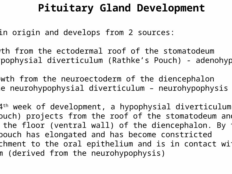
Pituitary Gland Development
Ectodermal in origin and develops from 2 sources:
1. An upgrowth from the ectodermal roof of the stomatodeumcalled hypophysial diverticulum (Rathke’s Pouch) - adenohypophysis
2. A downgrowth from the neuroectoderm of the diencephaloncalled the neurohypophysial diverticulum – neurohypophysis
During the 4th week of development, a hypophysial diverticulum(Rathke’s pouch) projects from the roof of the stomatodeum and liesadjacent to the floor (ventral wall) of the diencephalon. By the 5th
week, this pouch has elongated and has become constrictedat its attachment to the oral epithelium and is in contact with theinfundibulum (derived from the neurohypophysis)
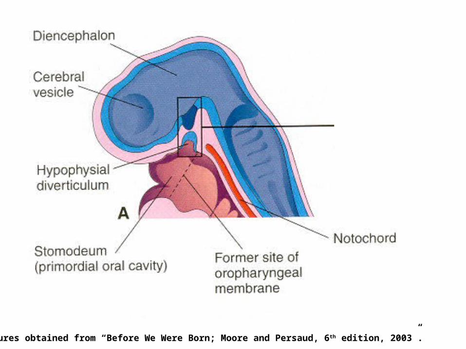
Figures obtained from “Before We Were Born; Moore and Persaud, 6 th edition, 2003”.

Figures obtained from “Before We Were Born; Moore and Persaud, 6 th edition, 2003”.
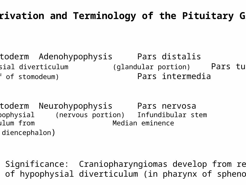
Derivation and Terminology of the Pituitary Gland
Oral Ectoderm Adenohypophysis Pars distalis(hypophysial diverticulum (glandular portion) Pars tuberalisfrom roof of stomodeum) Pars intermedia
Neuroectoderm Neurohypophysis Pars nervosa(neurohypophysial (nervous portion) Infundibular stemdiverticulum from Median eminence
floor of diencephalon)
Clinical Significance: Craniopharyngiomas develop from remnantsof stalk of hypophysial diverticulum (in pharynx of sphenoid bone)
Formation of the palate (weeks 7 to 9)
Palate develops from the primary palate and the secondary palate
The primary palate develops at about 28 days of gestation
Primary palate develops from the frontonasal and medial nasalprocesses and eventually forms the premaxillary portion of the maxilla
The secondary palate develops between 7th and 8th week of gestationand completes in the 3rd month
The critical period of palate development is from the end of 6th weektill the beginning of 9th week
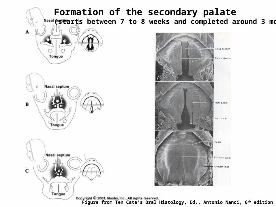
Formation of the secondary palate(starts between 7 to 8 weeks and completed around 3 months)
Figure from Ten Cate’s Oral Histology, Ed., Antonio Nanci, 6th edition
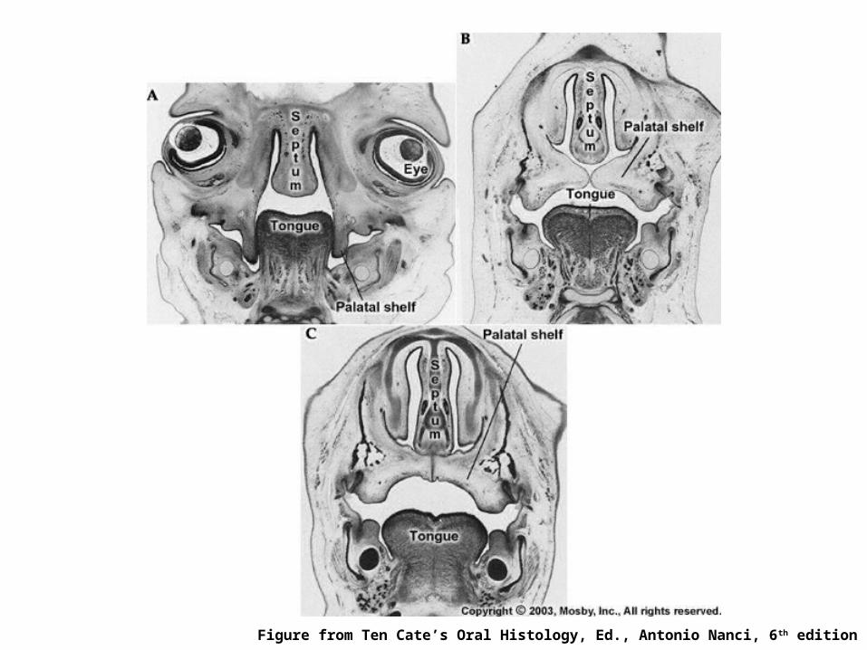
Figure from Ten Cate’s Oral Histology, Ed., Antonio Nanci, 6th edition
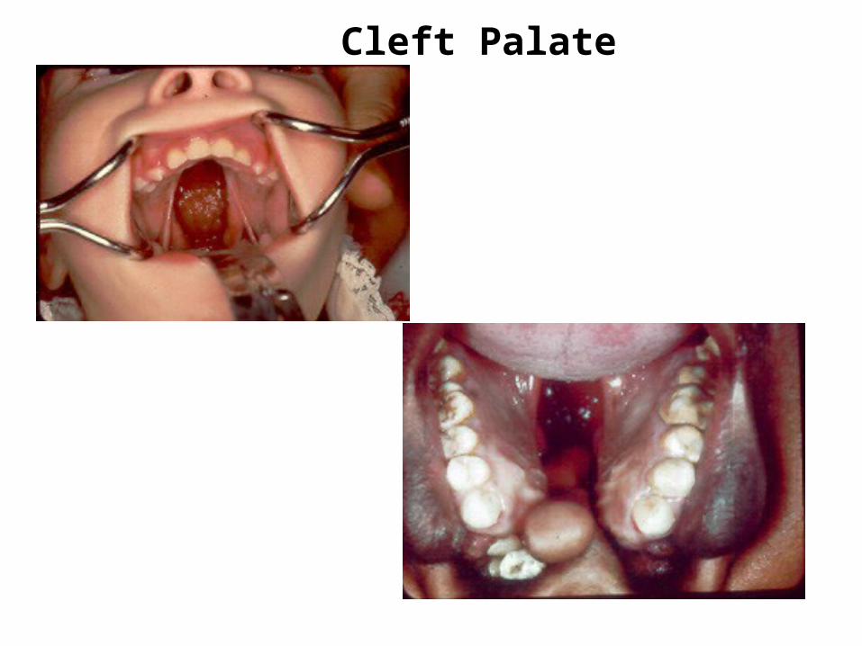
Cleft Palate
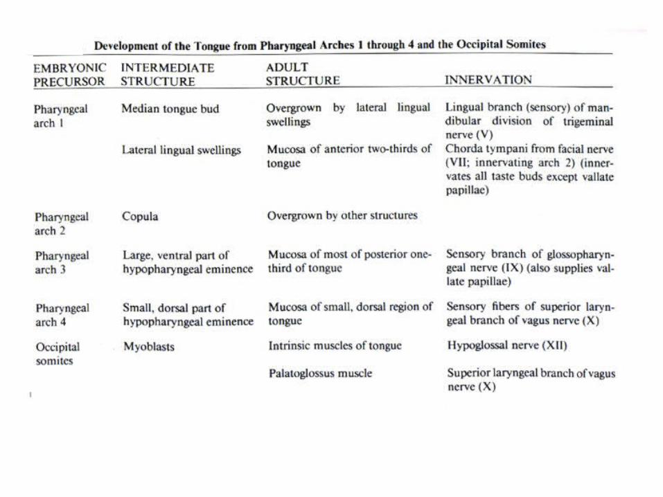
Formation of the Tongue
The tongue begins to develop at about 4 weeks. The oral part (anterior two-thirds) develops from two distal tongue buds (lateral lingual swellings) and a median tongue bud (tuberculum impar) [1st branchial arch].Innervation: V nerve The pharyngeal part develops from the copula and the hypobranchial eminence [2nd, 3rd and 4th branchial arches]. Innervation: IX cranial nerveThe line of fusion of the oral and pharyngeal parts of the tongue is roughly indicated in the adult by a V-shaped line called the terminal sulcus.At the apex of the terminal sulcus is the foramen cecum.
Muscles of the tongue develop form the occipital somites and innervated by hypoglossal nerve
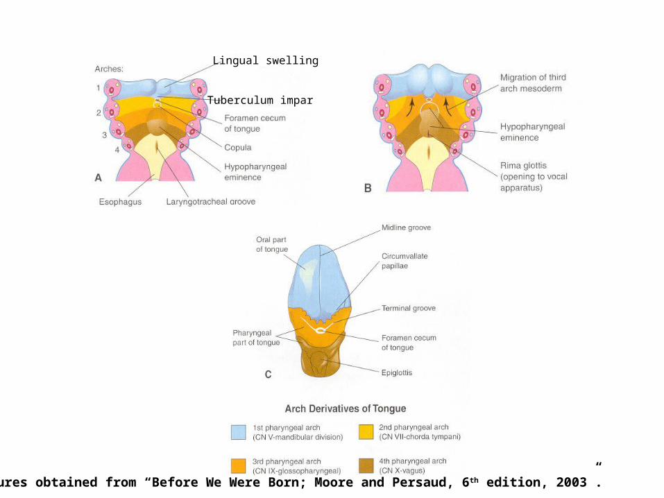
Lingual swelling
Tuberculum impar
Figures obtained from “Before We Were Born; Moore and Persaud, 6 th edition, 2003”.
The lingual papillae appear by the end of 8th week
Vallate and foliate papillae appear first, fungiform andfiliform (10-11 weeks) papillae appear later
Taste buds develop during the 11 to 13 weeks by inductiveinteraction between epithelial cells of the tongue and invadinggustatory nerve cells from chorda tympani, glossopharyngealand vagus nerves
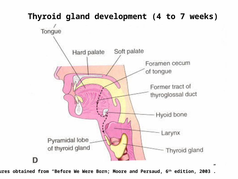
Thyroid gland development (4 to 7 weeks)
Figures obtained from “Before We Were Born; Moore and Persaud, 6 th edition, 2003”.
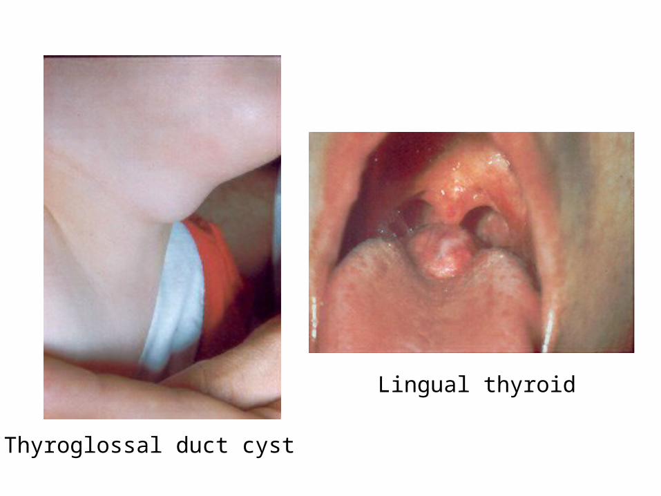
Thyroglossal duct cyst
Lingual thyroid
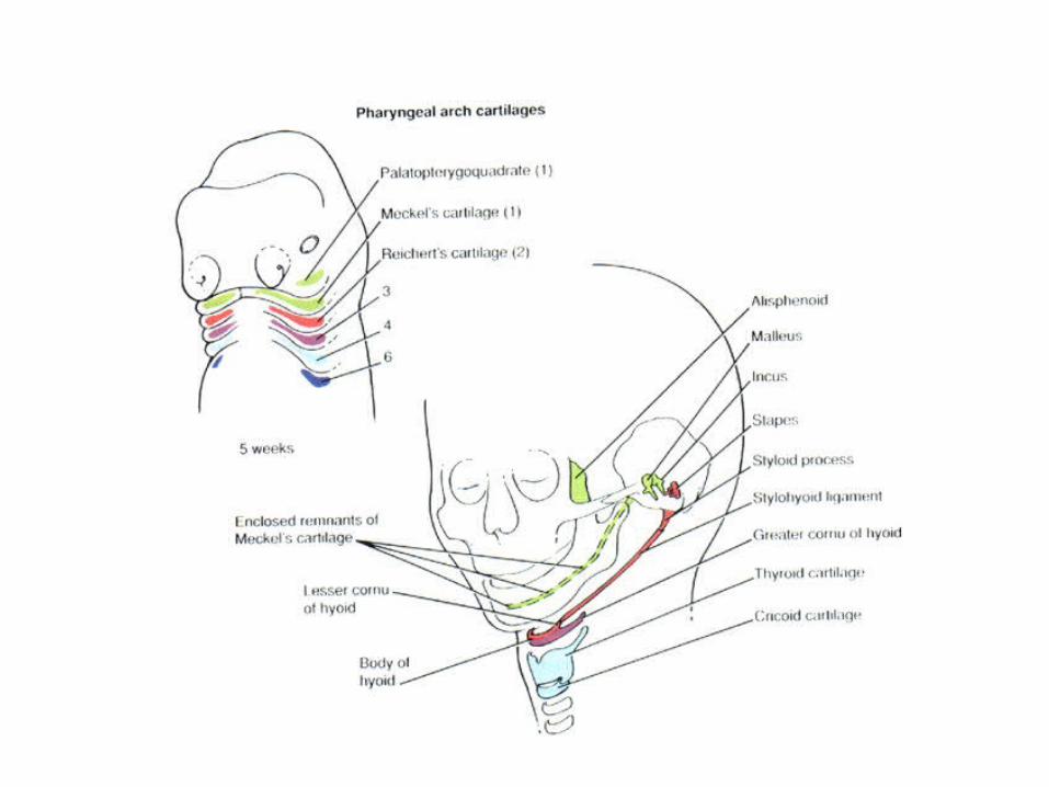
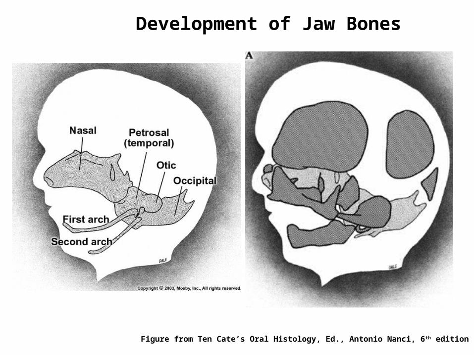
Figure from Ten Cate’s Oral Histology, Ed., Antonio Nanci, 6th edition
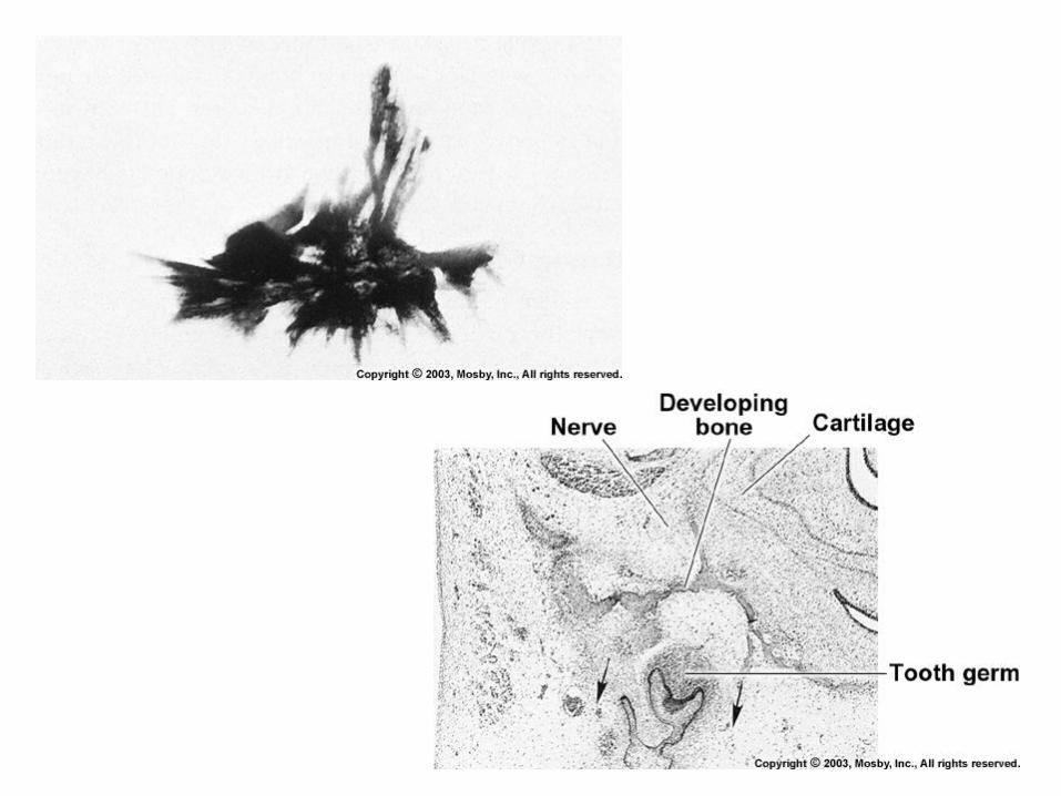
Development of Jaw Bones
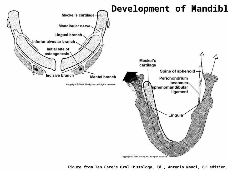
Figure from Ten Cate’s Oral Histology, Ed., Antonio Nanci, 6th edition
Development of Mandible

Figure from Ten Cate’s Oral Histology, Ed., Antonio Nanci, 6th edition
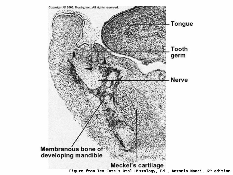
Figure from Ten Cate’s Oral Histology, Ed., Antonio Nanci, 6th edition
Fate of Meckel’s CartilagePosterior – malleus of the inner ear
Sphenomandibular ligament
Anteriorly, may contribute to mandibleby endochondral ossification (some evidence)
Rest are resorbed completely
Three secondary (growth) cartilages govern further growth ofmandible until birth
1. Condylar cartilage (most important)2. Coronoid cartilage3. Symphysial cartilage
Secondary Cartilages
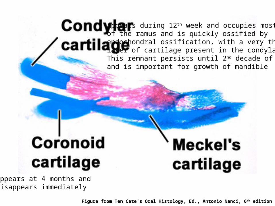
Appears during 12th week and occupies mostof the ramus and is quickly ossified byendochondral ossification, with a very thinlayer of cartilage present in the condylar head.This remnant persists until 2nd decade of lifeand is important for growth of mandible
Appears at 4 months anddisappears immediately
Figure from Ten Cate’s Oral Histology, Ed., Antonio Nanci, 6th edition
Development of Maxilla
Develops from one center of ossification in maxillary process ofthe 1st branchial arch
Center of ossification is angle between the divisions where theanterosuperior dental nerve is given off from inferior orbital nervefrom where it spreads posteriorly, anteriorly and superiorly
No arch cartilage is present, so maxilla develops in closeassociation with the nasal cartilage
One secondary cartilage also contributes to maxilladevelopment: zygomatic cartilage