hands-on workshop utilizing the prism cover test and … 2015 workshop 2-13... · hands-on workshop...
TRANSCRIPT

Hands-on workshop utilizing the
prism cover test and prism
therapeutics for the diplopic
patient
NANOS 2015

Introduction : Evaluation of the diplopic
patient is time consuming, demanding and frustrating
Understanding how to perform the prism cover test, determining fusional amplitudes, differentiating phoriafrom tropia, use of the PAT, placing press on prisms and prescribing prisms is an art

Goals:Explain the evaluation of diplopic patients where
prisms are useful in diagnosis and treatment
Compare different types of prisms available and their utility in examining and treating patients with diplopia
Describe the different prism cover tests and their use in differentiating phoria from tropia.
Faculty:Shelley Klein, CO – 4th and 6th nerve palsy
Erika Acera, CO – Thyroid orbitopathy
Shira Robbins, MD – Divergence insufficiency
David Granet, MD – Convergence insufficiency
Mitchell Strominger, MD – Diplopia following cataract extraction
No financial disclosures
Shira Robbins, MD AAP - Royalties
US Dept Health and Human Services - Consultant
Allergan – Consultant
Retrophin – Advisory Board
Equipment discussed and demonstrated is purely operational and not promotional

Special Thanks !Lloyd Powell,
President
Richmond Products
Bianca Granado
Marketing Manger
Richmond Products
Kathy Armstrong
President
Fresnel Prism and Lens Company

6
The Prism cover Test
Shelley Klein, CO
Tufts Medical Center
Boston, MA

The Prism Cover Test
Neutralization of deviation with prisms by
optically moving the image onto the fovea
7
F1 F2
F1

The Prism
• Triangular or wedge shaped piece of refracting material
• Thickest edge of the prism → BASE
• Thinnest edge of the prism → APEX
• A prism changes the direction of light without changing it’s focus
• Prism is created when the front surface is not parallel to the back surface
8
APEXBASE

The Prism
Prism is measured in units called “prism diopters (pd)”
Defined as 1pd yields a deviation of 1cm @ 1M.
In viewing an object through a prism, the image is displaced toward the apex.
On the retina, the image is moved towards the base.
9
OS
The Prism
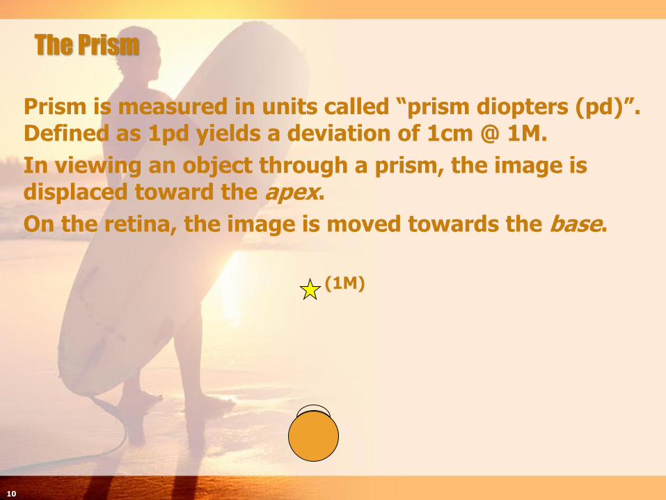
Prism is measured in units called “prism diopters (pd)”. Defined as 1pd yields a deviation of 1cm @ 1M.
In viewing an object through a prism, the image is displaced toward the apex.
On the retina, the image is moved towards the base.
(1M)
10

The Prism
Prism is measured in units called “prism diopters (pd)”. Defined as 1pd yields a deviation of 1cm @ 1M.
In viewing an object through a prism, the image is displaced toward the apex.
On the retina, the image is moved towards the base.
11
F1
The Prism
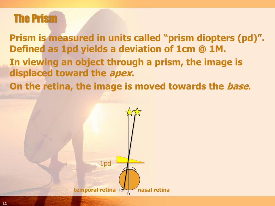
Prism is measured in units called “prism diopters (pd)”. Defined as 1pd yields a deviation of 1cm @ 1M.
In viewing an object through a prism, the image is displaced toward the apex.
On the retina, the image is moved towards the base.
1pd
temporal retina nasal retina
12
F1F2

The Prism
Prism is measured in units called “prism diopters (pd)”. Defined as 1pd yields a deviation of 1 cm @ 1 meter.
In viewing an object through a prism, the image is displaced toward the apex.
On the retina, the image is moved towards the base.
1cm at 1M
1pd
13
F1F2
The Prism
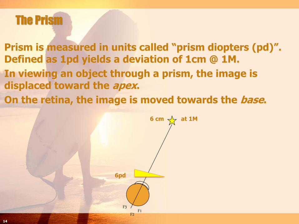
Prism is measured in units called “prism diopters (pd)”. Defined as 1pd yields a deviation of 1cm @ 1M.
In viewing an object through a prism, the image is displaced toward the apex.
On the retina, the image is moved towards the base.
6 cm at 1M
6pd
14
F1F2F3

The Cover Test
Prerequisites for a reliable Cover Test
- eye movement capability
- image formation and perception
- foveal fixation in each eye
- eliminating accommodative influences
- attention and cooperation
Most effective way to suspend fusion
Each eye should be occluded at least 2 secs
15
Types of Cover Tests
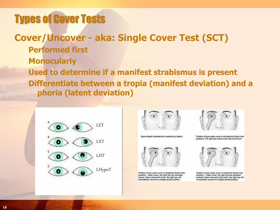
Cover/Uncover - aka: Single Cover Test (SCT)
Performed first
Monocularly
Used to determine if a manifest strabismus is present
Differentiate between a tropia (manifest deviation) and a phoria (latent deviation)
16
LET
LXT
LHT
LHypoT

Types of Cover Tests
Simultaneous Prism Cover Test (SPCT)
Measures the angle under normal binocular conditions
As the occluder covers the fixing eye, prisms are placed in front of the deviated eye at the same time to neutralize turn
Most useful with Accommodative Esotropia and Monofixation Syndrome (usually angle < 10pd)
Patient may be demonstrating partial control over a coexisting heterophoria through peripheral fusion binocularly.
“Cosmetic deviation”
17

Types of Cover Tests
Prism Cover Test (PCT)
a.k.a. Alternate Prism Cover Test (APCT)
Measures the total deviation (manifest and latent components)
Patients should not be allowed to establish binocularity
Tested at distance (6M) and near
Measurements at Near should include primary position and downgaze. (Measure through bifocal if present)
Measurements at Distance should include primary position plus
Side gazes to determine the presence of any lateral incomitance
Upgaze and downgaze to determine the presence of A or V patterns
If vertical is present head tilt R and L should be performed
If oblique dysfunction is present or suspected measure in the oblique positions
18

Different Prism Options
19
A. Loose prismsC. Fresnel Prism Trial Set
B. Horizontal and Vertical Prism Bars

Alternate Prism Cover Test
Deviation is measured using prisms
The apex of the prism is pointed towards the deviation
Thereby the base will be in the opposite direction.
20
Base OUT - BO
Base DOWN - BDBase IN - BI
Base UP - BU
Esotropia - LETExotropia - LXT
Left Hypertropia - LHT
Left Hypotropia - LHypoT
21
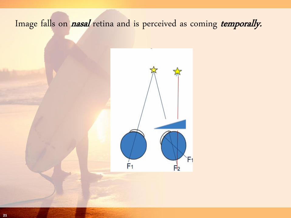
Image falls on nasal retina and is perceived as coming temporally.

22
Adding Base OUT prism moves the image in space towards the apex.While moving the image on the retina towards the base.

23
During PCT, as BO prism is added, the newly uncovered eye will move to pick up fixation. Neutralization of the turn is achieved when the correct amount of prism moves the image onto the fovea. Therebyeliminating the need for refixation.
This amount of prism needed is the measurement of the deviation.

Factors effecting prism and cover measurements
24
• Hold prisms in frontal plane position

Correct Frontal Plane Placement of Prism
25
F1F2F1
F2F1F1F2
F1F1
Right gaze(head turned L)
Left gaze(head turned R)Primary position
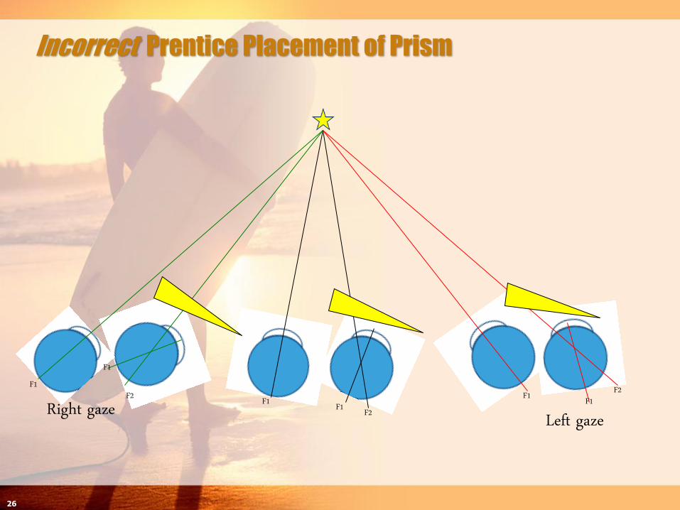
Incorrect Prentice Placement of Prism
26
F1F2F1
F2F1F1F2
F1F1
Right gaze Left gaze

Factors affecting prism and cover
measurements
27
• Hold prisms in frontal plane position
• High refractive errors > 5D

Factors affecting prism and cover
measurements
28
• Hold prisms in frontal plane position
• High refractive errors > 5D
• Stacking prisms > 20 pd, better to split between 2 eyes, OK to stack vertical and horizontal prism < 20pd otherwise, recommend splitting.
Factors affecting prism and cover
measurements
29
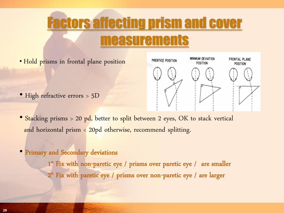
• Hold prisms in frontal plane position
• High refractive errors > 5D
• Stacking prisms > 20 pd, better to split between 2 eyes, OK to stack vertical and horizontal prism < 20pd otherwise, recommend splitting.
• Primary and Secondary deviations1° Fix with non-paretic eye / prisms over paretic eye / are smaller2° Fix with paretic eye / prisms over non-paretic eye / are larger

Primary and Secondary Deviations
Seen in paralytic or restrictive strabismus
Occurs when patient is fixating with the paralytic and/or restricted eye
2° deviations are larger because it takes greater innervation for the paretic/restricted eye to fix on a target
By Hering’s law, an equal amount of innervation is going to the contralateral yoke muscle
1° Fix with non-paretic eye/prisms over paretic eye/smaller
2° Fix with paretic eye/prisms over non-paretic eye/larger
30

Factors affecting prism and cover
measurements
31
• Hold prisms in frontal plane position
• High refractive errors > 5D
• Stacking prisms > 20 pd, better to split between 2 eyes, OK to stack vertical and horizontal prism < 20pd otherwise, recommend splitting.
• Primary and Secondary deviations1° Fix with non-paretic eye / prisms over paretic eye2° Fix with paretic eye / prisms over non-paretic eye
• Angle KappaPositive angle simulates an XTNegative angle simulates an ET

Angle Kappa……may simulate a strabismus
It is the angle formed by the pt’s visual and pupillary axes
Visual axis = line of sight connecting the fixation targetto he fovea
Pupillary axis = the line that passes perpendicular to the center of cornea
When the corneal light reflex is displaced……
Nasally = Positive angle simulates an XT
Temporally = Negative angle simulates an ET
32

Angle Kappa Generalizations
Positive angle kappa is most common
Negative angle kappa is more common in high myopes
Most angle kappas are physiologic especially in emmetropes and hyperopes (1.4 to 2.8 degrees is wnl)
Large angle kappa may be caused by retinal traction as in ROP with temporal dragging of the macula
33

The Prism Cover Test: important pearls to remember…
Don’t block pt’s view with your head!
When measuring in lateral gazes, make sure pt can see fixation target with adducted eye!
Dissociate maximally – pt’s have strong compensatory innervation to keep their eyes align
Don’t hurry/repeat APCT a few times if needed
For diagnostic purposes, measure 25 to 30 degrees from primary position, you may not uncover a paresis or restriction if you don’t expand the binocular field to it’s outer limits.
Some examiners like to measure till there is a reversal of the redress, personal preference
Sometimes it’s difficult to be certain of the end point of neutralizing the turn because of a rebound saccade. Do your best estimation.
Positioning of the prism is very important – make certain to hold the prism straight, so you won’t induce a vertical if you’re measuring a purely horizontal deviation and vice versa for a vertical deviation.
Primary and Secondary Deviation – more likely to be seen with a new onset palsy, when monitoring the progression of the palsy from OV to OV, be consistent with your measurements.
Be aware of an Angle Kappa – things may not be what they look like!
Sometimes you can’t or it’s difficult to do APCT – you may need to do a Krimsky
Poor fixation in a blind eye
Nystagmus
34

Use of PrismsMitchell Strominger, MD

Use of prismsGround in or Fresnel press on prisms
Pearls:
Give the least amount of prism that will accomplish steady fusion – start with ½ the measured amount in primary position and increase until steady fusion
Patients typically do not move their eye laterally more than 10 – 12 degrees
If bifocals, make sure that if no diplopia at distance that acceptable at near otherwise may need separate reading and distance glasses

Gound in prism:Split the prism 50:50
unless marked incomitance, then may need to prescribe asymmetrically or put all on one side
Keep amount to 6 prism diopters or less
AR coating might decrease glare

Fresnel press on prism:Inexpensive - $21.00
Temporary
Prism adaptation test
prior to surgery
prior to ground in

Fresnel press on prism:Determine amount
with loose prism
distance – primary
reading
Lines degrade vision
place on non preferred eye
Can tilt for horizontal and vertical components
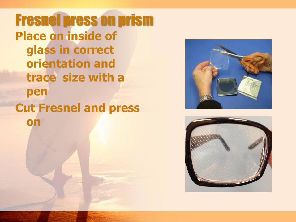
Fresnel press on prismPlace on inside of
glass in correct orientation and trace size with a pen
Cut Fresnel and press on

Case presentations

Thyroid Eye Disease
Erika Acera, CO

36yo with Graves Dz
RAI
Levothyroxine
s/p Bilateral orbital decompressions
c/o constant horizontal & vertical diplopia
Diplopia not getting better

• OS Limitation of abduction & supraduction• OD Mild limitations of abduction/supraduction/infraduction

Treatment of Thyroid OrbitopathyOcclusion
Prisms
Fresnel vs ground in
Surgery

Occlusion Techniques
Alternatives
Tape to lens
‘Pirate Patch’
Min Lens

Occlusion Techniques
Bangerter Occlusion Filters/Foils
Varying strengths to blur diplopic image

Treatment
Large angle ET & Hypotropia
Incomitant deviation
Binocular Single Vision
Base Out 40 & Base Up 25^ OS
Trial of Fresnels –Unable to correct with 1 prism due to large
deviation
need to split prism
Limitations
Aberrations / Blurred vision
Max 30^
Will only correct deviation in primary position – diplopia in other gaze positions

SurgeryBilateral Medial Rectus Recessions
Left Inferior Rectus Recession
Horizontal deviation eliminated
Minimal vertical deviation
Prisms more tolerable
6^ Base Up OS – Single vision
Fresnel prism placed on plano lenses

PrismsIncomitant deviation
Remind patients that they need to make head mvts not eye mvts when looking in different gaze positions
Large angle strabismusBlurred vision >12^
Optical aberrations
Loss of contrast
Comitant deviations
Small-moderate angle strabismus
Cost of Fresnel is much lower than ground-in prism
Fresnel effective in temporary situations – cranial nerve IV, VI palsies
Careful selection of patients for prismatic correction, management of patient’s Expectations and follow up to monitor the diplopia are critical to
the successful use of prisms

Shira Robbins, MD
Divergence Insufficiency

This 60 year-old woman with 2
week hx constant binocular
horizontal diplopia
corresponding to cataract
extraction left eye
D > N
Noted decreased left eye vision 3
years ago, left eye inturning x
18 mo no diplopia as vision so
poor
Case 1

PMHx/PSHx/Meds SLE diag 1977
Retinal exams Q6 mo
Blood work Q3 mo
Head trauma as child
Facial surgery post
trauma 10 years prior
2008 Cataract extraction
+ implant Right eye
2 weeks prior- Cataract
extraction + implant
Left eye
Plaquenil
Cortisone Injections
Paxil
ALLG - Sulfa

VA 20/25
20/80 ph 20/40 (mild myopia)
Color 8/8 OU
Pupils –APD, -anisocoria
SLE – Cornea clear centrally, AC D/Q, PCIOL OU good
position
DFE – Nl OU
Trigeminal fxn symm/nl
Exam

ET 15^
ET 15^
ET 15^ ET 15^
ET 15^
Near: orthoBSV 12 ^BODivergence FA or Nl
Mechanism of Divergence InsuffDeficit of a hypothetical divergence center in the
central nervous system
Tight inelastic medial rectus muscles preventing effective abduction
Aging related lax/sagging lateral recti have been proposed as causative = ARDE (Age related Divergence Esotropia), Sagging Eye Syndrome

Take Away PointsConsider in cases of adult onset diplopia where no
signs of CN6/INO exist, versions full

Convergence Insufficiency
David B Granet, MD

75yo with Parkinson’s
s/p CEIOL OU
c/o Intermittent horizontal diplopia when reading and when using the computer
Patches OD when doing near activities

ExamVA 20/25 OU
Near cc VA J2
EOM – full
SLE – wnl
DFE: wnl
Prism Cover Test
Distance: X4^
Near: add: X(T)’20
NPC: remote
Convergence FusionalAmplitudes
Distance:
Break 20^/Recover 16^
Near:
Unable – dissociated by prism


Treatment for CI
Orthoptic Exercises
Pencil Pushups
Stereograms
Computerized eye exercises (CVS program)
Base out Prisms to improve convergence amplitudes
Limitations – Parkinson’s Dz –physically unable to do exercises

Prisms for CI
Use Base In prisms to help with X(T) at near
How much BI prism to prescribe?Advise against correcting the full deviation at near
Want the patient to use some of their convergence ability
GOAL – enough BI prism to provide the patient with comfortable binocular single vision for near activities
Trial of Fresnel prisms on patient’s bifocals or reading/computer glasses
Patient preferred 5 ^ Base In prism OD; 5^ Base In prism OS -bifocals

Fresnel
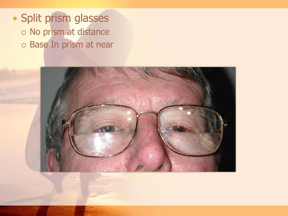
Split prism glasses No prism at distance
Base In prism at near
66
IVth Nerve Palsy
Shelley Klein, COTufts Medical Center
Boston, MA

67
AY 53 yo gentleman
Mar 6 2002
4 year history of diplopia relieved by head tilt to the left
Presumed breakdown of a congenital 4th nerve palsy
Given glasses with 2 base down OD and 2 base up OS
Harder to control over past year
20/20 OU
6 RHT – 25 RHT – 40 RHT
4+ RIOOA, minimal depression deficit on adduction

68
AY April 5 2002 RIO recession
November 2003
Diplopia only when tilting head to the right
3 RH

69
AY
December 22 2010 (8 years later), now 62 yo
Falling a lot with problems controlling diplopia
20/15 OU
5 to 10 degree head tilt to the left
-1.5 underaction of the RSO
PCT:
Ortho 3RH(T) 3RH(T)
builds to 25RHT
14RHT Ortho

70
AY: Fourth Nerve Schwannoma

71
AY
• Referred for stereotactic radiosurgery of a presumed schwannoma
• 4/29/11
- 13 Gy
- 50% isodose line via a plan of 14 isocenter each of 4 mm collimation
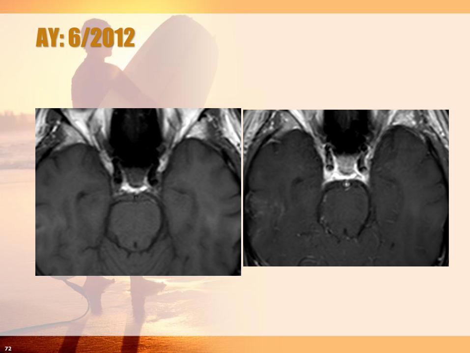
AY: 6/2012
72

AY: 6/2012
Recurring diplopia complaints
Did not want further surgery
2 RH(T) – 8 RH(T) – 12RHT
Downgaze 16 RHT
Now what?
Prism glasses
How much should we give? Which eye?
Bifocal v separate full frame readers
73

74
VIth Nerve Palsy
Shelley Klein, CO
Tufts Medical Center
Boston, MA

KC: 47 yof followed for a Left VIth Nerve Palsy
April 2008 was 1st seen by MBS, referred for progressive left head turn with ET requiring prism glasses. Diplopia worse in left gaze. No pain or discomfort.
POH:
Pt did not recall having a head turn as a child
Remembers 1st observing diplopia late 20s, was given prism Rx
Was told at that time she had Duane’s Syndrome
75

Findings from initial exam:AHP: Left head turn (can fuse with AHP)
Single Cover Test: D/N – LET in forced primary position
Fixation Preference: OD
DVA sc OD 20/20 OS 20/25
Stereopsis: 40 secs
Ductions: -4LLR, with no globe retraction on adduction
Forced Ductions: no significant restriction
Prism Cover Test: (Fix OD with prisms over OS)Distance: 3E 35LET 5LHypoT 55LET 6LHypoT
Near: 20LET’
76

Impression and Plan:
Almost complete Abduction deficit OS
Not typical Duane’s
no globe retraction on adduction
no constriction of LMR with forced ductions which one would expect with MR contracture of a longstanding ET
More suspicious of Left VI th NP
Mild anisometropic myopia OS
Ordered MRI
Proceed with treatment after results
77

MRI results: Thin LLR with a loop of the Basilar in the Left pre-pontine
cistern most probably compressing the VIth nerve
78
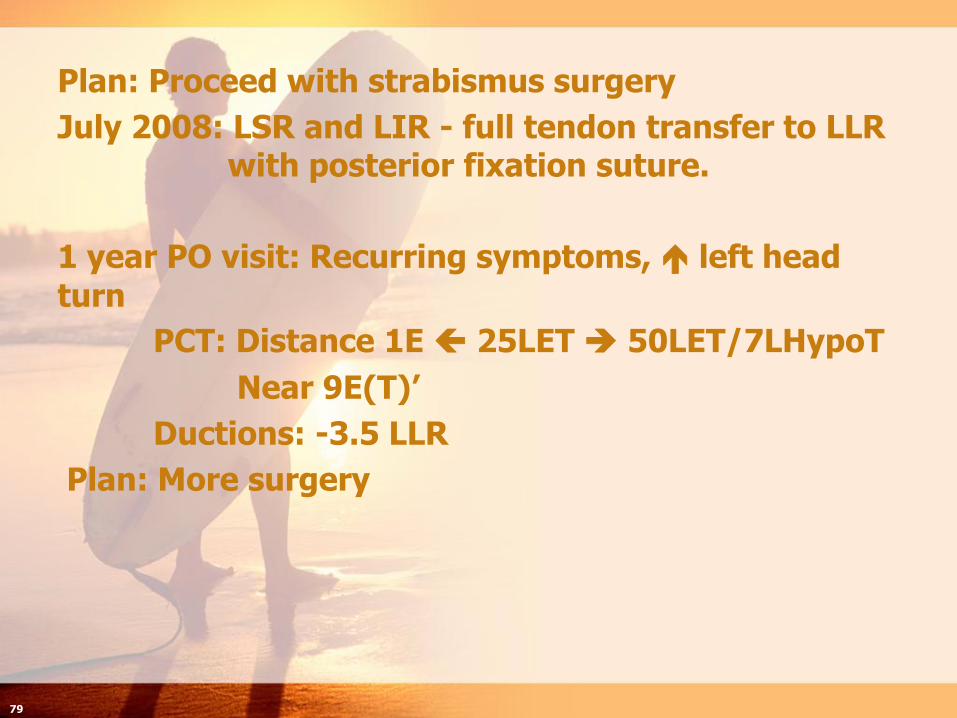
Plan: Proceed with strabismus surgery
July 2008: LSR and LIR - full tendon transfer to LLR with posterior fixation suture.
1 year PO visit: Recurring symptoms, left head
turn
PCT: Distance 1E 25LET 50LET/7LHypoT
Near 9E(T)’
Ductions: -3.5 LLR
Plan: More surgery
79
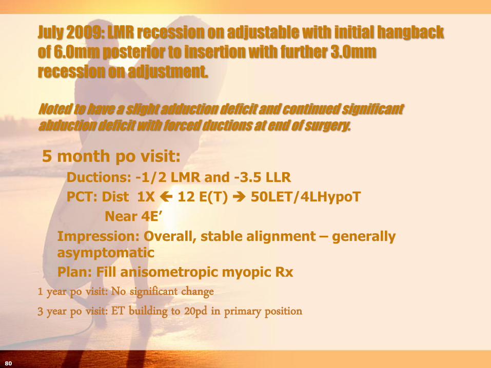
July 2009: LMR recession on adjustable with initial hangback
of 6.0mm posterior to insertion with further 3.0mm
recession on adjustment.
Noted to have a slight adduction deficit and continued significant
abduction deficit with forced ductions at end of surgery.
5 month po visit:
Ductions: -1/2 LMR and -3.5 LLR
PCT: Dist 1X 12 E(T) 50LET/4LHypoT
Near 4E’
Impression: Overall, stable alignment – generally asymptomatic
Plan: Fill anisometropic myopic Rx
80
1 year po visit: No significant change3 year po visit: ET building to 20pd in primary position
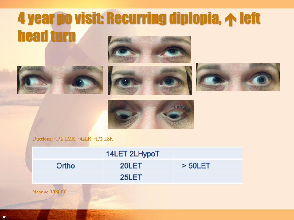
4 year po visit: Recurring diplopia, left
head turn
81
Ductions: -1/2 LMR, -4LLR, -1/2 LSR
Near sc 10E(T)’

What now?
Consider more surgery – maybe on OD
Prism glasses
Can we improve head position?
Can we reduce diplopia?
82
• What to consider in prescribing prisms:- Creating an exotropia in Right gaze?- Convergence amplitudes in Right gaze?- ? Bifocal with “V” pattern ET - ? Better to have separate readers- How much prism can I give to really help?

83
To be discussed in our break out
session………..

Diplopia Following Cataract Extraction
Mitchell Strominger, MD

Etiology of diplopia following cataract extractionacquired extraocular muscle dysfunction
intraoperative injury
direct injury of muscle with retrobulbur needle
myotoxicity from the anesthesia or injected antibiotics
inferior rectus muscle more common
superior rectus muscle involvement can occur
restrictive component but can see overaction
decompensation of longstanding, preexisting strabismus
sensory strabismus (exotropia)
strabismus with suppression (divergence insufficiency)
switched fixation
systemic disorder with ocular involvement
thyroid orbitopathy
supranuclear palsy (Parkinsonism)

86
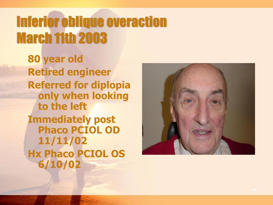
Inferior oblique overaction
March 11th 2003
80 year old
Retired engineer
Referred for diplopia only when looking to the left
Immediately post Phaco PCIOL OD 11/11/02
Hx Phaco PCIOL OS 6/10/02
87

Drawing
88
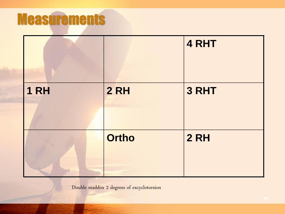
Measurements
4 RHT
1 RH 2 RH 3 RHT
Ortho 2 RH
Double maddox 2 degrees of excyclotorsion89

90

Inferior oblique muscle injury from local anesthesia for cataract
surgery
Four patients without preexisting strabismus who had diplopia following cataract surgery
Three had delayed onset hypertropia with fundus extorsionin the eye that underwent surgeryInferior oblique muscle overaction secondary to presumed
contracture
Inferior oblique recession in 2 cases
Prisms in one case
One had immediate-onset hypotropia with fundus intorsionin the eye that underwent surgeryInferior oblique muscle paresis
Hunter DG, Lam GC, Guyton DL. Ophthalmology 1995;102:501-509
91

Inferior Rectus Muscle Overaction After
Cataract Extraction
two patients, peribulbar, OS, no bridle sutures
Case
15 degree excyclotropia
Case 2
4 degrees excyclotropia
myotoxicity, degeneration,
regeneration, overaction
Ortho
16
Left
Hypo
24Left
Hypo
Munoz AJO 1994;118:664-666
Ortho
8 Left
Hypo
18
Left
Hypo
92

Superior Rectus Muscle Overaction After
Cataract Extraction
four patients, ipsilateral hypertropiaworse in upgaze than downgazetransient postoperative weakness of ipsilateral inferior
rectuscontracture or strengthening of ipsilateral antagonistanesthetic myopathy or direct trauma to inferior rectustwo patients recalled inability to look downward
immediately post op
„Grimmett and Lambert AJO 1992;114:72-80
93

Case 1 – What's the diagnosis68 year old complains of intermittent horizontal
diplopia 3 months following cataract extraction in the left eye
Notices it more when driving and looking far away at road signs
Old glasses had to be re-ground a few times, since couldn't follow optoms specifications. Something about not being offset.
- 5.00 OD 20/20 / -2.00 OS 20/20
12 E(T) distance with poor control / ortho near
94

Case 2 – What's the diagnosis58 year old with horizontal diplopia following
cataract extraction in the right eye
History of having worn glasses since age 4
Possible bifocals until high school
Remembers patching the left eye for years.
Vision therapy during elementary school
20/30 OD plano / 20/50 OS + 4.00
No stereo
4 ET distance and near with correction
3+ NS cataract left eye
95

Case 3 – What’s the diagnosis?70 year old onset of lines bending in the road while
driving only following removal of the cataract in the second eye
Remembers always having some double when looking to the left up until age 60, then improved. Never when looking straight ahead
Wife states that he always used to tilt his head when they first met and married, was getting worse for a while, but seems to have improved over the past 10 years.
20/20 OD -0.50 / 20/20 OS -0.50
Ortho – 10 RH(T) – 25 RHT
4+ right inferior oblique overaction
96

Case 4 – What's the diagnosis
63 year old complains of horizontal diplopia following bilateral cataract extraction
Sometimes had to close the left eye when extremely tired or out in the sun
Always wore glasses for distance but would take them off to read
Now doesn’t need glasses for either distance or near to see clearly
Without correction 20/20 OD / 20/40 OS (Mrx OS-1.50 20/20)
18 LX(T) distance / 18 LX(T) near – poor control
97
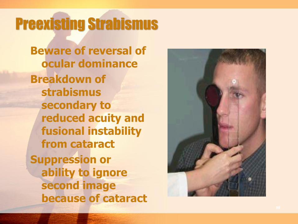
Preexisting Strabismus
Beware of reversal of ocular dominance
Breakdown of strabismus secondary to reduced acuity and fusional instability from cataract
Suppression or ability to ignore second image because of cataract
98
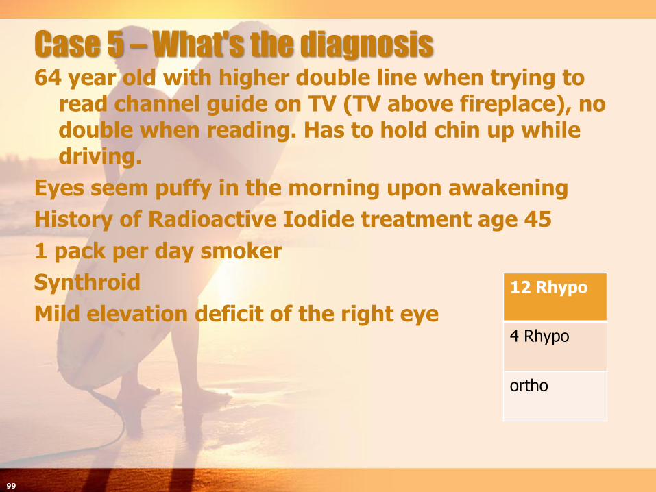
Case 5 – What's the diagnosis64 year old with higher double line when trying to
read channel guide on TV (TV above fireplace), no double when reading. Has to hold chin up while driving.
Eyes seem puffy in the morning upon awakening
History of Radioactive Iodide treatment age 45
1 pack per day smoker
Synthroid
Mild elevation deficit of the right eye
99
12 Rhypo
4 Rhypo
ortho

Case 6 – What's the diagnosis90 year old complains of the words running together
while reading following bilateral cataract extraction.
Prior to surgery only needed reading glasses, but states that they were quite thick
Paid top dollar for premium multifocal IOL’s and the vision is “great” for both distance and near.
Vision 20/25 both eyes for distance and near
Doesn’t seem to blink that much and shuffled into exam room
Ortho - distance, 12 X(T) near with poor NPC
100

Undiagnosed Systemic Disorders
occlusive effect of cataract masks the ocular deviationPSP / Parkinsonism
intraoperative tissue manipulation may aggravate an autoimmune or inflammatory conditionThyroid orbitopathy
101
