haloferax volcanii aglb and agld are involved in n...
TRANSCRIPT

doi:10.1016/j.jmb.2007.10.042 J. Mol. Biol. (2007) 374, 1224–1236
Available online at www.sciencedirect.com
Haloferax volcanii AglB and AglD are Involved inN-glycosylation of the S-layer Glycoprotein andProper Assembly of the Surface Layer
Mehtap Abu-Qarn1, Sophie Yurist-Doutsch1, Assunta Giordano2
Andrej Trauner3, Howard R. Morris3,4, Paul Hitchen3
Ohad Medalia1,5, Anne Dell3 and Jerry Eichler1⁎
1Department of Life Sciences,Ben Gurion University,Beersheva 84105, Israel2Istituto di ChimicaBiomolecolare, ConsiglioNazionale delle Ricerche,Pozzuoli (NA) 80078, Italy3Division of MolecularBiosciences, Faculty of LifeSciences, Imperial College,London SW7 2AZ, UK4M-SCAN Ltd,Wokingham,Berkshire, RG41 2TZ, UK5National Institute forBiotechnology in the Negev,Ben Gurion University,Beersheva 84105, Israel
Received 26 August 2007;received in revised form7 October 2007;accepted 16 October 2007Available online22 October 2007
*Corresponding author. E-mail addrURL: http://www.bgu.ac.il/life/
index.htm.Present address: J. Eichler, Depart
Ben Gurion University, PO Box 653,Israel.Abbreviations used: ER, endoplas
oligosacchryltransferase; MS, mass sMALDI TOF, Matrix-assisted laser dtime of flight; EM, electron microsco
0022-2836/$ - see front matter © 2007 E
In this study, the effects of deleting two genes previously implicated inHaloferax volcanii N-glycosylation on the assembly and attachment of anovel Asn-linked pentasaccharide decorating the H. volcanii S-layerglycoprotein were considered. Mass spectrometry revealed the pentasac-charide to comprise two hexoses, two hexuronic acids and an additional190 Da saccharide. The absence of AglD prevented addition of the finalhexose to the pentasaccharide, while cells lacking AglB were unable toN-glycosylate the S-layer glycoprotein. In AglD-lacking cells, the S-layerglycoprotein-based surface layer presented both an architecture andprotease susceptibility different from the background strain. By contrast,the absence of AglB resulted in enhanced release of the S-layer glycoprotein.H. volcanii cells lacking these N-glycosylation genes, moreover, grewsignificantly less well at elevated salt levels than did cells of the backgroundstrain. Thus, these results offer experimental evidence showing thatN-glycosylation endows H. volcanii with an ability to maintain an intactand stable cell envelope in hypersaline surroundings, ensuring survival inthis extreme environment.
© 2007 Elsevier Ltd. All rights reserved.
Keywords: Archaea; glycosyltransferase; N-glycosylation; oligosaccharyl-transferase; surface-layer glycoprotein
Edited by J. Karness: [email protected]/Eichler/
ment of Life Sciences,Beersheva 84105,
mic reticulum; OST,pectrometrometry;esorption/ionizationpy.
lsevier Ltd. All rights reserve
Introduction
N-linked protein glycosylation is one of the mostcommon and versatile post-translational modifica-tions and is experienced by proteins in all threedomains of life.1–4 The process is best understood inEukarya, where N-glycosylation begins with assem-bly of a dolichol pyrophosphate-linked oligosac-charide on the cytoplasmic face of the endoplasmicreticulum (ER).1,4–7 This lipid-linked oligosacchar-ide is then reoriented to face the ER lumen, whereadditional sugar subunits are added. The completed
d.

1225Haloferax volcanii N-glycosylation
oligosaccharide is then transferred, en bloc, to se-lected Asn residues of secretory pathway proteinsby the oligosacchryltransferase (OST) complex.8,9
Immediately following attachment of the oligosac-charide to a target protein, a series of glycan re-modeling steps that begin in the ER and continue inthe Golgi is initiated. The importance of N-glycosy-lation to Eukarya is reflected by the various rolesthis post-translational modification plays in proteinfolding, oligomerization and quality control.7While it has long been known that eukaryal
proteins experience N-glycosylation, it was onlyrecently shown that N-glycosylation also occurs inBacteria.3 The bacterial N-glycosylation pathwayhas been best described in the food-borne pathogen,Campylobacter jejuni, shown to contain an N-glyco-sylation machinery encoded by the 12 genes of thepgl locus.10,11 N-glycosylation in C. jejuni relies on asimpler pathway than does the parallel eukaryalprocess, with a heptameric polysaccharide beingassembled on a cytoplasmically oriented undeca-prenol lipid carrier lying in the plasma mem-brane.12,13 The oligosaccharide-loaded lipid is then“flipped” to face the cell exterior and the glycanmoiety is transferred to select Asn residues of targetproteins by PglB.14,15 In C. jejuni, at least 30 proteinshave been shown to containN-linked glycans, wherethey are implicated in pathogenicity viamediation ofhost–cell adherence, invasion and colonization.16–18
In contrast to the relatively well-defined path-ways of N-glycosylation in Eukarya and Bacteria,only little is known of the steps involved inarchaeal N-glycosylation, despite the fact that thefirst example of a prokaryal glycoprotein, theHalobacterium salinarum surface (S)-layer glycopro-tein, came from an archaeal source.19,20 Indeed,N-glycosylated proteins are far more abundant inArchaea than in Bacteria21,22 and include S-layerglycoproteins and flagellins, as well as variousmembrane and secreted proteins.2 Thus, whileN-glycosylation is relatively common in Archaea,the molecular mechanism of this processing eventhas only recently begun to attract attention.Chaban et al.23 reported on the effects of insertionalinactivation of two genes involved in the biogenesis
Figure 1. Archaeal and eukaryal homologues of H. volcanarchaeal and eukaryal homologues, generated by ClustalX (veT-coffee and drawn by TreeView (version 1.6.6; taxonomy.zoshown correspond to confidence levels obtained upon 1000Aeropyrum pernix (NP_147774.1; Aper), Archaeoglobus fulgidusHmar), Halobacterium sp. NRC-1 (NP_279416.1; HNRC), HaloHwal), Metallosphaera sedula (ZP_01601347.1; Msed), Methacetivorans (NP_618739.1; Mace), Methanosarcina barkeri (YP_Mhun), Natronomonas pharaonis (YP_326773.1; Npha), Pic(NP_127133.1; Paby), Sulfolobus solfataricus (NP_342803.1; SThermoplasma volcanium (NP_111403.1; Tvol), while the eukaryAep), Candida albicans (XP_715818.1; Calb), Dictyostelium disGgal), Homo sapiens (AAG09682.1; Hsap), Mus musculus (NPScer) andXenopus laevis (AAH44127.1; Xlae) (b) The deduced athe sequences of the Saccharomyces cerevisiae ALG5 protein (Nglucosyltransferase (AAG09682.1; Hsap). The nucleotide sequnucleotide sequence database (accession number AM698042)transmembrane domains.
and attachment of a trisaccharide found on fla-gellins and the S-layer glycoprotein of the metha-nogenMethanococcus voltae. At the same time, workin the Eichler laboratory showed that deletion ofHaloferax volcanii genes encoding homologues to theeukaryal OST catalytic subunit, Stt3, and a phos-phodolichol glucosyltransferase, Alg5, affectedS-layer glycoprotein glycosylation, as demon-strated via glycostaining and changes in apparentmolecular weight.24
Here, the roles played by the H. volcanii homo-logues of Stt3 and Alg5, now renamed AglB andAglD, respectively, according to the nomenclatureof Chaban et al.,23 in the N-glycosylation of theH. volcanii S-layer glycoprotein were investigated. Inaddition, the importance of correct N-glycosylationof the S-layer glycoprotein for H. volcanii physiologyand survival was explored.
Results
H. volcanii AglD is a homologue of eukaryal Alg5
Earlier work24 reported that H. volcanii encodeshomologues of eukaryal and bacterial proteinsinvolved in N-glycosylation. Of these, one of thetwo H. volcanii homologues of eukaryal Alg5detected, termed Alg5-A, was implicated in theglycosylation of a reporter glycoprotein, the S-layerglycoprotein. Now, employing the terminology forarchaeal genes involved in N-glycosylation (thearchaeal glycosylation genes) proposed by Chabanet al.,23 H. volcanii alg5-A has been renamed aglD.Conserved domain prediction25 indicates H. volcaniiAglD to contain a glycosyltransferase 2 familydomain (Pfam 00535, COG1216) as well as a secondregion predicted to belong to the Pfam03706 group,corresponding to an uncharacterized protein family(UPF0104) that includes members annotated asdolichol-P-glucose synthases that also contain aPfam 00535 domain.BLAST-based searches of several genomes
revealed the presence of AglD homologues in
ii AglD. (a) Phylogenetic tree of H. volcanii AglD and itsrsion 1.83) from a multiple sequence alignment created byology.gla.ac.uk/rod/treeview.html). The bootstrap valuestrial repeats. The archaeal sequences shown come from(NP_069415.1; Aful), Haloarcula marismortui (YP_136461.1;ferax volcanii (Hvol), Haloquadratum walsbyi (YP_657261.1;anopyrus kandleri (NP_614163.1; Mkan), Methanosarcina304067.1; Mbar), Methanospirillum hungatei (YP_503949.1;rophilus torridus (YP_024256.1; Ptor), Pyrococcus abyssisol), Thermococcus kodakarensis (YP_182777.1; Tkod) andal sequences shown come from Aedes aegypti (ABF18373.1;coideum (AAQ98885.1; Ddis), Gallus gallus (XP_417093.2;_079718.1; Mmus), Saccharomyces cerevisiae (NP_015097.1;mino acid sequence ofH. volcaniiAglDwas compared withP_015097.1; Scer) and Homo sapiens dolichyl-phosphate β-ence of H. volcanii aglD has been deposited in the EMBL. The bold, underlined residues correspond to predicted

Figure 1 (legend on previous page)
1226 Haloferax volcanii N-glycosylation
other archaeal species (Figure 1(a)), although nonehave been characterized functionally. Alignment† ofthe N-terminal halves of these AglD-like sequencesreveals several highly conserved motifs that may
† align.genome.jp; www.ch.embnet.org/software/TCoffee.html
participate in the proposed glycosyltransferaseactivity of the proteins (data not shown). WhileH. volcaniiAglDwas originally identified through itssimilarity to Saccharomyces cerevisiae ALG5 and itshomologues in other Eukarya, phylogenetic analysisreveals that archaeal AlgD proteins form a cladedistinct from their eukaryal homologues (Figure

Figure 2. Glycosylation of the H. volcanii S-layerglycoprotein differs in the presence or absence of aglD.(a) The protein contents of H. volcanii WR536 backgroundcells (bkgnd) as well as proteins of the same strain lackingeither Alg5-B (-alg5-B) or AglD (-aglD) were separated bySDS–7.5% (w/v) PAGE and subjected to Coomassiestaining (left panel) or overlay with HRP-conjugatedConA (right panel). (b) H. volcanii WR536 cells (bkgnd)or the same strain lacking AglD (-aglD) were grown in thepresence of [14C]glucose, -galactose or -mannose andsubjected to Coomassie staining (upper panel) or fluoro-graphy (lower panel). In both (a) and (b), only the S-layerglycoprotein is shown.
1227Haloferax volcanii N-glycosylation
1(a)). Indeed, as presented in Figure 1(b), H. volcaniiAglD can be clearly distinguished from S. cerevisiaeALG5 or its human homologue in terms of mem-brane topology. The topology prediction toolsTMHMM‡ and HMMTOP§ describe H. volcaniiAglD as an integral membrane protein presentinga large internally oriented N-terminal half and a C-terminal half comprising six membrane-spanningdomains including three short externally orientedloops. By contrast, the homologous human andyeast proteins are thought to contain a singletransmembrane domain, located almost immedi-ately downstream of the N terminus.
AglD participates in H. volcanii S-layerglycoprotein N-glycosylation
To better describe the contribution of AglD toH. volcanii protein glycosylation, a detailed exam-ination of both the N-glycosylation profile of theS-layer glycoprotein, a well-described reporter ofthis post-translational modification,26–29 and thecontribution of AglD to this processing event, wasundertaken. Initially, horseradish peroxidase (HRP)-conjugated concanavalin A (ConA) was employedin a lectin overlay protocol to probe the S-layerglycoprotein in background and aglD-deleted cells29as well as in cells lacking alg5-B, an aglD homologue(Figure 2(a)).24 While the lectin recognized theS-layer glycoprotein in all three cell types, the labeledband from the aglD-deleted strain migrating slightlyfaster on SDS–PAGE gels than did the protein fromthe background or Alg5B-lacking strains, in agree-ment with earlier observations.24 In a second experi-ment, the background and aglD-lacking strains weregrown in minimal medium and incubated with [14C]glucose, -galactose or -mannose. As in earlier ana-lyses of theH. volcanii glycoprotein pool28 and of theS-layer glycoprotein in particular,27,30 incubationwith the radiolabeled sugars led to labeling of theS-layer glycoprotein, albeit to differing degrees ineach strain (Figure 2(b)). Densitometric quantifica-tion of the relative amounts of radioactive glucose,galactose and mannose incorporated into compar-able amounts (as deemed by Coomassie staining anddensitometry) of the S-layer glycoprotein fromtriplicate samples of the background strain yieldeda 1.0:0.7:0.1 ratio, in agreement with earlier reports ofthe relative content of the three saccharides in theH. volcanii glycoprotein pool.28 When the same strainlacking aglDwas grown under similar conditions, allthree radiolabeled sugars were once again incorpo-rated into the S-layer glycoprotein, as revealed bySDS–PAGE and fluorography. In these cells, how-ever, glucose, galactose and mannose were found ina 1.0:0.1:0.1 ratio, averaged over three experiments.Thus, the efficiencies of [14C]glucose, -galactose and-mannose-based modification of the S-layer glyco-protein in the background and aglD-deleted strainsare clearly distinct.
‡www.cbs.dtu.dk/services/TMHMM-2.0§www.enzim.hu/hmmtop
AglD is involved in the addition of the finalhexose subunit to a pentasaccharide decoratingthe H. volcanii S-layer glycoprotein
Efforts next focused on mass spectrometric (MS)characterization of the glycosylation pattern of theS-layer glycoprotein reporter in AglD-lacking cells.SDS–PAGE gel pieces from the background andmutant strains containing the S-layer glycoproteinwere subjected to in-gel tryptic digestion and theobtained peptides were separated by liquid chroma-tography. MS/MS was then employed to revealpeptide sequences. As such, six peptide sequenceswere obtained, including two containing potentialN-glycosylation sites. Of these, the N-terminal1ERGNLDADSESFNK14 peptide (1581 m/z) was ofparticular interest, given that it contains a N-glyco-sylation site previously reported as being modi-fied.26 The glycan content of the Asn13-containingS-layer glycoprotein-derived peptide from the back-ground strain was determined by Matrix-assistedlaser desorption/ionization time of flight (MALDITOF) mass mapping of the nanoLC-purified trypticdigest, complemented by MS/MS analyses usingMALDI TOF/TOF and electrospray Q-TOF instru-mentation. The MALDI spectrum (Figure 3(a))revealed the presence of a pentasaccharide moiety

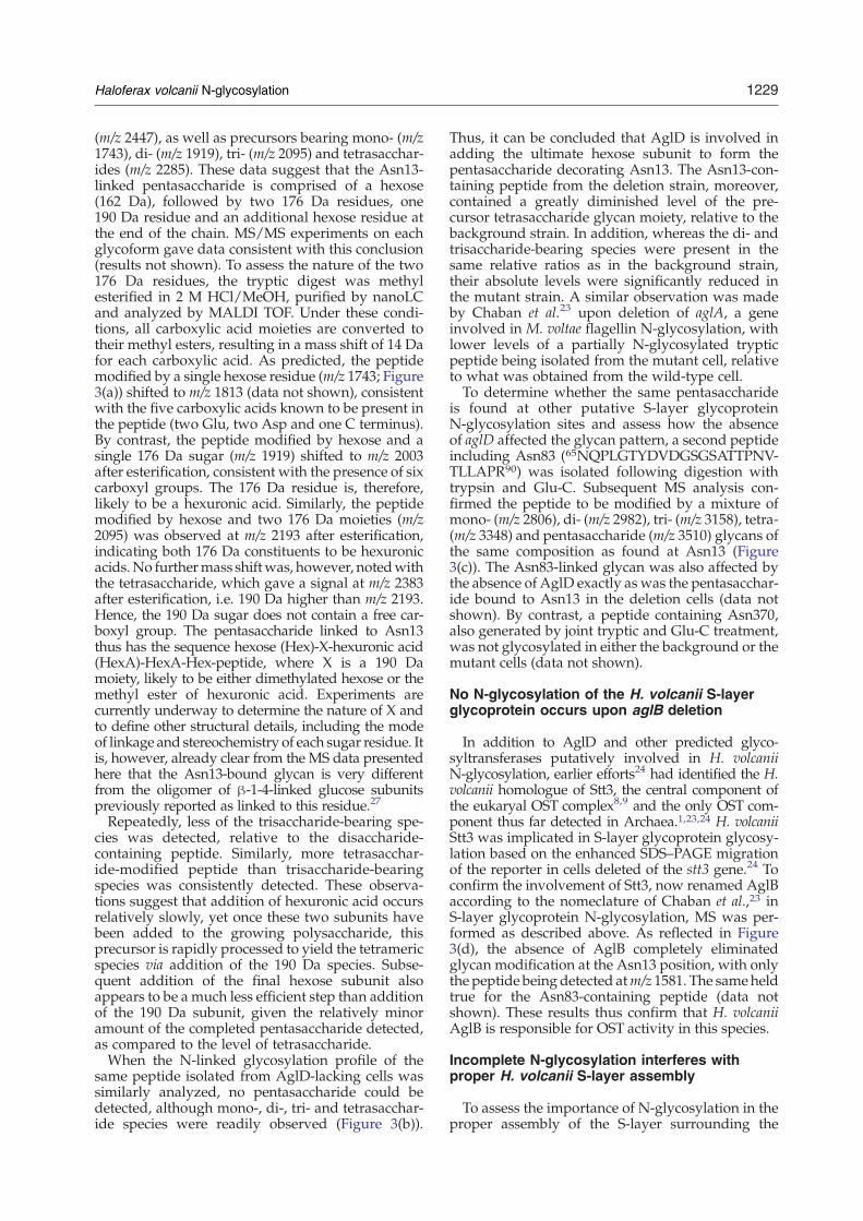
Figure 3. MALDI TOF analysis ofH. volcanii S-layer glycoprotein-derived glycopeptides. Shown areMALDI spectra obtained from the Asn13-containing tryptic glycopeptidesfrom (a) the background and (b) the algD deletion strains. (c) MALDI spectrum of the Asn83-containing tryptic/Glu-C glycopeptide from the background strain. (d) MALDIspectrum of the unmodified Asn13-containing peptide from the aglB deletion strain. The nature of the glycopeptide-associated sugar residues is shown in the inset of (b). Trypticpeptides were separated by offline nanoLC prior to analysis.
1228Haloferax
volcaniiN-glycosylation
1229Haloferax volcanii N-glycosylation
(m/z 2447), as well as precursors bearing mono- (m/z1743), di- (m/z 1919), tri- (m/z 2095) and tetrasacchar-ides (m/z 2285). These data suggest that the Asn13-linked pentasaccharide is comprised of a hexose(162 Da), followed by two 176 Da residues, one190 Da residue and an additional hexose residue atthe end of the chain. MS/MS experiments on eachglycoform gave data consistent with this conclusion(results not shown). To assess the nature of the two176 Da residues, the tryptic digest was methylesterified in 2 M HCl/MeOH, purified by nanoLCand analyzed by MALDI TOF. Under these condi-tions, all carboxylic acid moieties are converted totheir methyl esters, resulting in a mass shift of 14 Dafor each carboxylic acid. As predicted, the peptidemodified by a single hexose residue (m/z 1743; Figure3(a)) shifted to m/z 1813 (data not shown), consistentwith the five carboxylic acids known to be present inthe peptide (two Glu, two Asp and one C terminus).By contrast, the peptide modified by hexose and asingle 176 Da sugar (m/z 1919) shifted to m/z 2003after esterification, consistent with the presence of sixcarboxyl groups. The 176 Da residue is, therefore,likely to be a hexuronic acid. Similarly, the peptidemodified by hexose and two 176 Da moieties (m/z2095) was observed at m/z 2193 after esterification,indicating both 176 Da constituents to be hexuronicacids.No furthermass shiftwas, however, notedwiththe tetrasaccharide, which gave a signal at m/z 2383after esterification, i.e. 190 Da higher than m/z 2193.Hence, the 190 Da sugar does not contain a free car-boxyl group. The pentasaccharide linked to Asn13thus has the sequence hexose (Hex)-X-hexuronic acid(HexA)-HexA-Hex-peptide, where X is a 190 Damoiety, likely to be either dimethylated hexose or themethyl ester of hexuronic acid. Experiments arecurrently underway to determine the nature of X andto define other structural details, including the modeof linkage and stereochemistry of each sugar residue. Itis, however, already clear from the MS data presentedhere that the Asn13-bound glycan is very differentfrom the oligomer of β-1-4-linked glucose subunitspreviously reported as linked to this residue.27
Repeatedly, less of the trisaccharide-bearing spe-cies was detected, relative to the disaccharide-containing peptide. Similarly, more tetrasacchar-ide-modified peptide than trisaccharide-bearingspecies was consistently detected. These observa-tions suggest that addition of hexuronic acid occursrelatively slowly, yet once these two subunits havebeen added to the growing polysaccharide, thisprecursor is rapidly processed to yield the tetramericspecies via addition of the 190 Da species. Subse-quent addition of the final hexose subunit alsoappears to be a much less efficient step than additionof the 190 Da subunit, given the relatively minoramount of the completed pentasaccharide detected,as compared to the level of tetrasaccharide.When the N-linked glycosylation profile of the
same peptide isolated from AglD-lacking cells wassimilarly analyzed, no pentasaccharide could bedetected, although mono-, di-, tri- and tetrasacchar-ide species were readily observed (Figure 3(b)).
Thus, it can be concluded that AglD is involved inadding the ultimate hexose subunit to form thepentasaccharide decorating Asn13. The Asn13-con-taining peptide from the deletion strain, moreover,contained a greatly diminished level of the pre-cursor tetrasaccharide glycan moiety, relative to thebackground strain. In addition, whereas the di- andtrisaccharide-bearing species were present in thesame relative ratios as in the background strain,their absolute levels were significantly reduced inthe mutant strain. A similar observation was madeby Chaban et al.23 upon deletion of aglA, a geneinvolved in M. voltae flagellin N-glycosylation, withlower levels of a partially N-glycosylated trypticpeptide being isolated from the mutant cell, relativeto what was obtained from the wild-type cell.To determine whether the same pentasaccharide
is found at other putative S-layer glycoproteinN-glycosylation sites and assess how the absenceof aglD affected the glycan pattern, a second peptideincluding Asn83 (65NQPLGTYDVDGSGSATTPNV-TLLAPR90) was isolated following digestion withtrypsin and Glu-C. Subsequent MS analysis con-firmed the peptide to be modified by a mixture ofmono- (m/z 2806), di- (m/z 2982), tri- (m/z 3158), tetra-(m/z 3348) and pentasaccharide (m/z 3510) glycans ofthe same composition as found at Asn13 (Figure3(c)). The Asn83-linked glycan was also affected bythe absence of AglD exactly as was the pentasacchar-ide bound to Asn13 in the deletion cells (data notshown). By contrast, a peptide containing Asn370,also generated by joint tryptic and Glu-C treatment,was not glycosylated in either the background or themutant cells (data not shown).
No N-glycosylation of the H. volcanii S-layerglycoprotein occurs upon aglB deletion
In addition to AglD and other predicted glyco-syltransferases putatively involved in H. volcaniiN-glycosylation, earlier efforts24 had identified the H.volcanii homologue of Stt3, the central component ofthe eukaryal OST complex8,9 and the only OST com-ponent thus far detected in Archaea.1,23,24 H. volcaniiStt3 was implicated in S-layer glycoprotein glycosy-lation based on the enhanced SDS–PAGE migrationof the reporter in cells deleted of the stt3 gene.24 Toconfirm the involvement of Stt3, now renamed AglBaccording to the nomeclature of Chaban et al.,23 inS-layer glycoprotein N-glycosylation, MS was per-formed as described above. As reflected in Figure3(d), the absence of AglB completely eliminatedglycan modification at the Asn13 position, with onlythe peptide being detected atm/z 1581. The sameheldtrue for the Asn83-containing peptide (data notshown). These results thus confirm that H. volcaniiAglB is responsible for OST activity in this species.
Incomplete N-glycosylation interferes withproper H. volcanii S-layer assembly
To assess the importance of N-glycosylation in theproper assembly of the S-layer surrounding the

1230 Haloferax volcanii N-glycosylation
H. volcanii cell (apparently composed solely of the S-layer glycoprotein26), protease sensitivity of the S-layer glycoprotein was compared in the backgroundand the aglB and aglD deletion strains. Trypsinolysisof the cells (up to 3 h) revealed the S-layerglycoprotein in the AglD-lacking strain to be moreresistant to proteolysis than was its counterpart inthe background strain (Figure 4(a)), suggesting thatthe aberrant glycosylation of the S-layer glycopro-tein in the AglD-lacking strain affected the properassembly of the S-layer. By contrast, no difference inprotease susceptibility of the S-layer glycoprotein inthe background and AglB-lacking strains wasobserved (Figure 4(b)).Earlier examination of negatively stainedH. volcanii
envelope preparations revealed the S-layer as acrystalline-like lattice of hexameric complexes, eachcomprising six S-layer glycoproteins organizedaround an inwardly oriented, funnel-shaped pore31
Three-dimensional reconstruction of the H. volcaniiS-layer, as well as earlier electron microscopy (EM)evidence,32 suggested this cell wall structure to
Figure 4. Perturbed S-layer glycoprotein N-glycosylation aWR536 cells (open circles) or AglD-lacking cells of the same strat 37 °C. Aliquots were removed immediately prior to incubatioaddition of the protease and examined by SDS–7.5% PAGE.relative to that amount found immediately prior to proteolysithe average of three experiments ±SEM. Background cells, openAglB-lacking cells were examined instead of AglD-lacking celH. volcaniiWR536 cells (bkgnd); middle panel, the same cells la(-aglB). In each panel, the arrow points to the plasma membrsurface layer. The bar in the left panel represents 100 nm.
extend some 12.5 nm from the plasma membrane,implying the presence of a pseudo-periplasmicspace. Moreover, it was implied that the glycanmoieties of the S-layer glycoprotein provide physicalsupport for this architecture. Thus, to more directlyexamine whether differential N-glycosylation of theS-layer glycoprotein in the various strains led todifferences in S-layer architecture, cell envelopepreparations from background, aglB and aglD-deleted strains were prepared for EM analysisusing rapid freezing techniques. When isolated en-velope preparations from the background WR536strain were considered, both the plasma membraneand S-layer were readily detected (Figure 4(c), leftpanel). Examination of the space between these twoboundaries aswell as the edge of the S-layer revealeda lattice network. Cell envelopes obtained fromAglB-lacking cells presented a similar picture (Figure4(c), right panel). When, however, envelope pre-parations from H. volcanii cells deleted of aglD weresimilarly examined, no such symmetry was detected(Figure 4(c), middle panel).
ffectsH. volcanii S-layer architecture. (a) Background strainain (filled circles) were challenged with 1 mg/ml of trypsinn with trypsin and at subsequent 15–30 min intervals afterThe amount of S-layer glycoprotein remaining with time,s, was quantified densitometrically. Each point representscircles; AglD-lacking cells, filled circles. (b) As in (a), only
ls. (c) Electron micrographs of cells envelopes. Left panel,cking AglD (-aglD); right panel, the same cells lacking AglBane while the arrowhead points to the outer edge of the

1231Haloferax volcanii N-glycosylation
To determine whether the differential N-glyco-sylation of the S-layer glycoprotein in the variousH. volcanii strains reflected differences in S-layerstability, the extent of S-layer glycoprotein releaseinto the growth medium of each cell type wascompared. As reflected in Figure 5(a), less S-layerglycoprotein was released from AglD-lacking cells,relative to that amount released from the back-ground strain after 10 h. By contrast, significantlymore S-layer glycoprotein was released by cellslacking AglB within the same period. These relation-
Figure 5. The S-layer glycoprotein is more readilyreleased from H. volcanii cells lacking N-glycosylationcapability. (a) The levels of S-layer glycoprotein releasedinto the growthmedia fromH. volcaniiWR536 cells (bkgnd)or the same cells lacking either AglB (-aglB) or AglD (-aglD)after 10 h of growth were quantified densitometricallyfollowing trichloroacetic acid precipitation, SDS–PAGE andCoomassie staining. The amount of S-layer glycoproteinreleased from the background strain was set at a value of 1.Each bar represents the average of five separate growths foreach cell type. Error bars correspond to standard deviation.The asterisk corresponds to a value being statisticallysignificant to pb0.01, while the double asterisk correspondsto a statistical significance of pb0.001, as determined bystudent's t test. (b) BackgroundH. volcanii cells or the samecells lacking AglB were pulse radiolabeled with [35S]Cys/Met for 5 min and chased with excess Cys/Met. Thegrowth medium of each culture was removed, the proteincontent precipitated, separated by SDS–7.5% PAGE andvisualized by fluorography. Upper panel, background(bkgnd) cells; lower panel, AglB-lacking (-algB) cells.Shown is a representative of four repeats.
ships also held true when the amounts of S-layerglycoprotein released from each cell type were com-pared after 18 h of growth (data not shown).To further consider whether incomplete or absent
N-glycosylation affected S-layer turnover, metabolicpulse–chase radiolabeling was performed. Back-ground cells as well as the same cells deleted ofaglB or aglD were grown to the same optical densityin minimal medium and pulse radiolabeled with14 μCi of [35S]Cys/Met for 5 min, after which timeexcess cold Cys/Metwas added. Equivalent aliquotswere collected every 15min and the growthmediumwas isolated for analysis by SDS–PAGE and fluoro-graphy. Over the first 15–30 min of chase, radi-olabeled S-layer glycoprotein began to accumulate inthe medium of the background strain (Figure 5(b),upper panel). Over the next hour, a relatively con-sistent level of S-layer glycoprotein was detected. Acomparable picture was obtained when cells deletedof aglD were similarly considered (data not shown).By contrast, radiolabeled S-layer glycoprotein wasdetected in the medium of cells lacking AlgB fromthe onset of the chase period at higher levels thandetected in the background strain medium, with theamount of released 35S incorporating S-layer glyco-protein starting to decrease some 45 min followingthe onset of the chase (Figure 5(b), lower panel).Finally, analysis of the cellular fraction of the diffe-rent cell types confirmed that comparable amountsof radioactivity had been incorporated into theS-layer glycoprotein and other proteins during thepulse phase of the experiment (results not shown).Together, these studies indicate that partial pro-
cessing of S-layer glycoprotein N-glycosylation sitescompromises the proper assembly of the proteinshell surrounding H. volcanii cells. Moreover, whileN-glycosylation is not, per se, essential for properS-layer assembly, the absence of this post-transla-tional modification affects S-layer stability.
Growth in high salt is hindered by the absenceor perturbation of S-layer glycoproteinN-glycosylation
To determine whether the complete or partialabsence of N-glycosylation affected the ability ofH. volcanii to grow across a range of salt concentra-tions, cells of the background strain, aswell as cells ofthe same strain deleted of either aglB or aglD, weregrown in rich medium containing 1.75, 3.5 or 4.8 MNaCl. As reflected in Figure 6, the background straingrew at all three salt concentrations, although therate of growth decreased as the salt level increased.In the case of AglD-lacking cells (Figure 6(a)), growthwas slightly slower than the background strain inthe presence of 1.75 or 3.5 M NaCl, as reported.24
Growth of the deletion strain was significantly per-turbed relative to the background species in thepresence of 4.8 M NaCl. A similar picture wasobtainedwhenAglB-lacking cells were grown in richmedium containing a range of NaCl concentrations(Figure 6(b)). The deletion strain grew at essentiallythe same rate as background cells in the presence of
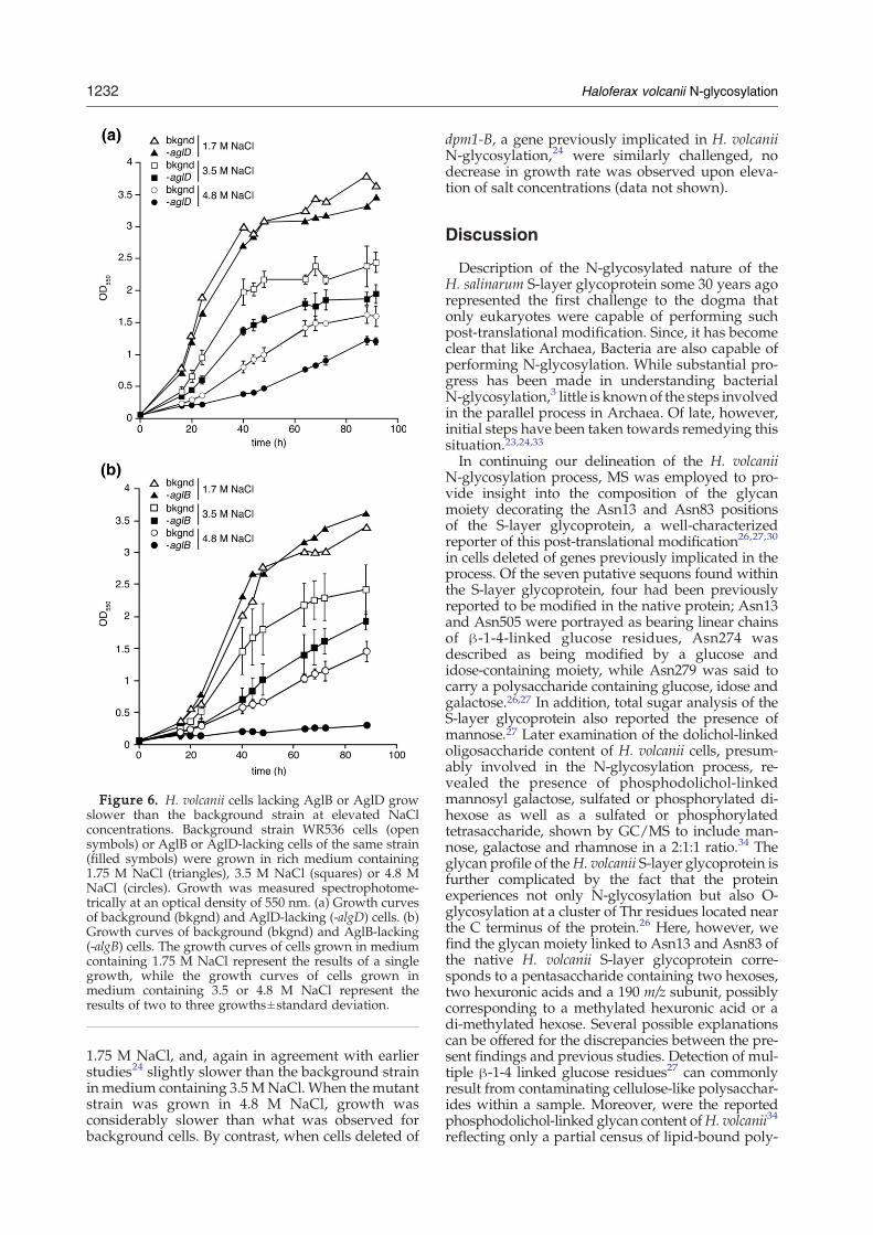
Figure 6. H. volcanii cells lacking AglB or AglD growslower than the background strain at elevated NaClconcentrations. Background strain WR536 cells (opensymbols) or AglB or AglD-lacking cells of the same strain(filled symbols) were grown in rich medium containing1.75 M NaCl (triangles), 3.5 M NaCl (squares) or 4.8 MNaCl (circles). Growth was measured spectrophotome-trically at an optical density of 550 nm. (a) Growth curvesof background (bkgnd) and AglD-lacking (-algD) cells. (b)Growth curves of background (bkgnd) and AglB-lacking(-algB) cells. The growth curves of cells grown in mediumcontaining 1.75 M NaCl represent the results of a singlegrowth, while the growth curves of cells grown inmedium containing 3.5 or 4.8 M NaCl represent theresults of two to three growths±standard deviation.
1232 Haloferax volcanii N-glycosylation
1.75 M NaCl, and, again in agreement with earlierstudies24 slightly slower than the background strainin medium containing 3.5 MNaCl.When the mutantstrain was grown in 4.8 M NaCl, growth wasconsiderably slower than what was observed forbackground cells. By contrast, when cells deleted of
dpm1-B, a gene previously implicated in H. volcaniiN-glycosylation,24 were similarly challenged, nodecrease in growth rate was observed upon eleva-tion of salt concentrations (data not shown).
Discussion
Description of the N-glycosylated nature of theH. salinarum S-layer glycoprotein some 30 years agorepresented the first challenge to the dogma thatonly eukaryotes were capable of performing suchpost-translational modification. Since, it has becomeclear that like Archaea, Bacteria are also capable ofperforming N-glycosylation. While substantial pro-gress has been made in understanding bacterialN-glycosylation,3 little is knownof the steps involvedin the parallel process in Archaea. Of late, however,initial steps have been taken towards remedying thissituation.23,24,33In continuing our delineation of the H. volcanii
N-glycosylation process, MS was employed to pro-vide insight into the composition of the glycanmoiety decorating the Asn13 and Asn83 positionsof the S-layer glycoprotein, a well-characterizedreporter of this post-translational modification26,27,30
in cells deleted of genes previously implicated in theprocess. Of the seven putative sequons found withinthe S-layer glycoprotein, four had been previouslyreported to be modified in the native protein; Asn13and Asn505 were portrayed as bearing linear chainsof β-1-4-linked glucose residues, Asn274 wasdescribed as being modified by a glucose andidose-containing moiety, while Asn279 was said tocarry a polysaccharide containing glucose, idose andgalactose.26,27 In addition, total sugar analysis of theS-layer glycoprotein also reported the presence ofmannose.27 Later examination of the dolichol-linkedoligosaccharide content of H. volcanii cells, presum-ably involved in the N-glycosylation process, re-vealed the presence of phosphodolichol-linkedmannosyl galactose, sulfated or phosphorylated di-hexose as well as a sulfated or phosphorylatedtetrasaccharide, shown by GC/MS to include man-nose, galactose and rhamnose in a 2:1:1 ratio.34 Theglycan profile of theH. volcanii S-layer glycoprotein isfurther complicated by the fact that the proteinexperiences not only N-glycosylation but also O-glycosylation at a cluster of Thr residues located nearthe C terminus of the protein.26 Here, however, wefind the glycan moiety linked to Asn13 and Asn83 ofthe native H. volcanii S-layer glycoprotein corre-sponds to a pentasaccharide containing two hexoses,two hexuronic acids and a 190 m/z subunit, possiblycorresponding to a methylated hexuronic acid or adi-methylated hexose. Several possible explanationscan be offered for the discrepancies between the pre-sent findings and previous studies. Detection of mul-tiple β-1-4 linked glucose residues27 can commonlyresult from contaminating cellulose-like polysacchar-ides within a sample. Moreover, were the reportedphosphodolichol-linked glycan content ofH. volcanii34
reflecting only a partial census of lipid-bound poly-

1233Haloferax volcanii N-glycosylation
saccharides in this species, then the absence of thepentasaccharide detected here linked to phospho-dolichol would be explained. Indeed, the previouslydefined lipid-linked glycan structures may be pre-ferentially involved in the biosynthesis of glyco-lipids35,28 or other H. volcanii glycoproteins.28,30Having defined N-linked glycans decorating the
H. volcanii S-layer glycoprotein, the actions of twoenzymes involved in N-glycosylation process, i.e.aglB and aglD, were characterized. AglD is involvedin catalyzing the addition of the final hexose to thepentasaccharide decorating at least two sequon Asnresidues, namely Asn13 and Asn83. In terms of itstopology, AglD includes a soluble, cytoplasm-facingN-terminal half, associated with the membrane viathe integral C-terminal portion of the molecule. Pre-dicted to span the membrane six times, AglD pre-sents a topology reminiscent of those eukaryal glyco-syltransferases containing active sites facing the ERlumen, responsible for the transfer of mannose orglucose residues from charged dolichol carriers tothe pyrophosphodolichol-bound heptasaccharidefollowing its reorientation from the cytoplasmic tothe lumenal face of the ER membrane. By contrast,those glycosyltransferases presenting domainshomologous to the soluble N-terminal portion ofAglD correspond to single membrane-spanning en-zymes that employ soluble nucleotide-activatedmonosaccharides in assembling the heptasaccharidefound on the cytoplasm-facing ER membrane-embedded dolichol pyrophosphate carrier.1,7 Fur-ther experimentation is, therefore, required before itcan be concluded whether the oligosaccharidesdecorating the H. volcanii S-layer glycoprotein areassembled from activated soluble sugars or fromlipid-linked subunits, thus defining the actual roleplayed by AglD in this processing event.AglB, as the sole component of the OST detected in
H. volcanii,24 is responsible for transferring a penta-saccharidemoiety, again presumably from adolicholphosphate carrier,34 to at least two S-layer glycopro-tein sites, namely Asn13 and Asn83. The observationthat H. volcanii AglB can transfer truncated glycanstructures to the S-layer glycoprotein, as in aglD–deleted cells, suggests a relaxed substrate specificityof the enzyme, as reported for eukaryal OSTs,36,37C. jejuni PglB12,13 and AglB from the methanoarch-aea M. voltae.23 However, unlike PglB,13 H. volcaniiAglB is capable of transferring not only fully assem-bled oligosaccharides, but also precursor glycans.Indeed, while differences in the relative amounts ofthe precursor oligosaccharide-bearing glycopeptidescould reflect the differential kinetics of the glyco-transferases involved in glycan assembly, such quan-titative differences could also reflect differentialprocessing by AglB (or the putative enzyme respon-sible for transferring dolichol-linked oligosaccharidesacross the archaeal plasma membrane) of theseprecursors. H. volcanii AglB is, moreover, unusual inthat it appears to transfer polysaccharides fromphospho- rather than pyrophosphodolichol carriers.34
The absence of aglD resulted in the generation of anS-layer displaying altered protease sensitivity and an
increased degree of disorder. Deletion of H. volcaniiaglB, and hence prevention of S-layer glycoproteinN-glycosylation, led to enhanced release of the pro-tein into the growth medium. Taken together, theresults imply that not only is complete N-glyco-sylation important for realizing correct H. volcaniiS-layer architecture but that stable association ofthe S-layer glycoprotein within the S-layer requiressuch modification. Accordingly, both aberrant andabsent N-glycosylation compromised the ability ofH. volcanii to grow at high salt concentrations. Thus,N-glycosylation can be considered a post-transla-tional modification designed to allow H. volcanii toremain intact in the face of the hypersaline environ-ment in which these cells exist. It should be noted,however, that the S-layer glycoprotein experiencesbothN andO-glycosylation.26 As such, the effects onthe S-layer glycoprotein, the S-layer and the intactcell observed upon either modulation or loss ofN-glycosylationmay present only a partial picture ofthe importance of S-layer glycoprotein glycosylationto H. volcanii cells.The observation that not all S-layer glycoprotein
sequons are modified (e.g. Asn370) raises thequestion of how such sites differ from sequons thatinclude modified Asn residues, such as Asn13 andAsn83. Unfortunately, the limited dataset consid-ered here does not allow for detailed assessment ofthe regions surrounding modified and unmodifiedH. volcanii S-layer glycoprotein sequons. Indeed,recent analysis of the amino acid composition ofthose archaeal sequons experimentally verified asbeing modified as well as the composition of the 20up- and downstream residues failed to reveal anyconsistent sequence traits, apart from a requirementfor the Asn-X-Ser/Thr motif, where X is any residueapart from Pro.33 As such, deciphering the rules go-verning whether a given archaeal sequon is modi-fied will have to wait for more experimental datato allow for development of appropriate predictivealgorithms.With the identification of gene products directly
involved in H. volcanii N-glycosylation and inM. voltae,23 a map of steps involved in this post-translational modification in Archaea and, therefore,across evolution, is beginning to emerge. Continuedeffortswill not only serve to fill the numerous gaps inour understanding of archaeal N-glycosylation butwill also offer insight into possible links between thisprotein processing event and the ability of archaealpolypeptides to remain folded and functional in theface of conditions that would normally lead toprotein denaturation, loss of solubility and aggrega-tion, conditions eventually resulting in cell death.
Materials and Methods
Data base accession code
The nucleotide sequence of H. volcanii AglD has beendeposited in the EMBL nucleotide sequence database(accession number AM698042).

1234 Haloferax volcanii N-glycosylation
Growth conditions
The H. volcanii background strain WR536 and the samestrain deleted for either alg5-A/aglD or stt3/aglB24 weregrown in complete medium containing 3.4 M NaCl,0.15 M MgSO4•7H20, 1 mM MnCl2, 4 mM KCl, 3 mMCaCl2, 0.3% (w/v) yeast extract, 0.5% (w/v) Tryptone,50 mM Tris–HCl (pH 7.2) at 40 °C.38
Metabolic labeling with [14C]glucose, -galactose or-mannose
To perform metabolic labeling with [14C]glucose,-galactose or -mannose, H. volcanii strain WR536 back-ground, aglB and aglD-depleted cells were grown inminimal medium.39 When the cultures reached an A550 of0.5, the cultures were supplemented with 12 μ Ci of [14C]glucose, -galactose or -mannose per ml cell culture andgrown at 40 °C for an additional 40 min. The proteincontents of the radiolabeled cells were separated by SDS–PAGE and examined by Coomassie staining and fluoro-graphy. [14C]Glucose (230–270 mCi/mmol), [14C]galac-tose (N200 mCi/mmol) and [14C]mannose (200–310 mCi/mmol) were obtained fromAmersham (Buckingham, UK).Densitometry was performed using IPLab Gel software(Signal Analytics, Vienna VA).
Pulse–chase radiolabeling
H. volcanii strain WR536, aglB- and aglD-depleted cellswere grown in minimal medium39 to A550=0.5. [
35S]Cys/Met (14.3 μCi/ml) was added for 5 min, after whichtime unlabeled Cys/Met was added to a final concentra-tion of 1 mM. Aliquots were removed immediately priorto and at 15 min intervals following addition of theunlabeled Cys/Met and centrifuged (8000g, 10 min, roomtemperature). The supernatant, corresponding to themedium, was removed and its protein content was pre-cipitated with 15% (w/v) trichloroacetic acid. The result-ing pellet was washed in ice-cold acetone and boiled inSDS–PAGE sample buffer. Following electrophoresis, theS-layer glycoprotein was visualized by fluorographyusing Kodak X-Omat film. The Redivue 35S radiolabelingmixture (N1000 Ci/mmol) came from Amersham (Buck-ingham, UK).
Concanavalin A overlay
ConA overlay blotting of the protein content ofH. volcanii WR536 cells and the same strain deleted foraglD or alg5-B24 was performed following protein sepa-ration by SDS–PAGE and transfer to a nitrocellulosemembrane (0.45 μm; Scheicher and Schuell, Dassel,Germany). The membrane was incubated with 10 μg/ml of horseradish peroxidase (HRP)-conjugated ConA(Sigma, St. Louis MO) in PBS containing 1 mM CaCl2,1 mM MnCl2, 0.5% (v/v) Tween 20 and 5% (w/v) low-fatmilk powder for 1 h. For visualization of ConA-labeledproteins, the membrane was developed using the ECLenhanced chemiluminescence kit (Amersham PharmaciaBiotech, Piscataway, NJ).
Mass spectrometry
For in-gel tryptic digestion of the H. volcanii S-layerglycoprotein from cells of the background strain, or the
same strain depleted of aglB or aglD, samples were run on10% pre-cast gels (Invitrogen, Paisley, UK) and stainedwith Novex Colloidal blue stain (Invitrogen). Relevantbands were excised, destained in 400 μl of 50% (v/v)acetonitrile in 0.1 M ammonium bicarbonate (pH 8.4) anddried using a SpeedVac drying apparatus. The gel sliceswere rehydrated in 20 μl of trypsin working solution(Promega sequencing grade modified trypsin, preparedaccording to manufacturer's instructions) and incubatedovernight at 37 °C. The supernatant was removed and thetryptic digest was halted by addition of 50 μl of 0.1% (v/v)trifluoroacetic acid for 10 min at 37 °C. The supernatantwas removed and the peptides were further extractedwith 200 μl of 60% (v/v) acetonitrile/0.1% trifluoroaceticacid for 15 min at 37 °C. The supernatant was removedand pooled with the previous supernatant. Both extractionsteps were repeated and the supernatants pooled. Thevolume of the combined supernatants was subsequentlyreduced using a SpeedVac drying apparatus. For doubledigest with Glu-C, aliquots of the tryptically digestedsamples were dried using a SpeedVac, then re-suspendedin 20 μl of Glu-C working solution (Roche sequencinggrade endoproteinase Glu-C, prepared according tomanufacturer's instructions). MALDI TOF MS was per-formed using a PerSeptive Biosystems Voyager DE STRmass spectrometer (Applied Biosystems, Foster City, CA)in the reflectron mode and set for delayed extraction.Samples were analyzed using the matrix α-cyano-4-hydroxy-cinnamic acid (Aldrich). Sequazyme peptidemass standards were used as external calibrants (AppliedBiosystems).For offline liquid chromatography MALDI TOF/TOF-
MS analysis, tryptic peptides were separated using theUltimate 3000 LC system (Dionex, Sunnyvale CA), fittedwith a Pepmap analytical C-18 nanocapillary (75 μminternal diameter ×15 cm length; Dionex). The digest wasloaded onto the column and eluted using solvent A (0.1%(v/v) formic acid in 2% acetonitrile and solvent B (0.1%formic acid in 90% acetonitrile), in the following gradient:0–60% solvent B (0– 36 min), 60–90% solvent B (36–37 min), 90% solvent B (37–40 min) and 100% solvent A(40–41 min). Eluting fractions were mixed with a cyano-hydroxy cinnamic acid matrix and spotted onto α metalMALDI target plate. MALDI TOF/TOF MS was per-formed using an Applied Biosystems 4800 mass spectro-meter in the positive reflectron mode and set for delayedextraction. MS/MS was performed with the CID settingturned on. Sequazyme peptide mass standards were usedas external calibrants.
Cryo-electron microscopy (EM)
Cell envelopes were prepared from H. volcanii strainWR536 background, aglB and aglD-depleted cells asdescribed.31 For cryo-EM, a 5 μl drop of cell envelopeswas applied onto glow-discharged 200 mesh carbon-coated copper grids (Quantifoil, Jena, Germany) andvitrified40 immediately after home-made 15 nm colloidalgold was added. Data were collected using a 300 kV FEIPolara transmission EM equipped with a field-emissiongun, and a Gatan post-column GIF 2002 energy filter. Thecell envelopes were imaged at 4400× magnification,yielding a 4 Å pixel size at the specimen level. Themicrographs were aligned to a common origin using thefiducial gold markers. To minimize systematic alignmenterrors, those gold markers in closest vicinity to amembrane were kept fixed in a least-squares fit, asimplemented by the TOM package.41

1235Haloferax volcanii N-glycosylation
Acknowledgements
J.E. is supported by the US Air Force Office forScientific Research (grant FA9550-07-10057). M.A.-Q.is the recipient of a fellowship from the IsraelMinistry for Foreign Affairs; O.M. is supported bythe Israel Science Foundation (grant 794/06) andA.D.and H.R.M. are supported by the Biotechnology andBiological Sciences Research Council and WellcomeTrust. A.D. is a Biotechnology and Biological SciencesResearch Council Professorial Fellow.
References1. Burda, P. & Aebi, M. (1999). The dolichol pathway of
N-linked glycosylation. Biochim. Biophys. Acta, 1426,239–257.
2. Eichler, J. & Adams, M. W. W. (2005). Post-transla-tional protein modification in Archaea. Microbiol. Mol.Biol. Rev. 69, 393–425.
3. Szymanski, C. M. & Wren, B. W. (2005). Proteinglycosylation in bacterial mucosal pathogens. NatureRev. Microbiol. 3, 225–237.
4. Weerapana, E. & Imperiali, B. (2006). Asparagine-linked protein glycosylation: from eukaryotic toprokaryotic systems. Glycobiology, 16, 91R–101R.
5. Kornfeld, R. & Kornfeld, S. (1985). Assembly ofasparagine-linked oligosaccharides.Annu. Rev. Biochem.54, 631–664.
6. Spiro, R. G. (2002). Protein glycosylation: nature, distri-bution, enzymatic formation, and disease implicationsof glycopeptide bonds. Glycobiology, 12, 43R–56R.
7. Helenius, A. & Aebi, M. (2004). Roles of N-linkedglycans in the endoplasmic reticulum. Annu. Rev.Biochem. 73, 1019–1049.
8. Kelleher, D. J.&Gilmore, R. (2006).An evolving viewofthe eukaryotic oligosaccharyltransferase. Glycobiology,16, 47R–62R.
9. Yan, A. & Lennarz, W. J. (2005). Unraveling the me-chanism of protein N-glycosylation. J. Biol. Chem. 280,3121–3124.
10. Szymanski, C. M., Yao, R., Ewing, C. P., Trust, T. J. &Guerry, P. (1999). Evidence for a system of generalprotein glycosylation in Campylobacter jejuni. Mol.Microbiol. 32, 1022–1030.
11. Linton, D., Allan, E., Karlyshev, A. V., Cronshaw, A. D.& Wren, B. W. (2002). Identification of N-acetylgalac-tosamine-containing glycoproteins PEB3 and CgpA inCampylobacter jejuni. Mol. Microbiol. 43, 497–508.
12. Glover, K. J., Weerapana, E., Numao, S. & Imperiali, B.(2005). Chemoenzymatic synthesis of glycopeptideswith PglB, a bacterial oligosaccharyl transferase fromCampylobacter jejuni. Chem. Biol. 12, 1311–1315.
13. Linton, D., Dorell, N., Hitchen, P. G., Amber, S.,Karlyshev, A. V., Morris, H. R. et al. (2005). Functionalanalysis of the Campylobacter jejuni N-linked proteinglycosylation pathway. Mol. Microbiol. 55, 1695–1703.
14. Wacker, M., Linton, D., Hitchen, P. G., Nita-Lazar, M.,Haslam, S. M., North, S. J. et al. (2002). N-linkedglycosylation in Campylobacter jejuni and its functionaltransfer into E. coli. Science, 298, 1790–1793.
15. Kelly, J., Jarrell, H., Millar, L., Tessier, L., Fiori,L. M., Lau, P. C. et al. (2006). Biosynthesis of theN-linked glycan in Campylobacter jejuni and additiononto protein through block transfer. J. Bacteriol. 188,2427–2434.
16. Szymanski, C. M., Burr, D. H. & Guerry, P. (2002).
Campylobacter protein glycosylation affects host cellinteractions. Infect. Immun. 70, 2242–2244.
17. Hendrixson, D. R. & DiRita, V. J. (2004). Identificationof Campylobacter jejuni genes involved in commensalcolonization of the chick gastrointestinal tract. Mol.Microbiol. 52, 471–484.
18. Karlyshev, A. V., Everest, P., Linton, D., Cawthraw, S.,Nevell, D. G. & Wren, B. W. (2004). The Campylobacterjejuni general glycosylation system is important forattachment to human epithelial cells and in thecolonization of chicks. Microbiology, 150, 1957–1964.
19. Mescher, M. F. & Strominger, J. L. (1976). Purificationand characterization of a prokaryotic glycoproteinfrom the cell envelope of Halobacterium salinarium.J. Biol. Chem. 251, 2005–2014.
20. Lechner, J. & Sumper, M. (1987). The primarystructure of a procaryotic glycoprotein. Cloning andsequencing of the cell surface glycoprotein gene ofhalobacteria. J. Biol. Chem. 262, 9724–9729.
21. Schaffer, C., Graninger, M. & Messner, P. (2001).Prokaryotic glycosylation. Proteomics, 1, 248–261.
22. Upreti, R. K., Kumar, M. & Shankar, V. (2003).Bacterial glycoproteins: functions, biosynthesis andapplications. Proteomics, 3, 363–379.
23. Chaban, B., Voisin, S., Kelly, J., Logan, S. M. & Jarrell,K. F. (2006). Identification of genes involved in thebiosynthesis and attachment of Methanococcus voltaeN-linked glycans: insight into N-linked glycosylationpathways in Archaea. Mol. Microbiol. 61, 259–268.
24. Abu-Qarn, M. & Eichler, J. (2006). Protein N-glycosy-lation in Archaea: defining Haloferax volcanii genesinvolved in S-layer glycoprotein glycosylation. Mol.Microbiol. 61, 511–525.
25. Marchler-Bauer, A., Anderson, J. B., Cherukuri, P. F.,DeWeese-Scott, C., Geer, L. Y., Gwadz, M. et al. (2005).CDD: a conserved domain database for proteinclassification. Nucl. Acids Res. 33, D192-D166.
26. Sumper, M., Berg, E., Mengele, R. & Strobel, I. (1990).Primary structure and glycosylation of the S-layerprotein ofHaloferax volcanii. J. Bacteriol. 172, 7111–7118.
27. Mengele, R. & Sumper, R. (1992). Drastic differences inglycosylation of related S-layer glycoproteins frommoderate and extreme halophiles. J. Biol. Chem. 267,8182–8185.
28. Zhu, B. C., Drake, R. R., Schweingruber, H. & Laine,R. A. (1995). Inhibition of glycosylation by ampho-mycin and sugar nucleotide analogs PP36 and PP55indicates that Haloferax volcanii beta-glucosylates bothglycoproteins and glycolipids through lipid-linkedsugar intermediates: evidence for three novel glyco-proteins and a novel sulfated dihexosyl-archaeol gly-colipid. Arch. Biochem. Biophys. 319, 355–364.
29. Eichler, J. (2000). Novel glycoproteins of the halophilicarchaeon Haloferax volcanii. Arch. Microbiol. 173,445–448.
30. Eichler, J. (2001). Post-translational modification of theS-layer glycoprotein occurs following translocationacross the plasma membrane of the haloarchaeonHaloferax volcanii. Eur. J. Biochem. 268, 4366–4373.
31. Kessel, M., Wildhaber, I., Cohen, S. & Baumeister, W.(1988). Three-dimensional structure of the regularsurface glycoprotein layer of Halobacterium volcaniifrom the Dead Sea. EMBO J. 7, 1549–1554.
32. Blaurock, A. E., Stoeckenius, W., Oesterhelt, D. &Scherfhof, G. L. (1976). Structure of the cell envelopeof Halobacterium halobium. J. Cell. Biol. 71, 1–22.
33. Abu-Qarn, M. & Eichler, J. (2007). An analysis ofamino acid sequences surrounding archaeal glyco-protein sequons. Archaea, 2, 73–81.
1236 Haloferax volcanii N-glycosylation
34. Kuntz, C., Sonnenbichler, J., Sonnenbichler, I., Sumper,M. & Zeitler, R. (1997). Isolation and characterizationof dolichol-linked oligosaccharides from Haloferaxvolcanii. Glycobiology, 7, 897–904.
35. Torreblanca, M., Rodriguez-Valera, F., Juez, G.,Ventosa, A., Kamekura, M. & Kates, M. (1986).Classification of non-alkaliphilic halobacteria basedon numerical taxonomy and polar lipid composition,and description of Haloarcula gen. nov. and Haloferaxgen. nov. Syst. Appl. Microbiol. 8, 89–99.
36. Turco, S. J., Stetson, B. & Robbins, P. W. (1977).Comparative rates of transfer of lipid-linked oligo-saccharides to endogenous glycoprotein acceptors invitro. Proc. Natl Acad. Sci. USA, 74, 4411–4414.
37. Munoz, M. D., Hernandez, L. M., Basco, R., Andaluz,E. & Larriba, G. (1994). Glycosylation of yeastexoglucanase sequons in alg mutants deficient in theglucosylation steps of the lipid-linked oligosacchar-
ide. Presence of glucotriose unit in Dol-PP-GlcNAc2-Man9Glc3 influences both glycosylation efficiencyand selection of N-linked sites. Biochim. Biophys.Acta, 1201, 361–366.
38. Mevarech, M. & Werczberger, R. (1985). Genetictransfer in Halobacterium volcanii. J. Bacteriol. 162,461–462.
39. Kauri, T., Wallace, R. & Kushner, D. J. (1990). Nutritionof the halophilic archaebacterium, Haloferax volcanii.Syst. Appl. Microbiol. 13, 14–18.
40. Dubochet, J., Adrian, M., Chang, J. J., Homo, J. C.,Lepault, J., McDowall, A. W. & Schultz, P. (1988).Cryo-electron microscopy of vitrified specimens.Quart. Rev. Biophys. 21, 129–228.
41. Nickell, S., Forster, F., Linaroudis, A., Net, W. D., Beck,F., Hegerl, R. et al. (2005). TOM software toolbox:acquisition and analysis for electron tomography.J. Struct. Biol. 149, 227–234.