gymnosperm foliage from the upper triassic of lunz, …
TRANSCRIPT

Geo.Alp, Vol. 7, S. 19–38, 2010
GYMNOSPERM FOLIAGE FROM THE UPPER TRIASSIC OF LUNZ, LOWER AUSTRIA: AN ANNOTATED CHECK LIST AND IDENTIFICATION KEY
Christian Pott1 & Michael Krings2
With 7 figures and 1 table
19
AbstractThe famous Lunz flora from Lower Austria is one of the richest and most diverse Late Triassic floras of the Northern He-misphere. The historical outcrops (mainly coal mines) are no longer accessible, but showy fossils can still be collected from natural exposures around the town of Lunz-am-See and from several of the old spoil tips. This paper presents an annotated check list with characterisations of all currently recognised gymnosperm foliage taxa in the Lunz flora. The descriptions are exemplified by illustrations of typical specimens and diagnostic features of the leaf morphology and epidermal anatomy. Moreover, a simple identification key for the taxa based on macromorphological features is provided that facilitates identification of newly collected specimens.
1 Naturhistoriska riksmuseet, Sektionen för paleobotanik, Box 50007, SE-104 05 Stockholm, Sweden; [email protected] Department für Geo- und Umweltwissenschaften, Paläontologie und Geobiologie, Ludwig-Maximilians-Universität, and Bayerische Staatssammlung für Paläontologie und Geologie, Richard-Wagner-Straße 10, 80333 München, Germany; [email protected]
1. Introduction
The Carnian (Late Triassic) flora from Lunz in Lo-wer Austria is one of only a few well-preserved flo-ras from the Alpine Triassic (Cleal, 1993; Dobruskina, 1998). The flora includes sphenophytes, ferns, cyca-daleans, bennettitaleans, conifers, and putative gink-gophytes (Dobruskina, 1989, 1998), and is currently comprised of more than 4,000 specimens (compres-sions) kept in various museum, geological survey, and university collections in Austria and beyond. The Lunz flora represents one of the richest and most diverse Late Triassic floras of the Northern Hemisphere. Alt-hough the classic outcrops (mainly coal mines) are long since closed, Lunz fossils can still be collected from several natural exposures around the town of Lunz-am-See (Figure 1), as well as from some of the old spoil tips in the vicinity of the coal mines. Apart from the unusually high proportion of fertile ele-
ments (i.e. reproductive structures) among the fossils (see e.g., Krasser, 1917, 1919; Kräusel, 1948, 1949, 1953; Pott et al., 2010), the most striking feature of the Lunz flora is the superabundance of exquisitely preserved gymnosperm foliage.
It has been suggested that the Lunz flora repre-sents a standard for Carnian floras that can be used for the identification, correlation, and comparison of coeval and slightly younger Mesozoic floras elsewhere (Dobruskina, 1989, 1998). In order to fully serve this purpose, however, a detailed documentation of the composition of the Lunz flora, together with user-friendly identification keys for, and descriptions of, the individual taxa are instrumental. Such tools have not been available to date since the various elements of the Lunz flora have been (formally) described in series of separate papers by different authors (e.g.,

20 Geo.Alp, Vol. 7, 2010
Brief overview of the genera and species
Thirteen gymnosperm foliage taxa, in the rank of species, are currently recognised in the Lunz flora, including five bennettitalean and five cycadalean foliage types, two putative ginkgophytes, and one conifer. In the following sections, brief characterisa-tions of the macromorphology of these foliage types are given. Information on the epidermal anatomy is provided for those taxa that have yielded cuticles and where species definition and discrimination from morphologically similar forms heavily rely on epider-mal features such as the architecture of the stomatal apparatus.
BENNETTITALES
Genus Pterophyllum Brongniart, 1825
Pterophyllum is a morphogenus used for bennet-titalean foliage characterised by segmented leaves with laterally or almost laterally inserted, almost parallel-sided leaf segments or leaflets (Figure 2), a striate rachis and cuticles displaying brachyparacy-tic (syndetocheilic) stomata (Pott et al., 2007e; Pott
Stur, 1871, 1885, 1888; Krasser, 1909a–b; Kräusel, 1921, 1943, 1949; Kräusel & Schaarschmidt, 1966), and subsequent synopses did not include detailed de-scriptions of individual taxa (e.g., Dobruskina, 1998). Moreover, some of the historical binominals that were established based on Lunz fossils are invalid, and only a few forms are sufficiently illustrated.
During the last six years, a research project focu-sing on the entirety of gymnosperm foliage fossils from Lunz has been conducted that resulted in a re-vision and detailed photographic documentation of most of the taxa based on both macromorphology and epidermal anatomy (Pott et al., 2007a–e). Based on the results from this project, we have compiled an annotated check list with brief descriptions for all currently recognised gymnosperm foliage taxa in the Lunz flora that is presented in this paper. The de-scriptions are accompanied by illustrations of typi-cal specimens and of characteristic features of the morphology and epidermal anatomy. Moreover, an identification key for the taxa is given. A synopsis at the end of the paper lists the various names histori-cally assigned to the gymnosperm foliage fossils from Lunz against the current binominals that are based on our revision (Table 1).
Figure 1Map of the area of Lunz-am-See in Lower Austria showing the historical fossil localities. 1–Hollenstein/Ybbs, 2–Ahornberg, 3–Holz-apfel, 4–Pramelreith, 5–Lunz am See, 6–Gaming, 7–Sankt Anton/Jeßnitz, 8–Wienerbruck, 9–Loich, 10–Kirchberg/Pielach, 11–Tradigist, 12–Schrambach, 13–Lilienfeld, 14–Kleinzell, 15–Ramsau, 16–Kaltenleutgeben.

21Geo.Alp, Vol. 7, 2010
mm long and 2–9 mm wide. Leaflets insert lateral-ly to the prominent and longitudinally striate rachis and are basally more or less constricted. Constric-tion is usually prominent in leaflets positioned in the proximal portion of the leaf, but rather indistinct or absent in distally positioned leaflets. Leaflet apices are obtuse to acutely rounded. The length/width-ra-tio of the leaflets is always >7:1; in some specimens, it reaches up to 22:1. The distal five leaf segments form the apex. The terminal leaflet does not differ in shape from the laterally positioned subterminal leaf-lets. Numerous parallel veins enter each leaflet and usually fork once near the base. Occasionally additi-onal bifurcations occur in the proximal portions of the leaflets.
Cuticles of Pterophyllum filicoides are well-known. The leaves are amphistomatic but with only a few stomata present on the adaxial side, and produce robust cuticles; costal and intercostal fields are di-stinguishable on both sides of the leaf. Occurrence of stomata is limited to the intercostal fields. Epidermal cells are narrow, rectangular, and elongate to isodi-ametric (square) in outline. Anticlinal cell walls are generally straight, but cells on the abaxial side may occasionally display faint and irregular undulations. Cells often bear a long and hollow papilla. The dia-cytic stomatal complexes are brachyparacytic; sto-matal pores are oriented perpendicularly to the veins, stomata are slightly sunken (see Pott et al., 2007e).
Pterophyllum brevipenne Kurr ex Schenk, 1864 emend. Pott et al., 2007
Estimated total leaf size: up to 25 cm long (probably not longer) and 6 cm wideCharacters: segmented, leaflets insert laterally to ra-chis, terminal leaflet differs from lateral ones, leaflet length/width ratio always <7:1.Figures: 3J; 4J, M
Pterophyllum brevipenne, leaves are petiolate and impari-segmented. They differ from P. filicoides in that they are oblong and more lanceolate or spatu-late to inverted-conical in outline. The largest leaf portions are up to 22.7 cm long and 6 cm wide. The lamina is subdivided into numerous narrow and short, spateolate leaflets, which are oppositely arranged and closely spaced. Leaflets are up to 27 mm long and 2.5–5 mm wide. Proximal leaflets are short, but increase in length toward the distal third of the leaf.
& McLoughlin, 2009; syndetocheilic in the sense of Thomas, 1930; Florin, 1933; Harris, 1969a; Van Konijnenburg-van Cittert, et al., 2001). Two species assignable to Pterophyllum, P. filicoides and P. brevipenne, occur in the Lunz flora. They represent by far the most common sterile gymnosperm foliage taxa, and are present on nearly every slab.
Pterophyllum filicoides (Schlotheim, 1822) Zeiller, 1906
Estimated total leaf size: up to 60 cm long (probably more) and 20 cm wideCharacters: segmented, leaflets insert laterally to rachis, terminal leaflet similar in shape and size to lateral ones, leaflet length/width ratio always >7:1.Figures: 3G, H; 4H, K, L
Pterophyllum filicoides leaves are petiolate impa-ri-segmented and oblong to broadly oval. The largest specimens (all incomplete) from Lunz are ~47 cm long and 20 cm wide. The lamina is subdivided into numerous long and narrow, parallel-sided to spate-olate leaflets, which are oppositely arranged, >100
Figure 2Midrib portion of leaves of Pterophyllum (above) and Nilssonia (below), illustrating the two different types of leaflet insertion (above: lateral insertion; below: adaxial insertion).

22 Geo.Alp, Vol. 7, 2010
Figure 3Gymnosperm foliage fossils from Lunz. A–Arberophyllum florinii (NHMW 1889/VI/0008), B–Nilssoniopteris angustior (GBAW 1909/002/0187), C–Nilssoniopteris lunzensis (NHMW 1888/I/0018), D–Nilssoniopteris haidingeri (NHMW 2006B0008/0042), E–Stachyotaxus (Elatocladus) lipoldii (NRM S148587), F–Pseudoctenis cornelii (NHMW 1887/I/0037), G, H–Pterophyllum filicoides (NRM S148314, GBAW 1909/003/0403), J–Pterophyllum brevipenne (NHMW 1884/D/1209), K–Nilssonia neuberi (GBAW 2006/004/0014), L–Nilssonia riegeri (GBAW 1909/003/0589), M–Nilssonia lunzensis (NRM S148602), N–Nilssonia sturii (GBAW 1909/003/0396). Scale bars 2 cm.

23Geo.Alp, Vol. 7, 2010

24 Geo.Alp, Vol. 7, 2010
the information available on the epidermal anatomy of Anomozamites is incomplete (Harris 1969a; Pott & McLoughlin, 2009) and does not provide features useful in the discrimination of Anomozamites from Nilssoniopteris. Boyd (2000) emended the diagnosis of Nilssoniopteris to include the lobed leaves that are intermediate between Nilssoniopteris and Anomozamites and display bennettitalean epidermal anatomy. In order to include also leaves dissected down to the rachis, Pott et al. (2007c) further expanded Boyd’s (2000) diagnosis of Nilssoniopteris.
Three morphospecies of Nilssoniopteris, i.e. N. haidingeri, N. angustior and N. lunzensis, have recently been described from Lunz (Pott et al., 2007c) based on specimens that were originally interpreted as marattialean ferns of the genus Macrotaeniopteris Schimper, 1869 by Krasser (1909a). Epidermal ana-tomy, especially stomatal morphology, demonstrates that they in fact belong to the Bennettitales.
Nilssoniopteris haidingeri (Krasser, 1909) Pott et al., 2007
Estimated total leaf size: up to 70 cm long and 15 cm wideCharacters: usually entire-margined, but may also be partly segmented; lanceolate, lamina/leaflets insert laterally to rachisFigures: 3D; 4E, G
Nilssoniopteris haidingeri leaves are quite large, (up to nearly 70 cm long and 15 cm), petiolate enti-re-margined or partially segmented, almost regular, broadly oval or oblong to lanceolate in outline, and have an obtuse-rounded apex. The rachis is marked-ly striate. The lamina is usually coarsely divided into several squarish segments that are oppositely to sub-oppositely arranged and insert laterally to the rachis. Segmentation is typically more profound in the pro-ximal portion of the lamina. Segments are 2–4 cm long and 3–13 cm wide, and generally increase in length toward the leaf apex; some may taper distal-ly and become slightly wider proximally. The width of the individual segments varies considerably; some are more than twice as wide as others. Numerous parallel veins enter each segment and run straight to the margin. Veins usually fork twice in the basal part of the segment.
The cuticles provide evidence that the leaves are amphistomatic; stomatal density is considerably hig-
The proximal one or two leaflets often lack counter-parts on the opposite side of the rachis. The leaflets are usually broadly attached to the rachis, but may occasionally display a distinct basal constriction. They are bluntly rounded apically. The length/width-ratio of the leaflets ranges from 4:1 to 6:1 but is always <7:1. The segments insert laterally to the prominent and longitudinally striate rachis. The apical portion of the leaf usually consists of three, sometimes up to five, leaflets; the terminal leaflet usually differs in morphology from the laterally positioned ones in that it is more rounded in outline and distinctly wi-der distally. Numerous parallel veins enter each of the leaflets. Veins usually fork once near the base. Additional vein bifurcations may sporadically occur; however, the occurrence of additional bifurcations is not limited to the proximal portion of the leaflet as it is in the very similar P. filicoides.
Leaves are amphistomatic but with only a few stomata present on the adaxial surface, and produce robust cuticles; costal and intercostal fields are di-stinguishable on both sides of the leaf. Occurrence of stomata is limited to the intercostal fields. Ada-xial stomatal density in Pterophyllum brevipenne is distinctly higher than that of P. filicoides. Epidermal cells are rectangular, and elongate to isodiametric (square) in outline. Anticlinal cell walls are straight. In contrast to P. filicoides, anticlinal cell walls in P. brevipenne are never sinuous or faintly undulating. Cells often bear a long and hollow papilla. The dia-cytic stomatal complexes are brachyparacytic; sto-matal pores are oriented perpendicularly to the veins, stomata are slightly sunken (see Pott et al., 2007e).
Genus Nilssoniopteris Nathorst, 1909 emend. Pott et al., 2007
Nathorst (1909) introduced the genus Nilssoniopteris for entire-margined cycadophyte leaves from the Jurassic of Europe. With regard to macromorpho-logy, some Nilssoniopteris fossils may resemble Anomozamites Schimper, 1870 emend. Harris, 1969a (see Pott & McLoughlin, 2009). Typical representatives of Nilssoniopteris are characterised by an entire-mar-gined leaf lamina. However, some specimens from Lunz show a lamina that is partially lobed or dis-sected up to the rachis. Fully segmented leaves are traditionally assigned to Anomozamites. However, several authors, e.g., Harris (1969a) and Boyd (2000), have illustrated intermediate types. Unfortunately,

25Geo.Alp, Vol. 7, 2010
lamina is inserted laterally to the striate rachis. It is very narrow close to the petiole, but then rapid-ly increases in width. Numerous parallel veins enter perpendicular to the lamina and run straight to the margin. Veins usually fork twice close to the rachis.
Cuticles reveal that the leaves are amphistomatic, and stomatal density is considerably higher on the abaxial side of the leaf. The leaves have relatively thin cuticles. Costal and intercostal fields are not clearly differentiated. Epidermal cells are isodiametric, usu-ally rectangular. Virtually every epidermal cell pos-sesses a distinct central cuticular thickening on the outer periclinal wall. This feature becomes more di-stinct towards the margin of the lamina. Anticlinal cell walls are smooth and well cutinised, partly with triangular cuticular thickenings in the cell corners. Stomata are brachyparacytic, slightly sunken, and occur sporadically in areas close to the rachis on the adaxial side, while stomata and subsidiary cells on the abaxial side are arranged in long rows that are ori-entated perpendicularly to the rachis; stomatiferous rows alternate with non-stomatiferous bands of cells (see Pott et al., 2007c).
Nilssoniopteris lunzensis (Stur ex Krasser, 1909) Pott et al., 2007
Estimated total leaf size: up to 25 cm long and 7 cm wideCharacters: segmented, leaflets insert laterally to ra-chis, terminal leaflet differ from lateral onesFigures: 3C; 4F
Nilssoniopteris lunzensis is characterised by rela-tively small petiolate leaves (up to 17.2 cm long and 6.2 cm wide). They are imparipinnate, lanceolate to oval in overall outline and possess a longitudinally striate rachis. The lamina is subdivided into individual segments, which insert laterally to the rachis. Leaf segments are broadly attached, slightly decurrent, irregularly to regularly opposite in position and up to 32.0 mm long and between 4.4 mm and 17.5 mm wide. The apex is formed by the uppermost three leaf segments. Individual leaf segments are more or less rectangular in outline and obtusely rounded apically. The apical leaf segment differs from the lateral seg-ments in being much narrower; however, it is rarely preserved. Numerous parallel veins enter each leaf segment. Veins usually fork once or twice immedi-ately after entering the segment. This species may
her on the abaxial side of the leaf. The leaves have robust cuticles. Costal and intercostal fields are di-stinct on the abaxial, but indistinct on the adaxial side of the leaf. The epidermal cells are rectangular and elongate to isodiametric in outline. Anticlinal walls are smooth. Epidermal cells on the abaxial side often bear a small, solid papilla. Stomata are slight-ly sunken and only occur close to the rachis on the adaxial side of the leaf, while they are regularly dis-tributed within the intercostal fields on the abaxial side. Stomata are brachyparacytic; stomatal pores are orientated perpendicular to the veins. Subsidiary cells are often slightly more heavily cutinised than the normal epidermal cells. The arrangement of epi-dermal cells in distinct rows gradually disappears towards the rachis. Epidermal cells positioned close to the lamina margin are much smaller than cells lo-cated in the middle portion of the lamina (see Pott et al., 2007c).
Remark: The material was originally assigned to Taeniopteris haidingeri Goeppert mnsc. nec Ett. by Stur (1885), which is a nomen nudum and not conspecific with T. haidingeri Ettingshausen, 1851 (a marattialean fern), as clearly stated by Stur (1885: ‘nec’). Krasser (1909a) transferred the material to a different genus (i.e. Macrotaeniopteris) and M. haidingeri is a valid name; it has a good diagnosis, il-lustrations were not necessary before 1912. Pott et al. (2007c) assigned all species of Macrotaeniopteris from Lunz to Nilssoniopteris based on the bennetti-talean nature of their cuticles but erroneously named this species Nilssoniopteris haidingeri (Stur ex Kras-ser); the correct indication of authorities is Nilssoniopteris haidingeri (Krasser) Pott et al., 2007.
Nilssoniopteris angustior (Stur ex Krasser, 1909) Pott et al., 2007
Estimated total leaf size: up to 35 cm long and 6 cm wideCharacters: segmented, leaflets insert laterally to rachisFigures: 3B; 4C, D
Nilssoniopteris angustior leaves appear to have been relatively large (the largest fragments are up to 29 cm long and 5.2 cm wide). They are petiolate, narrow, oblong to lanceolate in outline, and have an acute apex. The lamina is not subdivided into seg-ments, but occasionally growth aberrations of the leaf margin occur that resemble faint lobations. The

26 Geo.Alp, Vol. 7, 2010
Figure 4Gymnosperm foliage cuticles (ginkgophytes and bennettitaleans). A, B–Arberophyllum florinii (abaxial cuticle and stoma, NHMW 1886/I/0022/0001), C, D–Nilssoniopteris angustior (stoma, NHMW 1884/0015/0012; and abaxial cuticle, NHMW 1884/0015/0010), E–Nilssoniopteris haidingeri (stoma, NHMW 1885/D/3983/0003), F–Nilssoniopteris lunzensis (abaxial cuticle, NHMW 1885/D/4021/0001), G–Nilssoniopteris haidingeri (abaxial cuticle, GBAW 1909/002/0247/0008), H–Pterophyllum filicoides (abaxial cuticle, NHMW 1884/0021/0007), J–Pterophyllum brevipenne (abaxial cuticle, GBAW 2006/004/0003/0001), K, L–Pterophyllum filicoides (sinuous cell walls, GBAW 1909/003/0518/0005; and papillae, GBAW 1909/003/0518/0005), M–Pterophyllum brevipenne (adaxial cuticle, NHMW 1885/D/4087/0003). Scale bars 100 µm (A, D, F, G, J, M), 10 µm (B, C, E, H, K, L).

27Geo.Alp, Vol. 7, 2010

28 Geo.Alp, Vol. 7, 2010
Nilssonia sturii Krasser, 1909 emend. Pott et al., 2007
Estimated total leaf size: up to 80 cm long and 26 cm wideCharacters: segmented, leaflets inserted to the upper side of rachis, terminal leaflet unknown, veins un-forkedFigures: 3N; 5B, E, F
Nilssonia sturii leaves are petiolate, pinnate (seg-mented), of almost regular, oblong, more or less lanceolate shape; the apex remains unknown. The lamina is subdivided into numerous, irregularly op-positely positioned segments whose length continu-ously decreases towards the leaf tip. Segments are crescent- to sword-shaped, all of the same general shape, distally tapering and slightly widened at the base. The width of the individual segments may vary considerably, some segments being twice as wide as others. Segments are attached to the upper side of the rachis. Numerous parallel, unforked veins enter each segment and run straight to the segment tip. In adaxial surface view, the prominent rachis is nearly completely covered by the bases of the leaf segments. The largest incomplete leaf portions from Lunz are up to 54.5 cm long and 26.2 cm wide. Leaf segments are up to 13.2 cm long and ranging from 6.7 mm up to 18.8 mm in width at their base.
Cuticles are delicate, but well-known for this spe-cies. Leaves are hypostomatic. Costal and intercostals fields are distinguishable on the abaxial but not on the adaxial side. Epidermal cells are polygonal or rec-tangular in outline, elongate with acute or pointed ends. Anticlinal cell walls are smooth. Stomata are absent from the adaxial side, which does not show any other special features. The epidermis of the aba-xial side shows a clear differentiation into costal and intercostal fields. Every second or third cell bears a short, thick-walled, hollow papilla positioned at one end of the cell. Stomata are irregularly orien-ted within the intercostal fields. Actinocytic stomatal apparati are mono- to diacyclic, with 6–8 trapezoid to rectangular subsidiary cells (see Pott et al., 2007a).
be confused with Pterophyllum; however, leaflets are much wider in N. lunzensis than in both Pterophyllum species known from Lunz (see above).
Cuticles are well-known. The leaves are amphi-stomatic; however, stomatal density is considerably higher on the abaxial side of the leaf. Cuticles are ro-bust; costal and intercostal fields are not clearly dif-ferentiated. Epidermal cells are isodiametric, typically rectangular or squarish, not or only slightly elonga-ted. Anticlinal cell walls are smooth and sometimes have triangular cuticular thickenings in the corners. A central idiocuticular thickening may occur on the outer periclinal cell wall. Stomata sporadically occur on the adaxial side, while they are arranged in long rows orientated perpendicular to the rachis on the abaxial side. The stomatiferous rows alternate with non-stomatiferous bands of cells. Stomata are bra-chyparacytic, sunken, and the pores are orientated perpendicular to the cell rows. Long and hollow pa-pillae may occur on the epidermal cells of both leaf sides (see Pott et al., 2007c).
CYCADALES
Genus Nilssonia Brongniart, 1825
Brongniart (1825) introduced the genus Nilsonia for once-pinnate leaves from the Lower Jurassic of Scania (Sweden) that are characterised by a promi-nent venation. The spelling Nilsonia, which is some-times seen in the older literature, is a typographi-cal error, and the spelling Nilssonia is today widely accepted in literature. The most important character used to distinguish Nilssonia leaves from Pterophyllum is the insertion of the leaf segments to the ra-chis. Segments are inserted to the upper side of the rachis in Nilssonia, while they are laterally inserted in Pterophyllum (Figure 2). Additional characters of Nilssonia leaves include conical to tongue-shaped leaf segments and veins that do not fork. Cuticles show in most cases actinocytic (or more rarely cy-clocytic) stomata (haplocheilic in the sense of Florin, 1933) and often papillate surfaces.
Four species are currently recognised in the Lunz flora, including Nilssonia sturii, N. riegeri, N. lunzensis and N. neuberi. Nilssonia sturii is the most com-mon representative of the genus in the Lunz flora.

29Geo.Alp, Vol. 7, 2010
Leaves are amphistomatic and possess delicate cuticles. Costal and intercostals fields are distingu-ishable on the abaxial, but not on the adaxial side. Epidermal cells are polygonal or rectangular, elongate with acute or pointed ends. The adaxial and abaxial cuticles of Nilssonia riegeri are quite similar to those of N. sturii, morphologically as well as with regard to cell sizes, but N. riegeri differs from N. sturii in having stomata on the adaxial side. The monocyclic stomata of N. riegeri are oriented irregularly. Stomata are sur-rounded by an actinocytic ring of 6–8 trapezoid to rectangular subsidiary cells (see Pott et al., 2007a).
Nilssonia lunzensis Stur ex Pott et al., 2007
Estimated total leaf size: up to 50 cm long and 15 cm wideCharacters: segmented, leaflets inserted to the upper side of rachis, terminal leaflet rhomboidalFigure: 3M
Nilssonia lunzensis leaves are characterised by impari-pinnate, individual segments, which are atta-ched to the upper side of the rachis, strongly decur-rent basiscopically and tapering towards their tips, resulting in a rather open appearance of the leaf.
Nilssonia riegeri (Stur ex Krasser, 1909) Pott et al., 2007
Estimated total leaf size: up to 35 cm long and 9 cm wideCharacters: segmented, leaflets inserted to the upper side of rachis, terminal leaflet unknown, veins un-forkedFigure: 3L
Nilssonia riegeri leaves are oblong to lanceolate in outline, pinnate, and the narrow leaf segments are densely spaced. The lamina is subdivided into numerous, irregularly faced, narrow and lanceolate segments. Segments are attached to the upper side of the rachis in a way that they cover most of the ra-chis. Individual segments taper towards the tip, which results in an irregular outline of the leaf. Leaf seg-ments are narrow, basally slightly expanded, the tip is rounded. All segments are almost equal in width, bent slightly towards the leaf apex, more than five times as long as wide. Five to eight parallel, unforked veins enter the segments. Incomplete leaves are up to 16.0 cm long and 8.9 cm wide. Leaf segments are up to 5.2 cm long; their width ranges between 1.7 mm and 3.3 mm.
Figure 5Gymnosperm foliage cuticles (cycadaleans). A–Pseudoctenis cornelii (abaxial cuticle, NHMW 2007B0002/0005), B–Nilssonia sturii (aba-xial cuticle, GBAW 1909/002/0518/0007), C, D–Pseudoctenis cornelii (stomata, NHMW 2007B0002/0005), E, F, Nilssonia sturii (stoma and papilla, GBAW 1909/002/0518/0006). Scale bars 100 µm (A), 10 µm (B–F).

30 Geo.Alp, Vol. 7, 2010
Nilssonia neuberi Stur ex Pott et al., 2007
Estimated total leaf size: up to 150 cm long and 40 cm wideCharacters: segmented, leaflets inserted to the upper side of rachis, terminal leaflet unknown, veins un-forkedFigure: 3K
Nilssonia neuberi leaves clearly differ from all other Nilssonia species in the Lunz flora by their lar-ge size. Leaves are robust, regularly pinnate with leaf segments widely spaced. Individual leaf segments are slightly decurrent, attached to the upper side of the rachis, long and narrow in outline, and hardly tape-ring towards their tips. The striate rachis is remarkab-ly thin. Leaf petiole and apex remain unknown. Vena-tion is dense, and consisting of a large number of parallel, unforked veins that enter the leaf segments at 90° angles. The largest incomplete leaf portions are 52.5 cm long and 39.3 cm wide, with segments of each up to 23.3 cm long and 12.3–26.4 mm wide.
Cuticles are delicate and rather poorly preserved. Leaves are hypostomatic; costal and intercostals fields are distinguishable only on the abaxial side. Epidermal cells are rectangular, elongate, and nar-row to isodiametric in outline. Anticlinal cell walls are straight, periclinal walls smooth, some bearing a thick-walled hollow papilla. Haplocheilic stomata are randomly oriented, monocyclic, and sunken (see Pott et al., 2007a).
Genus Pseudoctenis Seward, 1911
The genus Pseudoctenis was introduced by Seward (1911) for Zamites-type leaves from the Jurassic of Sutherland, Great Britain. However, Seward did not provide a generic diagnosis, but only a comparison to Ctenis Lindley et Hutton, 1834 (cycadalean foli-age). Although Ctenis and Pseudoctenis are similar in macromorphology, Seward (1911) noted that they are easily distinguishable based on the occurrence of anastomoses in the venation of Ctenis. Harris (1950) concurs with Seward (1911) in that the Ctenis/Pseudoctenis series consists of two distinct groups.
The epidermal anatomy of the type species Pseudoctenis eathiensis Seward, 1911, and of two addi-
The differences in width of the individual segments, some being twice as wide as the adjacent, create an irregular appearance. The overall outline of the leaf is oblong to pointed-oval. Segment length gradually decreases towards the leaf apex. The apex consists of a large terminal segment that is rhomboidal in out-line. Leaf segments are bent towards the leaf apex. Numerous parallel veins enter each segment at an-gles of c. 80°, and run straight towards the segment tip without forking; each vein consists of two narrow vascular strands. Incomplete leaves from Lunz are up to 24.6 cm long and 13.9 cm wide. Individual leaf segments are up to 85.0 mm long and 16.8 to 42.1 mm wide.
Studying the cuticles of this form is difficult sin-ce they are very delicate. Leaves are amphistomatic. A differentiation into costal and intercostal fields is recognisable on both the adaxial and abaxial epider-mis. Cells are elongate, rectangular to polygonal or isodiametric in outline, occasionally ending acutely. Anticlinal cell walls are smooth and heavily cutini-sed. Stomata are confined to the intercostal fields, irregularly oriented and slightly sunken; they are mo-nocyclic with a ring of 6–7 polygonal subsidiary cells (see Pott et al., 2007a).
Figure 6Ginkgoites lunzensis (GBAW 1942/001/0002). Scale bar 1 cm.

31Geo.Alp, Vol. 7, 2010
Identification key
1 Lamina entire-margined ................................................... 2
Lamina partially or completely segmented ................. 3
2 Leaves lack petioles and rachis, tongue-shaped, narrow, up to 1.5 cm wide and 20 cm long ............... Arberophyllum florinii (A)
Leaves with distinct robust rachis, up to 6 cm wide .................................................................. Nilssoniopteris angustior (B)
3 Segments inserted laterally to rachis ........................... 4
Segments inserted to upper side of rachis .................. 9 (Nilssonia)
4 Segments broad, laminar, less than 3 times longer than wide .............................. 5
Segments narrow, more than 3 times longer than wide .............................................................................. 6
5 Leaves completely segmented, segments narrow, leaf length <20 cm ............................................................ Nilssoniopteris lunzensis (C)
Leaves only partially segmented, entire-margined at base and/or tip, large segments, leaves large, up to 60 cm long ................................................................ Nilssoniopteris haidingeri (D)
6 Segments actually represent individual leaves, each with a single central vascular strand (vein); leaves densely arranged, arcuated towards tip, leafy twigs small ................................................................ Elatocladus (Stachyotaxus) lipoldii (E)
Segments with more than 5 parallel veins ................. 7
7 Segments long, conical towards tip, segment base decurrent, typically loosely spaced, venation robust ................................................................... Pseudoctenis cornelii (F)
Segments parallel-sided or conical towards base, segment bases not decurrent, consistently densely spaced, venation dense, veins delicate ......... 8 (Pterophyllum)
8 Segments parallel-sided, long, always >7 times longer than wide, apical segment identical to lateral ones, leaves parallel-sided, >50 cm long ....... Pterophyllum filicoides (G)
Segments parallel-sided or conical at base, consistently appearing rounded, always <7 times longer than wide, apical segment distinctly different, leaf conical in shape, up to 25 cm long ...................... Pterophyllum brevipenne (H)
9 Leaves very large and robust, segments up to 25 cm long, parallel-sided ............................................... Nilssonia neuberi (J)
Segments distinctly shorter, conical ............................. 10
10 Segments acute, pointed, leaves delicate ................... Nilssonia riegeri
Segments wide, conical, apically obtuse-rounded .................................................................. 11
11 Segment bases distinctly decurrent, segment spacing typically wide ....................................................... Nilssonia lunzensis (K)
Segments not decurrent, segment spacing typically rather dense ........................................................ Nilssonia sturii (L)

32 Geo.Alp, Vol. 7, 2010
bears a small hollow papilla that overarches the pit mouth and covers the sunken guard cells (see Pott et al., 2007b).
GINKGOALES
Genus Arberophyllum Doweld, 2000
Arberophyllum forms an isolated taxon that differs in various morphological traits from other members of the Mesozoic ginkgophytes (Tralau, 1968; Dobrus-kina, 1998). The most characteristic features of Arberophyllum are strap-shaped leaves that lack petioles. The genus name Arberophyllum is a substitute for the original genus name Glossophyllum Kräusel, 1943, used i.a. by Kräusel (1943), since Glossophyllum (Müller Hal., 1851) Hampe, 1879 is preoccupied by a genus of mosses and the name of the fossil genus thus is a younger synonym and had to be replaced (for details, see Doweld 2000).
Arberophyllum florinii (Kräusel, 1943) Doweld, 2000
Estimated total leaf size: up to 20 cm long and 2 cm wideCharacters: tongue-shaped leaves without petioles, lamina entire-marginedFigures: 3A; 4A, B
Arberophyllum florinii leaves are common in the Lunz flora and usually yield excellently preserved cuticles. Kräusel (1943) assigned the species to the gymnosperm order Ginkgoales based on epidermal anatomy. The most characteristic features of A. florinii are tongue-shaped leaves, up to 20 cm long and 1.5 cm wide, that lack petioles and a central rachis. Leaves of A. florinii in the Lunz flora are very distinc-tive, also due to their thick, leathery cuticles that ea-sily chip off from the rock.
Cuticles reveal that the leaves are amphistoma-tic. Stomatiferous costal and non-stomatiferous in-tercostal fields are well-defined. Epidermal cells are polygonal to rectangular elongate or isodiametric. The anticlinal cell walls are straight and the outer periclinal walls smooth, producing only faint idiocu-ticular striae. Stomata are regularly distributed in the costal fields; stomatal pores are randomly oriented. The adaxial cuticle is thicker than the lower cuticle.
tional species, i.e. P. spectabilis Harris, 1932 and P. depressa Harris, 1932 establishes the cycadalean affinities of Pseudoctenis based on the presence of haplocheilic stomata (Harris, 1932; Van Konijnen-burg-van Cittert & Van der Burgh, 1989), which are especially valuable in distinguishing Pseudoctenis leaves from those bennettitalean foliage types that are similar in macromorphology, e.g., certain types of Pterophyllum and Zamites Brongniart, 1828. One leaf type assignable to the genus Pseudoctenis has been described from Lunz (Pott et al. 2007b) that repre-sents one of the earliest occurrences for the genus.
Pseudoctenis cornelii Pott et al., 2007
Estimated total leaf size: up to 70 cm long and 15 cm wideCharacters: loosely segmented, leaflets insert lateral-ly to rachis, terminal leaflet unknown, venation pro-minentFigures: 3F; 5A, C, D
Pseudoctenis cornelii leaf fragments are up to 14.5 cm long; based on the material at hand adult leaves of P. cornelii are estimated to have grown up to 70 cm long. The blade has a somewhat lax appearance be-cause the leaf segments are relatively loosely spaced. Tongue-shaped leaf segments extend from the rachis at angles between 80° and 90°. They are oppositely to sub-oppositely positioned, polymorphous (size and shape strongly depend on the position in the leaf), generally oblong in outline, tapering, and with roun-ded tips. The largest leaf segments may be >70 mm long and up to 6.5 mm wide. The segments are wholly adherent to the rachis and basiscopically decurrent. The venation is conspicuous. Seven to twelve parallel veins, forking once shortly after entering, enter each leaf segment from the rachis
Cuticles are well-preserved. The leaves are hy-postomatic. Both the adaxial and abaxial epidermis are differentiated into costal and intercostals fields. Epidermal cells are rectangular or elongate to isodi-ametric in outline; anticlinal walls are slightly undu-lated to sinuous. The intercostal fields of the abaxial cuticle are broad, between 350 and 450 µm wide, and com-posed of polygonal to broadly rectangular, isodiametric cells. Stomata are confined to the inter-costal fields, haplocheilic, regularly scattered across the intercostal fields, randomly oriented, and sur-rounded by 4–6 subsidiary cells. Each subsidiary cell

33Geo.Alp, Vol. 7, 2010
Table 1.Comparison of the historical and modern taxa names (green) of gymnosperm foliage from Lunz.
34 Geo.Alp, Vol. 7, 2010
PINALES
Genus Elatocladus Halle, 1913 emend. Harris, 1979
The systematic position of this Mesozoic conifer-like leaf type remains uncertain. Although affiliation with the Coniferales has been invoked, this issue is by no means settled (Florin, 1958; Harris, 1969b, 1979; Arndt, 2002). The general difficulties in assigning sterile leafy conifer or conifer-like twigs to clearly demarcated morphogenera were discussed in detail by Harris (1979). This author used criteria such as leaf proportions, leaf base and tip shapes, leaf diver-gence and manner of leaf insertion as discriminative features of genera. He favoured the assignment of sterile conifer shoots bearing divergent, elongate, dorsiventrally flattened, univeined leaves to Elatocladus in agreement with an earlier proposal by Ber-ry (1924). Rees and Cleal (2004) adopted a similar strategy and, in accordance with their study, we here follow Harris’ (1979) diagnosis of Elatocladus. Ho-wever, we note that very similar or conspecific leafy axes have been inferentially linked, although never convincingly found attached, to Palissya Endlicher, 1847 cones by several workers (Nathorst, 1908; Flo-rin, 1958; Parris et al., 1995; Schweitzer & Kirchner, 1996). Although Elatocladus has generally been used for younger (Jurassic–Cretaceous) shoots and leaves than those described below (Late Triassic), temporal separation is not a strong basis for differentiation of morphotaxa, and we note that Harris (1935) also recognised several species of this genus from Rhae-tian deposits of Greenland. Stachyotaxus Nathorst, 1886 in contrast includes both cones and shoots and is known only from Rhaetian strata. Shoots are dimorphic with a proximal part covered with small scale-like leaves and a distal part bearing longer en-siform leaves that are confined (distichously) in one level (Nathorst 1886, 1908; Harris, 1935). The cones consist of loose, spirally arranged bract-scale com-plexes each bearing a single seed inserted within a cup-like structure on the adaxial surface (cf. Pott & McLoughlin, in press).
Elatocladus (Stachyotaxus) lipoldii Kräusel, 1949
Estimated total size: up to 20 cm long and 4 cm wideCharacters: leafy twigs, leaves with prominent mid-veinFigure: 3E
Stomata are separated from one another by one to several ordinary epidermal cells. However, they are usually interconnected by idiocuticular striae. Guard cells are sunken and possess prominent circum-poral thickenings. The guard cells are surrounded by 5–7 subsidiary cells, which are more heavily cutinised than the normal epidermal cell. A distinct and solid papilla extends from each subsidiary cell and ove-rarches the pit mouth (see Pott et al., 2007d).
Genus Ginkgoites Seward, 1919
Based on works by himself, Seward (1919) intro-duced the genus Ginkgoites to accommodate fossil leaves that are similar in morphology to leaves of the extant Ginkgo biloba Linnæus, 1771, but that cannot be positively assigned to the extant genus Ginkgo Linnæus, 1771 with certainty.
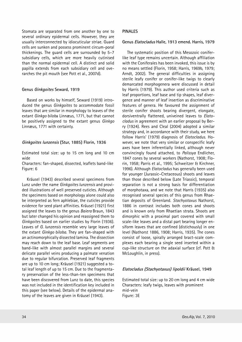
Ginkgoites lunzensis (Stur, 1885) Florin, 1936
Estimated total size: up to 15 cm long and 10 cm wideCharacters: fan-shaped, dissected, leaflets band-likeFigure: 6
Kräusel (1943) described several specimens from Lunz under the name Ginkgoites lunzensis and provi-ded illustrations of well preserved cuticles. Although the specimens based on morphology alone could also be interpreted as fern aphlebiae, the cuticles provide evidence for seed plant affinities. Kräusel (1921) first assigned the leaves to the genus Baiera Braun, 1843 but later changed his opinion and reassigned them to Ginkgoites based on earlier studies by Florin (1936). Leaves of G. lunzensis resemble very large leaves of the extant Ginkgo biloba. They are fan-shaped with an actinomorphically dissected lamina. The dissection may reach down to the leaf base. Leaf segments are band-like with almost parallel margins and several delicate parallel veins producing a palmate venation due to regular bifurcation. Preserved leaf fragments are up to 10 cm long; Kräusel (1921) suggested a to-tal leaf length of up to 15 cm. Due to the fragmenta-ry preservation of the less-than-ten specimens that have been discovered from Lunz to date, this species was not included in the identification key included in this paper (see below). Details of the epidermal ana-tomy of the leaves are given in Kräusel (1943).

35Geo.Alp, Vol. 7, 2010
another problem with regard to identification since they may represent immature foliage or mature folia-ge of juvenile plants or seedlings that does not display all the features characteristic of mature leaves from full-grown individuals of the same taxon (cf. Pott et al., 2007e; Pott & McLoughlin, 2009). In all of these instances, only the preparation of cuticles provides a data set of sufficient clarity for species identifica-tion. For detailed information on the epidermal anatomy of the species, the reader is referred to the publications that are indicated along with the species descriptions.
Acknowledgements
Hans Kerp, Institut für Geologie und Palaäonto-logie, Westfälische Wilhelms-Univeristät Münster, Germany, is thanked for all his help and support through the course of the whole project and for the co-supervision of CP’s PhD. We wish to thank the following people who provided access to the several collections under their care: David Cantrill, now Ro-yal Botanic Gardens, Melbourne, Australia; Thomas Denk, Else Marie Friis, Steve McLoughlin and Ove Johansson, Naturhistoriska riksmuseet, Stockholm, Sweden; Irene Zorn, Geologische Bundesanstalt, Vienna, Austria; Barbara Meller, Universität Wien, Vienna, Austria; Andreas Kroh and Matthias Harz-hauser, Naturhistorisches Museum, Vienna, Austria; Johanna van Konijnenburg-van Cittert, Netherlands Centre for Biodiversity Naturalis, Leiden University, The Netherlands; Johan van der Burgh, Institute of Environmental Biology, Utrecht University, The Ne-therlands; Martin Groß, Universalmuseum Joanneum, Graz, Austria; Lutz Kunzmann, Senckenberg Natur-historische Sammlungen Dresden, Germany; Stephan Schultka, Museum für Naturkunde, Berlin, Germany; Volker Wilde, Senckenberg Forschungsinsitut und Naturmuseum, Frankfurt am Main, Germany; Harald Steininger, Niederösterreichisches Landesmuseum, St. Pölten, Austria; Birgitt Aschauer, Waidhofen, Austria. Financial support was provided by the Deut-sche Forschungsgemeinschaft (DFG), Bonn, Germany (grants KR 2125/3-1 and KR 2125/3-2). We thank Johanna H. A. van Konijnenburg-van Cittert, Leiden, The Netherlands, and Mihai E. Popa, Bucharest, Ro-mania, for reviewing the manuscript.
The leafy coniferalean twigs from Lunz were assi-gned to Stachyotaxus lipoldii by Kräusel (1949), who also studied cuticles of the leaves. Originally, they were erroneously included in Pterophyllum by Stur (1885), but the leaves show only a single prominent midvein, which separates it from Pterophyllum that possesses several parallel veins per leaflet. Following the definitions and studies by Harris (1979), as well as those by several other workers, we feel that it would be more reasonable to transfer the leafy twigs from Lunz to Elatocladus. The twigs are stalked, up to 20 cm long, and bear several acute leaves (not leaflets in the sense of Kräusel, 1949), which are bent slightly forward towards the apex of the twigs. Leaf apices are somewhat acute, but mainly bluntly rounded. Leaves are up to 20 mm long and 3 mm wide. Kräusel (1949) described cuticles with haplocheilic stomata. Elatocladus lipoldii is the only conifer species known from the Lunz flora to date; it is a less common ele-ment than, e.g., Pterophyllum and Nilssonia leaves, but adds another typically Rhaetian genus to the in-ventory of the Lunz flora (cf. Pott et al., 2007e, 2008; Pott & McLoughlin, in press).
Comments on the key:
Drawings included in the key are simplified or ge-neralised, and slightly oversubscribed to underline the main characteristics of the species. A typical speci-men of each species, along with typical features of the cuticles, is illustrated in Figures 3–6.
The identification key largely relies on macro-morphological features because it was our intention to keep it as simple as possible and make it usable for a broad audience. Characteristics of the epider-mal anatomy are not included since cuticular analy-sis requires some effort and may not be available to everyone interested in the fossils from Lunz. The key was tested and works well for most hand specimens. It has to be taken into account, however, that some small and/or ill-preserved specimens may not be sa-fely identified down to species level based exclusively on macromorphology. The macromorphological diffe-rences between the species are sufficiently well re-cognisable in fossils that show a larger portion of the leaf. Fossils of entire but particularly small leaves pose

36 Geo.Alp, Vol. 7, 2010
Hampe, E. (1879). Enumeratio muscorum frondoso-rum Brasiliæ centralis, præcipue provinciarum Rio de Janeiro et Sao Paulo, adhuc cognitorum. – Vi-denskabelige Meddelelser fra Dansk Naturhistoriske Forening i Kjøbenhavn, 26: 73–163.
Harris, T. M. (1932): The fossil flora of Scoresby Sound East Greenland. Part 2: Description of seed plants incertae sedis together with a discussion of certain cycadophyte cuticles. – Meddelelser om Grønland, 85: 1–112.
Harris, T. M. (1935): The fossil flora of Scoresby Sound, East Greenland. Part 4: Ginkgoales, Coniferales, Lycopodiales and isolated fructifications. – Med-delelser om Grønland, 112: 1–176.
Harris, T. M. (1950): Notes on the Jurassic flora of Yorkshire, 46–48. – Annals and Magazine of Natu-ral History, Series 7, 3: 1001–1030.
Harris, T. M. (1969a): The Yorkshire Jurassic Flora. III. Bennettitales. – VI+186 p., Trustees of the British Museum (Natural History), London.
Harris, T. M. (1969b): Naming a fossil conifer. In: San-tarau, H. (ed.): J. Sen Memorial Volume, 243–252, Botanical Society of Bengal, Calcutta.
Harris, T. M. (1979): The Yorkshire Jurassic Flora. V. Co-niferales. – II+166 p., Trustees of the British Muse-um (Natural History), London.
Krasser, F. (1909a): Die Diagnosen der von Dionysius Stur in der obertriadischen Flora der Lunzer Schich-ten als Marattiaceenarten unterschiedenen Farne. – Sitzungsberichte der Kaiserlichen Akademie der Wissenschaften Wien, Abt. 1, 118: 13–43.
Krasser, F. (1909b): Zur Kenntnis der fossilen Flora der Lunzer Schichten. – Jahrbuch der Kaiserlich-König-lichen geologischen Reichsanstalt, 59: 1–26.
Krasser, F. (1917): Studien über die fertile Region der Cycadophyten aus den Lunzer-Schichten: Mikro-sporophylle und männliche Zapfen. Denkschriften der Kaiserlichen Akademie der Wissenschaften (Wien), Mathematisch-Naturwissenschaftliche Klasse, 94: 489–553.
Krasser, F. (1919): Studien über die fertile Region der Cycadophyten aus den Lunzer Schichten: Makro-sporophylle. – Denkschriften der Kaiserlichen Aka-demie der Wissenschaften (Wien), Mathematisch-Naturwissenschaftliche Klasse, 97: 1–32.
Kräusel, R. (1921): Über einige Pflanzen aus dem Keu-per von Lunz (Nieder-Österr.). – Jahrbuch der Preu-ßischen Geologischen Landesanstalt, 41: 192–209.
Kräusel, R. (1943): Die Gingkophyten der Trias von Lunz in Niederösterreich und von Neue Welt bei Basel. – Palaeontographica Abt. B, 87: 59–93.
References
Arndt, S. (2002): Morphologie und Systematik ausge-wählter mesozoischer Koniferen. – Palaeontogra-phica Abt. B, 262: 1–23.
Berry, E. W. (1924): Mesozoic plants from Patagonia. – American Journal of Science, Series 5, 7: 473–482.
Boyd, A. (2000): Bennettitales from Early Cretaceous floras of West Greenland: Pterophyllum and Nilssoniopteris. – Palaeontographica Abt. B, 255: 47–77.
Braun, C. F. W. (1843): Beiträge zur Urgeschichte der Pflanzen. – Beiträge zur Petrefacten-Kunde, 1–46.
Brongniart, A. (1825): Observations sur les végétaux fossiles renfermés dans les grès de Hoer en Scanie. – Annales des Sciences Naturelles, 4: 200–224.
Brongniart, A. (1828): Prodrome d’une histoire des vé-gétaux fossiles. – VIII+223 p., Levrault, Paris.
Cleal, C. J. (1993): Gymnospermophyta. – In: Benton, M. (ed.): The fossil record II, 795–808, Chapman & Hall, London.
Dobruskina, I. A. (1989): The alpine Lunz-Flora – a standard flora for the Carnian stage of the Triassic. – International Geological Review, 31: 1209–1215.
Dobruskina, I. A. (1998): Lunz flora in the Austrian Alps – a standard for Carnian floras. – Palaeogeography, Palaeoclimatology, Palaeoecology, 143: 307–345.
Doweld, A. B. (2000): Arberophyllum – a new generic name for Glossophyllum Kräusel, 1943 (Ginkgo-phyta). – Paleontological Journal, 34: 674–675.
Endlicher, S. (1847): Synopsis Coniferarum. – IV+368 p., Scheitlin & Zöllikofer, St. Gallen.
Ettingshausen, C. von (1851): Beiträge zur Flora der Vorwelt. – Haidinger’s Naturwissenschaftliche Ab-handlungen, 4: 65–99.
Florin, R. (1933): Zur Kenntnis der Spaltöffnungsap-parate der Bennettitales. – Kungliga Svenska Ve-tenskapsakademiens Handlingar, Tredje Serien, 12: 11–31.
Florin, R. (1936): Die fossilen Gingkophyten aus Franz-Joseph-Land nebst Erörterungen über vermeintliche Cordaitales mesozoischen Alters. I. Spezieller Teil. – Palaeontographica Abt. B, 81: 71–173.
Florin, R. (1958): On Jurassic taxads and conifers from north-western Europe and Eastern Greenland. – Acta Horti Bergiani, 17: 257–402.
Halle, T. G. (1913): The Mesozoic flora of Graham Land. Wissenschaftliche Ergebnisse der schwedischen Südpolar-Expedition 1901–1903 unter Mitwirkung zahlreicher Fachgenossenen. – 123 p., Generalsta-bens litografiska anstalt, Stockholm.

37Geo.Alp, Vol. 7, 2010
Pott, C., Krings, M., Kerp, H. (2007d): A surface micro-relief on the leaves of Glossophyllum florinii (?Gink-goales) from the Upper Triassic of Lunz, Austria. – Botanical Journal of the Linnean Society, 153: 87–95.
Pott, C., Krings, M., Kerp, H. (2008): The Carnian (Late Triassic) flora from Lunz in Lower Austria: palaeo-ecological considerations. – Palaeoworld, 17: 172–182.
Pott, C., Krings, M., Kerp, H., Friis, E. M. (2010): Re-construction of a bennettitalean flower from the Carnian (Upper Triassic) of Lunz, Lower Austria. – Review of Palaeobotany and Palynology, 159: 94–111.
Pott, C., McLoughlin, S. (2009): Bennettitalean foliage in the Rhaetian-Bajocian (latest Triassic-Middle Ju-rassic) floras of Scania, southern Sweden. – Review of Palaeobotany and Palynology, 158: 117–166.
Pott, C., McLoughlin, S. (in press): The Rhaeto-Liassic flora from Rögla, northern Scania, Sweden – Pa-laeontology.
Pott, C., Van Konijnenburg-van Cittert, J. H. A., Kerp, H., Krings, M. (2007e): Revision of the Pterophyllum species (Cycadophytina: Bennettitales) in the Car-nian (Late Triassic) flora from Lunz, Lower Austria. – Review of Palaeobotany and Palynology, 147: 3–27.
Rees, P. M., Cleal, C. J. (2004): Lower Jurassic floras from Hope Bay and Botany Bay, Antarctica. – Spe-cial Papers in Palaeontology, 72: 1–90.
Schenk, A. (1864): Beiträge zur Flora des Keupers und der rhätischen Formation. – Berichte der naturfor-schenden Gesellschaft zu Bamberg, 7: 1–91.
Schimper, W. P. (1869): Traité de paléontologie vé-gétale ou la flore du monde primitif dans ses rap-ports avec les formations géologiques et la flore du monde actuel. – IV+738 p. Baillière et fil), Paris.
Schimper, W. P. (1870–1872): Traité de paléontologie végétale ou la flore du monde primitif dans ses rap-ports avec les formations géologiques et la flore du monde actuel. – 996 p. Baillière et fils, Paris.
Schlotheim, E. F. von (1822): Nachträge zur Petre-factenkunde. 100 & 114 p., Becker’sche Verlags-buchhandlung, Gotha.
Schweitzer, H.-J., Kirchner, M. (1996): Die rhäto-juras-sischen Floren des Iran und Afghanistans: 9. Conife-rophyta. – Palaeontographica Abt. B, 238, 77–139.
Seward, A. C. (1911): The Jurassic flora of Sutherland. – Transactions of the Royal Society of Edinburgh, 47: 643–709.
Kräusel, R. (1948): Sturiella langeri nov. gen., nov. sp., eine Bennettitee aus der Trias von Lunz (Nieder-Österreich). – Senckenbergiana, 29: 141–149.
Kräusel, R. (1949): Koniferen und andere Gymnosper-men aus der Trias von Lunz, Nieder-Österreich. – Palaeontographica Abt. B, 89: 35–82.
Kräusel, R. (1953): Ein neues Dioonitocarpidium aus der Trias von Lunz. – Senckenbergiana, 34: 105–108.
Kräusel, R., Schaarschmidt, F. (1966): Die Keuperflora von Neuewelt bei Basel. IV. Pter-ophyllen und Tae-niopteriden. – Schweizer Paläontologische Abhand-lungen, 84: 3–44.
Lindley, J., Hutton, W. (1831–1836): The fossil flora of Great Britain, or figures and descriptions of the ve-getable remains found in a fossil state in the coun-try. – LIX+223 & XXVIII+208 p., James Ridgway, London.
Linnæus, C. (1771) Mantissa plantarum altera gene-rum editionis VI. & specierum editionis II. – 588 p., Laurentii salvii, Stockholm.
Müller, C. (1851): Synopsis muscorum frondosorum omnium hucusque cognitorum. Pars secunda. – 772 p., A. Foerstner, Berlin.
Nathorst, A. G. (1878–1886): Om floran i Skånes kol-förande bildningar. – Sveriges Geologiska Undersö-kning Serie C, 27, 33, 85: 1–126.
Nathorst, A. G. (1908): Paläobotanische Mitteilungen 7. Über Palissya, Stachyotaxus und Palaeotaxus. – Kungliga Svenska Vetenskapsakademiens Handlin-gar, 43: 3–20.
Nathorst, A. G. (1909): Über die Gattung Nilssonia Brongn. mit besonderer Berücksichtigung schwe-discher Arten. – Kungliga Svenska Vetenskapsaka-demiens Handlingar, 43: 3–37.
Parris, K. M., Drinnan, A. N., Cantrill, D. J. (1995): Palissya cones from the Mesozoic of Australia and New Zealand. – Alcheringa, 19: 87–111.
Pott, C., Kerp, H., Krings, M. (2007a): Morphology and epidermal anatomy of Nilssonia (cycadalean folia-ge) from the Upper Triassic of Lunz (Lower Austria). – Review of Palaeobotany and Palynology, 143: 197–217.
Pott, C., Kerp, H., Krings, M. (2007b): Pseudoctenis cornelii nov. spec. (cycadalean foliage) from the Car-nian (Upper Triassic) of Lunz, Lower Austria. – An-nalen des Naturhistorischen Museums Wien, 109A: 1–17.
Pott, C., Krings, M., Kerp, H. (2007c): The first record of Nilssoniopteris (Gymnospermophyta, Bennettitales) from the Carnian (Upper Triassic) of Lunz, Lower Austria. – Palaeontology, 50: 1299–1318.

38 Geo.Alp, Vol. 7, 2010
Tralau, H. (1968): Evolutionary trends in the genus Ginkgo. – Lethaia, 1: 63–101.
Van Konijnenburg-van Cittert, J. H. A., Mikuz, V., Pav-sic, J. (2001): Pterophyllum (Cycadopsida) from Car-nian beds in Poljane valley (Slovenia). – Geologija, 44: 317–323.
Van Konijnenburg-van Cittert, J. H. A., Van der Burg, J. (1989): The flora from the Kim-meridgian (Upper Jurassic) of Culgower, Sutherland, Scotland. – Re-view of Palaeo-botany and Palynology, 61: 1–51.
Zeiller, R. 1906. Bassin Houiller et Permien de Blanzy et du Creusot. Fascicule II. Flore fossile. – 265 p., Ministère des travaux publics, Paris.
Manuscript submitted: 12.4.2010Revised Manuscript accepted: 5.7.2010
Seward, A. C. (1919): Fossil Plants – A Textbook for Students of Botany and Geology. Volume IV. – XVI+543 p., Cambridge University Press, Cambridge.
Stur, D. (1871): Geologie der Steiermark. – XXXI+654p., Verlag des geognostisch-montanen Vereines für Steiermark, Graz.
Stur, D. (1885): Die obertriadische Flora der Lunzer-Schichten und des bituminösen Schiefers von Raibl. – Denkschriften der Kaiserlichen Akademie der Wis-senschaften (Wien), 3: 93–103.
Stur, D. (1888): Die Lunzer (Lettenkohle-)Flora in den „older mesozoic beds of the coalfield of eastern Virginia”. – Verhandlungen der Kaiserlich-Königlich Geologischen Reichsanstalt, 10: 203–217.
Thomas, H. H. (1930): Further observations on the cu-ticle structure of Mesozoic cycadean fronds. – The Journal of the Linnean Society of London/Botany, 48: 389–415.