guide to the common inshore of southern california - …...153 μm microplankton net, and smaller...
TRANSCRIPT
page 1
GUIDE TO THE COMMON INSHORE
rev. 2010-01
name_______________________________________________________
of Southern California - 4th Edition
Robert PerryMalibu High School, and
UCLA OceanGLOBE
Condensed for public educational purposes from the textbook,Ocean Images: the Plankton, available online at www.Blurb.com
Late megalopa larva of a crab.
page 2
table of contents:
PHYTOPLANKTON:PROTISTA: Diatoms . . . . . . . . . . . . . .. . . . . . . . . . . . . .PROTISTA: Dinoflagellates. . . . . . . . . . . . . . . . . . . . . . .
ZOOPLANKTON:PROTISTA: Protozoa . . . . . . . . . . . . . . . . . . . . . . . . . . .
ANIMALIA:Cnidaria . . . . . . . . . . . . . . . . . . . . . . . . . . . . . . . . .Platyhelminthes . . . . . . . . . . . . . . . . . . . . . . . . . . .Bryozoa . . . . . . . . . . . . . . . . . . . . . . . . . . . . . . . . .Rotifera . . . . . . . . . . . . . . . . . . . . . . . . . . . . . . . . .Polychaeta . . . . . . . . . . . . . . . . . . . . . . . . . . . . . . .Mollusca . . . . . . . . . . . . . . . . . . . . . . . . . . . . . . . .Crustaceans
Copepods . . . . . . . . . . . . . . . . . . . . . . . . . .Crustacean larvae . . . . . . . . . . . . . . . . . . . .Other holoplanktonic crustaceans . . . . . . . .
Echinodermata . . . . . . . . . . . . . . . . . . . . . . . . . . .Chordata . . . . . . . . . . . . . . . . . . . . . . . . . . . . . . . .
CALCULATING THE ABUNDANCE OF PLANKTON PERCUBIC METER OF SEAWATER. . . . . . . . . . . . . . . . . . . . . . . .
All photography © Robert Perrywww.MarineBioPhotography.com
All Rights Reserved.
This booklet has been made available fornon-profit, direct, face-to-face, educational
purposes only.
With special thanks to the crew of the R/V Sea World UCLA, 1995-2008.and the crew of the Condor Express, and the crew of the dive boat Peace.And a special thanks to the students who paddled for plankton at Zuma Beach as part of their research project at Malibu High School. Go Sharks !
37
10
111213131415
1617181920
21

page 3
PHYTOPLANKTON1- Introduction to the Diatoms.
Members of the Division Chrysophyta (orBacillariophyta) are known as diatoms. The word“Diatom” comes from the Greek Dia, = across, andtom, = to cut. This refers to the fact that diatoms areenclosed within two glass (SiO2) shells which splitacross the middle and separate from each otherduring reproduction. One shell is older (the epitheca)and is slightly larger than the other, younger shell (thehypotheca). The smaller fits inside the larger like a“pill box” or Altoid® mint container. As shown in thediagram to the right, during cell division they splitapart to reproduce. Each section grows a new,smaller half that fits inside the original. Thus, half ofthe new cells are smaller in size than the other half.This means that diatoms of the same species may bequite variable in their size.
Under the microscope most diatoms appear greenor golden, and their frustules are as clear as theglass which makes them. In the photos above youcan clearly see the chloroplasts inside the cellwall. When diatoms are exceptionally abundant inthe surface water, the ocean may appear green.Some species like Coscinodiscus (right) aresolitary, while others like Chaetoceros (left) formlong chains.
Diatoms, along with photosynthetic bacteria, are among the most important groups of producer organ-isms in the marine ecosystem. They inhabit the sun lit upper surface waters of most oceans and helpform the base of the food chain using photosynthesis to convert solar energy into chemical bondenergy. Many diatoms convert the carbohydrates that are produced during photosynthesis into oils.These oils are less dense than the surrounding seawater and, thus, help buoy the diatoms upward intothe illuminated photic zone of the ocean. Some diatom chain-forming species prevent sinking throughthe growth of spiny extensions (the setae or chaetae). During population explosions or “blooms” ofthese spiny diatoms they may damage the sensitive gills of fish and other animals.
Chaetoceros sp. a diatom chain. Coscinodiscus sp. a solitary diatom.
PHYTOPLANKTON - Diatoms
Diatom cell division.
setae
glass shell

page 4
Pseudo-nitzschia sp. a toxic diatom.
A small handful of diatom species are also toxic tomarine animals and humans. For example,Pseudo- nitzschia may produce a poisonous chemicalknown as Domoic Acid (DA) over the outside of its shells.DA is known to cause Amnesic Shellfish Poisoning (ASP),a neurological disorder that affects pelicans, sea lions, andother marine animals including humans. DA builds upinside the tissues of filter-feeding invertebrates such asmussels and clams, which, if eaten during periods of highconcentrations of DA, can cause amnesia and even deathin humans.
PHYTOPLANKTON - Diatoms
Aulacodiscus kittoniiFound with sand in nano- samples taken
from the surf zone.
Ceratulina pelagica Climacosphenia moniligera
Chaetoceros sp.Square cells with spines;
Count the number of cells.
Chaetoceros socialis
top viewside viewCoscinodiscus sp.
Mass of very small square cells with spines.
Corethron hystrix
chloroplastsvisible
chloroplastsvisible

page 5PHYTOPLANKTON - Diatoms
Coscinodiscus granii
Ditylum brightwellii
top view side viewNavicula distans
Odontella mobiliensis
Odontella longicruris
Planktoniella blandaPlanktoniella sol
Plagiogrammopsis vanheureckii
connecting sandgrains
Count the number of cells.

page 6PHYTOPLANKTON - Diatoms
Pseudo-nitzschia australis
Thalassionema nitzschioidesThalassiosira rotula
Striatellat unipunctata
Proboscia alata
Count the number of cells.

page 7PHYTOPLANKTON - Dinoflagellates
PHYTOPLANKTON2- Introduction to the Dinoflagellates.
Members of the Division Dinoflagellata (aka Dinophyta or Pyrrophyta) are very common in coastalplankton samples. The name comes from the greek word “Dinos” which means “to spin,” and “flagel-late” which means to have flagella. Flagella are whip-like “tails” that act like propellers and cause thesesingle celled organisms to rotate through the water. When observed immediately after capture, undera microscope, dinoflagellates move in a wonderful spiral pattern. Large species are caught with a153 μm microplankton net, and smaller species require a 20μm phytoplankton net.
Dinoflagellates appear reddish-brown, or greenish-brown under the microscope. Many dinoflagellateshave red or brown accessory pigments along withchlorophyll to carry out photosynthesis. Hence,when very abundant in the water, the phenomenonis called a “red tide.” Red tides are localized patchesof discolored water from dinoflagellate populationexplosions. They can become so abundant that theyblock out all sunlight except for the upper fewcentimeters of the ocean surface. Consequently thedinoflagellates in the shade become heterotrophicto survive. As heterotrophs, they may rapidlydeplete the oxygen content of the surroundingocean water causing sessile organisms to die. If thered tide includes very spiny species, they can also
Dinoflagellates are the second most abundant form of autotrophic life in the marine ecosystem. Assuch. they are at the base of the food chain and provide food for herbivorous zooplankton and sessilebenthic suspension feeders. Many dinoflagellate species are also toxic, and some are poisonous tohumans. Although it is unlikely that organisms high up on the food chain like humans swallow enoughtiny planktonic dinoflagellates directly to pose a threat, their toxins are “biomagnified” through the foodchain. Simply put, this means that it takes approximately 10 pounds of dinoflagellates to make onepound of zooplankton or benthic suspension feeder. Every unit of poison found naturally in adinoflagellate will magnify ten-fold in the next trophic level, a hundred times in the next level andso forth. Species such as Alexandrium and Gymnodinium are known to cause PSP (ParalyticShellfish Poisoning) in humans. PSP results when humans eat shellfish such as mussels, clams oroysters that are suspension feeders who have, in turn, been consuming these dinoflagellates.Symptoms of PSP include nausea, dizziness, a tingling numbness, respiratory failure and even death.It is not wise to consume these shellfish during periods of the year when toxic dinoflagellates areblooming.
clog animals’ gills or cause abrasions in gill tissue that then becomes susceptible to secondarybacterial infection. These factors combine to give red tides, and the dinoflagellates that cause them, abad reputation for killing fish and other animals in certain places in the world. Dinoflagellates possessexternal armored plates of cellulose. These cellulose plates protect the tiny single celled organismsfrom predators. Often these plates are extended to form long, sharp spines that further protect thecells. Another interesting fact is that most members of this group can produce their own biochemicallight. Most of the bioluminescence we view from boats or from breaking waves along the shore at nightis caused by dinoflagellates.
celluloseshell

page 8PHYTOPLANKTON - Dinoflagellates
Ceratium balechiiAkashiwo sanguinea
formerly known as Gymnodinium Ceratium azoricum
Ceratium carrienseCeratium divaricatum
previously confused with a gamete ofCeratium lineatum
Ceratium extensumCeratium furca
Ceratium fusus
Ceratium gravidum
horn connectionvery round
very long -up to 3 mm
very short
horns long, thinand symmetrical
twostumps
onestump

page 9PHYTOPLANKTON - Dinoflagellates
Dinophysis acuminata
Ceratium macroceros
Dinophysis tripos
Ceratium lineatum
Lingulodinium polyedra
Podolampas palmipes Prorocentrum micans
Protoperidinium sp.
Causes red tide in Southern California
horn connectionNOT round

page 10ZOOPLANKTON - Protozoa
ZOOPLANKTONPhylum Protozoa
These single celled organisms are different from the other Protists, the Diatoms and Dinoflagellates,because they are heterotrophs and do not contain photosynthetic pigments. Consequently they aremostly without color and appear clear under the microscope. Protozoans occur frequently in ourcoastal plankton hauls. Two groups of ameboid protozoans, the Radiolarians, and theForaminiferans, and one ciliate group, the Tintinnidae, are particularly common. Radiolarians feedthemselves by sending body extensions called pseudopods out of tiny holes in their shells. When theydie, their shells “rain” down and cover vast areas of the seabed many meters thick. Tintinnids use ciliato create feeding currents.
Discorbis sp. - a Foram(note chambers inside shell)
Acanthometron sp. - a RadiolarianUnidentified Radiolarian
Globigerina bulloides - a Foram
Parafavella sp. - a Tintinnid
Unidentified Tintinnid(also, a silicoflagellate Octactis octonaria
appears in the right corner)
ciliarycrown
page 11ZOOPLANKTON - Cnidaria
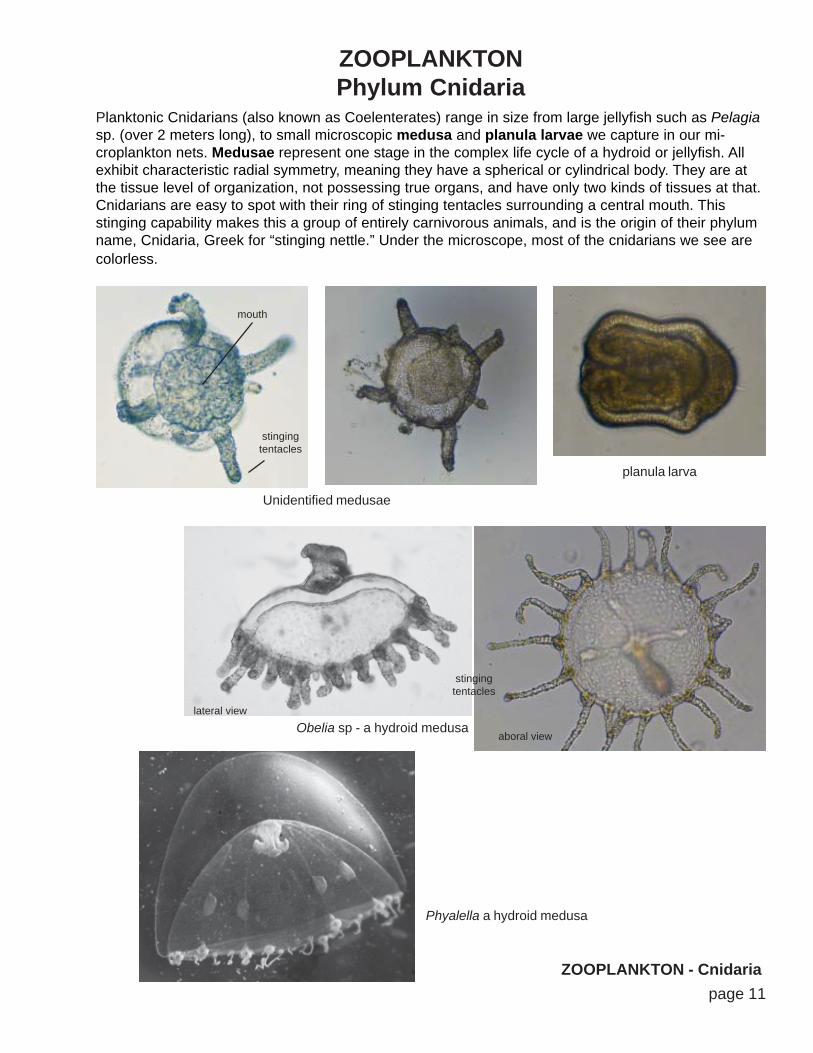
ZOOPLANKTONPhylum Cnidaria
Planktonic Cnidarians (also known as Coelenterates) range in size from large jellyfish such as Pelagiasp. (over 2 meters long), to small microscopic medusa and planula larvae we capture in our mi-croplankton nets. Medusae represent one stage in the complex life cycle of a hydroid or jellyfish. Allexhibit characteristic radial symmetry, meaning they have a spherical or cylindrical body. They are atthe tissue level of organization, not possessing true organs, and have only two kinds of tissues at that.Cnidarians are easy to spot with their ring of stinging tentacles surrounding a central mouth. Thisstinging capability makes this a group of entirely carnivorous animals, and is the origin of their phylumname, Cnidaria, Greek for “stinging nettle.” Under the microscope, most of the cnidarians we see arecolorless.
Unidentified medusae
Obelia sp - a hydroid medusa
Phyalella a hydroid medusa
lateral view
aboral view
stingingtentacles
stingingtentacles
mouth
planula larva

page 12
ZOOPLANKTONPhylum Platyhelminthes
ZOOPLANKTON - Platyhelminthes
Platyhelminthes, or “flatworms,” are compressed dorso-ventrally, like a piece of ribbon. Some peoplecall this “life in 2 Dimensions.” Flatworms are thought to be the first phylum to have evolved a truehead on one end of the body, and a tail on the opposite end. The evolution of a head means theflatworms were the first creatures on Earth to move head first through their environment. A head andtail also means that their right and left sides are mirror images, which is to say they were the firstorganisms on Earth to exhibit bilateral symmetry. Flatworms were also the first phylum to havetissues working together as rudimentary organs, but no organ systems exist in this group. Since mostof these animals live on the seafloor, it is only occassionally that we capture a flatworm in a planktonnet, but it does happen. Even after netting, refrigerating, stirring, and dropping onto a slide, they areoften very active under the microscope. Even more rarely we capture their Muller’s larvae.
Muller’s larva
Flatwormseyespots
visible
eyespotsvisible

page 13
ZOOPLANKTONPhylum Bryozoa
ZOOPLANKTON - Bryozoans & Rotifers
Phylum Rotifera
Bryozoans are ubiquitous benthic colonial animals that attach and spread over rocks, kelp, shells andmanmade marine structures. They are tiny wormlike creatures that live inside boxes and are barelyvisible to the naked eye. Bryozoans reproduce, develop and distribute themselves geographicallyusing a triangular-shaped cyphonautes larvae. Cyphonautes shells are transparent, double and theopen edge is fringed with active cilia. The internal organs are easy to see inside. Cyphonautes larvaedrift in the currents until ready to settle out to the bottom and take up a sessile benthic existance. Oncea larva lands it begins to divide asexually and continues to divide until an entire area is covered byidentical clones.
Cyphonautes larvae
Rotifers are small, worm-like animals with ciliary “wheel organs” on their heads that rotate and givethe group their name “rotifer.” Rotifers are very small, usually less than 200μm, which, for a complexmulticellular animal, brings it to about the size of a typical protozoan. Almost all rotifers are females.Males in some families have never been observed. Females lay fertile eggs without the need of a maleor fertilization, using a process known as parthenogenesis. Only one species of planktonic marinerotifer has been recorded along the coastline...they are more abundant in freshwater.
Brachionus plicatilis
Female w. egg inside Female towing egg outside
Looking down on head to seeciliary “wheel organs”
cilia
cilia
cilia
cilia
egg
egg
2 triangularshapedshells
cilia

page 14
ZOOPLANKTONPhylum Annelida, Class Polychaeta
ZOOPLANKTON - Polychaetes
Segmented worms are members of the phylum Annelida (latin, annulus: segment or ring). These“marine worms” or “bristle worms” are the most abundant worms in our local ocean and therefore themost common type of worm we capture in our coastal plankton hauls. The planktonic Annelids aremembers of the class Polychaeta (poly =many; chaeta = hairs or bristles) and are relatives of earth-worms and leeches. Anatomically they are distinguished from the other kinds of worms by having abody that is divided into segments from head to tail. Each segment has small extensions or “legs”(parapodia) on each side with numerous stiff bristles (chaeta or setae). The polychaetes we see in theplankton are the larval stages of benthic species, many of which are sedentary tubedwellers as adults.Very early polychaete trochophore larvae are essentially just a swimming “head.” As the worm elon-gates its body and grows, and it adds additional segments behind the head in a posterior direction.
Family Spionidaeearly trochophore mid trochophore
setae
setae
setae
cilia onhead
cilia ontail
setae
segmentedbody
late trochophore
other Polychaete trochophores

page 15
Gastropod veliger larvae
ZOOPLANKTONPhylum Mollusca
ZOOPLANKTON - Mollusks
Mollusks are a huge phylum in the ocean, with nearly 100,000 different species. The name Molluscacomes from the Greek, mollis, which means “soft,” and all members of this phylum have soft, muscularbodies that are usually protected by one or more external calcium shells (in adulthood). There areholoplanktonic mollusks known as pteropods and heteropods, and all of the bottom-dwelling mol-lusks have planktonic larval stages known as veligers. Gastropod (snails, slugs) larvae have a single,spiral shell and Bivalve (clams, mussels, oysters) larvae have two “clam shaped” shells. The veligerlarva, is taken from the latin word velum, which means “veil” or “wing.” The velum is a winged structurebordered by actively beating cilia. Veligers live in the plankton and grow until they become too big andheavy to float, when they spend their adulthood living on the seafloor.
Bivalve veliger larvae
Pteropods
cilia
foot
spiral shell w/no chambers
inside
cilia
“clam shaped”shell
cone shapedshell
page 16
ZOOPLANKTONPhylum Arthropoda - Class Crustacea
ZOOPLANKTON - Copepod crustaceans
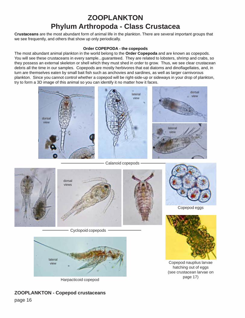
Crustaceans are the most abundant form of animal life in the plankton. There are several important groups thatwe see frequently, and others that show up only periodically.
Order COPEPODA - the copepodsThe most abundant animal plankton in the world belong to the Order Copepoda and are known as copepods.You will see these crustaceans in every sample...guaranteed. They are related to lobsters, shrimp and crabs, sothey possess an external skeleton or shell which they must shed in order to grow. Thus, we see clear crustaceandebris all the time in our samples. Copepods are mostly herbivores that eat diatoms and dinoflagellates, and, inturn are themselves eaten by small bait fish such as anchovies and sardines, as well as larger carnivorousplankton. Since you cannot control whether a copepod will be right-side-up or sideways in your drop of plankton,try to form a 3D image of this animal so you can identify it no matter how it faces.
Calanoid copepods
dorsalview
dorsalviewlateral
view
lateralview
Cyclopoid copepods
dorsalviews
Harpacticoid copepod
lateralview
Copepod eggs
Copepod nauplius larvaehatching out of eggs
(see crustacean larvae onpage 17)

page 17ZOOPLANKTON - Crustacean larvae
Crustacean larvaeAs you can see in the photo on page 16, some crustaceans begin life as a developing embryo insidean egg which is being carried attached to the female along with hundreds or thousands of other eggs.Other crustaceans shed their eggs into the water currents. Many crustaceans show up in our planktonsamples in their first stage after hatching from the egg, the nauplius stage. There may be severalnauplius stages, then a metamorphosis to a second larval stage known as the cypris larva. The cyprismay develop into a zoea, in crabs, for example, or a phyllosoma, in the case of the spiny lobster.
Nauplius larvae
dorsalview
lateralview
dorsalview
Cypris larvae
dorsalview
lateralview
Nauplius larvae
Crab zoea larvae
Zoea larvae ofEmerita analoga
Zoea larvae ofBlepharipoda occidentalis
Phyllosoma larvae ofCalifornia Spiny Lobster
Panulirus interruptus
rightlateralview
leftlateralview
frontalview
lateralview
lateralview
dorsalview

page 18
Crustaceans - Cladocerans
ZOOPLANKTON - Crustaceans
Evadne nordmanni
Podon polyphemoides
Penilia avirostris
dorsalview
lateralview
lateral viewfemale w/ brood
lateral viewfemale w/ brood
lateralview dorsal
view
ventralview

page 19ZOOPLANKTON - Echinoderms
ZOOPLANKTONPhylum Echinodermata
Echinoderms are entirely benthic animals, and they are among the dominant phyla of the abyssalbenthos. These spiny skinned animals are also very abundant on the seafloor in nearshore waters.Such common animals as sea stars, sea urchins, brittle stars and sand dollars belong to this group. Inorder to reproduce themselves and distribute the species, Echinoderms use planktonic larvae. Theselarvae are very unique and readily identifyable. Because our samples are taken from Zuma Beach, CA,an exposed sandy beach, we find a lot of sand dollar larvae.
Echinopluteus larvaeprobably Dendraster excentricus
Late echinoid larvaprobably Dendraster excentricus
Ophiopluteus larvaof a brittle star
Doliolaria larvaof a sea cucumber
Echinopluteus larvae
page 20
ZOOPLANKTONPhylum Chordata
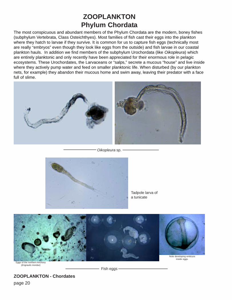
The most conspicuous and abundant members of the Phylum Chordata are the modern, boney fishes(subphylum Vertebrata, Class Osteichthyes). Most families of fish cast their eggs into the planktonwhere they hatch to larvae if they survive. It is common for us to capture fish eggs (technically mostare really “embryos” even though they look like eggs from the outside) and fish larvae in our coastalplankton hauls. In addition we find members of the subphylum Urochordata (like Oikopleura) whichare entirely planktonic and only recently have been appreciated for their enormous role in pelagicecosystems. These Urochordates, the Larvaceans or “salps,” secrete a mucous “house” and live insidewhere they actively pump water and feed on smaller planktonic life. When disturbed (by our planktonnets, for example) they abandon their mucous home and swim away, leaving their predator with a facefull of slime.
ZOOPLANKTON - ChordatesFish eggs
Eggs of the northern Anchovy(Engraulis mordax)
Note developing embryosinside eggs.
Oikopleura sp.
Tadpole larva ofa tunicate

page 21
3 Steps to Calculate the Amount of Planktonin a Cubic Meter of the Ocean:
The plankton net extractseverything from a cylindershaped chunk of water.
Step One
Step Two
CALCULATE THE VOLUME OF THE WATER THAT WAS SAMPLED BY THE NET USING THIS FORMULA:
r = radius of net (in meters)[squared]
L = Length of tow (in meters)
Multiply these numbers together and round-off to 2decimal places to get the number of cubic meters ofseawater the plankton net sampled. Write the answerin the preliminary data at the top of your plankton datasheet where it says “Calculated Net Volume.”
You will use the volume in Step Three calculations.
CALCULATE THE (AVERAGE) NUMBER OF EACH SPECIES PER DROP SAMPLED UNDER YOURMICROSCOPE:
Divide the number of each species you observed by thenumber of total drops you analyzed in class. Thiscalculation must be repeated for each species ofplankton you observed.
number observed
number of dropsanalyzed
______________
Record these calculations (two decimal places) next to each species of phytoplanktonand zooplankton you wrote on your data sheet, in the 3rd column.

page 22
Step Three (the final step)
3 Steps to Calculate the Amount of Planktonin a Cubic Meter of the Ocean (continued):
CALCULATE THE NUMBER OF PLANKTON PER CUBIC METER OF OCEAN WATER:
# m-3 =
# m-3
average numberper drop (step 2)
total drops inthe entire sampleX
volume (step 1)This is the final calculation that gets graphed and analyzed. It tells the number of each kind ofplankton that was living in a cubic meter chunk of ocean water on Thursday when the net tookthe sample.
Multiply the number per drop (from step 2) times the total number of drops in the entire sample(provided by instructor), then divide this by the volume you calculated in step 1.
Do this calculation for each species of phytoplankton and zooplankton you recorded, and writethe calculation (rounded to 2 decimal places) in the last column on your data sheet. Thiscolumn has been shaded grey and is marked “graph this column” to help you remember toenter these numbers into your spreadsheet database.