gray wolf exposure to emerging vector-borne...
TRANSCRIPT
RESEARCH ARTICLE
Gray Wolf Exposure to Emerging Vector-Borne
Diseases in Wisconsin with Comparison to
Domestic Dogs and Humans
Rocio F. Jara1¤a, Adrian P. Wydeven2¤b, Michael D. Samuel3*
1 Nelson Institute for Environmental Studies, University of Wisconsin-Madison, Madison, Wisconsin, United
States of America, 2 Wisconsin Department of Natural Resources, Retired, Ashland, Wisconsin, United
States of America, 3 U.S. Geological Survey, Wisconsin Cooperative Wildlife Research Unit, University of
Wisconsin-Madison, Madison, Wisconsin, United States of America
¤a Current address: Department of Biological Sciences, Sub-Antarctic Biocultural Conservation Program,
University of North Texas, Denton, Texas, United States of America.
¤b Current address: Timber Wolf Alliance, Northland College, Ashland, Wisconsin, United States of America
Abstract
World-wide concern over emerging vector-borne diseases has increased in recent years for
both animal and human health. In the United Sates, concern about vector-borne diseases in
canines has focused on Lyme disease, anaplasmosis, ehrlichiosis, and heartworm which
infect domestic and wild canids. Of these diseases, Lyme and anaplasmosis are also fre-
quently diagnosed in humans. Gray wolves (Canis lupus) recolonized Wisconsin in the
1970s, and we evaluated their temporal and geographic patterns of exposure to these four
vector-borne diseases in Wisconsin as the population expanded between 1985 and 2011. A
high proportion of the Wisconsin wolves were exposed to the agents that cause Lyme
(65.6%) and anaplasma (47.7%), and a smaller proportion to ehrlichiosis (5.7%) and
infected with heartworm (9.2%). Wolf exposure to tick borne diseases was consistently
higher in older animals. Wolf exposure was markedly higher than domestic dog (Canis famil-
iaris) exposure for all 4 disease agents during 2001–2013. We found a cluster of wolf expo-
sure to Borrelia burgdorferi in northwestern Wisconsin, which overlaps human and domestic
dog clusters for the same pathogen. In addition, wolf exposure to Lyme disease in Wiscon-
sin has increased, corresponding with the increasing human incidence of Lyme disease in a
similar time period. Despite generally high prevalence of exposure none of these diseases
appear to have slowed the growth of the Wisconsin wolf population.
Introduction
World-wide concern over emerging vector-borne diseases has increased in recent years for
both animal and human health [1,2]. In the United Sates (US), concern about canine vector-
borne diseases is mainly focused on Lyme disease, anaplasmosis, ehrlichiosis, and heartworm
[3] which infect domestic and wild canids. Infections with these pathogens can cause severe
PLOS ONE | DOI:10.1371/journal.pone.0165836 November 29, 2016 1 / 17
a11111
OPENACCESS
Citation: Jara RF, Wydeven AP, Samuel MD (2016)
Gray Wolf Exposure to Emerging Vector-Borne
Diseases in Wisconsin with Comparison to
Domestic Dogs and Humans. PLoS ONE 11(11):
e0165836. doi:10.1371/journal.pone.0165836
Editor: Brian Stevenson, University of Kentucky
College of Medicine, UNITED STATES
Received: July 14, 2016
Accepted: October 18, 2016
Published: November 29, 2016
Copyright: This is an open access article, free of all
copyright, and may be freely reproduced,
distributed, transmitted, modified, built upon, or
otherwise used by anyone for any lawful purpose.
The work is made available under the Creative
Commons CC0 public domain dedication.
Data Availability Statement: All relevant data are
within the paper and its Supporting Information
files.
Funding: We thank the Wisconsin Department of
Natural Resources for funding. This research was
also funded by the U.S. Geological Survey and the
Chilean National Commission for Scientific and
Technological Research (CONICYT - Spanish
acronym), which provided a graduate fellowship to
RJ. The Department of Forest and Wildlife Ecology,
University of Wisconsin-Madison, provided funds
for publication. The funders had no role in study
illness in domestic and wild canids, affecting survivorship of the host [4–8]. These diseases also
affect humans, with Lyme disease and anaplasmosis most frequently diagnosed in the North-
east and upper Midwest US [9–12]. In Wisconsin, Lyme disease in humans has been increas-
ing since the 1990’s and nationwide Lyme disease is currently the most common vector-borne
disease with more than 30,000 cases reported in 2012 [13]; however, true incidence could be
much higher due to underreporting [14]. Anaplasmosis became a reportable disease in 1999,
and since then the annual number of human anaplasmosis cases reported in Wisconsin has
grown steadily from 1 case in 2000 to> 500 during 2010–2104 [15].
In the US, Lyme disease is primarily caused by the bacterium B. burgdorferi sensu stricto(although a novel B. burgdorferi sensu lato genospecies (candidatus Borrelia mayonii) has been
recently described [16]) and anaplasmosis by the Rickettsiales Anaplasma phagocytophilum[17]. The black-legged tick Ixodes scapularis is the vector for both pathogens in the Midwest
and Northeast [18]; therefore, coinfection may also occur [3]. White-tailed deer (Odocoileusvriginanus) is an important host for adult I. scapularis ticks [18] while white footed mice (Pero-myscus leucopus) are the main reservoir for these disease agents [19–22]; however, many wild
mammals, including gray wolves (Canis lupus), can also serve as hosts. Serological studies in
Minnesota and Wisconsin found gray wolf exposure to B. burgdorferi ranging from 3% [23] to
48% [24]. Anaplasma phagocytophilum was reported in a captive timber wolf in Austria that
showed anorexia, depression, and fever [4]. In domestic dogs (Canis familiaris), geographic
patterns of prevalence of exposure to B. burgdorferi and A. phagocytophilum are similar to the
incidence pattern in humans, i.e., higher prevalence in the upper Midwest and Northeastern
US [3]. In particular, 10.2% and 10.5% of the domestic dogs tested in Wisconsin were positive
for exposure to B. burgdorferi and A. phagocytophilum respectively, between 2001 and 2007
[3].
Canine ehrlichiosis is caused by the bacterium Ehrlichia canis [25] and transmitted primar-
ily by the brown dog tick Rhipicephalus sanguineus [26]. In domestic dogs from the US, the
highest prevalence has been detected in the Southeast (0.6% prevalence). In Wisconsin, where
the brown dog tick is rare, the prevalence in domestic dogs is 0.3% [3]. Human infection with
this pathogen has only been reported in 6 patients in Venezuela [10]. Infection with this patho-
gen occurred in a captive gray wolf in Florida, US, that showed epistaxis, anorexia, weight loss
[7].
Heartworm is caused by the nematode Dirofilaria immitis transmitted by Aedes, Anopheles,and Culexmosquitoes. Feral dogs and coyotes (Canis latrans) are recognized reservoirs for this
parasite [27]. Infection is widely distributed in domestic dogs in the US with the highest preva-
lence in the Southeast [3]. In Wisconsin, only 0.6% of domestic dogs tested positive for infec-
tion with this parasite [3]. Prevalence in wild red wolves (Canis rufus) in Southeastern US has
reached 100%, whereas in Wisconsin, studies reported from 0% to 9% of gray wolf infection
[24] (Dorothy Ginnett and Jerold Theis, unpublished report to the DNR 2003). Human infec-
tion with D. immitis can occur, and is positively correlated with the prevalence in domestic
dogs at a state level [28]; however, most human cases are asymptomatic [29].
Gray wolves had become extirpated in Wisconsin in the late 1950s, but began to recolonize
the state in the mid-1970s after being listed as federally endangered in 1974, and state endan-
gered in 1975 [30]. The Wisconsin Department of Natural Resources (WDNR) began moni-
toring the wolf population in winter 1979–1980 when 25 wolves were counted in midwinter.
Subsequently the population grew to 782 wolves by winter 2011 [30,31]. Sampling animals for
disease testing was started in 1981, and archived samples of blood and serum for additional
disease testing was initiated in 1985 [32]. Gray wolves were removed from the state list of
endangered and threatened species in 2004 [30]. Wolves were down-listed to federally threat-
ened in 2003, and delisted in 2007, but during recent legal battles they have returned to the
Vector-Borne Diseases in Wisconsin Wolves
PLOS ONE | DOI:10.1371/journal.pone.0165836 November 29, 2016 2 / 17
design, data collection and analysis, decision to
publish, or preparation of the manuscript.
Competing Interests: The authors have declared
that no competing interests exist.
federal list of endangered species [33,34]. The disease status in wild wolves is of interest in
assessing the success of wolf recovery and recolonization, as well as assessing the effect of dis-
eases on population growth and role in wolf population dynamics.
Because wolves are free-ranging and have more contact with infectious vectors, such as
ticks, their probability of being exposed to these pathogens is likely higher than humans and
domestic dogs. Accordingly, we expect higher wolf exposure to these pathogens than for
humans and domestic dogs. Aside from a few reports in wild gray wolves, there are no large
temporal and spatial scale studies of wolf exposure to these vector-borne diseases. It is useful
to study vector-borne exposures in wolves to better understand health threats to wolf popula-
tions, but also because wolves can serve as a sentinel species for human and domestic dog
health risk. Wild sentinels may be more likely to reflect ecological changes in disease incidence
than public health reporting because diagnostic reporting and case definitions used to diag-
nose human Lyme disease change over time. In addition, preventive veterinary medicine (e.g.,
vaccination for Lyme disease and anthelmintics), diagnostic testing (annual exposure testing
using SNAP 4Dx Test and similar methods), tick removal, and antibiotic treatment mean that
domestic dogs may provide a biased assessment of natural pathogen exposure or risk. In addi-
tion, wolves might better indicate zones of higher risk because domestic dog movements coin-
cide with their owners (i.e., they visit similar areas and spend similar amounts of time in tick
friendly habitats). In Wisconsin, it is possible to integrate varied sources of information
including tick and mouse field surveys (e.g., for Lyme disease and anaplasmosis), human case
reports, and domestic and wild host prevalence reports [35].
The objective of our study is to evaluate the temporal and geographic patterns of gray wolf
exposure to agents causing four vector-borne diseases—Lyme disease, anaplasmosis, canine
ehrlichiosis and heartworm—in Wisconsin between 1985 and 2011. Specifically, we measured
seroprevalence in gray wolves over time and identified clusters of high exposure to these dis-
eases. In addition, we compared temporal and geographic patterns in wolves to humans and
domestic dogs to evaluate similarity in patterns of exposure/infection.
Material and Methods
We conducted this study using 373 blood or sera samples from wild Wisconsin wolves col-
lected between 1985 and 2011. The samples were from wolves captured as part of the annual
monitoring program conducted by the Wisconsin Department of Natural Resources (WDNR)
[30] or captured for depredation management by the U.S. Department of Agriculture Wildlife
Service (USDA-WS) [36]. Wolves captured as non-target species during the coyote trapping
season or found dead from various causes (vehicle collision, illegally killed, diseases) were also
included. Along with collecting sera samples, wolves were examined for ectoparasites, and
both Dermacentor and Ixodes ticks were commonly found on wolves. The samples came from
45 of 72 counties, representing mainly the northern forest region of Wisconsin (primary habi-
tat for gray wolves in the state). Blood samples of 10 cc or less were obtained from the cephalic,
saphenous, femoral, or jugular veins of live (captured/released) wolves using needles of gauges
20–25 (20–22 on adults, 22–25 on pups or immature adults). Samples collected by the WDNR
were obtained following a standard operating protocol (SOP) approved by the WDNR Animal
Care and Use Committee (ACUC). The USDA-WS followed a separate SOP, also approved by
the WDNR ACUC, for wolves captured for depredation management.
Wolves captured by the WDNR for monitoring and research purposes were mostly cap-
tured on national, state, or county forest, as well as state wildlife areas with permission of the
U.S. Forest Service or other public land managers. A few wolves were captured for monitoring
by the WDNR on private lands with authorization of the landowner. When federally listed as
Vector-Borne Diseases in Wisconsin Wolves
PLOS ONE | DOI:10.1371/journal.pone.0165836 November 29, 2016 3 / 17
threatened or endangered WDNR capture and monitoring of wolves was done under authority
of permits from the U.S. Fish and Wildlife Service. Most wolves captured at depredation site
by USDA-WS, operating under the authority of the WDNR, were captured on private land
with permission of the landowner. Prior to 2003, while wolves were listed as federally endan-
gered, depredating wolves were captured by USDA-WS and translocated to the Chequame-
gon-Nicolet National Forest or Menominee Indian reservation by WDNR with approvals by
the Forest Supervisor or Tribal Chairperson. After 2003, most depredating wolves were eutha-
nized by USDA-WS under threatened status listing authority, special permits, or delisting
authority as granted by the U.S. Fish and Wildlife Service. Euthanized depredating wolf speci-
mens were used for scientific, educational, or cultural (tribal) uses.
We tested blood (stored at room temperature on Nobuto strips) and sera samples (stored
frozen) using the SNAP 4Dx Test (IDEXX Laboratories, Westbrook, ME, USA) to determine
exposure of gray wolves to the four vector-borne disease agents. This test, designed for domes-
tic dogs, detects antibodies to B. burgdorferi, E. canis, and A. phagocytophilum indicating past
or current exposure; and the antigen for D. immitis, indicating current infection. Because the
test was designed for domestic dogs, we conducted a validation study for gray wolves [37] by
comparing frozen wolf serum with previous diagnostic results to those from the SNAP 4Dx
Test (S1 Text). We found near perfect agreement for Lyme disease (94.1% n = 17). However,
the agreement was weaker for both anaplasmosis (64.7% n = 17) and heartworm (68.5%
n = 35) suggesting our study might underestimate prevalence of exposure to A. phagocytophi-lum and infection with D. immitis in Wisconsin wolves. Unfortunately, none of the samples
was previously tested for antibodies against E. canis so validation for this agent was not
conducted.
Statistical analysis
We report prevalence of wolf exposure to these four pathogens (positive wolves/number
tested). We used separate logistic regression analysis to determine which predictor variables
best explain the prevalence of exposure to each pathogen. The response was the presence (1) or
absence (0) of antibodies or antigen (for heartworm) assessed with the SNAP 4Dx Test. The
predictor variables were age (pup, yearling, and adult), sex (male and female), and year (from
1985 to 2011). For E. canis, none of the pups tested positive. Therefore, we used a Z-test and
Bonferroni’s multiple comparisons to test for differences in % of exposure between pups, year-
lings, and adults. For each pathogen, we considered all 2-way interactions between the predic-
tor variables. We used backward elimination to arrive at a final model, dropping the least
significant variables with P> 0.05. We used χ2 tests to determine goodness of fit of the final
models. To test for coexposure between B. burgdorferi and A. phagocytophilum, we used Pear-
son’s Chi-squared test of independence with Yates’ continuity correction to evaluate potential
association between exposures to both pathogens.
To identify geographic clusters of disease, we used the Bernoulli spatial scan statistics [38]
implemented in SaTScanTM. We assigned each wolf to the county of capture and performed
spatial analysis for each disease at this geographical scale. We conducted the spatial analysis for
gray wolves, domestic dogs, and humans. We used the discrete Poisson model to analyze
domestic dogs using the cases for each pathogen and population at risk in every county which
we estimated using the formula provided by The American Veterinary Association [39]. For
humans we also used the discrete Poisson model to identify clusters of Lyme disease cases, the
only pathogen for which we had human data. We used logistic regression to further analyze
temporal trends in wolf exposure within the cluster and linear regression to analyze the annual
trend in human incidence within clusters. The response variable for the linear regression was
Vector-Borne Diseases in Wisconsin Wolves
PLOS ONE | DOI:10.1371/journal.pone.0165836 November 29, 2016 4 / 17
human incidence (100,000× cases /population) of Lyme disease and year (as a continuous vari-
able) was the explanatory variable.
To compare domestic dogs to gray wolves we used two sources of domestic dog data: preva-
lence of exposure and number of cases. Bowman et al. (2009) [3] reported the prevalence of
exposure to B. burgdorferi, A. phagocytophilum and E. canis or infection with D. immitis for
dogs in Wisconsin between 2001 and 2007 (no heartworm results for 2002 and 2003). The
SNAP 4Dx Test in that study is the same that we used for testing wolves. We used a Z-test with
normal approximation to compare proportions of positive results between wolves and domes-
tic dogs at a state level. We only used wolf data between 2001 and 2007 to make it comparable
to the domestic dog data set. The IDEXX laboratory website [40] provided positive domestic
dog cases by county between 2007 and 2013 (the county identifies where the individual was
tested). We used this data to compare spatial clustering between wolves and domestic dogs.
For the comparison of disease patterns in gray wolves and humans, we used the incidence
(cases/total population) of Lyme disease for humans in Wisconsin counties between 1989 and
2011 (Personal communication, Kristin Hardy, Wisconsin Department of Health Services).
Results
Wolf exposure to vector-borne disease agents
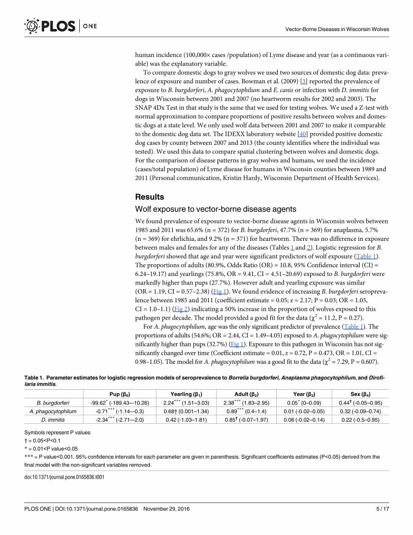
We found prevalence of exposure to vector-borne disease agents in Wisconsin wolves between
1985 and 2011 was 65.6% (n = 372) for B. burgdorferi, 47.7% (n = 369) for anaplasma, 5.7%
(n = 369) for ehrlichia, and 9.2% (n = 371) for heartworm. There was no difference in exposure
between males and females for any of the diseases (Tables 1 and 2). Logistic regression for B.
burgdorferi showed that age and year were significant predictors of wolf exposure (Table 1).
The proportions of adults (80.9%, Odds Ratio (OR) = 10.8, 95% Confidence interval (CI) =
6.24–19.17) and yearlings (75.8%, OR = 9.41, CI = 4.51–20.69) exposed to B. burgdorferi were
markedly higher than pups (27.7%). However adult and yearling exposure was similar
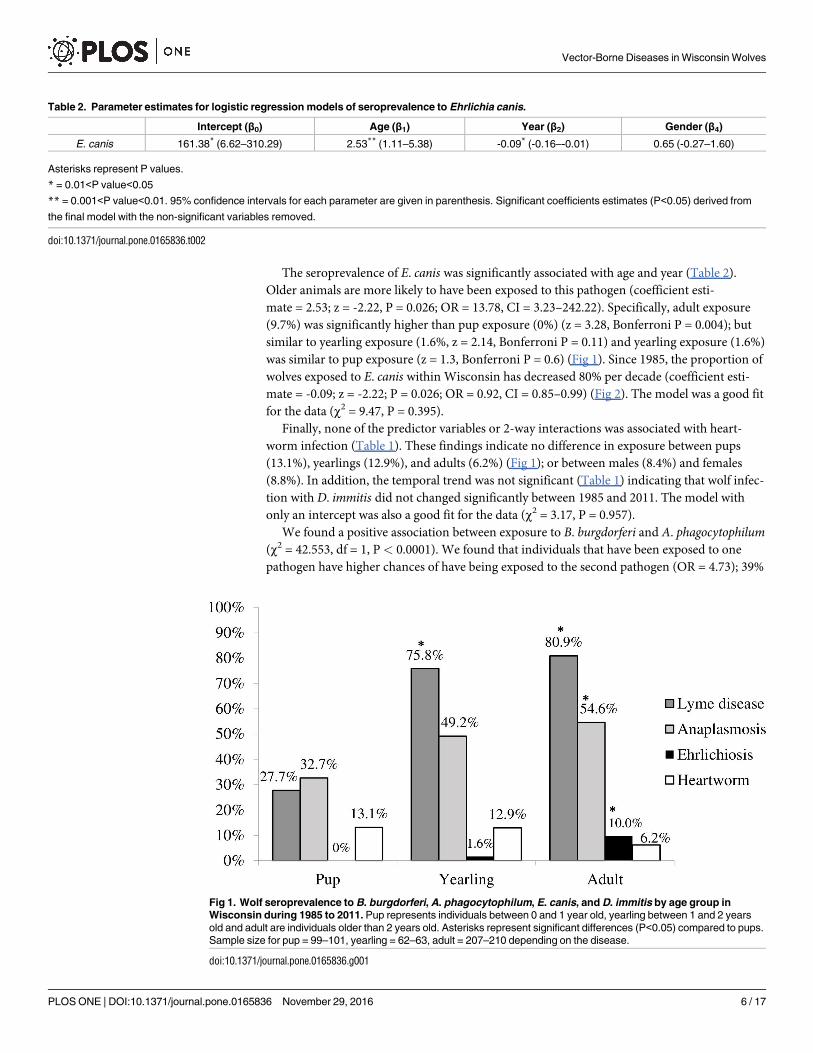
(OR = 1.19, CI = 0.57–2.38) (Fig 1). We found evidence of increasing B. burgdorferi seropreva-
lence between 1985 and 2011 (coefficient estimate = 0.05; z = 2.17; P = 0.03; OR = 1.05,
CI = 1.0–1.1) (Fig 2) indicating a 50% increase in the proportion of wolves exposed to this
pathogen per decade. The model provided a good fit for the data (χ2 = 11.2, P = 0.27).
For A. phagocytophilum, age was the only significant predictor of prevalence (Table 1). The
proportions of adults (54.6%; OR = 2.44, CI = 1.49–4.05) exposed to A. phagocytophilum were sig-
nificantly higher than pups (32.7%) (Fig 1). Exposure to this pathogen in Wisconsin has not sig-
nificantly changed over time (Coefficient estimate = 0.01, z = 0.72, P = 0.473, OR = 1.01, CI =
0.98–1.05). The model for A. phagocytophilum was a good fit to the data (χ2 = 7.29, P = 0.607).
Table 1. Parameter estimates for logistic regression models of seroprevalence to Borrelia burgdorferi, Anaplasma phagocytophilum, and Dirofi-
laria immitis.
Pup (β0) Yearling (β1) Adult (β2) Year (β3) Sex (β4)
B. burgdorferi -99.62* (-189.43–-10.26) 2.24*** (1.51–3.03) 2.38*** (1.83–2.95) 0.05* (0–0.09) 0.44† (-0.05–0.95)
A. phagocytophilum -0.71*** (-1.14–-0.3) 0.68† (0.001–1.34) 0.89*** (0.4–1.4) 0.01 (-0.02–0.05) 0.32 (-0.09–0.74)
D. immitis -2.34*** (-2.71–-2.0) 0.42 (-1.03–1.81) 0.85† (-0.07–1.97) 0.06 (-0.02–0.14) 0.22 (-0.5–0.95)
Symbols represent P values
† = 0.05<P<0.1
* = 0.01<P value<0.05
*** = P value<0.001. 95% confidence intervals for each parameter are given in parenthesis. Significant coefficients estimates (P<0.05) derived from the
final model with the non-significant variables removed.
doi:10.1371/journal.pone.0165836.t001
Vector-Borne Diseases in Wisconsin Wolves
PLOS ONE | DOI:10.1371/journal.pone.0165836 November 29, 2016 5 / 17
The seroprevalence of E. canis was significantly associated with age and year (Table 2).
Older animals are more likely to have been exposed to this pathogen (coefficient esti-
mate = 2.53; z = -2.22, P = 0.026; OR = 13.78, CI = 3.23–242.22). Specifically, adult exposure
(9.7%) was significantly higher than pup exposure (0%) (z = 3.28, Bonferroni P = 0.004); but
similar to yearling exposure (1.6%, z = 2.14, Bonferroni P = 0.11) and yearling exposure (1.6%)
was similar to pup exposure (z = 1.3, Bonferroni P = 0.6) (Fig 1). Since 1985, the proportion of
wolves exposed to E. canis within Wisconsin has decreased 80% per decade (coefficient esti-
mate = -0.09; z = -2.22; P = 0.026; OR = 0.92, CI = 0.85–0.99) (Fig 2). The model was a good fit
for the data (χ2 = 9.47, P = 0.395).
Finally, none of the predictor variables or 2-way interactions was associated with heart-
worm infection (Table 1). These findings indicate no difference in exposure between pups
(13.1%), yearlings (12.9%), and adults (6.2%) (Fig 1); or between males (8.4%) and females
(8.8%). In addition, the temporal trend was not significant (Table 1) indicating that wolf infec-
tion with D. immitis did not changed significantly between 1985 and 2011. The model with
only an intercept was also a good fit for the data (χ2 = 3.17, P = 0.957).
We found a positive association between exposure to B. burgdorferi and A. phagocytophilum(χ2 = 42.553, df = 1, P< 0.0001). We found that individuals that have been exposed to one
pathogen have higher chances of have being exposed to the second pathogen (OR = 4.73); 39%
Table 2. Parameter estimates for logistic regression models of seroprevalence to Ehrlichia canis.
Intercept (β0) Age (β1) Year (β2) Gender (β4)
E. canis 161.38* (6.62–310.29) 2.53** (1.11–5.38) -0.09* (-0.16–-0.01) 0.65 (-0.27–1.60)
Asterisks represent P values.
* = 0.01<P value<0.05
** = 0.001<P value<0.01. 95% confidence intervals for each parameter are given in parenthesis. Significant coefficients estimates (P<0.05) derived from
the final model with the non-significant variables removed.
doi:10.1371/journal.pone.0165836.t002
Fig 1. Wolf seroprevalence to B. burgdorferi, A. phagocytophilum, E. canis, and D. immitis by age group in
Wisconsin during 1985 to 2011. Pup represents individuals between 0 and 1 year old, yearling between 1 and 2 years
old and adult are individuals older than 2 years old. Asterisks represent significant differences (P<0.05) compared to pups.
Sample size for pup = 99–101, yearling = 62–63, adult = 207–210 depending on the disease.
doi:10.1371/journal.pone.0165836.g001
Vector-Borne Diseases in Wisconsin Wolves
PLOS ONE | DOI:10.1371/journal.pone.0165836 November 29, 2016 6 / 17
of wolves have been exposed to both pathogens (either subsequently or simultaneously) and
26.3% were negative for both pathogens, compared to 26% exposure to B. burgdorferi alone
and 8.4% to A. phagocytophilum alone.
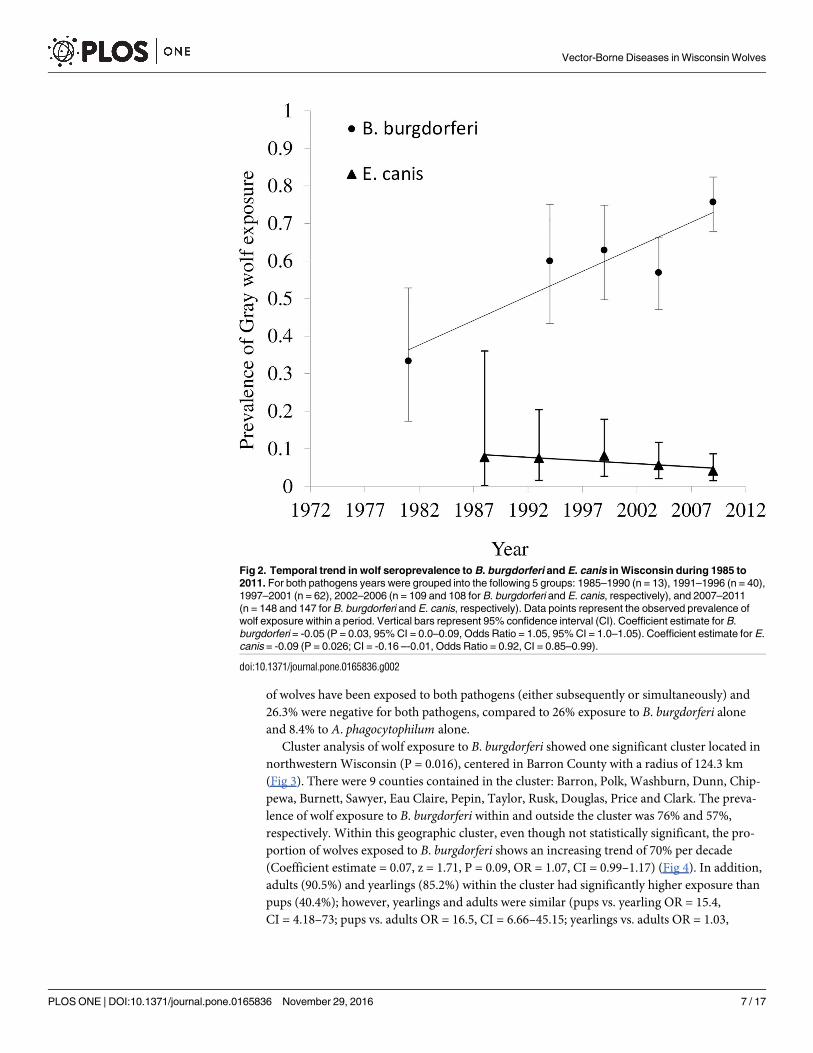
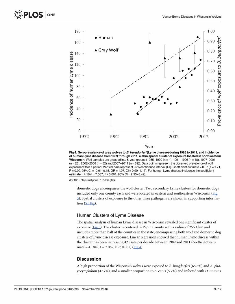
Cluster analysis of wolf exposure to B. burgdorferi showed one significant cluster located in
northwestern Wisconsin (P = 0.016), centered in Barron County with a radius of 124.3 km
(Fig 3). There were 9 counties contained in the cluster: Barron, Polk, Washburn, Dunn, Chip-
pewa, Burnett, Sawyer, Eau Claire, Pepin, Taylor, Rusk, Douglas, Price and Clark. The preva-
lence of wolf exposure to B. burgdorferi within and outside the cluster was 76% and 57%,
respectively. Within this geographic cluster, even though not statistically significant, the pro-
portion of wolves exposed to B. burgdorferi shows an increasing trend of 70% per decade
(Coefficient estimate = 0.07, z = 1.71, P = 0.09, OR = 1.07, CI = 0.99–1.17) (Fig 4). In addition,
adults (90.5%) and yearlings (85.2%) within the cluster had significantly higher exposure than
pups (40.4%); however, yearlings and adults were similar (pups vs. yearling OR = 15.4,
CI = 4.18–73; pups vs. adults OR = 16.5, CI = 6.66–45.15; yearlings vs. adults OR = 1.03,
Fig 2. Temporal trend in wolf seroprevalence to B. burgdorferi and E. canis in Wisconsin during 1985 to
2011. For both pathogens years were grouped into the following 5 groups: 1985–1990 (n = 13), 1991–1996 (n = 40),
1997–2001 (n = 62), 2002–2006 (n = 109 and 108 for B. burgdorferi and E. canis, respectively), and 2007–2011
(n = 148 and 147 for B. burgdorferi and E. canis, respectively). Data points represent the observed prevalence of
wolf exposure within a period. Vertical bars represent 95% confidence interval (CI). Coefficient estimate for B.
burgdorferi = -0.05 (P = 0.03, 95% CI = 0.0–0.09, Odds Ratio = 1.05, 95% CI = 1.0–1.05). Coefficient estimate for E.
canis = -0.09 (P = 0.026; CI = -0.16 –-0.01, Odds Ratio = 0.92, CI = 0.85–0.99).
doi:10.1371/journal.pone.0165836.g002
Vector-Borne Diseases in Wisconsin Wolves
PLOS ONE | DOI:10.1371/journal.pone.0165836 November 29, 2016 7 / 17
CI = 0.23–3.9). There was no difference in exposure within the cluster between males (74.7%)
and females (78.6%) (OR = 1.6, CI = 0.69–3.9).
In contrast, there was no temporal change outside the cluster in northern Wisconsin (pri-
mary region of wolf distribution within the state), although the prevalence of exposure has var-
ied over time (5 time periods) around an overall prevalence of 54.9% (Coefficient
estimate = 0.03, z = 1.22, P = 0.221, OR = 1.03, CI = 0.99–1.07). Outside the cluster, older
wolves were also more likely to be exposed to Lyme disease than pups (pups vs. yearling
OR = 12.7, CI = 3.79–50.03; pups vs. adults OR = 18.59, CI = 6.69–62.13; yearlings vs. adults
OR = 1.44, CI = 0.49–4.04). There was no difference between males (49.4%) and females
(64.5%) (OR = 2.12, CI = 0.95–4.94).
Exposure to A. phagocytophilum and E. canis and infection with D. immitis seems to be
homogeneous within the state we found no significant clusters of these agents across Wiscon-
sin (S1 Fig). These patterns indicate that risk of exposure is equally likely throughout the state.
Comparison of Domestic Dog and Wolf serology
In Wisconsin domestic dog exposure (number positive/number tested) between 2001 and
2007 [3] follows a similar pattern to that of wolves; higher exposure to B. burgdorferi (10.2%)
and A. phagocytophilum (10.5%), but low exposure to E. canis (0.3%) and infection with D.
immitis (0.6%). However, wolf exposure during the same period was markedly higher than
domestic dog exposure for B. burgdorferi (61%: z = 22.18, P< 0.0001), A. phagocytophilum(42.3%: z = 13.63, P< 0.0001), E. canis (6.9%: z = 14.65, P< 0.0001), and heartworm (10.7%:
z = 17.84, P < 0.0001) (S2 Fig).
There were three highly significant clusters of domestic dog exposure to B. burgdorferi in
Wisconsin. The most important cluster was centered in Dunn County with a radius of 262.5
km including 29 counties of western Wisconsin (Fig 3). This cluster of Lyme disease in
Fig 3. Spatial distribution of clusters of wolf seroprevalence to B. burgdorferi (1985–2011) and domestic dogs (2007–2013), and human cases
(1989–2011) in Wisconsin. The maps show the location and extent of the most likely cluster and secondary clusters of seroprevalence and the counties
encompassed by it are shaded. In addition, the log likelihood ratio (LLR), relative risk (RR), significance (P-value), expected number of cases (Expected) and
observed number of cases (Observed) are shown for each cluster.
doi:10.1371/journal.pone.0165836.g003
Vector-Borne Diseases in Wisconsin Wolves
PLOS ONE | DOI:10.1371/journal.pone.0165836 November 29, 2016 8 / 17
domestic dogs encompasses the wolf cluster. Two secondary Lyme clusters for domestic dogs
included only one county each and were located in eastern and southeastern Wisconsin (Fig
3). Spatial clusters of exposure to the other three pathogens are shown in supporting informa-
tion (S1 Fig).
Human Clusters of Lyme Disease
The spatial analysis of human Lyme disease in Wisconsin revealed one significant cluster of
exposure (Fig 3). The cluster is centered in Pepin County with a radius of 255.4 km and
includes more than half of the counties in the state, encompassing both wolf and domestic dog
clusters of Lyme disease exposure. Linear regression showed that human Lyme disease within
the cluster has been increasing 42 cases per decade between 1989 and 2011 (coefficient esti-
mate = 4.1849, t = 7.067, P < 0.001) (Fig 4).
Discussion
A high proportion of the Wisconsin wolves were exposed to B. burgdorferi (65.6%) and A. pha-gocytophilum (47.7%), and a smaller proportion to E. canis (5.7%) and infected with D. immitis
Fig 4. Seroprevalence of gray wolves to B. burgdorferi (Lyme disease) during 1985 to 2011, and incidence
of human Lyme disease from 1989 through 2011, within spatial cluster of exposure located in northwestern
Wisconsin. Wolf samples are grouped into 5-year groups (1985–1990 (n = 4), 1991–1996 (n = 18), 1997–2001
(n = 26), 2002–2006 (n = 52) and 2007–2011 (n = 69)). Data points represent the observed prevalence of wolf
exposure within a period. Vertical bars represent 95% confidence interval (CI). Coefficient estimate = 0.07 (z = 1.71,
P = 0.09, 95% CI = -0.01–0.15, OR = 1.07, CI = 0.99–1.17). For human Lyme disease incidence the coefficient
estimate = 4.18 (t = 7.067, P< 0.001, 95% CI = 2.95–5.42).
doi:10.1371/journal.pone.0165836.g004
Vector-Borne Diseases in Wisconsin Wolves
PLOS ONE | DOI:10.1371/journal.pone.0165836 November 29, 2016 9 / 17
(9.2%) between 1985 and 2011. Exposure to B. burgdorferi between 1988 and 1996 (57.4%) and
infection with D. immitis between 1991 and 1996 (2.5%) correspond with a previous study
based on a subset of the Wisconsin wolves for those time periods (47.8% and 2% for B. burg-dorferi and D. immitis respectively) [24]. Adult infection with D. immitis in 2003 (25%) and
pup infection in 2001 (15.4%) are also similar to a previous report (9%) in Wisconsin (D. Gin-
nett and J. Theis, unpublished WDNR 2003 report), although there may be overlap in samples
between both studies. We also found strong evidence of a positive association of gray wolf
coexposure to B. burgdorferi and A. phagocytophilum. These pathogens share vector (ticks) and
reservoir hosts (rodents); thus, this association is not surprising. The potential impact of coin-
fection/coexposure on wolves has not been studied; however, because A. phagocytophilummaybe an immunosuppressive agent, its presence could alter the intensity and duration of B.
burgdorferi infection [41–43].
Wolf seroprevalence is higher in adults than pups for B. burgdorferi, A. phagocytophilumand E. canis. There are two likely explanations for these patterns. First, if antibodies to these
agents are long lived, then adults have a longer risk of exposure than much younger pups. Sec-
ond, questing ticks transmitting these pathogens are sedentary; therefore, exposure to tick-
borne pathogens likely increases with mobility of the host. Because pups stay near den sites
while adults move greater distances tick-borne diseases are expected to be higher in older ani-
mals which are more likely to contact questing ticks. The fact that yearling exposure is similar
to adults suggests early exposure in life is common and movement may be an important risk
factor. In contrast, D. immitis, which has similar exposure rates for all ages, is transmitted by
mosquitoes, which actively search for a host.
We also found wolf exposure to B. burgdorferi has increased 50% per decade, and E. canishas decreased 80% per decade, while A. phagocytophilum exposure and D. immitis infection
have remained steady since 1985. Increased wolf exposure to B. burgdorferimight be due to
geographic expansion of endemic areas of infection, changes in the distribution of I. scapularis,and/or increased nymph density [44,45]. A cluster of Lyme disease is located in the area where
I. scapularis was first established in Wisconsin in 1968 [46], deer density is higher (compared
to other zones within wolf range) [47], and the habitat is suitable for I. scapularis [35]. Conse-
quently, I. scapularis is abundant in this area compared to other portions of the state, Lyme dis-
ease has increased since it was first reported in the late 1960s in northwestern Wisconsin, tick
density has also increased, and tick populations have expanded southward and eastward
[45,46,48]. Higher tick density and expanded geographic distribution generally agrees with the
higher proportion of wolves exposed to B. burgdorferi over time.
There is no clear explanation why adult wolves have a relatively high and yet declining
exposure to E. canis compared to dogs when the vector (R. sanguineus) is limited by cold win-
ter temperatures, typically found in domestic environments, and the primary reservoir
(domestic dogs) of this pathogen is widely distributed in Wisconsin. One potential explanation
is the discovery of the E.muris-like agent in humans and ticks in Wisconsin. This new agent
was first discovered in humans in 2009 [49] with subsequent human infections reported
between 2007 and 2013 [50]. A retrospective study of Ixodes ticks from northwestern Wiscon-
sin showed a 1% infection rate as early as the mid-1990s [51]. In 2011 a dog in Minnesota was
diagnosed with an E.muris-like agent infection, indicating this agent is present in the upper
Midwest and can be pathogenic to canines [52]. These observations suggest that Wisconsin
wolves may be exposed to ticks infected with the E.muris-like agent, potentially producing
cross-reacting antibodies on the SNAP 4Dx E. canis test. However, SNAP 4Dx test has not
been evaluated for E.muris-like agents and the infected dog in Minnesota was only positive for
Anaplasma [52]. Further research is needed to understand the apparent decline in E. canis
Vector-Borne Diseases in Wisconsin Wolves
PLOS ONE | DOI:10.1371/journal.pone.0165836 November 29, 2016 10 / 17
exposure in wolves, whether and when wolves have been exposed to E.muris, and whether E.
muris produces clinical infection in wild wolves.
We also note the potential for antibody sensitivity to decline with longer storage of both fro-
zen serum and Nobuto strips [53]. We were unable to formally assess potential degradation in
our study; however, as part of the SNAP test validation experiment (S1 Text), we obtained sim-
ilar results for B. burgdorferi serology from samples that were retested after up to 9 years in
storage. Nevertheless, we acknowledge that our results may underestimate increasing trends in
seroprevalence to these agents in the event of antibody degradation over time.
Stable exposure of wolves to A. phagocytophilum may be partly attributed to the potential
for B. burgdorferi to mask exposure to A. phagocytophilum by reducing antibody titers; a pat-
tern demonstrated in co-infected mice [54]. This masking effect may have reduced detection
of A. phagocytophilum, especially considering that wolf exposure to B. burgdorferi in Wisconsin
is very high, has increased, and we found coexposure between these two pathogens.
According to the SNAP 4Dx manufacturer [55] the analyte that detects exposure to A. pha-gocytophilum can cross react with Anaplasma platys, which infects domestic dogs, but its vector
has not been conclusively determined [56]. In the US, infection with A. platys and E. canis are
typically similar in domestic dogs, with higher prevalence in southern areas of the country
[57]. Because wolf exposure to E. canis in Wisconsin is much lower (5.7%) than A. phagocyto-philum (47.6%), we suspect only a small portion of the positive A. phagocytophilum results can
be attributed to A. platys exposure.
Heartworm infection also remained unchanged during our study. Several factors can affect
prevalence of heartworm including temperature [27]. Accordingly, infection with this parasite
is more common in southern areas where higher temperatures favor mosquito transmission.
These patterns suggest that further research is need to determine if higher temperatures due to
climate change might increase future heartworm prevalence in domestic and wild canids in
Wisconsin.
Wolves, Domestic Dogs, and Humans
Wolf exposure to B. burgdorferi, A. phagocytophilum, E. canis and infection with D. immitisbetween 2007 and 2011 was significantly higher than domestic dogs to these pathogens. This
difference likely arises because domestic dogs spend less time outdoors (less exposure time)
and because owners use preventive methods to remove ticks and/or have domestic dogs vacci-
nated to reduce pathogen transmission. We believe that captured wolves represent an unbiased
sample of animals for natural exposure to these diseases. Wolves captured for monitoring and
research purposes were generally captured in heavily forested portions of the state where most
wolves live [58]. While wolves living in mixed forest/farmland areas were less likely to be sam-
pled, these wolves also represent a small percentage of the wolf population, that were more
likely captured at livestock depredation sites [36]. We believe these samples provide a repre-
sentative sample of the distribution of Wisconsin wolves.
There is high domestic dog and wolf exposure, and human infection with B. burgdorferiand A. phagocytophilum in Wisconsin, which is considered an endemic area for these diseases
[3,13,59]. In addition, geographic patterns of exposure are similar between gray wolves,
domestic dogs, and humans for B. burgdorferi and A. phagocytophilum. During our study
period, wolf exposure to A. phagocytophilum increased only slightly and non-significantly.
This finding is not consistent with the increasing number of human anaplasmosis cases
reported each year in the US since the mid-1990s [59]. Increasing human anaplasmosis might
be due to increased recognition of this disease over time or changes in human behavior that
increase risk of exposure (e.g. spending more time in tick friendly habitat).
Vector-Borne Diseases in Wisconsin Wolves
PLOS ONE | DOI:10.1371/journal.pone.0165836 November 29, 2016 11 / 17
The increasing temporal trend of wolf exposure to B. burgdorferi corresponded with human
incidence of Lyme disease, although the rise in human incidence was primarily after 2001[60],
perhaps part due to under-reporting of human Lyme disease in the 1990s, followed by an
exponential “catch-up” phase after 2001 as increased reporting began to reflect the true
(higher) incidence. Ultimately, the increase in human cases is likely driven by ecological
changes in disease/vector density and transmission. Within the overlapping geographic cluster
of Lyme disease for wolves, domestic dogs, and humans in northwest Wisconsin, human inci-
dence has increased and wolf prevalence shows an increasing trend over time. This suggests
the increasing trend in human incidence in northern Wisconsin could be a consequence of
ecological changes in the vector or mammalian hosts and not simply increased recognition by
physicians. Domestic dog and human clusters are twice as big as the wolf cluster; however, all
three clusters are centered approximately in the same areas (Barron, Dunn, and Pepin coun-
ties). The differences in cluster size between wolves, domestic dogs, and humans might in part
be due to the limited range of wolves in Wisconsin compared to humans and domestic dogs.
In addition, domestic dog and human records of disease are based on the county of residence
and might not represent the exact location were the infection was acquired.
The geographic pattern of exposure to E. canis is similar between gray wolves and domestic
dogs (S1 Fig). We found partial overlap between wolf and domestic dog clusters in northwest-
ern Wisconsin; however, the wolf cluster is not significant. In Wisconsin there is generally low
wolf and domestic dog exposure to this pathogen compared to domestic dogs from southern
US [3]. It is surprising that wolf exposure to this pathogen was much higher than domestic
dogs between 2001 and 2007 because the brown dog tick, which transmits E. canis, is adapted
to life indoors [61]. Therefore, we expected domestic dogs to have higher exposure to this
pathogen; however, tick removal or preventive treatment by owners might reduce pathogen
transmission. The SNAP 4Dx Test used to detect E. canis antibodies can also cross react with
Ehrlichia chaffeensis and Ehrlichia ewingii [55,62]. However, exposure to these two pathogens
seems unlikely because they are transmitted by the Lone Star tick (Amblyomma americanum)
[63,64] which is not currently found within wolf range, although there are scattered reports in
southern Wisconsin (S. Paskewitz, personal communication) [65]. Human infection with E.
chaffeensis has been reported in Wisconsin; however, many of these patients could have con-
tracted the pathogen when traveling to higher risk areas in southern US [66]. Alternatively,
wolf exposure to E. canis may actually represent exposure to E.muris which has been in Wis-
consin since at least the mid-1990s [51]. More research is needed to determine whether the
SNAP 4Dx test has cross-reactivity with E.muris-like agents and whether wolves have been
historically exposed to E.muris.Although heartworm infection is common in red wolves and domestic dogs from the south-
ern US [3] it is uncommon in wolves and domestic dogs in Wisconsin, probably due to tem-
perature, the lack of wolves in southern Wisconsin, and other constraints. For domestic dogs,
significant clusters of exposure were spread throughout the state, and similarly, the non-signif-
icant wolf clusters of exposure were spread throughout wolf range in Wisconsin with almost
no overlap between species (S1 Fig). While heartworm is known to occasionally cause mortal-
ity in wolves, the impact of the other vector-borne diseases in wolves is poorly known [67].
The earliest detection of Lyme in Minnesota wolves was for 1980, shortly after wolves had
started entering Wisconsin from Minnesota [68]. The initial colonization of wolves into Wis-
consin was along the border with Minnesota where an endemic of Lyme disease existed and
high numbers of I. scapularis occur [30,68]. The wolf population in Wisconsin grew slowly ini-
tially, but by mid-1990s began displaying rapid growth, despite high prevalence of Lyme and
anaplasma exposure [68]. The slow population growth during the 1980s has been attributed to
outbreak of canine parvovirus and high human caused mortality [30], but perhaps some of
Vector-Borne Diseases in Wisconsin Wolves
PLOS ONE | DOI:10.1371/journal.pone.0165836 November 29, 2016 12 / 17
these vector-borne diseases were also contributing factors. But despite the continued presence
and expansion of Lyme and anaplasma exposure, the wolf population grew rapidly through
the 1990s and early 2000s and by 2011 had grown to minimum winter count of 782 wolves
[30,33], Thus, there is little evidence that these diseases are currently retarding growth of the
Wisconsin wolf population.
Conclusions
Lyme disease, anaplasmosis, ehrlichiosis, and heartworm are emerging vector-borne diseases
that affect canids, but their prevalence in wild gray wolves has not been systematically assessed.
We show Wisconsin wolves have high rates of exposure to B. burgdorferi and A. phagocytophi-lum, the agents causing Lyme disease and anaplasmosis, respectively. In contrast, wolves have
limited exposure to E. canis and D. immitis, the agents causing canine ehrlichiosis and heart-
worm, respectively. These patterns are similar to domestic dogs even though wolves are con-
stantly exposed to the vectors transmitting these pathogens. Wolf exposure to B. burgdorferi in
Wisconsin has increased, corresponding with the increasing human Lyme incidence, and sug-
gesting there might have been underreporting of the disease in humans before 2001. In addi-
tion, there is a cluster of wolf exposure to B. burgdorferi in northwestern Wisconsin, which
overlaps human and domestic dog clusters for the same pathogen. The high prevalence of B.
burgdorferi and A. phagocytophilum exposure, and probable underestimation of the latter and
heartworm infection, suggest that these diseases could represent a possible health risk to wolf
populations, although information on the effects of these diseases on wolves is lacking. During
our study period the estimated winter wolf population increased from 14–16 in 1985 to 782–
805 in 2011 as occupied wolf range increased >20 fold [30,31], suggesting that vector-borne
disease agents have not limited population growth. Due to climate change, future heartworm
risk to wolves and domestic dogs may increase, new diseases may arise, and virulence of exist-
ing diseases may change in Wisconsin. Thus periodic assessment of prevalence and impact of
vector-borne diseases will be an important part of future wolf conservation programs.
Supporting Information
S1 Data. Wolf Serology Data. Data elements: age, sex, County of collection, date of collection,
year of collection, and SNAP 4Dx test for Lyme, anaplasmosis, ehrlichiosis, and heart worm
(0 = negative and 1 = positive).
(XLSX)
S1 Fig. Spatial distribution of clusters of exposure to A. phagocytophilum, E. canis and
infection with D. immitis in Gray wolves (1985–2011) and domestic dogs (2007–2013) in
Wisconsin. The maps show the location and extent of the most likely cluster and secondary
clusters of infection and the counties encompassed by it are shaded. In addition, the log likeli-
hood ratio (LLR), relative risk (RR), significance (P-value), expected number of cases
(Expected) and observed number of cases (Observed) are shown for each cluster.
(PDF)
S2 Fig. Gray wolf and dog percent positive test results (number positive/number tested)
for antibodies to Borrelia burgdorferi, Anaplasma phagocytophilum and Echrlichia canisand antigen of Dirofilaria immitis in Wisconsin between 2001 and 2007. Wolf exposure to
each pathogen were siginificantly greater than dog exposure at α = 0.05 level. Sample size for
wolves = 178–178 and dogs = 51512–109745 depending on the pathogen.
(PDF)
Vector-Borne Diseases in Wisconsin Wolves
PLOS ONE | DOI:10.1371/journal.pone.0165836 November 29, 2016 13 / 17
S1 Text. SNAP 4Dx Validation.
(PDF)
Acknowledgments
We thank the Wisconsin Department of Natural Resources for access to samples and data pro-
vided by J. Wiedenhoeft. We thank L. C. Bartholomay for many helpful review comments that
improved the paper. We also thank Ronald N. Schultz for assistance with field work. We thank
the U.S. Geological Survey–National Wildlife Health Center for sharing wolf necropsy results.
Mention of trade names or products does not imply endorsement by the U.S. Government.
Author Contributions
Conceptualization: RFJ MDS.
Data curation: APW.
Formal analysis: RFJ MDS.
Funding acquisition: APW MDS RFJ.
Investigation: APW RFJ MDS.
Methodology: APW RFJ MDS.
Project administration: RFJ MDS.
Resources: APW RFJ MDS.
Supervision: MDS RFJ.
Visualization: RFJ MDS.
Writing – original draft: RFJ MDS.
Writing – review & editing: RJF MDS APW.
References1. Daszak P, Cunningham AA, Hyatt AD. Anthropogenic environmental change and the emergence of
infectious diseases in wildlife. Acta Trop. 2001; 78: 103–116. PMID: 11230820
2. Piesman J, Eisen L. Prevention of Tick-Borne Diseases. Annu Rev Entomol. 2008; 53: 323–343. doi:
10.1146/annurev.ento.53.103106.093429 PMID: 17877457
3. Bowman D, Little S, Lorentzen L, Shields J, Sullivan M, Carlin E. Prevalence and geographic distribution
of Dirofilaria immitis, Borrelia burgdorferi, Ehrlichia canis, and Anaplasma phagocytophilum in dogs in
the United States: Results of a national clinic-based serologic survey. Vet Parasitol. 2009; 160: 138–
148. doi: 10.1016/j.vetpar.2008.10.093 PMID: 19150176
4. Leschnik M, Kirtz G, Viranyi Z, Wille-Piazzai W, Duscher G. Acute granulocytic anaplasmosis in a cap-
tive timber wolf (Canis lupus occidentalis). J Zoo Wildl Med. 2012; 43: 645–648. doi: 10.1638/2011-
0224R.1 PMID: 23082534
5. Littman M, Goldstein R, Labato M, MR L, Moore G. ACVIM small animal consensus statement on Lyme
disease in dogs: diagnosis, treatment, and prevention. J Vet Intern Med. 2006; 20: 422–434. PMID:
16594606
6. Kohn B, Galke D, Beelitz P, Pfister K. Clinical Features of Canine Granulocytic Anaplasmosis in 18 Nat-
urally Infected Dogs. J Vet Intern Med. 2008; 22: 1289–1295. doi: 10.1111/j.1939-1676.2008.0180.x
PMID: 18783353
7. Harvey J, Simpson C, Gaskin J, Sameck J. Ehrlichiosis in wolves, dogs, and wolf-dog crosses. J Am
Vet Med Assoc. 1979; 175: 901–905. PMID: 521367
Vector-Borne Diseases in Wisconsin Wolves
PLOS ONE | DOI:10.1371/journal.pone.0165836 November 29, 2016 14 / 17
8. McCall J, Genchi C, Kramer L, Guerrero J, Venco L. Heartworm disease in animals and humans. Adv
Parasitol. 2008; 66: 193–285. doi: 10.1016/S0065-308X(08)00204-2 PMID: 18486691
9. Genchi C, Kramer L, Prieto G. Epidemiology of canine and feline dirofilariosis: a global view. In: Simon
F, Genchi C, editors. Heartworm infection in humans and animals. Salamanca: Ediciones Universidad
de Salamanca; 2001. pp. 121–133.
10. Perez M, Bodor M, Zhang C, Xiong Q, Rikihisa Y. Human infection with Ehrlichia canis accompanied by
clinical signs in Venezuela. Ann N Y Acad Sci. 2006; 1078: 110–117. doi: 10.1196/annals.1374.016
PMID: 17114689
11. Centers for Disease Control and Prevention. Lyme disease data and statistics. 2012; Available: http://
www.cdc.gov/lyme/stats/index.html. Accessed June 2012.
12. Centers for Disease Control and Prevention. Anaplasmosis, statistics and epidemiology. 2013. Avail-
able: http://www.cdc.gov/anaplasmosis/stats/. Accessed October 2013
13. Centers for Disease Control and Prevention. Lyme disease data and statistics. 2012. Available: http://
www.cdc.gov/lyme/stats/index.html. Accessed June 2012.
14. Centers for Disease Control and Prevention. Press Release, CDC provides estimate of Americans diag-
nosed with Lyme disease each year. 2013. Available: http://www.cdc.gov/media/releases/2013/p0819-
lyme-disease.html. Accessed Novemeber 2013.
15. Wisconsin Department of Health Services. Anaplasmosis and Ehrlichiosis. http://www.dhs.wisconsin.
gov/communicable/Tickborne/AandE/Index.htm. 2010. Available: http://www.dhs.wisconsin.gov/
communicable/Tickborne/AandE/Index.htm. Accessed Novemebr 2013.
16. Pritt B, Mead P, Johnson D, Neitzel D, Respicio-Kingry L, Davis J, et al. Identification of a novel patho-
genic Borrelia species causing Lyme borreliosis with unusually high spirochaetaemia: a descriptive
study. Lancet Infect Dis. 2016; 16: 556–564. doi: 10.1016/S1473-3099(15)00464-8 PMID: 26856777
17. Davidson W, Goff W. Anaplasmosis. In: Williams E, Barker I, editors. Infectious diseases of wild mam-
mals. Ames, Iowa: Iowa State University Press; 2001. pp. 455–466.
18. Brown R, Burgess E. Lyme borreliosis. In: Williams E, Barker I, editors. Infectious diseases of wild mam-
mals. Ames, Iowa: Iowa State University Press; 2001. pp. 435–454.
19. Levine J, Wilson M, Spielman A. Mice as reservoirs of the Lyme disease spirochete. Am J Trop Med
Hyg. 1985; 34: 355–360. PMID: 3985277
20. Donahue J, Piesman J, Spielman A. Reservoir competence of white-footed mice of Lyme disease spiro-
chetes. Am J Trop Med Hyg. 1987; 36: 92–96. PMID: 3812887
21. Mather T, Wilson M, Moore S, Ribeiro J, Spielman A. Comparing the relative potential of rodents as res-
ervoirs of the Lyme disease spirochete (Borrelia burgdorferi). Am J Epidemiol. 1989; 130: 143–150.
PMID: 2787105
22. Telford S, Dawson J, Katavolos P, Warner C, Kolbert C, Persing D. Perpetuation of the agent of human
granulocytic ehrlichiosis in a deer tick-rodent cycle. Proc Natl Acad Sci. 1996; 93: 6209–6214. PMID:
8650245
23. Kazmierczak J, Burgess E, Amundson T. Susceptibility of the gray wolf (Canis lupus) to infection with
the Lyme disease agent, Borrelia burgdorferi. J Wildl Dis. 1988; 24: 522–527. doi: 10.7589/0090-3558-
24.3.522 PMID: 3411709
24. Beheler-Amass K, Wydeven A, Thiel R. Wolf health and monitoring and mortality factors, appendix F.
1999. Available: http://www.timberwolfinformation.org/info/wolfmanplan/final/appendix/appendix_f.htm.
Accessed October 2013.
25. Rikihisa Y. The tribe Ehrlichieae and ehrlichial diseases. Clin Microbiol Rev. 1991; 4: 286–308. PMID:
1889044
26. Groves M, Dennis G, Amyx H, Huxsoll D. Transmission of Ehrlichia canis to dogs by ticks (Rhipicepha-
lus sanguineus). Am J Vet Res. 1975; 36: 937–940. PMID: 1147359
27. Brown H, Harrington L, Kaufman P, McKay T, Bowman D, Nelson C, et al. Key factors influencing
canine heartworm, Dirofilaria immitis, in the United States. Parasites and Vectors. 2012; 5: 1–9.
28. Theis J. Public health aspects of dirofilariasis in the United States. Vet Parasitol. 2005; 133: 157–180.
doi: 10.1016/j.vetpar.2005.04.007 PMID: 16039780
29. de Campos JRM, Barbas C, Filomeno L, Fernandez A, Minamoto H, Barbas Filho J, et al. Human pul-
monary dirofilariasis: analysis of 24 cases from São Paulo, Brazil. Chest. 1997; 112: 729–733. PMID:
9315807
30. Wydeven A, Wiedenhoeft J, Schultz R, Thiel R, Jurewicz R, Kohn B, et al. History, population growth,
and management of wolves in Wisconsin. In: Wydeven A, Van Deelen T, Heske E, editors. Recovery of
gray wolves in the Great Lakes Region of the United States. Springer New York; 2009. pp. 87–105.
Vector-Borne Diseases in Wisconsin Wolves
PLOS ONE | DOI:10.1371/journal.pone.0165836 November 29, 2016 15 / 17
31. Wydeven A, Wiedenhoeft J, Schultz R, Bruner J, Thiel R, Boles S, et al. Status of the timber wolf in Wis-
consin: performance report 1 July 2010 through 30 June 2011. Wisconsin Endangered Resources
Report # 141. Madison, Wisconsin; 2011.
32. Wisconsin Wolf Management Plan. Wisconsin Department of Natural Resources, PUBL-ER-099 99.
Madison, Wisconsin, USA; 1999.
33. Department of the Interior. Endangered and Threatened wildlife and plants; reinstatement of final rules
for the gray wolf in Wyoming and the Western Great Lakes in compliance with court orders. Fedruary
20, 2015. Federal Register Notice 50 CFR Part 17, Fish and Wildlife Service, Int. 2015.
34. ER O, Stenglein J, Shelley V, Rissman A, Browne-Nunez C, Voyles Z, et al. Pendulum swings in wolf
management led to conflict, illegal kills, and a legislated wolf hunt. Conserv Lett. 2014; 8: 351–360.
35. Guerra M, Walker E, Jones C, Paskewitz S, Cortinas M, Stancil A, et al. Predicting the risk of Lyme dis-
ease: habitat suitability for Ixodes scapularis in the North Central United States. Emerg Infect Dis. 2002;
8: 289–297. doi: 10.3201/eid0803.010166 PMID: 11927027
36. Ruid D, Paul W, Roell B, Wydeven A, Willging R, Jurewicz R, et al. Wolf–human conflicts and manage-
ment in Minnesota, Wisconsin, and Michigan. In: Wydeven A, Van Deelen T, Heske E, editors. Recov-
ery of gray wolves in the Great Lakes region of the United States. 2009. pp. 279–295.
37. Jara RF. Spatial and temporal patterns of Gray Wolf exposure to vector-borne diseases in Wisconsin.
M.Sc. Thesis, University of Wisconsin-Madison. 2013.
38. Kulldorff M. A spatial scan statistic. Commun Stat methods. 1997; 26: 1481–1496.
39. American Veterinary Medical Association. U.S. pet ownership statistics. 2013. Available: https://www.
avma.org/KB/Resources/Statistics/Pages/Market-research-statistics-US-pet-ownership.aspx.
Accessed June 2013.
40. IDEXX L. Diseases in your area. 2013. Available: http://www.dogsandticks.com/diseases_in_your_
area.php. Accessed June 2013.
41. Thomas V, Anguita J, Barthold S, Fikrig E. Coinfection with Borrelia burgdorferi and the agent of human
granulocytic ehrlichiosis alters murine immune responses, pathogen burden, and severity of Lyme
arthritis. Infect Immun. 2001; 69: 3359–3371. doi: 10.1128/IAI.69.5.3359-3371.2001 PMID: 11292759
42. Dumler J, Choi K, Garcia-Garcia J, Barat N, Scorpio D, Garyu J, et al. Human granulocytic anaplasmo-
sis and Anaplasma phagocytophilum. Emerg Infect Dis. 2005; 11: 1828–1834. doi: 10.3201/eid1112.
050898 PMID: 16485466
43. Nieto NC, Foley JE. Meta-analysis of coinfection and coexposure with Borrelia burgdorferi and Ana-
plasma phagocytophilum in humans, domestic animals, wildlife, and Ixodes ricinus-complex ticks. Vec-
tor-Borne Zoonotic Dis. 2009; 9: 93–102. doi: 10.1089/vbz.2008.0072 PMID: 18789001
44. Diuk-Wasser M, Gatewood A, Cortinas M, Yaremych-Hamer S, Tsao J, Kitron U, et al. Spatiotemporal
patterns of host-seeking Ixodes scapularis nymphs (Acari: Ixodidae) in the United States. J Med Ento-
mol. 2006; 43: 166–176. PMID: 16619595
45. Lee X, Hardy K, Johnson D, Paskewitz S. Hunter-killed deer surveillance to assess changes in the prev-
alence and distribution of Ixodes scapularis (Acari: Ixodidae) in Wisconsin. J Med Entomol. 2013; 50:
632–639. PMID: 23802460
46. Jackson J, DeFoliart G. Ixodes scapularis Say in northern Wisconsin. J Med Entomol. 1970; 7: 124–
125. PMID: 5435805
47. Wisconsin Department of Natural Resources. Fall deer population density 2012, per square mile deer
range. 2012. Available: http://dnr.wi.gov/topic/hunt/documents/falldeerperdr.pdf. Accessed October
2013.
48. Godsey JM, Amundson T, Burgess E, Schell W, Davis J, Kaslow R, et al. Lyme disease ecology in Wis-
consin: distribution and host preferences of Ixodes dammini, and prevalence of antibody to Borrelia
burgdorferi in small mammals. Am J Trop Med Hyg. 1987; 37: 180–187. PMID: 3605501
49. Pritt B, Sloan L, Johnson D, Munderloh U, Paskewitz S, McElroy K, et al. Emergence of a new patho-
genic Ehrlichia species, Wisconsin and Minnesota, 2009. N Engl J Med. 2011; 365: 422–429. doi: 10.
1056/NEJMoa1010493 PMID: 21812671
50. Johnson D, Schiffman E, Davis J, Neitzel D, Sloan L, Nicholson W, et al. Human infection with Ehrlichia
muris-like pathogen, United States, 2007–2013. Emerg Infect Dis. 2015; 21: 1794–1799. doi: 10.3201/
eid2110.150143 PMID: 26402378
51. Telford S III, Goethert H, Cunningham J. Prevalence of Ehrlichia muris in Wisconsin deer ticks collected
during the mid 1990s. Open Microbiol J. 2011; 5: 18–20. doi: 10.2174/1874285801105010018 PMID:
21643499
52. Hegarty B, Maggi R, Koskinen P, Beall M, Eberts M, Chandrashekar R, et al. Erlichia muris infection in
a dog from Minnesota. J Vet Intern Med. 2012; 26: 1217–1220. doi: 10.1111/j.1939-1676.2012.00968.x
PMID: 22816518
Vector-Borne Diseases in Wisconsin Wolves
PLOS ONE | DOI:10.1371/journal.pone.0165836 November 29, 2016 16 / 17
53. Curry P, Ribble C, Sears W, Orsel K, Hutchins W, Godson D, et al. Blood collected on filter paper for
wildlife serology: Evaluating storage and temperature challenges of field collections. J Wildl Dis. 2014;
40: 308–231.
54. Holden K, Hodzic E, Feng S, Freet K, Lefebvre R, Barthold S. Coinfection with Anaplasma phagocyto-
philum alters Borrelia burgdorferi population distribution in C3H/HeN mice. Infect Immun. 2005; 73:
3440–3444. doi: 10.1128/IAI.73.6.3440-3444.2005 PMID: 15908372
55. IDEXX L. SNAP 4Dx Test Clinical Reference Guide. 2013. Available: http://www.idexx.com/
pubwebresources/pdf/en_us/smallanimal/snap/4dx/snap-4dx-clinical-reference-guide.pdf. Accessed
November 2013.
56. Simpson R, Gaunt S, Hair J, Kocan K, Henk W, Casey H. Evaluation of Rhipicephalus sanguineus as a
potential biologic vector of Ehrlichia platys. Am J Vet Res. 1991; 52: 1537–1541. PMID: 1952347
57. Gaunt S, Beall M, Stillman B, Lorentzen L, Diniz P, Chandrashekar R, et al. Experimental infection and
co-infection of dogs with Anaplasma platys and Ehrlichia canis: hematologic, serologic and molecular
findings. Parasit Vectors. 2010; 3: 33. doi: 10.1186/1756-3305-3-33 PMID: 20377870
58. Mladenoff D, Clayton M, Pratt S, Sickley T. Change in occupied wolf habitat in the northern Great Lakes
region. In: Wydeven A, Van Deelen T, Heske E, editors. Recovery of gray wolves in the Great Lakes
Region of the United States. New York: Springer New York; 2009. pp. 119–138.
59. Centers for Disease Control and Prevention. Anaplasmosis, statistics and epidemiology. 2013. Avail-
able: http://www.cdc.gov/anaplasmosis/stats/. Accessed October 2013.
60. Wisconsin Department of Health Services. Lyme disease (Borrelia burgdorferi infection). 2013. Avail-
able: http://www.dhs.wisconsin.gov/communicable/Tickborne/Lyme/Index.htm. Accessed October
2013.
61. Dantas-Torres F. Biology and ecology of the brown dog tick, Rhipicephalus sanguineus. Parasit Vec-
tors. 2010; 3: 26–37. doi: 10.1186/1756-3305-3-26 PMID: 20377860
62. O’Connor T, Hanscom J, Hegarty B, Groat R, Breitschwerdt E. Comparison of an indirect immunofluo-
rescence assay, western blot analysis, and a commercially available ELISA for detection of Ehrlichia
canis antibodies in canine sera. Am J Vet Res. 2006; 67: 206–210. doi: 10.2460/ajvr.67.2.206 PMID:
16454622
63. Anziani O, Ewing S, Barker R. Experimental transmission of a granulocytic form of the tribe Ehrlichieae
by Dermacentor variabilis and Amblyomma americanum to dogs. Am J Vet Res. 1990; 51: 929–931.
PMID: 2368951
64. Anderson B, Sims K, Olson J, Childs J, Piesman J, Happ C, et al. Amblyomma americanum: a potential
vector of human ehrlichiosis. Am J Trop Med Hyg. 1993; 49: 239–244. PMID: 8357086
65. Centers for Disease Control and Prevention. Approximate Distribution of the Lone Star Tick. 2011.
Available: http://www.cdc.gov/ticks/maps/lone_star_tick.html. Accessed October 2013.
66. Wisconsin Department of Health Services. Ehrlichia chaffeensis infection. 2012. Available: http://www.
dhs.wisconsin.gov/communicable/tickborne/AandE/Echaffdatamaps.htm. Accessed October 2013.
67. Kreeger T. The internal wolf: physiology, pathology, and pharmacology. In: Mech L, Boitani L, editors.
Wolves:Behavior, Ecology and Conservation. Chicago, IL, USA: University of Chicago Press;
2003. pp. 192–217.
68. Thieking A, Goyal S, Bey R, Loken K, Mech D, Thiel R, et al. Seroprevalence of Lyme disease in gray
wolves from Minnesota and Wisconsin. J Wildl Dis. 1992; 28: 177–182. doi: 10.7589/0090-3558-28.2.
177 PMID: 1602567
Vector-Borne Diseases in Wisconsin Wolves
PLOS ONE | DOI:10.1371/journal.pone.0165836 November 29, 2016 17 / 17