germline mutations in the ccm1 gene, encoding krit1, cause cerebral cavernous malformations
TRANSCRIPT

Absence of N-Acetylaspartatein the Human Brain: Impacton Neurospectroscopy?Ernst Martin, MD,1 Andrea Capone, MD,2
Jacques Schneider, MD,1 Juergen Hennig, PhD,3 andThorsten Thiel, PhD1,3
N-acetylaspartate (NAA) contributes to the most promi-nent signal in proton magnetic resonance spectroscopy(1H-MRS) of the adult human brain. We report the ab-sence of NAA in the brain of a 3-year-old child with neu-rodevelopmental retardation and moderately delayed my-elination. Since normal concentration of NAA in bodyfluids is hardly detectable, 1H-MRS is a noninvasive tech-nique for identifying neurometabolic diseases with absentNAA. This report puts NAA as a neuronal marker toquestion.
Ann Neurol 2001;49:518–521
Proton magnetic resonance spectroscopy (1H-MRS) ofthe human brain provides noninvasive quantitative met-abolic information from amino acids (N-acetylaspartate[NAA], alanine, and glutamate), from amines (glu-tamine, choline, and creatine), from sugars (myoinositoland glucose), and from compounds involved in high-energy metabolism (creatine and lactate). During recentyears, pediatric neurospectroscopy has shed new light onmetabolic mechanisms of normal brain development1,2
and on metabolic diseases of the infant brain.3–6 Today,1H-MRS is a noninvasive clinical examination with anAmerican Medical Association billing code on U.S.Food and Drug Administration–approved equipment.
NAA contributes to the most prominent signal in1H-MRS of the human brain beyond the age of 3years. NAA is almost exclusively present in the centralnervous system, where it is predominantly located inpyramidal neurons, dendrites, and axons;7 in oligo-dendrocyte type 2 astrocyte progenitor cells; and inimmature8 and even mature oligodendrocytes.9 Beingspecific to neural tissue, NAA is said to signify viablebrain cells and denote neuronal density reflecting var-ious cellular compositions, which are in agreementwith histological findings.10 NAA is implicated inmany processes of the nervous system, such as theregulation of neuronal protein synthesis, brain lipidproduction, and the metabolism of aspartate and
N-acetyl-aspartyl-glutamate (NAAG).11 Moreover, itwas found to play a role in protecting neurons fromosmotic stress.12 NAAG has been observed in earlymaturing cells, predominantly in neocortical pyrami-dal cells, and in specific neuronal populations of thebasal ganglia and thalamus thought to use gamma-aminobutyric acid (GABA) as a neurotransmitter.13
Here, we are reporting for the first time the absenceof NAA and NAAG in the entire brain of a 3-year-oldboy with neurodevelopmental retardation and moder-ately delayed myelination.
Case ReportThis 3-year-old boy was born at term as the secondchild of a 20-year-old woman in an Eastern Europeancountry. He was brought to a foster home, from whichhe was adopted. At birth, weight and height were 2780gm and 54 cm, respectively. The head circumferencetaken at the age of 6 weeks was 37.5 cm. A neonatalblood screening for phenylketonuria and hypothyroid-ism, a urinanalysis for amino acids and mucopolysac-charides, a cranial ultrasound study, a serologic exam-ination for toxoplasmosis, and an ophthalmologicalexamination were performed before adoption. Resultsof these examinations were reported to be normal. Nomore information concerning the family history or thepregnancy was provided by the authorities.
A developmental delay, with slowing of milestones,soon became apparent, although there was no concernabout vision or hearing. Somatic growth for height andweight followed along the twenty-fifth percentile,whereas the head circumference progressively fell belowthe third percentile by the age of 19 months. At thisage, a developmental quotient of 0.5 necessitated sup-portive educational measures.
At the age of 3 years, he was able to sit unaided andto walk a few steps broad based. No neurologic deficitswere noted. He vocalized sounds but expressed nomeaningful words. He was not dysmorphic and couldunderstand simple commands. The electroencephalo-gram (EEG) was unremarkable. Results of the follow-ing biochemical investigations were normal: urinescreening for amino acids, organic acids, and oligo- andmucopolysaccharides; serum creatine kinase and lactatedeterminations; HIV serologic studies; and karyotypicstudies. The cerebrospinal fluid (CSF) was also unre-markable for protein, glucose, cell count, biogenicamines, folate, and pterines. CSF was examined by high-resolution magnetic resonance in vitro spectroscopy.
Methods and ResultsMagnetic resonance imaging (MRI) and 1H-MRS studieswere carried out on a 2 T whole body scanner (Bruker-Medical S200A, Fallanden, Switzerland) on two occasions: atthe age of 2 years and 3 months and at 3 years and 6months. On the first occasion, except for patchy T2-
From 1Neuroradiology and Magnetic Resonance, Department ofDiagnostic Imaging, 2the Department of Neurology, UniversityChildren’s Hospital Zurich, Zurich, Switzerland; and 3the Section ofMedical Physics, University Hospital Freiburg, Freiburg, Germany.
BRIEF COMMUNICATIONS
518 © 2001 Wiley-Liss, Inc.

hyperintensities in the centrum semiovale and the peritrigo-nal white matter of both hemispheres, most probably repre-senting areas of moderately delayed myelination, the MRIappeared surprisingly unremarkable. No signs of cerebral orcerebellar atrophy were present. At follow-up, the patchywhite matter changes were still existent despite a clear pro-gression in myelination on MRI (Fig 1).
Quantitative fully relaxed (repetition time [TR] 6000msec) single-voxel (PRESS) spectra were acquired from theparietal white matter, the occipital and frontal gray matter,the basal ganglia, and the cerebellum. Short echo times (echotime [TE] 30 msec) were chosen for absolute metabolitequantification using the LCModel algorithm14 and the un-suppressed water resonance as an internal reference. Long–echo time spectra (TE 270 msec) were also obtained to sep-arate signals from NAA at 2.02 ppm from overlappingresonances of glutamine, glutamate, and GABA. Sixty-fourscans were averaged using voxel sizes of 6 ml. All spectraobtained on both occasions demonstrate absence of NAA inall examined brain regions (Fig 2b–e). This becomes evenmore obvious in spectra with long echo times. No signal at2.02 ppm is detectable, and overlapping signals from glu-tamine, glutamate, and GABA have vanished due toj-coupling effects (Fig 2e).
The concentration of the other detectable metabolites arewithin age-appropriate normal limits (Table). No accumula-tion of the NAA precursor aspartate and insignificant amountsof lactate are found. At 3.75 ppm, an unusual resonance is
Fig 1. Axial T2-weighted image (left) shows mild patchy hy-perintensities in the frontal and parietal (peritrigonal) deepwhite matter. These changes are hypointense to the normalwhite matter on the inversion recovery image (right) and areinterpreted as moderately delayed myelination.
Fig 2. (a) An age-matched normal proton spectrum from theoccipital gray matter with the prominent N-acetylaspartate(NAA) resonance at 2.02 ppm. (b–e) Complete absence ofNAA in the spectra from different brain regions of the patientat age 3 years and 6 months. (b) Occipital gray matter. (c)Parieto-occipital white matter. (d) Basal ganglia. The absenceof NAA becomes even more obvious in the long–echo timespectrum. (e) Parieto-occipital white matter at TE 5 270msec. At 3.75 ppm the unusual resonance is indicated.
‹
Brief Communication: Martin et al: Absence of N-Acetylaspartate in the Human Brain 519

present in all spectra (see Fig 2). This resonance was not ob-served by in vitro CSF high-resolution spectroscopy.
DiscussionThe pool of NAA constitutes NAA and NAAG, withvariation of NAA concentrations in different brain re-gions. Biochemical and spectroscopic studies have dem-onstrated low concentrations of NAA in preterm in-fants, which increase during human brain maturationin parallel with the progress of myelination to reachalmost adult levels by the age of 3 years.2 While theneuronal density decreases during late gestation andearly postnatal life, increasing NAA concentrationsmight reflect the process of differentiation and matu-ration of dendrites, axons, and synapses together withneuronal soma. Although there are still uncertaintiesabout the precise function of NAA,15 most publica-tions in this field are based on the assumption thatNAA is a neuronal marker. Moreover, the cerebral con-centration of NAA has been correlated with mental de-velopment,16 and low concentration has been proposedto signify reduced numbers of neurons in pediatric pa-tients with mental retardation and developmentalanomalies.17 Low NAA levels were said to indicateneurodegenerative disease,18 cognitive impairment,19
and adverse neurodevelopmental outcome in neonateswith hypoxic-ischemic encephalopathy.20
To our knowledge, no living subject, human or ani-mal, has yet been reported with undetectable concentra-tions of NAA and NAAG in the brain. In line with thepresent theory on the role of NAA, the absence of NAAin all brain regions would imply substantial disintegra-tion and extensive loss of viable neuroaxonal tissue. Wehave no indication that this is the case in this child fromMRI, nor would it be compatible with his sensorimotorand even cognitive performance. We therefore proposethat the concentration of NAA, as determined by in vivo1H-MRS, can no longer be taken as an indicator of vi-able neuronal tissue and that the functional role of NAAin the brain must be reevaluated in order to correctlyinterpret neurospectroscopic results in a clinical setting.
Considering the metabolism of NAA, we hypothe-size a block of the biosynthesis of NAA at the level ofacetyl-CoA-L-aspartate-N-acetyltransferase (ANAT),which converts L-aspartate to NAA, because we foundno signal from degradation products in the spectra(i.e., acetate, L-aspartate, or NAAG). A block of ANATwould cause an accumulation of NAA precursors (e.g.,L-aspartate or acetyl-CoA). Both precursors are in-volved in many metabolic pathways and therefore arenot expected to accumulate. We might speculate thatthe resonance at 3.75 ppm, which is normally notpresent in brain spectra of healthy individuals, signifiesan as yet unidentified metabolic precursor. The neuro-imaging findings are consistent with moderately de-layed myelination, since the normal concentrations ofcholine and myoinositol indicate neither gliosis nor ac-tive demyelination, as seen in leukoencephalopathies.To date, there are only two other reports of instancesin which neurospectroscopy has provided key evidenceof a new neurometabolic disease.4,6
Our results from MRI and 1H-MRS and from the asyet normal biochemical and genetic findings let us con-clude that this boy may suffer from a new neurometa-bolic disease. Moreover, they emphasize the crucial roleof in vivo 1H-MRS in detecting neurometabolic dis-eases with low levels or an absence of NAA in thebrain, since the concentration of NAA in body fluids isalmost undetectably low.
This study was supported by a grant of the Swiss National ResearchFoundation, No. 32-52647.97.
We thank Professor Eugen Boltshauser and Professor Beat Stein-mann from the University Children’s Hospital Zurich for fruitfuldiscussions and Professor Ron A. Wevers, University Medical Cen-tre Nijmegen, for in vitro spectroscopy of CSF.
References1. Kreis R, Ernst T, Ross B. Development of the human brain: In
vivo quantification of metabolite and water content with protonmagnetic resonance spectroscopy. Magn Reson Med 1993;30:424–437
Table. Metabolite Concentrations (mmol/kg Wet Weight) from Different Regions of the Boy’s Brain
BrainMetabolites
OccipitalGray Matter
ParietalWhite Matter Cerebellum Basal Ganglia
Basal GangliaControl Values
NAA 1 NAAG 0.95 6 0.39 0.79 6 0.29 1.37 6 0.76 0.00 6 0.00 10.99 6 0.54Myolnositol 3.44 6 0.86 3.82 6 0.38 6.55 6 0.91 1.92 6 0.96 3.67 6 0.71Lactate 1.13 6 0.28 0.83 6 0.27 0.67 6 0.79 0.84 6 0.52 0.70 6 0.37Creatine 6.68 6 0.53 6.18 6 0.37 9.69 6 0.87 7.19 6 0.65 7.42 6 0.44Glutamate 4.84 6 0.58 3.45 6 0.34 2.97 6 1.01 4.54 6 0.54 4.71 6 0.56Glutamine 3.08 6 0.92 3.24 6 0.64 4.20 6 1.76 1.45 6 1.10 3.21 6 0.54Choline 0.74 6 0.09 1.11 6 5.14 1.64 6 0.18 1.36 6 0.15 1.35 6 0.11Aspartate 0.46 6 0.31 0.00 6 0.00 0.00 6 0.00 0.00 6 0.00 0.00 6 0.00
NAA 5 N-acetylaspartate; NAAG 5 N-acetyl-aspartyl-glutamate.
Age-matched normal control values from the basal ganglia (last column) are shown for comparison.
520 Annals of Neurology Vol 49 No 4 April 2001

2. Pouwels PJ, Brockmann K, Kruse B, et al. Regional age depen-dence of human brain metabolites from infancy to adulthood asdetected by quantitative localized proton MRS. Pediatr Res1999;46:474–485
3. Bruhn H, Kruse B, Korenke G, et al. Proton NMR spectros-copy of cerebral metabolic alterations in infantile peroxisomaldisorders. J Comput Assist Tomogr 1992;16:335–344
4. Stoeckler S, Holzbach U, Hanefeld F, et al. Creatine deficiencyin the brain: a new, treatable inborn error of metabolism. Pe-diatr Res 1994;36:409–413
5. Tzika AA, Ball WS, Vigneron DB, et al. Clinical proton MRspectroscopy of neurodegenerative disease in childhood. Am JNeuroradiol 1993;14:1267–1281
6. Van der Knaap MS, Wevers RA, Struys EA, et al. Leukoen-cephalopathy associated with a disturbance in the metabolismof polyols. Ann Neurol 1999;46:925–928
7. Simmons ML, Frondoza CG, Coyle JT. Immunocytochemicallocalization of N-acetyl-aspartate with monoclonal antibodies.Neuroscience 1991;45:37–45
8. Urenjak J, Williams SR, Gadian DG, Noble M. Specific ex-pression of N-acetylaspartate in neurons, oligodendrocyte-type-2 astrocyte progenitors, and immature oligodendrocytes invitro. J Neurochem 1992;59:55–61
9. Bhakoo KK, Pearce D. In vitro expression of N-acetylaspartateby oligodendrocytes: implications for proton magnetic reso-nance spectroscopy signal in vivo. J Neurochem 2000;74:254–262
10. Ebisu T, Rooney WD, Graham SH, et al. N-Acetylaspartate asan in vivo marker of neuronal viability in kainate-induced sta-tus epilepticus: H-1 magnetic resonance spectroscopic imaging.J Cereb Blood Flow Metab 1994;14:373–382
11. Birken DL, Oldendorf WH. N-acetyl-L-aspartic acid: a litera-ture review of a compound prominent in 1H-NMR spectro-scopic studies of brain. Neurosci Biobehav Rev 1989;13:23–31
12. Taylor DL, Davies SE, Obrenovitch TP, et al. Investigationinto the role of N-acetylaspartate in cerebral osmoregulation.J Neurochem 1995;65:275–281
13. Moffett JR, Namboodiri MA. Differential distribution ofN-acetylaspartylglutamate and N-acetylaspartate immunoreac-tivities in rat forebrain. J Neurocytol 1995;24:409–433
14. Provencher S. Estimation of metabolite concentrations from lo-calized in vivo NMR spectra. Magn Reson Med 1993;30:672–679
15. Clark JB. N-acetylaspartate: a marker for neuronal loss or mi-tochondrial dysfunction. Dev Neurosci 1998;20(4–5):271–276
16. Jung RE, Brooks WM, Yeo RA, et al. Biochemical markers ofintelligence: a proton MR spectroscopy study of normal humanbrain. Proc R Soc Lond B Biol Sci 1999;266:1375–1379
17. Hashimoto T, Tayama M, Miyazaki M, et al. ReducedN-acetylaspartate in the brain observed on in vivo proton mag-netic resonance spectroscopy in patients with mental retarda-tion. Pediatr Neurol 1995;13:205–208
18. Hanefeld F, Kruse B, Bruhn H, Frahm J. In vivo proton mag-netic resonance spectroscopy of the brain in a patient with L-2-hydroxyglutaric acidemia. Pediatr Res 1994;35:614–616
19. Meyerhoff DJ, Mackay S, Bachman L, et al. Reduced brainN-acetylaspartate suggests neuronal loss in cognitively impairedhuman-immunodeficiency-virus–seropositive individuals: invivo H-1 magnetic resonance spectroscopic imaging. Neurology1993;43:509–515
20. Groenendaal F, Veenhoven RH, van der Grond J, et al. Cere-bral lactate and N-acetyl-aspartate/choline ratios in asphyxiatedfull-term neonates demonstrated in vivo using proton magneticresonance spectroscopy. Pediatr Res 1994;35:148–151
A Novel TRK A (NTRK1)Mutation Associated withHereditary Sensory andAutonomic NeuropathyType VHenry Houlden, MRCP,1 R. H. M. King, PhD,2
A. Hashemi-Nejad, FRCS (Orth),3 N. W. Wood, MD,1
C. J. Mathias, MD,4 Mary Reilly, MD,1 andP. K. Thomas, DSc1
A boy with recurrent pyrexial episodes from early lifesustained a painless ankle injury and was found to have acalcaneus fracture and, later, neuropathic joint degenera-tion of the tarsus. Examination revealed distal loss ofpain and temperature sensation and widespread anhidro-sis. Sural nerve biopsy demonstrated severe reduction insmall-caliber myelinated fiber density but only modestreduction in unmyelinated axons, the pattern of type Vhereditary sensory and autonomic neuropathy (HSAN V).DNA analysis showed that he was homozygous for a mu-tation in the NTRK1/high-affinity nerve growth factor(TrkA) gene, his parents being heterozygous. Mutationsin this gene are known to be responsible for HSAN IV(congenital insensitivity to pain with anhidrosis). Thetwo disorders are therefore likely to be allelic.
Ann Neurol 2001;49:521–525
Swanson1 reported two brothers with congenital insen-sitivity to pain, anhidrosis, and mild mental retarda-tion. Postmortem examination was performed on oneof them at the age of 12 years,2 and it revealed anabsence of Lissauer’s tracts and reduced numbers ofsmall dorsal root ganglion cells. Goebel et al3 later re-ported a normal total density of myelinated nerve fi-bers but a possible reduction in those of smaller size.Unmyelinated axons were virtually absent. This auto-somal recessive disorder has been termed congenital in-sensitivity to pain with anhidrosis (CIPA),1 or type IVhereditary sensory and autonomic neuropathy (HSANIV).4 Mutations in the gene for the high-affinity nerve
From the 1 University Department of Clinical Neurology, Instituteof Neurology; 2Department of Clinical Neurosciences, Royal Freeand University College Medical School; 3Royal National Orthopae-dic Hospital; and 4Autonomic Research Unit, Institute of Neurol-ogy, London, United Kingdom.
Received Sep 25, 2000, and in revised form Dec 14. Accepted forpublication Dec 16, 2000.
Address correspondence to Dr Thomas, University Department ofClinical Neurology, Institute of Neurology, Queen Square, LondonWCIN 3BG, United Kingdom. E-mail: [email protected]
© 2001 Wiley-Liss, Inc. 521
growth factor receptor, TrkA (NTRKA), have beenfound in a small number of families.5–7
A phenotypically similar disorder has been catego-rized as type V HSAN.4 The original singleton casesshowed a congenital loss of pain sensation, impairedsweating, preserved muscle strength, and retained ten-don reflexes.4,8 Donaghy et al9 described a similar au-tosomal recessive disorder. In all these cases, nerve bi-opsy showed a selective loss of small myelinated fibers.Unmyelinated axon density was only slightly reduced.The main difference between HSAN IV and HSAN Vis, therefore, the pattern of nerve fiber loss and thegreater severity of the anhidrosis in the former. Thepresent report suggests that HSAN IV and V are notdistinct disorders but different manifestations of muta-tions in the NTRK1 gene.
Case ReportA Pakistani boy aged 9 years was born to healthy butconsanguinous parents (Fig 1A). The pregnancy andbirth were normal, as were early developmental mile-stones. During early childhood in Pakistan, he had re-current episodes of pyrexia, up to 40°C, during hot
weather. His father noticed that during these his skinwas dry and he failed to sweat. After moving to theUnited Kingdom when aged 6 years, his pyrexial epi-sodes became less frequent. They were treated by put-ting him under a cold shower. Bladder and bowelfunction was normal. When he was younger, he hadhad recurrent syncopal attacks.
In 1998, the patient fell, injuring his right ankle. Nopain was experienced until 2 to 3 days later. His anklebecame swollen. A fracture of the calcaneus was diag-nosed and treated by a plaster cast. Further radiologicalinvestigations because of persistent ankle swellingshowed damage to the neck of the talus and a scleroticlesion in the cuboid. Magnetic resonance imagingdemonstrated synovial thickening and joint effusions.Tuberculous infection was excluded.
Neurologically, cranial nerve function, motor func-tion in the limbs, and tendon reflexes were normal.Plantar responses were flexor. Light touch and jointposition sense was normal. Pinprick and temperaturesense was lost distally in the limbs. Deep pain sensibil-ity in his feet was absent bilaterally. His peripheralnerves were not thickened.
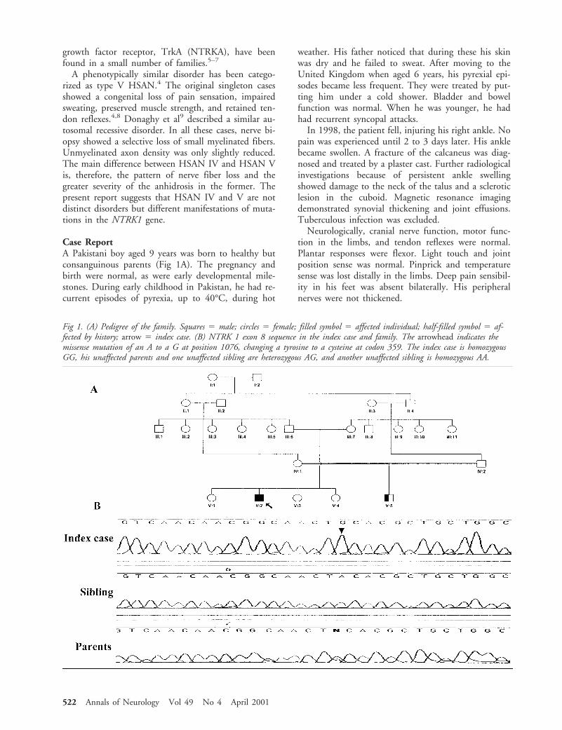
Fig 1. (A) Pedigree of the family. Squares 5 male; circles 5 female; filled symbol 5 affected individual; half-filled symbol 5 af-fected by history; arrow 5 index case. (B) NTRK 1 exon 8 sequence in the index case and family. The arrowhead indicates themissense mutation of an A to a G at position 1076, changing a tyrosine to a cysteine at codon 359. The index case is homozygousGG, his unaffected parents and one unaffected sibling are heterozygous AG, and another unaffected sibling is homozygous AA.
522 Annals of Neurology Vol 49 No 4 April 2001

In the family history (see Fig 1A) a male cousin, alsoborn to consanguinous parents, is known to have an-hidrosis.
ResultsNerve Conduction StudiesPeroneal motor nerve conduction was normal (conduc-tion velocity 46 m/sec, distal motor latency 3.5 msec, Fwave latency 39 msec), but compound muscle actionpotential amplitude was reduced (1.4 mV, knee andankle stimulation). Sural and superficial peroneal sen-sory action potential amplitudes were slightly reduced(8 mV, 6 mV) with normal conduction velocities (41m/sec, 44 m/sec).
Autonomic Function TestsThere was no evidence of orthostatic hypotension, andthere was normal sinus arrhythmia. Pressor test re-sponses were mildly impaired (simple spelling, cutane-ous cold challenge, and hyperventilation). No galvanicskin responses were detectable in the feet on inspiratorygasps. Plasma adrenaline and dopamine levels werenormal when resting or on tilting. Plasma noradrena-line levels were slightly reduced (139 pmg/ml; 168pmg/ml tilted; normal 200–500 pmg/ml).
Nerve BiopsySural nerve fascicular biopsy was undertaken and thespecimen processed by standard techniques.10 Myelin-ated fiber density was 7,669/mm2 (normal value forsame age 10,979/mm2). There was a relative deficiencyof small myelinated fibers (Fig 2,3). No actively degen-erating fibers, signs of regenerative activity, or hyper-trophic changes were detected. Myelin thickness wasnormal, as assessed by g ratio (axon diameter/total fiberdiameter) distributions. On electron microscopic exam-ination, unmyelinated axon density was slightly re-duced, at 26,920/mm2, compared with an age-matchedcontrol value of 35,700/mm2.11 No abnormal axonalor Schwann cell inclusions were seen. The blood vesselsand connective tissues appeared normal.
Genetic AnalysisDNA was extracted from blood samples obtained fromaffected and unaffected family members. The 17 exonsand flanking intronic regions of the NTRK1 gene wereamplified by polymerase chain reaction (PCR).7,13
PCR products were purified (Qiaquick purification kitQiagen, Hilden, Germany) and sequenced on anABI377 automated sequencer (BigDye Terminator cy-cle sequencing kit, Perkin-Elmer, Foster City, USA).
A novel missense mutation was identified in exon 8at codon 359 causing a tyrosine-to-cysteine amino acidchange. This was homozygous in the index case andheterozygous in both parents and one unaffected
daughter; another unaffected daughter was homozy-gous for the normal base (see Fig 1B). This mutationwas not found in 30 Pakistani and 50 Caucasian con-trol subjects by sequencing. Two further novel base
Fig 2. Portions of transverse sections of (A) a sural nerve bi-opsy specimen from the patient and (B) a postmortem speci-men from Case 13 of Jacobs and Love11 showing lack of smallmyelinated fibers in A. Thionin and acridine orange, 3 480.
Fig 3. Myelinated fiber size distribution for the patient (histo-gram) and Case 13 from Jacobs and Love11 (continuous line)showing depletion of small myelinated fibers in the patient.
Brief Communication: Houlden et al: A Novel TRK A (NTRK1) Mutation 523

changes were identified in exon 15, both creating anamino acid change: histidine 598 tyrosine and glycine607 valine. These two changes were in disequilibriumwith each other, consistent with close genetic proxim-ity. Both base changes were present in Pakistani (3 het-erozygotes in 30 individuals) and Caucasian (1 ho-mozygote and 13 heterozygotes in 80 individuals)control subjects. Two other polymorphisms werepresent in the family and control subjects, both silentchanges, exon 14 CAA–CAG 1656 and exon15 GCC–GCT base 1869. Polymorphism segregationwas consistent with recessive inheritance.
Polymorphic markers located close to the NTRK1gene were analyzed in the family. These markers wereD1S2878, D1S484, D1S196, D1S2726, D1S252,D1S218, and D1S498. Each marker was amplified, di-luted, and pooled according to protocol (Perkin-ElmerABI Linkage Mapping Set version 2.0) and run on anAB1377 automated sequencer and analyzed using ABIGenescan and Genotyper software. This showed ge-netic linkage to the NTRK1 gene in an autosomal re-cessive fashion with homozygosity in markers acrossthe NTRK1 region.
DiscussionThe index case in the present family showed a combi-nation of widespread congenital anhidrosis and distallyaccentuated loss of pain and temperature sensibility.He had minor abnormalities of vasomotor functionwithout orthostatic hypotension. Recurrent syncopalattacks had occurred in early childhood, usually pro-voked by injury or emotional factors. It is of interestthat syncope was a feature in one of the original casesof CIPA reported by Swanson.1 As was pointed out byDyck et al,4 the designation CIPA is a misnomer. Theneurological changes are manifestations of a peripheralneuropathy, as evidenced by the distal distribution ofthe sensory loss and sudomotor dysfunction.
Sural nerve biopsy showed a reduction in small my-elinated fiber density, with preservation of density forlarger-caliber fibers and relative preservation of unmy-elinated axons. This is the pattern described for HSANV, as distinct from the virtual absence of unmyelinatedaxons in HSAN IV.4 As already stated, HSAN IV isdue to mutations in the NTRK1 gene; functional stud-ies have shown that these result in inactivation of theNTRK1/nerve growth factor receptor.13
The exon 8 tyrosine 359 cysteine NTRK1 mutation ispathogenic and likely to cause partial loss of gene func-tion and a less severe deficiency of unmyelinated axonsbut a greater effect on small myelinated fibers. Alterna-tively, NTRK1 expression could be modulated by thepresence of rare polymorphisms in the gene. Other re-ported mutations in the NTRK1 gene lead to a pheno-type of HSAN IV. The majority of these mutationscause aberrant splicing or truncation and usually result
in a more significant effect on NTRK1 mRNA than doesthe missense mutation reported.7,13,14 This codon hasbeen mutated to a stop codon in a Japanese patient witha more severe clinical phenotype and sural nerve biopsyfindings consistent with HSAN IV.15 It will be impor-tant to analyze the NTRK1 gene in other families withHSAN V to define the spectrum of the mutations, themechanism of action, and the way in which particularmutations lead to different pathological phenotypes.
An unresolved question, alluded to by Donaghy etal.9 and Dyck,16 is how to explain the severe distal lossof pain sensibility in face of the relatively modest lossof small myelinated fibers in HSAN IV and a similarmild reduction in unmyelinated axons in HSAN V.Landrieu et al.17 reported a mother and daughter with“congenital indifference to pain” in whom other formsof sensation and autonomic function were preserved, aswere sensory action potentials. Nerve biopsy findingswere normal, with normal numbers and size distribu-tions of myelinated and unmyelinated axons. An agno-sia for pain is unlikely because the mother had experi-enced pain from dental treatment. An abnormality oftransmitter function or of central pain pathways wouldthus have to be considered, and such an abnormalitymay thus be an additional deficit in HSAN IV and V.
This study was supported by the Wellcome Trust (H.H., P.K.T.,and R.H.M.K.).
We thank Dr Jean Jacobs for access to the control nerve specimen,Michelle Nourallah for performing the morphometric studies, andJohn Muddle for writing the programs and analyzing the results.
References1. Swanson AG. Congenital insensitivity to pain: a unique syn-
drome in two male siblings. Arch Neurol 1963;8:299–306.2. Swanson AG, Buchan GG, Alvord EC Jr. Autonomic changes
in congenital insensitivity to pain: absence of small primary sen-sory neurons in ganglia, roots and Lissauer’s tract. Arch Neurol1965;12:12–18.
3. Goebel HH, Veit S, Dyck PJ. Confirmation of virtual unmy-elinated fiber absence in hereditary sensory neuropathy type IV.J Neuropathol Exp Neurol 1980;39:670–675.
4. Dyck PJ, Mellinger JF, Reagan TJ, et al. Not “indifference topain” but varieties of hereditary sensory and autonomic neurop-athy. Brain 1983;106:373–390.
5. Indo Y, Tsuruta M, Hayashida Y, et al. Mutations in theTRKA/NGF receptor gene in patients with congenital insensi-tivity to pain with anhidrosis. Nat Genet 1996;13:485–488.
6. Yotsumoto S, Setoyama M, Hozumi H, et al. A novel pointmutation affecting the tyrosine kinase domain of the TRKAgene in a family with congenital insensitivity to pain with an-hidrosis. J Invest Dermatol 1999;112:810–814.
7. Mardy S, Miura Y, Endo F, et al. Congenital insensitivity topain with anhidrosis: novel mutations in the TRKA (NTRK1)gene encoding a high-affinity receptor for nerve growth factor.Am J Hum Genet 1999;64:1570–1579.
8. Low PA, Burke WJ, McLeod JG. Congenital sensory neuropa-thy with selective loss of small myelinated fibers. Ann Neurol1978;3:179–182.
524 Annals of Neurology Vol 49 No 4 April 2001

9. Donaghy M, Hakin RN, Bamford JM, et al. Hereditary sensoryneuropathy with neurotrophic keratitis: description of an auto-somal recessive disorder with a selective reduction of small my-elinated nerve fibres and a discussion of the classification of thehereditary sensory neuropathies. Brain 1987;110:563–583.
10. Tournev I, King RHM, Workman J, et al. Peripheral nerveabnormalities in the congenital cataracts facial dysmorphismneuropathy (CCFDN) syndrome. Acta Neuropathol 1999;98:165–170.
11. Jacobs JM, Love S. Qualitative and quantitative morphology ofhuman sural nerve at different ages. Brain 1985;108:897–924.
12. Roa BB, Warner LE, Garcia CA, et al. Myelin protein zero(MPZ) gene mutations in nonduplication type 1 Charcot-Marie-Tooth disease. Hum Mutat 1996;7:36–45.
13. Greco A, Villa R, Fusetti L, et al. The Gly571Arg mutation,associated with the autonomic and sensory disorder congenitalinsensitivity to pain with anhidrosis, causes the inactivation ofthe NTRK1/nerve growth factor receptor. J Cell Physiol 2000;182:127–133.
14. Miura Y, Mardy S, Awaya Y, et al. Mutation and polymor-phism analysis of the TRKA (NTRK1) gene encoding a high-affinity receptor for nerve growth factor in congenital insensi-tivity to pain with anhidrosis (CIPA) families. Hum Genet2000;106:116–124.
15. Iwanaga R, Matsuishi T, Ohnishi A, et al. Serial magnetic res-onance images in a patient with congenital sensory neuropathywith anhidrosis and complications resembling heat stroke.J Neurol Sci 1996;142:79–84.
16. Dyck PJ. Neuronal atrophy and degeneration predominantly af-fecting peripheral sensory and autonomic neurons. In: Dyck PJ,Thomas PK, Griffin JW, et al, eds. Peripheral neuropathy.Philadelphia: W.B. Saunders, 1993;1065–1093.
17. Landrieu P, Said G, Allaire C. Dominantly transmitted congen-ital indifference to pain. Ann Neurol 1990;27:574–578.
Subthalamic Infusion of anNMDA Antagonist PreventsBasal Ganglia MetabolicChanges and NigralDegeneration in a RodentModel of Parkinson’sDiseaseFabio Blandini, MD,1 Giuseppe Nappi, MD,1,2
and J. Timothy Greenamyre, MD, PhD3
Using permanent cannulas connected to subcutaneouspumps, we infused selective glutamate antagonists intothe subthalamic nucleus of rats. Pumps were implantedimmediately after the intrastriatal injection of 6-hydroxy-dopamine and delivered micro-quantities of the N-methyl-D-aspartate antagonist MK-801 or the a-amino-3-hydroxy-5-methylisoxazole antagonist NBQX for 4weeks. Subthalamic infusion of MK-801, but not ofNBQX, prevented the basal ganglia metabolic changesand motor abnormalities caused by nigrostriatal lesion.Animals treated with MK-801 also exhibited marked re-duction of nigral cell loss. We conclude that pharmaco-logical modulation of subthalamic activity may haveboth symptomatic and neuroprotective effects in Par-kinson’s disease.
Ann Neurol 2001;49:525–529
In Parkinson’s disease (PD), nigrostriatal degenerationtriggers a cascade of changes in basal ganglia circuitry,which leads to overactivity of the subthalamic nucleus(STN) and its projection nuclei, medial globus pallidus(MGP) and substantia nigra pars reticulata (SNr).1 Re-cent evidence suggests that STN overactivity may alsocontribute to the progression of PD. In addition to itsmain targets, the STN also sends excitatory projectionsto the substantia nigra pars compacta (SNc).2,3 There-fore, subthalamic disinhibition might cause glutamater-gic overstimulation of residual SNc neurons. A numberof factors, including oxidative stress and mitochondrialdefects, might reduce the ability of nigral neurons to
From the 1Laboratory of Functional Neurochemistry, NeurologicalInstitute C. Mondino, Pavia, Italy; 2Institute of Nervous and Men-tal Diseases, La Sapienza University, Rome, Italy; and 3Departmentsof Neurology and Pharmacology, Emory University, Atlanta, GA.
Received Jul 7, 2000. Accepted for publication Dec 18, 2000.
Address correspondence to Dr Blandini, Laboratory of FunctionalNeurochemistry, Neurological Institute C. Mondino, Via Palestro, 327100 Pavia, Italy. E-mail: [email protected]
© 2001 Wiley-Liss, Inc. 525

cope with a potentially harmful agent, such as gluta-mate.1 This enhanced glutamatergic input to the SNcmay therefore aggravate—via a mechanism known as“indirect excitotoxicity”4—the progression of the dis-ease, leading to a vicious cycle in which STN overac-tivity and nigral damage support each other.5 Indeed,in rats, ablation of the STN counteracts the SNc de-generation caused by intrastriatal administration of6-hydroxydopamine (6-OHDA)6 or 3-nitropropionicacid.7
The aim of this study was to investigate whetherchronic reduction of STN glutamatergic transmissioninterferes with the development of the nigrostriatal le-sion and the resulting basal ganglia functional changescaused by 6-OHDA. For this purpose, we infused se-lective antagonists of either the N-methyl-D-aspartate(NMDA) or the a-amino-3-hydroxy-5-methylisoxazolepropionic acid (AMPA) glutamate receptor subtypeinto the STN of rats immediately after the striatal in-jection of 6-OHDA. As opposed to the direct injectionof 6-OHDA into the SNc, which causes massive andrapid cell loss, intrastriatal injection of 6-OHDA causesa partial SNc lesion that evolves gradually. This tech-nique is therefore recommended when evaluating neu-roprotective strategies for experimental PD.8
Material and MethodsMale Sprague-Dawley rats (250–300 gm) were used. All an-imal care and use was in accordance with National Institutesof Health guidelines and was approved by the InstitutionalAnimal Care and Use Committee (IACUC) of Emory Uni-versity. The NMDA blocker MK-801 and the AMPA blockerNBQX, in its water-soluble form (1,2,3,4-tetrahydroxy-6-nitro-2,3-dioxo-benzo quinoxaline-7-sulfonamide disodium),were purchased from RBI (Natick, MA). Drugs were dissolvedin saline solution.
Surgical ProceduresAnimals were anesthetized with ketamine (75 mg/kg) andxylazine (10 mg/kg) and placed in a stereotactic apparatus.They received a 3.5-ml injection of 6-OHDA (2.5 mg/mlplus 0.2 mg/ml ascorbate) into the right striatum (1 mm an-
terior, 3 mm lateral with respect to bregma, and 4.5 mmventral with respect to dura) at a rate of 0.5 ml/min. Imme-diately after, a 28-gauge infusion cannula was lowered intothe ispilateral STN (3.7 mm posterior, 2.4 mm lateral withrespect to bregma, and 7.9 mm ventral with respect to dura).The cannula base was secured to the skull with dental ce-ment and anchor screws and connected to a subcutaneousmini-osmotic pump placed on the back of the animal (AlzetBrain Infusion kit, Alza, Palo Alto, CA).
DrugsPumps were loaded with 10 mM MK-801, 50 mM NBQX,or saline solution (control animals) and delivered into theSTN at a rate of 4.2 nl/min for 4 weeks. Given the absenceof previous studies with similar experimental conditions, wechose concentrations of MK-801 and NBQX known to behighly effective in in vitro conditions.9,10
Behavioral TestingAt the third week, animals were tested with systemic am-phetamine (3 mg/kg, intraperitoneally) to evaluate the be-havioral effect (turning behavior) of the evolving nigrostriatallesion. Rotational response to the drug was expressed as thenumber of full (360-degree) turns per minute.
HistochemistryAt the end of the fourth week, animals were killed by de-capitation. Brains were rapidly removed, frozen on dry ice,and stored at 270°C. Frozen coronal sections (25 mm) con-taining striatum, globus pallidus (GP, the rodent structurehomologous to the lateral globus pallidus), entopeduncularnucleus (EP, the rodent structure homologous to the MGP),STN, SNr, and SNc were cut and mounted on slides. Sec-tions were stained for activity of cytochrome oxidase (CO), afunctional marker of neuronal activation,11,12 using a metal-enhanced histochemical technique described recently.13 Thecell loss in the SNc was evaluated by means of Nissl staining,which was also used to verify the correct location of striatalinjections and subthalamic cannulas.
Image AnalysisA densitometric comparison of CO activity in the two hemi-spheres was carried out using a computerized video-based
Table. Cytochrome Oxidase Activity in the Striatum, Globus Pallidus (GP), Entopeduncular Nucleus (EP), Subthalamic Nucleus(STN), and Substantia Nigra Pars Reticulata (SNr) of Rats with Unilateral Nigrostriatal Lesion and Ipsilateral IntrasubthalamicInfusion of Saline Solution (Control Animals), MK-801, or NBQX
Striatum GP EP STN SNr
Intact Lesioned Intact Lesioned Intact Lesioned Intact Lesioned Intact Lesioned
Control animals (n 5 9) 413 6 22 455a 6 21 97 6 9 133a 6 13 82 6 6 116b 6 11 526 6 46 587 6 45 221 6 31 283a 6 35MK-801 (n 5 7) 420 6 23 419 6 24 135 6 17 152 6 16 94 6 19 111 6 21 647 6 62 497a 6 48 260 6 36 253 6 36NBQX (n 5 6) 423 6 19 455c 6 19 128 6 9 168b 6 13 89 6 15 124b 6 9 544 6 64 593 6 55 210 6 19 291b 6 21
ap , 0.005 vs intact side (Student’s t test for paired data).bp , 0.01 vs intact side (Student’s t test for paired data).cp , 0.05 vs intact side (Student’s t test for paired data).
Values (mean 6 SEM) are expressed as optical density units (31000).
526 Annals of Neurology Vol 49 No 4 April 2001

image analysis system (Imaging Research, St. Catharines,Ontario, Canada). The same system was used to count theNissl-positive cells in the SNc. In both cases, the averagevalue of at least four adjacent sections was considered as rep-resentative of each area.
StatisticsComparisons between groups were made using one-way anal-ysis of variance (ANOVA) followed by a Fisher’s post-hoctest. Side-to-side comparisons within the same group weremade by Student’s t test for paired data. Minimum level ofstatistical significance was set at p , 0.05.
ResultsOnly animals in which both the striatal 6-OHDA in-jection and subthalamic cannula were correctly locatedwere considered for the data analysis.
CO ActivityControl animals showed significant increases in COstaining in the striatum, GP, EP, and SNr ipsilaterallyto the nigrostriatal lesion (Table). CO activity was alsoincreased in the ipsilateral STN, although not signifi-cantly. Animals that received subthalamic infusion ofMK-801 showed a significant reduction of CO activityin the STN and no significant asymmetries in any ofthe other nuclei evaluated (Fig 1). Like control ani-mals, the animals that received subthalamic NBQXshowed significant increases in CO activity in the stri-atum, GP, EP, and SNr ispilaterally to the nigrostriatallesion as well as a slight, nonsignificant increase in theipsilateral STN.
Nigral DamageControl animals showed discrete cell loss (30%) in theright SNc compared with the left SNc. Animals treatedwith subthalamic MK-801 but not with NBQXshowed a significant reduction of the nigral cell losswith respect to control animals (Fig 2A).
Rotational BehaviorControl animals showed consistent rotational behaviorin response to systemic amphetamine (Fig 2B). The re-sponse was significantly reduced in the animals that re-ceived subthalamic MK-801 but not NBQX.
DiscussionGlutamate antagonists have repeatedly proven benefi-cial in animal models of PD.14 In this study, we con-tinuously infused selective ionotropic glutamate antag-onists into the STN with the aim of investigatingwhether chronic blockade of glutamatergic transmis-sion at the level of the STN could influence the SNcdamage and resulting functional changes caused by6-OHDA.
In control animals, the intrastriatal injection of6-OHDA caused discrete SNc cell loss. CO activitywas significantly increased in the striatum, GP, EP,and SNr ipsilaterally to the lesion and nonsignificantlyin the STN. Subthalamic infusion of MK-801 but notNBQX decreased CO activity in the nucleus, pre-vented the metabolic changes in the other basal ganglianuclei, and reduced the degree of the SNc cell loss.The rotational response to amphetamine observed in
Fig 1. Brain coronal sectionsstained for cytochrome oxidase activ-ity. The sections, which containsubthalamic nucleus (STN), globuspallidus (GP), and substantia nigrapars reticulata (SNr), were obtainedfrom a control animal (A) andfrom an animal that, in additionto the intrastriatal injection of6-hydroxydopamine, received a4-week intrasubthalamic infusion ofMK-801. (B) Note how, in thecontrol specimen, the staining ismore intense in the STN, GP, andSNr on the lesioned side. Con-versely, the animal treated withMK-801 shows a clear reduction ofcytochrome oxidase activity in theSTN (where the drug was infused)and no metabolic asymmetries inGP and SNr.
Brief Communication: Blandini et al: Subthalamic Infusion of Glutamate Antagonists in Rats with Nigrostriatal Lesion 527

control animals was also abolished in animals treatedwith MK-801.
Our metabolic data confirm that nigrostriatal lesion,even of limited degree, leads to overactivity of basalganglia output nuclei.1 Such overactivity is sustainedby the STN, since the reduction of STN activitycaused by MK-801 abolished these changes.
The origin of STN overactivity is currently disputed.According to classical models of basal ganglia organiza-tion, the activity of GP—which normally inhibitsSTN—should decrease following nigrostriatal lesion.This would cause STN disinhibition.15 In fact, wefound that GP metabolic activity increased, rather thandecreased, after nigrostriatal lesion. This confirms pre-vious observations and suggests that STN overactivityhas different sources.16,17 For example, given the recip-rocal connections between STN and SNc,3,18 degener-ation of SNc might affect the STN directly. In addi-tion, enhanced excitatory inputs originating from boththe brainstem and the thalamus reach the STN in ratsbearing a nigrostriatal lesion.19
The striatum is not typically considered a target ofSTN projections, although a subthalamo-striatal path-way has been described.2 It is interesting to note thatthe metabolic changes found in the striatum of bothcontrol animals and animals treated with NBQX weresimilar to those observed in the typical STN projectionnuclei. Moreover, the intrasubthalamic infusion ofMK-801 prevented the striatal metabolic changes.These findings confirm a previous observation20 andsuggest that modifications in STN activity can affectthe striatum directly.
The fact that reducing STN activity through localinfusion of MK-801 protected SNc neurons confirmsthat the STN plays a role in the progression of thenigral damage. Thus, once nigrostriatal degenerationbegins (irrespective of cause), STN overactivity ensues,and glutamatergic stimulation of SNc neurons contrib-utes to subsequent cell loss. It is interesting that nosymptomatic or protective effects were seen when theAMPA receptor blocker NBQX was used, which pointsto a more important role of NMDA receptors in me-diating the functional responses of the STN.
In conclusion, chronic blockade of NMDA receptor-mediated transmission in the STN had both symptom-atic and neuroprotective effects in a rodent model ofPD. Pharmacological manipulation of the STN, throughselective drugs capable of modulating glutamatergictransmission, may therefore represent a valuable tool forthe treatment of PD.
This study was supported by United States Public Health Servicegrant NS33779 (J.T.G.).
We thank Dr Ranjita Betarbet, Dr Roberto Fancellu, and MonicaGarcia-Osuna for their assistance.
References1. Blandini F, Nappi G, Tassorelli C, Martignoni E. Functional
changes of the basal ganglia circuitry in Parkinson’s disease.Progr Neurobiol 2000;62:63–88.
2. Kita H, Kitai ST. Efferent projections of the subthalamic nu-cleus in the rat: light and electron microscopic analysis with thePHA-L method. J Comp Neurol 1987;260:435–452.
3. Smith Y, Charara A, Parent A. Synaptic innervation of mid-brain dopaminergic neurons by glutamate-enriched terminals inthe squirrel monkey. J Comp Neurol 1996;364:231–253.
4. Albin RL, Greenamyre JT. Alternative excitotoxic hypotheses.Neurology 1992;42:733–738.
5. Rodriguez MC, Obeso A, Olanow W. Subthalamic nucleus-mediated excitotoxicity in Parkinson’s disease: a target for neu-roprotection. Ann Neurol 1998;44:S175–S188.
6. Piallat B, Benazzouz A, Benabid AL. Subthalamic nucleus le-sion in rats prevents dopaminergic nigral neuron degenerationafter striatal 6-OHDA injection: behavioural and immunohis-tochemical studies. Eur J Neurosci 1996;8:1408–1414.
7. Nakao N, Ekini N, Nakai K, Itakura T. Ablation of the sub-thalamic nucleus supports the survival of nigral dopaminergicneurons after nigrostriatal lesions induced by the mitochondrialtoxin 3-nitropropionic acid. Ann Neurol 1999;45:640–651.
8. Sauer H, Oertel WH. Progressive degeneration of nigrostriatal
Fig 2. (A) Nigral damage. Bars represent the mean (6 SEM)reduction in the number of Nissl-positive neurons of the le-sioned substantia nigra pars compacta (SNc) compared withthe intact SNc. Animals that received intrasubthalamic infu-sion of MK-801 showed a significant reduction of the nigraldamage (ANOVA, F 5 3.7, p , 0.05; Fisher’s post-hoc test(asterisk), p , 0.05 vs control animals). (B) Rotational be-havior. Animals treated with MK-801 showed significant re-duction in the rotational response to amphetamine comparedwith control animals (ANOVA, F 5 3.55, p , 0.05; Fish-er’s post-hoc test (asterisk), p , 0.05 vs control animals).
528 Annals of Neurology Vol 49 No 4 April 2001

dopamine neurons following intrastriatal terminal lesions with6-hydroxydopamine: a combined retrograde tracing and immu-nocytochemical study in the rat. Neuroscience 1994;59:401–415.
9. Bon CLM, Paulsen O, Greenfield SA. Association between thelow threshold calcium spike and activation of NMDA receptorsin guinea-pig substantia nigra pars compacta neurons. EurJ Neurosci 1998;10:2009–2015.
10. Li S, Stys PK. Mechanisms of ionotropic glutamate receptor-mediated excitotoxicity in isolated spinal cord white matter.J Neurosci 2000;20:1190–1198.
11. Wong-Riley MTT. Cytochrome oxidase: an endogenous meta-bolic marker for neuronal activity. Trends Neurosci 1989;12:94–101.
12. Blandini F, Garcia-Osuna M, Greenamyre JT. Subthalamic ab-lation reverses changes in basal ganglia oxidative metabolismand motor response to apomorphine induced by nigrostriatallesion in rats. Eur J Neurosci 1997;9:1407–1413.
13. Divac I, Mojsilovic-Petrovic J, Lopez-Figueroa MO, et al. Im-proved contrast in histochemical detection of cytochromeoxidase: metallic ions protocol. Neurosci Methods 1995;56:105–113.
14. Blandini F, Porter RHP, Greenamyre JT. Glutamate and Par-kinson’s disease. Mol Neurobiol 1996;12:1–17.
15. Albin RL, Young AB, Penney JB. The functional anatomy ofbasal ganglia disorders. Trends Neurosci 1989;12:366–375.
16. Vila M, Levy R, Herrero MT, et al. Metabolic activity of thebasal ganglia in parkinsonian syndromes in human and non-human primates: a cytochrome oxidase histochemistry study.Neuroscience 1996;71:903–912.
17. Hassani OK, Mouroux M, Feger J. Increased subthalamic neu-ronal activity after nigral dopaminergic lesion independent ofdisinhibition via the globus pallidus. Neuroscience 1996;72:105–115.
18. Hassani OK, Feger J, Yelnik J, Francois C. Evidence for a do-paminergic innervation of the subthalamic nucleus in the rat.Brain Res 1997;749:88–94.
19. Orieux G, Francois C, Feger J, et al. Metabolic activity of ex-citatory parafascicular and pedunculopontine inputs to the sub-thalamic nucleus in a rat model of Parkinson’ disease. Neuro-science 2000;97:79–88.
20. Blandini F, Greenamyre JT. Effect of subthalamic nucleus le-sion on mitochondrial enzyme activity in rat basal ganglia.Brain Res 1995;669:59–66.
Germline Mutations in theCCM1 Gene, EncodingKrit1, Cause CerebralCavernous MalformationsMiguel Lucas, MD1, Alzenira F. Costa, PhD,1
Mariano Montori, MD,2 Francisca Solano, BS,1
Marıa D. Zayas, PhD,1 and Guillermo Izquierdo, MD,3
Mutations in the Krit1 gene have been recently discov-ered as the cause of hereditary cerebral cavernous angi-oma. We sought the possibility that de novo, noninher-ited mutations of Krit1 also cause cavernous angioma. Apatient with two cerebral malformations carries a het-erozygous deletion of two base pairs (741delTC) in exonVI of the Krit1 gene. The deletion initiates a frameshiftmutation that, 23 amino acids downstream, encodes aTAA stop triplet replacing a CAT triplet of histidine atexon VII (H271X). Magnetic resonance images of theparents were normal, neither parent carries the 741delTCmutation, and both bear the wild-type sequence of exonVI. These findings document a de novo germline muta-tion in Krit1 gene that causes cerebral cavernous malfor-mations.
Ann Neurol 2001;49:529–532
Krit1 is an ankyrin repeat–containing protein that in-teracts with Krev-1/rap1a,1 a protein described as amember of the Ras family of GTPases with probabletumor-suppressing activity in the cell. Truncating mu-tations in CCM1, the gene encoding Krit1 protein,cause hereditary cavernous angiomas,2,3 and a great va-riety of mutations have been described.2–4.
Familial forms of cerebral cavernous angiomas aremanifested as multiple lesions and sporadic forms as aunique lesion, suggesting a “Knudson’s double-lossmechanism,” that evoke hereditary and sporadic formsof tumorigenesis caused by tumor-suppressor genes.5
Although it is generally believed that some percentageof the patients with cavernous malformation representsnew mutations, to our knowledge, de novo mutationscausing cavernous angioma have not been previously
From the 1Servicio de Biologıa Molecular, Hospital UniversitarioVirgen Macarena, Seville, Spain; 2Servicio de Neurologıa HospitalMiguel Servet, Zaragoza, Spain; and 3Servicio de Neurologıa, Hos-pital Universitario Virgen Macarena, Seville, Spain.
Received Aug 15, 2000, and in revised form Jan 3, 2001. Acceptedfor publication Jan 3, 2001.
Address correspondence to Dr Lucas, Servicio de Biologıa Molecu-lar, Hospital Universitario Virgen Macarena, Avenida Dr Fedrianis/n, 41009 Seville, Spain. E-mail: [email protected]
© 2001 Wiley-Liss, Inc. 529

reported. We discovered a very illustrative single-strandconformation polymorphism (SSCP) in exon VI of theindex patient of an a priori familial form. The absenceof cavernous malformations in the parents and theirnormal SCCP patterns suggested a case of noninheritedcavernous angioma, and therefore we sought a possiblemutation of the Krit1 gene. This possibility seems in-teresting in relation to the abovementioned pathogenicmechanism.
Patients and MethodsThe main clinical characteristic of the proband consisted ofheadaches, whereas the parents and sister were almost symp-toms free, except for the mother, who complained aboutheadaches. Two cavernomas were discovered through T2-weighted magnetic resonance imaging (MRI) studies of thepatient. MRI results were normal for the patient’s parentsand healthy sister.
CCM1 haplotypes of individuals of the CV36 family wereanalyzed with the protocols and primers as previously de-scribed.6 The polymorphic microsatellite markers spanningthe CCM1 interval were the following: DTS492, D7S2410,D7S1813, D7S689, D7S657, D7S527, and D7S479. A frag-ment of Krit1 gene containing exon VI was amplified bypolymerase chain reaction (PCR) techniques with primersforward (5’TTGTTAGATTGTGATGTA) and reverse(59AACATAATAAAAACTTTC), as described recently.7
The nomenclature of cDNA refers to the work of Serebriiskiet al,1 although part of the Krit1 gene was missed in this re-port, and additional exons have been identified very recently.8
Genomic DNA was initially screened by analysis of SSCP.PCR fragments were separated by electrophoresis in 10%
Fig 1. Magnetic resonance images (MRIs) of the patient and the parents. The screening of the whole brain by T2-weigthed MRIsshowed two cavernomas in the patient (middle), in contrast to the normal images of father (left) and mother (right). We did notfind other malformations in the screening of the whole brains of the parents or the sister.
Fig 2. (Top) Single-strand conformation polymorphism (SSCP)of exon VI of Krit1 gene of the Spanish family CVE36. Ali-quots of DNA were amplified in the polymerase chain reaction(PCR) mixture containing [a32P]-dCTP. Other reagents aredescribed in Patients and Methods. The products of the PCRencompassing the exon VI were run under nondenaturing con-ditions in 10% acrylamide containing 10% glycerol for 16hours at 20°C in a 20-by-30 cm gel. Bands were revealedafter 48-hour exposure to x-ray films. The lower part of thefigure shows the haplotypes of the critical region of CCM1 inindividuals of CV36 family. The corresponding markers aregiven in the first column. Note that both the affected and thehealthy sibling inherited the same chromosome from the fatherand a different chromosome from the mother. Square 5 male;circle 5 female.
530 Annals of Neurology Vol 49 No 4 April 2001

acrylamide in the absence and in the presence of 10% glyc-erol as described.2
DNA sequencing of exon VI was carried out in genomicDNA of the proband and parents with the terminaldideoxynucleotides method (fmol kit, Promega, Lyon,France). The forward primer was 59CGAATATACAG AAT-GGATG, and the above-described oligonucleotide was theprimer of the reverse strand. Manual sequencing reactions
were performed with 32P-59-primers labeled with [g32P]ATPand polynucleotide kinase. Partially automated sequenceanalyses of both the sense the and antisense strands extend-ing to 30 to 40 bases of introns 5 and 6 were performedwith 59-labeled IRD800 primers according to the protocolsof the SequiTherm kit (Epicentre Technologies, Madison,WI). Electrophoretic separation in acrylamide gels and anal-ysis of the sequencing fragments were performed in a LI-COR DNA4000 sequencer with the software provided bythe manufacturer (LI-COR Inc, Lincoln, Nebraska).
ResultsT2-weighted MRIs demonstrated the presence of twocerebral cavernous malformations (CCMs) in the pro-band of the CV36 family. CCMs were not found ineither parent (Fig 1) or the healthy sister (not shown)in a whole-brain screening.
The analysis of CCM1 haplotypes showed that thesiblings inherited the same chromosome from the fa-ther but different chromosomes from the mother (Fig2). The haplotypes also revealed the lack of recombi-nation in this region. Our first step in identifying thepossible mutation was to obtain an illustrative SSCP inthe PCR product of exon VI (see bands in Fig 2). Astriking finding was that none of the progenitorsshared the SSCP, suggesting a noninherited mutationin exon VI of Krit1.
Sequencing of the corresponding genomic regionsincluded the sense and antisense strands of exon VIand at least 30 bases at the 39 end of intron 5 and 30bases at the 59 end of intron 6. The sequencing ofexon VI identified a 2-bp (TC/AG) deletion that is 48bp from the splice junction with intron 6. The deletionof nucleotides 741T and 742C shifts the reading frameand predicts a mutated sequence of 23 amino acids(RNSAIFHYMGFFRKPQPSTQTIS) that ends with aTAA termination triplet. The 741delTC mutation sub-stitutes the CAT triplet of histidine at position 271with the stop codon (H271X). The sequencing of thesense and antisense strands clearly demonstrated thedeletion in the genomic DNA of the proband and thenormal sequence in parents. We did not find othermutations either in the coding region or in the bound-ary intronic sequences of exon VI of the proband orthe relatives. Comparisons among the parents and pro-band of a set of polymorphic markers of independentloci excluded a possible false paternity or maternityascription.
DiscussionThe clinical symptoms of the index patient were com-patible with a mutation of maternal origin because, inaddition to antecedents of epilepsy in the grandfather,both the proband and her mother complained of head-aches. The finding of two CCMs in the proband butnot in relatives suggested a noninherited CCM. None-
Fig 3. Comparison of the mutated and wild-type sequences ofthe sense (upper panel) and antisense (lower panel) strandssurrounding the TC/AG deletion. The exon VI of genomicDNA of the father (upper traces), proband (middle traces),and mother (lower traces) were amplified by polymerase chainreaction techniques. The sequences of the products were deter-mined with the 59-IRD800–labeled forward (sense) and re-verse (antisense) primers (see Patients and Methods) in anLI-COR 4000 autosequencer. The TC/GA pair (underlinedin the wild-type chromosome) deletion displaces the sequencesof the mutated strand. The amino acid sequences of the mu-tated and wild-type chromosomes are given above the nucleo-tide sequence of the sense strands. Ambiguities for mutatedalleles are as follows: W, A or T; R, A or G; S, C or G; Y,C or T. Small letters indicate questionable base.
Brief Communication: Lucas et al: De Novo Mutation of Krit1 Gene 531

theless, a familial form of CCM was not excluded bythe aforementioned considerations, given the variablepenetrance of this disease.
The proband did not inherit the disease allele fromeither parent, indicating that the patient represents a denovo mutation. This conclusion is supported by thefollowing data: (1) the finding of the SSCP of exon VIin the patient but not in the parents or sister, (2) thedemonstration of the 741delTC deletion in exon VI ofthe proband and the wild-type sequence in both par-ents and the healthy sister, (3) the demonstration ofthe deletion of the AG tandem in the antisense strand,(4) the absence of recombination in the interval ofCCM1, and (4) the exclusion of a false maternity orpaternity ascription.
The pathogenic mechanism of CCMs could be sim-ilar to the formation of the neurocutaneous tumor oftuberous sclerosis type 2 (TSC2), where tuberin, theproduct of the TSC2 gene, functions as a tumor sup-pressor protein by acting as a GTPase activator forKrev-1/rap1a.9 The existence of either multiple or sin-gle lesions in hereditary and sporadic forms, respective-ly10 suggests the “Knudson double-loss mechanism” incavernous angioma and evokes the hereditary and spo-radic forms of retinoblastoma.5 The great variety ofmutations described in the hereditary form of cavern-ous angioma2–4 suggest a high mutation rate of theKrit1 gene. The results herein described are the firstevidence of CCMs caused by a spontaneous mutationin the Krit1 gene. Nonetheless, a sporadic cavernousmalformation caused by a late or somatic mutation in asingle cell in the nervous system can be excluded. Ineffect, such an event is not detectable in the genomicDNA extracted from peripheral leukocytes. The patientlikely has a germline mutation, as evidenced by the de-letion in peripheral leukocyte genomic DNA and theCCMs. Thus, this patient likely has a condition that isheritable by her offspring. In effect, the proband in thisreport represents a “founder” for a new lineage of in-dividuals with familial cavernous malformation byvirtue of a new mutation.
This study was supported by grant 99/0407 fromFondo de Investigaciones Sanitarias
References1. Serebriiskii I, Estojak J, Sonoda G, et al. Association of Krev/
rap1a with Krit1, a novel ankyrin repeat–containing protein en-coded by a gene mapping to 7q21–22. Oncogene 1997;15:1043–1049.
2. Laberge-le Couteulx S, Jung HH, Labauge P, et al. Truncatingmutations in CCM1, encoding Krit1, cause hereditary cavern-ous angiomas. Nat Genet 1999;23:189–193.
3. Sahoo T, Johnson EW, Thomas JW, et al. Mutations in the-gene encoding Krit1, a Krev-1/rap1a binding protein, cause ce-rebral cavernous malformations (CCM1) Hum Mol Genet1999;8:2325–2333.
4. Zhang J, Clatterbuck RE, Rigamonti D, Dietz HC. Mutations
in KRT1 in familial cerebral cavernous malformations. Neuro-surgery 2000;4:1272–1276.
5. Knudson AG. Hereditary cancer: two hits revisited. J CancerRes Clin Oncol 1996;122:135–140.
6. Jung HH, Labauge P, Laberge S, et al. Spanish families withcavernous angioma do not share the Hispano-AmericanCCM1 haplotype. J Neurol Neurosurg Psychiatry 1999;67:551–552.
7. Lucas M, Solano F, Zayas MD, et al. Spanish families withcerebral cavernous angioma do not bear the 742C3T HispanicAmerican mutation of the KRIT1 gene. Ann Neurol 2000;47:836.
8. Sahoo T, Serebriiski I, Kotova E, et al. Identification of theauthentic full length amino acid sequence of Krit1 (CCM1) uti-lizing a combination of computational gene-prediction toolsand RT-PCR. Am J Hum Genet 2000;67(suppl 2):261.
9. Wienecker R, Konig A, DeClue JE. Identification of tuberin,the tuberous sclerosis–2 product: tuberin possesses specificRap1GAP activity. J Biol Chem 1995;270:16409–16414.
10. Labauge P, Laberge S, Brunereau L, et al. Hereditary cerebralcavernous angiomas: clinical and genetic features in 57 Frenchfamilies. Lancet 1998;352:1892–1897
Abnormal Desmin Proteinin Myofibrillar MyopathiesCaused by Desmin GeneMutationsMian Li, MD, PhD, and Marinos C. Dalakas, MD
Muscle proteins were extracted in various sodium dode-cyl sulfate buffers from 6 patients with myofibrillar my-opathy (MFM) and previously identified with mutationsin the desmin gene (desmin myopathy; DesM), 6 withMFM without mutations, and 14 disease controls tosearch for alterations in biochemistry and solubility ofmutated desmin filaments. In the 1% posthigh-speed pel-let fraction, desmin was detected with immunoblots onlyin DesM and not the other MFM. We conclude that mu-tant desmin forms insoluble aggregates that are specificfor the DesM and can be detected with Western blots.
Ann Neurol 2001;49:533–536
From the Neuromuscular Diseases Section, National Institute ofNeurological Diseases and Stroke, National Institute of Health, Be-thesda, MD.
Received Jul 12, 2000, and in revised form Jan 9, 2001. Acceptedfor publication Jan 9, 2001.
Address correspondence to Dr Dalakas, Neuromuscular DiseasesSection, NINDS, NIH, Building 10, Room 4N248, 10 CenterDrive, MSC 1382, Bethesda, MD 20892-1382.E-mail: [email protected]
532 Annals of Neurology Vol 49 No 4 April 2001

Myofibrillar myopathies (MFM) are a heterogenousgroup of inherited skeletal myopathies often associatedwith cardiomyopathies. Myofibrillar proteins, includingdesmin, actin, gelsolin, and dystrophin, accumulate inskeletal muscle fibers of biopsy specimens, but desminis more consistently and abundantly found.1–3 Desmin,a 53 kD type II intermediate filament protein, main-tains the structural and functional integrity of the myo-fibrils and functions as a cytoskeletal protein linkingindividual myofibrils at the Z-band level to each otherand to the sarcolemma. Mutations in the desmin genecause a skeletal and cardiac myopathy, which is a dis-tinct subset among the MFM group termed desmin my-opathy (DesM).4–7 Because in DesM the deposits ofdesmin are histologically similar to those in the otherMFM, it is fundamental to identify at the protein levelwhether in DesM the aggregated mutant desmin is dif-ferent from the nonmutant desmin accumulated in themuscle fibers of other patients with MFM. Elucidatingsuch differences may help us to understand the under-lying disease mechanism and provide a screening toolfor the diagnosis of DesM.
Patients and MethodsWe studied desmin protein in muscle biopsy specimens from6 patients with DesM caused by the recently reporteddesmin gene mutations4 and from 6 patients with otherMFM without identifiable mutations in the desmin or a-B-crystalline gene. Desmin protein deposits on the muscle bi-opsies were prominent in both groups. For controls we stud-ied muscles from patients with sporadic-inclusion-bodymyositis (3), Duchenne muscular dystrophy (2), unidentifi-able muscular dystrophy (1), mitochondrial myopathy (2),vacuolar myopathy due to phosphofructokinase deficiency(2), paraneoplastic desmatomyositis (1), and human musclewith normal muscle morphology (3).
Fractionation of Proteins from Cell Extracts byDifferential CentrifugationProtein fractionation was based on the method of Coligan etal.8 In brief, six-m-thick cryosections of muscle were centri-fuged, washed in phosphate-buffered saline (pH 7.4), andresuspended in 20 ml sodium dodecyl sulfate (SDS) lysisbuffer of low concentration, 0.1% (w/v), or high concentra-tion, 1% (w/v), containing 50 mM Tris-HCl (pH 7.5), 500mM NaCl, 1% Nonidet P40, 0.5% sodium deoxycholate, 2mM EGTA, 10% glycerol, 25 mg/ml phenylmethylsulfonylfluoride, 100 mM leupeptin, and 10 mg/ml aprotinine. Thecell suspension was homogenized, incubated at 25°C for 5hours, and centrifuged twice at low speed (600g) to removenuclei, unbroken cell debris, and large insoluble particles.The supernatant containing solubilized proteins, such asdesmin monomer, and partially solubilized proteins, such astetramer aggregates, was collected as the postlow-speed super-natant fraction. To obtain an aggregate-enriched desmin pro-tein and polymers fraction, a high-speed centrifugation at100,000g for 90 minutes was performed.8 The pellet was
then resuspended in the sample buffer (NP0003; Novex, SanDiego, CA) as the posthigh-speed pellet fraction.
ImmunoblottingFifteen microliters of 20 mg protein from either the postlow-speed supernatant fraction or the high-speed pellet fractionwas prepared in 5 ml sample buffer (NP0003; Novex) andincubated at 95°C for 5 minutes. The solubilized proteinswere processed in 10% SDS-polyacrylamide gel, transferredonto a polyvinylidene difluride membrane (Millipore, Bed-ford, MA), and probed with a mouse monoclonal IgG an-tibody against 1) desmin (catalog No. MDE II; AccurateCo., Westbury, NY) in 1:100 dilution, 2) a-tropomyosinof striated muscle (Accurate Co.) in 1:50 dilution, and 3)HSP27 (Chemicon International Inc., Temecula, CA) in1:200 dilution. Antibody binding was detected withperoxidase-conjugated anti-mouse polyclonal IgG (Amer-sham International, Buckinghamshire, England). Enhancedchemiluminescence reagent (Amersham) was used to detectthe immunoreactive bands. Antibody specificity for the stri-ated muscle desmin was concurrently examined in extractsfrom skeletal muscle, cardiac muscle (Chemicon), and ovar-ian tumor containing smooth muscle. Some experimentswere replicated using two additional mouse monoclonal an-tibody against desmin (clone DE-R-11; Novocastra Labo-ratories; and clone ZSD1; Zymed, Inc., San Francisco, CA).
ResultsDesmin in the Postlow-Speed Supernatant Fraction(0.1% SDS)As is shown in Figure 1A, the antidesmin antibody wasspecific for the desmin of the skeletal (see Fig 1, lanes2, 3) and cardiac (see Fig 1, lane 4) muscle but not forthe smooth muscle expressed in the tumor (see Fig 1,lane 1). Desmin migrated at a molecular weight of 57kD, which is slightly higher than its predicted 53 kD.Specificity for striated muscle reactivity was internallycontrolled with reaction to a-tropomyosin (see Fig 1A,arrowhead, lanes 2–4).
Immunoblotting of proteins from postlow-speed su-pernatant fraction could not distinguish between thedesmin of the normal or the disease control musclesand the desmin of MFM and DesM muscles, exceptfor 2 pateints (see Fig 1B). One of these patients hadDesM with a 32-amino-acid deletion,4 correspondingto the lower molecular weight protein band (see Fig1B, lane 7), which represents the mutant protein en-coded by the mutant allele. The other patient (see Fig1B, lane 9) had MFM but no detectable desmin geneabnormality.
Desmin in the Posthigh-Speed Pellet Fraction(0.1% SDS)Immunoblotting of proteins in this fraction distin-guished the desmin of MFM muscles from the others.As is shown in Figure 2, no reaction was noted in thenormal control or the other muscle diseases (see Fig 2,
© 2001 Wiley-Liss, Inc. 533

left, lanes 1–7). In contrast, strong immunoreactivitywas noted in 5 of 6 DesM muscles (see Fig 2, left,lanes 8, 9, right lanes 3, 4, 6, 7, 10) and in 3 of 6MFM muscles (see Fig 2, right, lanes 1, 5, 11). In spiteof the marked differences in the desmin protein be-tween the controls and the MFM muscles, the HSP27,which also forms aggregates during nonspecific stress,did not reveal differences between the two groups (seeFig 2, arrowhead).
Desmin in the Posthigh-Speed Pellet Fraction(1% SDS)When the concentration of ionic detergent was in-creased to 1% SDS in the extraction buffer, immuno-reactivity for desmin remained strong in the high-speedpellet fraction in 4 of 6 patients with DesM (Fig 3,lanes 3–5, 7) but in none of the other MFM withoutdesmin gene mutations (see Fig 3, lanes 1, 8–11).
DiscussionWe found that the mutant desmin intermediate filamentprotein exhibits altered biochemical properties and solu-bility, which are different from those of the wild-type
desmin protein. This finding offers an explaination forthe formation of insoluble aggregates within the musclefibers of DesM and provides a tool for confirming ordetecting mutant desmin protein by Western blots inthe majority (up to 66%) of patients with DesM.
In vitro, transient transfection assay revealed thatmutant desmin proteins when introduced into fibro-blasts do not form the normal intermediate filamen-tous network but become desmin aggregates.4 Al-though the normally insoluble desmin intermediatefilament or polymer aggregates denature into solubleprotein monomer with a strong detergent,9 the trun-cated intermediate filament proteins, such as desmin orkeratin, remain in the insoluble fraction.10,11 In takingadvantage of these biochemical alterations, the postlow-speed supernatant fraction, which consists of crude celllysates, demonstrated no differences in the desmin pro-tein between controls and the MFM or DesM musclesbecause mutations in 5 of the 6 DesM patients werein-frame point mutations that do not change the mo-lecular weight of the protein. When differential centrif-ugation combined with increasing concentration ofionic detergent was used, clear biochemical differences
Fig 1. Western blot analysis of desmin of postlow-speed supernatant fraction prepared from control muscles (A) and myofibrillarmyopathy (MFM; B). (A) Control specimens. Ovarian tumor tissue (lane 1) and muscle biopsy specimen (lane 2) from a patientwith paraneoplastic dermatomyositis, muscle biopsy specimens from a patient with s-IBM (lane 3), and cardiac muscle from a nor-mal human subject (lane 4). Monoclonal antibodies against desmin recognize a 57 kD protein (arrow) in striated muscles (lanes2–4) but not the smooth muscle (lane 1). Monoclonal antibodies against a-tropomyosin of the striated muscle recognize a 35 kDprotein (arrowhead, lanes 2–4), which was used as an internal control for loading the same amount of muscle protein. (B) MFMwith desmin gene mutations (lanes 3, 4, 6–8, 10) or without identifiable mutations (lanes 1, 2, 5, 9, 11). In addition to the 57kD protein (arrow), the monoclonal antibodies against desmin also recognize a protein with lower molecular weight in 1 patient(m4, lane 7) that correspond to the described deletion of 32 amino acids.4 As described elsewhere,4 the missense point mutations inm1 (lane 3) was in codon 337, changing the sequence from GCC to CCC; in m2 (lane 4) in codon 451, changing the ATC toATG; in m3 and m5 (lanes 6, 8) in codon 360 and 393, resulting in G to C and A to T substitutions; and in m6 (lane 10) incodon 406, changing the sequence from CGG to TGG. No reactivity to desmin was seen in 1 patient (lane 9), although reactivityof HSP27 (arrowhead, lane 9) was present.
534 Annals of Neurology Vol 49 No 4 April 2001

were observed in the muscle protein extracts betweenMFM and controls (Fig 2). The distinction becamemore specific with 1% SDS, because under these con-ditions only the desmin from patients with DesM im-munoreacted. Such immunoreactivity corresponds tothe highly insoluble desmin aggregates we have ob-served not only in vitro within the muscle fibers of theDesM biopsies but also in the transfected cells invitro,4 owing to the dominant negative effect of themutant desmin protein.
During nonspecific cellular insults such as heat stress,HSP27 also forms aggregates and shows an early re-sponse for redistribution towards the insoluble frac-tion.12,13 Using HSP27 as an internal control, however,we demonstrated that the desmin protein abnormality isnot a nonspecific phenomenon of protein aggregationwithin the muscle fibers but rather a primary insult un-derlying defects in the desmin protein as a consequenceof desmin gene mutations. In contrast, in MFM withnormal desmin gene, the desmin protein aggregates aresecondary, resulting from primary defects in other un-known or known proteins connected with the interme-diate filamentous network, such as a-B-crystallin.14,15
Previous reports suggested a distinction between thedesmin of MFM patients and normal controls on thebasis of a 49 kD band reactivity in the desmin pro-tein.16,17 In these studies, however, the degree of solu-
Fig 2. Western blot analysis of desmin of posthigh-speed pellet fraction (0.1% sodium dodecyl sulfate) from muscle biopsy specimens.Samples are from control patients (left, lanes 1–7) and 2 myofibrillar myopathy (MFM) patients with desmin gene mutations (lanes8, 9). Samples from other MFM patients are shown at right (lanes 3, 4, 6–8, 10 for DesM and lanes 1, 2, 5, 9, 11 for MFMwithout mutations). Samples in lanes 8–10 in the left panel are the same as samples 10, 7, 2, respectively, in the right panel.(Left) No desmin reactivity was noted in non-MFM control muscles (lanes 1–7) compared to MFM with desmin gene mutations(lanes 8, 9). (Right) Strong desmin reactivity is evident (arrow) in 5 of the 6 specimens from MFM with desmin gene mutationsand 3 of the 6 MFM without identifiable mutations.
Fig 3. Western blot analysis of desmin of posthigh-speed pelletfraction (1% sodium dodecyl sulfate) from muscle biopsy speci-mens from myofibrillar myopathy (MFM) with desmin genemutation (lanes 2–7) and MFM without identifiable genemutations (lanes 1, 8–11). Strong desmin reactivity is notedin 4 of the 6 MFM with desmin gene mutations (lanes 3–5,7) but not in the MFM without identifiable mutations.
Brief Communication: Li et al: Li and Dalakas: Desmin Protein in Myofibrillar Myopathies 535

bility was not considered, and correlation with desmingene analysis was not performed to confer specificityfor the altered desmin in patients with DesM.
In summary, enhanced desmin protein aggregates inthe DesM muscle, as illustrated in this study, reflectthe in vivo process of increased insoluble desmin pro-tein, which cannot form normal filamentous network,owing to the desmin gene mutations. Although highlyinsoluble desmin appears to be specific for the DesM,desmin, which is more soluble than the mutateddesmin but less soluble than the desmin in disease con-trols, also forms aggregates characteristic for MFMprobably because of defects in other intermediatefilament-associated proteins. Similar alterations in theprotein solubility govern the disease pathogenesis invarious neurodegenerative disorders in which aggregatedproteins are formed within the targeted tissues.18–20
Dr. Lev Goldfarb has identified our previously reported mutationsin the studied patients (4,5).
References1. Goebel HH. Desmin-related myopathies. Curr Opin Neurol
1997;10:426–429.2. Nakano S, Engel AG, Akiguchi I, Kimura J. Myofibrillar myop-
athy. III. Abnormal expression of cyclin-dependent kinases andnuclear proteins. J Neuropathol Exp Neurol 1996;55:549–562.
3. Amato AA, Kagan-Hallet K, Jackson CE, et al. The wide spec-trum of myofibrillar myopathy suggests a multifactorial etiologyand pathogenesis. Neurology 1998;51:1646–1655.
4. Dalakas MC, Park K-Y, Semino-Mora C, et al. Desmin myop-athy, a skeletal myopathy with cardiomyopathy caused by mu-tations in the desmin gene. N Engl J Med 2000;342:770–780.
5. Goldfarb LG, Park KY, Cervenakova L, et al. Missense muta-tions in desmin associated with familial cardiac and skeletal my-opathy. Nature Genet 1998;19:402–403.
6. Munoz-Marmol AM, Strasser G, Isamat M, et al. A dysfunc-tional desmin mutation in a patient with severe generalized my-opathy. Proc Natl Acad Sci USA 1998;95:11312–11317.
7. Sjoberg G, Saavedra-Matiz CA, Rosen DR, et al. A missense mu-tation in the desmin rod domain is associated with autosomaldominant distal myopathy, and exerts a dominant negative effecton filament formation. Hum Mol Genet 1999;8:2191–2198.
8. Coligan JE, Dunn BM, Ploegh HL, et al. Current protocals inprotein science, vol 1. New York: John Wiley and Sons, Inc.,1997:6.1.6.
9. Darnell J, Lodish H, Baltimore D. Molecular cell biology, 2nded. Scientific American Books, Inc., 1990:894.
10. Raats JM, Gerards WL, Schreuder MI, et al. Biochemical andstructural aspects of transiently and stably expressed mutantdesmin in vimentin-free and vimentin-containing cells. EurJ Cell Biol 1992;58:108–127.
11. Coulombe PA, Hutton ME, Vassar R, Fuchs E. A function forkeratins and a common thread among different types of epidermol-ysis bullosa simplex diseases. J Cell Biol 1991;115:1661–1674.
12. Caspers G-J, Bhat SP. The expanding small heat-shock proteinfamily, and structure predictions of the conserved “a-crystallindomain.” J Mol Evol 1995;40:238–248.
13. Suzuki A, Sugiyama Y, Hayashi Y, et al. MKBP, a novel mem-ber of the small heat shock protein family, binds and activatesthe myotonic dystrophy protein kinase. J Cell Biol 1998;140:1113–1124.
14. Vicart P, Caron A, Guicheney P, et al. A missense mutation inthe alphaB-crystallin chaperone gene causes a desmin-relatedmyopathy. Nature Genet 1998;20:92–95.
15. Engel AG. Myofibrillar myopathy. Ann Neurol 1999;46:681–683.16. Arbustini E, Morbini P, Grasso M, et al. Restrictive cardiom-
yoapthy, atrioventricular block and mild to subclinical myop-athy in patients with desmin-immunoreactive material deposits.J Am Coll Cardiol 1998;31:645–653.
17. Lobrinus JA, Janzer RC, Kuntzer T, et al. Familial cardiomy-opathy and distal myopathy with abnormal desmin accumula-tion and migration. Neuromuscul Disord 1998;8:77–86.
18. Dobson CM. Protein misfolding, evolution and disease. TIBS1999;24:329–332.
19. Lowe J, Blanchard A, Morrell K, et al. Ubiquitin is a commenfactor in intermediate filament inclusion bodies of diverse typein man, including those of Parkinson’s disease, Pick’s disease,and Alzheimer’s disease, as well as Rosenthal fibres in cerebellarastrocytomas, cytoplasmic bodies in muscles, and Mallory bod-ies in alcoholic liver disease. J Pathol 1988;155:9–15.
20. Hashimoto M, Takeda A, Hsu LJ, et al. Role of cytochrome cas a stimulator of a-synuclein aggregation in Lewy body disease.J Biol Chem 1999;274:28849–28852.
Effects of Riluzole onCortical Excitability inPatients with AmyotrophicLateral SclerosisKatja Stefan, MD, Erwin Kunesch, MD,Reiner Benecke, MD, and Joseph Classen, MD
2Using transcranial magnetic stimulation, the effect of ri-luzole (RLZ) on cortical excitability was studied in patientswith amyotrophic lateral sclerosis (ALS). Paired-pulse in-hibition (PPI) and paired-pulse facilitation (PPF) were re-duced. RLZ partially restored deficient PPI in the first of 4consecutive 3-month periods of testing, but left PPF un-changed. These findings substantiate the view that attenu-ation of glutamate-related excitotoxicity is an importantfactor contributing to the beneficial effect of RLZ in ALS.
Ann Neurol 2001;49:537–540
Riluzole (RLZ) modestly slows disease progression inamyotrophic lateral sclerosis (ALS) patients.1,2 RLZ hasbeen shown to interact with glutamate-mediated neu-
From the Department of Neurology, University of Rostock, Germany
Received Sep 29, 2000 and in revised form Jan 18, 2001. Acceptedfor publication Jan 24, 2001.
Address correspondence to Dr Classen, Klinik fur Neurologie undPoliklinik, Universitat Rostock Gehlsheimer Str. 20, 18055 Ros-tock, Germany. E-mail: [email protected]
536 Annals of Neurology Vol 49 No 4 April 2001

rotransmission at multiple pre-and postsynaptic sites.3
We used transcranial magnetic stimulation (TMS) toinvestigate serially the effects of RLZ on several param-eters of cortical excitability in ALS patients.
MethodsThe study was approved by the Ethics Committee of theUniversity of Rostock and informed consent was obtainedfrom all participants. Patients were included if they fulfilledthe following criteria: 1. Clinical and EMG-evidence of ALSas defined by the El Escorial criteria;4 2. TMS yielded amotor-evoked potential (MEP) of at least 0.5 mV in the ex-tensor digitorum communis muscle (EDC); 3. The patientsmust be drug-naıve with respect to RLZ.
Twenty-two patients aged 35 to 81 years (64 6 12 years;mean 6 SD) with ALS were screened for the study. Patientswere treated with 100 mg RLZ daily.
Ten patients (51 6 11 years) with pure upper (hereditaryspastic paraplegia (n 5 2) or lower motoneuron disorders(spinal muscular atrophy (n 5 3), X-linked recessive bul-bospinal neuronopathy (n 5 1), multifocal motor neuropa-thy with conduction block (n 5 2), pure motor neuropathies(n 5 2) served as disease controls. Additionally, 13 age- andsex-matched healthy control subjects (52 6 18 years) weretested.
Electromyogram (EMG) was recorded from the right ex-tensor digitorum communis muscle. Raw signals were ampli-fied, bandpass filtered between 20 and 2000 Hz, sampled at5 kHz, and analyzed off-line.
Focal TMS was performed using a figure eight–shapedmagnetic coil connected with a Magstim 200 magnetic stim-ulator (Magstim, Whitland, Dyfed, UK). For the paired-pulse experiments two stimulators were connected to thesame coil through a BISTIM module (Magstim).
At the optimal position for eliciting a MEP in the restingright EDC the resting motor threshold (RMT) was deter-mined as the stimulator intensity producing a response of atleast 50 mV in at least 5 of 10 consecutive trials.
Paired-pulse inhibition (PPI) and facilitation (PPF) werestudied using a double-shock paradigm.5 Test stimulus in-tensity was adjusted to evoke an unconditioned MEP re-sponse with an amplitude of approximately 0.5 to 1.0 mV inthe relaxed target muscle (TSI*). The intensity of the condi-tioning stimulus was set at 20% below RMT. Interstimulusintervals (ISIs) of 3 ms and 13 ms were selected, for PPI orPPF, respectively. EMG activity was monitored by visual andauditory feedback. The cortical silent period (SP) induced byTMS at 150% RMT was elicited while subjects held a tonicvoluntary contraction of approximately 30% of their individ-ual maximal force. Ten artifact-free trials were collected.
ALS patients were tested at the time of enrollment intothe study and subsequently during treatment with RLZ.TMS testing under RLZ was done at least 5 days after ini-tiation of RLZ therapy and in subsequent intervals of max-imally 3-month duration over 1 year and as long as the pa-tients were able and willing to return to the neurologicaldepartment for outpatient visits and as long as an amplitudeof at least 0.5 mV could be evoked in the relaxed EDC.
TMS-evoked MEP amplitudes were expressed as a fractionof the maximal muscle compound action potential elicited
by supramaximal electrical stimulation of the radial nerve(MEP%). The duration of the SP was determined as thetime from stimulus onset to the time of reoccurrence of vol-untary EMG activity.6 MEP amplitudes of mean condi-tioned responses (CR3 or CR13, for ISI of 3 ms and 13 ms,respectively) were expressed as percentage of the mean am-plitude of the unconditioned test response. PPI and PPF arethen given as (1) PPI 5 100%-CR3, (2) PPF 5 CR13-100%. Results were compared between the ALS patients andthe control groups employing unpaired two-tailed t tests. Fortime series, data from individual subjects were binned in3-month intervals if more than one data point for a 3-monthperiod was available for a subject.
ResultsOf 22 ALS patients screened for the study, 9 patientsdid not meet the inclusion criteria because TMS failedto evoke an MEP in the resting EDC of sufficient size(n 5 7), or the patients were already under RLZ whenfirst seen (n 5 2). The mean duration of symptoms atthe time of the screening visit was 8.4 6 5.9 months.Over the course of the study, all patients with initiallysuspected ALS eventually fulfilled the El Escorial crite-ria of definite ALS.
Results from all included ALS patients at baseline,matched healthy controls, and disease controls are pre-sented in Table 1. CR3 was significantly increased inpatients with ALS as compared to control groups, cor-responding to a decrease of PPI (from a mean of44.2% in controls to 3.1% in ALS patients). PPF wassignificantly reduced in ALS patients.
Of the 13 patients originally included, 3 were un-available for retesting under RLZ therapy. In the re-maining 10 ALS patients, baseline measurements ofcortical excitability were compared to measurementsobtained at least 5 days after initiating the RLZ ther-apy. Results are shown in Table 2. RLZ partially re-stored reduced PPI, on average, by 33.8%. In contrastto its effect on PPI, RLZ induced no change in PPF orthe duration of the cortical silent period. TSI* andRMT under RLZ therapy were similar as before initi-ation of RLZ.
The increase of PPI associated with RLZ was presentfor only the first 3 months of therapy. Thereafter, PPIdecreased again in 6 of 8 patients and returned to nearthe baseline levels (Fig a). PPF and duration of SP re-mained essentially unchanged during continuous ther-apy with RLZ.
PPI was within the normal range at the initial base-line assessment in 2 patients. In these patients PPI de-creased after a 5- to 6-month follow-up. This decreasecould partially be reversed by increasing the total dailydose from 100 to 150 mg RLZ (see Fig b).
DiscussionBecause the principal mechanism of action of RLZ iswell known,3 the effect of RLZ on distinct abnormal-
© 2001 Wiley-Liss, Inc. 537

ities in paired-pulse studies in ALS patients providesfurther insight into the pathogenesis of this disease aswell as on the mechanisms by which RLZ may exert itsbeneficial effect in ALS.
Implications for the Pathogenesis of ALSAs other authors,7–12 we have demonstrated an abnor-mal reduction of PPI in ALS patients. The impairmentof PPI in ALS patients has been taken as evidence fora primary role of inhibitory interneurons in the patho-genesis of ALS,9,13 consistent with histopathologicalevidence of degradation of inhibitory interneurons.14
During the first 3 months of therapy, RLZ partiallyrestored PPI in ALS patients, including full normaliza-tion in some. This finding would support a primaryrole of inhibitory interneurons only if RLZ were to ex-ert a direct effect at inhibitory interneurons. However,RLZ does not directly interfere with g-aminobutyricacid (GABA) release.15 Because the magnitude of PPIis also enhanced by antiglutamatergic substances,16,17
the RLZ-induced increase of PPI in ALS patients likelyis a consequence of its antiglutamatergic property. Byremoval of excess glutamatergic excitation of PTCs orexcitatory interneurons in the presence of RLZ, theseneurons would again become sensitive to inhibitoryGABAergic influences. Because inhibition could befully normalized in some patients, a severe loss of in-hibitory interneurons is unlikely and little effect on dis-ease progression would be expected from attempts atrestoring intracortical inhibition by GABAergic agents.
PPF was reduced in ALS patients in agreement withat least two previous studies.7,10 Furthermore, PPF re-mained unaffected by RLZ while PPI was substantiallyincreased. The failure of RLZ to reduce PPF may re-flect an insufficient reduction of glutamatergic stimu-lation of excitatory interneurons or of PTCs, implyingthat in unmedicated patients glutamatergic stimulationof these neurons, or their glutamate-mediated depolar-ization, would be grossly enhanced.
Findings by Ridding and colleagues18 may provide acomplementary or alternative mechanism by which ourresults could be explained. These authors showed thatPPI and PPF both decrease with voluntary activation.
Importantly, this phenomenon is generated cortically.It may thus be concluded that the neuronal elementsmediating PPI and PPF are inhibited when pathwaysleading to PTC activity, or PTCs themselves, are ac-tive. The decrease of PPI and PPF in ALS could beexplained by overactivity of the neuronal elements in-hibiting the interneurons mediating PPI and PPF. RLZmay reduce the overactivity of these neuronal elements:PPI would then be increased and PPF would be en-hanced. At the same time, an antiglutamatergic effectof RLZ at the postsynaptic PTC membrane would de-crease PPF, overall leaving PPF unchanged by RLZ inALS patients.
Together, our results suggest that the decreases ofPPI and PPF reflect either spontaneous or stimulation-induced overactivity at excitatory synapses. The originof this overactivity may well include insufficient clear-ance of basal glutamate release from the synapticcleft.19
Implications for the Mechanism of the ClinicalBenefit Exerted by RLZOf several neurophysiological abnormalities in ALS pa-tients, PPI was the only one that was partially normal-ized by RLZ. A significant effect of RLZ on PPI wasobserved exclusively duringthe first of 4 consecutive3-month intervals of chronic drug therapy. Thus, theduration of the effect on PPI corresponded to the du-ration of the effect of RLZ on disease progression as
Table 1. Transcranial Magnetic Stimulation Parameters of Amyotrophic Lateral Sclerosis (ALS) Patients,Disease Controls, and Healthy Controls
Parameter Healthy Controls p value ALS Patients p value Disease Controls
RMT (%) 40.1 6 8.0 n.s. 37.7 6 10.8 n.s. 43.3 6 6.7MEP (%) 55.1 6 19.9 p , 0.05 39.6 6 23.1 p , 0.02 61.2 6 23.4SP (ms) 152 6 26 n.s. 146 6 35 n.s. 159 6 44CR3 (%) 55.8 6 15.3 p , 0.001 96.9 6 35.9 p , 0.001 54.9 6 14.6CR13 (%) 155.1 6 23.3 p , 0.002 128.7 6 27.6 n.s. 152.8 6 40.8
Data shown represents 6 SD. p values refer to comparisons between ALS patients and healthy controls, and between ALSpatients and disease controls, respectively.n.s. 5 not significant.
Table 2. Effect of Riluzole on Transcranial MagneticStimulation Parameters in AmyotrophicLateral Sclerosis Patients
Parameter 2RLZ 1RLZ p value
RMT (%) 35.7 6 10.0 36.9 6 12.2 n.s.MEP (%) 44.5 6 24.3 44.2 6 26.8 n.s.SP (ms) 149 6 34 141 6 31 n.s.CR3 (%) 95.8 6 41.4 62.0 6 31.7 p , 0.05CR13 (%) 131.3 6 28.9 130.3 6 27.4 n.s.
2RLZ 5 before riluzole therapy; 1RLZ 5 measurement at25 6 24 days of continuous riluzole therapy.n.s. 5 not significant.
538 Annals of Neurology Vol 49 No 4 April 2001

known from clinical studies.1,2 This temporal congru-ency may suggest that the restoration of PPI is a neu-rophysiological marker of the beneficial clinical effectof RLZ and closely related to the underlying mecha-nism of this effect.
Future studies should address the question ofwhether RLZ therapy could be tailored to the needs ofindividual patients.
Supported by DFG Cl 95/3-1 and a grant-in-aid by Aventis (for-merly Rhone-Poulenc Rorer), Germany.
References1. Bensimon G, Lacomblez L, Meininger V. A controlled trial of
riluzole in amyotrophic lateral sclerosis. ALS/Riluzole StudyGroup. N Engl J Med 1994;330:585–591.
2. Lacomblez L, Bensimon G, Leigh PN, et al. A confirmatorydose-ranging study of riluzole in ALS. ALS/Riluzole StudyGroup-II. Neurology 1996;47:S242–50.
3. Doble A. The pharmacology and mechanism of action of ri-luzole. Neurology 1996;47:S233–S241.
4. Brooks BR. El Escorial World Federation of Neurology criteriafor the diagnosis of amyotrophic lateral sclerosis. Subcommitteeon Motor Neuron Diseases/Amyotrophic Lateral Sclerosis ofthe World Federation of Neurology Research Group on Neu-romuscular Diseases and the El Escorial “Clinical limits ofamyotrophic lateral sclerosis” workshop contributors. J NeurolSci 1994;124:96–107.
5. Kujirai T, Caramia MD, Rothwell JC, et al. Corticocortical in-hibition in human motor cortex. J Physiol (Lond) 1993;471:501–519.
6. Triggs WJ, Macdonell RA, Cros D, et al. Motor inhibition andexcitation are independent effects of magnetic cortical stimula-tion. Ann Neurol 1992;32:345–351.
7. Hanajima R, Ugawa Y, Terao Y, et al. Ipsilateral cortico-cortical inhibition of the motor cortex in various neurologicaldisorders. J Neurol Sci 1996;140:109–116.
8. Yokota T, Yoshino A, Inaba A, Saito Y. Double cortical stim-ulation in amyotrophic lateral sclerosis. J Neurol NeurosurgPsychiatry 1996;61:596–600.
9. Ziemann U, Winter M, Reimers CD, et al. Impaired motorcortex inhibition in patients with amyotrophic lateral sclerosis.Evidence from paired transcranial magnetic stimulation. Neu-rology 1997;49:1292–1298.
10. Salerno A, Georgesco M. Double magnetic stimulation of themotor cortex in amyotrophic lateral sclerosis. Electroencepha-logr Clin Neurophysiol 1998;107:133–139.
11. Sommer M, Tergau F, Wischer S, et al. Riluzole does not havean acute effect on motor thresholds and the intracortical excit-ability in amyotrophic lateral sclerosis. J Neurol 1999;246:22–26.
12. Caramia MD, Palmieri MG, Desiato MT, et al. Pharmacologicreversal of cortical hyperexcitability in patients with ALS. Neu-rology 2000;54:58–64.
13. Enterzari-Taher M, Eisen A, Stewart H, Nakajima M. Abnor-malities of cortical inhibitory neurons in amyotrophic lateralsclerosis. Muscle Nerve 1997;20:65–71.
14. Nihei K, McKee AC, Kowall NW. Patterns of neuronal degen-eration in the motor cortex of amyotrophic lateral sclerosis pa-tients. Acta Neuropathol (Berl) 1993;86:55–64.
15. MacIver MB, Amagasu SM, Mikulec AA, Monroe FA. Riluzoleanesthesia: use-dependent block of presynaptic glutamate fibers.Anesthesiology 1996;85:626–634.
16. Schwenkreis P, Liepert J, Tegenthoff M, Malin J-P. Influenceof the glutamate antagonist riluzole on intracortical inhibitoryand excitatory phenomena. Electroencephalogr Clin Neuro-physiol 1997;103:54–16.
17. Ziemann U, Chen R, Cohen LG, Hallett M. Dextromethor-phan decreases the excitability of the human motor cortex.Neurology 1998;51:1320–1324.
18. Ridding MC, Taylor JL, Rothwell JC. The effect of voluntarycontraction on cortico-cortical inhibition in human motor cor-tex. J Physiol (Lond) 1995;487:541–548.
19. Rothstein JD, Martin LJ, Kuncl RW. Decreased glutamatetransport by the brain and spinal cord in amyotrophic lateralsclerosis. N Engl J Med 1992;326:1464–1468.
Fig. (a) Time course of effect of riluzole on paired-pulse inhi-bition (3 ms). Box plots of PPI in ALS patients before and atdifferent 3-month intervals following initiation of RLZ ther-apy. Top row shows number of patients at different intervals.Asterisk indicates significant difference of PPI before and at 1-to 3-month interval (paired t test; p , 0.05). E 5 outlierat 4- to 6-month interval. (b) Partial reversal of delayed de-crease of paired-pulse inhibition in 2 ALS patients by intensi-fied riluzole-therapy. In two patients with initially normalPPI, CR3 increased between 5 and 6 months after initiatingRLZ therapy equivalent to decrease of PPI. RLZ dosage wasthen increased from the standard dose of 100 mg (light gray)to 150 mg (dark gray). This resulted in a decrease of CR3(partial renormalization of PPI).
© 2001 Wiley-Liss, Inc. 539

Gluten Sensitivity inSporadic and HereditaryCerebellar AtaxiaKhalafalla O. Bushara, MD,1 Stephan U. Goebel, MD,3
Holly Shill, MD,1 Lev G. Goldfarb, MD,2
and Mark Hallett, MD1
Gluten sensitivity, with or without classical celiac diseasesymptoms and intestinal pathology, has been suggested asa potentially treatable cause of sporadic cerebellar ataxia.Here, we investigated the prevalence of abnormally highserum immunoglobulin A (IgA) and IgG anti-gliadin an-tibody titers and typical human lymphocyte antigen(HLA) genotypes in 50 patients presenting with cerebel-lar ataxia who were tested for molecularly characterizedhereditary ataxias. A high prevalence of gluten sensitivitywas found in patients with sporadic (7/26; 27%) and au-tosomal dominant (9/24; 37%) ataxias, including patientswith known ataxia genotypes indicating a hitherto unrec-ognized association between hereditary ataxias and glutensensitivity. Further studies are needed to determinewhether gluten sensitivity contributes to cerebellar degen-eration in patients with hereditary cerebellar ataxia. Pa-tients with hereditary ataxia (including asymptomaticpatients with known ataxia genotype) should be consid-ered for screening for gluten sensitivity and gluten-freediet trials.
Ann Neurol 2001;49:540–543
Celiac disease (CD) is an intestinal syndrome charac-terized by immunologic intolerance to wheat protein(gluten) leading to mucosal atrophy and malabsorp-tion. CD has long been associated with progressiveneurological deficits, predominantly cerebellar ataxia(with or without myoclonus) and peripheral neuropa-thy.2–4 Nervous system involvement in CD is believedto be the result of the disease rather than nutritionaldeficiencies related to malabsorption, such as vitamin Eor vitamin B12 deficiencies.3,5 Earlier reports havemainly documented the occurrence of neurologicaldeficits as complications of prediagnosed CD.2–4
However, more recent studies suggest that apparently
idiopathic cerebellar ataxia may be the presentingmanifestation of gluten sensitivity.6–8 The diagnosis ofgluten sensitivity, defined as “a state of heightened im-munological responsiveness to ingested gluten in genet-ically predisposed individuals,” was made by demon-strating abnormally high titers of antibodies to gliadins(the major constituents of the gluten group of pro-teins)9 and supported by the presence of typical humanlymphocyte antigen (HLA) class II genotypes (QD2,DR4 and QD8) known to be strongly associated withCD.10 Because the majority of patients with glutensensitivity and ataxia lacked the classical CD symptomsor mucosal atrophy, it has been concluded that intes-tinal pathology is not a prerequisite for developingcerebellar degeneration, a situation similar to that ofdermatitis herpetiformis where gluten sensitivity pre-dominantly targets extraintestinal tissue.11
The recognition of gluten sensitivity in patients withataxia is important because it is potentially treatableand would also identify patients at risk for developinggastrointestinal malignancies reported in about 9% ofuntreated CD patients.12 Previous reports of patientswith ataxia and gluten sensitivity have implied a dis-tinct type of disease, although the cerebellar syndromedescribed in these patients can be clinically indistin-guishable from that of other ataxias including heredi-tary ataxias.8 In the current study, we investigated theprevalence of gluten sensitivity in a cohort of 50 pa-tients with cerebellar ataxia, both sporadic and familial.
Materials and MethodsPatients were screened for IgG and IgA anti-gliadin antibodies(AGA) using enzyme-linked immunosorbent assay (ELISA)(SCIMEDX, Denville, NJ). We also measured anti-reticulin(ARA) and anti-endomysial (EmA) antibodies using indirectimmunofluorescence (SCIMEDX) and anti-tissue transglu-taminase (anti-tTG) antibodies using ELISA (Inova Diagnos-tics, Fairport, NY).13
Patients were tested for abnormal trinucleotide repeats ex-pansion in genes of spinocerebellar ataxia (SCA) types 1, 2,3, 6, 7, 8 and Friedreich’s ataxia (FA). We also performedHLA typing, complete blood count, erythrocyte sedimenta-tion rate, serum immunoglobulin assay (to exclude selectiveIgA deficiency), vitamin E, B12, folic acid, methylmalonicacid, plasma homocysteine, routine renal, liver, and thyroidfunction tests, rheumatoid factor, Sjogren syndrome, and an-tinuclear, thyroid, and paraneoplastic antibodies (Anti Hu,Ri, and Yo) tests. All patients had brain magnetic resonanceimaging (MRI) performed and, when clinically indicated,underwent electromyography and nerve conduction studies(EMG/NCS). Upper gastrointestinal endoscopy and duode-nal biopsy were performed in patients with positive anti-gliadin antibodies (see Results).
ResultsTwenty-six patients had sporadic disease with no fam-ily history of ataxia and negative genetic tests (Table
From the 1Human Motor Control Section, and 2Clinical Neuroge-netics Unit, National Institute of Neurological Disorders andStroke; and 3Digestive Diseases Branch, National Institute of Dia-betes, Digestive and Kidney Diseases, National Institutes of Health,Bethesda, MD.
Received Nov 2, 2000 and in revised form Jan 24, 2001. Acceptedfor publication Jan 24, 2001.
Address correspondence to Dr Mark Hallett, NIH, NINDS, Build-ing 10, Room 5N226, 10 Center Drive MSC1428, Bethesda, MD20892-1428. E-mail: [email protected]
540 Annals of Neurology Vol 49 No 4 April 2001

1). Twenty-four patients (of 24 different families) hada known ataxia gene defect and/or family history sug-gestive of an autosomal-dominant inheritance pattern(Table 2). Seven of 26 patients with sporadic ataxia(27%) and 9 of 24 patients with hereditary ataxia(37%) had positive IgG, IgA, or both AGA (IgG orIgA-AGA levels . 25 U/mL). None had EmA-IgA an-tibodies. Three patients had anti-tTG antibodies (1with sporadic and 2 with hereditary ataxia) and werealso positive for AGA and ARA (patients 20, 49, and50; see Tables 1 and 2). Intestinal biopsy (obtained in15 of 16 patients with positive AGA) showed normalduodenal mucosa (8 patients) or inflammatory changeswith lymphoplasmocellular infiltration and focal villousblunting but no crypt hyperplasia (patients 20, 23, 24,25, 45, 49, and 50). There was no apparent differencein gastrointestinal symptoms, age of ataxia onset, inci-dence of peripheral neuropathy, myoclonus, or otherneurological findings between AGA-positive and AGA-negative or between hereditary and sporadic ataxiagroups (see Tables 1 and 2). HLA type DQ2 or DR4/DQ8 was present in 85% and 88% of AGA positivecompared to 28% and 46% of AGA negative sporadicand hereditary ataxia, respectively. Except for the ab-normalities shown in Tables 1 and 2, hematological,biochemical, and autoimmune profiles were normal. Inall patients, cerebellar atrophy (with or without brain
stem or global brain atrophy) was evident on brainMRI.
DiscussionIn agreement with previous studies, our results indicatea high prevalence of gluten sensitivity (as indicated bypositive AGA) in patients with cerebellar ataxia (16/50;32%). Also consistent with previous studies, we foundthat symptoms of malabsorption, villous atrophy,EmA-IgA, and anti-tTG antibodies are absent in themajority of AGA-positive patients. EmA-IgA wereshown to be specific for severe mucosal disease andtheir absence is consistent with a predominantly ex-traintestinal pathology.14
The new and unexpected finding of the currentstudy is that the prevalence of gluten sensitivity wassimilarly high in both sporadic and hereditary ataxiasincluding those with identified gene defects. This indi-cates a previously unrecognized association betweengluten sensitivity and autosomal dominant ataxias. Incontrast to our findings, Pellecchia and colleaguesfound negative AGA in 23 patients with hereditaryataxia (6 with SCA2 and 17 with FA).8 Although thereason for this discrepancy is currently unclear, it isunlikely to be attributed entirely to the difference inhereditary ataxia types studied, as all of our SCA2 pa-
Table 1. Patients with Sporadic Cerebellar Ataxia
Patient’snumber Age Sex
Duration(years) GI
Ataxia: gait/limb/eye/speech Other findings
AGA: IgA/IgG HLA Other lab
1 53 M 2 2 1/1/1/0 Myoclonus, extensor plantars 2/2 2 1RF2 79 M 2 2 1/1/0/0 2 2/2 2 23 74 F 5 1/1/1/1 Brisk reflexes, restless legs 2/2 2 24 75 M 10 2 1/0/1/0 2 2/2 DQ2, DR4 1RF5 31 F 4 2 1/1/0/0 ADN 2/2 DQ2 IDDM6 62 F 10 2 1/1/1/1 2 2/2 2 27 63 F 2 2 1/1/1/1 2 2/2 2 28 67 F 6 1/1/0/0 2 2/2 2 29 68 F 11 1 1/1/1/1 Extensor plantars 2/2 2 2
10 55 F 20 1 1/1/1/0 Brisk reflexes, myoclonus 2/2 DQ2-DR4 211 18 M 8 2 1/1/0/0 Spaticity, brisk reflexes 2/2 N/A Thrombocytopenia12 62 F 5 2 1/1/1/0 Extensor plantars 2/2 2 Temporal meningioma13 70 M 3 2 1/0/1/0 ADN 2/2 2 214 13 F 3 1 1/1/1/1 Optic atrophy, extensor plantars 2/2 2 215 62 M 20 2 1/1/1/1 Extensor plantars, ADN 2/2 2 216 64 F 12 2 1/1/1/0 2 2/2 2 217 28 F 9 2 1/1/1/1 Ophthalmoplegia 2/2 DQ2 High ESR18 53 M 5 2 1/1/1/1 2 2/2 2 219 31 F 2 2 1/1/1/1 2 2/2 DQ2 220 71 M 5 2 1/1/1/1 2 1/1, R, T 2 Anemia, high ESR21 51 M 4 2 1/1/1/0 2 2/1 DQ2 High ESR22 55 F 4 2 1/0/1/0 2 1/2 DQ2 223 62 M 8 2 1/1/1/1 Hypertonia, extensor plantars 1/1, R DR4 224 50 M 3 2 1/1/1/1 2 1/2, R 2 225 64 F 10 2 1/1/0/0 Cervical dystonia, AN 1/2 DQ2 226 49 M 4 2 1/1/1/0 2 2/1 DQ2 2
GI 5 gastrointestinal symptoms; gait 5 gait ataxia; limb 5 limb ataxia; eye 5 nystagmus, dymetric, or slow saccades; speech 5 dysarthria; 15 present; 0 5 absent; Other findings 5 abnormal findings on clinical examination or electromyography/nerve conduction study; AN 5axonal neurpathy; ADN 5 axonal/demyelinating neuropathy; AGA 5 anti-gliadin antibodies; R 5 positive anti-reticulin; T 5 positive anti-tissue-transglutaminase antibodies; Other lab 5 abnormal laboratory findings; RF 5 rheumatoid factor; ESR 5 erythrocyte sedementation rate;IDDM 5 insulin-dependent diabetes.
Brief Communication: Bushara et al: Gluten Sensitivity in Cerebellar Ataxia 541

tients (5 patients belonging to 5 different families)were AGA positive.
Evidence from clinical and pathological studies inceliac disease suggests a causal relationship betweengluten sensitivity and cerebellar degeneration.2–4 Pur-kinje cell loss has been the most consistently reportedneuropathological finding in celiac disease.15 Antibod-ies to gliadin have been found in the cerebrospinalfluid of gluten-sensitive patients with cerebellar ataxiaand myoclonus (Ramsay Hunt syndrome)16 and re-cently, Hadjivassiliou reported lymphocytic cerebellarinfiltration suggesting an immune-mediated direct in-sult to the nervous system.7 In support of this view isthe improvement of ataxia documented in some pa-tients treated with a gluten-free diet, indicating the po-tential reversibility of cerebellar dysfunction.7,17 Onthe other hand, the majority of patients reportedshowed no appreciable clinical response to a gluten-freediet. Although the reason for this is unclear, it has beenattributed to factors such as dietary noncompliance (es-pecially in patients with no gastrointestinal symptoms)or the irreversibility of neural damage resulting fromlong disease duration prior to treatment.6,7
The lack of response to dietary restrictions in previ-ous reports and our finding in patients with hereditaryataxia a high frequency of positive AGA may indicatethe irrelevance of gluten sensitivity to the cerebellar de-
generation or that positive AGA is the consequence ofcerebellar degeneration caused by other factors. How-ever, one possibility is that gluten sensitivity may con-tribute to the degenerative process in hereditary ataxias.Further studies are needed to clarify this important is-sue given the lack of effective treatment for hereditaryataxia. Patients with hereditary cerebellar ataxia (in-cluding asymptomatic patients with known ataxia genedefect) should be considered for screening for glutensensitivity and trials of gluten-free diet to determinewhether dietary intervention improves ataxia, slows itsprogress, or delays its onset in those with gluten sensi-tivity.
References1. Marsh MN. The natural history of gluten sensitivity: defining,
refining and re-defining. Q J Med 1995;88:9–13.2. Cooke WT, Smith WT. Neurological disorders associated with
adult coeliac disease. Brain 1966;89:683–722.3. Muller AF, Donnelly MT, Smith CM, et al. Neurological com-
plications of celiac disease: a rare but continuing problem. Am JGastroenterol 1996;91:1430–1435.
4. Bhatia KP, Brown P, Gregory R, et al. Progressive myoclonicataxia associated with coeliac disease. The myoclonus is of cor-tical origin, but the pathology is in the cerebellum. Brain 1995;118:1087–1093.
5. Ward ME, Murphy JT, Greenberg GR. Celiac disease andspinocerebellar degeneration with normal vitamin E status.Neurology 1985;35:1199–1201.
Table 2. Patients with Hereditary Cerebellar Ataxia
Patient’snumber Age Sex
Duration(years) FH
Ataxia: gait/limb/eye/speech Other findings Genetics
AGA: IgA/IgG HLA Other lab
27 16 F 2 1 1//1/1/1 2 SCA1 2/2 2 228 39 F 9 1 1//1/1/1 2 SCA3 2/2 2 239 62 M 12 1 1/1/1/0 2 SCA3 2/2 DR4 Low B1230 34 F 10 1 1/1/0/0 2 SCA3 2/2 2 231 43 F 3 1 1//1/1/1 Extensor plantars SCA3 2/2 2 1 RF32 27 F 9 2 1/1/1/0 Extensor plantars SCA3 2/2 DR4 Low vitamin E33 31 F 6 1 1/1/0/0 2 SCA5a 2/2 2 234 71 F 9 1 1/1/0/0 2 SCA6 2/2 DR4 235 61 F 3 1 1/1/0/1 2 SCA6 2/2 2 236 62 M 5 1 1/1/1/1 2 SCA6 2/2 DQ2 237 27 M 3 1 1//1/1/1 2 AD 2/2 DQ2 238 31 F 15 2 1/1/0/0 2 AD 2/2 2 239 43 M 13 1 1/1/1/0 2 AD 2/2 DQ2 240 29 F 7 2 1//1/1/0 Extensor FA 2/2 DQ2 241 20 F 4 2 1//1/1/0 Extensor plantars FA 2/2 2 High ESR42 32 F 12 1 1//1/1/0 2 AD 2/1 DR4 243 48 M 10 1 1//1/1/0 Ophthalmoplegia SCA2 1/2 DQ2 244 37 F 11 1 1//1/1/1 Extensor plantars SCA2 1/2 DQ2 245 39 M 7 1 1/1/1/1 Ophthamloplegia SCA2 1/2 DQ2 246 30 M 10 2 1/1/1/1 2 SCA2 1/2 DR4 247 65 F 20 1 1//1/1/0 2 SCA2 1/2 DQ2 248 64 F 13 1 1/1/0/1 2 SCA3 1/2 DQ2 Anemia, high49 63 F 5 1/0/0/0 2 SCA8 1/2R,T DR4-DQ8 High ESR50 43 F 10 1 1/1/0/0 ADN AD 1/1,R,T 2 Low vitamin E
aMember of the family reported in 17.FH 5 family history of ataxia; Gait 5 gait ataxia; limb 5 limb ataxia; eye 5 nystagmus, dymetric, or slow saccades; speech 5 dysarthria; 15 present; 0 5 absent; Other findings 5 abnormal findings on clinical examination or electromyography/nerve conduction study; ADN 5axonal/demyelinating neuropathy; AD 5 autosomal dominant; AGA 5 anti-gliadin antibodies; R 5 positive anti-reticulin; T 5 positiveanti-tissue-transglutaminase antibodies; Other lab 5 abnormal laboratory findings; RF 5 rheumatoid factor; ESR 5 erythrocyte sedimentationrate.
542 Annals of Neurology Vol 49 No 4 April 2001

6. Hadjivassiliou M, Gibson A, Davies JG, et al. Does cryptic glu-ten sensitivity play a part in neurological illness? Lancet 996;347:369–371.
7. Hadjivassiliou M, Grunewald RA, Chattopadhyay AK, et al.Clinical, radiological, neurophysiological, and neuropathologicalcharacteristics of gluten ataxia. Lancet 1998;352:1582–1585.
8. Pellecchia MT, Scala R, Filla A, et al. Idiopathic cerebellarataxia associated with celiac disease: lack of distinctive neuro-logical features. J Neurol Neurosurg Psychiatry 1999;66:32–35.
9. Wieser H. The precipitating factor in coeliac disease. BaillieresClin Gastroenterol 1995;9:191–207.
10. Kaknoff MF. Genetic basis of coeliac disease-role of HLAgenes. In: Marsh MN, ed. Coeliac disease. Boston: Blackwell,1992:215–238.
11. Hadjivassiliou M, Grunewald RA, Davies-Jones GA. Glutensensitivity: a many headed hydra. BMJ 1999;318:1710–1711.
12. Cooper BT, Holmes GK, Ferguson R, Cooke WT. Celiac dis-ease and malignancy. Medicine (Baltimore) 1980;59:249–261.
13. Dieterich W, Ehnis T, Bauer M, et al. Identification of tissuetransglutaminase as the autoantigen of celiac disease. Nat Med1997;3:797–801.
14. Volta U, Molinaro N, De Franchis R, et al. Correlation be-tween IgA anti-endomysial antibodies and subtotal villous atro-phy in dermatitis herpetiformis. J Clin Gastroenterol 1992;14:298–301.
15. Kinney HC, Burger PC, Hurwitz BJ, et al. Degeneration of thecentral nervous system associated with celiac disease. J NeurolSci 1982;53:9–22.
16. Chinnery PF, Reading PJ, Milne D, et al. CSF antigliadin an-tibodies and the Ramsay Hunt syndrome. Neurology 1997;49:1131–1133.
17. Collin P, Pirttila T, Nurmikko T, et al. Celiac disease, brainatrophy, and dementia. Neurology 1991;41:372–375.
Brief Communication: Bushara et al: Gluten Sensitivity in Cerebellar Ataxia 543