geraline c. lin and richard a. glennon- hallucinogens: an update
TRANSCRIPT
National Institute on Drug Abuse
RESEARCHMONOGRAPH SERIES
Hallucinogens:An Update
146U.S. Department of Health and Human Services • Public Health Service • National Institutes of Health
Hallucinogens: An Update
Editors:
Geraline C. Lin, Ph.D.National Institute on Drug Abuse
Richard A. Glennon, Ph.D.Virginia Commonwealth University
NIDA Research Monograph 1461994
U.S. DEPARTMENT OF HEALTH AND HUMAN SERVICESPublic Health ServiceNational Institutes of Health
National Institute on Drug Abuse5600 Fishers LaneRockville, MD 20857
ACKNOWLEDGEMENT
This monograph is based on the papers from a technical review on“Hallucinogens: An Update” held on July 13-14, 1992. The review meetingwas sponsored by the National Institute on Drug Abuse.
COPYRIGHT STATUS
The National Institute on Drug Abuse has obtained permission from thecopyright holders to reproduce certain previously published material asnoted in the text. Further reproduction of this copyrighted material ispermitted only as part of a reprinting of the entire publication or chapter.For any other use, the copyright holder’s permission is required. All othermaterial in this volume except quoted passages from copyrighted sources isin the public domain and may be used or reproduced without permissionfrom the Institute or the authors. Citation of the source is appreciated.
Opinions expressed in this volume are those of the authors and do notnecessarily reflect the opinions or official policy of the National Institute onDrug Abuse or any other part of the U.S. Department of Health and HumanServices.
The U.S. Government does not endorse or favor any specific commercialproduct or company. Trade, proprietary, or company names appearing inthis publication are used only because they are considered essential in thecontext of the studies reported herein.
National Institute on Drug AbuseNIH Publication No. 94-3872Printed 1994
NIDA Research Monographs are indexed in the Index Medicus. They areselectively included in the coverage of American Statistics Index, Biosciencesinformation Service, Chemical Abstracts, Current Contents, PsychologicalAbstracts, and Psychopharmacology Abstracts.
ii
ContentsPreface
Geraline C. Lin . . . . . . . . . . . . . . . . . . . . . . . . . . . . . . . . . . . . . . . . 1
Classical Hallucinogens: An Introductory OverviewRichard A. Glennon . . . . . . . . . . . . . . . . . . . . . . . . . . . . . . . . . . . . . 4
Are Hallucinogens Psychoheuristic?Stephen Szára . . . . . . . . . . . . . . . . . . . . . . . . . . . . . . . . . . . . . . . . 33
Lysergamides RevisitedRobert C. Pfaff, Xuemei Huang, Danuta Marona-Lewicka, RobertOberlender, and David E. Nichols . . . . . . . . . . . . . . . . . . . . . . . . . 52
Structure-Activity Relationships of Classic Hallucinogens and theirAnalogs
Peyton Jacob III and Alexander T. Shulgin . . . . . . . . . . . . . . . . . . 74
Human Hallucinogenic Drug Research: Regulatory, Clinical, andScientific Issues
Rick J. Strassman . . . . . . . . . . . . . . . . . . . . . . . . . . . . . . . . . . . . . . 92
Serotonin Receptor Involvement in an Animal Model of the AcuteEffects of Hallucinogens
Mark A. Geyer and Kirsten M. Krebs . . . . . . . . . . . . . . . . . . . . . 124
The Stimulus Effects of Serotonergic Hallucinogens in AnimalsJerrold C. Winter . . . . . . . . . . . . . . . . . . . . . . . . . . . . . . . . . . . . . 157
Electrophysiological Studies on the Actions of HallucinogenicDrugs at 5-HT2 Receptors in Rat Brain
George K. Aghajanian . . . . . . . . . . . . . . . . . . . . . . . . . . . . . . . . . 183
Neurochemical Evidence That Hallucinogenic Drugs Are 5-HT1C
Receptor Agonists: What Next?Elaine Sanders-Bush . . . . . . . . . . . . . . . . . . . . . . . . . . . . . . . . . . 203
Autoradiographic Approaches to Studying Hallucinogens orOther Drugs
Nathan M. Appel . . . . . . . . . . . . . . . . . . . . . . . . . . . . . . . . . . . . . 214
iii

Hallucinogens Acting at 5-HT Receptors: Towards a MechanisticUnderstanding at Atomic Resolution
Harel Weinstein, Daqun Zhang, and Juan A. Ballesteros . . . . . . 241
Molecular Modeling of the Interaction of LSD and OtherHallucinogens with 5-HT2 Receptors
Richard B. Westkaemper and Richard A. Glennon . . . . . . . . . . . 263
Structure and Function of Serotonin 5-HT2 ReceptorsJean C. Shih, Kevin Chen, and Timothy K. Gallaher . . . . . . . . . 284
SummaryRichard A. Glennon . . . . . . . . . . . . . . . . . . . . . . . . . . . . . . . . . . . 298
iv
Preface
Geraline C. Lin
Despite a general trend of declining substance abuse by high schoolseniors and college students in the United States from 1985 to 1991, themost recent (1992) National Institute on Drug Abuse (NIDA) NationalHigh School Senior Survey (currently known as Monitoring the Future)has found that annual prevalence of lysergic acid diethylamide (LSD) usehas risen for a third consecutive year from 1989 to 1992 among collegestudents and young adults aged 19 to 28. Moreover, from 1991 to 1992,an increase in LSD use by high school seniors comparable to the increaseby college students and a trend of increasing annual prevalence of LSDuse by 10th and 8th graders (although at a lower rate for the latter) werealso observed.
Prompted by these observations and other independent sources indicatingan increase in LSD use, the fact that the last comprehensive, indepthreview of research in this area by NIDA was conducted well over10 years ago, and the impressive advances made in and the tremendousresearch opportunities afforded by molecular biology and otherneuroscience disciplines during the past decade, NIDA undertook thepresent technical review to examine current knowledge on hallucinogenresearch and to identify research priorities in this area.
The technical review meeting entitled “Hallucinogens: An Update” washeld July 13 and 14, 1992, in Bethesda, MD. The objectives of themeeting were: (1) to update current knowledge on hallucinogen research;(2) to identify future preclinical and clinical research needs; (3) to discussproblems and possible solutions associated with hallucinogen research,especially relating to human studies; (4) to explore the potentialtherapeutic utility, if any, of classical hallucinogens; and (5) to addressissues related to substance abuse such as how hallucinogen research cancontribute, directly and indirectly, to drug abuse research and helpprevent, ameliorate, and resolve problems associated with hallucinogenabuse.
The meeting covered qualitative and quantitative studies in both animalsand humans on a wide range of classical hallucinogens, includinginvestigational new drug (IND) clinical studies on N,N-dimethyl-tryptamine (DMT). Presentations addressed behavioral, drugdiscrimination (DD), and operant conditioning experiments performed
1
with whole animals as well as electrophysiological and neurochemicalstudies’ exploring receptors, second messenger systems, and structure-function relationships of the 5-hydroxytryptamine, (5-HT2) receptor at themolecular level. It might be noted, as an aside, that progress in serotoninresearch has been moving at a rapid pace. Since this technical reviewwas held, there have been some changes in serotonin receptornomenclature. The originally defined 5-HT2 receptors mentioned in thismonograph are now referred to as 5-HT2A receptors, whereas 5-HT1C
receptors are now termed 5-HT2C receptors. Both receptors, therefore, areconsidered as members of the same subfamily.
Applications of autoradiography, position emission tomography (PET)scanning, and other imaging techniques for identifying anatomic loci ofaction also were presented at the review. Other topics addressedstructure-activity relationships (SAR) of ergolines, use of moleculargraphic models of 5-HT2 receptors for elucidating the action ofhallucinogens (i.e., whether it be agonist, partial agonist, or antagonist),and identifying amino acid residues important in ligand binding. Adiscussion of the potential psychoheuristic value of hallucinogens alsotook place. Human studies of hallucinogens have recently resumed. Adescription of the effects of DMT in humans is provided in thismonograph, and a Hallucinogens Rating Scale (or, more accurately, aDMT-like rating scale) is described (Strassman, this volume). Finally,the meeting concluded with a summary highlighting challenges andopportunities and identifying future research needs.
This monograph represents a state-of-the-art information resourceconcerning classical hallucinogens. It is hoped that this monograph willserve to stimulate further research in this area. Hallucinogen research, inaddition to its relevance to hallucinogen abuse due to the unique actionsof hallucinogens on human perception, cognition, and behavior, alsoaffords an opportunity to unveil some fundamental brain processesthrough which these functions are organized and manifested. Therefore,an understanding of the mechanism of the action of hallucinogens notonly would allow for opportunities to develop strategies and/or modalitiesfor combating hallucinogen abuse but also would have profoundconsequences on individual and public health.
The monograph should be valuable to members of the scientificcommunity who are involved in drug abuse research and neuroscienceresearch in general; to those interested in the field of classicalhallucinogens, including professionals in mental health, psychiatry,
2

public health, and education; and to Government agencies with regulatoryresponsibility, drug enforcement responsibility, or both.
AUTHOR
Geraline C. Lin, Ph.D.Biomedical BranchDivision of Basic ResearchNational Institute on Drug AbuseNational Institutes of HealthParklawn Building, Room 10A-195600 Fishers LaneRockville, MD 20857
3
Classical Hallucinogens:An Introductory OverviewRichard A. Glennon
INTRODUCTION
Classical hallucinogens may be broadly divided into two categories:indolylalkylamines and phenylalkylamines. The indolylalkylamines maybe further divided into:
simple tryptamines (e.g., N,N-dimethyltryptamine [DMT],5-methoxy DMT, psilocin);
-methyltryptamines (e.g., a-MeT, 5-methoxy a-MeT);ergolines (e.g., (+)lysergic acid diethylamide [(+)LSD]); andß-carbolines (e.g., harmala alkaloids).
Phenylalkylamines may be subdivided into:
phenylethylamines (e.g., mescaline) andl phenylisopropylamines (e.g., 1-(2,5-dimethoxy-4X-phenyl)-2-aminopropanes where X = methyl, bromo, or iodo (i.e., DOM, DOB,and DOI, respectively).
For general reviews, see Nichols and Glennon (1984).
What constitutes a hallucinogenic agent? There have been variousattempts to define the term “hallucinogenic,” but none of the definitionsseems to adequately, accurately, and completely describe the actions ofthese agents. Perhaps one of the better definitions-actually, a set ofcriteria-is that provided by Hollister (1968): (1) in proportion to othereffects, changes in thought, perception, and mood should predominate;(2) intellectual or memory impairment should be minimal; (3) stupor,narcosis, or excessive stimulation should not be an integral effect;(4) autonomic nervous system side effects should be minimal; and(5) addictive craving should be absent.
It is recognized that not all classical hallucinogens necessarily produceidentical effects. In fact, it has been said that a dose of a given agent mayproduce different effects in the same individual upon different occasions
4
of administration (Naranjo 1973), and that the human subject is as much acontributor to the final definition of a drug’s action as is the drug itself(Shulgin and Shulgin 1991). Clearly, there exist some differences ineffect. How can these differences be rationalized? There are severallikely explanations: (1) effects may be dose-dependent (and additionalexamination of more doses of more agents in more subjects may revealgreater similarity than difference); (2) side effects may contributesubstantially to the observed differences; (3) the agents may notconstitute a mechanistically homogeneous group of compounds; and/or(4) the classical hallucinogens may act via similar but nonidenticalmechanisms that share a common mechanistic component.
Because there is some evidence for similarity of effect, the lastexplanation (the common component hypothesis) provides a frameworkfor mechanistic investigations. That is, the effects produced byhallucinogens may be likened to response patterns formed by certainneurohumoral “keys” played on a piano with the resulting chords beingmanifested as differently perceived behavioral effects (Glennon 1984).Identification of a common key may be important to further under-standing of these agents. Agents lacking a common component are likelyacting via a different mechanism; such agents may need to be categorizedseparately, and such categorization may influence future treatmentmodalities.
HALLUCINOGENIC AGENTS: METHODS OF INVESTIGATION
Hallucinogenic agents have been investigated using both human andnonhuman subjects. Obviously, only human subjects possess thefaculties required to accurately assess and describe the subjective effectsof these agents. However, relatively few hallucinogens have beenexamined in humans (however, see Shulgin and Shulgin 1991), and legalconstraints discourage new clinical studies. Investigations involvingnonhuman subjects are much more common, allow the examination ofgreater numbers of agents in large numbers of subjects, and are certainlyless restrictive in terms of governmental regulation. However, there areobvious limitations to this approach, the most prominent being that it isnot known if animals experience subjective effects identical to thoseexperienced by humans. On the other hand, lacking the sophisticatedbehavioral repertoire of humans, animals may (?) be better able to focuson the common effects produced by these agents.
5
Animal studies tend to fall into two categories: investigative(i.e., observational) and interpretive. The former simply categorizes theeffects of known hallucinogens in animal subjects (e.g., effect onelectroencephalographic patterns, social behavior, and sleep cycles) in anattempt to catalog their pharmacological effects without furtherinterpretation. The latter addresses possible mechanisms involved in theproduction of these effects. Mechanistic interpretation must necessarilybe conservative, and identified mechanisms may or may not be related tothe hallucinogenic activity of the agents under investigation.
Another type of investigation involving animals is the development ofanimal models to identify novel hallucinogens. Such studies begin withexamination of known hallucinogens to determine what effects arecommon to a series of agents but absent upon administration of inactiveagents. Once such an effect has been identified, the model ideally ischallenged with other hallucinogens and nonhallucinogens and ultimatelywith novel agents. It never can be assured that novel agents identified inthis manner will be hallucinogenic until they have been evaluated inhuman subjects. Nevertheless, robust and reliable animal models can bevaluable for further mechanistic investigations by allowing experi-mentation not appropriate (or allowed) in humans. Here also, greaternumbers and doses of agents can be evaluated in relatively large subjectpopulations.
Thus, studies involving human and nonhuman subjects have their ownpeculiar limitations, advantages, and disadvantages. The ideal situationlikely would be investigations involving both types of subjects.
MODELS OF HALLUCINOGENIC ACTIVITY
Animal Models
Over the years there have been numerous reports of animal models thatmight be useful for examining hallucinogenic agents (reviewed: Glennon1992). Animal models are of two types: behavioral and nonbehavioral.The behavioral models are further divided into analog models and assaymodels (Stoff et al. 1978); others have referred to these models as“isomorphic models” and “parallel models,” respectively (Jacobs andTrulson 1978). Analog models are correlational; that is, they rely onsome drug-induced animal behavior for which there is an intrinsicsimilarity in human effect (e.g., exploratory behavior and stereotypy).
6
Assay models are inferential; that is, there need not be a relationshipbetween the animal and human behavior so long as the test drugs producea dose-related effect that parallels human hallucinogenic potency. It hasbeen whimsically suggested that if hallucinogens elicited tail-bitingbehavior in rodents in a dose-dependent manner with a potency thatparallels human hallucinogenic potency, then tail-biting could be a usefulassay model of hallucinogenic activity (Stoff et al. 1978).
Nonbehavioral animal models may be of an analog or assay nature butsimply rely on effects that are not necessarily behavioral (e.g., contractionof isolated muscle tissue in a muscle bath). Some common explicit orimplicit animal models include (1) the serotonin syndrome;(2) ear-scratch reflex or scratch reflex stereotypy; (3) head-twitchresponse; (4) rabbit hyperthermia; (5) limb-flick behavior in cats orlimb-jerk in monkeys; (6) startle reflex; (7) investigatory behavior;(8) disruption of fixed-ratio responding, the so-called hallucinogenicpause; and (9) drug discrimination (DD) using animals trained to standardhallucinogens (reviewed: Glennon 1992). Combinations of these andother assays have been employed as test batteries (Otis et al. 1978; Stoffet al. 1978) with the hope that a combination of tests might prove morereliable. To date, however, there is no foolproof animal model thatallows reliable predictions of hallucinogenic activity. That is not to saythat the use of animal models is not worthwhile; indeed, they haveenhanced the understanding of hallucinogenic agents significantly,Unfortunately, each model has resulted in some false positives (i.e., hasidentified an agent known to be inactive in humans as being potentiallyhallucinogenic) and/or false negatives (i.e., has identified a knownhallucinogen as being potentially inactive).
Nonanimal Models
Nonanimal techniques have been employed to investigate hallucinogenicagents and, in particular, the structure-activity relationships (SAR) ofsuch agents. These may be classified as stochastic interaction models,conservative molecule models, and mechanistic models (Kier andGlennon 1978). Stochastic interaction models are simulations ofdrug-receptor interactions in the absence of any understanding of thereceptor involved (i.e., the model features interactions between an activedrug molecule and some hypothetical receptor feature). The conservativemolecule approach is an investigation of the structural influence(e.g., physicochemical or quantum chemical properties) of active agentson hallucinogenic activity. This approach is amechanistic and is simply
7
an attempt to correlate hallucinogenic activity/potency with chemicalstructure. The mechanistic model is similar to the conservative moleculeapproach except that it allows development of quantitative relationshipsbetween properties of drugs and pharmacological activities with potentialmechanistic relevance (e.g., the influence of lipophilicity on receptoraffinity for a series of active agents).
Although all three of these models may be of some predictive ormechanistic value, each requires animal or human data for initial inputand, as such, cannot be considered a substitute for animal models. One ofthe more exciting techniques explored recently is the modeling ofdrug-receptor interactions using graphics models of neurotransmitterreceptors. Because the precise three-dimensional structures ofneurotransmitter receptors are unknown at this time, different models,and indeed different hypothetical modes of drug-receptor interaction, arepossible (reviewed: Westkaemper and Glennon 1991). Thus, thesemodels will require buttressing and validation by empirical methods suchas site-directed mutagenesis, ligand binding utilizing chimeric receptors,or both. Nevertheless, such investigations have propelled the study ofhallucinogens to the submolecular level.
ENIGMATIC AGENTS
Certain agents are continually identified by various animal models asbeing “active,” when in fact there are little or no supporting human data.These agents fall into three broad categories. First, there are agentsknown to lack hallucinogenic activity in humans when administered in asingle dose. Amphetamine, an example of such an agent, is active inseveral animal models (e.g., rabbit hyperthermia). Second, there areagents that generally are regarded as lacking hallucinogenic propertiesand that may even be widely used therapeutically, but for which there arescattered accounts of hallucinogenic episodes in humans. Lisuride istypical of this type of enigmatic agent. Third, there are those agents forwhich human data are very limited. Quipazine, for example, is active inmany, if not most, animal models. It is this last category of agents that ismost troublesome. Until additional clinical studies are conducted, it cannever be known with certainty if these types of agents truly are withouthallucinogenic effects. Nevertheless, these agents should continue to beused in future studies with animals in order to challenge new models aswell as to gain additional insight about the agents themselves.
8
THE DRUG DISCRIMINATION PARADIGM
The DD paradigm was listed above along with other animal models ofhallucinogenic activity. In fact, it has never been claimed that theparadigm is a model of hallucinogenic activity; however, it has been quitesuccessful in qualitatively and quantitatively identifying hallucinogenicagents. The author has used this method extensively. Because some ofthe results described below require an understanding of this method, abrief description will be provided here (see Glennon et al. 1991a for areview and additional detail).
In the DD paradigm, animals are trained to elicit a particular responsewhen administered a specific dose of a hallucinogenic agent and to elicita different response when administered vehicle. Thus, animals can betrained to discriminate a drug from nondrug condition by, for example,responding on one of two levers in a two-lever operant procedure. Onceanimals have been trained to discriminate a specific hallucinogen fromsaline, various pharmacological investigations can be conducted(e.g., determination of median effective dose [ED,] values, time of onset,and duration of action).
Of particular interest are tests of stimulus generalization and tests ofstimulus antagonism. In the former, also referred to as challenge tests orsubstitution tests, doses of different agents are administered to animalstrained to discriminate a specific hallucinogen from saline. Such studiesallow the identification of other agents that produce stimulus effectssimilar to those of a common training drug. That is, the animals are ineffect identifying novel agents that presumably are perceived to possesssimilar properties. The results of these studies also allow for interagentpotency comparisons for those agents identified as being active.
Tests of stimulus antagonism are quite similar and are based on thepresumption that administration of the appropriate neurotransmitterantagonist in combination with the training drug will result in nondrug(i.e., vehicle-appropriate) responding. Such studies are useful for theidentification of potential antagonists or, given the appropriateneurotransmitter antagonist, may be useful in identifying mechanisms ofaction. The DD paradigm has proven to be quite effective for theinvestigation of hallucinogenic agents as well as other drugs of abuse,including amphetamine, cocaine, phencyclidine (PCP), opioids,barbiturates, and ethanol (Glennon et al. 1991a).
9
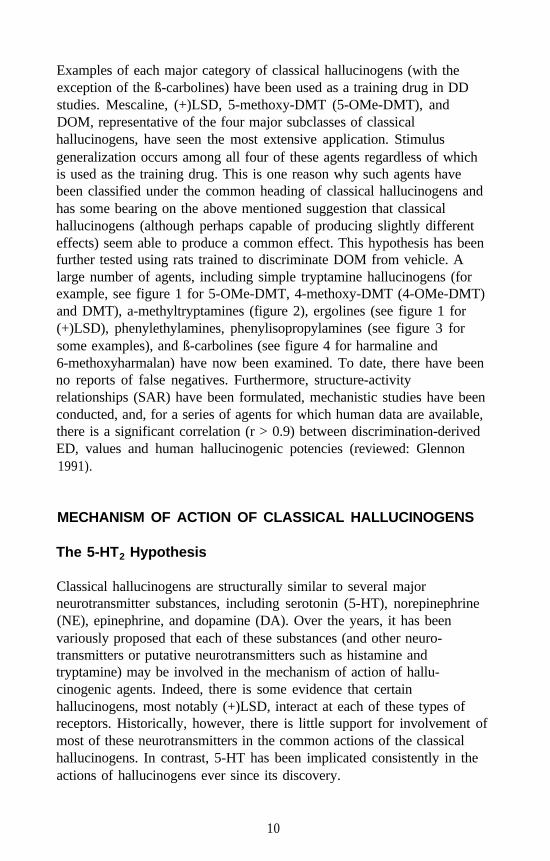
Examples of each major category of classical hallucinogens (with theexception of the ß-carbolines) have been used as a training drug in DDstudies. Mescaline, (+)LSD, 5-methoxy-DMT (5-OMe-DMT), andDOM, representative of the four major subclasses of classicalhallucinogens, have seen the most extensive application. Stimulusgeneralization occurs among all four of these agents regardless of whichis used as the training drug. This is one reason why such agents havebeen classified under the common heading of classical hallucinogens andhas some bearing on the above mentioned suggestion that classicalhallucinogens (although perhaps capable of producing slightly differenteffects) seem able to produce a common effect. This hypothesis has beenfurther tested using rats trained to discriminate DOM from vehicle. Alarge number of agents, including simple tryptamine hallucinogens (forexample, see figure 1 for 5-OMe-DMT, 4-methoxy-DMT (4-OMe-DMT)and DMT), a-methyltryptamines (figure 2), ergolines (see figure 1 for(+)LSD), phenylethylamines, phenylisopropylamines (see figure 3 forsome examples), and ß-carbolines (see figure 4 for harmaline and6-methoxyharmalan) have now been examined. To date, there have beenno reports of false negatives. Furthermore, structure-activityrelationships (SAR) have been formulated, mechanistic studies have beenconducted, and, for a series of agents for which human data are available,there is a significant correlation (r > 0.9) between discrimination-derivedED, values and human hallucinogenic potencies (reviewed: Glennon1991).
MECHANISM OF ACTION OF CLASSICAL HALLUCINOGENS
The 5-HT2 Hypothesis
Classical hallucinogens are structurally similar to several majorneurotransmitter substances, including serotonin (5-HT), norepinephrine(NE), epinephrine, and dopamine (DA). Over the years, it has beenvariously proposed that each of these substances (and other neuro-transmitters or putative neurotransmitters such as histamine andtryptamine) may be involved in the mechanism of action of hallu-cinogenic agents. Indeed, there is some evidence that certainhallucinogens, most notably (+)LSD, interact at each of these types ofreceptors. Historically, however, there is little support for involvement ofmost of these neurotransmitters in the common actions of the classicalhallucinogens. In contrast, 5-HT has been implicated consistently in theactions of hallucinogens ever since its discovery.
10
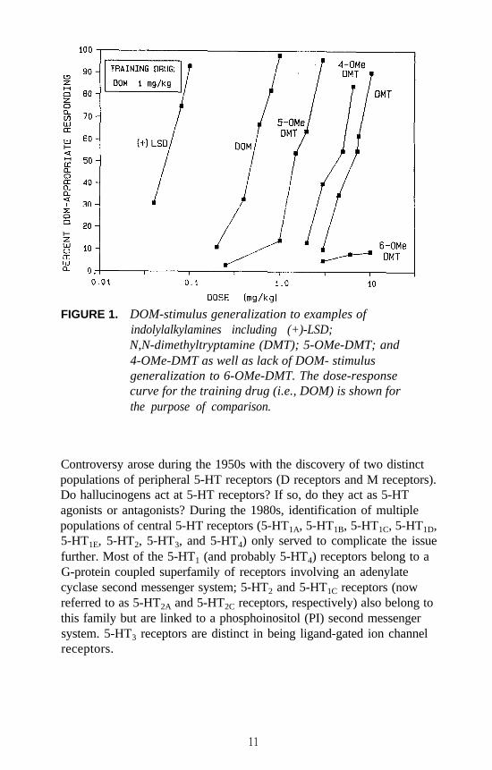
FIGURE 1. DOM-stimulus generalization to examples ofindolylalkylamines including (+)-LSD;N,N-dimethyltryptamine (DMT); 5-OMe-DMT; and4-OMe-DMT as well as lack of DOM- stimulusgeneralization to 6-OMe-DMT. The dose-responsecurve for the training drug (i.e., DOM) is shown forthe purpose of comparison.
Controversy arose during the 1950s with the discovery of two distinctpopulations of peripheral 5-HT receptors (D receptors and M receptors).Do hallucinogens act at 5-HT receptors? If so, do they act as 5-HTagonists or antagonists? During the 1980s, identification of multiplepopulations of central 5-HT receptors (5-HT1A, 5-HT1B, 5-HT1C, 5-HT1D,5-HT1E, 5-HT2, 5-HT3, and 5-HT4) only served to complicate the issuefurther. Most of the 5-HT1 (and probably 5-HT4) receptors belong to aG-protein coupled superfamily of receptors involving an adenylatecyclase second messenger system; 5-HT2 and 5-HT1C receptors (nowreferred to as 5-HT2A and 5-HT2C receptors, respectively) also belong tothis family but are linked to a phosphoinositol (PI) second messengersystem. 5-HT3 receptors are distinct in being ligand-gated ion channelreceptors.
11
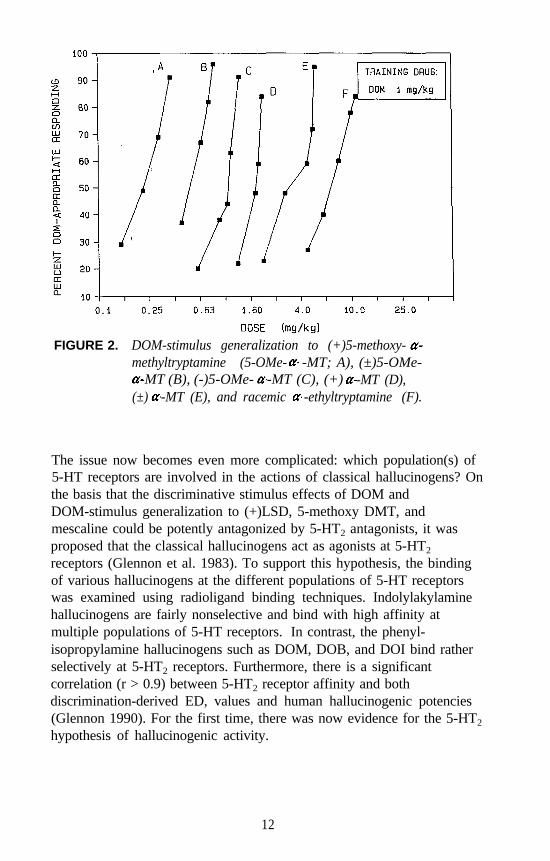
FIGURE 2. DOM-stimulus generalization to (+)5-methoxy-methyltryptamine (5-OMe- -MT; A), (±)5-OMe-
-MT (B), (-)5-OMe- -MT (C), (+) -MT (D),(±) -MT (E), and racemic -ethyltryptamine (F).
The issue now becomes even more complicated: which population(s) of5-HT receptors are involved in the actions of classical hallucinogens? Onthe basis that the discriminative stimulus effects of DOM andDOM-stimulus generalization to (+)LSD, 5-methoxy DMT, andmescaline could be potently antagonized by 5-HT2 antagonists, it wasproposed that the classical hallucinogens act as agonists at 5-HT2
receptors (Glennon et al. 1983). To support this hypothesis, the bindingof various hallucinogens at the different populations of 5-HT receptorswas examined using radioligand binding techniques. Indolylakylaminehallucinogens are fairly nonselective and bind with high affinity atmultiple populations of 5-HT receptors. In contrast, the phenyl-isopropylamine hallucinogens such as DOM, DOB, and DOI bind ratherselectively at 5-HT2 receptors. Furthermore, there is a significantcorrelation (r > 0.9) between 5-HT2 receptor affinity and bothdiscrimination-derived ED, values and human hallucinogenic potencies(Glennon 1990). For the first time, there was now evidence for the 5-HT2
hypothesis of hallucinogenic activity.
12
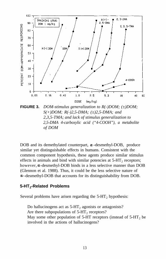
FIGURE 3. DOM-stimulus generalization to R(-)DOM; (±)DOM;S(+)DOM; R(-)2,5-DMA; (±)2,5-DMA; and2,3,5-TMA; and lack of stimulus generalization to2,5-DMA 4-carboxylic acid (“4-COOH”), a metaboliteof DOM
DOB and its demethylated counterpart, -desmethyl-DOB, producesimilar yet distinguishable effects in humans. Consistent with thecommon component hypothesis, these agents produce similar stimuluseffects in animals and bind with similar potencies at 5-HT2 receptors;however, -desmethyl-DOB binds in a less selective manner than DOB(Glennon et al. 1988). Thus, it could be the less selective nature of
-desmethyl-DOB that accounts for its distinguishability from DOB.
5-HT2-Related Problems
Several problems have arisen regarding the 5-HT2 hypothesis:
Do hallucinogens act as 5-HT2 agonists or antagonists?Are there subpopulations of 5-HT2 receptors?May some other population of 5-HT receptors (instead of 5-HT2 beinvolved in the actions of hallucinogens?
13
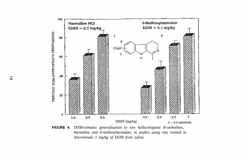
FIGURE 4. DOM-stimulus generalization to two hallucinogenic ß-carbolines,harmaline and 6-methoxyharmalan, in studies using rats trained todiscriminate 1 mg/kg of DOM from saline.
On the basis of previous DD studies, it was originally suggested thatclassical hallucinogens act as 5-HT2 agonists. However, Pierce andPeroutka (1988) recently challenged this concept and have suggested thathallucinogens, particularly (+)LSD, act as 5-HT2 antagonists. Thisagonist-versus-antagonist controversy was reexamined, and it wasconcluded that hallucinogens are not 5-HT2 antagonists; classicalhallucinogens are agonists, or at least partial agonists, at 5-HT2 receptors(Glennon 1990). Certain hallucinogens, including LSD, may possess alow intrinsic activity; thus, given in combination with a full agonist, suchagents might occasionally appear to behave as antagonists in somepharmacological assays.
Lyon and colleagues (1987) have proposed that 5-HT2 receptors exist in alow-affinity state and an agonist high-affinity state (i.e., the two-statehypothesis). [3H]Ketanserin, a 5-HT2 antagonist, labels both states of thereceptors, whereas the agonist radioligands [3H]DOB and [125I]DOIapparently label the agonist high-affinity state. Pierce and Peroutka(1989) later conducted related investigations using [77Br]DOB andproposed an alternative explanation: there exist two different populations(i.e., subpopulations) of 5-HT2 receptors (the two-site hypothesis). Theresults of recent cloning studies favor the two-state concept in that asingle 5-HT2 receptor is expressed that behaves in a manner reminiscentof a two-state receptor population (reviewed: Weinshank et al. 1992). Itmight be noted, however, that the possibility of two different(overlapping) binding domains has not yet been excluded; that is,agonists and antagonists may bind in a slightly different manner at thesame population (or state) of 5-HT2 receptors.
Finally, there is the issue of involvement of other (or additional)populations of 5-HT receptors in the actions of hallucinogens. This isdiscussed below.
Involvement of 5-HT1C Receptors
Shortly after the 5-HT2 hypothesis was proposed (Glennon et al. 1983,1984), Pazos and coworkers (1984) described their discovery of 5-HT1C
receptors. The binding of hallucinogens at these receptors wassubsequently examined, and little difference between their 5-HT2 and5-HT1C affinities was found (Titeler et al. 1988); indeed, later studieshave shown less than a tenfold difference in receptor affinity for a largeseries of phenylalkylamine derivatives (Glennon et al. 1992). As with5-HT2 receptor affinities, 5-HT1C affinities also are correlated both with
15
discrimination-derived ED, values and human hallucinogenic potencies.In addition, Burris and Sanders-Bush (1988) reported that DOM acts as a5-HT1C agonist. Furthermore the 5-HT2 hypothesis was based, in part,on the finding that 5-HT2 antagonists (such as ketanserin andpirenperone) antagonize the stimulus effects of hallucinogens. It is nowrecognized that these 5-HT2 antagonists are described more accurately as5-HT2 and 5-HT1C antagonists. Thus, the likelihood exists that 5-HT2
and/or 5-HT1C receptors are involved in the actions of hallucinogenicagents. It may be this interaction that constitutes the common “key”mentioned at the beginning of this chapter, and it may be this commoninteraction that allows animals to reliably discriminate classicalhallucinogens from the vehicle.
It is quite difficult to ascribe a specific role for 5-HT1C versus 5-HT2
receptors in the mechanism of action of hallucinogens due to the lack ofagents that display selectivity for one of these populations of receptorsover the other. Nearly all agents that bind at 5-HT2 receptors bind at5-HT1C receptors. However, there are a few agents that might offer somehope in resolving this problem. The DA 5-HT1A antagonist spiperonebinds with approximately 500-fold selectivity for 5-HT2 versus 5-HT1C
receptors. An attempt was made to antagonize the stimulus effects ofDOM using various doses of spiperone with the intention that it might bemore difficult to antagonize the DOM stimulus if the stimulus was5-HT1C mediated. Unfortunately, the results of these studies wereinconclusive due to the severe disruptive effects of low doses ofspiperone in combination with DOM (Glennon 1991).
Another agent of interest is l-(3-trifluoromethylphenyl)piperazine(TFMPP). Although TFMPP binds at multiple populations of 5-HTreceptors, evidence suggests that TFMPP is a 5-HT1C agonist but a 5-HT2
antagonist (or, at best, a 5-HT2 partial agonist). Administration ofTFMPP to animals trained to discriminate DOM from saline failed toresult in stimulus generalization. In a parallel study, administration ofDOM (or DOI) to animals trained to discriminate TFMPP from vehicleresulted in only partial generalization followed at slightly higher DOM(or DOI) doses by disruption of behavior. Thus, the results were againinconclusive. In a third series of studies, rats trained to discriminate0.5 milligrams per kilogram (mg/kg) of TFMPP from the vehicle wereadministered doses of DOM in combination with 0.2 mg/kg of TFMPP(ED, dose = 0.17 mg/kg). The rationale for this investigation was thatlower (i.e., nondisruptive) doses of DOM should potentiate the effect ofthe near-ED, dose of TFMPP if both agents act via a common
16
mechanism. The results (shown in figure 5) were somewhat surprising inthat low doses of DOM, rather than potentiating the effect, actuallyantagonized the effect of TFMPP. For all practical purposes, the resultsof this study also were inconclusive; however, they suggest that DOMand TFMPP are likely producing their stimulus effects via differentmechanisms.
There is one additional piece of information that perhaps has somebearing on the 5-HT2 versus 5-HT1C controversy. Several years ago,Glennon and Hauck (1985) reported that the DOM stimulus generalizesto lisuride. This was a rather unexpected finding. Subsequently, theauthor and coworkers reevaluated lisuride as a potential DOM antagonistand found that it attenuates the DOM stimulus by 50 percent at very lowdoses (i.e., at one-sixtieth of the dose that results in stimulusgeneralization). This led to speculation that lisuride may be acting as apartial agonist (Glennon 1991). Sanders-Bush has recently demonstrated(this volume) that, whereas lisuride is a pure 5-HT1C antagonist, itbehaves as a partial agonist at 5-HT2 receptors. These results areconsistent with the present DD studies. Thus, although hallucinogensunquestionably bind at both populations of receptors and whereas amechanistic role for 5-HT1C receptors cannot yet be eliminated, it wouldappear on the basis of all the above mentioned studies that the DOMstimulus involves primarily a 5-HT2 mechanism.
Before leaving the topic of 5-HT2 and 5-HT1C receptors, it might bementioned that certain of the animal models described earlier appear toinvolve actions mediated by these receptors. For example, thehead-twitch response has been proposed to involve such a mechanism(Glennon 1992). In retrospect, some of these models may be lessfarfetched and more mechanistically relevant than once suspected.
Involvement of Other 5-HT Receptors
It was recently reported that there may be functional interactions betweendifferent populations of 5-HT receptors such that action at one maymodulate activation of another (reviewed: Glennon et al. 1991b). Thus,interaction of an agonist at one population of 5-HT receptors maymodulate the effect of the interaction of a second agonist at a differentpopulation of receptors. This could have far-reaching consequences. Forexample, what is the effect of a nonselective agonist that interacts at morethan one population of receptors at the same time? What about anonselective agent that is an agonist at one population and an antagonist
17
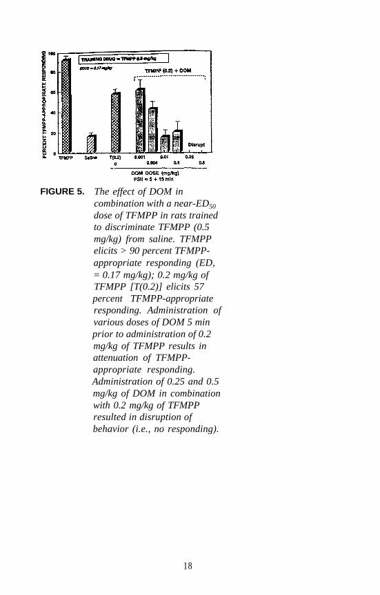
FIGURE 5. The effect of DOM incombination with a near-ED50
dose of TFMPP in rats trainedto discriminate TFMPP (0.5mg/kg) from saline. TFMPPelicits > 90 percent TFMPP-appropriate responding (ED,= 0.17 mg/kg); 0.2 mg/kg ofTFMPP [T(0.2)] elicits 57percent TFMPP-appropriateresponding. Administration ofvarious doses of DOM 5 minprior to administration of 0.2mg/kg of TFMPP results inattenuation of TFMPP-appropriate responding.Administration of 0.25 and 0.5mg/kg of DOM in combinationwith 0.2 mg/kg of TFMPPresulted in disruption ofbehavior (i.e., no responding).
18
at another? Experiments necessary to sort out these types of interactionscould be rather labor intensive and their interpretation quite complicated.Worse yet are cases where such types of interactions are possible butunrecognized.
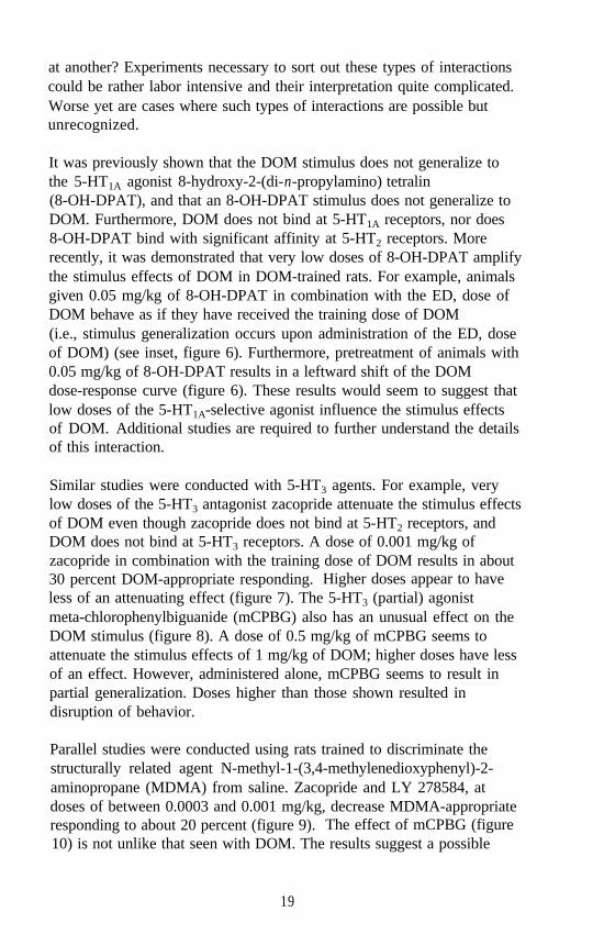
It was previously shown that the DOM stimulus does not generalize tothe 5-HT1A agonist 8-hydroxy-2-(di-n-propylamino) tetralin(8-OH-DPAT), and that an 8-OH-DPAT stimulus does not generalize toDOM. Furthermore, DOM does not bind at 5-HT1A receptors, nor does8-OH-DPAT bind with significant affinity at 5-HT2 receptors. Morerecently, it was demonstrated that very low doses of 8-OH-DPAT amplifythe stimulus effects of DOM in DOM-trained rats. For example, animalsgiven 0.05 mg/kg of 8-OH-DPAT in combination with the ED, dose ofDOM behave as if they have received the training dose of DOM(i.e., stimulus generalization occurs upon administration of the ED, doseof DOM) (see inset, figure 6). Furthermore, pretreatment of animals with0.05 mg/kg of 8-OH-DPAT results in a leftward shift of the DOMdose-response curve (figure 6). These results would seem to suggest thatlow doses of the 5-HT1A-selective agonist influence the stimulus effectsof DOM. Additional studies are required to further understand the detailsof this interaction.
Similar studies were conducted with 5-HT3 agents. For example, verylow doses of the 5-HT3 antagonist zacopride attenuate the stimulus effectsof DOM even though zacopride does not bind at 5-HT2 receptors, andDOM does not bind at 5-HT3 receptors. A dose of 0.001 mg/kg ofzacopride in combination with the training dose of DOM results in about30 percent DOM-appropriate responding. Higher doses appear to haveless of an attenuating effect (figure 7). The 5-HT3 (partial) agonistmeta-chlorophenylbiguanide (mCPBG) also has an unusual effect on theDOM stimulus (figure 8). A dose of 0.5 mg/kg of mCPBG seems toattenuate the stimulus effects of 1 mg/kg of DOM; higher doses have lessof an effect. However, administered alone, mCPBG seems to result inpartial generalization. Doses higher than those shown resulted indisruption of behavior.
Parallel studies were conducted using rats trained to discriminate thestructurally related agent N-methyl-1-(3,4-methylenedioxyphenyl)-2-aminopropane (MDMA) from saline. Zacopride and LY 278584, atdoses of between 0.0003 and 0.001 mg/kg, decrease MDMA-appropriateresponding to about 20 percent (figure 9). The effect of mCPBG (figure10) is not unlike that seen with DOM. The results suggest a possible
19
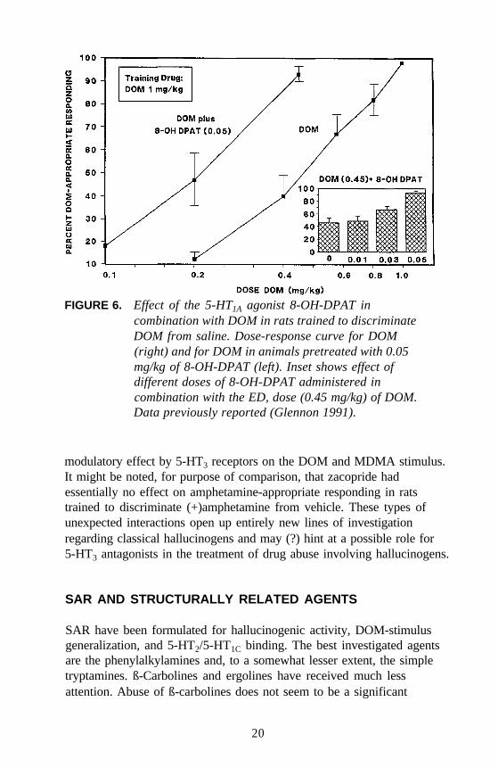
FIGURE 6. Effect of the 5-HT1A agonist 8-OH-DPAT incombination with DOM in rats trained to discriminateDOM from saline. Dose-response curve for DOM(right) and for DOM in animals pretreated with 0.05mg/kg of 8-OH-DPAT (left). Inset shows effect ofdifferent doses of 8-OH-DPAT administered incombination with the ED, dose (0.45 mg/kg) of DOM.Data previously reported (Glennon 1991).
modulatory effect by 5-HT3 receptors on the DOM and MDMA stimulus.It might be noted, for purpose of comparison, that zacopride hadessentially no effect on amphetamine-appropriate responding in ratstrained to discriminate (+)amphetamine from vehicle. These types ofunexpected interactions open up entirely new lines of investigationregarding classical hallucinogens and may (?) hint at a possible role for5-HT3 antagonists in the treatment of drug abuse involving hallucinogens.
SAR AND STRUCTURALLY RELATED AGENTS
SAR have been formulated for hallucinogenic activity, DOM-stimulusgeneralization, and 5-HT2/5-HT1C binding. The best investigated agentsare the phenylalkylamines and, to a somewhat lesser extent, the simpletryptamines. ß-Carbolines and ergolines have received much lessattention. Abuse of ß-carbolines does not seem to be a significant
20
FIGURE 7. Effect of the 5-HT3 antagonist zacopride administeredin combination with the training dose of DOM to ratstrained to discriminate 1 mg/kg of DOM from saline.
problem, and it is perhaps this reason that accounts for the lack of interestor urgency to study these agents. Ergolines, on the other hand, can offer,a significant synthetic challenge and relatively few agents are readilyavailable. The SAR of classical hallucinogens has been reviewed(Nichols and Glennon 1984).
Many investigations of classical hallucinogens are limited to a smallhandful of standard agents (e.g., LSD, mescaline, DOM). Far fewerstudies have examined some of the more novel or structurally distinctagents, or have examined series of agents. It would seem prudent toexamine additional agents and structurally related analogs in order todefine exactly what structural features contribute to activity. A classicexample is the phenylisopropylamine amphetamine. The amphetaminestructural backbone is contained in, for example, the hallucinogen DOMand the designer drug MDMA; and yet each of these three agentsproduces effects in animals and humans that are clearly distinguishablefrom one another (Shulgin and Shulgin 1991). As the structure of one ofthese agents is gradually modified to one of the others, at what point does
21
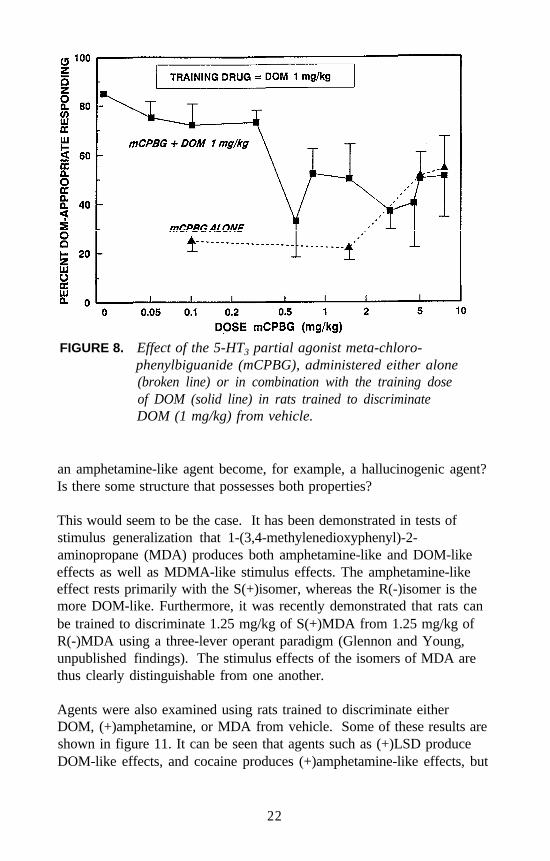
FIGURE 8. Effect of the 5-HT3 partial agonist meta-chloro-phenylbiguanide (mCPBG), administered either alone(broken line) or in combination with the training doseof DOM (solid line) in rats trained to discriminateDOM (1 mg/kg) from vehicle.
an amphetamine-like agent become, for example, a hallucinogenic agent?Is there some structure that possesses both properties?
This would seem to be the case. It has been demonstrated in tests ofstimulus generalization that 1-(3,4-methylenedioxyphenyl)-2-aminopropane (MDA) produces both amphetamine-like and DOM-likeeffects as well as MDMA-like stimulus effects. The amphetamine-likeeffect rests primarily with the S(+)isomer, whereas the R(-)isomer is themore DOM-like. Furthermore, it was recently demonstrated that rats canbe trained to discriminate 1.25 mg/kg of S(+)MDA from 1.25 mg/kg ofR(-)MDA using a three-lever operant paradigm (Glennon and Young,unpublished findings). The stimulus effects of the isomers of MDA arethus clearly distinguishable from one another.
Agents were also examined using rats trained to discriminate eitherDOM, (+)amphetamine, or MDA from vehicle. Some of these results areshown in figure 11. It can be seen that agents such as (+)LSD produceDOM-like effects, and cocaine produces (+)amphetamine-like effects, but
22
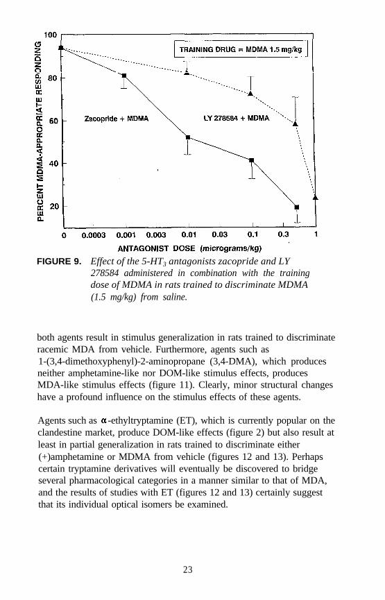
FIGURE 9. Effect of the 5-HT3 antagonists zacopride and LY278584 administered in combination with the trainingdose of MDMA in rats trained to discriminate MDMA(1.5 mg/kg) from saline.
both agents result in stimulus generalization in rats trained to discriminateracemic MDA from vehicle. Furthermore, agents such as1-(3,4-dimethoxyphenyl)-2-aminopropane (3,4-DMA), which producesneither amphetamine-like nor DOM-like stimulus effects, producesMDA-like stimulus effects (figure 11). Clearly, minor structural changeshave a profound influence on the stimulus effects of these agents.
Agents such as -ethyltryptamine (ET), which is currently popular on theclandestine market, produce DOM-like effects (figure 2) but also result atleast in partial generalization in rats trained to discriminate either(+)amphetamine or MDMA from vehicle (figures 12 and 13). Perhapscertain tryptamine derivatives will eventually be discovered to bridgeseveral pharmacological categories in a manner similar to that of MDA,and the results of studies with ET (figures 12 and 13) certainly suggestthat its individual optical isomers be examined.
23
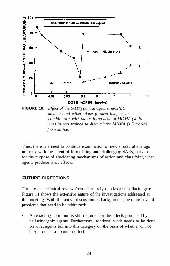
FIGURE 10. Effect of the 5-HT3 partial agonist mCPBGadministered either alone (broken line) or incombination with the training dose of MDMA (solidline) in rats trained to discriminate MDMA (1.5 mg/kg)from saline.
Thus, there is a need to continue examination of new structural analogsnot only with the intent of formulating and challenging SARs, but alsofor the purpose of elucidating mechanisms of action and classifying whatagents produce what effects.
FUTURE DIRECTIONS
The present technical review focused entirely on classical hallucinogens.Figure 14 shows the extensive nature of the investigations addressed atthis meeting. With the above discussion as background, there are severalproblems that need to be addressed:
An exacting definition is still required for the effects produced byhallucinogenic agents. Furthermore, addtional work needs to be doneon what agents fall into this category on the basis of whether or notthey produce a common effect.
24
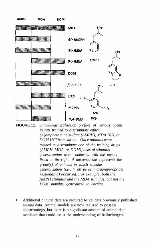
FIGURE 11. Stimulus-generalization profiles of various agentsin rats trained to discriminate either(+)amphetamine sulfate (AMPH), MDA HCl, orDOM HCl from saline. Once animals weretrained to discriminate one of the training drugs(AMPH, MDA, or DOM), tests of stimulusgeneralization were conducted with the agentslisted on the right. A darkened bar represents thegroup(s) of animals in which stimulusgeneralization (i.e., > 80 percent drug-appropriateresponding) occurred. For example, both theAMPH stimulus and the MDA stimulus, but not theDOM stimulus, generalized to cocaine.
Additional clinical data are required to validate previously publishedanimal data. Animal models are now realized to possessshortcomings, but there is a significant amount of animal dataavailable that could assist the understanding of hallucinogens.
25
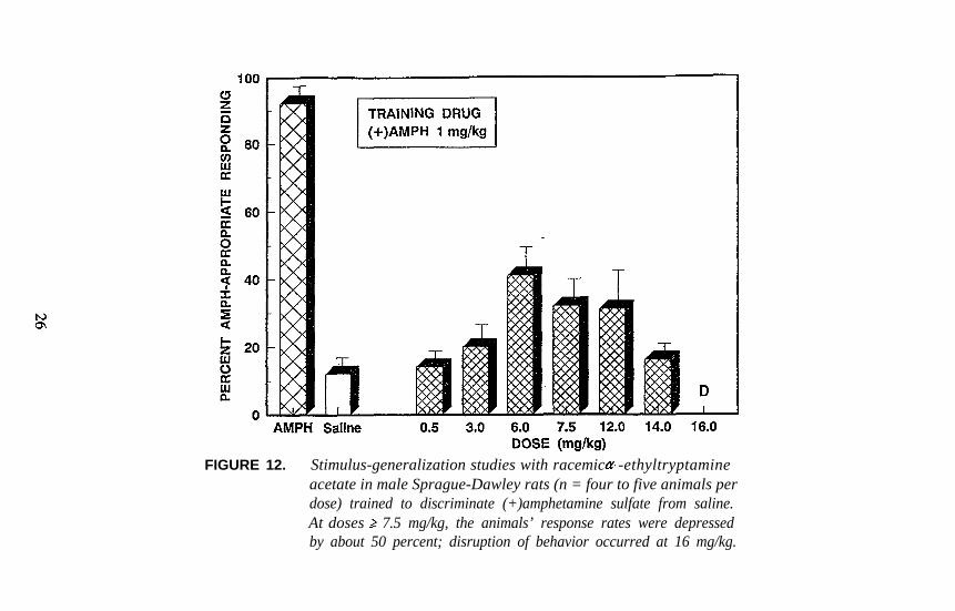
FIGURE 12. Stimulus-generalization studies with racemic -ethyltryptamineacetate in male Sprague-Dawley rats (n = four to five animals perdose) trained to discriminate (+)amphetamine sulfate from saline.At doses 7.5 mg/kg, the animals’ response rates were depressedby about 50 percent; disruption of behavior occurred at 16 mg/kg.
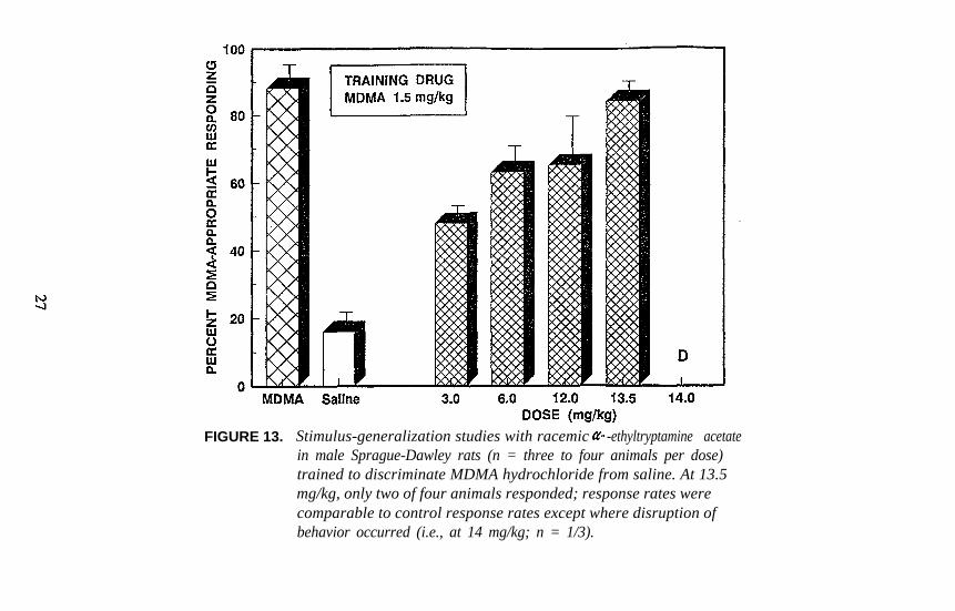
FIGURE 13. Stimulus-generalization studies with racemic -ethyltryptamine acetatein male Sprague-Dawley rats (n = three to four animals per dose)trained to discriminate MDMA hydrochloride from saline. At 13.5mg/kg, only two of four animals responded; response rates werecomparable to control response rates except where disruption ofbehavior occurred (i.e., at 14 mg/kg; n = 1/3).
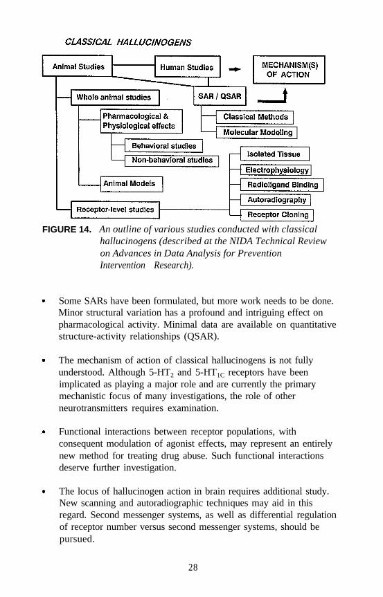
FIGURE 14. An outline of various studies conducted with classicalhallucinogens (described at the NIDA Technical Reviewon Advances in Data Analysis for PreventionIntervention Research).
Some SARs have been formulated, but more work needs to be done.Minor structural variation has a profound and intriguing effect onpharmacological activity. Minimal data are available on quantitativestructure-activity relationships (QSAR).
The mechanism of action of classical hallucinogens is not fullyunderstood. Although 5-HT2 and 5-HT1C receptors have beenimplicated as playing a major role and are currently the primarymechanistic focus of many investigations, the role of otherneurotransmitters requires examination.
Functional interactions between receptor populations, withconsequent modulation of agonist effects, may represent an entirelynew method for treating drug abuse. Such functional interactionsdeserve further investigation.
The locus of hallucinogen action in brain requires additional study.New scanning and autoradiographic techniques may aid in thisregard. Second messenger systems, as well as differential regulationof receptor number versus second messenger systems, should bepursued.
28
Is there a prototypic classical hallucinogen? Many investigationswith hallucinogens involve the same small number of agents, inparticular LSD. Should LSD be considered a prototype agent? Or isit possible that investigation of one or two agents in great depth maylead researchers astray by providing information that is unique to aspecific agent, rather than information that may be more germane toclassical hallucinogens as a group? The same may be said for animalmodels. Perhaps future studies should not rely solely oninvestigating the same small number of standard agents nor rely onlyon a few pharmacological test procedures.
NOTE
Since the original submission of this manuscript, the terms “5-HT2 and5-HT1C receptors” have been replaced by “5-HT2A and 5-HT2C receptors,”respectively.
REFERENCES
Burris, K.D., and Sanders-Bush, E. Hallucinogens directly activateserotonin 5-HT1C receptors in choroid plexus. Soc Neurosci Abstr14:553,1988.
Glennon, R.A. Hallucinogenic phenylisopropylamines: Stereochemicalaspects. In: Smith, D.F., ed. Handbook of Stereoisomers: Drugs inPsychopharmacology. Boca Raton, FL: CRC Press, 1984. pp.327-368.
Glennon, R.A. Do hallucinogens act as 5-HT2 agonists or antagonists?Neuropsychopharmacology 56:509-517, 1990.
Glennon, R.A. Discriminative stimulus properties of hallucinogens andrelated designer drugs. In: Glennon, R.A.; Jarbe, T.; and Frankenheim,J., eds. Drug Discrimination: Applications to Drug Abuse Research.National Institute on Drug Abuse Research Monograph No. 116.DHHS Pub. No. (ADM)92-1878. Washington, DC: Supt. of Docs.,U.S. Govt. Print. Off., 1991. pp. 25-44.
Glennon, R.A. Animal models for assessing hallucinogenic agents. In:Boulton, A.A.; Baker, G.B.; and Wu, P., ed. Animal Models of DrugAddiction. Clifton, NJ: Humana Press, 1992. pp. 345-386.
Glennon, R.A., and Hauck, A.E. Mechanistic studies on DOM as adiscriminative stimulus. Pharmacol Biochem Behav 23:937-941,1985.
29
Glennon, R.A.; Young, R.; and Rosecrans, J.A. Antagonism of thestimulus effects of the hallucinogen DOM and the purported serotoninagonist quipazine by 5-HT2 antagonists. Eur J Pharmacol 91:189-192, 1983.
Glennon, R.A.; Titeler, M.; and McKenney, J.D. Evidence for theinvolvement of 5-HT2 receptors in the mechanism of action ofhallucinogenic agents. Life Sci 35:2505-2511, 1984.
Glennon, R.A.; Titeler, M.; and Lyon, R.A. A preliminary investigationof the psychoactive agent 4-bromo-2,5-dimethoxyphenylethylamine:A potential drug of abuse. Pharmacol Biochem Behav 30:597-601,1988.
Glennon, R.A.; Jarbe, T.; and Frankenheim, J., eds. Drug Discrimination:Applications to Drug Abuse Research. National Institute on DrugAbuse Research Monograph No. 116. DHHS Pub. No.(ADM)92-1878. Washington, DC: Supt. of Docs., U.S. Govt. Print.Off., 1991a.
Glennon, R.A.; Darmani, N.A.; and Martin, B.R. Multiple populations ofserotonin receptors may modulate the behavioral effects ofserotonergic agents. Life Sci 48:2493-2498, 1991b .
Glennon, R.A.; Raghupathi, R.; Bartyzel, P.; Teitler, M.; and Leonhardt,S. Binding of phenylalkylamine derivatives at 5-HT1C and 5-HT2
serotonin receptors: Evidence for a lack of selectivity. J Med Chem35:734-740, 1992.
Hollister, L.E. Chemical Psychoses. Springfield, IL: Charles C. Thomas,1968. pp. 17-18.
Jacobs, B.L., and Trulson, M.E. An animal behavioral model for studyingthe actions of LSD and related hallucinogens. In: Stillman, R.C., andWillette, R.E., eds. The Psychopharmacology of Hallucinogens. NewYork: Pergamon Press, 1978. pp. 301-314.
Kier, L.B., and Glennon, R.A. Progress with several models for the studyof SAR of hallucinogenic agents. In: Barnett, G.; Trsic, M.; andWillette, R.E., eds. QuaSAR: Quantitative Structure ActivityRelationships of Analgesics, Narcotic Antagonists, and Hallucinogens.National Institute on Drug Abuse Research Monograph No. 22.DHHS Pub. No. (ADM)78-729. Washington, DC: Supt. of Docs.,U.S. Govt. Print. Off., 1978. pp 159-185.
Lyon, R.A.; Davis, K.H.; and Titeler, M. [3H]DOB(4-bromo-2,5-dimethoxyphenylisopropyl-amine) labels a guanylnucleotide-sensitive state of cortical 5-HT2 receptors. Mol Pharmacol31:194-199, 1987.
Naranjo, C. The Healing Journey. New York: Pantheon Books, 1973.
30
Nichols, D.E., and Glennon, R.A. Medicinal chemistry andstructure-activity relationships of hallucinogens. In: Jacobs, B.L., ed.Hallucinogens: Neurochemical, Behavioral, and ClinicalPerspectives. New York: Raven Press, 1984. pp. 95-142.
Otis, L.S.; Pryor, G.T.; Marquis, W.J.; Jensen, R.; and Petersen, K.Preclinical identification of hallucinogenic compounds. In: Stillman,R.C., and Willette, R.E., eds. The Psychopharmacology ofHallucinogens. New York: Pergamon Press, 1978. pp. 126-149.
Pazos, A.; Hoyer, D.; and Palacios, J.M. Binding of serotonergic ligandsto the porcine choroid plexus. Characterization of a new type of 5-HTrecognition site. Eur J Pharmacol 106:539-546, 1984.
Pierce, P.A., and Peroutka, S.J. Antagonism of 5-hydroxytryptamine-2receptor-mediated phosphatidylinositol turnover by d-lysergic aciddiethylamide. J Pharmacol Exp Ther 247:918-925, 1988.
Pierce, P.A., and Peroutka, S.J. Evidence for distinct5-hydroxytryptamine-2 receptor binding site subtypes in corticalmembrane preparations. J Neurochem 52:656-658, 1989.
Shulgin, A.T., and Shulgin, A. PIHKAL: A Chemical Love Story.Berkeley, CA: Transform Press, 1991. p. xxi.
Stoff, D.M.; Gillin, J.C.; and Wyatt, R.J. Animal models of drug-inducedhallucinations. In: Stillman, R.C., and Willette, R.E., eds. ThePsychopharmacology of Hallucinogens. New York: Pergamon Press,1978. pp. 259-267.
Titeler, M.; Lyon, R.A.; and Glennon, R.A. Radioligand bindingevidence implicates the brain 5-HT2 receptors as a site of action forLSD and phenylisopropylamine hallucinogens. Psychopharmacology94:213-216, 1988.
Weinshank, R.L.; Adham, N.; Zgombick, J.; Bard, J.; Branchek, T., andHartig, P.R. Molecular analysis of serotonin receptor subtypes. In:Langer, S.Z.; Brunello, N.; Racagni, G.; and Mendlewicz, J., eds.Serotonin Receptor Subtypes: Pharmacological Significance andClinical Implications. Basel: Karger, 1992. pp. 1-12.
Westkaemper, R.B., and Glennon, R.A. Approaches to molecularmodeling studies and specific application to serotonin ligands andreceptors. Pharmacol Biochem Behav 40:1019-1030, 1991.
ACKNOWLEDGMENTS
The author’s laboratory work was supported in part by PHS grantDA 01642. The contribution of Rodney Higgs, responsible for some ofthe drug discrimination studies reported here, is gratefully acknowledged.
31

AUTHOR
Richard A. Glennon, Ph.D.Professor of Medicinal ChemistryDepartment of Medicinal ChemistrySchool of Pharmacy, Box 980540Medical College of VirginiaVirginia Commonwealth UniversityRichmond, VA 23298-0540
32
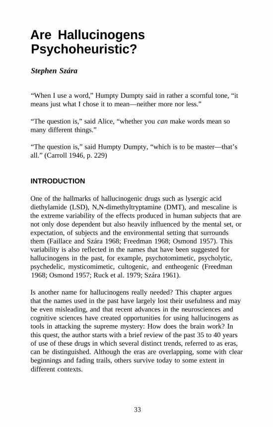
Are HallucinogensPsychoheuristic?
Stephen Szára
“When I use a word,” Humpty Dumpty said in rather a scornful tone, “itmeans just what I chose it to mean—neither more nor less.”
“The question is,” said Alice, “whether you can make words mean somany different things.”
“The question is,” said Humpty Dumpty, “which is to be master—that’sall.” (Carroll 1946, p. 229)
INTRODUCTION
One of the hallmarks of hallucinogenic drugs such as lysergic aciddiethylamide (LSD), N,N-dimethyltryptamine (DMT), and mescaline isthe extreme variability of the effects produced in human subjects that arenot only dose dependent but also heavily influenced by the mental set, orexpectation, of subjects and the environmental setting that surroundsthem (Faillace and Szára 1968; Freedman 1968; Osmond 1957). Thisvariability is also reflected in the names that have been suggested forhallucinogens in the past, for example, psychotomimetic, psycholytic,psychedelic, mysticomimetic, cultogenic, and entheogenic (Freedman1968; Osmond 1957; Ruck et al. 1979; Szára 1961).
Is another name for hallucinogens really needed? This chapter arguesthat the names used in the past have largely lost their usefulness and maybe even misleading, and that recent advances in the neurosciences andcognitive sciences have created opportunities for using hallucinogens astools in attacking the supreme mystery: How does the brain work? Inthis quest, the author starts with a brief review of the past 35 to 40 yearsof use of these drugs in which several distinct trends, referred to as eras,can be distinguished. Although the eras are overlapping, some with clearbeginnings and fading trails, others survive today to some extent indifferent contexts.
33
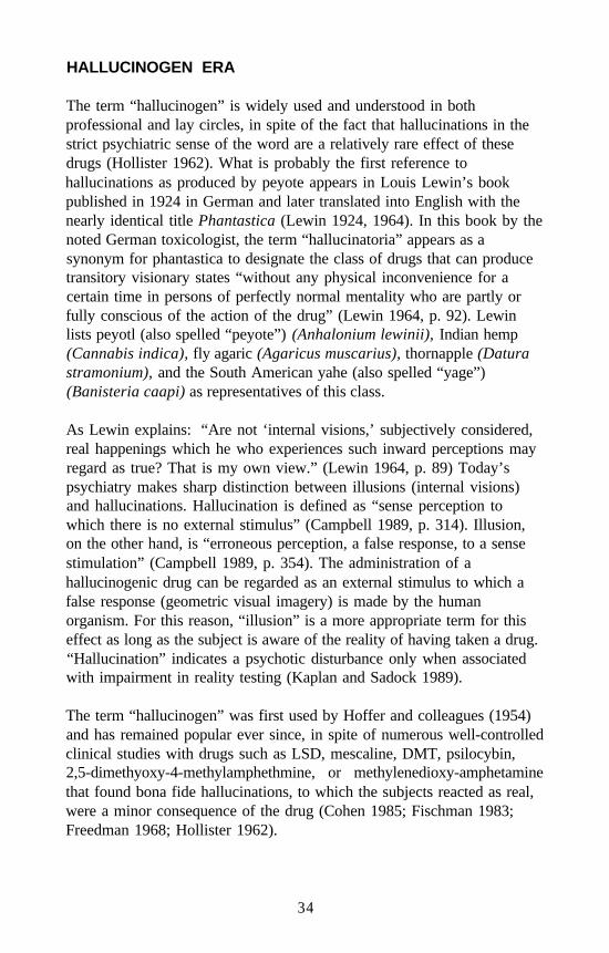
HALLUCINOGEN ERA
The term “hallucinogen” is widely used and understood in bothprofessional and lay circles, in spite of the fact that hallucinations in thestrict psychiatric sense of the word are a relatively rare effect of thesedrugs (Hollister 1962). What is probably the first reference tohallucinations as produced by peyote appears in Louis Lewin’s bookpublished in 1924 in German and later translated into English with thenearly identical title Phantastica (Lewin 1924, 1964). In this book by thenoted German toxicologist, the term “hallucinatoria” appears as asynonym for phantastica to designate the class of drugs that can producetransitory visionary states “without any physical inconvenience for acertain time in persons of perfectly normal mentality who are partly orfully conscious of the action of the drug” (Lewin 1964, p. 92). Lewinlists peyotl (also spelled “peyote”) (Anhalonium lewinii), Indian hemp(Cannabis indica), fly agaric (Agaricus muscarius), thornapple (Daturastramonium), and the South American yahe (also spelled “yage”)(Banisteria caapi) as representatives of this class.
As Lewin explains: “Are not ‘internal visions,’ subjectively considered,real happenings which he who experiences such inward perceptions mayregard as true? That is my own view.” (Lewin 1964, p. 89) Today’spsychiatry makes sharp distinction between illusions (internal visions)and hallucinations. Hallucination is defined as “sense perception towhich there is no external stimulus” (Campbell 1989, p. 314). Illusion,on the other hand, is “erroneous perception, a false response, to a sensestimulation” (Campbell 1989, p. 354). The administration of ahallucinogenic drug can be regarded as an external stimulus to which afalse response (geometric visual imagery) is made by the humanorganism. For this reason, “illusion” is a more appropriate term for thiseffect as long as the subject is aware of the reality of having taken a drug.“Hallucination” indicates a psychotic disturbance only when associatedwith impairment in reality testing (Kaplan and Sadock 1989).
The term “hallucinogen” was first used by Hoffer and colleagues (1954)and has remained popular ever since, in spite of numerous well-controlledclinical studies with drugs such as LSD, mescaline, DMT, psilocybin,2,5-dimethyoxy-4-methylamphethmine, or methylenedioxy-amphetaminethat found bona fide hallucinations, to which the subjects reacted as real,were a minor consequence of the drug (Cohen 1985; Fischman 1983;Freedman 1968; Hollister 1962).
34
The report by Hoffer and colleagues (1954) is considered by many as thestart of a new era in psychiatric research, taking the suggestion seriouslythat these drugs reproduce, in normal subjects, some symptoms ofschizophrenia or similar psychoses. The drugs, therefore, arepsychotomimetic; the terms “psychosomimetic” and “psychotogenic” arealso used in this sense. During the mid-1950s, chlorpromazine was madeavailable to treat psychotic patients. Serotonin, norepinephrine (NE), andgamma aminobutyric acid (GABA) were found in synapses in the brain.Reports started to appear implicating the action of these drugs onsynapses as the most likely mechanism of psychoactivity (for a review,see Cooper et al. 1974). Psychopharmacology as a discipline was born.There was much excitement, and expectations, among psychiatrists thattheir profession might finally become scientifically based, and that theknowledge gained would help to develop more effective treatment fortheir patients.
PSYCHOTHERAPEUTIC ERA
The psychotomimetic era for hallucinogens gradually gave way to aseemingly perverse movement that claimed that hallucinogens couldactually help certain psychiatric patients and advocated apsychotherapeutic use of these drugs. The justification was provided bysome of the unique and peculiar effects seen and/or experienced in certainsituations, such as the loss of ego boundaries and regression to a moreprimitive, childlike functioning of the ego that seems to facilitate therecall of early childhood memories that have been forgotten or repressed.These effects are utilized by the psycholytic approach to the treatment ofchronic alcoholism. It was rationalized that abolishing the distinctionbetween subject and object (ego boundary) and conscious andunconscious self (regression) would cause a lessening of alienation fromthe world, a rediscovery of the self, and a learning of a new set of values;thus a new beginning could be achieved (Savage et al. 1962). Combiningthis approach with hypnosis gave rise to the hypnodelic strategy for thesame purpose. The therapeutic results, however, only lasted for a fewmonths at best, and longterm followup indicated relapse of drinkingbehavior to essentially pretreatment levels (Faillace 1966; Faillace et al.1970; Levine and Ludwig 1965).
35
PSYCHEDELIC ERA
Another peculiar effect of these drugs is a dramatic change in perception:it appears to the person as if the eyes (the “doors of perception”) havebeen cleansed and the person could see the world as new in all respects—“as Adam may have seen it on the day of creation” as Aldous Huxley(1954, p. 17) pointed out in his popular and influential book. This newreality is perceived and interpreted by some individuals as manifestationof the true nature of their mind; hence, the term “psychedelic” wassuggested by Osmond (1957). This interpretation has been embraced notonly by professional therapists but also by some segments of the public,and gave rise to the “Summer of Love” in San Francisco in 1967 withfree distribution of LSD. This perception resulted in the formation ofnumerous cults, communes, and drug-oriented religious groups(Freedman 1968), permeated the lyrics and style of popular music (acidrock), and was viewed by some as one of the contributing sources of theoccasional resurgence of popularity of illegal drug use (Cohen 1966,Szára 1968).
BEHAVIORISTIC ERA
In a review of the clinical use of psychotomimetic drugs, Faillace referredto a group of investigators as “behaviorists...for lack of a better term”(1966, p. 15). This group, he said, “is not principally interested intreatment but is trying objectively to determine the actions of these drugs.A great many investigators from many divergent disciplines are includedin this group” (Faillace 1966, p. 16). He then cited four groups asexamples.
1. Hoch and coworkers in New York explored the effects of LSD,mescaline, and other similar drugs on psychotic patients andconcluded that these drugs aggravate schizophrenic symptoms. Thegroup showed that the drugs brought forth the same type ofpsychodynamic material in their patients and there was nothingparticularly specific for any of these drugs.
2. Isbell and coworkers at the Addiction Research Center, then inLexington, KY, conducted a series of investigations on formernarcotic addicts. They observed rapid development of tolerance tothe effects of LSD and also showed the development of cross-tolerance between LSD and psilocybin. This group demonstrated the
36
feasibility of obtaining good dose-response relationships utilizing anarray of physiological tests (e.g., pupil size, blood pressure). Thegroup developed a standardized questionnaire, the AddictionResearch Center Inventory (ARCI), that became widely accepted andis used for assessing the subjective effects of psychoactive drugs in aquantitative fashion.
3. Delay and coworkers in Paris carried out an intensive study ofpsilocybin and showed that the phosphoryl group does not contributeto its psychoactive effects because the dephosphorylated derivative,psilocin, has equal potency in humans. They observed anintensification of psychotic and psychoneurotic symptoms in 90mental patients and 47 so-called normals, and suggested thatpsilocybin may offer diagnostic possibilities in difficult clinical cases.
4. Faillace referred to the work of the author and colleagues at SaintElizabeth’s Hospital in Washington, DC, as the last example ofnontherapeutically oriented clinical research with hallucinogenicdrugs. This work focused primarily on tryptamine derivatives suchas DMT. In the course of investigation of the metabolism of thesecompounds that included the N,N-diethyl- and N,N-dipropyl-derivatives of tryptamine (DET and DPT, respectively), theconclusion was reached that 6-hydroxylation of the indole ring mightbe an important biological mechanism for the psychoactivity of thesecompounds. To provide further evidence, the Saint Elizabeth’sHospital laboratories synthesized a number of derivatives of thesecompounds that were blocked by substitution at the 6-position so asto prevent hydroxylation at this position (Kalir and Szára 1963). Oneof these, the 6-fluoro derivative of DET, was shown in clinical teststo produce autonomic symptoms and mood changes without thecharacteristic perceptual and thinking disturbances usually observedwith psychotomimetic agents. Although there are some doubtswhether 6-hydroxylation is responsible for psychoactive metabolites(Rosenberg et al. 1963), this fluorinated derivative of DET might beuseful as an active placebo in clinical studies with hallucinogens(Faillace 1966).
ERA OF LEGAL LIMBO
All these studies were done before 1966, the year that was a turning pointin research with these drugs. In response to public anxiety about drug
37
abuse, Congress passed the Drug Abuse Control Amendment (PublicLaw 89-74) that went into effect in May 1966. This amended law bannedpublic use and sale of peyote, mescaline, LSD, DMT, and several othersimilar drugs. Pharmaceutical companies were forced to stopmanufacturing LSD and turn over their supplies of the drug to theNational Institute of Mental Health (NIMH).
That same month the author was invited to give a paper at the 122ndAnnual Meeting of the American Psychiatric Association held in AtlanticCity. The original stated, in part:
This publicity pressure threatens serious scientific research not onlywith LSD but with the entire class of hallucinogenic drugs. Wecannot put blame on the drugs; we can only put blame on the mannerand the ways they are being used. It is my belief that it would bemost unfortunate if we were to permit undue hysteria to destroy avaluable tool of science and evaporate an eventual hope for the manyhopeless (Szára 1967, p. 1517).
Many other investigators voiced similar concerns (Cohen 1966; Dahlberg1966; Freedman 1966; Klee 1966) before congressional committees andother appropriate forums (Szára and Hollister 1973), but the situationremains the same today. Clinical research with these drugs essentiallystopped, with the exception of Strassman’s work on DMT (Strassman,this volume) and some treatment-oriented work with LSD such as that ondying cancer patients (Yensen 1985). Some figures on human studieswith LSD during the period of 1953-73 are shown in table 1.
THE PSYCHOHEURISTIC ERA
After more than 20 years of deliberate legal neglect and constraints, it istime, especially in view of the current focus on the “Decade of theBrain,”1 to recognize and emphasize the potentially immense heuristicvalue of these drugs in helping to explore the neurobiological bases ofsome fundamental dimensions of psychic functions. With this in mind,the author suggests changing the point of view or attitude of professionalsand of the public by calling these drugs by a name other thanhallucinogens, psychotomimetics, or psychedelics. These names allsuggest that, when these drugs are consumed, they will do something:produce hallucinations (rarely), mimic psychoses (questionably), or“manifest the mind” (whatever that means). In other words, these names
38
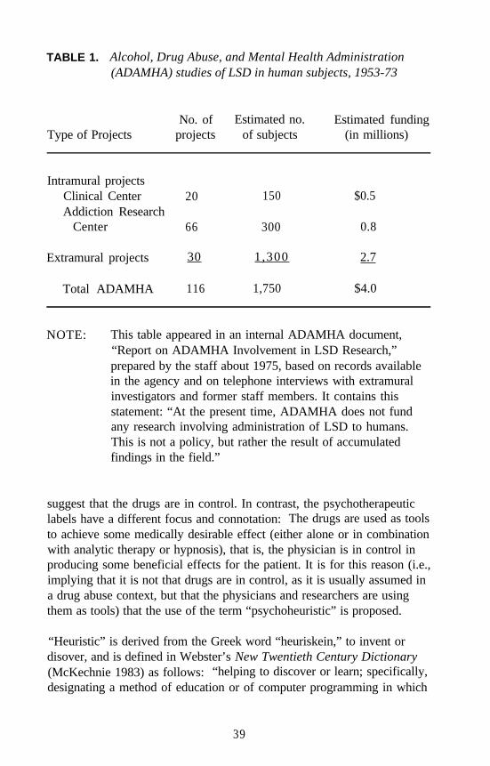
TABLE 1. Alcohol, Drug Abuse, and Mental Health Administration(ADAMHA) studies of LSD in human subjects, 1953-73
Type of ProjectsNo. of Estimated no. Estimated funding
projects of subjects (in millions)
Intramural projectsClinical CenterAddiction Research
Center
20 150 $0.5
66 300 0.8
Extramural projects 30 1,300 2.7
Total ADAMHA 116 1,750 $4.0
NOTE: This table appeared in an internal ADAMHA document,“Report on ADAMHA Involvement in LSD Research,”prepared by the staff about 1975, based on records availablein the agency and on telephone interviews with extramuralinvestigators and former staff members. It contains thisstatement: “At the present time, ADAMHA does not fundany research involving administration of LSD to humans.This is not a policy, but rather the result of accumulatedfindings in the field.”
suggest that the drugs are in control. In contrast, the psychotherapeuticlabels have a different focus and connotation: The drugs are used as toolsto achieve some medically desirable effect (either alone or in combinationwith analytic therapy or hypnosis), that is, the physician is in control inproducing some beneficial effects for the patient. It is for this reason (i.e.,implying that it is not that drugs are in control, as it is usually assumed ina drug abuse context, but that the physicians and researchers are usingthem as tools) that the use of the term “psychoheuristic” is proposed.
“Heuristic” is derived from the Greek word “heuriskein,” to invent ordisover, and is defined in Webster’s New Twentieth Century Dictionary(McKechnie 1983) as follows: “helping to discover or learn; specifically,designating a method of education or of computer programming in which
39
the pupil or machine proceeds along empirical lines, using rules ofthumb, to find solutions or answers.”
The Random House College Dictionary (Stein 1980) gives the followingdefinitions for heuristic: (1) “Serving to indicate or point out; stimulatinginterest as a means of furthering investigation; (2) (of a teaching method)encouraging the student to discover for himself.”
It is in the first, general sense of the dictionaries’ definitions that the wordpsychoheuristic is meant to be used: “helping to discover” and“stimulating interest as a means of furthering investigation” into themechanism(s) by which some of the unique psychological effects areproduced by these drugs and, beyond that, to serve as keys to unlock themysteries of the brain/mind relationship.
UNIQUE CHARACTERISTICS OF PSYCHOHEURISTICAGENTS
What are the unique characteristics of the effects of these drugs that pointto their potential as psychoheuristic agents? The vivid, mostly geometricvisual illusions are one of the hallmarks of LSD, DMT, and other majorpsychedelics. These illusions are sometimes so intense that they are seenas superimposed on any outside surface, be it a plain white wall orpeople’s faces. As pointed out earlier in this chapter, these visual patternsare seldom perceived as having real outside existence; so they are, strictlyspeaking, illusions rather than hallucinations. Nevertheless, they aresufficiently striking and sometimes spectacular, so that they have been ofsome interest to psychologists (Klüver 1967; Oster 1970; Siegel 1977), tophysiologists (Evarts 1957; Purpura 1957), and even to mathematicians(Cowan 1988). Some other unique characteristics might be the alterationof time perception, synesthesia, dehabituation, the extreme individualvariability of many of their actions, the religious or mysticomimeticproperties, and the so-called cultogenic effects. The reader can probablyname a number of others.
However, most of these effects are interpretations and/or secondaryconsequences of the drugs’ disturbances of some fundamentalphysiological or psychological processes that underlie humans’ capacityto attend, to be aware of, and to regulate their relationships with thephysical and social environment. Thus, the author’s recommendation isto use these drugs in a heuristic mode to explore the biological correlates
40
and perhaps the mechanism(s) of the fundamental process that isfrequently referred to by the psychoanalytic term of disturbance of “egoboundaries” or “oceanic feeling.” This aspect has been emphasizedespecially by psychiatrists, among others (Fischman 1983). Freedman, inhis much-quoted landmark paper On the Use and Abuse of LSD, puts itthis way:
It is my impression that one basic dimension of behavior latentlyoperative at any level of function and compellingly revealed inLSD states is “portentousness”—the capacity of the mind to seemore than it can tell, to experience more than it can explicate, tobelieve in and be impressed with more than it can rationallyjustify, to experience boundlessness and “boundaryless” events,from the banal to the profound. (Freedman 1968, p. 331)
Grof, who has perhaps more clinical research experience than anyone elsein the world with LSD and other hallucinogens such as DPT, hasconcluded that the major psychedelics do not produce specificpharmacologic states (i.e., toxic psychosis) but are unspecific amplifiersof mental processes (Grof 1980). In other words, rather than producingeffects that are specific for the drug, they activate mostly unconsciousmental processes from various deep levels. These mental processes arespecific for the personality of the individual. The major focus of Grof’stherapeutically oriented work was to interpret unconscious memories forthe perinatal experience of pain and trauma as an example of what hecalled “temporal expansion of consciousness,” and to deal with theso-called transpersonal experiences as a result of spatial expansion ofconsciousness. The common denominator, he said, “in this rich andramified group of phenomena is the feeling of the individual that hisconsciousness expanded beyond the usual ego boundaries and limitationsof time and space” (Grof 1980, p. 94).
BOUNDARIES IN THE MIND AND THE BRAIN
The subjective phenomenon of loss of ego boundaries is not restricted topsychedelic experiences. In the twilight states of falling asleep andwaking, people go through such experiences every day, although noteveryone is fully aware of them. LSD and similar drugs have the uniqueproperty of producing similar twilight states while a person is fully awakeand aware of them. This characteristic makes these drugs specially suited
41
to exploring the full extent of these general phenomena, including theirpostulated biological bases.
The generality of these phenomena is underscored by a broadpsychological theory of boundaries proposed recently by Hartmann, awell-known sleep researcher. He put forward this suggestion in his bookBoundaries in the Mind and claims that these boundaries represent amajor dimension of personality that had largely been neglected(Hartmann 1991). In the course of studies on people with nightmares,Hartmann was struck by the observation that such people have a group ofcommon characteristics that could be described as open, unguarded,sensitive, fluid, artistic, and vulnerable. The description that seemed bestto encompass all these people was that they had “thin boundaries” inmany different senses. In contrast, among the control subjects, Hartmannfound a significant group of people who could be characterized as having“thick boundaries” in the sense that they have a very solid, separate senseof self; they keep emotionally distant from most others; and they do notbecome overinvolved, sometimes appearing inflexible, even rigid. Thisdistinction seems to hold in many areas of interpersonal relations in theseextreme groups, but there were people who were in between.
The initial evaluation was based on Rorschach tests (popularly known asinkblot tests) of individuals participating in Hartmann’s sleep electro-encephalogram (EEG) studies. Watson (1985) has found EEG correlatesof this dimension: thin boundary individuals producing significantlylarger numbers of phasic integrated potentials (PIP), also known asponto-geniculo-occipital (PGO) spikes, on the boundaries of rapid eyemovement (REM) and non-REM sleep stages.
Hartmann (1991) also has developed a 145-item questionnaire that couldbe used to quantify the thick and thin dimensions. The questionnairecovers 12 categories of psychological phenomena such as childhoodexperiences, interpersonal relations, habits, opinions, and sleep-wake anddream-recall patterns. Most people do not score in the extreme in eachcategory but thick in some areas and thin in others. Among 300 subjects,there was some statistically significant correlation of this dimension tosome of the scales of the Minnesota Multiphasic Personality Inventory(MMPI), but a closer analysis indicated that the boundary scale isdefinitely not measuring sickness or psychopathology.
The concept of boundaries, Hartmann (1991) claims, should be helpful inunderstanding and preventing the potential consequences of some
42
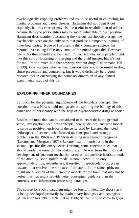
psychologically crippling problems and could be useful in counseling formarital problems and career choices. Hartmann did not point it outexplicitly, but this concept may also be useful in rehabilitation of addicts,because thin-type personalities may be more vulnerable to peer pressure.Hartmann does mention that among the various psychoactive drugs, thepsychedelic types are the only ones that produce a temporary thinning ofinner boundaries. None of Hartmann’s thick boundary subjects hasreported ever taking LSD; only some of the mixed types did. However,one of his thin boundary subject said: “I can see why some people mightlike this sort of loosening or merging and the vivid images, but it’s notfor me. I’m too much like that anyway, without drugs.” (Hartmann 1991,p. 239). One wonders whether this questionnaire would be useful in drugabuse prevention and counseling, but it would definitely be a goodresearch tool in quantifying the boundary dimension in any clinicalexperimental study of this trait.
EXPLORING INNER BOUNDARIES
So much for the potential significance of the boundary concept. Thequestion arises: How should one go about exploring the biology of thisdimension of personality with the help of psychoheuristic drugs as tools?
Besides the tools that can be considered to be heuristic in the generalsense, investigators need new concepts, new guidelines, and new modelsto serve as positive heuristics in the sense used by Lakatos, the notedphilosopher of science, who focused on conceptual and strategicproblems in the 1960s and 1970s in defining new research programs(Lakatos and Musgrave 1970). Lakatos’ use of heuristics is in thesecond, specific dictionary sense: Defining some concrete rules thatshould guide the research. His striking example was from the historicaldevelopment of quantum mechanics based on the positive heuristic modelof the atom by Bohr. Bohr’s model is now known to be onlyapproximately true; nevertheless, it resulted in spectacular progress inresearch that resolved the structure of the atom. In the same spirit, onemight use a version of the hierarchic models for the brain that may not beperfect but that might provide better conceptual guidance than thecurrently used information-processing paradigm.
The source for such a paradigm might be found in hierarchy theory as itis being developed primarily by evolutionary biologists and ecologists(Allen and Starr 1988; O’Neill et al. 1986; Salthe 1985) to come to grips
43
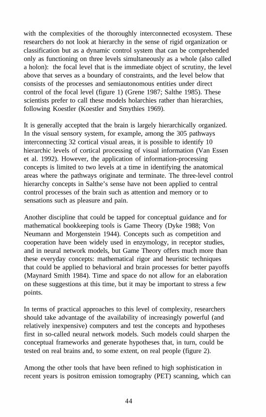
with the complexities of the thoroughly interconnected ecosystem. Theseresearchers do not look at hierarchy in the sense of rigid organization orclassification but as a dynamic control system that can be comprehendedonly as functioning on three levels simultaneously as a whole (also calleda holon): the focal level that is the immediate object of scrutiny, the levelabove that serves as a boundary of constraints, and the level below thatconsists of the processes and semiautonomous entities under directcontrol of the focal level (figure 1) (Grene 1987; Salthe 1985). Thesescientists prefer to call these models holarchies rather than hierarchies,following Koestler (Koestler and Smythies 1969).
It is generally accepted that the brain is largely hierarchically organized.In the visual sensory system, for example, among the 305 pathwaysinterconnecting 32 cortical visual areas, it is possible to identify 10hierarchic levels of cortical processing of visual information (Van Essenet al. 1992). However, the application of information-processingconcepts is limited to two levels at a time in identifying the anatomicalareas where the pathways originate and terminate. The three-level controlhierarchy concepts in Salthe’s sense have not been applied to centralcontrol processes of the brain such as attention and memory or tosensations such as pleasure and pain.
Another discipline that could be tapped for conceptual guidance and formathematical bookkeeping tools is Game Theory (Dyke 1988; VonNeumann and Morgenstein 1944). Concepts such as competition andcooperation have been widely used in enzymology, in receptor studies,and in neural network models, but Game Theory offers much more thanthese everyday concepts: mathematical rigor and heuristic techniquesthat could be applied to behavioral and brain processes for better payoffs(Maynard Smith 1984). Time and space do not allow for an elaborationon these suggestions at this time, but it may be important to stress a fewpoints.
In terms of practical approaches to this level of complexity, researchersshould take advantage of the availability of increasingly powerful (andrelatively inexpensive) computers and test the concepts and hypothesesfirst in so-called neural network models. Such models could sharpen theconceptual frameworks and generate hypotheses that, in turn, could betested on real brains and, to some extent, on real people (figure 2).
Among the other tools that have been refined to high sophistication inrecent years is positron emission tomography (PET) scanning, which can
44
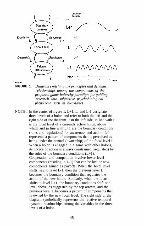
FIGURE 1. Diagram sketching the principles and dynamicrelationships among the components of theproposed game-holarchy paradigm for guidingresearch into subjective psychobiologicalphenomena such as boundaries.
NOTE: In the center of figure 1, L+1, L, and L-1 designatethree levels of a holon and refer to both the left and theright side of the diagram. On the left side, in line with Lis the focal level of a currently active holon, abovewhich and in line with L+1 are the boundary conditions(rules and regulations) for awareness and action. L-1represents a pattern of components that is perceived asbeing under the control (ownership) of the focal level L.When a holon is engaged in a game with other holons,its choice of action is always constrained (regulated) bythe rules of the boundary conditions (L+1).Cooperation and competition involve lower levelcomponents (residing in L-1) that can be lost or newcomponents gained as payoffs. When the focal levelshifts, say to level L-1, then the previous level Lbecomes the boundary condition that regulates theaction of the new holon. Similarly, when the focusshifts to level L+1, the boundary conditions shift onelevel above, as suggested by the top arrows, and theprevious level L becomes a pattern of components thatis owned by the new focal level. The right side of thediagram symbolically represents the relative temporaldynamic relationships among the variables in the threelevels of a holon.
45
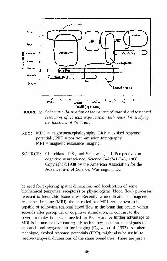
FIGURE 2. Schematic illustration of the ranges of spatial and temporalresolution of various experimental techniques for studyingthe functions of the brain.
KEY: MEG = magnetoencephalography, ERP = evoked responsepotentials, PET = positron emission tomography,MRI = magnetic resonance imaging.
SOURCE: Churchland, P.S., and Sejnowski, T.J. Perspectives oncognitive neuroscience. Science 242:741-745, 1988.Copyright ©1988 by the American Association for theAdvancement of Science, Washington, DC.
be used for exploring spatial dimensions and localization of somebiochemical (enzymes, receptors) or physiological (blood flow) processesrelevant to hierarchic boundaries. Recently, a modification of magneticresonance imaging (MRI), the so-called fast MRI, was shown to becapable of following regional blood flow in the brain that occurs withinseconds after perceptual or cognitive stimulation, in contrast to theseveral minutes time scale needed for PET scan. A further advantage ofMRI is its noninvasive nature; this technology uses intrinsic signals ofvenous blood oxygenation for imaging (Ogawa et al. 1992). Anothertechnique, evoked response potentials (ERP), might also be useful toresolve temporal dimensions of the same boundaries. These are just a
46
few examples; many other methods could be profitably employed asrequired by the questions at hand (Churchland and Sejnowski 1988).
Last, but not least, there are some useful placebos that should sharpen anyresearch design. Bromo-LSD could serve as a nonpsychoactive analogfor LSD in a certain dosage range, and 6-fluoro DET can be used as anactive placebo for the shorter acting psychoheuristic drug DET asdemonstrated more than 25 years ago by Faillace and colleagues (1967).
SUMMARY
The author argues in this chapter for a reconsideration of the perceptionof hallucinogens as being only toxic, damaging, and therefore strictlycondemnable for being abused. The author advocates that hallucinogensbe viewed as powerful psychoheuristic tools that, in combination withother necessary conceptual (such as holarchic theory) and laboratory tools(such as PET scan or MRI), may help solve a major mystery of nature:the workings of human brains and minds.
NOTE
The National Institute on Drug Abuse is among the many Federaldepartments and agencies participating in programs and activities toobserve the “Decade of the Brain” beginning January 1, 1990.Additional information about this effort can be found in the bill (H.J.Res. 174, March 8, 1989), which became Public Law 101-58 onJuly 25, 1989.
REFERENCES
Allen, T.F.H., and Starr, T.B. Hierarchy: Perspectives for EcologicalComplexity. Chicago: University of Chicago Press, 1988.
Campbell, R.J. Psychiatric Dictionary. 6th ed. New York: OxfordUniversity Press, 1989.
Carroll, L. Alice in Wonderland and Through the Looking Glass.Kingsport, TN: Grosset & Dunlap, 1946.
Churchland, P.S., and Sejnowski, T.J. Perspectives on cognitiveneuroscience. Science 242:741-745, 1988.
47
Cohen, S. Statement to the Senate Subcommittee on ExecutiveReorganization Hearing on Organization of Government ProgramsRelating to LSD. Washington, DC, May 24-26, 1966.
Cohen, S. The varieties of psychotic experience. J Psychoactive Drugs17(4):291-296, 1985.
Cooper, J.R.; Bloom, F.E.; and Roth, R.H. The Biochemical Basis ofNeuropharmacalogy. 2d ed. New York: Oxford University Press,1974.
Cowan, J.D. Brain mechanisms underlying visual hallucinations. In:Pines, D., ed. Emerging Syntheses in Science. Redwood City, CA:Addison-Wesley, 1988. pp. 123-131.
Dahlberg, C.C. Statement to the Senate Subcommittee on ExecutiveReorganization Hearing on Organization of Government ProgramsRelating to LSD, Washington, DC, May 24-26, 1966.
Dyke, C. The Evolutionary Dynamics of Complex Systems: A Study inBiosocial Complexity. Oxford, UK: Oxford University Press, 1988.
Evarts, A.V. A review of the neurophysiological effects of lysergic aciddiethylamide (LSD) and other psychotomimetic agents. Ann N Y AcadSci 66(3):479-495, 1957.
Faillace, L. Clinical use of psychotomimetic drugs. Compr Psychiatry7(1):13-20, 1966.
Faillace, L.A., and Szára, S. Hallucinogenic drugs: Influence of mentalset and setting. Dis Nerv System 29:124-126, 1968.
Faillace, L.A.; Vourlekis, A.; and Szára, S. Clinical evaluation of somehallucinogenic tryptamine derivatives. J Nerv Ment Dis 145(4):306-313,1967.
Faillace, L.A.; Vourlekis, A.; and Szára, S. Hallucinogenic drugs in thetreatment of alcoholism: A two-year follow-up. Compr Psychiatry11(1):51-56, 1970.
Fischman, L.G. Dreams, hallucinogenic drug states, and schizophrenia: Apsychological and biological comparison. Schizophr Bull 9(1):73-94,1983.
Freedman, D.X. Statement to the Senate Subcommittee on ExecutiveReorganization Hearing on Organization of Government ProgramsRelating to LSD. Washington, DC, May 24-26, 1966.
Freedman, D.X. On the use and abuse of LSD. Arch Gen Psychiatry18:330-347, 1968.
Grene, M. Hierarchies in biology. Am Sci 75:504-510, 1987.Grof, S. Realms of the human unconscious: Observations from LSD
research. In: Walsh, R.N., and Vaughan, F., eds. Beyond Ego:Transpersonal Dimensions in Psychology. Los Angeles: Jeremy P.Tarcher, Inc., 1980. pp. 87-99.
48
Hartmann, E. Boundaries in the Mind: A New Psychology of Personality.New York: Basic Books, 1991.
Hoffer, A.; Osmond, H.; and Smythies, J. Schizophrenia: A newapproach. II. Results of a year’s research. J Ment Sci 100:29-45, 1954.
Hollister, L.E. Drug-induced psychoses and schizophrenic reactions, acritical comparison. Ann N Y Acad Sci 96:80-88, 1962.
Huxley, A. The Doors of Perception. New York: Harper & Row, 1954.Kalir, A., and Szára, S. Synthesis and pharmacological activity of
fluorinated tryptamine derivatives. J Med Chem 6:716-719, 1963.Kaplan, H.I., and Sadock, B.J. Comprehensive Textbook of Psychiatry/V.
5th ed. Baltimore: Williams & Wilkins, 1989.Klee, G.D. Statement to the Senate Subcommittee on Executive
Reorganization Hearing on Organization of Government ProgramsRelating to LSD. Washington, DC, May 24-26, 1966.
Klüver, H. Mescal and Mechanisms of Hallucination. Chicago:University of Chicago Press, 1967.
Koestler, A., and Smythies, J.R. Beyond Reductionism: New Perspectivein the Life Sciences. Boston: Beacon Press, 1969.
Lakatos, I., and Musgrave, A., eds. Criticism and the Growth ofKnowledge. Cambridge, UK: Cambridge University Press, 1970.
Levine, J., and Ludwig, A.M. Alterations in consciousness produced bycombinations of LSD, hypnosis, and psychotherapy,Psychopharmacologia (Berlin) 7:123-137, 1965.
Lewin, L. Phantastika: Die Beteubenden und Erregenden Genussmittel(Narcotic and Stimulating Substances). Berlin: Verlag G. Stilke, 1924.
Lewin, L. Phantastica: Narcotic and Stimulating Drugs, Their Use andAbuse. Wirth, P.H.A., trans. New York: E.P. Dutton & Co., 1964.
Maynard Smith, J. Game theory and the evolution of behaviour. BehavBrain Sci 7:95-125, 1984.
McKechnie, J.L., ed. New Twentieth Century Dictionary of the EnglishLanguage. Unabridged. 2d ed. New York: Simon & Schuster, 1983.
Ogawa, S.; Tank, D.W.; Menon, R.; Ellerman, J.M.; Kim, S.-G.; Merkle,H.; and Ugubril, K. Intrinsic signal changes accompanying sensorystimulation: Functional brain mapping with magnetic resonanceimaging. Proc Natl Acad Sci USA 89:595l-5992, 1992.
O’Neill, R.V.; DeAngelis, D.L.; Waide, J.B.; and Allen, T.F.H. AHierarchical Concept of Ecosystems. Princeton, NJ: PrincetonUniversity Press, 1986.
Osmond, H. A review of the clinical effects of psychotomimetic agents.Ann N Y Acad Sci 66(3):418-434, 1957.
Oster, G. Phosphenes. Sci Am 222(2):83-87, 1970.
49
Purpura, D.P. Experimental analysis of the inhibitory action of lysergicacid diethylamide on cortical dendritic activity. Ann N Y Acad Sci66(3):515-536, 1957.
Rosenberg, D.E.; Isbell, H.; and Miner, E.J. Comparison of a placebo,N-dimethyltryptamine and 6-hydroxy-N-dimethyltryptamine in man.Psychopharmacologia (Berlin) 4:39-42, 1963.
Ruck, C.A.P.; Bigwood, J.; Staples, D.; Ott, J.; and Wasson, G.Entheogens. J Psychedelic Drugs 11(1-2):145-146, 1979.
Salthe, S.N. Evolving Hierarchical Systems. New York ColumbiaUniversity Press, 1985.
Savage, C.; Terril, J.; and Jackson, D.O. LSD transcendence and the newbeginning. J Nerv Ment Dis 135:425-439, 1962.
Siegel, R.K. Hallucinations. Sci Am 237(4):132-140, 1977.Stein, J., ed. The Random House College Dictionary. Rev. ed. New York:
Random House, 1980.Szára, S. “Psychosomimetic or Mysticomimetic?” Paper presented at
National Institute of Mental Health Seminar, Bethesda, MD,November 14, 1961.
Szára, S. The hallucinogenic drugs—curse or blessing? Am J Psychiatry123:1513-1518, 1967.
Szára, S. “A Scientist Looks at the Hippies.” Report prepared for theNational Institute of Mental Health, 1968.
Szára, S., and Hollister, L. “NIMH and Legal Drug Control.” Reportprepared for the National Institute of Mental Health, 1973.
Van Essen, D.C.; Anderson, C.H.; and Felleman, D.J. Informationprocessing in the primate visual system: An integrated systemsperspective. Science 255:419-423, 1992.
Von Neumann, J., and Morgenstein, O. Theory of Games and EconomicBehavior. Princeton, NJ: Princeton University Press, 1944.
Watson, R. “Phasic Integrated Potentials and Ego Boundary Deficit.”Paper presented at a joint meeting of the Sleep Research Society andthe Association of Sleep Disorders Centers, Seattle, WA, July 6-8,1985.
Yensen, R. LSD and psychotherapy. J Psychoactive Drugs 17(4):267-277, 1985.
50

AUTHOR
Stephen Szára, M.D., D.Sc.ChiefBiomedical Research Branch (Ret.)National Institute on Drug Abuse10901 Jolly WayKensington, MD 20895
51
Lysergamides RevisitedRobert C. Pfaff, Xuemei Huang, Danuta Marona-Lewicka,Robert Oberlender, and David E. Nichols
INTRODUCTION
In discussions of hallucinogens in past years, most of the focus has beenon phenethylamines and phenylisopropylamines, with a modest amounton tryptamines. A large gap always has been the lack of discussion oflysergamides. Lysergic acid diethylamide (LSD) is one’ of the classichallucinogenic agents, but substituted lysergamides always have seemedto be largely ignored. The lysergamides have been investigated onseveral historical occasions, but the late 1950s witnessed most of therecent work (Abrahamson 1959; Cerletti and Doepfner 1958; Gogertyand Dille 1957; Isbell et al. 1959). Within the past 8 years, there hasbeen an attempt to fill in some obvious gaps that exist in theunderstanding of lysergamide-type hallucinogens.
The lysergamides are derived from the ergot fungus, well knownthroughout history as a source of various types of medications. Extractsof ergot have been recognized as legitimate pharmaceutical preparationssince at least the Middle Ages. Lysergic acid is derived from hydrolysisof the ergot alkaloids. Although present-day methods of ergot productionutilize submerged culture fermentation rather than cultivation on rye orother cereal grains, Claviceps is the ultimate alkaloid source.
It is also known that lysergamides have been employed as psychoactivepreparations, with some of these uses stretching back to antiquity. Forexample, arguments have been made that the Greek mysteries at Eleusiswere related to the ingestion of a preparation that contained lysergamidesderived from an ergot fungus that infested the grass in that region.Another example is ololiuqui, a South American Indian and Mexicanpsychoactive preparation that was prepared from the seeds of certainmorning glories, Rivea corymbosa. Ergine, or lysergic acid amide(figure 1), is the primary psychoactive component of morning gloryseeds. In 1943, Hofmann discovered the unusual properties of LSD, thediethyl amide or lysergic acid.
52
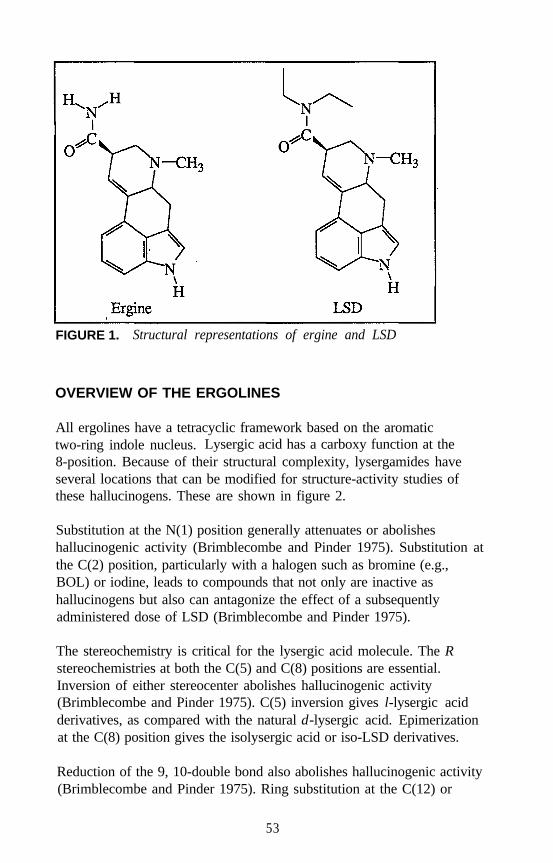
FIGURE 1. Structural representations of ergine and LSD
OVERVIEW OF THE ERGOLINES
All ergolines have a tetracyclic framework based on the aromatictwo-ring indole nucleus. Lysergic acid has a carboxy function at the8-position. Because of their structural complexity, lysergamides haveseveral locations that can be modified for structure-activity studies ofthese hallucinogens. These are shown in figure 2.
Substitution at the N(1) position generally attenuates or abolisheshallucinogenic activity (Brimblecombe and Pinder 1975). Substitution atthe C(2) position, particularly with a halogen such as bromine (e.g.,BOL) or iodine, leads to compounds that not only are inactive ashallucinogens but also can antagonize the effect of a subsequentlyadministered dose of LSD (Brimblecombe and Pinder 1975).
The stereochemistry is critical for the lysergic acid molecule. The Rstereochemistries at both the C(5) and C(8) positions are essential.Inversion of either stereocenter abolishes hallucinogenic activity(Brimblecombe and Pinder 1975). C(5) inversion gives l-lysergic acidderivatives, as compared with the natural d-lysergic acid. Epimerizationat the C(8) position gives the isolysergic acid or iso-LSD derivatives.
Reduction of the 9, 10-double bond also abolishes hallucinogenic activity(Brimblecombe and Pinder 1975). Ring substitution at the C(12) or
53
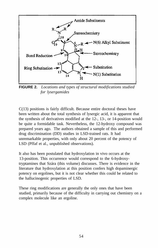
FIGURE 2. Locations and types of structural modifications studiedfor lysergamides
C(13) positions is fairly difficult. Because entire doctoral theses havebeen written about the total synthesis of lysergic acid, it is apparent thatthe synthesis of derivatives modified at the 12-, 13-, or 14-position wouldbe quite a formidable task. Nevertheless, the 12-hydroxy compound wasprepared years ago. The authors obtained a sample of this and performeddrug discrimination (DD) studies in LSD-trained rats. It hadunremarkable properties, with only about 20 percent of the potency ofLSD (Pffaf et al., unpublished observations).
It also has been postulated that hydroxylation in vivo occurs at the13-position. This occurrence would correspond to the 6-hydroxy-tryptamines that Szára (this volume) discusses. There is evidence in theliterature that hydroxylation at this position confers high dopaminergicpotency on ergolines, but it is not clear whether this could be related tothe hallucinogenic properties of LSD.
These ring modifications are generally the only ones that have beenstudied, primarily because of the difficulty in carrying out chemistry on acomplex molecule like an ergoline.
54
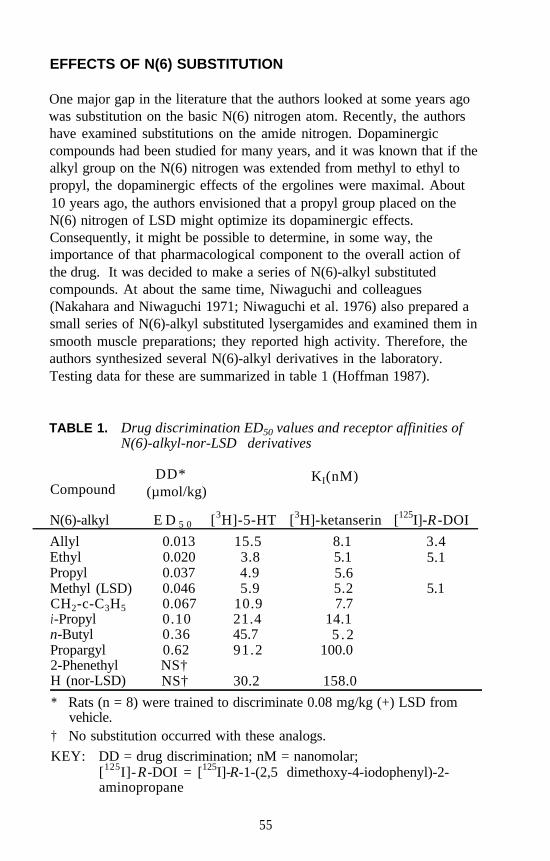
EFFECTS OF N(6) SUBSTITUTION
One major gap in the literature that the authors looked at some years agowas substitution on the basic N(6) nitrogen atom. Recently, the authorshave examined substitutions on the amide nitrogen. Dopaminergiccompounds had been studied for many years, and it was known that if thealkyl group on the N(6) nitrogen was extended from methyl to ethyl topropyl, the dopaminergic effects of the ergolines were maximal. About10 years ago, the authors envisioned that a propyl group placed on theN(6) nitrogen of LSD might optimize its dopaminergic effects.Consequently, it might be possible to determine, in some way, theimportance of that pharmacological component to the overall action ofthe drug. It was decided to make a series of N(6)-alkyl substitutedcompounds. At about the same time, Niwaguchi and colleagues(Nakahara and Niwaguchi 1971; Niwaguchi et al. 1976) also prepared asmall series of N(6)-alkyl substituted lysergamides and examined them insmooth muscle preparations; they reported high activity. Therefore, theauthors synthesized several N(6)-alkyl derivatives in the laboratory.Testing data for these are summarized in table 1 (Hoffman 1987).
TABLE 1. Drug discrimination ED50 values and receptor affinities ofN(6)-alkyl-nor-LSD derivatives
DD* KI(nM)Compound (µmol/kg)
N(6)-alkyl
AllylEthylPropylMethyl (LSD)CH2-c-C3H5i-Propyln-ButylPropargyl2-PhenethylH (nor-LSD)
E D 5 0 [3H]-5-HT
0.013 15.50.020 3.80.037 4.90.046 5.90.067 10.90.10 21.40.36 45.70.62 91.2NS†NS† 30.2
[3H]-ketanserin [125I]-R -DOI
8.1 3.45.1 5.15.65.2 5.17.7
14.15 . 2
100.0
158.0
* Rats (n = 8) were trained to discriminate 0.08 mg/kg (+) LSD fromvehicle.
† No substitution occurred with these analogs.KEY: DD = drug discrimination; nM = nanomolar;
[125I]-R-DOI = [125I]-R-1-(2,5 dimethoxy-4-iodophenyl)-2-aminopropane
55
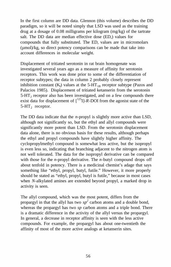
In the first column are DD data. Glennon (this volume) describes the DDparadigm, so it will be noted simply that LSD was used as the trainingdrug at a dosage of 0.08 milligrams per kilogram (mg/kg) of the tartratesalt. The DD data are median effective dose (ED,) values forcompounds that fully substituted. The ED, values are in micromolars(µmol)/kg, so direct potency comparisons can be made that take intoaccount differences in molecular weight.
Displacement of tritiated serotonin in rat brain homogenate wasinvestigated several years ago as a measure of affinity for serotoninreceptors. This work was done prior to some of the differentiation ofreceptor subtypes; the data in column 2 probably closely representinhibition constant (KI) values at the 5-HT1B receptor subtype (Pazos andPalacios 1985). Displacement of tritiated ketanserin from the serotonin5-HT2 receptor also has been investigated, and on a few compounds thereexist data for displacement of [125I]-R-DOI from the agonist state of the5-HT2 receptor.
The DD data indicate that the n-propyl is slightly more active than LSD,although not significantly so, but the ethyl and allyl compounds weresignificantly more potent than LSD. From the serotonin displacementdata alone, there is no obvious basis for these results, although perhapsthe ethyl and propyl compounds have slightly higher affinity. Thecyclopropylmethyl compound is somewhat less active, but the isopropylis even less so, indicating that branching adjacent to the nitrogen atom isnot well tolerated. The data for the isopropyl derivative can be comparedwith those for the n-propyl derivative. The n-butyl compound drops offabout tenfold in potency. There is a medicinal chemist’s adage that sayssomething like “ethyl, propyl, butyl, futile.” However, it more properlyshould be stated as “ethyl, propyl, butyl is futile,” because in most caseswhen N-alkylated amines are extended beyond propyl, a marked drop inactivity is seen.
The allyl compound, which was the most potent, differs from thepropargyl in that the allyl has two sp2 carbon atoms and a double bond,whereas the propargyl has two sp carbon atoms and a triple bond. Thereis a dramatic difference in the activity of the allyl versus the propargyl.In general, a decrease in receptor affinity is seen with the less activecompounds. For example, the propargyl has about one-twentieth theaffinity of most of the more active analogs at ketanserin sites.
56
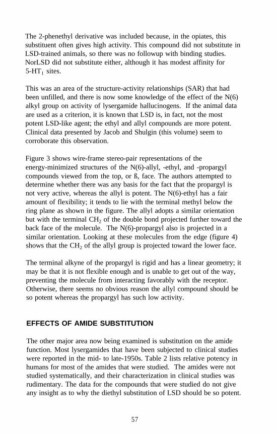
The 2-phenethyl derivative was included because, in the opiates, thissubstituent often gives high activity. This compound did not substitute inLSD-trained animals, so there was no followup with binding studies.NorLSD did not substitute either, although it has modest affinity for5-HT1 sites.
This was an area of the structure-activity relationships (SAR) that hadbeen unfilled, and there is now some knowledge of the effect of the N(6)alkyl group on activity of lysergamide hallucinogens. If the animal dataare used as a criterion, it is known that LSD is, in fact, not the mostpotent LSD-like agent; the ethyl and allyl compounds are more potent.Clinical data presented by Jacob and Shulgin (this volume) seem tocorroborate this observation.
Figure 3 shows wire-frame stereo-pair representations of theenergy-minimized structures of the N(6)-allyl, -ethyl, and -propargylcompounds viewed from the top, or ß, face. The authors attempted todetermine whether there was any basis for the fact that the propargyl isnot very active, whereas the allyl is potent. The N(6)-ethyl has a fairamount of flexibility; it tends to lie with the terminal methyl below thering plane as shown in the figure. The allyl adopts a similar orientationbut with the terminal CH2 of the double bond projected further toward theback face of the molecule. The N(6)-propargyl also is projected in asimilar orientation. Looking at these molecules from the edge (figure 4)shows that the CH2 of the allyl group is projected toward the lower face.
The terminal alkyne of the propargyl is rigid and has a linear geometry; itmay be that it is not flexible enough and is unable to get out of the way,preventing the molecule from interacting favorably with the receptor.Otherwise, there seems no obvious reason the allyl compound should beso potent whereas the propargyl has such low activity.
EFFECTS OF AMIDE SUBSTITUTION
The other major area now being examined is substitution on the amidefunction. Most lysergamides that have been subjected to clinical studieswere reported in the mid- to late-1950s. Table 2 lists relative potency inhumans for most of the amides that were studied. The amides were notstudied systematically, and their characterization in clinical studies wasrudimentary. The data for the compounds that were studied do not giveany insight as to why the diethyl substitution of LSD should be so potent.
57
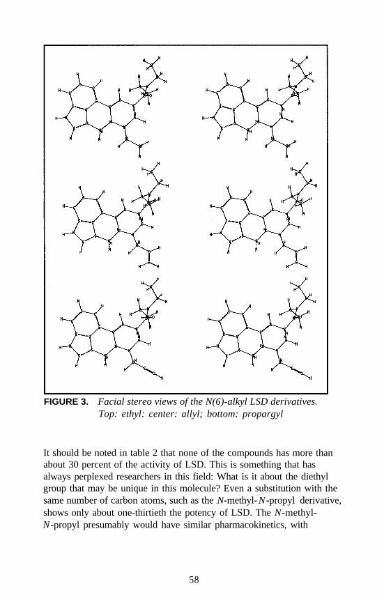
FIGURE 3. Facial stereo views of the N(6)-alkyl LSD derivatives.Top: ethyl: center: allyl; bottom: propargyl
It should be noted in table 2 that none of the compounds has more thanabout 30 percent of the activity of LSD. This is something that hasalways perplexed researchers in this field: What is it about the diethylgroup that may be unique in this molecule? Even a substitution with thesame number of carbon atoms, such as the N-methyl-N-propyl derivative,shows only about one-thirtieth the potency of LSD. The N-methyl-N-propyl presumably would have similar pharmacokinetics, with
58
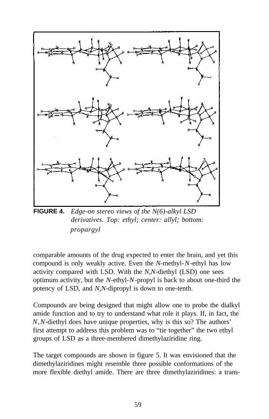
FIGURE 4. Edge-on stereo views of the N(6)-alkyl LSDderivatives. Top: ethyl; center: allyl; bottom:
propargyl
comparable amounts of the drug expected to enter the brain, and yet thiscompound is only weakly active. Even the N-methyl-N-ethyl has lowactivity compared with LSD. With the N,N-diethyl (LSD) one seesoptimum activity, but the N-ethyl-N -propyl is back to about one-third thepotency of LSD, and N,N-dipropyl is down to one-tenth.
Compounds are being designed that might allow one to probe the dialkylamide function and to try to understand what role it plays. If, in fact, theN,N-diethyl does have unique properties, why is this so? The authors’first attempt to address this problem was to “tie together” the two ethylgroups of LSD as a three-membered dimethylaziridine ring.
The target compounds are shown in figure 5. It was envisioned that thedimethylaziridines might resemble three possible conformations of themore flexible diethyl amide. There are three dimethylaziridines: a trans-
59
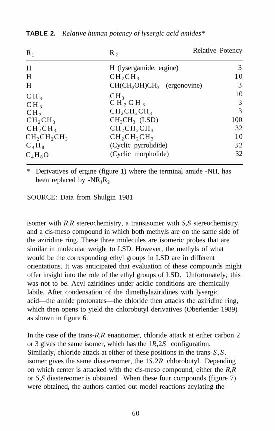
TABLE 2. Relative human potency of lysergic acid amides*
R1 R 2Relative Potency
HHH
C H 3
C H 3C H 3CH 2 CH 3
C H 2 C H 3
H (lysergamide, ergine) 3C H 2 C H 3 10CH(CH2OH)CH3 (ergonovine) 3
C H 310
C H 2 C H 3 3CH2CH2CH3 3CH2CH3 (LSD) 100CH 2 CH 2 CH 3 32CH 2 CH 2 CH 3 1 0(Cyclic pyrrolidide) 3 2(Cyclic morpholide) 32
CH2CH2CH3
C 4 H 8
C 4 H 8 O
* Derivatives of ergine (figure 1) where the terminal amide -NH, hasbeen replaced by -NR1R2
SOURCE: Data from Shulgin 1981
isomer with R,R stereochemistry, a transisomer with S,S stereochemistry,and a cis-meso compound in which both methyls are on the same side ofthe aziridine ring. These three molecules are isomeric probes that aresimilar in molecular weight to LSD. However, the methyls of whatwould be the corresponding ethyl groups in LSD are in differentorientations. It was anticipated that evaluation of these compounds mightoffer insight into the role of the ethyl groups of LSD. Unfortunately, thiswas not to be. Acyl aziridines under acidic conditions are chemicallylabile. After condensation of the dimethylaziridines with lysergicacid—the amide protonates—the chloride then attacks the aziridine ring,which then opens to yield the chlorobutyl derivatives (Oberlender 1989)as shown in figure 6.
In the case of the trans-R,R enantiomer, chloride attack at either carbon 2or 3 gives the same isomer, which has the 1R,2S configuration.Similarly, chloride attack at either of these positions in the trans-S ,S .isomer gives the same diastereomer, the 1S ,2R chlorobutyl. Dependingon which center is attacked with the cis-meso compound, either the R,Ror S,S diastereomer is obtained. When these four compounds (figure 7)were obtained, the authors carried out model reactions acylating the
60
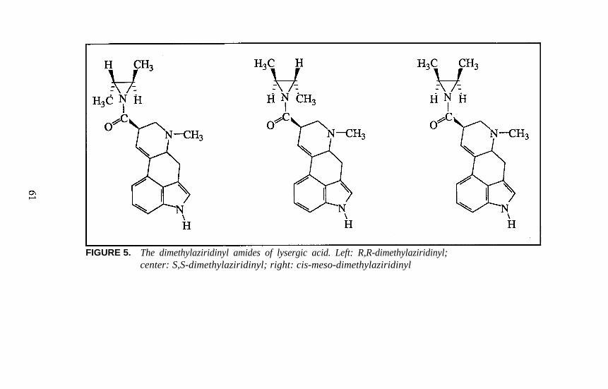
FIGURE 5. The dimethylaziridinyl amides of lysergic acid. Left: R,R-dimethylaziridinyl;center: S,S-dimethylaziridinyl; right: cis-meso-dimethylaziridinyl
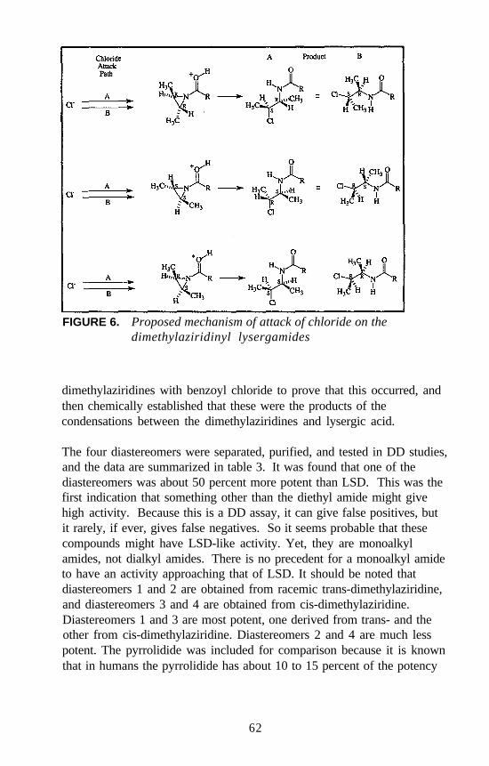
FIGURE 6. Proposed mechanism of attack of chloride on thedimethylaziridinyl lysergamides
dimethylaziridines with benzoyl chloride to prove that this occurred, andthen chemically established that these were the products of thecondensations between the dimethylaziridines and lysergic acid.
The four diastereomers were separated, purified, and tested in DD studies,and the data are summarized in table 3. It was found that one of thediastereomers was about 50 percent more potent than LSD. This was thefirst indication that something other than the diethyl amide might givehigh activity. Because this is a DD assay, it can give false positives, butit rarely, if ever, gives false negatives. So it seems probable that thesecompounds might have LSD-like activity. Yet, they are monoalkylamides, not dialkyl amides. There is no precedent for a monoalkyl amideto have an activity approaching that of LSD. It should be noted thatdiastereomers 1 and 2 are obtained from racemic trans-dimethylaziridine,and diastereomers 3 and 4 are obtained from cis-dimethylaziridine.Diastereomers 1 and 3 are most potent, one derived from trans- and theother from cis-dimethylaziridine. Diastereomers 2 and 4 are much lesspotent. The pyrrolidide was included for comparison because it is knownthat in humans the pyrrolidide has about 10 to 15 percent of the potency
62
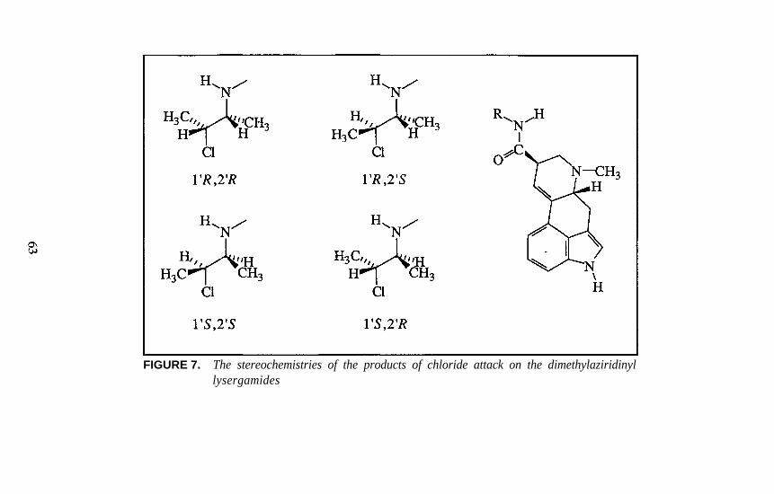
FIGURE 7. The stereochemistries of the products of chloride attack on the dimethylaziridinyllysergamides
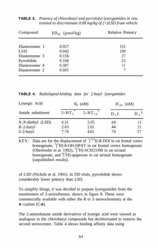
TABLE 3. Potency of chlorobutyl and pyrrolidyl lysergamides in ratstrained to discriminate 0.08 mg/kg of (+)LSD from vehicle
Compound ED50 (µmol/kg) Relative Potency
Diastereomer 1 0.027 155LSD 0.042 100Diastereomer 3 0.156 27Pyrrolidide 0.168 25Diastereomer 4 0.387 11Diastereomer 2 0.605 7
TABLE 4. Radioligand-binding data for 2-butyl lysergamides
Lysergic Acid KI (nM) IC50 (nM)
Amide substituent 5-HT 2* 5-HT 1 A
†D 1 ‡ D 2
§
N,N-diethyl (LSD) 6.31 5.05 60 13R -2-butyl 2.63 2.01 44 15S -2-butyl 7.76 4.61 70 37
KEY: Data are for the displacement of *[125I]-R-DOI in rat frontal cortexhomogenate, †[3H]-8-OH-DPAT in rat frontal cortex homogenate(Oberlender et al. 1992), ‡[3H]-SCH23390 in rat striatalhomogenate, and §[3H]-spiperone in rat striatal homogenate(unpublished results).
of LSD (Nichols et al. 1991). In DD trials, pyrrolidide showsconsiderably lower potency than LSD.
To simplify things, it was decided to prepare lysergamides from theenantiomers of 2-aminobutane, shown in figure 8. These werecommercially available with either the R or S stereochemistry at the
-carbon (C- ).
The 2-aminobutane amide derivatives of lysergic acid were viewed asanalogous to the chlorobutyl compounds but dechlorinated to remove thesecond stereocenter. Table 4 shows binding affinity data using
64
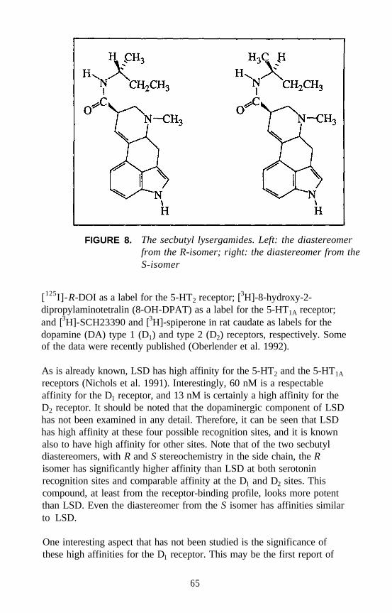
FIGURE 8. The secbutyl lysergamides. Left: the diastereomerfrom the R-isomer; right: the diastereomer from theS-isomer
[125I]-R-DOI as a label for the 5-HT2 receptor; [3H]-8-hydroxy-2-dipropylaminotetralin (8-OH-DPAT) as a label for the 5-HT1A receptor;and [3H]-SCH23390 and [3H]-spiperone in rat caudate as labels for thedopamine (DA) type 1 (D1) and type 2 (D2) receptors, respectively. Someof the data were recently published (Oberlender et al. 1992).
As is already known, LSD has high affinity for the 5-HT2 and the 5-HT1A
receptors (Nichols et al. 1991). Interestingly, 60 nM is a respectableaffinity for the D1 receptor, and 13 nM is certainly a high affinity for theD2 receptor. It should be noted that the dopaminergic component of LSDhas not been examined in any detail. Therefore, it can be seen that LSDhas high affinity at these four possible recognition sites, and it is knownalso to have high affinity for other sites. Note that of the two secbutyldiastereomers, with R and S stereochemistry in the side chain, the Risomer has significantly higher affinity than LSD at both serotoninrecognition sites and comparable affinity at the D1 and D2 sites. Thiscompound, at least from the receptor-binding profile, looks more potentthan LSD. Even the diastereomer from the S isomer has affinities similarto LSD.
One interesting aspect that has not been studied is the significance ofthese high affinities for the D1 receptor. This may be the first report of
65
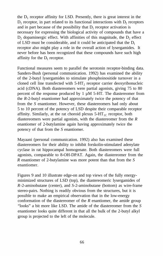
the D1 receptor affinity for LSD. Presently, there is great interest in theD1 receptor, in part related to its functional interactions with D2 receptorsand in part because of the possibility that D1 receptor activation isnecessary for expressing the biological activity of compounds that have aD2 dopaminergic effect. With affinities of this magnitude, the D2 effectof LSD must be considerable, and it could be anticipated that the D1
receptor also might play a role in the overall action of lysergamides. Itnever before has been recognized that these compounds have such highaffinity for the D1 receptor.
Functional measures seem to parallel the serotonin receptor-binding data.Sanders-Bush (personal communication. 1992) has examined the abilityof the 2-butyl lysergamides to stimulate phosphoinositide turnover in acloned cell line transfected with 5-HT2 receptor carrier deoxyribonucleicacid (cDNA). Both diastereomers were partial agonists, giving 75 to 80percent of the response produced by 1 µM 5-HT. The diastereomer fromthe R-2-butyl enantiomer had approximately twice the potency of thatfrom the S enantiomer. However, these diastereomers had only about5 to 10 percent of the potency of LSD despite their comparable receptoraffinity. Similarly, at the rat choroid plexus 5-HT1C receptor, bothdiastereomers were partial agonists, with the diastereomer from the Renantiomer of 2-butylamine again having approximately twice thepotency of that from the S enantiomer.
Mayaani (personal communication. 1992) also has examined thesediastereomers for their ability to inhibit forskolin-stimulated adenylatecyclase in rat hippocampal homogenate. Both diastereomers were fullagonists, comparable to 8-OH-DPAT. Again, the diastereomer from theR enantiomer of 2-butylamine was more potent than that from the Senantiomer.
Figures 9 and 10 illustrate edge-on and top views of the fully energy-minimized structures of LSD (top), the diastereomeric lysergamides ofR-2-aminobutane (center), and S-2-aminobutane (bottom) as wire-framestereo-pairs. Nothing is readily obvious from the structures, but it ispossible to make an empirical observation that in the low-energyconformation of the diastereomer of the R enantiomer, the amide group“looks” a bit more like LSD. The amide of the diastereomer from the Senantiomer looks quite different in that all the bulk of the 2-butyl alkylgroup is projected to the left of the molecule.
66
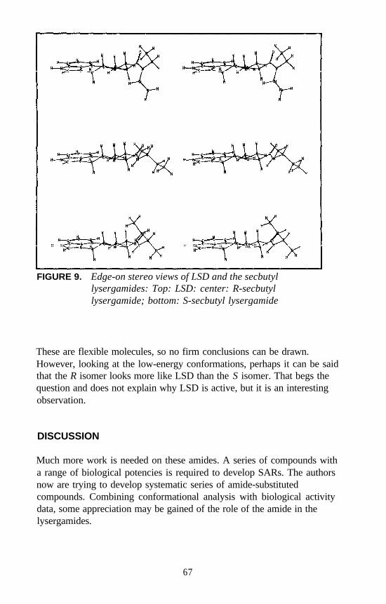
FIGURE 9. Edge-on stereo views of LSD and the secbutyllysergamides: Top: LSD: center: R-secbutyllysergamide; bottom: S-secbutyl lysergamide
These are flexible molecules, so no firm conclusions can be drawn.However, looking at the low-energy conformations, perhaps it can be saidthat the R isomer looks more like LSD than the S isomer. That begs thequestion and does not explain why LSD is active, but it is an interestingobservation.
DISCUSSION
Much more work is needed on these amides. A series of compounds witha range of biological potencies is required to develop SARs. The authorsnow are trying to develop systematic series of amide-substitutedcompounds. Combining conformational analysis with biological activitydata, some appreciation may be gained of the role of the amide in thelysergamides.
67
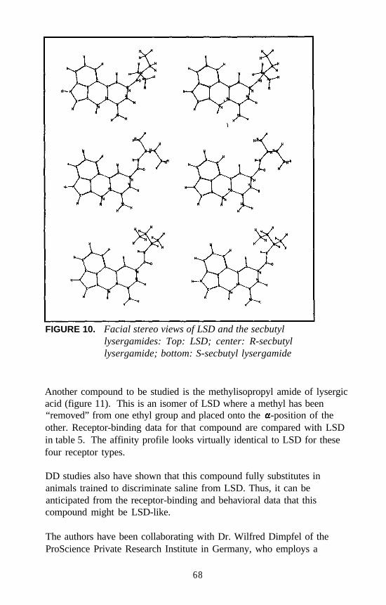
FIGURE 10. Facial stereo views of LSD and the secbutyllysergamides: Top: LSD; center: R-secbutyllysergamide; bottom: S-secbutyl lysergamide
Another compound to be studied is the methylisopropyl amide of lysergicacid (figure 11). This is an isomer of LSD where a methyl has been“removed” from one ethyl group and placed onto the -position of theother. Receptor-binding data for that compound are compared with LSDin table 5. The affinity profile looks virtually identical to LSD for thesefour receptor types.
DD studies also have shown that this compound fully substitutes inanimals trained to discriminate saline from LSD. Thus, it can beanticipated from the receptor-binding and behavioral data that thiscompound might be LSD-like.
The authors have been collaborating with Dr. Wilfred Dimpfel of theProScience Private Research Institute in Germany, who employs a
68
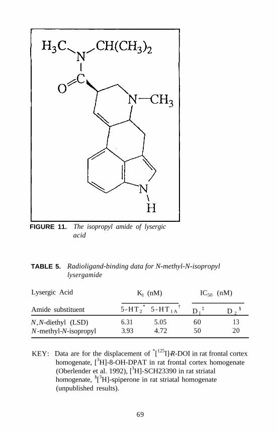
FIGURE 11. The isopropyl amide of lysergicacid
TABLE 5. Radioligand-binding data for N-methyl-N-isopropyllysergamide
Lysergic Acid KI (nM) IC50 (nM)
Amide substituent 5-HT 2* 5 - H T 1 A
†D l
‡ D 2§
N,N-diethyl (LSD) 6.31 5.05 60 13N -methyl-N-isopropyl 3.93 4.72 50 20
KEY: Data are for the displacement of *[125I]-R-DOI in rat frontal cortexhomogenate, [3H]-8-OH-DPAT in rat frontal cortex homogenate(Oberlender et al. 1992), [3H]-SCH23390 in rat striatalhomogenate, §[3H]-spiperone in rat striatal homogenate(unpublished results).
69
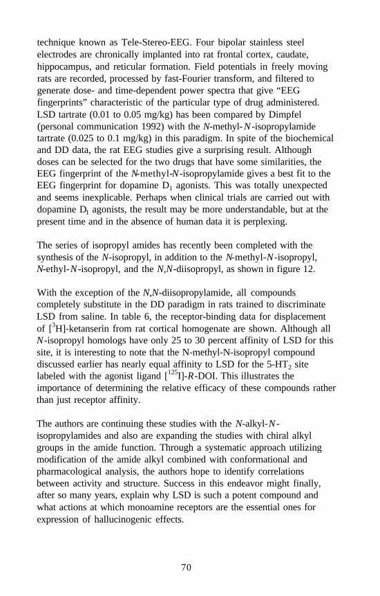
technique known as Tele-Stereo-EEG. Four bipolar stainless steelelectrodes are chronically implanted into rat frontal cortex, caudate,hippocampus, and reticular formation. Field potentials in freely movingrats are recorded, processed by fast-Fourier transform, and filtered togenerate dose- and time-dependent power spectra that give “EEGfingerprints” characteristic of the particular type of drug administered.LSD tartrate (0.01 to 0.05 mg/kg) has been compared by Dimpfel(personal communication 1992) with the N-methyl-N -isopropylamidetartrate (0.025 to 0.1 mg/kg) in this paradigm. In spite of the biochemicaland DD data, the rat EEG studies give a surprising result. Althoughdoses can be selected for the two drugs that have some similarities, theEEG fingerprint of the N-methyl-N-isopropylamide gives a best fit to theEEG fingerprint for dopamine D1 agonists. This was totally unexpectedand seems inexplicable. Perhaps when clinical trials are carried out withdopamine D1 agonists, the result may be more understandable, but at thepresent time and in the absence of human data it is perplexing.
The series of isopropyl amides has recently been completed with thesynthesis of the N-isopropyl, in addition to the N-methyl-N -isopropyl,N-ethyl-N-isopropyl, and the N,N-diisopropyl, as shown in figure 12.
With the exception of the N,N-diisopropylamide, all compoundscompletely substitute in the DD paradigm in rats trained to discriminateLSD from saline. In table 6, the receptor-binding data for displacementof [3H]-ketanserin from rat cortical homogenate are shown. Although allN-isopropyl homologs have only 25 to 30 percent affinity of LSD for thissite, it is interesting to note that the N-methyl-N-isopropyl compounddiscussed earlier has nearly equal affinity to LSD for the 5-HT2 sitelabeled with the agonist ligand [125I]-R-DOI. This illustrates theimportance of determining the relative efficacy of these compounds ratherthan just receptor affinity.
The authors are continuing these studies with the N-alkyl-N -isopropylamides and also are expanding the studies with chiral alkylgroups in the amide function. Through a systematic approach utilizingmodification of the amide alkyl combined with conformational andpharmacological analysis, the authors hope to identify correlationsbetween activity and structure. Success in this endeavor might finally,after so many years, explain why LSD is such a potent compound andwhat actions at which monoamine receptors are the essential ones forexpression of hallucinogenic effects.
70
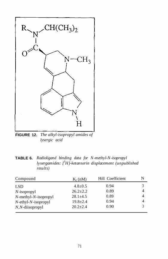
FIGURE 12. The alkyl-isopropyl amides oflysergic acid
TABLE 6. Radioligand binding data for N-methyl-N-isopropyllysergamides: [3H]-ketanserin displacement (unpublishedresults)
Compound KI (nM) Hill Coefficient N
LSD 4.8±0.5 0.94 3N-isopropyl 26.2±2.2 0.89 4N-methyl-N-isopropyl 28.1±4.5 0.89 4N-ethyl-N-isopropyl 19.8±2.4 0.94 4N,N-diisopropyl 20.2±2.4 0.90 3
71
REFERENCES
Abrahamson, H.A. Lysergic acid diethylamide (LSD-25): XXIX. Theresponse index as a measure of threshold activity of psychotropicdrugs in man. J Psych 48:65-78, 1959.
Brimblecombe, R.W., and Pinder, R.M. Hallucinogenic Agents. Bristol,UK: Wright-Scientechnica, 1975.
Cerletti, A., and Doepfner, W. Comparative study on the serotoninantagonism of amide derivatives of lysergic acid and of ergotalkaloids. J Pharmacol Exp Ther 122:124-136, 1958.
Gogerty, J.H., and Dille, J.M. Pharmacology of d-lysergic acidmorpholide (LSM). J Pharmacol Exp Ther 120:340-348, 1957.
Hoffman, A.J. “Synthesis and Pharmacological Evaluation of N(6)-AlkylNorlysergic Acid N,N-Diethylamide Derivatives.” Doctoral thesis.West Lafayette, IN: Purdue University, 1987.
Isbell, H.; Miner, E.J.; and Logan, C.R. Relationships ofpsychotomimetic to antiserotonin potencies of congeners of lysergicacid diethylamide. Psychopharmacologia 1:20-28, 1959.
Nakahara, Y., and Niwaguchi, T. Studies on lysergic acid diethylamideand related compounds. I. Synthesis of d-N6-demethyl-lysergic aciddiethylamide. Chem Pharm Bull 19:2337-2341, 1971.
Nichols, D.E.; Oberlender, R.; and McKenna, D.J. Stereochemicalaspects of hallucinogenesis. In: Watson, R.R., ed. Biochemistry andPhysiology of Substance Abuse. Boca Raton, FL: CRC Press, 1991.pp. 1-39.
Niwaguchi, T.; Nakahara, Y.; and Ishii, H. Studies on lysergic aciddiethylamide and related compounds. IV. Syntheses of various amidederivatives of norlysergic acid and related compounds. YakugakuZasshi 96:673-678, 1976.
Oberlender, R.A. “Stereoselective Aspects of Hallucinogenic DrugAction and Drug Discrimination Studies of Entactogens.” Doctoralthesis. West Lafayette, IN: Purdue University, 1989.
Oberlender, R.; Pfaff, R.C.; Johnson, M.P.; Huang, X.; and Nichols, D.E.Stereoselective LSD-like activity in d-lysergic acid amides of(R)- and(S)-2-aminobutane. J Med Chem 35:203-211, 1992.
Pazos, A., and Palacios, M. Quantitative autoradiographic mapping ofserotonin receptors in rat brain. I. Serotonin-1 receptors. Brain Res346:205-230, 1985.
Shulgin, A.T. Hallucinogens. In: Wolff, M.E., ed. Burger’s MedicinalChemistry, Part III. 4th ed. New York: Wiley, 1981. pp. 1109-1137.
72
ACKNOWLEDGEMENTS
This research was supported by U.S. Public Health Service grant DA-02189 from the National Institute on Drug Abuse. The synthetic work ofStewart Frescas and radioligand binding studies by Arthi Kanthasamyalso are acknowledged.
AUTHORS
Robert C. Pfaff, Ph.D.Associate Professor of ChemistryDepartment of ChemistrySt. Joseph’s CollegeRensselaer, IN 47978
Xuemei HuangGraduate AssistantDepartment of Pharmacology and Toxicology
Danuta Marona-LewickaVisiting Assistant Professor of PharmacologyDepartment of Medicinal Chemistry and Pharmacognosy
David E. Nichols, Ph.D.Professor of Medicinal Chemistry and PharmacologyDepartments of Medicinal Chemistry and Pharmacognosy andPharmacology and Toxicology
Purdue UniversityWest Lafayette, IN 47907
Robert Oberlender, Ph.D.Assistant Professor of Medicinal ChemistryDepartment of Pharmaceutics and Medicinal ChemistryUniversity of the PacificStockton, CA 95211
73
Structure-Activity Relationshipsof the Classic Hallucinogens andTheir AnalogsPeyton Jacob III and Alexander T. Shulgin
The path that leads to the appearance of a new psychotropic drug in thepractice of medicine usually consists of four stages: discovery of activity,the development of animal behavioral models that can be correlated tothis activity, the study of mechanisms of action and nature of toxicity,and the demonstration of effectiveness and benefit. The final studies ofeffectiveness for drugs intended for human use must be done in humansubjects. This is the essence of the Phases 1 through 4 studies found inthe Food and Drug Administration (FDA) regulations associated with theinvestigatory new drug (IND) application. With many drug families, theresults of the animal model studies (steps 2 and 3) can allow prediction ofnew drug structures (step 1). However, with research in the hallucino-genic drugs (where the desired pharmacological activity can be demon-strated only in humans), the confirmation of activity must occur ofnecessity in humans. Therefore, it is of potential value for future researchin this area to bring together in a single review the known humanpotencies of the classic hallucinogens and their analogs.
Two words in the title of this chapter must be defined: hallucinogen andclassic. A hallucinogen is a drug that changes a person’s state ofawareness by modifying sensory inputs, loosening cognitive and creativerestraints, and providing access to material normally hidden in memory ormaterial of an unconscious nature. The changes thus gained are notmasked by amnesia, although they will last only a finite period of time,and they are demonstrable only in humans. A generation ago these drugswere inaccurately called psychotomimetics, things that imitate psychosis.Today the term “hallucinogen” is allowed as a euphemism, although thatterm is also inaccurate because hallucinations are not part of the usualsyndrome. In another generation, the synonym psychedelics may becomeacceptable in the medical and scientific literature.
Several chemical families that include drugs which have been clinicallyassociated with the term “hallucinogen” have been excluded by designfrom this review, These substances have been the topics of other
74
conferences or monographs sponsored. by the National Institute on DrugAbuse (NIDA). Among these exclusions are marijuana, tetrahydro-cannabinol (THC), ketamine, and related parasympatholytics such as theDatura alkaloids and the JB compounds, opiates, and agents related to3,4-methylenedioxymethamphetamine (MDMA).
The term “classic” depends on the views of the person who defines it. Acompound achieves a classic status when it has served as the focus of aconsiderable amount of research attention, With compounds such asmescaline, lysergic acid diethylamide (LSD), 2,5-dimethylthio-phenethylamine (DOM), dimethyltryptamine (DMT), and psilocybin, thestatus as classic hallucinogens might be due to the extensive animal andclinical research that has appeared in the literature. But with othercompounds such as thiomescaline, 2,5-dimethoxy-4-methylphen-ethylamine (2C-D), 2,5-dimethoxy-4-methylthiophenethylamine (2C-T),and 3,4,5-trimethoxyamphetamine (TMA), the bulk of the publishedliterature has been focused at a structural and chemical level. Theanalogs of these nine prototypic classic hallucinogens are reviewed here,and nine tables list structurally related variants that have been explored inhumans.
The oldest classic hallucinogen known to Western science is mescaline,the major alkaloid of the dumpling cactus peyote. Mescaline was isolatedfrom the peyote cactus, the pharmacology was defined in 1896, and thestructure of mescaline verified by synthesis in 1919. It is used as thepotency standard against which all other phenethylamine bases have beencompared. Table 1 shows the relative potency of mescaline along withthe several alkyl homologs that have been studied with the oxygen atomsmaintained in the vicinal 3,4,5-orientation.
A generalization is that there is an increase in potency with increasing thelength of the alkyl group on the 4-position oxygen atom but not with suchchanges with the meta-oriented groups. The naming of these syntheticcompounds has exploited the coincidence that mescaline carries themethoxy group at the 4-position and both words begin with the samesyllable. The names “escaline,” “proscaline,” and “buscaline” followeasily as the groups become ethoxy, propoxy, and butoxy. The names forthe diethoxy homologs (here and in table 2) incorporate the nomenclatureprefix from being symmetrical (sym) or asymmetrical (asym) and havingtwo (bis) ethoxy groups (bescaline).
75
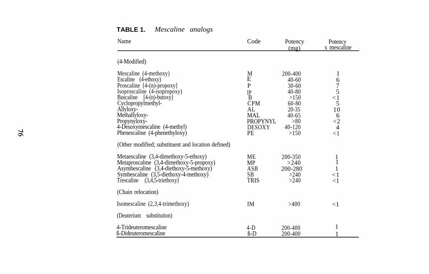
TABLE 1. Mescaline analogs
Name Code Potency Potency(mg) x mescaline
(4-Modified)
Mescaline (4-methoxy)Escaline (4-ethoxy)Proscaline [4-(n)-propoxy]Isoproscaline (4-isopropoxy)Buscaline [4-(n)-butoxy]Cyclopropylmethyl-Allyloxy-Methallyloxy-Propynyloxy-4-Desoxymescaline (4-methyl)Phenescaline (4-phenethyloxy)
MEPIPBCPMALMALPROPYNYLDESOXYPE
200-40040-6030-6040-80>150
60-8020-3540-65
>8040-120
>150
1675
<15
106
<24
<1
(Other modified; substituent and location defined)
Metaescaline (3,4-dimethoxy-5-ethoxy) MEMetaproscalme (3,4-dimethoxy-5-propoxy) MP
200-350 1>240 1
Asymbescaline (3,4-diethoxy-5-methoxy)Symbescaline (3,5-diethoxy-4-methoxy)Trescaline (3,4,5-triethoxy)
ASB 200-280 1SB >240 <1TRIS >240 <1
(Chain relocation)
Isomescaline (2,3,4-trimethoxy)
(Deuterium substitution)
4-Trideuteromescalineß-Dideuteromescaline
IM >400 <1
4-D 200-400 1ß-D 200-400 1
A comment is appropriate for the use of the symbols “>” and “<” in thesetables. When a dosage weight is given as >250 milligrams (mg), theimplicit statement is that no activity had been found at 250 mg. It is notknown whether the compound is active at any dose, but, if it is, it will beat a dose greater than 250 mg. Thus, the potency relative to mescalineshown as < 1 means that if activity is found, it will be less than that ofmescaline. There is no implication that the compound is or is not active.
There have been reports of mescaline analogs with a methoxy groupremoved. The analog 3,4-dimethoxyphenethylamine (DMPEA) hasachieved some notoriety with the report of the observation of a pink spotin the thin-layer chromatographic analysis of the extracts of the urine ofschizophrenic patients. The association of the spot with the diagnosis ofschizophrenia has remained controversial, but the chemical identity hasbeen shown to be DMPEA. Efforts to evoke some central nervoussystem (CNS) disturbance with this compound (orally, to > 1.5 grams (g);intravenously [IV], to > 10 mg) have produced no effects. Because of itsclose structural resemblance to the neurotransmitter dopamine (DA)(3,4-dihydroxyphenethylamine), this result has been disappointing. The4-ethoxy homolog, 3-methoxy-4-ethoxyphenethylamine (2,3,4-tri-methoxyphenethylamine) (MEPEA) has been assayed to 300 mg but haslittle if any activity.
An additional compound is isomescaline (2,3,4-trimethoxyphenethyl-amine). There is a fascinating report in the literature concerning itsactivity. It has been stated to be inactive in normal subjects but topromote a distinct intoxication in schizophrenic patients. If this effectwere confirmed, it might play an interesting role as a marker or abiochemical probe of schizophrenia.
The two last compounds in table 1 are the only known deuterium analogsthat have been explored in humans, and neither can be distinguished frommescaline. Other uniquely deuterated isotopomers that may be of interestare 3,5-(bis-trideuteromethoxy)-4-methoxyphenethylamine (3,5-D);2,6-dideuteromescaline (2,6-D), and -dideuteromescaline ( -D). Thelast compound, being deuterated at the most probable primary site formetabolic attack, might be of a different potency due to the kinetics ofa-proton removal, and a study of the (R)- -monodeuteroisotopomers[(R) -D] and (S)- -monodeuteroisotopomers [(S) -D] might beinformative. None of these latter compounds has as yet been studied.
77
The substitution of a sulfur atom for the 4-oxygen atom of mescalineyields the remarkably potent analog thiomescaline. Although thiscompound has not been widely studied in clinical trials, it has been thestarting point of extensive synthetic studies that have further emphasizedthe importance of the 4-position of the aromatic ring of thephenethylamines (see table 2).
Homologation at the 4-position again increases or maintains potency untilthe chain reaches a length of three carbon atoms (4-thioproscaline), andthen activity begins to disappear. None of the unsaturated (allylthio,methallylthio) or related electron-rich alkylthio counterparts (cyclo-propylmethylthio) has been studied for comparison to the relativelypotent oxygen counterparts (table 1). They should be reasonably simpleto synthesize and might be exceptionally potent.
One additional degree of structural variation is introduced with the sulfuratom replacement for the oxygen. It may occupy either of two positions,para or meta, to the ethylamine side chain. The meta-sulfur positionalisomers still emphasize the importance of the nature of the alkylsubstituent on the para-heteroatom. The three possible thioanalogs ofisomescaline were without activity.
The two remaining prototypes for structure-activity analysis are closerelatives to well-known amphetamine counterparts. The well-studieddrug DOM has a 2-carbon homolog, 4-methyl-2,5-dimethoxyphen-ethylamine (2C-D). The bromo counterpart 2,5-dimethoxy-4-bromo-amphetamine (DOB) has a 2-carbon homolog, 2,5-dimethoxy-4-bromo-phenethylamine (2C-B). Both phenethylamines have engendered largefamilies of analogs, and both amphetamines are presented in table 6.
The simplest 4-alkyl-substituted hallucinogenic compound is 2C-D. Thisbase has been widely explored in the United States as a prototype for theexploration of new compound. In Germany 2C-D has been used as apsychotherapeutic agent in its own right, at larger dosages, usually underthe code name of LE-25. The base without this para-alkyl group is2,5-dimethoxyphenethylamine (2C-H), but if this group is homologatedto an ethyl, the extraordinarily powerful and effective compound2,5-dimethoxy-4-ethylphenethylamine (2C-E) is found. Potencycontinues to increase with further chain lengthening, but the positivenature of the observed psychopharmacological effects is lessened.
78
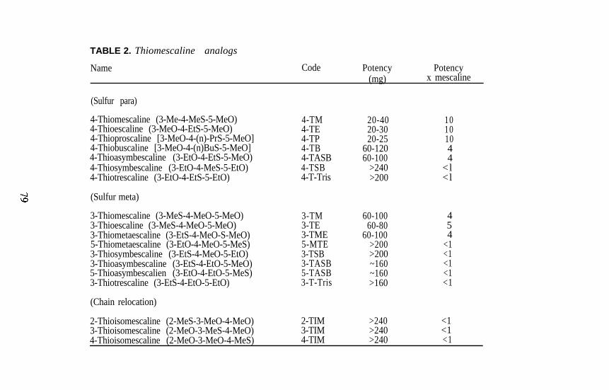
TABLE 2. Thiomescaline analogs
Name Code Potency(mg)
Potencyx mescaline
(Sulfur para)
4-Thiomescaline (3-Me-4-MeS-5-MeO)4-Thioescaline (3-MeO-4-EtS-5-MeO)4-Thioproscaline [3-MeO-4-(n)-PrS-5-MeO]4-Thiobuscaline [3-MeO-4-(n)BuS-5-MeO]4-Thioasymbescaline (3-EtO-4-EtS-5-MeO)4-Thiosymbescaline (3-EtO-4-MeS-5-EtO)4-Thiotrescaline (3-EtO-4-EtS-5-EtO)
4-TM 20-40 104-TE 20-30 104-TP 20-25 104-TB 60-120 44-TASB 60-100 44-TSB >240 <l4-T-Tris >200 <l
(Sulfur meta)
3-Thiomescaline (3-MeS-4-MeO-5-MeO) 3-TM 60-1003-Thioescaline (3-MeS-4-MeO-5-MeO) 3-TE 60-803-Thiometaescaline (3-EtS-4-MeO-S-MeO) 3-TME 60-1005-Thiometaescaline (3-EtO-4-MeO-5-MeS) 5-MTE >2003-Thiosymbescaline (3-EtS-4-MeO-5-EtO) 3-TSB >2003-Thioasymbescaline (3-EtS-4-EtO-5-MeO) 3-TASB ~1605-Thioasymbescalien (3-EtO-4-EtO-5-MeS) 5-TASB ~1603-Thiotrescaline (3-EtS-4-EtO-5-EtO) 3-T-Tris >160
(Chain relocation)
454
<1<1<1<1<1
2-Thioisomescaline (2-MeS-3-MeO-4-MeO)3-Thioisomescaline (2-MeO-3-MeS-4-MeO)4-Thioisomescaline (2-MeO-3-MeO-4-MeS)
2-TIM >240 <13-TIM >240 <14-TIM >240 <1
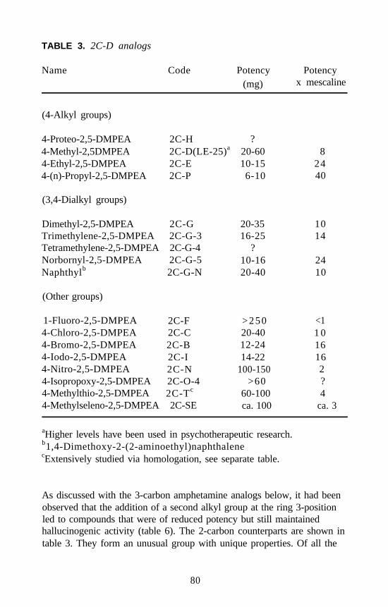
TABLE 3. 2C-D analogs
Name Code Potency(mg)
Potencyx mescaline
(4-Alkyl groups)
4-Proteo-2,5-DMPEA4-Methyl-2,5DMPEA4-Ethyl-2,5-DMPEA4-(n)-Propyl-2,5-DMPEA
2C-H ?2C-D(LE-25)a 20-602C-E 10-152C-P 6-10
(3,4-Dialkyl groups)
Dimethyl-2,5-DMPEA 2C-GTrimethylene-2,5-DMPEA 2C-G-3Tetramethylene-2,5-DMPEA 2C-G-4Norbornyl-2,5-DMPEA 2C-G-5Naphthylb 2C-G-N
20-3516-25
?10-1620-40
(Other groups)
1-Fluoro-2,5-DMPEA 2C-F4-Chloro-2,5-DMPEA 2C-C4-Bromo-2,5-DMPEA 2C-B4-Iodo-2,5-DMPEA 2C-I4-Nitro-2,5-DMPEA 2C-N4-Isopropoxy-2,5-DMPEA 2C-O-44-Methylthio-2,5-DMPEA 2C-Tc
4-Methylseleno-2,5-DMPEA 2C-SE
>250 <120-40 1 012-24 1614-22 16
100-150 2>60 ?
60-100 4ca. 100 ca. 3
82440
1014
2410
aHigher levels have been used in psychotherapeutic research.b1,4-Dimethoxy-2-(2-aminoethyl)naphthalenecExtensively studied via homologation, see separate table.
As discussed with the 3-carbon amphetamine analogs below, it had beenobserved that the addition of a second alkyl group at the ring 3-positionled to compounds that were of reduced potency but still maintainedhallucinogenic activity (table 6). The 2-carbon counterparts are shown intable 3. They form an unusual group with unique properties. Of all the
80
phenethylamine/amphetamine pairs explored so far, the phenethylamineis of a lower potency than the amphetamine homolog, and the potency ofeach increases yet further with the increasing of the molecular weight ofthe 4-alkyl group. However, with these 3,4-dialkyl analogs, as the twoalkyl groups become increasingly large and complex, not only do thephenethylamines become more potent than the amphetamine homologs,but the absolute potency also tends to increase with increased mass andbulk of these alkyl groups. The most potent compound yet found in thisstructural family is the illustrated norbornyl material 3,6-dimethoxy-4-(2-aminoethyl)-benzobornane (2C-G-5), with a total of 5 aliphaticcarbons arranged between the 3- and 4-positions.
Many directions can be pursued here, both synthetically and pharmaco-logically. So far, all the compounds that have been prepared-those thathave been evaluated psychopharmacologically and those whoseevaluation has not yet been completed-are symmetrically substitutedabout the 3,4-axis. The use of a Diels-Alder reaction with benzoquinoneas the dieneophyle ensures an almost unlimited degree of variation in the3,4-dialkyl-2,5-dimethoxyphenethylamines. With the increase in masssuggesting greater potency, there may be some remarkable compoundshere. Two further avenues of promising exploration are obvious. Anasymmetric substitution is possible in which the 3- and 4-position groupsare different from one another. Also, one must investigate the opticalisomers of the racemates produced (as with 2C-G-5).
Two additional centrally active structural variations of the prototype2C-D have been observed and explored to a small degree. Thep-oxygenated analogs are known as the ß-methoxy-ß-arylethylamine(BOX) series, and the materials with ethoxy groups in place of either ofthe methoxy groups are called the tweetios. Neither family is entered intable 3, but both are logical extensions of it.
The BOX compounds are ß-oxy analogs of phenethylamines, masked asthe methyl ether. The X then is the initial or identifier of the 2C analogthat has been oxygenated. This manipulation introduces an oxygenheteroatom at a position identical to that found in the neurotransmittersnorepinephrine (NE) and epinephrine. But it also introduces a new chiralcenter, and in the corresponding amphetamine derivatives a threo-erythrosystem of diastereoisomers that resembles that of ephedrine andpseudoephedrine would be produced. The ß-methoxy analogs of 2C-ß,ß,2,5-trimethoxy-4-bromophenethylamine (BOB), and 2C-D (ß,2,5-trimethoxy-4-methylphenethylamine [BOD]) are a little more potent than
81
their oxygen-free counterparts but not as interesting subjectively. Therehas been no research done on the pure enantiomers.
The two tweetio analogs (2-ethoxy and 5-ethoxy) of both 2C-D and 2C-Bhave been explored and have dramatically reduced activity. The5-tweetio (5-ethoxy) compounds are of twofold lessened potency, and the2-tweetio (2-ethoxy) materials are down by another factor of five. Thebis-etios (2,5-diethoxy homologs of 2C-D and 2C-B) are not known to beactive at all.
Near the bottom of table 3 is a sulfur compound, 2C-T. Although onlymodest in activity, its homologs show a wide and varied psycho-pharmacology and constitute yet another family of hallucinogenics.These 2C-T analogs are listed and compared in table 4.
The optimum alkyl substitution is two to three carbons, with 2C-T-2,2C-T-4, and 2C-T-7 (the S-ethyl, S-isopropyl, and S-propyl) being bothpotent and LSD-like. The placement of a methoxy on the S-ethyl groupof 2C-T-2 yields the active methoxyethylthio derivative 2C-T-13, and thereplacement of this methoxy group with a fluorine gives the potentß-fluoroethylthio-2,5-dimethoxyphenethylamine (2C-T-21). Thisstructure is interesting because it is the first hallucinogenic drug with sixseparate elements in its formula (C, H, N, O, S, and F) and is potentiallyvaluable as a vehicle for 18F studies of brain kinetics with positronemission tomography (PET). In this case the fluorine atom is intrinsic tothe expressed central activity.
There are two classic amphetamine hallucinogens that have provided thestarting point for extensive structure-activity investigations. The first,based on the well-known 3-carbon homolog of mescaline 3,4,5-tri-methoxyamphetamine (TMA), is shown in table 5. Here all compoundsare characterized by the presence of an oxygen atom on the 4-position ofthe benzene ring, where the l-position is always defined as the point ofattachment of the aminoalkyl side chain. All six possible positionalisomers of TMA have been prepared and compared. Two isomers thatstand out from the others are the 2,4,5- and the 2,4,6-isomers, TMA-2and TMA-6. Both are active in the 20 to 50 mg range orally. The first ofthese has been broadly modified, with the most productive area of changebeing the nature of the alkoxy group in the 4-position to give 2,5-dimethoxy-4-ethoxyamphetamine (MEM) or the cycling of it into the5-membered dioxole ring to give 2-methoxy-4,5-methylenedioxy-amphetamine (MMDA-2). This latter methylenedioxy base also has been
82
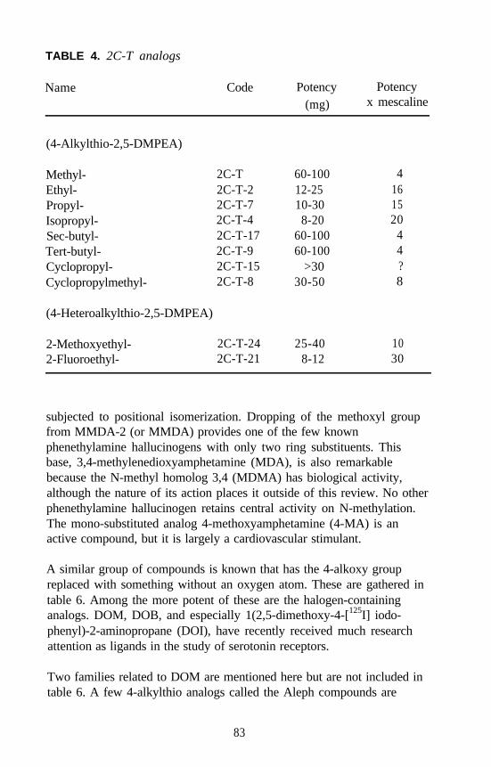
TABLE 4. 2C-T analogs
Name Code Potency(mg)
Potencyx mescaline
(4-Alkylthio-2,5-DMPEA)
Methyl-Ethyl-Propyl-Isopropyl-Sec-butyl-Tert-butyl-Cyclopropyl-Cyclopropylmethyl-
(4-Heteroalkylthio-2,5-DMPEA)
2C-T 60-100 42C-T-2 12-25 162C-T-7 10-30 152C-T-4 8-20 202C-T-17 60-100 42C-T-9 60-100 42C-T-15 >30 ?2C-T-8 30-50 8
2-Methoxyethyl- 2C-T-24 25-402-Fluoroethyl- 2C-T-21 8-12
1030
subjected to positional isomerization. Dropping of the methoxyl groupfrom MMDA-2 (or MMDA) provides one of the few knownphenethylamine hallucinogens with only two ring substituents. Thisbase, 3,4-methylenedioxyamphetamine (MDA), is also remarkablebecause the N-methyl homolog 3,4 (MDMA) has biological activity,although the nature of its action places it outside of this review. No otherphenethylamine hallucinogen retains central activity on N-methylation.The mono-substituted analog 4-methoxyamphetamine (4-MA) is anactive compound, but it is largely a cardiovascular stimulant.
A similar group of compounds is known that has the 4-alkoxy groupreplaced with something without an oxygen atom. These are gathered intable 6. Among the more potent of these are the halogen-containinganalogs. DOM, DOB, and especially 1(2,5-dimethoxy-4-[125I] iodo-phenyl)-2-aminopropane (DOI), have recently received much researchattention as ligands in the study of serotonin receptors.
Two families related to DOM are mentioned here but are not included intable 6. A few 4-alkylthio analogs called the Aleph compounds are
83
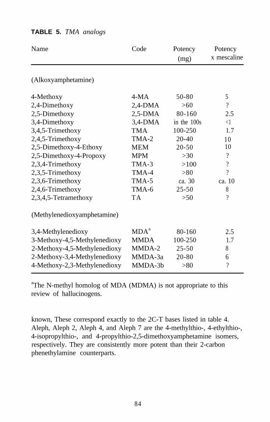
TABLE 5. TMA analogs
Name Code Potency Potency(mg) x mescaline
(Alkoxyamphetamine)
4-Methoxy 4-MA2,4-Dimethoxy 2,4-DMA2,5-Dimethoxy 2,5-DMA3,4-Dimethoxy 3,4-DMA3,4,5-Trimethoxy TMA2,4,5-Trimethoxy TMA-22,5-Dimethoxy-4-Ethoxy MEM2,5-Dimethoxy-4-Propoxy MPM2,3,4-Trimethoxy TMA-32,3,5-Trimethoxy TMA-42,3,6-Trimethoxy TMA-52,4,6-Trimethoxy TMA-62,3,4,5-Tetramethoxy TA
(Methylenedioxyamphetamine)
3,4-Methylenedioxy3-Methoxy-4,5-Methylenedioxy2-Methoxy-4,5-Methylenedioxy2-Methoxy-3,4-Methylenedioxy4-Methoxy-2,3-Methylenedioxy
MDAa
MMDAMMDA-2MMDA-3aMMDA-3b
50-80>60
80-160in the 100s100-25020-4020-50
>30>100>80
ca. 3025-50
>50
5?2.5<11.71010???
ca. 108?
80-160 2.5100-250 1.725-50 820-80 6
>80 ?
aThe N-methyl homolog of MDA (MDMA) is not appropriate to thisreview of hallucinogens.
known, These correspond exactly to the 2C-T bases listed in table 4.Aleph, Aleph 2, Aleph 4, and Aleph 7 are the 4-methylthio-, 4-ethylthio-,4-isopropylthio-, and 4-propylthio-2,5-dimethoxyamphetamine isomers,respectively. They are consistently more potent than their 2-carbonphenethylamine counterparts.
84
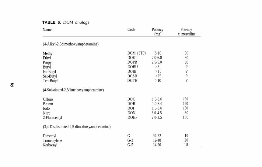
TABLE 6. DOM analogs
Name Code Potency(mg)
Potencyx mescaline
(4-Alkyl-2,5dimethoxyamphetamine)
Methyl DOM (STP) 3-10 50Ethyl DOET 2.0-6.0 80Propyl DOPR 2.5-5.0 80Butyl DOBU >3 ?Iso-Butyl DOIB >10 ?Set-Butyl DOSB >25 ?Tert-Butyl DOTB >10 ?
(4-Substituted-2,5dimethoxyamphetamine)
Chloro DOC 1.5-3.0 150Bromo DOB 1.0-3.0 150Iodo DOI 1.5-3.0 150Nitro DON 3.0-4.5 802-Fluoroethyl DOEF 2.0-3.5 100
(3,4-Disubstituted-2,5-dimethoxyamphetamine)
Dimethyl G 20-32 10Trimethylene G-3 12-18 20Norbornyl G-5 14-20 18
The second group has a 2,4,6-substitution pattern. The majority of thecompounds listed in the last few tables has carried the 3,4,5- or the2,4,5-substitution pattern. The similarity of potency between TMA-2 andTMA-6 (the latter with the 2,4,6 substitution pattern, see table 5) hasopened up a new family of hallucinogenic amphetamines, one of theauthors’ current areas of research. With this group also, the 4-positionappears to dictate the potency and nature of response. It seems that eachof the 2,4,5-substituted materials may have an active 2,4,6-counterpart.The isomer that corresponds to DOM (2,6-dimethoxy-4-methylam-phetamine [pseudo-DOM]) is active at 15 to 25 mg orally. Syntheticprocedures are now in hand to prepare the pseudo analogs of the 2C-Tfamily with various alkylthio groups at the 4-position.
The first six tables have been devoted to the phenethylaminehallucinogens; the remaining three list the second “kingdom” ofpharmacologically related compounds, the tryptamine hallucinogens.Table 7 lists the known active tryptamines other than the psilocybe group.The N,N-dialkyltryptamines are the oldest and most thoroughly studied.Those with low molecular weight groups, DMT and N,N-diethyltryp-tamine [DET], are presumably inactivated through metabolic deaminationand hence must be administered parenterally or with some amine oxidaseinhibitor. The presence of groups with increased bulk, such as isopropylgroups, on the nitrogen atom allows these compounds to be active orally.
The indole ring can carry a single oxygen substituent in the aromatic ring,and activity can be retained. The 4-substituted indoles are discussedbelow. The 5-hydroxylation of DMT (the substitution position of theneurotransmitter serotonin, 5-hydroxytryptamine [5-HT]) yieldsbufotenine, which is probably not a hallucinogen. Converting this to itsmethyl ether yields a group of N,N-dialkyl tryptamines whoseparenteral/oral availabilities closely parallel the DMT counterparts,except that there is generally an appreciable increase in potency. Themasking of the two vulnerable locales of serotonin, the O-methylation toallow entry into the CNS and the -methylation to avert enzymaticdeamination, provide , O-dimethylserotonin. This is an orally activehallucinogen of uniquely high potency. Any other substitution on theindole ring (6-, 7-, or multisubstitution) gives inactive compounds.Almost nothing is known about the oral versus parenteral requirements,potency, or nature of action of tryptamines (5-proteo and 5-methoxy)with mixed alkyl groups on the basic nitrogen atom. This is the secondarea of the authors’ current research.
86
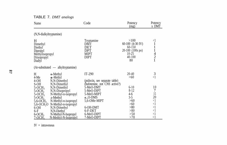
TABLE 7. DMT analogs
Name Code Potency(mg)
Potencyx DMT
(N,N-dialkyltryptamine)
HDimethylDiethylDipropylMethylisopropylDiisopropylDiallyl
(Ar-substituted — alkyltryptamine)
H -Methyl4-Me -Methyl4-OH N,N-DimethylS-OH N,N-Dimethyl5-OCH3 N,N-Dimethyl5-OCH3 N,N-Disopropyl5-OCH3 N-Methyl-n-isopropyl5-OCH3 a-Methyl5,6-OCH3 N-Methyl-n-isopropyl5,6-OCH2O N-Methyl-n-isopropyl6-OH N,N-Dimethyl6-F N,N-Diethyl6-OCH3 N-Methyl-N-Isopropyl
TryptamineDMTDETDPTMIPTDIPT
IT-290
(psilocin, see separate table)(bufotenine, not CNS active?)5-MeO-DMT5-MeO-DIPT5-MeO-MIPT
,O-DMS5,6-OMe-MIPT
6-OH-DMT6-F-DET6-MeO-DIPT7-MeO-DIPT
>100 <160-100 (4-30 IV) 1
60-150 120-100 (100s po) 1
10-25 440-100 2
80 1
20-40 3>60 <1
6-108-124-63-5>60>60>80>80>50
1071520<1<1<1<1<1
7-OCH3 N-Methyl-N-Isopropyl
IV = intravenous
>70 <1
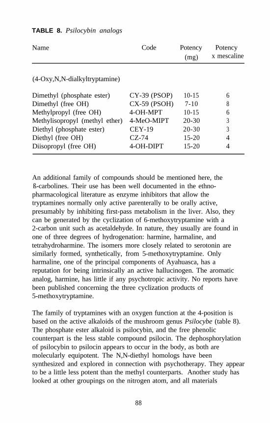
TABLE 8. Psilocybin analogs
Name Code Potency Potency(mg) x mescaline
(4-Oxy,N,N-dialkyltryptamine)
Dimethyl (phosphate ester) CY-39 (PSOP) 10-15 6Dimethyl (free OH) CX-59 (PSOH) 7-10 8Methylpropyl (free OH) 4-OH-MPT 10-15 6Methylisopropyl (methyl ether) 4-MeO-MIPT 20-30 3Diethyl (phosphate ester) CEY-19 20-30 3Diethyl (free OH) CZ-74 15-20 4Diisopropyl (free OH) 4-OH-DIPT 15-20 4
An additional family of compounds should be mentioned here, theß-carbolines. Their use has been well documented in the ethno-pharmacological literature as enzyme inhibitors that allow thetryptamines normally only active parenterally to be orally active,presumably by inhibiting first-pass metabolism in the liver. Also, theycan be generated by the cyclization of 6-methoxytryptamine with a2-carbon unit such as acetaldehyde. In nature, they usually are found inone of three degrees of hydrogenation: harmine, harmaline, andtetrahydroharmine. The isomers more closely related to serotonin aresimilarly formed, synthetically, from 5-methoxytryptamine. Onlyharmaline, one of the principal components of Ayahuasca, has areputation for being intrinsically an active hallucinogen. The aromaticanalog, harmine, has little if any psychotropic activity. No reports havebeen published concerning the three cyclization products of5-methoxytryptamine.
The family of tryptamines with an oxygen function at the 4-position isbased on the active alkaloids of the mushroom genus Psilocybe (table 8).The phosphate ester alkaloid is psilocybin, and the free phenoliccounterpart is the less stable compound psilocin. The dephosphorylationof psilocybin to psilocin appears to occur in the body, as both aremolecularly equipotent. The N,N-diethyl homologs have beensynthesized and explored in connection with psychotherapy. They appearto be a little less potent than the methyl counterparts. Another study haslooked at other groupings on the nitrogen atom, and all materials
88
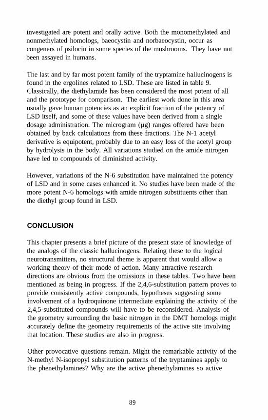
investigated are potent and orally active. Both the monomethylated andnonmethylated homologs, baeocystin and norbaeocystin, occur ascongeners of psilocin in some species of the mushrooms. They have notbeen assayed in humans.
The last and by far most potent family of the tryptamine hallucinogens isfound in the ergolines related to LSD. These are listed in table 9.Classically, the diethylamide has been considered the most potent of alland the prototype for comparison. The earliest work done in this areausually gave human potencies as an explicit fraction of the potency ofLSD itself, and some of these values have been derived from a singledosage administration. The microgram (µg) ranges offered have beenobtained by back calculations from these fractions. The N-1 acetylderivative is equipotent, probably due to an easy loss of the acetyl groupby hydrolysis in the body. All variations studied on the amide nitrogenhave led to compounds of diminished activity.
However, variations of the N-6 substitution have maintained the potencyof LSD and in some cases enhanced it. No studies have been made of themore potent N-6 homologs with amide nitrogen substituents other thanthe diethyl group found in LSD.
CONCLUSION
This chapter presents a brief picture of the present state of knowledge ofthe analogs of the classic hallucinogens. Relating these to the logicalneurotransmitters, no structural theme is apparent that would allow aworking theory of their mode of action. Many attractive researchdirections are obvious from the omissions in these tables. Two have beenmentioned as being in progress. If the 2,4,6-substitution pattern proves toprovide consistently active compounds, hypotheses suggesting someinvolvement of a hydroquinone intermediate explaining the activity of the2,4,5-substituted compounds will have to be reconsidered. Analysis ofthe geometry surrounding the basic nitrogen in the DMT homologs mightaccurately define the geometry requirements of the active site involvingthat location. These studies are also in progress.
Other provocative questions remain. Might the remarkable activity of theN-methyl N-isopropyl substitution patterns of the tryptamines apply tothe phenethylamines? Why are the active phenethylamines so active
89
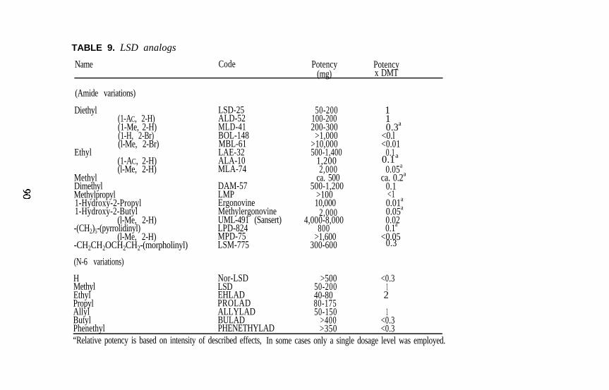
TABLE 9. LSD analogs
Name Code Potency(mg)
Potencyx DMT
(Amide variations)
Diethyl(1-AC, 2-H)(1-Me, 2-H)(1-H, 2-Br)(l-Me, 2-Br)
Ethyl(1-AC, 2-H)
Methyl(l-Me, 2-H)
ALA-10MLA-74
Dimethyl DAM-57Methylpropyl LMP1-Hydroxy-2-Propyl Ergonovine1-Hydroxy-2-Butyl Methylergonovine
(l-Me, 2-H) UML-491 (Sansert)-(CH2)5-(pyrrolidinyl) LPD-824
(l-Me, 2-H) MPD-75-CH2CH2OCH2CH2-(morpholinyl) LSM-775
(N-6 variations)
Nor-LSDLSDEHLADPROLADALLYLAD
HMethylEthylPropylAllyl
LSD-25ALD-52MLD-41BOL-148MBL-61LAE-32
50-200100-200200-300
>1,000>10,000500-1,400
1,2002,000
ca. 500500-1,200
>10010,000
2,0004,000-8,000
800>1,600
300-600
>50050-20040-8080-17550-150
>400
12
1<0.3BULAD
PHENETHYLAD >350 <0.3
110.3a
<0.l<0.01
0.10.1a
0.05a
ca. 0.2a
0.1<10.01a
0.05a
0.020.1a
<0.050.3
<0.3
ButylPhenethyl“Relative potency is based on intensity of described effects, In some cases only a single dosage level was employed.
whereas common wisdom would predict that they should be deaminatedand thus inactive? Also, with these compounds and their amphetaminehomologs, why is it only the 4-position that allows such extensivestructural manipulation without much attenuation in activity? In thetryptamine world, might there not be a host of active compounds to befound pursuing the amphetamine-like structure of ,O-DMS, and perhapsfurther homologs with one or two alkyl groups on the basic nitrogen?
The answers to these and other related questions might provide valuableinformation to help explain the remarkable activity of this class ofpsychotropic agents.
NOTE
Some of the data presented here have not been published previously. Acomprehensive bibliography documenting the known data reviewed inthis article would contain hundreds of citations. For leading reviews thatprovide these references, the following should be consulted.
Shulgin, A.T. Psychotomimetic drugs: Structure activity relationships.Iversen, L.L., Iversen, S.D.; and Snyder, S. H., eds. Handbook ofPsychopharmacology. Vol. 11. New York: Plenum Press, 1978.pp. 243-333.
Shulgin, A.T. Hallucinogens. In: Wolff, M.E., ed. Burger’s MedicinalChemistry, 4th ed. New York: Wiley and Co., 1981a. pp. 1109-1137.
Shulgin, A.T. Chemistry of Psychotomimetics. In: Hoffmeister, F., andStille, G., eds. Handbook of Experimental Pharmacology, Vol. 55/3.Berlin: Springer-Verlag, 1981b. pp. 3-29.
Shulgin, A.T., and Shulgin, A. PIHKAL: A Chemical Love Story.Berkeley, CA: Transform Press, 1991.
AUTHORS
Peyton Jacob III, Ph.D.Research Chemist3787 Highland RoadLafayette, CA 94549
Alexander T. Shulgin, Ph.D.1483 Shulgin RoadLafayette, CA 94549
91
Human Hallucinogenic DrugResearch: Regulatory, Clinical,and Scientific IssuesRick J. Strassman
INTRODUCTION
It is likely that no other group of drugs has been subject to such intensiveclinical study and to then nearly complete neglect in such a short a periodof time as the hallucinogens. Mescaline, the most well-knownpsychoactive alkaloid of the peyote cactus, was studied in a limitedfashion (Kluver 1928) before the discovery of lysergic acid diethylamide(LSD). However, Sandoz Laboratories’ description of the microgrampotency of LSD to induce psychotomimetic effects (Stoll 1947) generatedtremendous excitement in psychiatry and may be seen as an equallyimportant precursor to the modern era of biological psychiatry, as was thecontemporaneous discovery of chlorpromazine’s antipsychotic properties.
Thousands of subjects, both normal volunteers and psychiatric patients,received LSD and similar drugs in research in the United States andEurope. Szára (this volume) has summarized the research done andmonetary effort expended toward understanding LSD’s effects during theearly 1950s through the early 1970s. Important insights into humancognitive, emotional, electroencephalographic, and perceptual functionsin normal individuals and in persons with a broad range of psychiatricdisorders were detailed, based on early psychopharmacologicinvestigations of LSD’s effects (Hoffer and Osmond 1967). Drug-assisted psychotherapy, although not clearly proven efficacious beforeresearch with these drugs was stopped, had shown some promising results(Pahnke et al. 1970). Neither optimal patient selection nor psycho-therapeutic approaches had been decided upon, nor had a firm theoreticalbasis for this type of work been developed (Grinspoon and Bakalar1979).
There are several recently published popular histories of the psychedelicera: its beginnings, flourishing, and eventual decline within the scientific,legislative, and media communities (Lee and Shlain 1985; Stevens 1987).Unsupervised use of these drugs by millions of young adults and the
92
domestic instability caused by the Vietnam War combined to make useand abuse of hallucinogens a significant public health concern. In orderto restrict their use by the public, hallucinogens were placed intoSchedule I of the Controlled Substances Act of 1970. Drugs in thisschedule have no known medical use, cannot be used safely even undermedical supervision, and are highly abusable. However, the regulatoryprocess involved in working with Schedule I compounds discouraged allbut a few investigators from human research with these compounds.Recently, however, mescaline’s psychological and electroencepha-lographic effects have been studied by a German group (Oepen et al.1989). In addition, a group of Swiss clinical psychiatrists has beenadministering 3,4-methylenedioxymethamphetamine (MDMA) and LSDto psychiatric patients since 1985, but no data have been forthcomingfrom purely clinical use (Widmer 1992).
The confusing and often contradictory nature of the approaches taken inunderstanding and studying these compounds is reflected in the manynames they have received. These names include but are not limited to:“hallucinogenic,” “psychedelic,” “psychotomimetic,” “mysticomimetic,”“entheogenic” (generating the divine), “psychodysleptic,” “illusogenic,”“phanerothyme” (“soul-feeling”), “psycholytic,” “phantasticant,” and“psychotogenic” (Stafford 1982). “Hallucinogenic,” probably the mostwidely used term in the medical literature, is used in this chapter.However, it should be remembered that frank hallucinations with thesedrugs are rare, and hallucinogen is as much a term of convenience as ofaccuracy (Shulgin, this volume).
A tremendous amount of publicity and scientific discourse took placeregarding the adverse effects of hallucinogenic drugs. As early as 1960,Cohen had published data on nearly 5,000 individuals exposed to LSDeither as research subjects or in drug-assisted psychotherapy. These caseswere obtained from his and others’ research caseloads. He concluded thatthe incidence of psychotic reactions lasting longer than 48 hours was0.08 percent in controls and 0.18 percent in patients; the incidence ofsuicide attempts was 0 percent in controls and 0.12 percent in patients(Cohen 1960). Thousands of cases subsequently were reported ofindividuals presenting to emergency rooms and psychiatric clinics fortreatment of adverse reactions. These included acute panic reactions, thebad trip, and “flashbacks,” or spontaneous eruptions into awareness ofone or more features of a previous drug experience.
93
These reports have been reviewed carefully, and it was concluded thatnearly all of these contained serious methodological flaws (Strassman1984). These unavoidable limitations to these descriptions of adverseeffects included lack of knowledge of the actual identity or dose of drug,confounding combinations of other drugs or alcohol, sparse premorbiddata on the subjects’ psychiatric history, and selection bias. Furthermore,it appeared that in carefully screened, prepared, supervised, and followed-up subjects, both experimental and psychotherapeutic, the incidence ofprolonged or delayed adverse reactions was extremely low (Strassman1984).
The necessary interfacing between human psychopharmacology and basicneuropharmacology has proceeded for the psychotherapeutic drugs suchas minor tranquilizers, antidepressants, neuroleptics, and lithium. Theseparallel courses of investigation have provided clinicians with newfamilies of drugs based upon data generated in lower animal models.Examples include the 5-hydroxytryptamine (5-HT1A) partial agonist andanxiolytic agent buspirone and the selective serotonin reuptake inhibitorsfluoxetine and sertraline. Such interfacing work, however, has not beenpossible with the hallucinogens because of the difficulty associated withhuman research with them.
In spite of restrictions on human use of hallucinogens, basic research hasproceeded at a rapid rate. For example, the original work describing the5-HT1 and 5-HT2 subtypes used LSD (Peroutka and Snyder 1979); thelater discovery of the 5-HT1C subtype was made with LSD (Yagaloff andHartig 1985); and phenethylamine hallucinogens are now the standardradioligands for localization of 5-HT2/5-HT1C subtypes in mammalianbrain (Appel et al. 1990).
Several converging factors now provide a suitable backdrop for theresumption of human research with hallucinogens (Freedman 1984). Theaforementioned advances in serotonin neuropharmacology havegenerated one of the major thrusts in current neuroscience research; thatis, the role of serotonin in normal and aberrant brain function. Muchhuman psychopharmacologic data are being generated on the effects of5-HT agonists and antagonists, including L-tryptophan, L-5-hydroxy-tryptophan, fenfluramine, chlorimipramine, buspirone, b-chloro-(1-piperazinyl)pyrazine (MK-212), meta-chlorophenylpiperazine(MCPP), ritanserin, metergoline, methysergide, and cyproheptadine (e.g.,Murphy et al. 1991). No 5-HT active compounds generate moreprofound or unusual effects on mental function than the hallucinogens,
94
and a firmer theoretical foundation is now available to begin assessing therelative contributions of specific 5-HT subtypes in their mechanism ofaction. In addition, the use of hallucinogenic drugs by young adults isgrowing, unlike the use of other illicit psychoactive drugs (NIDA 1991).Therefore, public health issues are addressed by resumption of carefulstudy of the hallucinogens with the possibility of developing safe,effective, and rapid antidotes to drug-induced acute adverse reactions.Finally, the approaches taken by proponents of the psychedelic revolutionmay have faded sufficiently into the background of the American psycheto allow the resumption of systematic, hypotheses-based research into theeffects and mechanisms of action, and perhaps therapeutic utility, of thesemost interesting drugs.
EPIDEMIOLOGY AND PUBLIC HEALTH SIGNIFICANCE
Hallucinogen use in this country had remained relatively constant overthe last 20 years but now appears to be increasing. The 1990 high schooldrug use survey (Johnson et al. 1991a) found that approximately10 percent of high school seniors polled had used hallucinogens at leastonce, an increase of 0.4 percent since 1989. These lifetime prevalencerates are about the same as those for cocaine, seven to eight times higherthan for heroin, almost twice as high as for sedatives, and three timeshigher than for steroids. LSD ranks first in the categories “most intense”and “longest” high among respondents. The comparable populationestimates for young adults and college students indicate a 0.8 percent risein lifetime prevalence rates for LSD use (0.6 percent increase in annualprevalence rates) in 1990 compared to 1989; females’ use had beenconverging on males, but “in 1990, an important increase in LSD useamong males widened the difference again” (Johnson et al. 1991b, p. 92).Lifetime prevalence rates in young adults for LSD are three times thosefor phencyclidine (PCP) and more than half those for cocaine. The 1990National Household Survey of Drug Abuse (NIDA 1991) reports thatlifetime prevalence of hallucinogen use is 7.6 percent, compared with11.3 percent for cocaine. The number of individuals in this country,therefore, who have used an hallucinogen at least once is estimated atbetween 13 and 17 million. Whites use hallucinogens more (8.7 percentlifetime prevalence) than either African Americans (3.0 percent) orHispanics (5.2 percent), with a consistent male-to-female ratio of2-3:1 across all ethnic groups.
95
The compound used in the study described below, N,N-dimethyl-tryptamine (DMT), is used by a seemingly small proportion ofhallucinogen abusers. However, DMT is a prototypical tryptaminehallucinogen. Variations on its structure have generated other short-duration parenterally active drugs such as N,N-diethyltryptamine (Száraet al. 1966) and N,N-dipropyltryptamine (Soskin et al. 1973).Furthermore, designer drugs developed to circumvent existing schedulinglaws and/or produce specific qualitative effects derive from thiscompound (Repke et al. 1985). Other recent developments also areimportant. Ayahuasca, an Amazonian plant extract (McKenna et al.1984a), has become popular in some circles on the coasts, being importedfrom Peru and Brazil. It contains DMT, found in one of the plants(Psychotria viridis), and monoamine oxidase (MAO) inhibitingß-carboline harmala alkaloids in another plant (Banisteriopsis caapi),allowing the DMT to become orally active (McKenna et al. 19848).
Space limitations prevent a detailed discussion of the relationshipbetween hallucinogen use and other psychiatric disorders. However, thegreater incidence of use of hallucinogens by individuals withschizophrenia than by a normal population (Mueser et al. 1990), thepossible role of hallucinogens in the development of borderlinepsychopathology (Dulit et al. 1990), the higher incidence of hallucinogenuse in nondrinkers who are first- and second-degree relatives ofalcoholics compared to controls (Schuckit and Sweeney 1987), and therole of hallucinogens in precipitating schizophrenic and other psychoses(Vardy and Kay 1983) all bespeak the importance these drugs have forissues of public psychiatric health.
REGULATORY ISSUES
A detailed account of the process by which an investigational new drug(IND) permit was acquired for DMT has been published (Strassman1991). This chapter summarizes the salient points.
There were several reasons DMT, rather than longer-acting, more popularhallucinogens, was chosen for a research proposal. First, it is a veryshort-acting hallucinogen, with peak effects obtained within 2 to5 minutes after intramuscular (IM) injection (Sai-Halasz et al. 1958).This attribute is an advantage in the potentially anxiety-provokingenvironment provided by a modem clinical research center, where arenewal of human studies with hallucinogens must take place. That is, no
96
matter how troubled a subject’s experience on DMT might be, it wouldbe short-lived and relatively manageable.
Second, DMT is an endogenous hallucinogen whose existence in thehuman nervous system has never been adequately explicated (Gillin et al.1976). Thus, an investigation into the mechanisms of action and effectsof DMT might shed some light on endogenous hallucinatory states.Along this line of reasoning, antidotes to DMT might show efficacy astreatments of disorders with hallucinations such as schizophrenia. Theimportance of the 5-HT2 receptor subtype in the mechanism of action ofthe novel antipsychotic drugs clozapine (Meltzer and Nash 1991) andritanserin (Gelders et al. 1986) bespeaks the relevance of this approach.Finally, DMT abuse is not a widespread phenomenon. Thus, beginninghuman hallucinogen research with DMT would not cause the intensepublic scrutiny that might result from knowledge that LSD once againwas being used in humans.
In order to use a drug in humans for either nonindicated purposes or apurpose with no currently established use, it is necessary to apply to theFood and Drug Administration (FDA) for an IND permit. However, for aSchedule I hallucinogen, none of which has been used in humans formany years, there is the additional problem of locating an acceptablesource of the compound. A Drug Master File for the compound must beestablished for the FDA to determine basic safety and manufacturing dataconcerning any drug for human use. This file contains manufacturingand quality control data for the drug to which FDA may refer. FDA also is charged with concurrently establishing whether the proposed course ofstudy will yield worthwhile data.
The proposed study was to establish normative dose-response data forDMT in a group of experienced hallucinogen users in a double-blind,randomized, placebo-controlled study design.
Experienced hallucinogen users were chosen for three reasons:experienced subjects would be less likely to panic than naive subjects inresponse to the sudden onset and intensely hallucinogenic effects of ashort-acting drug such as DMT; experienced subjects would be morecapable of carefully describing the effects of DMT than would subjectswith no experience with these compounds; and liability for thesubsequent development of substance abuse and/or other psychiatricpathology would be less sustainable in subjects with a prior history of useof these drugs.
97
The literature on the biological and psychological effects on humans andanimals of hallucinogens in general and DMT in particular was reviewed,primarily from the perspective of recent advances in 5-HT neuropharm-acology and psychopharmacology. Several hypotheses were thengenerated regarding what variables would be affected by DMT, what theinterpretation of these effects would be, and subsequent experiments thatwould help prove or disprove these interpretations. The variables ofinterest were easily observable and verifiable. They included severalneuroendocrine functions, cardiovascular effects, autonomic effects(pupil diameter and core temperature), and rating scale scores.
The first step in the application process for use of DMT was to submit aprotocol to the Human Research Review Committee of the University ofNew Mexico School of Medicine’s Institutional Review Board (IRB).The IRB is responsible for the protection of human subjects involved inany clinical research. Although the IRB is concerned primarily with risk-to-benefit ratios, it also may offer suggestions regarding the scientificquality of the proposed study. This latter function usually is subsumedby the Scientific Advisory Committee of the General Clinical ResearchCenter (GCRC) of the University of New Mexico Medical School, wherethe actual study was to take place.
The IRB places great emphasis on the nature of the informed consentdocument and, at first, was inclined to have this document state that DMT“had no known medical use,” one of the criteria for placement intoSchedule I. However, the suggestion was made that, if FDA approvalwere subsequently obtained, this would not be the case, as DMT wouldhave been approved as a medically useful compound (i.e., as a probe ofhuman serotonergic psychopharmacology and neuroendocrinology). TheIRB accepted this reasoning and did not insist on including this phrase,which would have unnecessarily alarmed the subjects who were alreadyapproaching the study with some trepidation. The IRB also wanted astatement in the informed consent document regarding what effectssubjects might experience while under the influence of DMT.
Interviews had previously been conducted with 19 experienced DMTfree-base smokers in order to draft a new rating scale for this study (seebelow). Thus, a relatively balanced account of the subjective effects ofDMT could be provided without going into too much detail. Thesubjects interviewed spoke most frequently of positively charged effects,so these predominated in the informed consent. However, panic, fear,confusion, and other unpleasant experiences also were listed as possible
98
outcomes. Prolonged ( > 24 hour) reactions to hallucinogenic drugs dooccur in carefully controlled settings, although such reactions never havebeen reported for DMT. However, their overall incidence is quite low.The informed consent document, then, included a statement concerningthe fact that subjects might experience prolonged adverse responses to thedrug and that psychiatric hospitalization was available.
Finally, the IRB was told that local approval was necessary beforeapplying for Federal permission to begin this study. Once Federalpermission was acquired, the principal investigator (PI) would notify theIRB and begin, and not before.
The issues of confidentiality and anonymity also were of concern, sinceadmitting to the use of Schedule I drugs is a crime, and a relatively high-functioning group was expected to volunteer for the study based on thesocioeconomic status of the initial interviewees. Meetings with theuniversity hospital counsel, directors of medical records and hospitaladmissions, GCRC head nurse, and administrative assistants led to acoding scheme by which subjects’ confidentiality and anonymity weremaintained, with only the PI having the key to the code.
The next step was presenting the protocol to the GCRC ScientificAdvisory Committee for a thorough scientific critique. Issues ofinformed consent and ethical review were not taken up, except in ageneral way, as these concerns were the purview of the IRB.
After approval from both university committees charged with scientificand ethical issues, State Pharmacy Board approval was necessary toobtain and use a Schedule I drug. The protocol and the appropriateapprovals had previously been submitted to the Drug EnforcementAdministration (DEA) in Washington, DC, but approval had not yet beenreceived. However, the State Pharmacy Board approved the protocolafter it was explained that Federal DEA approval could not be receiveduntil State approval was obtained and that the project could not beginuntil all the requisite Federal permits were in hand.
The Federal approval process required simultaneous interaction with theFDA and the DEA. The applications for a Schedule I permit for DMTwere sent to DEA, and the IND application was sent to FDA at the sametime. Nearly 2 years were required to keep both of these processes ontrack until they were finally approved.
99
The application process to the DEA was complicated by requests forDMT for two separate reasons, possibly from two different sources, andof two different grades. DMT was required for the development of alaboratory assay for DMT in human blood and could be purchased fromone of several chemical supply houses. However, laboratory grade DMTcould not be used for human studies, even if it were purified to FDAstandards. Thus, an additional source of pharmaceutical quality DMTwas necessary for human administration. DEA was reluctant to approvethe Schedule I application to possess laboratory grade DMT until the INDwas received from the FDA. FDA could not decide whether to approvethe lND request until pharmaceutical quality DMT was on hand and itsappropriateness for human use could be confirmed. FDA finally wrote abrief letter to DEA saying that the science of the study was reasonableand that human studies of DMT would not begin until the FDA haddeemed it safe to begin, but that DEA could approve the Schedule Ipermit to obtain laboratory grade DMT (so assay development couldbegin) and pharmaceutical grade DMT (to which the appropriate qualitycontrol procedures could be applied).
The next major effort involved finding a source of DMT approved by theFDA. The FDA required a pedigree of the DMT if it were to beadministered parenterally, the only way DMT alone is active. Thepedigree is a description of all precursors, intermediates, and impurities inthese compounds, en route to the final synthesis of the DMT. A detailedchemical analysis of the final product also is required. Additionally, themanufacturer must provide FDA with details concerning themanufacturing and cleanup procedures to assure FDA that “goodmanufacturing procedures (GMP)” were followed. The National Instituteon Drug Abuse (NIDA) had some DMT “on the shelf,” as did SigmaChemical (from which the laboratory grade DMT was purchased). Eithersample could be purified to > 99 percent purity, but FDA would notaccept DMT without a pedigree. Neither NIDA nor Sigma had thepedigree information. Several other possible sources also proved to beeither legally or economically unfeasible.
Finally, the DEA determined that regulations allow the synthesis of smallbatches of Schedule I drugs by previously licensed research laboratoriesif the drug was for research rather than fiduciary purposes. The“coincidental activities” clause frees synthesizers of small batches ofscheduled drugs from the much more rigorous and costly securityarrangements required for manufacturers with primarily commercialinterests. A colleague with a Schedule I permit for DMT then agreed to
100
make a small batch of DMT for this study and to provide the requisitedocumentation, a collaboration with which FDA was satisfied inprinciple.
The DMT was formulated for intravenous (IV) administration by theinpatient pharmacy of the University of New Mexico Hospital. TheDMT then had to be put through the quality control steps required byFDA. Since pharmaceutical companies work with much larger lots ofdrugs than this small batch, it took some creative problem solving todetermine the number of vials of injectable DMT that needed to be testedfor pyrogenicity, sterility, and content uniformity. The last issueconcerns both the concentration of representative samples of the productbeing within 5 to 10 percent of the hypothetical value (in this case40 milligrams per milliliter [mg/ml]) and the maintenance of thisconcentration over time.
Satisfied with the quality of the DMT for human use and the scientificrigor of the study, FDA gave approval to begin. DEA was notified at thistime. Amendments to this protocol, if not substantive, were discussedand approved by telephone. More substantial amendments, such ascombining DMT with another drug, required both written and telephoneconsultation as well as formally requesting the manufacturer of theadditional drug to provide written authorization for FDA to access itsDrug Master File and IND information. DEA was sent copies of allchanges to the original DMT protocol.
CLINICAL ISSUES
Recruitment, Screening, and Orientation of Subjects
Experienced hallucinogen users were recruited for this study by word ofmouth, except for the last subject who responded to an announcementsent through the psychology department graduate student training officeof a local university. Although this particular issue has not yet arisen, itwas decided to take no more than two subjects from any particulardepartment or division to prevent the development of cliques, anunfortunate occurrence in previous human hallucinogen studies usinggraduate students. Subjects initially called the PI to discuss the protocol.They were asked about current medication use or drug abuse and whetherthey had experience with hallucinogens. Those who were takingmedication chronically, had an intercurrent medical illness, or were
101
currently abusing cocaine or alcohol were not accepted for furtherscreening. Occasional marijuana use was accepted. The subjects werethen interviewed in person to screen more carefully for current medicaland psychiatric conditions and to assess the level of experience withhallucinogenic drugs. This interview assessed adaptive and maladaptiveresponses to dysphoric experiences with these drugs. That is, theinvestigators were interested in knowing how subjects managed bad tripsand whether they repressed and denied, or acknowledged, the relevanceof these experiences to their personal lives. Examples of this type ofadaptive response to an acute adverse reaction are being more carefulwith whom the person takes hallucinogens and deciding not to take thesedrugs in a busy metropolitan setting.
Subjects would have been withdrawn if they currently suffered from asevere Axis I disorder (e.g., schizophrenia, major affective disorder,substance abuse, or anxiety disorder) or had a history of psychosis notdue to either drugs or fever. One subject with an adjustment disorderrelated to divorce proceedings was included, as he appeared to bemanaging these stresses adaptively. A past history of major depression orsubstance abuse was not necessarily cause for exclusion from the study ifsubjects were in good remission for at least 1 year, understood the natureof their episode(s), and were not in life circumstances that couldprecipitate a recurrence during the study.
Subjects also were required to have relatively good object relations, asdetermined by stability of work, school, and interpersonal functions. Inthis study, the subject with the most chaotic life circumstances was theperson who developed an intercurrent depression midway through theprotocol and had to be withdrawn from further participation. Suchindividuals are not believed suitable for this type of project until theirpersonal and/or work lives are more stable.
Subjects were interested in why this work was being done, the expectedresults, and the hypotheses. Honesty and tact are very important whenorienting and preparing subjects for hallucinogenic drug studies. Suchsubjects, during the intoxication, may be exquisitely sensitive tointerpersonal nuances, attempts to obscure or lie, and personal discomfortwith particular issues. Any approach with these subjects that hints ofdeviousness, dishonesty, manipulation, or callousness is multipliedseveral times over when the actual drug intoxication is underway. Thus,starting off on the right foot is not only desirable but will preventparanoid, panic, and dysphoric reactions later during the actual study.
102
At that time, the nature of the study was described, and subjects receiveda copy of the informed consent to review. The subjects were asked tothink over their participation and were told to call the PI with theirdecision or any further questions. Prospective subjects were instructed todiscuss their participation with their spouse or significant other. Thehandful of subjects with fearful and reluctant spouses needed more timeto decide to participate; some required a meeting between the spouse andstudy staff to prevent study participation from generating unnecessaryfriction within a couple. Such friction could not help but carry over intothe drug sessions and affect the quality of drug effects.
The informed consent document stated that the primary effects of DMTwere psychological, although high doses can cause a transient moderatelyelevated blood pressure. Auditory and/or visual hallucinations mightoccur, and other unusual effects were likely. Sense of time might bealtered; very powerful emotions, both pleasurable or unpleasant, might beexperienced, including opposite feelings or thoughts at the same time.Extreme sensitivity to the environment and people, including frankparanoia, could occur; on the other hand, subjects might not noticeanything at all in the environment. Separation of mental processes fromall physical sensations could occur. Extreme changes of mood are notuncommon. The incidence of prolonged ( > 24 hr) adverse reactions tohallucinogenic drugs in well-screened, prepared, and followed-upsubjects was described as quite low. If prolonged adverse reactions toDMT did develop, 24-hour emergency psychiatric consultation wasavailable. Psychiatric hospitalization could also be arranged.
Only 3 of the original 12 subjects had previous experience with DMT,and all were extremely interested in knowing as much about it aspossible. Each subject received a copy of a chapter on the history,botany, chemistry, and effects of short-acting tryptamines written for aneducated lay audience (Stafford 1982, pp. 308-331).
Prospective subjects then received a structured clinical interview (SCID)of the Diagnostic and Statistical Manual of Mental Disorders III-Revised(DSM-III-R), Outpatient Version (Spitzer et al. 1987), by a trainedpsychiatric research nurse. First-degree relatives’ psychiatric historieswere obtained during the SCID. Diagnoses were confirmed bydiscussion with two research psychiatrists. A medical history, a physicalexamination, and laboratory screening tests then were performed by thePI. Laboratory tests included a complete blood count with differential,24-item chemistry panel, thyroid functions (including thyroid stimulating
103
hormone), routine urinalysis, and electrocardiogram (EKG). Subjectswere withdrawn who demonstrated any significant abnormalities in thisscreening, including thyroid disorder, high blood pressure (diastolic over90 mm Hg), heart disease/abnormal EKG, hepatitis, peptic ulcer disease,diabetes, or other serious medical problems.
The PI administered the medical work-up personally to establish a hands-on relationship with subjects that would facilitate their ability to morecomfortably regress under the effects of DMT under supervision in aclinical research setting, and because the personal relationship mightprove helpful. This was also the first opportunity for subjects to meet theresearch nurse who was to assist in the drug sessions. She introducedherself, drew the requisite screening blood samples, performed the EKG,and gave subjects a brief tour of the inpatient unit. This allowed subjectsto initiate a relationship with the nurse and become accustomed to hermanner of performing potentially painful, uncomfortable, andembarrassing procedures (even more so in the midst of the DMTintoxication). Subsequently, the PI and nurse discussed initialimpressions of the subjects.
Nature of the Research Team
The importance of one clinical nurse being involved with subjects fromstart to finish of the protocol cannot be overemphasized. The nurse’spresence is important to the development of subjects’ trust and ability toattend completely to the effects of the drug, just as the subjects’ trust andconfidence in the PI as the only one ever to administer the drug isimportant. On the rare occasion when the nurse was unavailable forinpatient studies, things never went as smoothly, subjects were not asrelaxed, and verbal and nonverbal signals between the substituting nurseand PI were not as clear and unimpeded as with the regular nurse. All ofthese issues are significant when subjects are in an extremely regressed,suggestible, and, at times, disoriented state.
In addition, the nurse preferably should be of the opposite gender to thatof the PI. The regressed state induced by hallucinogens greatly enhancesthe subjects’ tendency to project and overly identify with theexperimental team. The presence of two authority figures of oppositegenders may help provide a soothing transference template for subjectswho may be experiencing helpless infantile feelings.
104
The PI for human hallucinogen studies should be a psychiatrist withexperience in the interviewing, diagnosis, and treatment of psychotic(drug-induced or otherwise) states. The PI should have extensivepsychotherapeutic expertise, as the transference and countertransferenceissues involved in dealing with childlike and regressed conditionsrequires great tact and sensitivity. If at all possible, the PI should alsohave had a successful course of psychotherapy/psychoanalysis in order tolearn about his/her interpersonal style, nuances, idiosyncracies, and theireffect on others.
DMT Sessions and Their Supervision
Before entering into the double-blind, placebo controlled, randomizedarm of the study, all subjects received nonblind administrations of0.04 and 0.4 milligrams per kilogram (mg/kg) IV DMT, usually onconsecutive days. The DMT solution (40 mg/ml) was drawn into a steriletuberculin syringe at a dose range of 0.04 to 0.4 mg/kg, and diluted withsterile saline to 1.0 ml before administration. Tolerance was not aconcern, as previous work demonstrated no tolerance to twice daily IMadministration of fully hallucinogenic doses of DMT for 5 consecutivedays. The drug was administered IV rather than IM as had been reportedpreviously (Szára 1957) because of the results of the initial dose-findingstudies using a subject with experience smoking DMT free base, the usualform and route of administration by recreational users (Stafford 1982).The onset and intensity of effect of 1.0 mg/kg IM DMT fumarate weredescribed by this subject as significantly less intense and hallucinogenicthan a previous experience with the smoked drug. Further dose-findingstudies with this subject and another experienced DMT smokerestablished that 0.4 mg/kg IV DMT fumarate produced a rush andhallucinogenic effects comparable to or slightly greater than those seenwith a full dose of smoked DMT free base.
Subjects were admitted to a dimly lit (about 100 lux) room by 9:00 a.m.after having fasted since midnight. A butterfly style small gauge metalneedle was inserted into a forearm vein and kept patent with heparinizedsaline. Subjects were allowed to relax for 30 minutes before the drug wasgiven. DMT was infused over 30 seconds and flushed with 5 ml ofsterile saline over the next 15 seconds. Blood pressure and heart ratewere taken with an automatic cuff several times before and frequentlyduring the 30 minutes following drug administration. IV diphen-hydramine and diazepam and sublingual nitroglycerin were available forsevere allergic, emotional, and hypertensive reactions, respectively. The
105
research team (a nurse and psychiatrist) sat quietly on either side of thesubject, attentive to verbal and nonverbal cues, but did not offer anydirection or advice unless absolutely necessary during the first severalminutes, the time of peak effects of the drug. Verbal and emotionalsupport were provided as necessary, but drug sessions were notexploratory or therapeutic in intent nor nature. The experienced nature ofthe subjects, their relatively stable object relations, and the consistentemphasis on objective data collection was helpful in focusing subjects’expectations toward these goals. As drug effects resolved, discussionfocused on the nature of the subject’s experiences, The butterfly catheterwas removed at 30 minutes postinjection, and the Hallucinogen RatingScale (HRS, see below) was administered. After the subjects ate a snackor meal, they were discharged.
Subjects felt the onset of effects before the 45-second infusion wascompleted. Peak hallucinogenic effects of 0.4 mg/kg doses occurredusually before the first vital sign check at 2 minutes postinjection, auseful temporal reference point for subjects. Peak effects began resolvingat 90 to 120 seconds postinjection. Subjects remained moderatelyintoxicated by the 0.4 mg/kg dose for another 5 to 15 minutes and feltrelatively normal by 25 to 30 minutes postinjection.
The nonblind days served several purposes. They allowed assessment ofthe cardiovascular and subjective effects of both subclinical and fullyhallucinogenic doses of DMT in a novel and potentially anxiety-provoking setting. These days also provided the research team andsubjects an opportunity to become acquainted and familiar with eachothers’ styles without the extensive blood drawing and use of the rectalthermistor that could exaggerate any potentially paranoid reactions.Subjects were also able to calibrate themselves to what to expectregarding minimal and maximal effects of the drug, one of the purposesof the double-blind study being to determine whether subjects coulddistinguish among incremental doses of DMT. Finally, these nonblinddays provided subjects with the opportunity to drop out of the studybefore extensive data had been collected. None withdrew, although onesubject was dropped because his diastolic blood pressure rose to >100mm Hg after a low dose.
Double-blind study days were quite similar to the nonblind days. Two IVlines were inserted, the second for frequent blood drawing, as was aflexible reusable rectal thermistor for core temperature measurement.Pupil diameter was measured against a standard card with black circles of
106
1 millimeter gradations near the subject’s face. DMT and sterile salineplacebo dosages of 0.05, 0.1, 0.2, and 0.4 mg/kg were administered.Dosages of 0.4 mg/kg were assumed to be fully hallucinogenic, 0.05 mgwould be barely distinguishable from placebo, and the intermediate doseswere equally spaced on a log scale. Blood samples were drawn and laterassayed for beta-endorphin, adrenocorticotropic hormone (ACTH),cortisol, prolactin, growth hormone, and melatonin. Pupil diameter andvital signs (VS) (i.e., heart rate and blood pressure) were measured 2, 5,10, 15, 30, and 60 minutes after drug administration. Since the last bloodsample was at 60 minutes postinjection, the HRS was administered30 minutes later than for the nonblind sessions. This second 30-minuteperiod was usually taken up by more relaxed conversation, althoughwhen possible, attention was repeatedly turned back toward the subjects’drug experience.
Study sessions occurred at an interval of at least 2 weeks from January toSeptember, 1991. The one female subject was studied during the earlyfollicular phase of her cycle. A negative result to a pregnancy test drawnthe night before her study days was always ensured before drugadministration.
SCIENTIFIC ISSUES
Serotonergic Mediation of Biologic and Behavioral Effects
Hallucinogenic drugs have multiple effects on central neurotransmission.The area currently of greatest interest is serotonin and its receptors,specifically the 5-HT2, 5-HT1A and 5-HT1C, subtypes (Heym and Jacobs1987). Noradrenergic (Horita and Hamilton 1969) and dopaminergic(Ahn and Makman 1979) effects of hallucinogens have been describedbut are not now receiving comparable attention.
Hallucinogenic drugs, as primarily serotonergic agonists or partialagonists, have diffuse and relatively well documented physiologicaleffects. Growth hormone, prolactin, beta-endorphin, ACTH, and cortisolare all affected by serotonergic stimuli (Van de Kar 1991). Many of thesehormones rise with the administration of hallucinogens in lower animals(Koenig et al. 1987) and humans (Demsich and Neubauer 1979) and arebelieved modulated by 5-HT1A, 5-HT1C, and/or 5-HT2 receptor subtypes.Cardiovascular variables (McCall and Humphrey 1982), core temperature(Horita and Dillie 1954), and pupillary diameter (Greiner et al. 1958) also
107
are affected by serotonergic hallucinogens, with the majority of datasuggesting 5-HT2 mechanisms of action.
Studies in Animals. Animal behavioral studies use several models ofhallucinogenicity to pharmacologically characterize known hallucinogensand to assess whether novel compounds have hallucinogenic properties.These models include drug discrimination (DD) (Appel et al. 1982)(consistent with 5-HT1A [Spencer et al. 1987] and 5-HT2/5-HT1C
[Glennon, this volume] effects); the serotonin syndrome (mediated by the5-HT1A subtype [Smith and Peroutka 1986]) and one of its components,head shakes (mediated by the 5-HT2 subtype [Peroutka et al. 1981]); theabortive limb flick in cats (Jacobs et al. 1976); and the characteristicexploratory behavior patterns of rodents placed in a novel environment(Geyer, this volume). All these models, although generating muchrelevant data, lack requisite specificity. For example, apomorphine(Geyer et al. 1979) and quipazine (Kuhn et al. 1978) appearhallucinogenic in some of these models, and psilocybin (Koerner andAppel 1983) is sometimes not found to be so.
The 5-HT2/5-HT1C subtype is now of most interest regarding mediation ofhallucinogenic drug effects. For example, ritanserin, a potent andselective 5-HT2/5-HT1C antagonist (Sahin-Erdemeli et al. 1991), blocksthe discriminable effects of LSD in lower animals (Colpaert et al. 1985).It also blocks certain neuroendocrine (Lee et al. 1991) and subjective(Seibyl et al. 1991) effects of serotonergic agonists in humans, effects thatmay be mediated by 5-HT2/5-HT1C receptors. Finally, anecdotal reportsindicate that ritanserin pretreatment blocks the hallucinogenic effects ofLSD in humans. However, the 5-HT1A receptor subtype also is important.DMT (Deliganis et al. 1991) and LSD (Pierce and Peroutka 1989) havealmost equal affinities for the two subtypes.
An endogenous human hallucinogen, 5-MeO-DMT (Smythies et al.1979), used in many animal studies of hallucinogenic drug effects, hasgreater affinity for the 5-HT1A receptor than for the 5-HT2 receptor(McKenna et al. 1990). Additionally, the discriminative effects of5-MeO-DMT, which generalize to LSD (Young et al. 1982) and theselective 5-HT2/5-HT1C agonist l-(2,5-dimethoxy-4-methylphenyl)-2-aminopropane (DOM) (Glennon et al. 1979; Glennon et al. 1986), areblocked by the 5-HT1A antagonist pindolol (Spencer et al. 1987), which isessentially devoid of 5-HT2/5-HT1C effects (Hoyer 1988). The 5-HT1A
receptor may therefore contribute necessary effects that produce thecharacteristic hallucinogenic drug syndrome. Although DMT’s
108
neuroendocrine and behavioral effects were inconsistently blocked bycyproheptadine (Meltzer et al. 1980), the lack of specificity of thisantagonist makes assessment of relative contributions of the 5-HT2 and5-HT1 subtypes difficult.
Experimental Results of DMT Administration. Full details of theeffects of DMT administration on multiple biological variables have beenpresented (Strassman and Qualls 1994). Generally, DMT was physicallywell tolerated by the subjects participating in this study. There were noepisodes of nausea or vomiting with the dose range used, something thatcannot be always said for longer-acting drugs (Denber 1956). Onesubject’s diastolic blood pressure rose to >100 mm Hg after a screening,nonblind dose of 0.04 mg/kg; that subject was withdrawn from furtherparticipation. As one other subsequent subject had to be dropped fromfurther study after a similar rise in response to 0.05 mg/kg DMT, this testdose of drug is necessary if higher doses are to be used.
As expected, based upon lower animal and clinical data, there were dose-dependent elevations in several neuroendocrine variables in response toDMT. Beta-endorphin, ACTH, cortisol, and prolactin were stimulated.The rise in POMC peptides was nearly 4 to 5 times baseline, with peakeffects occurring within 5 minutes of DMT administration. Growthhormone rose relative to placebo, but the effects of the 0.05 mg/kg dosewere no different than those of the 0.4 mg/kg dose. Melatonin levels insix subjects whose 0.4 mg/kg dose blood samples were analyzeddemonstrated no daytime stimulation of this pineal hormone. Thenegative melatonin data are inconsistent with DMT’s biological effectsbeing mediated by nonspecific stress rather than by selective serotoninreceptor subtype activation. The rise in beta-endorphin seen in this studyis more than twice that seen after a 28.5 mile mountain race in whichdaytime melatonin levels doubled (Strassman et al. 1989). The earlierstudy proposed that massive sympathetic drive (i.e., catacholaminerelease) was responsible for the observed rise in melatonin. In the presentstudy, a greater rise in beta-endorphin was seen in circumstances elicitingno effect on melatonin levels. These two sets of data make it unlikelythat a stress effect (i.e., purely sympathetic activation) drove theneuroendocrine variables; if that were the case, melatonin levels wouldhave risen, which they did not.
Additionally, there were dose-dependent elevations in mean arterial bloodpressure, heart rate, rectal temperature, and pupil diameter. For nearly allof these biological dependent variables, the dose at which statistically
109
significant differences could be seen between DMT and placebo was0.2 mg/kg, the threshold dose for hallucinogenic effects.
Although sublingual nitroglycerin and IV diphenhydramine wereavailable at the bedside for every session, there was never an occasion touse them for hypertensive or allergic reactions. However, the authorbelieves these drugs and IV diazepam should be readily available duringall hallucinogenic drug sessions.
Subjective Effects
The study protocol was designed to generate normative dose-responsedata for DMT in nonpsychiatrically ill volunteers. The data were toinclude biological and psychological variables. There was little difficultydeciding upon which biological variables to study, based on human andbasic data regarding 5-HT regulation of multiple physiological processes.
Rating Scale. The author decided to develop a new rating scale tocarefully assess the subjective effects induced by the powerful, short-acting hallucinogen DMT rather than using previously developedinstruments. The goal was to devise a scale developed from a populationdifferent than that used to develop normative data for the standard drugeffect questionnaire, the Addiction Research Center Inventory (ARCI)(Haertzen and Hickey 1987). The ARCI subjects were former narcoticaddicts serving prison sentences for violations of narcotics laws, and theywere not informed what drugs were administered or what effects wereexpected. The subjects were not necessarily experienced with, or proneto use hallucinogens.
Other LSD rating scales’ development also suffered from both a lack oftruly informed consent and subjects’ lack of previous experience withhallucinogens (Linton and Langs 1962; Abramson et al. 1955). Theauthor believed it important to develop a questionnaire based on whatrespondents got out of using DMT (and other hallucinogens). The LSDscale on the ARCI is also known as the dysphoria scale, and althoughusers of hallucinogens usually experience some dysphoria whileintoxicated, they do not take these drugs for their dysphoric effects.Thus, a less pathological approach to scale development was also thoughtuseful.
Nineteen experienced DMT free base smokers, who were alsoexperienced with a wide variety of other hallucinogens, were interviewed
110
to provide a detailed description of the physical, perceptual, emotional,and cognitive effects of smoked DMT. A draft of the new scale, theHRS, was developed. It was administered to subjects in 13 nonblind lowdose (0.04 mg/kg) and 12 high dose (0.4 mg/kg) DMT sessions. One lowdose subject did not receive a high dose because of his blood pressureresponse to 0.04 mg/kg DMT. The HRS was modified based on theexperiences reported during these sessions and upon suggestions of thesubjects, all but two of whom had college degrees. The finalquestionnaire contained 126 questions. It generally took 15 to20 minutes to fill out. The HRS was administered to subjects after alldrug effects had resolved; subjects usually would fill out thequestionnaire while eating or relaxing in bed. The brief duration of drugeffect and incapacitating nature of the higher doses precluded subjectsanswering the questionnaire during the acute intoxication.
Two methods were used to create a more manageable number of groupsof questions. The first method for developing factors was the clinicalmethod. Based upon initial interviews with DMT smokers, the subjects’narrative accounts of their experiences, and a mental status descriptiveapproach to mental functions, six clusters of conceptually coherentquestions were created. These clusters were: (1) affect,(2) somatesthesia/interoception, (3) intensity, (4) perception,(5) cognition, and (6) volition.
The second method of developing clusters was based on principalcomponent factor analysis with varimax rotation, allowing the computerto create relevant factors equal in number to the clinical clusters. Adetailed description of the development of the HRS and the derivation offactors/clusters has been published previously (Strassman et al. 1994).
Results. These data were described in more detail by Strassman andcolleagues (1994). Behaviorally, DMT caused no troublesome effects inthe dose range 0.04 to 0.4 mg/kg. No subject dropped out after thenonblind screening administration of the low (0.04 mg/kg) and high(0.4 mg/kg) doses. Only one subject was withdrawn halfway through thestudy because of the development of a major depressive episode.However, he had a history of recurrent major depression, acute episodesbeing triggered by stressful personal circumstances similar to those hewas experiencing while in the study. Followup of 11 of the 12 subjectshas not revealed any delayed negative effects of participation. The onesubject lost to followup had gone through the protocol with nodifficulties.
111
A brief summary of each dose’s effects follows.
0.4 mg/kg: All subjects described an intense, rapidly developing rushthat was both pleasurable and transiently anxiety-provoking, feltthroughout the body and mind. Visual effects, noted by all subjects,ranged from intensely colored, rapidly moving, concrete, formed, more orless recognizable images with eyes open or closed, to abstract geometricpatterns that were not obviously representational. With eyes open, thevisual field was overlaid by geometric patterns, with undulatingmovement and intensification of colors of objects. Auditoryhallucinations were noted in more than half the subjects but were notclearly familiar (i.e., music or voices). Subjects described usually high-pitched, whining, chattering, crinkling/crunching, or at times comicalnoises, such as “boing” and “sproing” sounds heard in cartoons.Somatesthetic effects were of a highly stimulatory fear-response nature,although all subjects distinguished between their physical reaction to thedrug and the less emotional subjective response to this reaction.
As effects resolved, the physical sensations became pleasant and relaxing.Some subjects (N=2) described a sexual effect of the highest dose, asensation of heat developing in the genital area; no one experiencedorgasm or ejaculated. After the initial anxiety associated with the onsetof high dose effects, all but one subject (11/12) described exciting,euphoric, and highly positively charged feelings, often related to thevisual hallucinatory display. One-third to one-half of the subjects alsodescribed the emotional valence as bland, impersonal, and journalistic innature, the experience developing and resolving so quickly that there didnot seem to be time for subjects to react or modify it in any way.Subjects described their thoughts and evaluative capacities as relativelyunimpaired and likened the experience to a dream in which, althoughawareness of the outside world was abolished, they were alert andattentive to the hallucinatory effects, remembering them quite well and indetail.
The first nonblind high dose was more anxiety-ridden (particularly for thefirst 30 seconds postinjection) than was the subsequent high dose.Subjects were better prepared to lose control after having been in thatstate once before. Their understanding that the drug experience wasphysically safe and that they would not lose their minds was strengthenedby having had the high dose before. Finally, their confidence in theresearch team to unobtrusively support their relatively helpless state grewas their participation in the study progressed.
112
0.2 mg/kg: This generally was the threshold dose for hallucinogeniceffects. The majority of subjects had some visual hallucinations, butauditory hallucinations were rare. Some found this to be their dose ofchoice, being less disorienting and anxiety provoking in onset than the0.4 mg/kg dose, but generating enough perceptual and affective effects tobe interesting and pleasurable.
0.1 mg/kg: This dose was not enjoyed by about half of the subjects.They felt the somatesthetic sensations of excitation and a tense, dysphoric“drive to discharge” more striking than the perceptual or affectivechanges.
0.05 mg/kg: One-third of the subjects mistook this dose for placebo.Those who were able to distinguish the dose from saline remarked on itsuniformly relaxing, comfortable, and warm physical effects. One formerheroin user likened it to the “soft cotton batting” of heroin. There wereno perceptual (auditory or visual) effects at this dose.
Either method of grouping questions, the clinical clustering or theprincipal components factor analysis, provided better statistical separationof doses than did the biological factors described above. Two factors,intensity and cognition, separated all five treatments. Two of theprincipal component factors also could distinguish among the fivetreatments. The clinical clusters were more capable of separating placebofrom 0.05 mg/kg effects; the principal component factors were moresensitive in distinguishing between hallucinogenic doses (0.2 mg/kg) andnonhallucinogenic (0.1 mg/kg) doses.
SUMMARY AND AREAS FOR FUTURE RESEARCH
This study demonstrated that if experienced hallucinogen users arecarefully screened, prepared, supervised, and followed up, the naturallyoccurring short-acting hallucinogen DMT can be safely administeredrepeatedly in a clinical research setting. Normative dose-response datafor multiple biological variables were generated, includingneuroendocrine, cardiovascular, and autonomic (temperature and pupildiameter) variables. Data generated from a new rating scale, the HRS,provide a detailed account of subjective effects using severalfactors/clusters that demonstrate better resolution of dose effects than didthe biological variables. Statistically significant effects of DMT onbiological variables relative to placebo were almost always associated
113
with hallucinogenic effects. These biological and psychological data willprovide the bases for further pharmacologic characterization ofhallucinogenic drugs’ properties.
Selective Blockade
A logical series of followup studies involve selective blockade of 5-HTreceptor subtypes before DMT administration. Ritanserin or a similar5-HT1C/5-HT2 antagonist could be used in this manner, and effects onDMT’s biological and psychological perturbations could be assessed. Ifritanserin does block DMT’s neuroendocrine and/or behavioral effects,basic neuropharmacologic data will be confirmed in humans regardingthe primacy of the 5-HT2/5-HT1C receptor(s) in mediating hallucinogenicdrug effects. Second, a selective and safe antagonist might be availablefor hallucinogenic drug crises (Sadzot et al. 1989). Third, if ritanserinpretreatment enhances any effects of DMT, as might be hypothesizedbased on the enhancement of neuroendocrine responses to L-tryptophanby ritanserin in humans (Charig et al. 1986), an understanding of thefunctional interactions between 5-HT2/5-HT1C and 5-HT1A receptors inhumans will be advanced. Fourth, less related to drug abuse but also ofclinical import, would be justification for the previously mentioned use ofritanserin in endogenous hallucinatory states.
Pindolol, a beta-blocker with potent 5-HT1A antagonistic effects, has beenused in humans to block presumptive 5-HT1A agonists’ neuroendocrineand other biological effects (Anderson and Cowen 1992; Lesch et al.1990). No data exist on whether pindolol blocks serotonergic agonists’psychological effects in humans. Modulation of DMT’s biological orpsychological effects would also provide data validating or refuting therole of the 5-HT1A receptor in the mechanism of action of hallucinogens.
Tolerance
Repeated or chronic administration of behaviorally active drugs oftenresults in decreased responsiveness to their acute effects. Tolerance toLSD’s behavioral effects was established in the rat (Freedman et al. 1958)and behaviorally and physiologically in humans (Isbell et al. 1956).Current emphasis is on receptor downregulation, particularly with respectto the 5-HT2 subtype, which can occur within 2 to 3 days (Buckholtz etal. 1990).
114
DMT is unique in human hallucinogenic drug research in that tolerance toits effects has never been demonstrated (Gillin et al. 1976). Theseinvestigators administered a fully hallucinogenic IM dose of DMT twicea day for 5 days and found no tolerance to psychological, cardiovascular,or pupillary effects. Additionally, DMT’s relatively undiminished effectsin subjects tolerant to LSD (Rosenberg et al. 1964) is puzzling and maybe associated with differential 5-HT1A versus 5-HT2/5-HT1C effects. Illicitusers describe variable tolerance depending on frequency of smokedadministration. In animals, heroic regimes may be necessary to inducetolerance (Kovacic and Domino 1976). DMT’s electroencephalographic(EEG) effects, in fact, may be potentiated by repeated administration(Gillin et al. 1973). In addition, 5-MeO-DMT’s prolactin-raising effectsare enhanced with repeated administration (Simonovic and Meltzer1979), in contrast to LSD’s effect on rat corticosterone showing tolerance(Halaris et al. 1976). Clearly the situation is complex and requires humanstudies with special attention to dose and interval.
Gender Differences
Animal data suggest a role for sex steroids in modulating several facets ofserotonergic neurotransmission. Female rats are more sensitive thanmales to 5-HT2-mediated hyperthermia (Bigeon et al. 1979) and show’ amore robust prolactin response to the hallucinogen 5-MeO-DMT(McBride et al. 1990)
Human data also suggest sex differences in several parameters of 5-HTfunction. However, many studies demonstrating effects of gender onserotonergic variables do not control for menstrual state; for example,womens’ less robust cortisol response to 5-hydroxy-tryptophan (Maes etal. 1989). Recent data do suggest menstrual phase effects of responsivityto 5-HT agonists (O’Keene et al. 1991). Prevalence of hallucinogen usein males in all ethnic groups is two to three times that in females (seeabove). Is this due to unpredictable effects of hallucinogens in womenbecause of differential sensitivity across the menstrual cycle? A study ofhallucinogenic drugs’ effects across the cycle will provide additional datarelating human and animal psychopharmacology and neuroendocrin-ology. More importantly, it will also establish norms by which data onhuman male and female hallucinogenic drug responses can be compared.
115
Other Hallucinogens
Now that systematic dose-response data for the biological andpsychological effects of DMT in experienced hallucinogen users havebeen generated, a similar approach may be taken with other drugs. Thesecould include psilocybin, the primary hallucinogen in “magic”mushrooms (Beug and Bigwood 1982); LSD; and phenylethlyaminessuch’ as DOM, DOB, and DOI. These latter compounds are generallyincluded with the classical hallucinogens, but little or no systematicallyacquired human data have been published to support or refute thisclassification. The biological and psychological data obtained with DMTcould be compared and contrasted to that generated by similar dose-response studies with other similar compounds.
REFERENCES
Abramson, H.A.; Jarvik, M.E.; Kaufman, M.R.; Kornetsky, C.; Levine,A.; and Wagner, M. Lysergic acid diethylamide (LSD-25): I.Physiological and perceptual responses. J Psychol 39:3-60,1955.
Ahn, H., and Makman, M. Interaction of LSD and other hallucinogenswith dopamine-sensitive adenylate cyclase in primate brain: Regionaldifferences. Brain Res 162:77-88, 1979.
Anderson, I., and Cowen, P.J. Effect of pindolol on endocrine andtemperature responses to buspirone in healthy volunteers.Psychopharmacology 106:428-432, 1992.
Appel, J.; White, F.; and Holohean, A. Analyzing mechanism(s) ofhallucinogenic drug action with drug discrimination procedures.Neurosci Biobehav Rev 6:529-536, 1982,
Appel, N.M.; Mitchell, W.M.; Garlick, R.K.; Glennon, R.A.; Teitler, M.;and De Souza, E.B. Autoradiographic characterization of (±)-1-(2,5-dimethoxy-4-[125I]iodophenyl)-2-aminopropane ([125I]DOI) binding to5-HT2 and 5-HT1C receptors in rat brain. J Pharmacol Exp Ther255:843-857, 1990.
Beug, M., and Bigwood, J. Psilocybin and psilocin levels in twentyspecies from seven genera of wild mushrooms in the PacificNorthwest, U.S.A. J Ethnopharmacol 5:271-285, 1982.
Bigeon, A.; Segal, M.; and Samuel, D. Sex differences in behavioral andthermal responses to pargyline and tryptophan. Psychopharmacology61:77-80, 1979.
116
Buckholtz, N.; Zhou, D.; Freedman, D.X.; and Potter, W. Lysergic aciddiethylamide (LSD) administration selectively downregulatesserotonin, receptors in rat brain. Neuropsychopharmacology 3:137-148, 1990.
Charig, E.; Anderson, I.; Robinson, J.; Nutt, D.; and Cowen P.J.L-tryptophan and prolactin release: Evidence for interaction between5-HT1 and 5-HT2 receptors. Human Psychopharmacol 1:93-97, 1986.
Cohen, S. Lysergic acid diethylamide: Side effects and complications.J Nerv Ment Dis 130:30-40, 1960.
Colpaert, F.; Meert, T.; Niemegeers, C.; and Janssen, P. Behavioral and5-HT antagonist effects of ritanserin: A pure and selective antagonistof LSD discrimination in rat. Psychopharmacology 86:45-54, 1985.
Deliganis, A.; Pierce, P.; Peroutka, S.J. Differential interactions ofdimethyltryptamine (DMT) with 5-HT1A and 5-HT2 receptors.Biochem Pharmacol 41:1739-1744, 199l.
Demisch, L., and Neubauer, R. Stimulation of human prolactin secretionby mescaline. Psychopharmacology 64:361-363, 1979.
Denber, H. Studies on mescaline: VII. The role of anxiety in themescaline-induced state and its influence on the therapeutic result.J Nerv Ment Dis 124:74-77, 1956.
Dulit, R.; Fyer, M.; Haas, G.; Sullivan, T.; and Frances, A. Substance usein borderline personality disorder. Am J Psychiatry 147:1002-1007,1990.
Freedman, D.X.; Aghajanian, G.K.; Ornitz, E.; and Rosner, B. Patterns oftolerance to lysergic acid diethylamide and mescaline in rats. Science127:1173-1174, 1958.
Freedman, D.X. LSD: The bridge from animal to human. In: Jacobs, B.,ed. Hallucinogens: Neurochemical, Behavioral and ClinicalPerspectives. New York: Raven Press, 1984. pp. 203-226.
Gelders, Y.; Vanden Bussche, G.; Reyntjens, A.; and Janssen, P.Serotonin-S2 receptor blockers in the treatment of chronicschizophrenia. Supplement. Clin Neuropharmacol 94:325-327, 1986.
Geyer, M. A.; Light, R.; Rose, G.; Petersen, L.; Horwitt, D.; Adams, L.;and Hawkins, R. A characteristic effect of hallucinogens oninvestigatory responding in rats. Psychopharmacology 65:35-40,1979.
Gillin, J.C.; Cannon, E.; Magyar, R.; Schwartz, M.; Wyatt, R.J. Failure ofN,N-dimethyltryptamine to evoke tolerance in cats. Biol Psychiatry7:213-220, 1973.
Gillin, J.C.; Kaplan, J.; Stillman, R.; and Wyatt, R.J. The psychedelicmodel of schizophrenia: The case of N,N-dimethyltryptamine. Am JPsychiatry 133:203-208, 1976.
117
Glennon, R.A.; Rosecrans, J.; Young, R.; and Gaines, J. Hallucinogens asdiscriminative stimuli: Generalization of DOM to a 5-methoxy-N,N-dimethyltryptamine stimulus. Life Sci 24:993-998, 1979.
Glennon, R.A.; McKenney, J.; Lyon, R.; and Titeler, M. 5-HT1 and5-HT2 binding characteristics of 1-(2,5-dimethoxyphenyl)-2-aminopropane analogues. J Med Chem 29:194-199, 1986.
Greiner, T.; Burch, N.; and Edelberg, R. Psychopathology andpsychophysiology of minimal LSD-25 dosage. A preliminary dosage-response study. Arch Neurol Psychiatry 79:208-210, 1958.
Grinspoon, L., and Bakalar, J.B. Psychedelic Drugs Reconsidered. NewYork: Basic Books, 1979.
Haertzen, C.A., and Hickey, J.E. Addiction Research Center Inventory(ARCI): Measurement of euphoria and other drug effects. In: Bozarth,M.A., ed. Methods of Assessing the Reinforcing Properties of AbusedDrugs. New York: Springer-Verlag, 1987. pp. 489-524.
Halaris, A.; Freedman, D.X.; and Fang, V. Plasma corticoids and braintryptophan after acute and tolerance dosage of LSD. Life Sci 17: 1467-1472, 1976.
Heym, J., and Jacobs, B. Serotonergic mechanisms of hallucinogenicdrug effects. Monogr Neural Sci 13:55-81, 1987.
Hoffer, A., and Osmond, H. The Hallucinogens. New York: AcademicPress, 1967.
Horita, A., and Dillie, J. The pyretogenic effect of lysergic aciddiethylamide. Science 120:1110-1111, 1954.
Horita, A., and Hamilton, A. Lysergic acid diethylamide: Dissociation ofits behavioral and hyperthermic effects by D,L-alpha-methyl-p-tyrosine. Science 164:78-79, 1969.
Hoyer, D. Functional correlates of serotonin 5-HT1 recognition sites.J Recept Res 8159-81, 1988.
Isbell, H.; Belleville, R.; Fraser, H.; Wikler, A.; and Logan, C. Studies onlysergic acid diethylamide (LSD-25): I. Effects in former morphineaddicts and development of tolerance during chronic intoxication.Arch Gen Psychiatry 76:468-478, 1956.
Jacobs, B.; Trulson, M.; and Stem, W. An animal behavior model forstudying the actions of LSD and related hallucinogens. Science194:741-743, 1976.
Johnson, L.; O’Malley, P.; and Bachman, J. Drug Use Among AmericanHigh School Seniors, College Students and Young Adults, 1975-1990.Vol I, High School Seniors. DHHS Pub. No.(ADM)91-1813.Washington, DC: Supt. of Docs., U.S. Govt. Print. Off., 1991a.
118
Johnson, L.; O’Malley, P.; and Bachman, J. Drug Use Among AmericanHigh School Seniors, College Students and Young Adults, 1975-1990.Vol II, College Students and Young Adults. DHHS Pub.No.(ADM)91-1835. Washington, DC: Supt. of Docs., US. Govt.Print. Off., 1991b.
Kluver, H. Mescal. London: Kegan Paul, 1928.Koenig, J.; Gudelsky, G.; and Meltzer, H.Y. Stimulation of corticosterone
and beta-endorphin secretion in the rat by selective 5-HT receptorsubtype activation. Eur J Pharmacol 137:1-8, 1987.
Koerner, J., and Appel, J. Psilocybin as a discriminative stimulus: Lackof specificity in an animal behavior model for “hallucinogens.”Psychopharmacology 76:130-135, 1983.
Kovacic, B., and Domino, E. Tolerance and limited cross-tolerance to theeffects of N,N-dimethyltryptamine (DMT) and lysergic aciddiethylamide-25 (LSD) on food-rewarded bar pressing in the rat.J Pharmacol Exp Ther 197:495-502, 1976.
Kuhn, D.; White, F.; and Appel, J. The discriminative stimulus propertiesof LSD: Mechanisms of action. Neuropharmacology 17:257-263,1978.
Lee, M.A., and Shlain, B. Acid Dreams. New York: Grove Press, 1985.Lee, M.-Y.; Nash, J.F.; Barnes, M.; and Meltzer, H.Y. Inhibitory effect of
ritanserin on the 5-hydroxytryptophan-mediated cortisol, ACTH andprolactin secretion in humans. Psychopharmacology 103:258-264,1991.
Lesch, K.-P.; Sohnle, K.; Poten, B.; Schoellnhammer, G.; Rupprecht, R.;and Schulte, H. Corticotropin and cortisol secretion after central5-hydroxytryptamine-1A (5-HT1A) receptor activation: Effects of5-HT receptor and ß-adrenoceptor antagonists. J Clin EndocrinolMetab 70:670-674, 1990.
Linton, H.B., and Langs, R.J. Subjective reactions to lysergic aciddiethylamide (LSD-25) measured by a questionnaire. Arch GenPsychiatry 6:352-368, 1962.
Maes, M.; Vandewoude, M.; Schotte, C.; Maes, L.; Martin, M., andBlock, P. Sex-linked differences in cortisol, ACTH and prolactinresponses to 5-hydroxy-tryptophan in healthy controls and minor andmajor depressed patients. Acta Psychiatr Scand 80:584-590, 1989.
McBride, A.; Tierney, H.; DeMeo, M.; Chen, J.; and Mann, J. Effects ofage and gender on CNS serotonergic responsivity in normal adults.Biol Psychiatry 27:1143-1155, 1990.
McCall, R., and Humphrey, S. Involvement of serotonin in the centralregulation of blood pressure: Evidence for a facilitating effect onsympathetic nerve activity. J Pharmacol Exp Ther 222:94-102, 1982.
119
McKenna, D.J.; Repke, D.; Lo, L.; and Peroutka, S.J. Differentialinteractions of indolealkylamines with 5-hydroxytryptamine receptorsubtypes. Neuropharmacology 29:193-198, 1990.
McKenna, D.J.; Towers, G.; and Abbott, F. Monoamine oxidaseinhibitors in South American hallucinogenic plants: Tryptamine andß-carboline constituents of ayahuasca. J Ethnopharmacol 10:195-223,1984a.
McKenna, D.J.; Towers, G.; and Abbott, F. Monoamine oxidaseinhibitors in South American hallucinogenic plants: Part 2:Constituents of orally-active myristicaceous hallucinogens.J Ethnopharmacol 12:179-211, 1984b.
Meltzer, H.Y.; Wiita, B.; Tricou, B.; Simonovic, M.; and Fang, V. Effectsof serotonin precursors and serotonin agonists on plasma hormonelevels. In: Ho, B.; Schoolar, J.; and Usdin, E., eds. Serotonin inBiological Psychiatry. New York: Raven Press, 1980. pp. 117-139.
Meltzer, H.Y., and Nash, J.F. Effects of antipsychotic drugs on serotoninreceptors. Pharmacol Rev 43:587-604, 1991.
Mueser, K.; Yarnold, P.; Levinson, D.; Singh, H.; Bellack, A.; Kee, K.;Morrison, R.; and Yadalam, K. Prevalence of substance abuse inschizophrenia: Demographic and clinical correlates. SchizophreniaBull 16:31-56, 1990.
Murphy, D.L.; Lesch, K.-P.; Aulakh, C.S.; and Pigott, T.A. Serotonin-selective arylpiperazines with neuroendocrine, behavioral,temperature, and cardiovascular effects in humans. Pharmacol Rev43:527-552, 1991.
National Institute on Drug Abuse, Division of Epidemiology andPrevention Research. National Household Survey on Drug Abuse:Population Estimates 1990. DHHS Pub. No.(ADM)91-1732.Washington, DC: Supt. of Docs., U.S. Govt. Print. Off., 1991.
Oepen, G.; Fuenfgeld, M.; Harrington, A.; Hermie, L.; and Botsch, H.Right hemisphere involvement in mescaline-induced psychosis.
Psychiatry Res 29:335-336, 1989.O’Keene, V.; O’Hanlon, M.; Webb, M.; and Dinan, T.
d-Fenfluramine/prolactin response throughout the menstrual cycle:Evidence for an oestrogen-induced alteration. Clin Endocrinol 34:289-292, 1991.
Pahnke, W.N.; Kurland, A.A.; Unger, S.; Savage, C.; and Grof, S. Theexperimental use of psychedelic (LSD) psychotherapy. JAMA212:1856-1863, 1970.
Peroutka, S.J.; Lebovitz, R.; and Snyder, S.H. Two distinct serotoninreceptors with distinct physiological functions. Science 212:827-829,1981.
120
Peroutka, S.J., and Snyder, S.H. Multiple serotonin receptors: Differentialbinding of [3H]5-hydroxytryptamine, [3H]lysergic acid diethylamideand [3H]spiroperidol. Mol Pharmacol 16:687-690, 1979.
Pierce, P., and Peroutka, S.J. Hallucinogenic drug interactions withneurotransmitter receptor binding sites in human cortex.Psychopharmacology 97:118-122, 1989.
Repke, D.; Grotjahn, D.; and Shulgin, A.T. Psychotomimetic N-methyl-N-isopropyltryptamines: Effects of variation of aromatic oxygensubstituents. J Med Chem 28:89-2896, 1985.
Rosenberg, D.; Isbell, H.; Miner, E.; and Logan, C. The effect of N,N-dimethyltryptamine in human subjects tolerant to lysergic aciddiethylamide. Psychopharmacologia 5:217-227, 1964.
Sadzot, B.; Baraban, J.; Glennon, R.A.; Lyon, R.; Leonhardt, S.; Jan,C.-R.; and Titeler, M. Hallucinogenic drug interactions at the humanbrain 5-HT2 receptors: Implications for treating LSD-inducedhallucinogenesis. Psychopharmacology 98:495-499, 1989.
Sahin-Erdemeli, I.; Schoeffter, P.; and Hoyer, D. Competitive antagonismby recognized 5-HT2 receptor antagonists at 5-HT1C receptors in pigchoroid plexus. Naunyn Schmiedebergs Arch Pharmacol 344:137-142, 1991.
Sai-Halasz, A.; Brunecker, G.; and Szara, S.I. Dimethyltryptamine: Einneues Psychoticum. Psychiat Neurol (Basel) 135:285-301, 1958.
Schuckit, M., and Sweeney, S. Substance use and mental health problemsamong sons of alcoholics and controls. J Stud Alcohol 48:528-534,1987.
Seibyl, J.; Krystal, J.; Price, L.; Woods, S.; D’Amico, C.; Heninger, G.;and Charney, D. Effects of ritanserin on the behavioral,neuroendocrine, and cardiovascular responses to meta-chlorophenylpiperazine in healthy human subjects. Psychiatry Res38:227-236, 1991.
Simonovic, M., and Meltzer, H.Y. Repeated administration of5-methoxy-N,N-dimethyltryptamine to male rats potentiatesstimulation of prolactin secretion by serotonin agonists. Eur JPharmacol 58:399-405, 1979.
Smith, L., and Peroutka, S.J. Differential effects of 5-hydroxy-tryptamine1A selective drugs on the 5-HT behavioral syndrome.Pharmacol Biochem Behav 24:1513-1519, 1986.
Smythies, J.; Morin, R.; and Brown, G. Identification ofdimethyltryptamine and O-methylbufotenin in human cerebrospinalfluid by combined gas chromatography/mass spectrometry. BiolPsychiatry 14:549-556, 1979.
121
Soskin, R.; Grof, S.; and Richards, W. Low doses of dipropyltryptaminein psychotherapy. Arch Gen Psychiatry 28:817-821, 1973.
Spencer, D., Jr.; Glaser, T.; and Traber, J. Serotonin receptor subtypemediation of the interoceptive discriminative stimuli induced by5-methoxy-N,N-dimethyltryptamine. Psychopharmacology 93:158-166, 1987.
Spitzer, R.; Williams, J.; and Gibbon, M. Structured Clinical Interview ofDSM-III-R: Outpatient Version. New York: Biometric ResearchDepartment, New York State Psychiatric Institute, 1987.
Stafford, P. Psychedelics Encyclopedia. Rev. ed. Boston: HoughtonMifflin, 1982.
Stevens, J. Storming Heaven. New York: Atlantic Monthly Press, 1987.Stoll, W.A. Lysergsaure-diathylamid, ein Phantaskum aus der
Mutterkorngruppe. Schweiz Arch Neurol Psych 60:279-323, 1947.Strassman, R.J. Adverse reactions to psychedelic drugs. A review of the
literature. J Nerv Ment Dis 172:577-595, 1984.Strassman, R.J.; Appenzeller, O.; Lewy, A.J.; Qualls, C.R.; and Peake,
G.T. Increase in plasma melatonin, beta-endorphin, and cortisol after a28.5-mile mountain race: Relationship to performance and lack ofeffect of naltrexone. J Clin Endocrinol Metab 69:540-545, 1989.
Strassman, R.J. Human hallucinogenic drug research in the United States:A present-day case history and review of the process. J PsychoactiveDrug 23:29-38, 1991.
Strassman, R.J., and Qualls, C.R. Dose response study of N,N-dimethyltryptamine in humans: I. Neuroendocrine, autonomic andcardiovascular effects. Arch Gen Psychiatry 51:85-97, 1994.
Strassman, R.J.; Qualls, C.R.; Uhlenhuth, E.H.; and Kellner, R. Dose-response study of N,N-dimethyltryptamine in humans. II. Subjectiveeffects measured by a new rating scale. Arch Gen Psychiatry 51:98-108, 1994.
Szára, S.I. The comparison of the psychotic effect of tryptaminederivatives with the effects of mescaline and LSD-25 in self-experiments. In: Garattini, S., and Ghetti, V., eds. Psychotropic Drugs.New York: Elsevier, 1957. pp. 460-466.
Szára, S.I.; Rockland, L.; Rosenthal, D.; and Handlon, J. Psychologicaleffects and metabolism of N,N-diethyltryptamine in man. Arch GenPsychiatry 15:320-329, 1966.
Van de Kar, L. Neuroendocrine pharmacology of serotonergic (5-HT)neurons. Annu Rev Pharmacol Toxicol 31:289-320, 1991.
Vardy, M., and Kay, S. LSD psychosis or LSD-induced schizophrenia.Arch Gen Psychiatry 40:877-883, 1983.
122
Widmer, S. Clinical work using MDMA in Switzerland since 1985.Multidisciplinary Association for Psychedelic Studies Newsletter(Charlotte, NC) 3:13, 1992.
Yagaloff, K., and Hartig, P. 125I-Lysergic acid diethylamide binds to anovel serotonergic site on rat choroid plexus epithelial cells.J Neurosci 5:3178-3183, 1985.
Young, R.; Rosecrans, J.; and Glennon, R.A. Comparative discriminativestimulus effects of 5-methoxy-N,N-dimethyltryptamine and LSD. LifeSci 30:2057-2062, 1982.
ACKNOWLEDGMENTS
This investigation was supported by National Institute on Drug Abusegrant RO3-DA06524; the Scottish Rite Foundation for SchizophreniaResearch, NMJ; University of New Mexico General Clinical ResearchCenter grant RR00997-13; the Scott Rogers Fund of the University ofNew Mexico; and University of New Mexico Department of Psychiatryresearch funds. The authors would like to thank Cynthia Geist, R.N., andthe nursing staff of the General Clinical Research Center, 5E, Universityof New Mexico Hospital; Clifford Qualls, Ph.D., for statistical support;Robert Kellner, M.D., Ph.D., and Eberhardt Uhlenhuth, M.D., for HRSdevelopment consultation; David Schade, M.D., and Joy McLeod forlaboratory support; Curtis Wright, M.D., M.P.H, of the Pilot DrugEvaluation Staff at FDA; Margaret Brophy of the ODRR at DEA; andPaul Salkovskis, Ph.D., Oxford University for helpful discussionsregarding HRS statistical analyses.
AUTHOR
Rick J. Strassman, M.D.Associate ProfessorDepartment of PsychiatryUniversity of New Mexico2400 Tucker Avenue, NE.Albuquerque, NM 87131
123
Serotonin Receptor Involvementin an Animal Model of the AcuteEffects of HallucinogensMark A. Geyer and Kirsten M. Krebs
INTRODUCTION
To facilitate the study of hallucinogenic drugs in the whole animal,investigators have explored a variety of animal behavioral measures thatpotentially could parallel hallucinogenic activity in humans (see Glennon,this volume), In the development of such models, d-lysergic aciddiethylamide (LSD), the most well-known and potent of thehallucinogens, has been used most frequently as the prototype drug.Structurally an ergot alkaloid compound, LSD acts on the serotonin(5-hydroxytryptamine [5-HT]) system as a partial agonist at both 5-HT1A
and 5-HT1C/2 receptors (Peroutka and Snyder 1979; Sanders-Bush et al.1988). In attempting to model the behavioral effects of hallucinogens inhumans, considerable research has focused on examining theresponsiveness of animals to discrete phasic stimuli. Such studies haverevealed that LSD reduces the rate of habituation of reflex responding(Geyer et al. 1978; Izquierdo 1975; Miliaressis and St. Laurent 1974) andproduces alterations in both conditioned and unconditioned respondingconsistent with enhanced reactivity to irrelevant stimuli without directalterations of sensory thresholds (Key 1964a,b; Key and Bradley 1960).Unfortunately, none of these effects has been demonstrated to be bothcommon to a variety of hallucinogens and uncharacteristic ofnonhallucinogens (Geyer et al. 1978; Stoff et al. 1978), criteria that arevital to an animal model of hallucinogenic activity.
More recently, researchers have attempted to use the elicitation byhallucinogens of animal behaviors that are not typically observed innormal animals as indicators of hallucinogenic action. While problems ofnonselectivity still apply to most such behaviors, including increasedinitial startle responses, flat body posture, forepaw treading, and lower lipretraction (Arnt and Hyttel 1989; Berendsen and Broekkamp 1991; Braffand Geyer 1980; Johansson et al. 1990), other behaviors may be morespecifically associated with hallucinogens. For example, behaviors suchas head twitches and shakes have been elicited by hallucinogenic
124
compounds but not by nonhallucinogenic 5-HT agonists of the 5-HT1A
receptor such as 8-hydroxy-2-dipropylaminotetralin (8-OH-DPAT)(Berendsen and Broekkamp 1991; Darmani et al. 1990a,b). However,most of these responses are elicited only by relatively high doses ofhallucinogenic drugs and are difficult to quantitate by automatedprocedures. Further, such measures provide no information about thepsychological processes influenced by the hallucinogens and are difficultto relate directly to the subjective effects of hallucinogens in humans.
Among the most widely used and successful approaches to assessing thehallucinogenic activity of drugs is the drug discrimination (DD)procedure discussed in more detail elsewhere in this volume (Glennon;Winter). While this procedure has demonstrated considerable predictivevalidity, the psychological nature of the effect(s) being detected isdifficult to define. Furthermore, the procedure requires that the animalsbe treated repeatedly with the hallucinogen being studied. Such repeatedadministrations prevent the application of the procedure to the study ofthe acute effects of hallucinogens, which is important because of therapidity with which tolerance occurs to the effects of hallucinogens inhumans.
HALLUCINOGEN EFFECTS ON EXPLORATORY BEHAVIOR
Recent studies of the effects of acute administrations of hallucinogens onspontaneous behaviors of experimental animals have revealed consistentand specific effects on the normal behavioral repertoire of the animalsthat can be studied efficiently with automated procedures. The initialevaluations of the effects of LSD on locomotor activity yieldedseemingly inconsistent results. Depending on the specific parametersused in each particular study, hallucinogens were found to increase,decrease, or produce no change in the amount of locomotor orinvestigatory behavior.
This apparent inconsistency is now understood as being a reflection of thefact that the effects of hallucinogens on spontaneous activity aredemonstrably dependent on the size of the experimental chamber(Hughes 1973), the nature and degree of stimulation from the testenvironment (Cunha and Masur 1978), the animal’s degree of familiaritywith the test environment (Adams and Geyer 1985a; Tilson et al. 1975),and the manner in which the animals are handled prior to testing (Geyerand Light 1979). Thus, the changes in locomotor or investigatory
125
behavior produced by hallucinogenic drugs are critically dependent onthe precise nature of the environmental context in which the animals aretested. Furthermore, this variability in the effects of hallucinogens onmeasures of locomotion suggests that locomotion per se is not directlyaffected by these drugs. Rather, the changes in locomotion appear to besecondary to the effects of hallucinogens on the animal’s sensitivity toenvironmental stimuli. As in humans, hallucinogens do not lead toconsistent effects on the level of arousal as reflected in measures of motoractivity; rather, hallucinogens alter the manner in which the organism’sbehavior is influenced by the environment.
To examine this hypothesis more directly, several researchers havestudied the effects of hallucinogens on the exploratory behavior ofanimals. One of the most common tests of exploration uses a holeboard,which is simply a chamber with holes placed in the floor and/or walls.These holes serve as specific stimuli that burrowing animals such as ratsreadily investigate. Hence, measures of the frequency or duration of“holepokes” are viewed as indices of the animal’s investigatorytendencies (File and Wardill 1975). In an extensive series of studies,LSD and several other hallucinogens were found to producedose-dependent alterations in the temporal distribution of holepokes,consisting of an initial reduction and subsequent increase during a24-minute test (Geyer et al. 1979). This temporal distribution ofresponding was found to be unaltered by variations in preinjection time,suggesting that it was related to drug-induced alterations inresponsiveness to the environment rather than to the time-course of drugaction. Additional studies using the holeboard confirmed that the initialdecrease in exploration was attributable to an interaction between theeffects of the drug, the animal’s exposure to the stimuli associated withhandling, and the introduction of the rat to the novel chamber (Geyer andLight 1979). These effects were therefore interpreted as an LSD-inducedenhancement of the animal’s responsiveness to the test environment.
A BEHAVIORAL PROFILE OF HALLUCINOGEN EFFECTS
Subsequent studies have more thoroughly characterized the effects ofhallucinogenic drugs on the responsiveness of rats to environmentalstimuli by simultaneously monitoring both locomotor and investigatoryresponses in the behavioral pattern monitor (BPM) system. The BPM is acombination of an open-field activity monitor and a holeboard. Bytracking the movements of the animals within a Cartesian coordinate
126
system of infrared photobeams, the BPM enables analyses of quantitativeand qualitative changes in patterns of locomotor and investigatoryactivity (Geyer 1990). The BPM is a 30.5 x 61.0 centimeter (cm) blackPlexiglas™ chamber illustrated schematically in figure 1. Ten 2.5 cmholes are placed in the chamber (three in each long wall, one in one shortwall, and three in the floor). Photocells in each hole detect investigatoryholepokes. A touchplate 15.2 cm above the floor allows detection ofrearings when contact is made by the animal between the metal floor andthe metal touchplate. A 4 x 8 grid of infrared photobeams detects theanimal’s position in an X-Y plane. Every 200 milliseconds (ms) thecomputer sampled the status of all the beams in each chamber, enablingreconstruction of the path taken by the animal and calculation of a varietyof descriptive statistics that reflect changes in the amount or pattern of theactivity.
When rats were tested by placing them directly into a novel BPMchamber (forced exploration), a consistent effect of LSD was adose-dependent reduction of exploration in the first half of an hour-longtest session (Adams and Geyer 1982). In order to ensure that thebehaviors being studied were as natural as possible in a laboratory setting,all studies were conducted in the dark phase of the animal’s day-nightcycle when nocturnal animals such as rats are typically most active. Tomaximize the exploratory behavior of the control animals and minimizethe influences of extraneous factors, the animals were tested in darkchambers that were kept scrupulously clean. In addition, it has provenessential that the animals be handled as gently and consistently aspossible as exemplified by the demonstrated influence of handling on thebehavioral effects of LSD (Geyer and Light 1979). During the weekbefore testing, animals were also acclimated to the handling associatedwith transport to the laboratory and injections.
Under these controlled conditions the number of photobeam interruptions(movements) was decreased very consistently by reasonably low doses ofLSD as illustrated in figure 2a (Adams and Geyer 1982, 1985a). At thesame time both holepokes and rearings, two measures of investigatorybehavior, were also decreased by LSD (figure 3). These resultsconfirmed the findings of Geyer and Light (1979) and extended thosefindings to include activity and rearings as well as holepokes. Thesedecreases were limited to the initial 30 minutes of the test session,although a variety of other drugs decreased activity throughout thehour-long test session (Mittman and Geyer 1989). Further, thesedecreases in locomotor and investigatory behavior were specifically
127
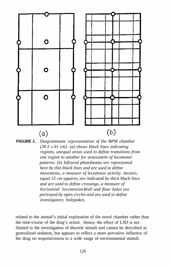
FIGURE 1. Diagrammatic representation of the BPM chamber(30.5 x 61 cm). (a) shows black lines indicatingregions, unequal areas used to define transitions fromone region to another for assessment of locomotorpatterns. (b) Infrared photobeams are representedhere by thin black lines and are used to definemovements, a measure of locomotor activity. Sectors,equal 15 cm squares, are indicated by thick black lincsand are used to define crossings, a measure ofhorizontal locomotion.Wall and floor holes areportrayed by open circles and are used to defineinvestigatory holepokes.
related to the animal’s initial exploration of the novel chamber rather thanthe time-course of the drug’s action. Hence, the effect of LSD is notlimited to the investigation of discrete stimuli and cannot be described asgeneralized sedation, but appears to reflect a more pervasive influence ofthe drug on responsiveness to a wide range of environmental stimuli.
128
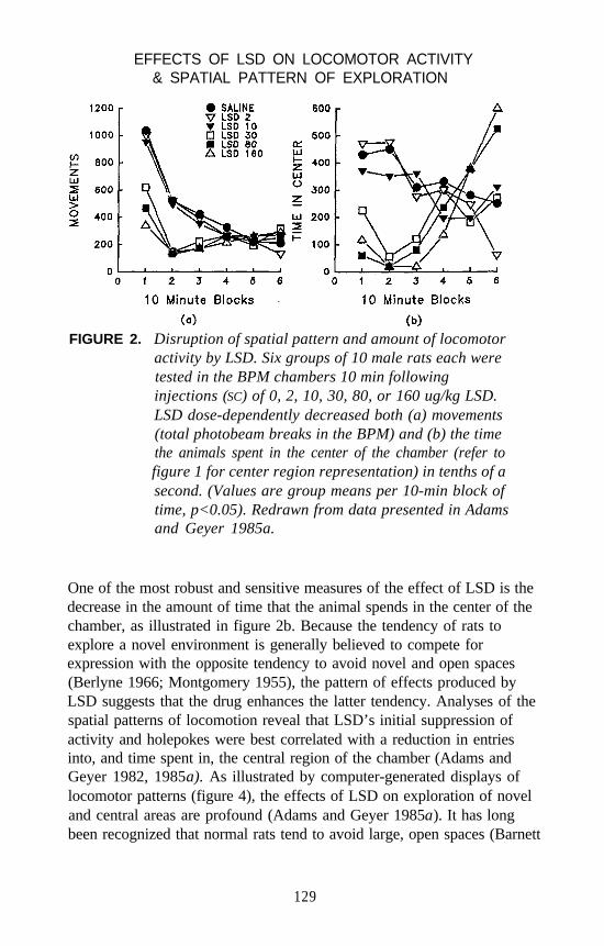
EFFECTS OF LSD ON LOCOMOTOR ACTIVITY& SPATIAL PATTERN OF EXPLORATION
FIGURE 2. Disruption of spatial pattern and amount of locomotoractivity by LSD. Six groups of 10 male rats each weretested in the BPM chambers 10 min followinginjections (SC) of 0, 2, 10, 30, 80, or 160 ug/kg LSD.LSD dose-dependently decreased both (a) movements(total photobeam breaks in the BPM) and (b) the timethe animals spent in the center of the chamber (refer tofigure 1 for center region representation) in tenths of asecond. (Values are group means per 10-min block oftime, p<0.05). Redrawn from data presented in Adamsand Geyer 1985a.
One of the most robust and sensitive measures of the effect of LSD is thedecrease in the amount of time that the animal spends in the center of thechamber, as illustrated in figure 2b. Because the tendency of rats toexplore a novel environment is generally believed to compete forexpression with the opposite tendency to avoid novel and open spaces(Berlyne 1966; Montgomery 1955), the pattern of effects produced byLSD suggests that the drug enhances the latter tendency. Analyses of thespatial patterns of locomotion reveal that LSD’s initial suppression ofactivity and holepokes were best correlated with a reduction in entriesinto, and time spent in, the central region of the chamber (Adams andGeyer 1982, 1985a). As illustrated by computer-generated displays oflocomotor patterns (figure 4), the effects of LSD on exploration of noveland central areas are profound (Adams and Geyer 1985a). It has longbeen recognized that normal rats tend to avoid large, open spaces (Barnett
129
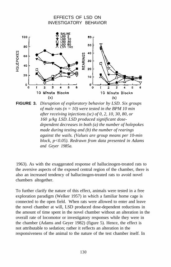
EFFECTS OF LSD ONINVESTIGATORY BEHAVIOR
FIGURE 3. Disruption of exploratory behavior by LSD. Six groupsof male rats (n = 10) were tested in the BPM 10 minafter receiving injections (sc) of 0, 2, 10, 30, 80, or160 µ/kg LSD. LSD produced significant dose-dependent decreases in both (a) the number of holepokesmade during testing and (b) the number of rearingsagainst the wails. (Values are group means per 10-minblock, p<0.05). Redrawn from data presented in Adamsand Geyer 1985a.
1963). As with the exaggerated response of hallucinogen-treated rats tothe aversive aspects of the exposed central region of the chamber, there isalso an increased tendency of hallucinogen-treated rats to avoid novelchambers altogether.
To further clarify the nature of this effect, animals were tested in a freeexploration paradigm (Welker 1957) in which a familiar home cage isconnected to the open field. When rats were allowed to enter and leavethe novel chamber at will, LSD produced dose-dependent reductions inthe amount of time spent in the novel chamber without an alteration in theoverall rate of locomotor or investigatory responses while they were inthe chamber (Adams and Geyer 1982) (figure 5). Hence, the effect isnot attributable to sedation; rather it reflects an alteration in theresponsiveness of the animal to the nature of the test chamber itself. In
130
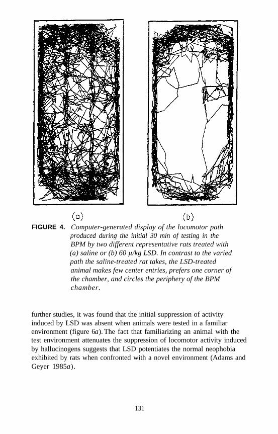
FIGURE 4. Computer-generated display of the locomotor pathproduced during the initial 30 min of testing in theBPM by two different representative rats treated with(a) saline or (b) 60 µ/kg LSD. In contrast to the variedpath the saline-treated rat takes, the LSD-treatedanimal makes few center entries, prefers one corner ofthe chamber, and circles the periphery of the BPMchamber.
further studies, it was found that the initial suppression of activityinduced by LSD was absent when animals were tested in a familiarenvironment (figure 6a The fact that familiarizing an animal with the).test environment attenuates the suppression of locomotor activity inducedby hallucinogens suggests that LSD potentiates the normal neophobiaexhibited by rats when confronted with a novel environment (Adams andGeyer 1985a).
131
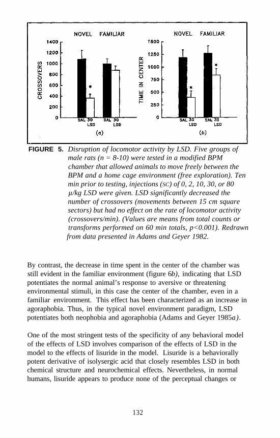
FIGURE 5. Disruption of locomotor activity by LSD. Five groups ofmale rats (n = 8-10) were tested in a modified BPMchamber that allowed animals to move freely between theBPM and a home cage environment (free exploration). Tenmin prior to testing, injections (SC) of 0, 2, 10, 30, or 80µ/kg LSD were given. LSD significantly decreased thenumber of crossovers (movements between 15 cm squaresectors) but had no effect on the rate of locomotor activity(crossovers/min). (Values are means from total counts ortransforms performed on 60 min totals, p<0.001). Redrawnfrom data presented in Adams and Geyer 1982.
By contrast, the decrease in time spent in the center of the chamber wasstill evident in the familiar environment (figure 6b), indicating that LSDpotentiates the normal animal’s response to aversive or threateningenvironmental stimuli, in this case the center of the chamber, even in afamiliar environment. This effect has been characterized as an increase inagoraphobia. Thus, in the typical novel environment paradigm, LSDpotentiates both neophobia and agoraphobia (Adams and Geyer 1985a).
One of the most stringent tests of the specificity of any behavioral modelof the effects of LSD involves comparison of the effects of LSD in themodel to the effects of lisuride in the model. Lisuride is a behaviorallypotent derivative of isolysergic acid that closely resembles LSD in bothchemical structure and neurochemical effects. Nevertheless, in normalhumans, lisuride appears to produce none of the perceptual changes or
132
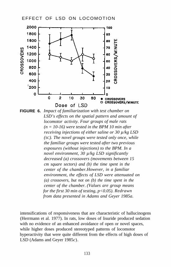
EFFECT OF LSD ON LOCOMOTION
FIGURE 6. Impact of familiarization with test chamber onLSD’s effects on the spatial pattern and amount oflocomotor activity. Four groups of male rats(n = 10-16) were tested in the BPM 10 min afterreceiving injections of either saline or 30 µ/kg LSD(SC). The novel groups were tested only once, whilethe familiar groups were tested after two previousexposures (without injections) to the BPM. In anovel environment, 30 µ/kg LSD significantlydecreased (a) crossovers (movements between 15cm square sectors) and (b) the time spent in thecenter of the chamber.However, in a familiarenvironment, the effects of LSD were attenuated on(a) crossovers, but not on (b) the time spent in thecenter of the chamber. (Values are group meansfor the first 30 min of testing, p<0.05). Redrawnfrom data presented in Adams and Geyer 1985a.
intensifications of responsiveness that are characteristic of hallucinogens(Herrmann et al. 1977). In rats, low doses of lisuride produced sedationwith no evidence of an enhanced avoidance of open or novel spaces,while higher doses produced stereotyped patterns of locomotorhyperactivity that were quite different from the effects of high doses ofLSD (Adams and Geyer 1985c).
133
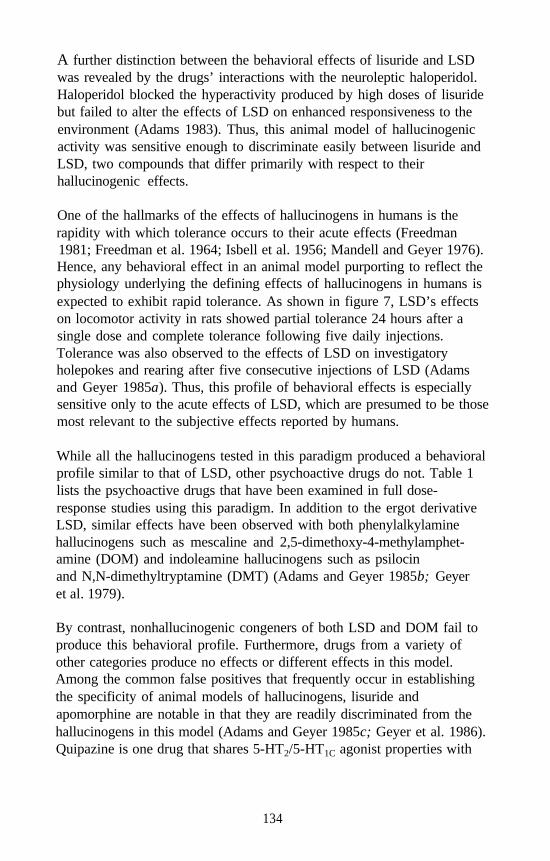
A further distinction between the behavioral effects of lisuride and LSDwas revealed by the drugs’ interactions with the neuroleptic haloperidol.Haloperidol blocked the hyperactivity produced by high doses of lisuridebut failed to alter the effects of LSD on enhanced responsiveness to theenvironment (Adams 1983). Thus, this animal model of hallucinogenicactivity was sensitive enough to discriminate easily between lisuride andLSD, two compounds that differ primarily with respect to theirhallucinogenic effects.
One of the hallmarks of the effects of hallucinogens in humans is therapidity with which tolerance occurs to their acute effects (Freedman1981; Freedman et al. 1964; Isbell et al. 1956; Mandell and Geyer 1976).Hence, any behavioral effect in an animal model purporting to reflect thephysiology underlying the defining effects of hallucinogens in humans isexpected to exhibit rapid tolerance. As shown in figure 7, LSD’s effectson locomotor activity in rats showed partial tolerance 24 hours after asingle dose and complete tolerance following five daily injections.Tolerance was also observed to the effects of LSD on investigatoryholepokes and rearing after five consecutive injections of LSD (Adamsand Geyer 1985a). Thus, this profile of behavioral effects is especiallysensitive only to the acute effects of LSD, which are presumed to be thosemost relevant to the subjective effects reported by humans.
While all the hallucinogens tested in this paradigm produced a behavioralprofile similar to that of LSD, other psychoactive drugs do not. Table 1lists the psychoactive drugs that have been examined in full dose-response studies using this paradigm. In addition to the ergot derivativeLSD, similar effects have been observed with both phenylalkylaminehallucinogens such as mescaline and 2,5-dimethoxy-4-methylamphet-amine (DOM) and indoleamine hallucinogens such as psilocinand N,N-dimethyltryptamine (DMT) (Adams and Geyer 1985b; Geyeret al. 1979).
By contrast, nonhallucinogenic congeners of both LSD and DOM fail toproduce this behavioral profile. Furthermore, drugs from a variety ofother categories produce no effects or different effects in this model.Among the common false positives that frequently occur in establishingthe specificity of animal models of hallucinogens, lisuride andapomorphine are notable in that they are readily discriminated from thehallucinogens in this model (Adams and Geyer 1985c; Geyer et al. 1986).Quipazine is one drug that shares 5-HT2/5-HT1C agonist properties with
134
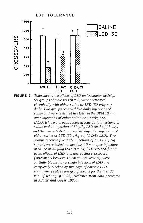
L S D T O L E R A N C E
FIGURE 7. Tolerance to the effects of LSD on locomotor activity.Six groups of male rats (n = 6) were pretreatedchronically with either saline or LSD (30 µ/kg SC)daily. Two groups received five daily injections ofsaline and were tested 24 hrs later in the BPM 10 minafter injections of either saline or 30 µ/kg LSD[ACUTE]. Two groups received four daily injections ofsaline and an injection of 30 µ/kg LSD on the fifth day,and then were tested on the sixth day after injections ofeither saline or LSD (30 µ/kg SC) [1 DAY LSD]. Twogroups received five daily injections of LSD (30 µ/kgSC) and were tested the next day 10 min after injectionsof saline or 30 µ/kg LSD (n = 14) [5 DAYS LSD].Theacute effects of LSD, e.g. decreasing crossovers(movements between 15 cm square sectors), werepartially blocked by a single injection of LSD andcompletely blocked by five days of chronic LSDtreatment. (Values are group means for the first 30min of testing, p<0.05). Redrawn from data presentedin Adams and Geyer 1985a.
135
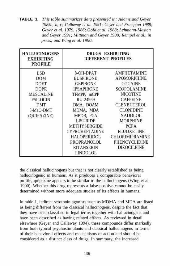
TABLE 1. This table summarizes data presented in: Adams and Geyer1985a, b, c; Callaway et al. 1991; Geyer and Frampton 1988;Geyer et al. 1979, 1986; Gold et al. 1988; Lehmann-Mastenand Geyer 1991; Mittman and Geyer 1989; Rempel et al., inpress; and Wing et al. 1990.
HALLUCINOGENSEXHIBITING
PROFILE
DRUGS EXHIBITINGDIFFERENT PROFILES
LSDDOMDOETDOPR
MESCALINEPSILOCIN
DMT5-MeO-DMT
(QUIPAZINE)
8-OH-DPAT AMPHETAMINEBUSPIRONE APOMORPHINEGEPIRONE COCAINE
IPSAPIRONE SCOPOLAMINETFMPP, mCPP NICOTINE
RU-24969 CAFFEINEDMA, DOAM CLENBUTEROLMDMA, MDA CLONIDINEMBDB, PCA NADOLOLLISURIDE MORPHINE
METHYSERGIDE PCPACYPROHEPTADINE FLUOXETINE
HALOPERIDOL CHLORIMIPRAMINEPROPRANOLOL PHENCYCLIDINE
RITANSERIN DIZOCILPINEPINDOLOL
II
the classical hallucinogens but that is not clearly established as beinghallucinogenic in humans. As it produces a comparable behavioralprofile, quipazine appears to be similar to the hallucinogens (Wing et al.1990). Whether this drug represents a false positive cannot be easilydetermined without more adequate studies of its effects in humans.
In table 1, indirect serotonin agonists such as MDMA and MDA are listedas being different from the classical hallucinogens, despite the fact thatthey have been classified in legal terms together with hallucinogens andhave been described as having related effects. As reviewed in detailelsewhere (Geyer and Callaway 1994), these compounds differ markedlyfrom both typical psychostimulants and classical hallucinogens in termsof their behavioral effects and mechanisms of action and should beconsidered as a distinct class of drugs. In summary, the increased
136
avoidance of novel and open spaces induced by LSD in rats appears tosatisfy several requirements for an animal model of hallucinogenicactivity. First, the effect is common to all the hallucinogens tested atdoses that generally correspond to the potencies of these drugs inhumans. Second, this profile of effects has not been reproduced by anyof the nonhallucinogenic drugs tested, including inactive congeners ofhallucinogens. Third, tolerance to this increased avoidance effect of LSDdevelops at a rate is comparable to that observed in humans.
SEROTONERGIC MECHANISMS OF HALLUCINOGENEFFECTS
In order to investigate the mechanisms of action of hallucinogens, anumber of studies have examined the 5-HT receptor subtypes thatcontribute to the hallucinogen-induced behavioral profile of an increasedavoidance of novel and open spaces. In the authors’ laboratory, threeapproaches to this investigation have been used. First, selective 5-HTagonists have been tested. Second, 5-HT receptor antagonists, such asthey are, have been tested for their ability to block the effects of therespective agonists. Third, due to the limited set of 5-HT antagonists,cross-tolerance paradigms have been used to examine the involvement ofparticular receptor subtypes in the behavioral effects of the agonists.
Serotonin Agonists
Of the phenalkylamine hallucinogens which are more selective for the5-HT1C/5-HT2 receptor sites, mescaline and 2,5-dimethoxy-4-iodo-amphetamine (DOI) as well as other 5-HT1C/5-HT2 agonists produceddose-related decreases in the initial levels of exploratory activity in thesame BPM paradigm used to characterize the effects of LSD (Wing et al.1990). In each case, these decreases were accompanied by preferentialdecreases in entries into and time spent in the center of the chamber. Aswith LSD, this suppression of exploratory behavior was attenuatedsignificantly when rats were familiarized with the testing chamber priorto the administration of DOI (Wing et al. 1990). Similarly, as shown infigure 8, acute administration of the 5-HT1C/5-HT2 agonist DOMsuppressed locomotion and both entries into and time spent in the centerof the BPM chamber (Adams and Geyer 1985b). Familiarization with thechamber diminished the effect of DOM on exploratory locomotion butnot on the rats’ tendency to enter the center of the chamber (figure 8).
137
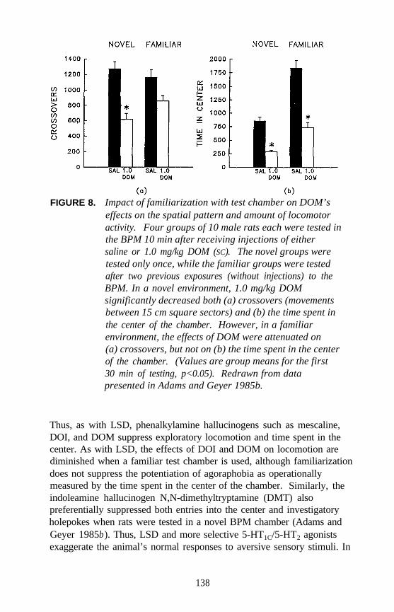
FIGURE 8. Impact of familiarization with test chamber on DOM’seffects on the spatial pattern and amount of locomotoractivity. Four groups of 10 male rats each were tested inthe BPM 10 min after receiving injections of eithersaline or 1.0 mg/kg DOM (SC). The novel groups weretested only once, while the familiar groups were testedafter two previous exposures (without injections) to theBPM. In a novel environment, 1.0 mg/kg DOMsignificantly decreased both (a) crossovers (movementsbetween 15 cm square sectors) and (b) the time spent inthe center of the chamber. However, in a familiarenvironment, the effects of DOM were attenuated on(a) crossovers, but not on (b) the time spent in the centerof the chamber. (Values are group means for the first30 min of testing, p<0.05). Redrawn from datapresented in Adams and Geyer 1985b.
Thus, as with LSD, phenalkylamine hallucinogens such as mescaline,DOI, and DOM suppress exploratory locomotion and time spent in thecenter. As with LSD, the effects of DOI and DOM on locomotion arediminished when a familiar test chamber is used, although familiarizationdoes not suppress the potentiation of agoraphobia as operationallymeasured by the time spent in the center of the chamber. Similarly, theindoleamine hallucinogen N,N-dimethyltryptamine (DMT) alsopreferentially suppressed both entries into the center and investigatoryholepokes when rats were tested in a novel BPM chamber (Adams andGeyer 1985b). Thus, LSD and more selective 5-HT1C/5-HT2 agonistsexaggerate the animal’s normal responses to aversive sensory stimuli. In
138
the novel environment, this potentiation of sensory responsiveness isexpressed as initial decreases in exploratory behavior. In a familiarenvironment, the effect is expressed in a more specific decrease in visitsto the open area in the center of the chamber.
The 5-HT1A agonists, which are not generally considered hallucinogenic,have also been tested in this behavioral paradigm. Buspirone,8-OH-DPAT, gepirone, and ipsapirone all produced decreases inlocomotor activity in a novel environment. The decreases weresuperficially similar to those produced by the 5-HT1C/5-HT2 agonists(Mittman and Geyer 1989). Unlike the mixed 5-HT1A and 5-HT1C/5-HT2
agonist LSD, the more selective 5-HT1A compounds produced decreasesin center entries that were strictly proportional to the decreases in othermeasures of activity and were typically maintained throughout the testsession. Furthermore, in contrast to the effects of LSD or phenalkyl-amine hallucinogens, the effects of the 5-HT1A agonists were notattenuated when the animals were tested in a familiar environment(figure 9). These decreases were more indicative of a general suppressionof motor behavior rather than of a specific potentiation of the animals’neophobic and agoraphobic responses to threatening stimuli (Mittmanand Geyer 1989). Thus, hallucinogenic 5-HT agonists such as the5-HT1C/5-HT2 agonists DMT, DOI, and mescaline were found to producebehavior similar to that produced by LSD, while 5-HT1A agonists thatwere not hallucinogenic produced behavior more akin to a generalizedsedation rather than the behavioral profile elicited by LSD administration.
Serotonin Antagonists
The effects of the 5-HT1C/5-HT2 and 5-HT1A agonists in this paradigm canbe distinguished pharmacologically as well as behaviorally. Mittman andGeyer (1991) showed that the effects of the 5-HT1C/5-HT2 agonist DOI onexploratory locomotion and time spent in the center were blockedeffectively by the 5-HT1C/5-HT2 antagonist ritanserin as shown infigure 10. Similar effects were obtained with the 5-HT1C/5-HT2
antagonist ketanserin using a variety of 5-HT1C/5-HT2 hallucinogens(Wing et al. 1990). By contrast, the effects of the 5-HT1A agonist8-OH-DPAT were completely insensitive to ketanserin (Wing et al.1990).
However, administration of the 5-HT1 antagonist (and beta-adrenergicantagonist) propranolol did not yield receptor-specific effects (figure 11).When propranolol was used as an antagonist to the 5-HT1A agonist
139
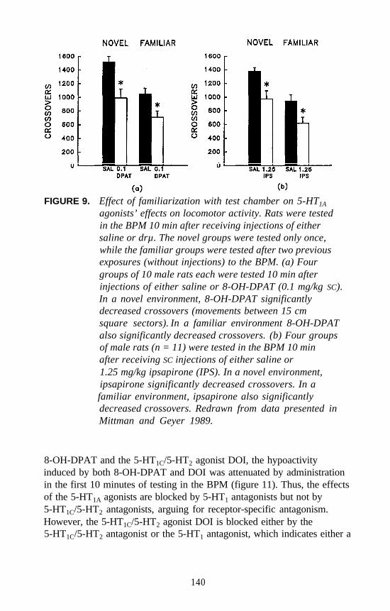
FIGURE 9. Effect of familiarization with test chamber on 5-HT1A
agonists’ effects on locomotor activity. Rats were testedin the BPM 10 min after receiving injections of eithersaline or drµ. The novel groups were tested only once,while the familiar groups were tested after two previousexposures (without injections) to the BPM. (a) Fourgroups of 10 male rats each were tested 10 min afterinjections of either saline or 8-OH-DPAT (0.1 mg/kg SC).In a novel environment, 8-OH-DPAT significantlydecreased crossovers (movements between 15 cmsquare sectors). In a familiar environment 8-OH-DPATalso significantly decreased crossovers. (b) Four groupsof male rats (n = 11) were tested in the BPM 10 minafter receiving SC injections of either saline or1.25 mg/kg ipsapirone (IPS). In a novel environment,ipsapirone significantly decreased crossovers. In afamiliar environment, ipsapirone also significantlydecreased crossovers. Redrawn from data presented inMittman and Geyer 1989.
8-OH-DPAT and the 5-HT1C/5-HT2 agonist DOI, the hypoactivityinduced by both 8-OH-DPAT and DOI was attenuated by administrationin the first 10 minutes of testing in the BPM (figure 11). Thus, the effectsof the 5-HT1A agonists are blocked by 5-HT1 antagonists but not by5-HT1C/5-HT2 antagonists, arguing for receptor-specific antagonism.However, the 5-HT1C/5-HT2 agonist DOI is blocked either by the5-HT1C/5-HT2 antagonist or the 5-HT1 antagonist, which indicates either a
140
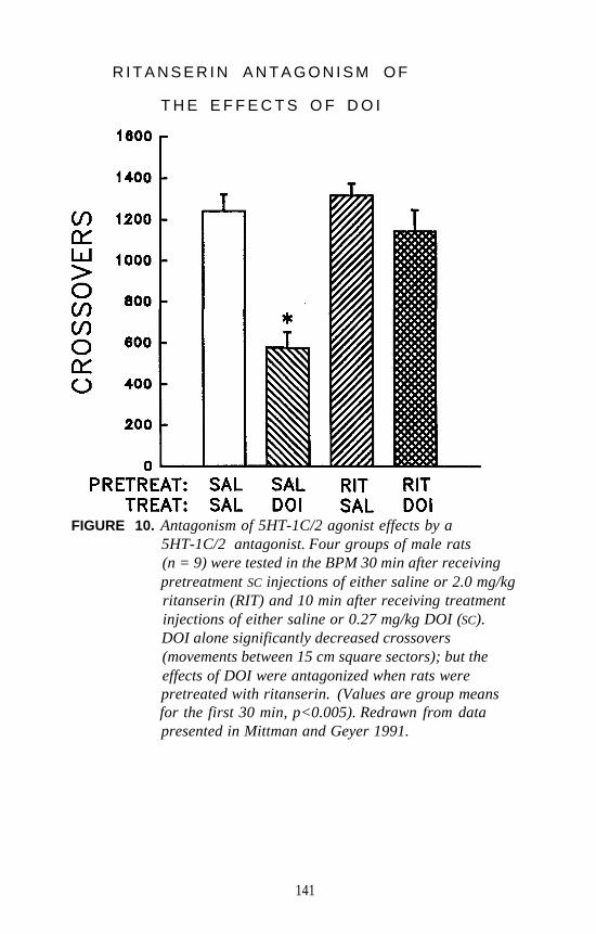
R I T A N S E R I N A N T A G O N I S M O F
T H E E F F E C T S O F D O I
FIGURE 10. Antagonism of 5HT-1C/2 agonist effects by a5HT-1C/2 antagonist. Four groups of male rats(n = 9) were tested in the BPM 30 min after receivingpretreatment SC injections of either saline or 2.0 mg/kgritanserin (RIT) and 10 min after receiving treatmentinjections of either saline or 0.27 mg/kg DOI (SC).DOI alone significantly decreased crossovers(movements between 15 cm square sectors); but theeffects of DOI were antagonized when rats werepretreated with ritanserin. (Values are group meansfor the first 30 min, p<0.005). Redrawn from datapresented in Mittman and Geyer 1991.
141
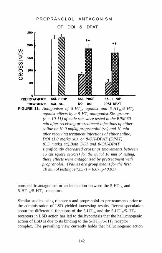
PROPRANOLOL ANTAGONISM
OF DOI & DPAT
FIGURE 11. Antagonism of 5-HT1A agonist and 5-HT1C/5-HT2
agonist effects by a 5-HT1 antagonist.Six groups(n = 10-11) of male rats were tested in the BPM 30min after receiving pretreatment injections of eithersaline or 10.0 mg/kg propranolol (SC) and 10 minafter receiving treatment injections of either saline,DOI (1.0 mg/kg SC), or 8-OH-DPAT (DPAT)(0.5 mg/kg SC).Both DOI and 8-OH-DPATsignificantly decreased crossings (movements between15 cm square sectors) for the initial 10 min of testing;these effects were antagonized by pretreatment withpropranolol. (Values are group means for the first10 min of testing; F(2,57) = 8.07, p<0.01).
nonspecific antagonism or an interaction between the 5-HT1A and5-HT1C/5-HT2 receptors.
Similar studies using ritanserin and propranolol as pretreatments prior tothe administration of LSD yielded interesting results. Recent speculationabout the differential functions of the 5-HT1A and the 5-HT1C/5-HT2
receptors in LSD action has led to the hypothesis that the hallucinogenicaction of LSD is due to its binding to the 5-HT1C/5-HT2 receptorcomplex. The prevailing view currently holds that hallucinogenic action
142
is due to actions mediated via the 5-HT1C/5-HT2 receptor complex. Asevidence for this hypothesis, some studies have correlated the 5-HT2
binding affinity of a drug with its hallucinogenic potency (Glennon et al.1984; Titeler et al. 1988).
However, because of the paucity of dose-finding studies in humans, thesestudies have been unable to thoroughly examine the indoleamine andergot alkaloid hallucinogens, although LSD and some indoleamines havebeen examined. These studies have focused instead primarily on thephenalkylamine hallucinogens such as mescaline, DOI, and DOM. LSDappears to be more closely related to the indoleamines, which includeDMT and psilocybin (Mandell and Geyer 1976). Correlations between5-HT1C/5-HT2 function and hallucinogenic potency have yet to be studiedfor the indoleamines or the ergots as groups. Therefore, the relativecontributions of 5-HT1A and 5-HT1C/5-HT2 receptors in hallucinogenicaction have not been established definitively, especially for theindoleamine and ergot alkaloid hallucinogens.
Because the prevailing view is that classical hallucinogens act primarilyvia agonist actions at 5-HT1C/5-HT2 receptors, it was predicted that the5-HT1C/5-HT2 antagonist ritanserin would block the effects of LSD,whereas the 5-HT1 antagonist propranolol would not. Instead, ritanserinonly partially blocked the effects of LSD, as did propranolol. Only thecombination of ritanserin and propranolol completely blocked the effectsof LSD (Mittman and Geyer 1991). These results suggest a role for the5-HT1A receptor as a contributor to the behavioral effects of LSD.Perhaps, as suggested by the antagonist studies, there is an interactionbetween the 5-HT1A and 5-HT1C/5-HT2 receptors. To explore further thecontributions of the receptors to the effects of LSD, an approach based onthe apparent tolerance associated with 5-HT agonists was used.
TOLERANCE AND CROSS-TOLERANCE TESTS
The previous attempts to use antagonists to block specific receptor siteswhen testing LSD, a mixed 5-HT1A and 5-HT1C/5-HT2 agonist, wereproblematic primarily because of the lack of selective 5-HT1A antagonists.Unfortunately the common 5-HT1 antagonist propranolol is an antagonistat both 5-HT1A and 5-HT1B sites and is a ß-adrenergic blocker as well.Without selective antagonists, it was difficult to assess the respectivecontributions of the receptor subtypes to the effects of LSD on locomotorand investigatory behavior.
143
Alternatively, another approach is that of tolerance. Chronic admini-stration of a selective 5-HT1A agonist such as 8-OH-DPAT producestolerance to the acute effects of 8-OH-DPAT (Berendsen and Broekkamp1991; Johansson et al. 1990; Larsson et al. 1990; Pranzatelli and Pluchino1991). Similarly, tolerance has also been reported to the acute effects ofDOI, a 5-HT1C/5-HT2 agonist, after repeated administration (Berendsenand Broekkamp 1991; Darmani et al. 1990a; Pranzatelli and Pluchino1991). In the serotonin system, it appears that receptor regulation is notan adequate explanation for tolerance, as behavioral tolerance is seen inthe absence of receptor change (Trulson 1985). Nevertheless, bindingstudies have demonstrated that chronic administration of both8-OH-DPAT and DOI result in decreased numbers of 5-HT1A and5-HT1C/5-HT2 receptor sites, respectively (Buckholtz et al. 1988; Larssonet al. 1990; McKenna et al. 1989; Pranzatelli 1991).
It is well established that tolerance to LSD in humans is striking for therapidity with which it occurs (Freedman et al. 1964; Isbell et al. 1956;Mandell and Geyer 1976). In animals, tolerance or partial tolerance tothe acute effects of LSD administration has also been demonstrated formost behaviors (Adams and Geyer 1985a; Appel and Freedman 1968;Bridger 1975; Carter and Appel 1978; Geyer and Light 1979; Murray etal. 1977; Rech et al. 1975; Trulson 1985; Winter 1971), although somebehavioral paradigms may be resistant to tolerance induction (Braff andGeyer 1980; Bridger 1975; Murray et al. 1977).
Cross-tolerance regimens were used to elucidate the contribution of5-HT1A receptor action to the effects of LSD on locomotor activity.These experiments were designed to test the hypothesis that the effects ofLSD in the BPM model are influenced by 5-HT1A function and by5-HT1C/5-HT2 function, thus supporting a combination of 5-HT1A and5-HT1C/5-HT2 receptor action.
Tolerance to 8-OH-DPAT and DOI
First, it was determined whether tolerance to the selective agonists wouldexhibit cross-tolerance to a different selective agonist. The hypothesis,based on earlier research (Berendsen and Broekkamp 1991; Darmani etal. 1990a; Johansson et al. 1990; Nash et al. 1989; Pranzatelli andPluchino 1991), was that the 5-HT1A and 5-HT1C/5-HT2 agonists8-OH-DPAT and DOI, respectively, would both show tolerance but thatcross-tolerance between the two drugs would not occur.
144
The most robust acute effect of 8-OH-DPAT administration, that ofdecreasing the number of movements made by the rat in the BPM in thefirst 10 minutes of an hour-long test session, was attenuated when8-OH-DPAT was administered repeatedly every 12 hours for 5 days(figure 12). That is, tolerance was observed to the effects of 8-OH-DPATwhen it was given chronically.
A similar pattern of tolerance was observed with DOI (figure 12). Adiminished effect of the challenge dose of DOI occurred in animalspretreated chronically with DOI. As predicted, the repeated admini-stration of DOI produced no significant cross-tolerance to the effects ofan acute dose of 8-OH-DPAT (figure 12). However, the chronicadministration of 8-OH-DPAT produced some cross-tolerance to theeffects of a challenge dose of DOI (figure 12). Thus, the expectedtolerance to the effects of 8-OH-DPAT and DOI occurred. AlthoughDOI produced no cross-tolerance to 8-OH-DPAT, some cross-tolerancewas found when DOI was tested in rats pretreated with 8-OH-DPAT(Krebs and Geyer 1994). Thus, the tolerance regimens were sufficient toproduce significant tolerance, but this tolerance was not completelyspecific pharmacologically to each receptor subtype.
These effects are similar to what was observed when the 5-HT1 antagonistpropranolol was able to block some of the effects of the 5-HT1C/5-HT2
agonist DOI (figure 11). The partial cross-tolerance to DOI after chronic8-OH-DPAT administration may reflect an interaction between 5-HT1A
and 5-HT1C/5-HT2 receptors. Such an interaction has been suggestedpreviously (Amt and Hyttel 1989; Darmani et al. 1990b; Krebs and Geyer1994) and will require further studies to elucidate.
Tolerance to LSD and 8-OH-DPAT
A parallel study examined cross-tolerance between the effects of8-OH-DPAT and LSD. Based on previous studies (Adams and Geyer1985a), tolerance for the effects of LSD was expected. Based on itsaffinity to 5-HT1A receptors, chronic LSD pretreatment was expected toproduce cross-tolerance to the effects of 8-OH-DPAT treatment. Themost important hypothesis was that 8-OH-DPAT pretreatment wouldproduce cross-tolerance to the effects of LSD, suggesting either that5-HT1A receptors contribute to the effects of LSD or that 5-HT1A and5-HT1C/5-HT2 receptors interact functionally. In either case, reciprocalcross-tolerance between 8-OH-DPAT and LSD was hypothesized. Asbefore, tolerance to the effects of 8-OH-DPAT was evident (figure 13).
145

D O I & 8 - O H - D P A T T O L E R A N C E
& C R O S S T O L E R A N C E
FIGURE 12. Asymmetrical cross-tolerance between 5-HT1A and5-HT1C/5-HT2 agonists. Nine groups of male rats(n = 7-8) were pretreated chronically every 12 hrs for5 days with either saline, DOI (1.0 mg/kg SC), or8-OH-DPAT (0.5 mg/kg SC).Thirty-six hrs after thelast pretreatment, rats were tested in the BPM 10 minafter receiving treatment injections of either saline,1.0 mg/kg DOI, or 0.5 mg/kg 8-OH-DPAT (DPAT).Both DOI and 8-OH-DPAT significantly decreasedmovements, a measure of global motor activity; theseeffects were significantly attenuated when thecorresponding pretreatmeqts of DOI and 8-OH-DPATwere given, respectively. Also, cross-toleranceoccurred to the effects of DOI when 8-OH-DPAT wasthe pretreatment, but no attenuation of 8-OH-DPAT’seffects occurred when DOI was the pretreatment.(Values are percentages of group means ofmovements for corresponding control groups, for thefirst 10 min of testing, p<0.05). An * represents asignificant difference from control, while a +represents a significant difference from the acute drµgroup. Each symbol indicates significant tolerance orcross-tolerance, while both symbols together indicatepartial tolerance or cross-tolerance. Redrawn fromdata presented in Krebs and Geyer 1994.
146
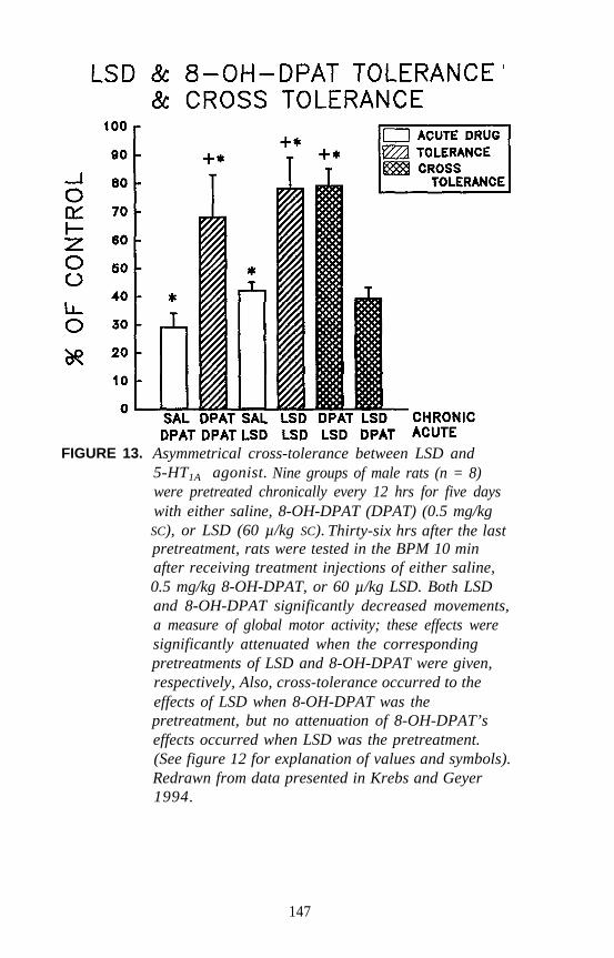
FIGURE 13. Asymmetrical cross-tolerance between LSD and5-HT1A agonist. Nine groups of male rats (n = 8)were pretreated chronically every 12 hrs for five dayswith either saline, 8-OH-DPAT (DPAT) (0.5 mg/kg
SC), or LSD (60 µ/kg SC). Thirty-six hrs after the lastpretreatment, rats were tested in the BPM 10 minafter receiving treatment injections of either saline,0.5 mg/kg 8-OH-DPAT, or 60 µ/kg LSD. Both LSDand 8-OH-DPAT significantly decreased movements,a measure of global motor activity; these effects weresignificantly attenuated when the correspondingpretreatments of LSD and 8-OH-DPAT were given,respectively, Also, cross-tolerance occurred to theeffects of LSD when 8-OH-DPAT was thepretreatment, but no attenuation of 8-OH-DPAT’seffects occurred when LSD was the pretreatment.(See figure 12 for explanation of values and symbols).Redrawn from data presented in Krebs and Geyer1994.
147
Also, the effects of LSD on movements showed tolerance after thechronic regimen was performed (figure 13). In animals chronicallypretreated with 8-OH-DPAT every 12 hours for 5 days, the effects ofLSD were significantly diminished. Thus, cross-tolerance to LSD wasobserved after the chronic administration of 8-OH-DPAT.
This result was consistent with the previous observation that propranololpartially blocks this effect of LSD, and further suggests that5-HT1A-related systems participate in these behavioral effects of LSD.However, even after chronic 8-OH-DPAT pretreatment a significanteffect of LSD was still evident, indicating that the cross-tolerance wasonly partial. The reciprocal comparison did not evidence cross-tolerance.As seen in figure 13, animals pretreated chronically with LSD did notexhibit cross-tolerance to the effects of 8-OH-DPAT (Krebs and Geyer1994). This nonreciprocal cross-tolerance is hypothesized to haveoccurred because the dose of LSD used did not occupy 5-HT1A receptorsites as effectively as did the dose of 8-OH-DPAT, although the doseswere equated in terms of the acute behavioral effects of the two drugs.The major finding of asymmetrical partial cross-tolerance between8-OH-DPAT and LSD indicates that the effects of LSD on locomotorbehavior, as defined by movements, are influenced by 5-HT1A receptorfunction.
Tolerance to LSD and DOI
A parallel cross-tolerance study was performed with DOI and LSD.Because the previous study indicated that DOI tolerance was marginalwhen animals were treated every 12 hours for 5 days (figure 12), thechronic regimen was increased to injections every 12 hours for 8 days.As shown in figure 14, tolerance to the effects of both DOI and LSD wasreplicated. The effects of LSD were diminished somewhat in animalschronically pretreated with DOI, indicating that partial cross-tolerance tothe effects of LSD occurred (figure 14). However, there was noreciprocal cross-tolerance when animals were chronically pretreated withLSD and challenged with DOI (Krebs and Geyer 1994). Therefore, themajor finding of partial cross-tolerance to LSD after chronic DOIadministration indicates that the effects of LSD on locomotion areinfluenced by the action of the 5-HT1C/5-HT2 system.
Taken together, evidence of partial cross-tolerance between 8-OH-DPATand LSD and between DOI and LSD indicates that the behavioral effectsof LSD are influenced by a combination of 5-HT1A and 5-HT1C/5-HT2
148
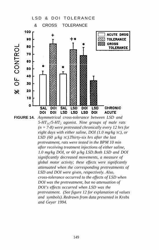
L S D & D O I T O L E R A N C E
& CROSS TOLERANCE
FIGURE 14. Asymmetrical cross-tolerance between LSD and5-HT1C/5-HT2 agonist. Nine groups of male rats(n = 7-8) were pretreated chronically every 12 hrs foreight days with either saline, DOI (1.0 mg/kg SC), orLSD (60 µ/kg SC).Thirty-six hrs after the lastpretreatment, rats were tested in the BPM 10 minafter receiving treatment injections of either saline,1.0 mg/kg DOI, or 60 µ/kg LSD.Both LSD and DOIsignificantly decreased movements, a measure ofglobal motor activity; these effects were significantlyattenuated when the corresponding pretreatments ofLSD and DOI were given, respectively. Also,cross-tolerance occurred to the effects of LSD whenDOI was the pretreatment, but no attenuation ofDOI’s effects occurred when LSD was thepretreatment. (See figure 12 for explanation of valuesand symbols). Redrawn from data presented in Krebsand Geyer 1994.
149
receptor action or that 5-HT1A and 5-HT1C/5-HT2 receptors interact in themodulation of this behavior. Accordingly, the prevailing view that theaction of the 5-HT1C/5-HT2 receptor is primarily responsible forhallucinogenic activity may be true for the phenalkylamine hallucin-ogens, but this interpretation cannot completely explain the action of theless selective 5-HT1A/5-HT1C/5-HT2 ergot alkaloid LSD.
There is some evidence of an interaction between the 5-HT1A and5-HT1C/5-HT2 receptors. In one study it was found that the effect of the5-HT1A agonist 8-OH-DPAT was significantly increased when rats weretreated simultaneously with DOI, a 5-HT1C/5-HT2 agonist. Interestingly,the effect of DOI on head twitches was inhibited by simultaneoustreatment with 8-OH-DPAT (Arnt and Hyttel 1989). Moreover, theincreased forepaw treading induced by cotreatment with 8-OH-DPATand DOI was partially inhibited by high doses of either the 5-HT2
antagonists, ritanserin and ketanserin, or the 5-HT1 and ß-adrenoceptorantagonist (-)-alprenolol. Furthermore, the effects of the 5-HT2
antagonists were synergistic with those of (-)-alprenolol in their ability toblock the forepaw treading induced by the agonist combination (Arnt andHyttel 1989). This synergistic relationship was interpreted as anasymmetrical interaction between the 5-HT1A and 5-HT1C/5-HT2
receptors.
Another study has replicated some of these findings by demonstratingthat the head twitch response induced by the 5-HT1C/5-HT2 agonist DOIwas significantly inhibited by simultaneous administration of the 5-HT1A
agonist 8-OH-DPAT, but not by the 5-HT1B/5-HTC agonist1-(3-trifluromethylphenyl)piperazine (TFMPP) (Darmani et al. 1990b).These researchers argue for a pharmacological interaction between the5-HT1A and 5-HT2 receptors that manifests itself in a 5-HT1A inhibitoryaction on 5-HT2 receptor-modulated behavior (Darmani et al. 1990b) .
These results, taken together with the asymmetrical cross-tolerance results(Krebs and Geyer 1994) and the propranolol antagonism of DOIdiscussed earlier, suggest that there may indeed be interactions betweenthe 5-HT1A and 5-HT1C/5-HT2 receptors. With further investigation, theBPM system may be able to shed light on the nature of this interaction bymore thoroughly characterizing the effects of 5-HT1A agonists andhallucinogens. With more detailed analyses of phenalkylamine,indoleamine, and ergot alkaloid hallucinogens, clarification of themechanisms of action of these drugs should be possible.
150
REFERENCES
Adams, L.M. “LSD-Induced Alterations in Spatial and Temporal Patternsof Exploratory Activity: Possible Method of Its Effects on Mood andPerception.” Ph.D. dissertation, University of California at San Diego,1983.
Adams, L.M., and Geyer, M.A. LSD-induced alterations of locomotorpatterns and exploration in rats. Psychopharmacology 77:179-185,1982.
Adams L.M., and Geyer, M.A. A proposed animal model forhallucinogens based on LSD’s effects on patterns of exploration inrats. Behav Neuroscience 99:881-900, 1985a.
Adams, L.M., and Geyer, M.A. Effects of DOM and DMT in a proposedanimal model of hallucinogenic activity. Prog Neuro-Psychopharmacol Biol Psychiatry 91121-132, 1985b .
Adams, L.M., and Geyer, M.A. Patterns of exploration in rats distinguishlisuride from lysergic acid diethylamide. Pharmacol Biochem Behav23:461-468, 1985c.
Appel, J.B., and Freedman, D.X. Tolerance and crosstolerance amongpsychotomimetic drugs. Psychopharmacologia (Berl) 13:267-274,1968.
Amt, J., and Hyttel, J. Facilitation of 8-OH-DPAT-induced forepawtreading of rats by the 5-HT2 agonist DOI. Eur J Pharmacol161:45-51, 1989.
Barnett, S.A. The Rat: A Study in Behavior. Chicago: Aldine, 1963.Berendsen, H.H.B., and Broekkamp, C.L.E. Attenuation of 5-HT1A and
5-HT2 but not 5-HT1C receptor mediated behaviour in rats followingchronic treatment with 5-HT receptor agonists, antagonists oranti-depressants. Psychopharmacology 105:219-224, 1991.
Berlyne, D.E. Curiosity and exploration. Science 153:25-33, 1966.Braff, D.L., and Geyer, M.A. Acute and chronic LSD effects on rat
startle: Data supporting an LSD-rat model of schizophrenia. BiolPsychiatry 15:909-916, 1980.
Bridger, W.H. Good trip or bad trip: The roles of tolerance and stress inhallucinogenic drug action. In: Mandell, A.J., ed. Advances inBiochemical Psychopharmacology. Vol. 13. New York: Raven Press,1975. pp. 1-25.
Buckholtz, N.S.; Zhou, D.; and Freedman, D.X. Serotonin, agonistadministration down-regulates rat brain serotonin, receptors. Life Sci42:2439-2445, 1988.
151
Callaway, C.W.; Johnson, M.P.; Gold, L.H.; Nichols, D.E.; and Geyer,M.A. Amphetamine derivatives induce locomotor hyperactivity byacting as indirect serotonin agonists. Psychopharmacology104:293-301, 1991.
Carter, R.B., and Appel, J.B. LSD and 5-HTP: Tolerance andcrosstolerance relationships. Eur J Pharmacol 50:145-148, 1978.
Cunha, J.M., and Masur, J. Evaluation of psychotropic drugs with amodified open field test. Pharmacology 16:259-267, 1978.
Darmani, N.A.; Martin, B.R.; and Glennon, R.A. Withdrawal fromchronic treatment with (+)-DOI causes super-sensitivity to 5-HT2
receptor-induced head-twitch behaviour in mice. Eur J Pharmacol186:115-118, 1990a.
Darmani, N.A.; Martin, B.R.; Pandey, U.; and Glennon, R.A. Dofunctional relationships exist between 5-HT1A and 5-HT2 receptors?Pharmacol Biochem Behav 36:901-906, 1990b .
File, S.E., and Wardill, A.G. Validity of head-dipping as a measure ofexploration in a modified holeboard. Psychopharmacology 44:53-59,1975.
Freedman, D.X. Mode of action of hallucinogenic drugs. In: Van Praag,H.M., ed. Handbook of Biological Psychiatry. Vol. 4. New York:Marcel Dekker, Inc., 1981. pp. 859-884.
Freedman, D.X.; Appel, J.B.; Hartman, F.R.; and Molliver, M.E.Tolerance to behavioral effects of LSD-25 in rat. J Pharm Exp Ther143:309-313, 1964.
Geyer, M.A. Approaches to the characterization of drug effects onlocomotor activity in rodents. In: Adler, M.W., and Cowan, A., eds.Testing and Evaluation of Drugs of Abuse. New York: Wiley-Liss,1990. pp. 81-99.
Geyer, M.A., and Callaway, C.W. Behavioral pharmacology of ring-substituted amphetamine analogs. In: Cho, A.K., and Segal, D.S., eds.Amphetamine and its Analogs: Neuropsychopharmacology,Toxicology and Abuse. New York: Academic Press, 1994. pp. 177-208.
Geyer, M.A., and Light, R.K. LSD-induced alterations of investigatoryresponding in rats. Psychopharmacology 65:41-47, 1979.
Geyer, M.A.; Petersen L.R.; Rose G.J.; Horwitt, D.D.; Light, R.K.;Adams, L.M.; Zook, J.A.; Hawkins, R.L.; and Mandell, A.J. Theeffects of lysergic acid diethylamide and mescaline-derivedhallucinogens on sensory-integrative function: Tactile startle.J Pharmacol Exp Ther 207:837-847, 1978.
152
Geyer, M.A.; Light, R.K.; Rose, G.J.; Petersen, L.R.; Horwitt, D.D.;Adams, L.M.; and Hawkins, R.L. A characteristic effect ofhallucinogens on investigatory responding in rats.Psychopharmacology 65:35-40, 1979.
Geyer, M.A.; Russo, P.V.; and Masten, V.L. Multivariate assessment oflocomotor behavior: Pharmacological and behavioral analyses.Pharmacol Biochem Behav 25:277-288, 1986.
Glennon, R.A.; Titeler, M.; and McKenney, J.D. Evidence for 5-HT2
involvement in the mechanism of action of hallucinogenic agents. LifeSci 35:2505-2511, 1984.
Herrmann, W.M.; Horowski, R.; Dannehl, K.; Kramer, U.; and Lurati, K.Clinical effectiveness of lisuride hydrogen maleate: A double-blindtrial versus methysergide. Headache 17:54-60, 1977.
Hughes, R.N. Effects of LSD on exploratory behavior and locomotion inrats. Behav Biol 9:357-365, 1973.
Isbell, H.; Belleville, R.E.; Fraser, H.F.; Wikler, A.; and Logan, C.R.Studies on lysergic acid diethylamide (LSD-25): I. Effects in formermorphine addicts and development of tolerance during chronicintoxication. AMA Arch Neurol Psychiatry 76:468-478, 1956.
Izquierdo, I. Relations between orienting, pseudoconditioned andconditioned responses in the shuttle-box: A pharmacological analysisby means of LSD and dibenamine. Behav Biol 15:193-205, 1975.
Johansson, C.E.; Meyerson, B.J.; and Hoglund, A.U. The long-termeffects of 8-hydroxy-2-(di-n-propyl-amino)tetralin (8-OH-DPAT) oncopulatory and exploratory behavior in male rats. Eur J Pharmacol178:1-9, 1990.
Key, B.J. Alteration in the generalization of visual stimuli induced bylysergic acid diethylamide in cats. Psychopharmacology 6:327-337,1964a.
Key, B.J. The effects of LSD-25 on the interaction between conditionaland nonconditional stimuli in a simple avoidance situation.Psychopharmacology 6:319-326, 1964b.
Key, B.J., and Bradley, P.B. The effects of drugs on conditioning andhabituation to arousal stimuli in animals. Psychopharmacology1:450-462, 1960.
Krebs, K.M., and Geyer, M.A. Crosstolerance studies of serotoninreceptors involved in behavioral effects of LSD. Psychopharmacology113:429-437, 1994.
153
Larsson, L.G.; Renyi, L.; Ross, S.B.; Svensson, B.; and Angeby-Moller,K. Different effects on the responses of functional pre- andpostsynaptic 5-HT1A receptors by repeated treatment of rats with the5-HT1A receptor agonist 8-OH-DPAT. Neuropharmacology 29:85-91,1990.
Mandell, A.J., and Geyer, M.A. Hallucinations: Chemical andphysiological. In: Grenell, R.G., and Gabay, S., eds. BiologicalFoundations of Psychiatry. New York: Raven Press, 1976.pp. 729-753.
McKenna, D.J.; Nazarali, A.J.; Himeno, A.; and Saavedra, J.M. Chronictreatment with (+)DOI, a psychotomimetic 5-HT2 agonist,downregulates 5-HT2 receptors in rat brain.Neuropsychopharmacology 2:81-87, 1989.
Miliaressis, T.E., and St. Laurent, J. Effets de l’amide de l’acidelysergique-25 sur la reaction de sursaut chez le rat. Can J PhysiolPharmacol 52:126-129, 1974.
Mittman, S.M., and Geyer, M.A. Effects of 5-HT1A agonists on locomotorand investigatory behaviors in rats differ from those of hallucinogens.Psychopharmacology 98:321-329, 1989.
Mittman, S.M., and Geyer, M.A. Dissociation of multiple effects of acuteLSD on exploratory behavior in rats by ritanserin and propranolol.Psychopharmacology 105:69-76, 1991.
Montgomery, K.C. The relation between fear induced by novelstimulation and exploratory behavior. J Comp Physiol Psychol48:254-260, 1955.
Murray, T.F.; Craigmill, A.L.; and Fischer, G.J. Pharmacological andbehavioral components of tolerance to LSD and mescaline in rats.Pharmacol Biochem Behav 7:239-244, 1977.
Nash, J.F.; Meltzer, H.Y.; and Gudelsky, G.A. Selective crosstolerance to5-HT1A and 5-HT2 receptor-mediated temperature and corticosteroneresponses. Pharmacol Biochem Behav 33:781-785, 1989.
Peroutka, S.J., and Snyder S.H. Multiple serotonin receptors: Differentialbinding of [3H]5-hydroxytryptamine, [3H]lysergic acid diethylamideand [3H]spiroperidol. Mol Pharmacol 16:687-699, 1979.
Pranzatelli, M.R. Regulation of 5-HT2 receptors in rat cortex: Studieswith a putative selective agonist and an antagonist. BiochemPharmacol 42:1099-1105, 1991.
Pranzatelli, M.R., and Pluchino, R.S. The relation of central 5-HT1A and5-HT2 receptors: Low dose agonist-induced selective tolerance in therat. Pharmacol Biochem Behav 39:407-413, 1991.
154
Rech, R.H.; Tilson, H.A.; and Marquis, W.J. Adaptive changes inbehavior after repeated administration of various psychoactive drugs.In: Mandell, A.J., ed. Neurobiological Mechanisms of Adaptation andBehavior. New York: Raven Press, 1975. pp. 263-286.
Rempel, N.L.; Callaway, C.W.; and Geyer, M.A. Serotonin,, receptoractivation mimics behavioral effects of presynaptic serotonin release.Neuropsychopharmacology 8:201-212, 1993.
Sanders-Bush E.; Burris K.D.; and Knoth K. Lysergic acid diethylamideand 2,5-dimethoxy-4-methylamphetamine are partial agonists atserotonin receptors linked to phosphoinositide hydrolysis.J Pharmacol Exp Ther 246:924-928, 1988.
Stoff, D.M.; Gillin, J.C.; and Wyatt, R.J. Animal models of drug-inducedhallucinations. In: Stillman, R.C., and Willette, R.E., eds. ThePsychopharmocology of Hallucinogens. New York: Pergamon Press,1978. pp. 259-267.
Tilson, H.A.; Baker, T.G.; Chamberlain, J.H.; Marquis, W.J.; and Rech,R.H. Behavioral and neuropharmacological analysis of amphetamineand 2,5-dimethoxy-methlamphetamine in rats. Psychopharmacologia44:229-239, 1975.
Titeler, M.; Lyon, R.A.; and Glennon, R.A. Radioligand bindingevidence implicates the brain 5-HT2 receptor as a site of action forLSD and phenylisopropylamine hallucinogens. Psychopharmacology94:213-216, 1988.
Trulson, M.E. Separation of tolerance to the behavioral effects of LSDfrom changes in serotonin receptor binding in cats. Eur J Pharmacol111:385-388, 1985.
Welker, W.I. Free vs. forced exploration of a novel situation by rats.Psycho1 Rep 3:95-108, 1957.
Wing, L.L.; Tapson, G.S.; and Geyer, M.A. Mediation of acutebehavioral effects of hallucinogens in rats. Psychopharmacology100:417-425, 1990.
Winter, J.C. Tolerance to a behavioral effect of lysergic acid diethylamideand crosstolerance to mescaline in the rat: Absence of a metaboliccomponent. J Pharmacol Exp Ther 178:625-630, 1971.
ACKNOWLEDGMENTS
This chapter was prepared with support from National Institute on DrugAbuse grant DA02925. Mark A. Geyer was supported by a ResearchScientist Development Award (MH00188) from the National Institute ofMental Health.
155

AUTHORS
Mark A. Geyer, Ph.D.Professor of Psychiatry
Kirsten M. Krebs, M.A.Department of Psychology
School of MedicineUniversity of California at San DiegoLa Jolla, CA 92093-0804
156
The Stimulus Effects ofSerotonergic Hallucinogensin AnimalsJerrold C. Winter
INTRODUCTION
Mankind has known of naturally occurring hallucinogens for thousandsof years (Schultes and Hofmann 1980), but less than one-tenth of onemillennium has passed since it could be said with confidence that a purechemical of known structure is hallucinogenic (Heffter 1897; Spath1919). That chemical was mescaline, a representative of thephenethylamine subclass of the serotonergic hallucinogens. The othersubclass, the indoleamines, is perhaps best represented by lysergic aciddiethylamide (LSD), a drug whose hallucinogenic properties werediscovered by Albert Hofmann on April 16, 1943 (Hofmann 1959). Thatboth the indoleamines and the phenethylamines are properly classifiedtogether as serotonergic hallucinogens has become apparent only in thelast 2 decades.
The remarkable alterations in thought and perception produced byhallucinogens have been described by many. In some instances, carefullyselected human volunteers have received these drugs in rigidly controlledstudies (Isbell et al. 1959; Martin and Sloan 1977; Strassman, thisvolume). In others, investigators have told of the effects of hallucinogensfollowing self-administration. These reporters have ranged from personswithout scientific training (DeCurtis 1987) to distinguished scientistssuch as Hofmann (1959), Szára (1957), and Shulgin (this volume;Shulgin et al. 1986). But to seek out the most intimate mechanisms bywhich hallucinogens work their magic, and in the process to makediscoveries of ultimate benefit to humanity, researchers are obliged to useother species.
In turning to animals to aid in understanding of hallucinogens, majorethical problems are avoided, but, concomitantly, there arise questions ofrelevance. Is there a suitable animal model of hallucination, aphenomenon thought by many to be a uniquely human experience? Mostof those who work with animals find comfort in the knowledge that the
157
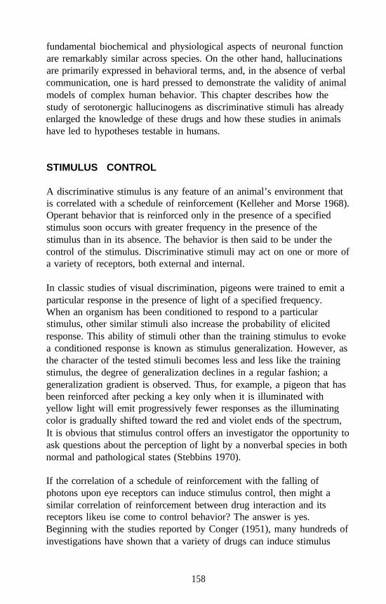
fundamental biochemical and physiological aspects of neuronal functionare remarkably similar across species. On the other hand, hallucinationsare primarily expressed in behavioral terms, and, in the absence of verbalcommunication, one is hard pressed to demonstrate the validity of animalmodels of complex human behavior. This chapter describes how thestudy of serotonergic hallucinogens as discriminative stimuli has alreadyenlarged the knowledge of these drugs and how these studies in animalshave led to hypotheses testable in humans.
STIMULUS CONTROL
A discriminative stimulus is any feature of an animal’s environment thatis correlated with a schedule of reinforcement (Kelleher and Morse 1968).Operant behavior that is reinforced only in the presence of a specifiedstimulus soon occurs with greater frequency in the presence of thestimulus than in its absence. The behavior is then said to be under thecontrol of the stimulus. Discriminative stimuli may act on one or more ofa variety of receptors, both external and internal.
In classic studies of visual discrimination, pigeons were trained to emit aparticular response in the presence of light of a specified frequency.When an organism has been conditioned to respond to a particularstimulus, other similar stimuli also increase the probability of elicitedresponse. This ability of stimuli other than the training stimulus to evokea conditioned response is known as stimulus generalization. However, asthe character of the tested stimuli becomes less and less like the trainingstimulus, the degree of generalization declines in a regular fashion; ageneralization gradient is observed. Thus, for example, a pigeon that hasbeen reinforced after pecking a key only when it is illuminated withyellow light will emit progressively fewer responses as the illuminatingcolor is gradually shifted toward the red and violet ends of the spectrum,It is obvious that stimulus control offers an investigator the opportunity toask questions about the perception of light by a nonverbal species in bothnormal and pathological states (Stebbins 1970).
If the correlation of a schedule of reinforcement with the falling ofphotons upon eye receptors can induce stimulus control, then might asimilar correlation of reinforcement between drug interaction and itsreceptors likeu ise come to control behavior? The answer is yes.Beginning with the studies reported by Conger (1951), many hundreds ofinvestigations have shown that a variety of drugs can induce stimulus
158
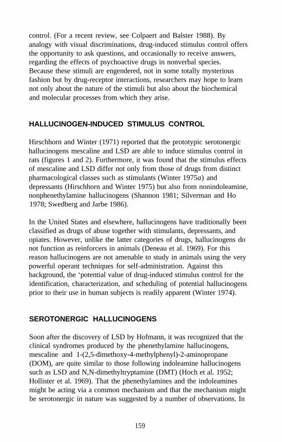
control. (For a recent review, see Colpaert and Balster 1988). Byanalogy with visual discriminations, drug-induced stimulus control offersthe opportunity to ask questions, and occasionally to receive answers,regarding the effects of psychoactive drugs in nonverbal species.Because these stimuli are engendered, not in some totally mysteriousfashion but by drug-receptor interactions, researchers may hope to learnnot only about the nature of the stimuli but also about the biochemicaland molecular processes from which they arise.
HALLUCINOGEN-INDUCED STIMULUS CONTROL
Hirschhorn and Winter (1971) reported that the prototypic serotonergichallucinogens mescaline and LSD are able to induce stimulus control inrats (figures 1 and 2). Furthermore, it was found that the stimulus effectsof mescaline and LSD differ not only from those of drugs from distinctpharmacological classes such as stimulants (Winter 1975a) anddepressants (Hirschhorn and Winter 1975) but also from nonindoleamine,nonphenethylamine hallucinogens (Shannon 1981; Silverman and Ho1978; Swedberg and Jarbe 1986).
In the United States and elsewhere, hallucinogens have traditionally beenclassified as drugs of abuse together with stimulants, depressants, andopiates. However, unlike the latter categories of drugs, hallucinogens donot function as reinforcers in animals (Deneau et al. 1969). For thisreason hallucinogens are not amenable to study in animals using the verypowerful operant techniques for self-administration. Against thisbackground, the ‘potential value of drug-induced stimulus control for theidentification, characterization, and scheduling of potential hallucinogensprior to their use in human subjects is readily apparent (Winter 1974).
SEROTONERGlC HALLUCINOGENS
Soon after the discovery of LSD by Hofmann, it was recognized that theclinical syndromes produced by the phenethylamine hallucinogens,mescaline and 1-(2,5-dimethoxy-4-methylphenyl)-2-aminopropane(DOM), are quite similar to those following indoleamine hallucinogenssuch as LSD and N,N-dimethyltryptamine (DMT) (Hoch et al. 1952;Hollister et al. 1969). That the phenethylamines and the indoleaminesmight be acting via a common mechanism and that the mechanism mightbe serotonergic in nature was suggested by a number of observations. In
159
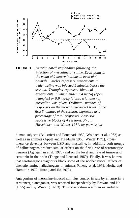
FIGURE 1. Discriminated responding following theinjection of mescaline or saline. Each point isthe mean of 2 determinations in each of 4animals. Circles represent experiments inwhich saline was injected 5 minutes before thesession. Triangles represent identicalexperiments in which either 7.4 mg/kg (opentriangles) or 9.9 mg/kg (closed triangles) ofmescaline was given. Ordinate: number ofresponses on the mescaline-correct lever in thefirst 5 minutes of the session, expressed as apercentage of total responses. Abscissa:successive blocks of 4 sessions. FromHirschhorn and Winter 1971, by permission
human subjects (Balistrieri and Fontanari 1959; Wolbach et al. 1962) aswell as in animals (Appel and Freedman 1968; Winter 1971), cross-tolerance develops between LSD and mescaline. In addition, both groupsof hallucinogens produce similar effects on the firing rate of serotonergicneurons (Aghajanian et al. 1970) and on the level and rate of turnover ofserotonin in the brain (Tonge and Leonard 1969). Finally, it was knownthat serotonergic antagonists block some of the nonbehavioral effects ofphenethylamine hallucinogens in animals (Cheng et al. 1973; Horita andHamilton 1972; Huang and Ho 1972).
Antagonism of mescaline-induced stimulus control in rats by cinanserin, aserotonergic antagonist, was reported independently by Browne and Ho(1975) and by Winter (19753). This observation was then extended to
160
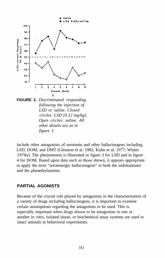
FIGURE 2. Discriminated respondingfollowing the injection ofLSD or saline. Closedcircles: LSD [0.12 mg/kg].Open circles: saline. Allother details are as infigure 1.
include other antagonists of serotonin and other hallucinogens includingLSD, DOM, and DMT (Glennon et al. 1982; Kuhn et al. 1977; Winter1978a). The phenomenon is illustrated in figure 3 for LSD and in figure4 for DOM. Based upon data such as those shown, it appears appropriateto apply the term “serotonergic hallucinogens” to both the indoleaminesand the phenethylamines.
PARTIAL AGONISTS
Because of the crucial role played by antagonists in the characterization ofa variety of drugs including hallucinogens, it is important to examinecertain assumptions regarding the antagonists to be used. This isespecially important when drugs shown to be antagonists in one oranother in vitro, isolated tissue, or biochemical assay systems are used inintact animals in behavioral experiments.
161
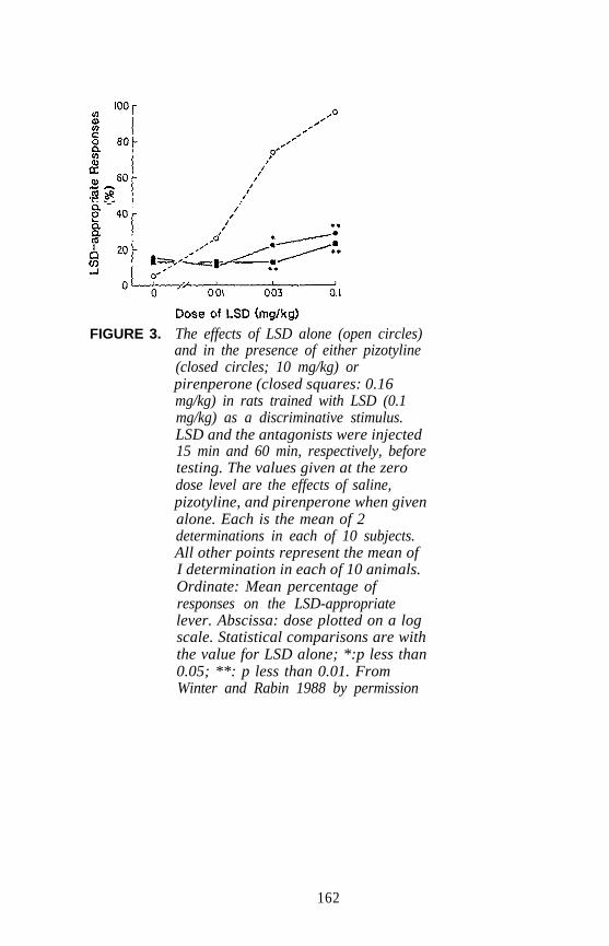
FIGURE 3. The effects of LSD alone (open circles)and in the presence of either pizotyline(closed circles; 10 mg/kg) orpirenperone (closed squares: 0.16mg/kg) in rats trained with LSD (0.1mg/kg) as a discriminative stimulus.LSD and the antagonists were injected15 min and 60 min, respectively, beforetesting. The values given at the zerodose level are the effects of saline,pizotyline, and pirenperone when givenalone. Each is the mean of 2determinations in each of 10 subjects.All other points represent the mean ofI determination in each of 10 animals.Ordinate: Mean percentage ofresponses on the LSD-appropriatelever. Abscissa: dose plotted on a logscale. Statistical comparisons are withthe value for LSD alone; *:p less than0.05; **: p less than 0.01. FromWinter and Rabin 1988 by permission
162
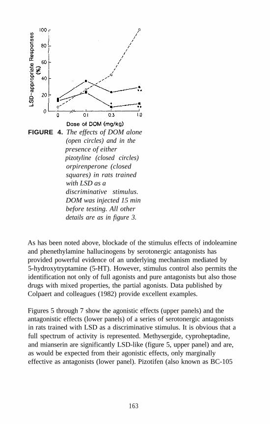
FIGURE 4. The effects of DOM alone(open circles) and in thepresence of eitherpizotyline (closed circles)orpirenperone (closedsquares) in rats trainedwith LSD as adiscriminative stimulus.DOM was injected 15 minbefore testing. All otherdetails are as in figure 3.
As has been noted above, blockade of the stimulus effects of indoleamineand phenethylamine hallucinogens by serotonergic antagonists hasprovided powerful evidence of an underlying mechanism mediated by5-hydroxytryptamine (5-HT). However, stimulus control also permits theidentification not only of full agonists and pure antagonists but also thosedrugs with mixed properties, the partial agonists. Data published byColpaert and colleagues (1982) provide excellent examples.
Figures 5 through 7 show the agonistic effects (upper panels) and theantagonistic effects (lower panels) of a series of serotonergic antagonistsin rats trained with LSD as a discriminative stimulus. It is obvious that afull spectrum of activity is represented. Methysergide, cyproheptadine,and mianserin are significantly LSD-like (figure 5, upper panel) and are,as would be expected from their agonistic effects, only marginallyeffective as antagonists (lower panel). Pizotifen (also known as BC-105
163
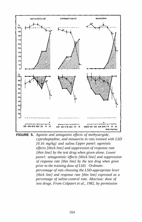
FIGURE 5. Agonist and antagonist effects of methysergide,cyproheptadine, and minaserin in rats trained with LSD[0.16 mg/kg] and saline.Upper panel: agonisticeffects [thick line] and suppression of response rate[thin line] by the test drug when given alone. Lowerpanel: antagonistic effects [thick line] and suppressionof response rate [thin line] by the test drug when givenprior to the training dose of LSD. Ordinate:percentage of rats choosing the LSD-appropriate lever[thick line] and response rate [thin line] expressed as apercentage of saline-control rate. Abscissa: dose oftest drugs. From Colpaert et al., 1982, by permission
164
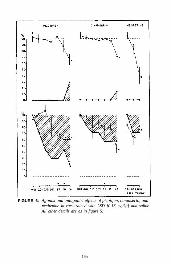
FIGURE 6. Agonist and antagonist effects of pizotifen, cinanserin, andmetitepine in rats trained with LSD [0.16 mg/kg] and saline.All other details are as in figure 5.
165
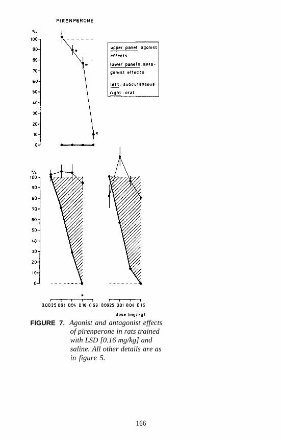
FIGURE 7. Agonist and antagonist effectsof pirenperone in rats trainedwith LSD [0.16 mg/kg] andsaline. All other details are asin figure 5.
166
and as pizotyline), cinanserin, and metitepine (figure 6) are clearly lessagonistic, but only pizotifen approaches being a complete antagonist.Finally, figure 7 provides convincing evidence that pirenperone is devoidof LSD-like stimulus effects and is a complete antagonist of LSD at dosesthat have only modest effects upon rate of responding.
Of the three purported antagonists showing the highest degree ofLSD-like stimulus effects in the rat, two-methysergide andcyproheptadine-are claimed to be sometimes associated withhallucinations in humans. It is clear that drug-induced stimulus controlprovides the means to assess full agonists, pure antagonists, and thosedrugs that exhibit intermediate degrees of activity. It is likely thatextrapolation from animals to humans is most problematic for thelast-named group of drugs.
INTERMEDIATE RESULTS AND THE BERRY-ATORHYPOTHESIS
One of the most remarkable features of drug-induced stimulus control isthat animals trained with a drug of one pharmacological class do notalways exhibit drug-appropriate responses when tested with a drug ofanother class even at doses known to have stimulus properties. Forexample, rats trained with morphine sulfate (6 milligrams per kilogram[mg/kg]) versus saline emit primarily saline-appropriate responses whentested with ethanol at a dose of 630 mg/kg, despite the fact that stimuluscontrol is readily established in rats trained with a 630 mg/kg dose ofethanol versus saline (Winter 1975c). However, observations such asthese should not diminish the appreciation of what have been called“intermediate results” (Winter 1978b).
In the larger context of stimulus control, it was noted above that ageneralization gradient is observed. As the tested stimulus becomes lessand less like the conditioned stimulus, the probability of responseemission becomes less and less. With respect to drug-induced stimuluscontrol, a clear example of a stimulus gradient is seen when subjectstrained with saline versus a specified dose of a drug are tested with arange of lesser doses of that drug. As expected, intermediate degrees ofdrug-appropriate responding occur.
167
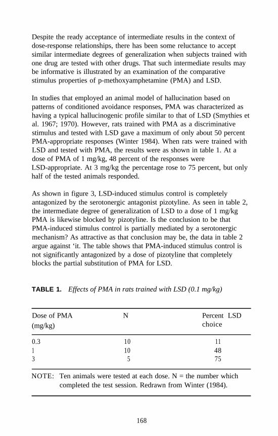
Despite the ready acceptance of intermediate results in the context ofdose-response relationships, there has been some reluctance to acceptsimilar intermediate degrees of generalization when subjects trained withone drug are tested with other drugs. That such intermediate results maybe informative is illustrated by an examination of the comparativestimulus properties of p-methoxyamphetamine (PMA) and LSD.
In studies that employed an animal model of hallucination based onpatterns of conditioned avoidance responses, PMA was characterized ashaving a typical hallucinogenic profile similar to that of LSD (Smythies etal. 1967; 1970). However, rats trained with PMA as a discriminativestimulus and tested with LSD gave a maximum of only about 50 percentPMA-appropriate responses (Winter 1984). When rats were trained withLSD and tested with PMA, the results were as shown in table 1. At adose of PMA of 1 mg/kg, 48 percent of the responses wereLSD-appropriate. At 3 mg/kg the percentage rose to 75 percent, but onlyhalf of the tested animals responded.
As shown in figure 3, LSD-induced stimulus control is completelyantagonized by the serotonergic antagonist pizotyline. As seen in table 2,the intermediate degree of generalization of LSD to a dose of 1 mg/kgPMA is likewise blocked by pizotyline. Is the conclusion to be thatPMA-induced stimulus control is partially mediated by a serotonergicmechanism? As attractive as that conclusion may be, the data in table 2argue against ‘it. The table shows that PMA-induced stimulus control isnot significantly antagonized by a dose of pizotyline that completelyblocks the partial substitution of PMA for LSD.
TABLE 1. Effects of PMA in rats trained with LSD (0.1 mg/kg)
Dose of PMA(mg/kg)
0.313
N
1010
5
Percent LSDchoice
114875
NOTE: Ten animals were tested at each dose. N = the number whichcompleted the test session. Redrawn from Winter (1984).
168
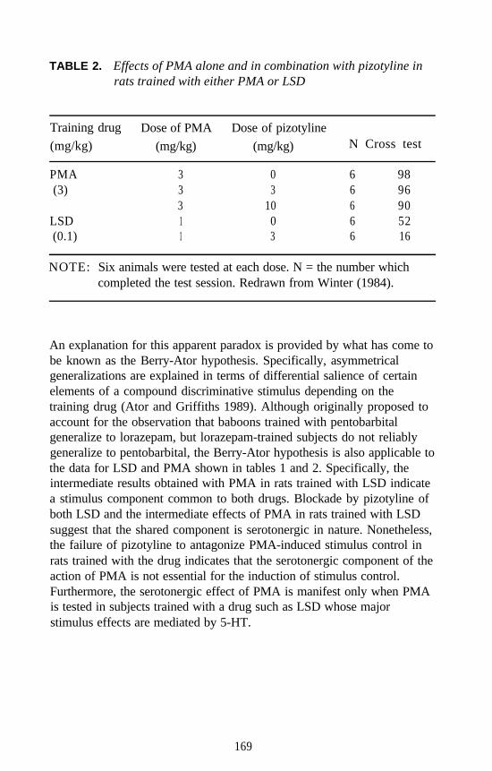
TABLE 2. Effects of PMA alone and in combination with pizotyline inrats trained with either PMA or LSD
Training drug Dose of PMA Dose of pizotyline(mg/kg) (mg/kg) (mg/kg) N Cross test
PMA 3 0 6 98(3) 3 3 6 96
3 10 6 90LSD 1 0 6 52(0.1) 1 3 6 16
NOTE: Six animals were tested at each dose. N = the number whichcompleted the test session. Redrawn from Winter (1984).
An explanation for this apparent paradox is provided by what has come tobe known as the Berry-Ator hypothesis. Specifically, asymmetricalgeneralizations are explained in terms of differential salience of certainelements of a compound discriminative stimulus depending on thetraining drug (Ator and Griffiths 1989). Although originally proposed toaccount for the observation that baboons trained with pentobarbitalgeneralize to lorazepam, but lorazepam-trained subjects do not reliablygeneralize to pentobarbital, the Berry-Ator hypothesis is also applicable tothe data for LSD and PMA shown in tables 1 and 2. Specifically, theintermediate results obtained with PMA in rats trained with LSD indicatea stimulus component common to both drugs. Blockade by pizotyline ofboth LSD and the intermediate effects of PMA in rats trained with LSDsuggest that the shared component is serotonergic in nature. Nonetheless,the failure of pizotyline to antagonize PMA-induced stimulus control inrats trained with the drug indicates that the serotonergic component of theaction of PMA is not essential for the induction of stimulus control.Furthermore, the serotonergic effect of PMA is manifest only when PMAis tested in subjects trained with a drug such as LSD whose majorstimulus effects are mediated by 5-HT.
169
CORRELATIONS BETWEEN HUMANS AND ANIMALS
How much can a rat or other nonverbal species tell us about the humanexperience with hallucinogens? In the present context, what candrug-induced stimulus control by serotonergic hallucinogens tell us abouttheir mechanisms of action in humans? Can drug-induced stimuluscontrol provide the means to reliably predict whether a drug fresh fromthe synthetic chemist will produce hallucinations in human subjects? Oneapproach to answering these questions is to examine the ability of animalsto classify drugs correctly as being either hallucinogenic or nonhall-ucinogenic on the basis of their stimulus properties. A convenient way topursue this approach is in terms of false negatives and false positives.
FALSE NEGATIVES
A false negative is a drug known to be hallucinogenic in man, yet fails tomimic other known hallucinogens of the same class trained asdiscriminative stimuli in animals. “Of the same class” is emphasizedbecause of the well-established fact, noted above, that drugs such as LSD,phencyclidine, tetrahydrocannabinol, and atropine are all properly calledhallucinogens, yet each is pharmacologically distinct. Thus, the falsenegatives to be considered at this time are those within the classdesignated as serotonergic hallucinogens.
In theory, false negatives have a finite probability of occurrence. Forexample, imagine two drugs, A and B, that share a commonhallucinogen-inducing effect in humans. Further imagine that the sharedeffect of drugs A and B is reflected in a distinctive discriminativestimulus in animals. However, drug A does not generalize to drug B,because drug B suppresses behavior in a chosen animal species at doseslower than those required to elicit the hallucinogen-specific stimulus. Theprinciple is clear: an animal in which all behavior has been obliteratedcannot answer questions about discriminative stimuli.
In practice, the clearest example of a false negative among theserotonergic hallucinogens was provided by Koerner and Appel(1982).Rats trained to discriminate between 4-phosphoryloxy-DMT (psilocybin)and saline generalized to 4-hydroxy-DMT (psilocin) and LSD but failedto generalize to mescaline.
170
Koerner and Appel (1982) regarded their data as evidence of “theinability of existing drug discrimination procedures to detect‘hallucinogenicity’ in drugs (p. 134).” This assessment may be too harshin view of the numerous demonstrations of the similarities between thestimulus effects of various indoleamine and phenethylaminehallucinogens and, more specifically, the blockade of the stimulus effectsof both psilocybin and mescaline by the serotonergic antagonist,cinanserin (Silverman and Ho 1978). A gentler interpretation is that,despite their similarities, each member of the class of serotonergichallucinogens is a distinct pharmacological entity, and as such each wouldbe expected to have distinctive features in its stimulus complex.Ironically, the more thorough the research, the more likely it is that suchdistinctive features will be detected. As a practical matter, one mightwish to characterize an unknown drug against several serotonergichallucinogens. It should be noted as well that in a subsequent study byAppel and Callahan (1989), mescaline-trained rats generalized fully topsilocybin.
FALSE POSITIVES
False positives are of greater concern than false negatives with respect tothe discriminative power of drug-induced stimulus control, but at thesame time they are of perhaps greater heuristic value. A false positive is adrug known to be devoid of hallucinogenic activity in humans but whichnonetheless fully mimics known serotonergic hallucinogens in animals.
Before discussing false positives, it must be noted that interpretation ofanimal data is sometimes hampered by the inadequacy of availableclinical data. For example, 2, 3, 4-trimethoxyphenethylamine(2,3,4-TMPEA) is a structural isomer of mescaline that was reported tohave no psychotropic effects (Shulgin 1964; Shulgin and Shulgin 1991).On that basis it was used to test the hypothesis that the differentpharmacologic properties of hallucinogens and nonhallucinogens in manare reflected in distinctive stimuli in rats. Only after it was found that ratsappeared unable to distinguish between the effects of mescaline and2,3,4-TMPEA (Winter 1973) was the author’s attention drawn to acitation of 2,3,4-TMPEA hallucinogenic activity in schizophrenic patients(Slotta and Muller 1936). The proper interpretation of this set ofobservations or their implications for schizophrenia remains uncertain.
171
One may argue on theoretical grounds, as argued above for falsenegatives, that true false positives should occasionally arise. If oneimagines two drugs, C and D, to possess a shared hallucination-inducingproperty and drug D to also be a very potent emetic agent, one mayremain unaware of D’s hallucinogenic activity for the simple reason thathallucinogenic doses are never reached in vomiting human subjects. Onthe other hand, if the emetic effect is species-specific, C and D would beexpected to generalize to one another in an emesis-resistant species andthus yield a false positive.
Despite the foregoing caveats, false positives have been identified.Understanding them is essential to a valid judgment of the efficacy ofdrug-induced stimulus control as a practical tool for the study ofserotonergic hallucinogens. Although a number of candidates presentthemselves, only lisuride and 2-(1-piperazinyl) quinoline maleate(quipazine) are discussed in detail in this chapter.
Lisuride is an ergot that is usually classified as a dopaminergic agonist.As such, it has a therapeutic role in the treatment of Parkinson’s disease.Lisuride is generally regarded as being nonhallucinogenic and quitedifferent from LSD in its actions. Despite their distinctive effects inhumans, complete symmetrical generalization of LSD and lisuride hasbeen reported (Holohean et al. 1982; White and Appel 1982a,b).
The observation that the stimulus effects of LSD and lisuride aredifferentially antagonized by serotonergic and dopaminergic antagonists(Cunningham and Appel 1987; White and Appel 1982c) is of obviouspharmacologic interest. However, it does not resolve the question oflisuride as a false positive. What is required is a demonstration thatanimals can behaviorally differentiate the two drugs. A series ofexperiments by Callahan and Appel (1990) provided just such ademonstration.
Rats were presented with three levers simultaneously. Each lever wasassociated with one of three conditions: saline, LSD, or lisuride. Thedoses of LSD and lisuride were chosen to be comparable in their stimuluseffects when tested in animals trained with lisuride and LSD, respectively,versus saline. As shown in figure 8, the rats learned to discriminatebetween the three training conditions. Furthermore, when dose-responsetests were done with a range of doses of LSD and lisuride, there was agraded distribution of responses between the saline-appropriate lever andthe LSD- and lisuride-appropriate levers, respectively. Thus, in the
172
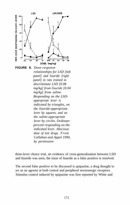
FIGURE 8. Dose-responserelationships for LSD [leftpanel] and lisuride [rightpanel] in rats trained todiscriminate LSD [0.08mg/kg] from lisuride [0.04mg/kg] from saline.Responding on the LSD-appropriate lever isindicated by triangles, onthe lisuride-appropriatelever by squares, and onthe saline-appropriatelever by circles. Ordinate:percent responding on theindicated lever. Abscissa:dose of test drugs. FromCallahan and Appel 1990,by permission
three-lever choice trial, no evidence of cross-generalization between LSDand lisuride was seen; the issue of lisuride as a false positive is resolved.
The second false positive to be discussed is quipazine, a drug thought toact as an agonist at both central and peripheral serotonergic receptors.Stimulus control induced by quipazine was first reported by White and
173
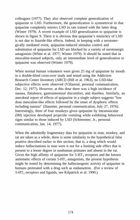
colleagues (1977). They also observed complete generalization ofquipazine to LSD. Furthermore, the generalization is symmetrical in thatquipazine completely mimics LSD in rats trained with the latter drug(Winter 1979). A recent example of LSD generalization to quipazine isshown in figure 9. There it is obvious that quipazine’s mimickry of LSDis not due to lisuride-like effects. Indeed, in keeping with a serotoner-gically mediated event, quipazine-induced stimulus control andsubstitution of quipazine for LSD are blocked by a variety of serotonergicantagonists (White et al. 1977; Winter 1979). It should be noted that inmescaline-trained subjects, only an intermediate level of generalization toquipazine was observed (Winter 1979).
When normal human volunteers were given 25 mg of quipazine by mouthin a double-blind cross-over study and tested using the AddictionResearch Center Inventory (ARCI) (Hill et al. 1963), no LSD-likesubjective effects were observed (Villarreal, personal communication,Dec. 12, 1977). However, at this dose there was a high incidence ofnausea, flatulence, gastrointestinal discomfort, and diarrhea. Similarly, ananecdotal report of effects of quipazine in a single subject suggests “lowdose mescaline-like effects followed by the onset of dysphoric effectsincluding nausea!’ (Daumier, personal communication, July 27, 1976).Interestingly, three of four monkeys given quipazine by intramuscular(IM) injection developed projectile vomiting while exhibiting behavioralsigns similar to those induced by LSD (Schlemmer, Jr., personalcommunication, Jan. 14, 1977).
When the admittedly fragmentary data for quipazine in man, monkey, andrat are taken as a whole, there is some similarity to the hypothetical falsepositive described earlier in this section; that is, a drug which wouldinduce hallucinations in man were it not for a limiting side effect that ispresent to a lesser degree in nonhuman primates and absent in the rat.Given the high affinity of quipazine for 5-HT3 receptors and the knownantiemetic effects of certain 5-HT3 antagonists, the present hypothesismight be tested by determining the hallucinogenic activity of quipazine inhumans pretreated with a drug such as ondansetron. (For a review of5-HT3 receptors and ligands, see Kilpatrick et al. 1990.)
174
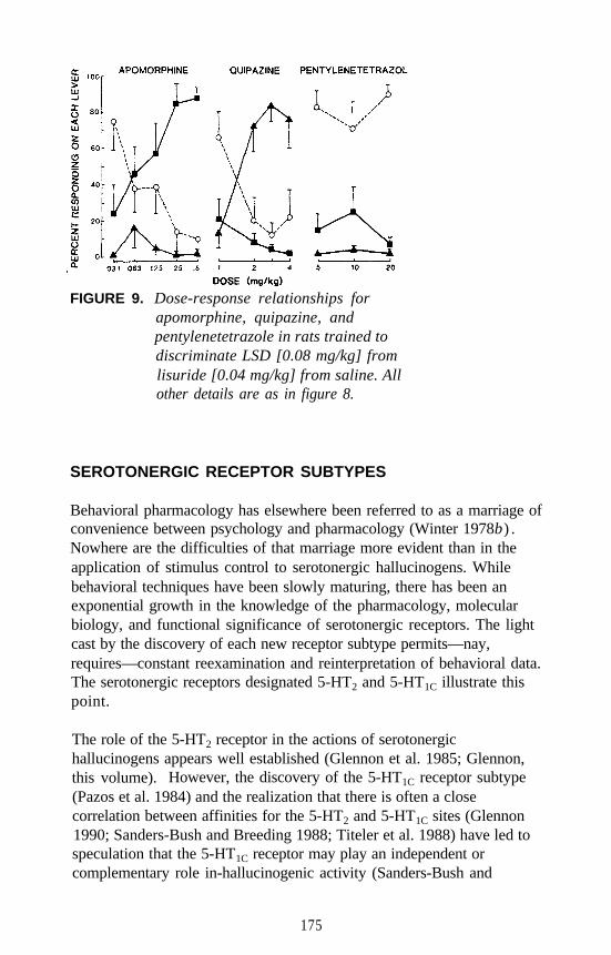
FIGURE 9. Dose-response relationships forapomorphine, quipazine, andpentylenetetrazole in rats trained todiscriminate LSD [0.08 mg/kg] fromlisuride [0.04 mg/kg] from saline. Allother details are as in figure 8.
SEROTONERGIC RECEPTOR SUBTYPES
Behavioral pharmacology has elsewhere been referred to as a marriage ofconvenience between psychology and pharmacology (Winter 1978b) .Nowhere are the difficulties of that marriage more evident than in theapplication of stimulus control to serotonergic hallucinogens. Whilebehavioral techniques have been slowly maturing, there has been anexponential growth in the knowledge of the pharmacology, molecularbiology, and functional significance of serotonergic receptors. The lightcast by the discovery of each new receptor subtype permits—nay,requires—constant reexamination and reinterpretation of behavioral data.The serotonergic receptors designated 5-HT2 and 5-HT1C illustrate thispoint.
The role of the 5-HT2 receptor in the actions of serotonergichallucinogens appears well established (Glennon et al. 1985; Glennon,this volume). However, the discovery of the 5-HT1C receptor subtype(Pazos et al. 1984) and the realization that there is often a closecorrelation between affinities for the 5-HT2 and 5-HT1C sites (Glennon1990; Sanders-Bush and Breeding 1988; Titeler et al. 1988) have led tospeculation that the 5-HT1C receptor may play an independent orcomplementary role in-hallucinogenic activity (Sanders-Bush and
175
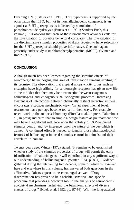
Breeding 1991; Titeler et al. 1988). This hypothesis is supported by theobservation that LSD, but not its nonhallucinogenic congeners, is anagonist at 5-HT1C receptors as indicated by stimulation ofphosphoinositide hydrolysis (Burris et al. 199 1; Sanders-Bush, thisvolume.) It is obvious that each of these biochemical advances calls forthe investigation of possible behavioral correlates. The investigation ofthe discriminative stimulus properties of drugs reputed to have selectivityfor the 5-HT1C receptor should prove informative. One such agentpresently under study is m-chlorophenylpiperazine (MCPP) (Winter andRabin 1992).
CONCLUSION
Although much has been learned regarding the stimulus effects ofserotonergic hallucinogens, this area of investigation remains exciting inits promise. The observation that atypical antipsychotic drugs such asclozapine have high affinity for serotonergic receptors has given new lifeto the old idea that there may be a connection between exogenoushallucinogens and endogenous hallucinogenic processes. Increasingawareness of interactions between chemically distinct neurotransmittersencourages a broader mechanistic view. On an experimental level,researchers have perhaps become too set in their ways. For example,recent work in the author’s laboratory (Fiorella et al., in press; Palumbo etal., in press) indicates that so simple a design feature as pretreatment timemay have a significant influence upon the stability of DOM-inducedstimulus control and, by inference, upon the nature of the cue which istrained. A continued effort is needed to identify those pharmacologicalfeatures of hallucinogen-induced stimulus control in animals and theircorrelates in humans.
Twenty years ago, Winter (1972) stated, “It remains to be establishedwhether study of the stimulus properties of drugs will permit the earlyidentification of hallucinogens or will contribute in any significant way toour understanding of hallucinogens.” (Winter 1974, p. 831). Evidencegathered during the intervening two decades, some of which is reviewedhere and elsewhere in this volume, has answered both questions in theaffirmative. Others appear to be encouraged as well: “Drugdiscrimination has proven to be a reliable, sensitive, and specificprocedure that provides a powerful tool in the analysis of neuropharm-acological mechanisms underlying the behavioral effects of diverseclasses of drugs.” (Koek et al. 1992, pp. 97-98). With the long-awaited
176
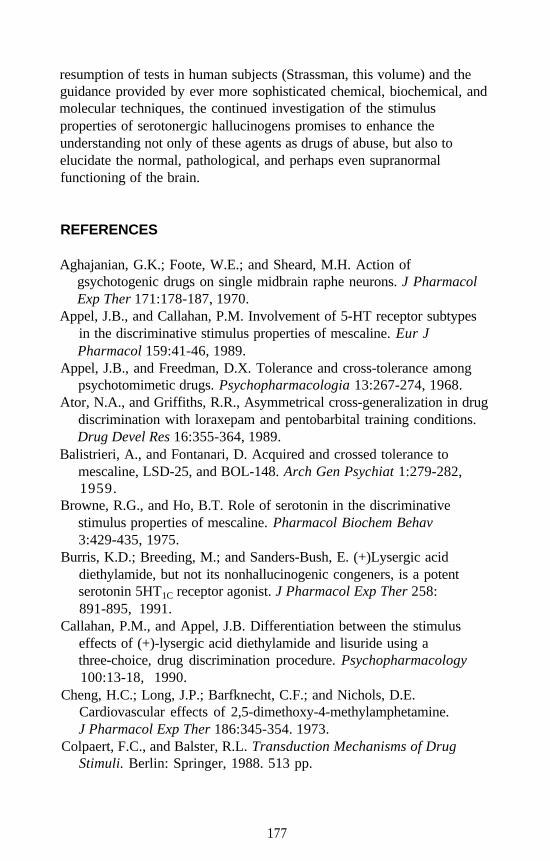
resumption of tests in human subjects (Strassman, this volume) and theguidance provided by ever more sophisticated chemical, biochemical, andmolecular techniques, the continued investigation of the stimulusproperties of serotonergic hallucinogens promises to enhance theunderstanding not only of these agents as drugs of abuse, but also toelucidate the normal, pathological, and perhaps even supranormalfunctioning of the brain.
REFERENCES
Aghajanian, G.K.; Foote, W.E.; and Sheard, M.H. Action ofgsychotogenic drugs on single midbrain raphe neurons. J PharmacolExp Ther 171:178-187, 1970.
Appel, J.B., and Callahan, P.M. Involvement of 5-HT receptor subtypesin the discriminative stimulus properties of mescaline. Eur JPharmacol 159:41-46, 1989.
Appel, J.B., and Freedman, D.X. Tolerance and cross-tolerance amongpsychotomimetic drugs. Psychopharmacologia 13:267-274, 1968.
Ator, N.A., and Griffiths, R.R., Asymmetrical cross-generalization in drugdiscrimination with loraxepam and pentobarbital training conditions.Drug Devel Res 16:355-364, 1989.
Balistrieri, A., and Fontanari, D. Acquired and crossed tolerance tomescaline, LSD-25, and BOL-148. Arch Gen Psychiat 1:279-282,1959.
Browne, R.G., and Ho, B.T. Role of serotonin in the discriminativestimulus properties of mescaline. Pharmacol Biochem Behav3:429-435, 1975.
Burris, K.D.; Breeding, M.; and Sanders-Bush, E. (+)Lysergic aciddiethylamide, but not its nonhallucinogenic congeners, is a potentserotonin 5HT1C receptor agonist. J Pharmacol Exp Ther 258:891-895, 1991.
Callahan, P.M., and Appel, J.B. Differentiation between the stimuluseffects of (+)-lysergic acid diethylamide and lisuride using athree-choice, drug discrimination procedure. Psychopharmacology100:13-18, 1990.
Cheng, H.C.; Long, J.P.; Barfknecht, C.F.; and Nichols, D.E.Cardiovascular effects of 2,5-dimethoxy-4-methylamphetamine.J Pharmacol Exp Ther 186:345-354. 1973.
Colpaert, F.C., and Balster, R.L. Transduction Mechanisms of DrugStimuli. Berlin: Springer, 1988. 513 pp.
177
Colpaert, F.C.; Niemegeers, J.E.; and Janssen, P.A.J. A drugdiscrimination analysis of lysergic acid diethylamide: In vivo agonistand antagonist effects of purported 5-hydroxytryptamine antagonistsand of pirenperone, an LSD-antagonist. J Pharmacol Exp Ther221:206-214, 1982.
Conger, J.J. The effects of alcohol on conflict behavior in the albino rat.Quart J Studies Alc 12:1-29, 1951.
Cunningham, K.A., and Appel, J.B. Neuropharmacological reassessmentof the discriminative stimulus properties of d-lysergic aciddiethylamide. Psychopharmacology 91:67-73, 1987.
DeCurtis, A. Interview with George Harrison. Rolling Stone November.5, 1987, pp. 47-50.
Deneau, G.; Yanagita, T.; Seevers, M.H. Self-administration ofpsychoactive substances by the monkey. Psychopharmacologia16:30-48, 1969.
Fiorella, D.; Rabin, R.A.; and Winter, J.C. The time-dependent stimuluseffects of l-[2,5-dimethoxy-4-methylphenyl]-2-aminopropane [DOM]:Implications for drug-induced stimulus control as a method for thestudy of hallucinogenic agents. Psychopharmacology, in press.
Glennon, R.A. Do classical hallucinogens act as 5-HT2 agonists orantagonists? Neuropsychopharmacol 3:509-517, 1990.
Glennon, R.A.; Rosecrans, J.A.; Young, R. The use of the drugdiscrimination paradigm for studying hallucinogenic agents. In:Colpaert, F.C., and Slangen, J.L., eds. Drug Discrimination:Applications in CNS Pharmacology. Amsterdam: Elsevier, 1982.
Glennon, R.A.; Titeler, M.; McKenney, J.D. Evidence for 5-HT2
involvement in the mechanism of action of hallucinogens. Life Sci35:2505-2511, 1985.
Heffter, A. Ueber Pellote. Naunyn-Schmiedebergs Arch Exp Pharmakol40:418-425, 1897.
Hill, H.E.; Haertzen, C.A.; Wolbach, A.B., Jr.; and Miner, E.J. TheAddiction Research Center Inventory: Standardization of scales whichevaluate subjective effects of morphine, amphetamine, pentobarbital,alcohol, LSD-25, pyrahexyl, and chlorpromazine.Psychopharmacologia 4:167-205, 1963.
Hirschhorn, I.D., and Winter, J.C. Mescaline and lysergic aciddiethylamide (LSD) as discriminative stimuli. Psychopharmacologia22:64-71, 1971.
Hirschhorn, I.D., and Winter, J.C. Differences in the stimulus propertiesof barbital and hallucinogens. Pharmacol Biochem Behav 3:343-347,1975.
178
Hoch, P.J.; Cattell, J.P.; and Pennes, H.H. Effects of mescaline andlysergic acid [d-LSD-25]. Amer J Psychiat 108:579-584, 1952.
Hofmann, A. Psychotomimetic drugs, chemical and pharmacologicalaspects. Acta Physiol Pharmacol Neerl 8:240-258, 1959.
Hollister, LE.; MacNicol, M.F.; and Gillespie, H.K. An hallucinogenicamphetamine analog [DOM] in man. Psychopharmacologia 14:62-73,1969.
Holohean, A.M.; White, F.J.; and Appel, J.B. Dopaminergic andserotonergic mediation of the discriminable effects of ergot alkaloids.Eur J Pharmacol 81:595-602, 1982.
Horita, A., and Hamilton, A.E. On the hyperthermic action of2,5-dimethoxy-4-methylamphetamine. Proc West Pharmacol Soc15:104-105, 1972.
Huang, J.-T., and Ho, B.T. The pressor action of2,5-dimethoxy-4-methylamphetamine in rats. J Pharm Pharmacol24:656-657, 1972.
Isbell, H.; Miner, E.J.; and Logan, C.R. Cross tolerance betweenD-2-brom-lysergic acid diethylamide (BOL- 148) and theD-diethylamide of lysergic acid (LSD-25). Psychopharmacologia1:109-116, 1959.
Kelleher, R.T., and Morse, L.H. Determinants of the specificity ofbehavioral effects of drugs. In: Reviews of Physiology, Biochemistry,and Behavior. Vol. 60. New York: Springer-Verlag, 1968. pp. 2-56.
Kilpatrick, G.J.; Bunce, K.T.; and Tyers, M.B. 5-HT3 receptors. Med ResRev 10:441-475, 1990.
Koek, W.; Jackson, A.; and Colpaert, F.C. Behavioral pharmacology ofantagonists at 5-HT2/5-HT1C receptors. Neurosci Biobehav Rev16:95-105, 1992.
Koerner, J., and Appel, J.B. Psilocybin as a discriminative stimulus: Lackof specificity in an animal behavior model of hallucinogensPsychopharmacology 76:130-135, 1982.
Kuhn, D.M.; White, F.J.; and Appel, J.B. Discriminative stimulusproperties of hallucinogens: Behavioral assay of drug action. In: Lal,H., ed. Discriminative Stimulus Properties of Drugs. New York:Plenum, 1977.
Martin, W.R., and Sloan, J.W. Pharmacology and classification ofLSD-like hallucinogens. In: Martin, W.R., ed. Handbook ofExperimental Pharmacology. Vol. 45. Berlin: Springer-Verlag, 1977,pp. 306-368.
Pazos, A.; Hoyer; D.; and Palacios, J.M. The binding of serotonergicligands to the porcine choroid plexus: Characterization of a new typeof serotonin recognition site. Eur J Pharmacol 106:539-546, 1984.
179
Palumbo, P.A.; Fiorella, D.; Rabin, R.A.; and Winter, J.C. Comparativestability of stimulus control induced by LSD and DOM. PharmacolBiochem Behav, in press.
Sanders-Bush, E., and Breeding, M.: Putative selective 5-HT2 antagonistsblock serotonin 5-HT1C receptors in choroid plexus. J Pharmacol ExpTher 247:169-173, 1988.
Sanders-Bush, E., and Breeding, M. Choroid plexus epithelial cells inprimary culture: A model of 5-HT1C receptor activation byhallucinogenic drugs. Psychopharmacology 108:340-346, 1991.
Schultes, R.E., and Hofmann, A. The Botany and Chemistry ofHallucinogens. 2d ed. Springfield, IL: Charles C. Thomas, 1980.
Shannon, H.E. Evaluation of phencyclidine analogs on the basis of theirdiscriminative stimulus properties in the rat. J Pharmacol Exp Ther216:543-551, 1981.
Shulgin, A.T. Psychotomimietic amphetamines:Methoxy-3,4,-dialkoxyamphetamines. Experientia 20:366-367, 1964.
Shulgin, A.T., and Shulgin, A. PIHKAL: A Chemical Love Story.Berkeley, CA: Transform Press, 1991. pp. 690-692.
Shulgin, A.T.; Shulgin, L.A.; and Jacob, P. A protocol for the evaluationof new psychoactive drugs in man. Meth Find Expt Clin Pharmacol8:313-320, 1986.
Silverman, P.B., and Ho, B.T. Stimulus properties of DOM:Commonality with other hallucinogens. In: Colpaert, F.C., andRosecrans, J.A., eds. Stimulus Properties of Drugs: Ten Years ofProgress. Amsterdam: Elsevier/North Holland, 1978. pp. 189-198.
Slotta, K.H., and Muller, J. Ueber den Abbau des Mescalins undMescalin-ahnlicher Stoffe in Organismus. Hoppe-Seyler’s Z. PhysiolChem 238:14-22, 1936.
Smythies, J.R.; Beaton, J.; Benington, F.; and Morin, R. Behavioraleffects of some derivatives of amphetamine and LSD and theirsignificance. Nature 226:644-645, 1970.
Smythies, J.R.; Johnson, U.S.; Bradley, R.J.; Benington, R.; Morin, D.;and Clark, L.C. Some new behavior-disrupting amphetamines andtheir significance. Nature 216:128-129, 1967.
Spath, E. Ueber die Anhalonium-Alkaloide. I. Anhalin und Mezcalin.Monatsh Chem 40:129-152, 1919.
Stebbins, W.C. Animal Psychophysics: The Design and Conduct ofSensory Experiments. New York: Appleton-Century-Crofts, 1970.
Swedberg, M.D.B., and Jarbe, T.U.C. Drug discrimination procedures:Differential characteristics of the drug A vs drug B and the drug A vsdrug B vs no drug cases. Psychopharmacology 90:341-346, 1986.
180
Szara, S. The comparison of the psychotic effect of tryptamine derivativeswith the effects of mescaline and LSD in self-experiments. In:Garattini, S., and Ghetti, V., eds. Psychotropic Drugs. Amsterdam:Elsevier, 1957, pp. 460-467.
Titeler, M.; Lyon, R.A.; and Glennon, R.A. Radioligand binding evidenceimplicates the brain 5-HT2 receptor as a site of action for LSD andphenylisopropylamine hallucinogens. Psychopharmacology 94:213-216, 1988.
Tonge, Sk, and Leonard, B.E. The effects of some hallucinogenic drugsupon the metabolism of 5-hydroxytryptamine in the brain. Life Sci8:805-814, 1969.
White, F.J. and Appel, J.B. Training dose as a factor in LSD-salinediscrimination. Psychopharmacology 76:20-25, 1982a.
White, F.J., and Appel, J.B. Lysergic acid diethylamide and lisuride:Differentiation of their neuropharmacological actions. Science216:535-537, 1982b.
White, F.J., and Appel, J.B. The role of dopamine and serotonin in thediscriminative stimulus effects of lisuride. J Pharmacol Exp Ther221:421-427, 1982c .
White, F.J.; Kuhn, D.M.; and Appel, J.B. Discriminative stimulusproperties of quipazine. Neuropharmacology 16:827-832, 1977.
Winter, J.C. Tolerance to a behavioral effect of lysergic acid diethylamideand cross-tolerance to mescaline in the rat: Absence of a metaboliccomponent. J Pharmacol Exp Ther 178:625-630, 1971.
Winter, J.C. “Discriminable Properties of Drugs and State-DependentLearning.” Paper presented at the 56th Annual Meeting of theFederation of American Societies for Experimental Biology (FASEB),Atlantic City, April 14, 1972.
Winter, J.C. A comparison of the stimulus properties of mescaline and2,3,4-trimethoxyphenylethylamine. J Pharmacol Expt Ther185:101-107, 1973.
Winter, J.C. Hallucinogens as discriminative stimuli. Federation Proc33:1825-1832, 1974.
Winter, J.C. The effects of DOM, DOET, d-amphetamine, and cocaine inrats trained with mescaline as a discriminative stimulus.Psychopharmacologia 44:29-32, 1975a.
Winter, J.C. Blockade of the stimulus properties of mescaline by aserotonin antagonist. Arch Int Pharmacodyn 214:250-253, 1975b .
Winter, J.C. The stimulus properties of morphine and ethanol.Psychopharmacologia 44:209-214, 1975c.
181
Winter, J.C. Stimulus properties of phenethylamine hallucinogens andlysergic acid diethylamide: The role of 5-hydroxytryptamine.J Pharmacol Exp Ther 204:416-423, 1978a.
Winter, J.C. Drug-induced stimulus control. In: Blackman, D.E., andSanger, D.J., eds. Contemporary Research in BehavioralPharmacology. New York: Plenum, 1978b. pp. 209-237.
Winter, J.C. Quipazine-induced stimulus control in the rat.Psychopharmacology 60:265-269, 1979.
Winter, J.C. The stimulus properties ofp-methoxyamphetamine: Anonessential serotonergic component. Pharmacol Biochem Behav20:201-203, 1984.
Winter, J.C., and Rabin, R.A. Interactions between serotonergic agonistsand antagonists in rats trained with LSD as a discriminative stimulus.Pharmacol Biochem Behav 30:617-624, 1988.
Winter, J.C. and R.A. Rabin: The discriminative stimulus properties ofm-chlorophenylpiperazine (MCPP). Pharmacol Biochem Behav45:221-223, 1993.
Wolbach, A.B.; Isbell, H.; and Miner, E.J. Cross tolerance betweenmescaline and LSD-25 with a comparison of the mescaline and LSDreactions. Psychopharmacologia 3:1-14, 1962.
ACKNOWLEDGMENTS
Preparation of this manuscript was supported in part by National Instituteon Drug Abuse grant DA-03385.
AUTHOR
Jerrold C. Winter, Ph.D.Professor of Pharmacology and ToxicologyDepartment of Pharmacology and TherapeuticsSchool of Medicine and Biomedical SciencesState University of New York at BuffaloBuffalo, NY 14214-3000
182
Electrophysiological Studies onthe Actions of HallucinogenicDrugs at 5=HT2 Receptors in RatBrain
George K. Aghajaniun
INTRODUCTION
There has been a longstanding interest in the role of the brain serotonin(5-hydroxytryptamine [5-HT]) system in the electrophysiological actionsof psychedelic hallucinogenic drugs such as d-lysergic acid diethylamide(LSD) and mescaline. Early studies focused on the actions of LSD onserotonin-containing (serotonergic) neurons in the raphe nuclei of thebrain stem. LSD and other indoleamine hallucinogens were found tohave potent direct inhibitory effects upon serotonergic raphe neurons(Aghajanian et al. 1972). However, it was soon discovered thatmescaline and other phenethylamine hallucinogens failed to share thisaction (Haigler and Aghajanian 1973). Later, it was shown that the directinhibitory effect of LSD and other indoleamine hallucinogens onserotonergic neurons is mediated by 5-HT1A receptors, and that this actionis shared by a number of selective 5-HT1A agonists that are nothallucinogenic (Sprouse and Aghajanian 1987, 1988). Thus, thereappears to be no correlation between the activity of various drugs at5-HT1A receptors in the raphe nuclei and the presence or absence ofhallucinogenic properties.
In contrast, a good correlation exists between the affinity of bothindoleamine and phenethylamine hallucinogens for 5-HT2 receptors andthe potency of these drugs as hallucinogens in humans (Glennon et al.1984; Titeler et al. 1988) As described below, electrophysiologicalstudies in a number of brain regions show that indoleamine andphenethylamine hallucinogens have common electrophysiological actionat 5-HT2 receptors. There has, however, been some debate on whetherthe hallucinogens act as agonists, partial agonists (Glennon 1990;Sanders-Bush et al. 1988; Sheldon and Aghajanian 1990) or antagonists(Pierce and Peroutka 1990) at 5-HT2 receptors. Recent electrophysio-logical studies bearing on this issue are described in this chapter.
183
The discovery of the structural and functional similarities between the5-HT2 and 5-HT1C receptors (Hartig 1989) has also led to studiesexamining the effect of the hallucinogens at 5-HT1C sites. Recent datafrom studies in the choroid plexus showed that the hallucinogens act aspartial agonists at 5-HT1C receptors that mediate an activation ofphosphoinositide turnover (Burris et al 1991; Sanders-Bush and Breeding1991). The electrophysiological studies are reviewed from the standpointof how they may distinguish between actions at 5-HT2 and 5-HT1C
receptors.
Quantitative autoradiographic studies show high concentrations of 5-HT2
sites in the forebrain including the neocortex (layers IV/V), piriformcortex, claustrum, nucleus accumbens (NACC), and olfactory tubercle(OT) (Pazos et al. 1985). In contrast, with the exception of a few areas(e.g., facial nucleus and the n. tractus solitarius), relatively lowconcentrations of 5-HT2 receptors are found in the brain stem. Single-cellstudies on the physiological properties of 5-HT2 receptors in severalrepresentative brain regions are described in the following sections.
PHYSIOLOGICAL ACTIONS OF HALLUCINOGENS
Locus Coeruleus
The systemic administration of indoleamine and phenethylaminehallucinogens (e.g., LSD and mescaline) results in decreased spontaneousactivity of noradrenergic cells in the locus coeruleus (LC) of anesthetizedrats but, paradoxically, a facilitation of the activation of these cells bysensory stimuli (Aghajanian 1980; Rasmussen and Aghajanian 1986).These effects in the LC are of theoretical interest since, in contrast to theraphe nuclei (see introduction), they are shared by the two major classesof psychedelic hallucinogens. Mediation by 5-HT2 receptors is suggestedby the fact that effects of the hallucinogens on LC neurons can bereversed by low intravenous (IV) doses of selective 5-HT2 antagonistssuch as ritanserin, LY-53857 (figure 1) (Rasmussen and Aghajanian1986), and ketanserin (Gorea and Adrien 1988). Antipsychotic drugswith affinity for 5-HT2 binding sites are also able to reverse the actions ofhallucinogens in the LC independently of their actions at dopamine (DA)and adrenergic receptors (Rasmussen and Aghajanian 1988). The relativepotencies of antipsychotic drugs in blocking the actions of hallucinogensin LC neurons correlates highly with their affinity for 5-HT2 receptors(Rasmussen and Aghajanian 1988). Of particular note is the fact that at
184
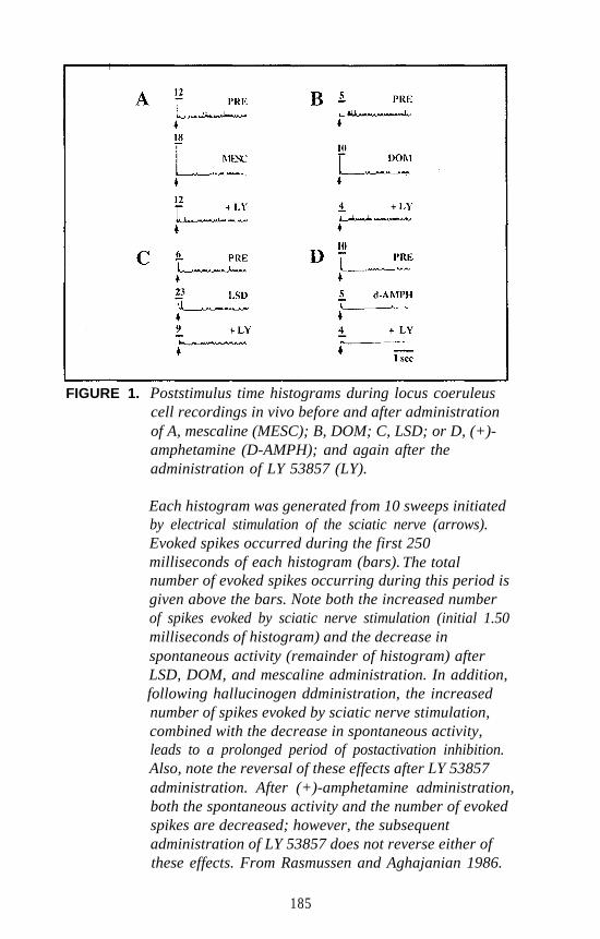
FIGURE 1. Poststimulus time histograms during locus coeruleuscell recordings in vivo before and after administrationof A, mescaline (MESC); B, DOM; C, LSD; or D, (+)-amphetamine (D-AMPH); and again after theadministration of LY 53857 (LY).
Each histogram was generated from 10 sweeps initiatedby electrical stimulation of the sciatic nerve (arrows).Evoked spikes occurred during the first 250milliseconds of each histogram (bars). The totalnumber of evoked spikes occurring during this period isgiven above the bars. Note both the increased numberof spikes evoked by sciatic nerve stimulation (initial 1.50milliseconds of histogram) and the decrease inspontaneous activity (remainder of histogram) afterLSD, DOM, and mescaline administration. In addition,following hallucinogen ddministration, the increasednumber of spikes evoked by sciatic nerve stimulation,combined with the decrease in spontaneous activity,leads to a prolonged period of postactivation inhibition.Also, note the reversal of these effects after LY 53857administration. After (+)-amphetamine administration,both the spontaneous activity and the number of evokedspikes are decreased; however, the subsequentadministration of LY 53857 does not reverse either ofthese effects. From Rasmussen and Aghajanian 1986.
185
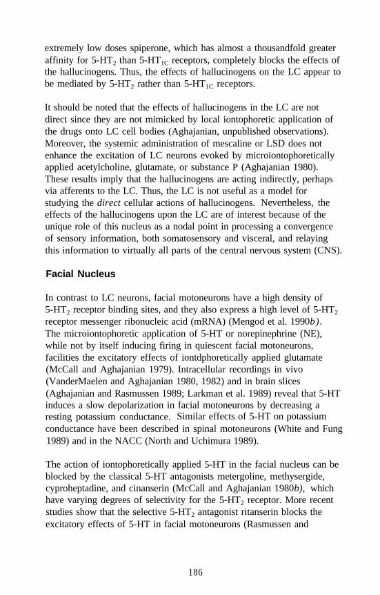
extremely low doses spiperone, which has almost a thousandfold greateraffinity for 5-HT2 than 5-HT1C receptors, completely blocks the effects ofthe hallucinogens. Thus, the effects of hallucinogens on the LC appear tobe mediated by 5-HT2 rather than 5-HT1C receptors.
It should be noted that the effects of hallucinogens in the LC are notdirect since they are not mimicked by local iontophoretic application ofthe drugs onto LC cell bodies (Aghajanian, unpublished observations).Moreover, the systemic administration of mescaline or LSD does notenhance the excitation of LC neurons evoked by microiontophoreticallyapplied acetylcholine, glutamate, or substance P (Aghajanian 1980).These results imply that the hallucinogens are acting indirectly, perhapsvia afferents to the LC. Thus, the LC is not useful as a model forstudying the direct cellular actions of hallucinogens. Nevertheless, theeffects of the hallucinogens upon the LC are of interest because of theunique role of this nucleus as a nodal point in processing a convergenceof sensory information, both somatosensory and visceral, and relayingthis information to virtually all parts of the central nervous system (CNS).
Facial Nucleus
In contrast to LC neurons, facial motoneurons have a high density of5-HT2 receptor binding sites, and they also express a high level of 5-HT2
receptor messenger ribonucleic acid (mRNA) (Mengod et al. 1990b).The microiontophoretic application of 5-HT or norepinephrine (NE),while not by itself inducing firing in quiescent facial motoneurons,facilities the excitatory effects of iontdphoretically applied glutamate(McCall and Aghajanian 1979). Intracellular recordings in vivo(VanderMaelen and Aghajanian 1980, 1982) and in brain slices(Aghajanian and Rasmussen 1989; Larkman et al. 1989) reveal that 5-HTinduces a slow depolarization in facial motoneurons by decreasing aresting potassium conductance. Similar effects of 5-HT on potassiumconductance have been described in spinal motoneurons (White and Fung1989) and in the NACC (North and Uchimura 1989).
The action of iontophoretically applied 5-HT in the facial nucleus can beblocked by the classical 5-HT antagonists metergoline, methysergide,cyproheptadine, and cinanserin (McCall and Aghajanian 1980b), whichhave varying degrees of selectivity for the 5-HT2 receptor. More recentstudies show that the selective 5-HT2 antagonist ritanserin blocks theexcitatory effects of 5-HT in facial motoneurons (Rasmussen and
186
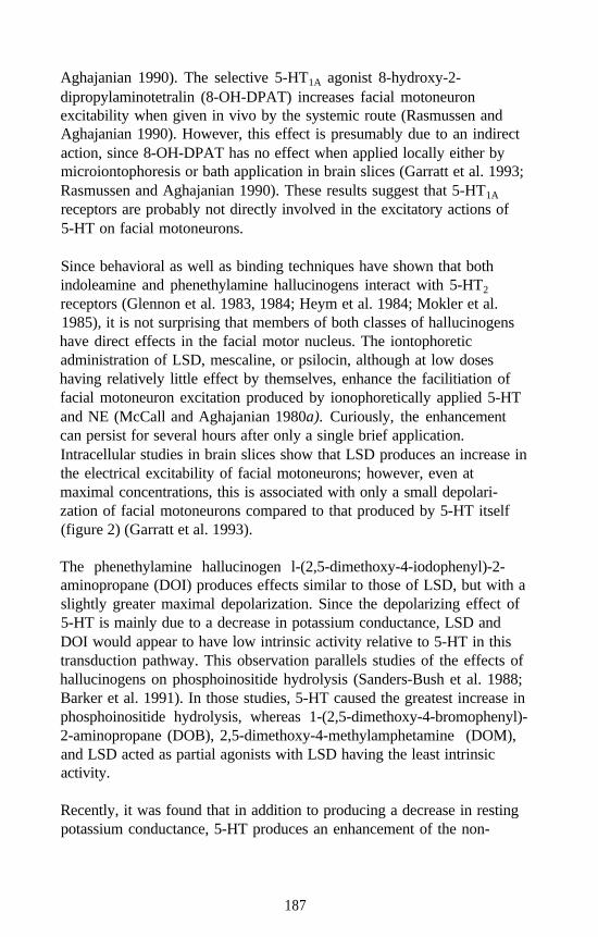
Aghajanian 1990). The selective 5-HT1A agonist 8-hydroxy-2-dipropylaminotetralin (8-OH-DPAT) increases facial motoneuronexcitability when given in vivo by the systemic route (Rasmussen andAghajanian 1990). However, this effect is presumably due to an indirectaction, since 8-OH-DPAT has no effect when applied locally either bymicroiontophoresis or bath application in brain slices (Garratt et al. 1993;Rasmussen and Aghajanian 1990). These results suggest that 5-HT1A
receptors are probably not directly involved in the excitatory actions of5-HT on facial motoneurons.
Since behavioral as well as binding techniques have shown that bothindoleamine and phenethylamine hallucinogens interact with 5-HT2
receptors (Glennon et al. 1983, 1984; Heym et al. 1984; Mokler et al.1985), it is not surprising that members of both classes of hallucinogenshave direct effects in the facial motor nucleus. The iontophoreticadministration of LSD, mescaline, or psilocin, although at low doseshaving relatively little effect by themselves, enhance the facilitiation offacial motoneuron excitation produced by ionophoretically applied 5-HTand NE (McCall and Aghajanian 1980a). Curiously, the enhancementcan persist for several hours after only a single brief application.Intracellular studies in brain slices show that LSD produces an increase inthe electrical excitability of facial motoneurons; however, even atmaximal concentrations, this is associated with only a small depolari-zation of facial motoneurons compared to that produced by 5-HT itself(figure 2) (Garratt et al. 1993).
The phenethylamine hallucinogen l-(2,5-dimethoxy-4-iodophenyl)-2-aminopropane (DOI) produces effects similar to those of LSD, but with aslightly greater maximal depolarization. Since the depolarizing effect of5-HT is mainly due to a decrease in potassium conductance, LSD andDOI would appear to have low intrinsic activity relative to 5-HT in thistransduction pathway. This observation parallels studies of the effects ofhallucinogens on phosphoinositide hydrolysis (Sanders-Bush et al. 1988;Barker et al. 1991). In those studies, 5-HT caused the greatest increase inphosphoinositide hydrolysis, whereas 1-(2,5-dimethoxy-4-bromophenyl)-2-aminopropane (DOB), 2,5-dimethoxy-4-methylamphetamine (DOM),and LSD acted as partial agonists with LSD having the least intrinsicactivity.
Recently, it was found that in addition to producing a decrease in restingpotassium conductance, 5-HT produces an enhancement of the non-
187
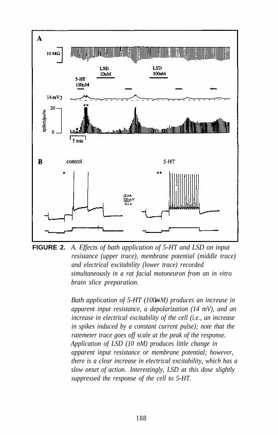
FIGURE 2. A. Effects of bath application of 5-HT and LSD on inputresistance (upper trace), membrane potential (middle trace)and electrical excitability (lower trace) recordedsimultaneously in a rat facial motoneuron from an in vitrobrain slice preparation.
Bath application of 5-HT (100 M) produces an increase inapparent input resistance, a depolarization (14 mV), and anincrease in electrical excitability of the cell (i.e., an increasein spikes induced by a constant current pulse); note that theratemeter trace goes off scale at the peak of the response.Application of LSD (10 nM) produces little change inapparent input resistance or membrane potential; however,there is a clear increase in electrical excitability, which has aslow onset of action. Interestingly, LSD at this dose slightlysuppressed the response of the cell to 5-HT.
188
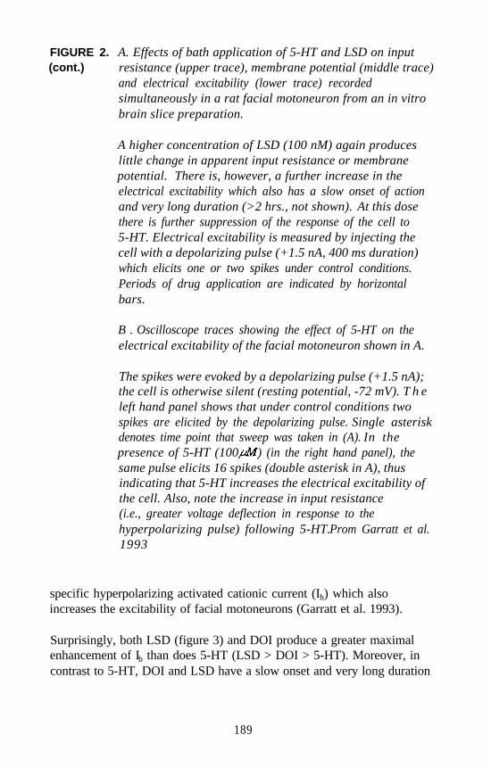
FIGURE 2. A. Effects of bath application of 5-HT and LSD on input(cont.) resistance (upper trace), membrane potential (middle trace)
and electrical excitability (lower trace) recordedsimultaneously in a rat facial motoneuron from an in vitrobrain slice preparation.
A higher concentration of LSD (100 nM) again produceslittle change in apparent input resistance or membranepotential. There is, however, a further increase in theelectrical excitability which also has a slow onset of actionand very long duration (>2 hrs., not shown). At this dosethere is further suppression of the response of the cell to5-HT. Electrical excitability is measured by injecting thecell with a depolarizing pulse (+1.5 nA, 400 ms duration)which elicits one or two spikes under control conditions.Periods of drug application are indicated by horizontalbars.
B . Oscilloscope traces showing the effect of 5-HT on theelectrical excitability of the facial motoneuron shown in A.
The spikes were evoked by a depolarizing pulse (+1.5 nA);the cell is otherwise silent (resting potential, -72 mV). T h eleft hand panel shows that under control conditions twospikes are elicited by the depolarizing pulse. Single asteriskdenotes time point that sweep was taken in (A). In thepresence of 5-HT (100 ) (in the right hand panel), thesame pulse elicits 16 spikes (double asterisk in A), thusindicating that 5-HT increases the electrical excitability ofthe cell. Also, note the increase in input resistance(i.e., greater voltage deflection in response to thehyperpolarizing pulse) following 5-HT.Prom Garratt et al.1993
specific hyperpolarizing activated cationic current (Ih) which alsoincreases the excitability of facial motoneurons (Garratt et al. 1993).
Surprisingly, both LSD (figure 3) and DOI produce a greater maximalenhancement of Ib than does 5-HT (LSD > DOI > 5-HT). Moreover, incontrast to 5-HT, DOI and LSD have a slow onset and very long duration
189
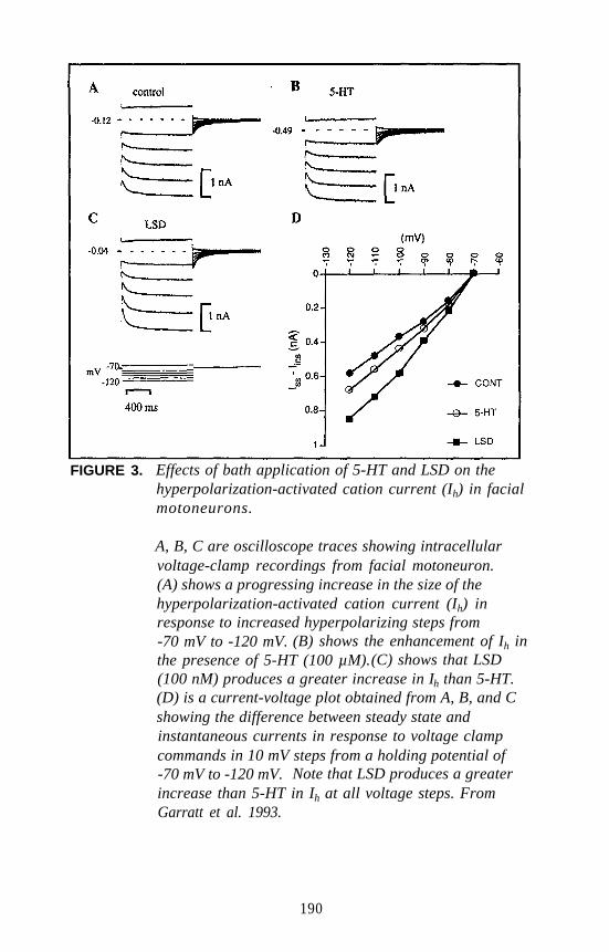
FIGURE 3. Effects of bath application of 5-HT and LSD on thehyperpolarization-activated cation current (Ih) in facialmotoneurons.
A, B, C are oscilloscope traces showing intracellularvoltage-clamp recordings from facial motoneuron.(A) shows a progressing increase in the size of thehyperpolarization-activated cation current (Ih) inresponse to increased hyperpolarizing steps from-70 mV to -120 mV. (B) shows the enhancement of Ih inthe presence of 5-HT (100 µM).(C) shows that LSD(100 nM) produces a greater increase in Ih than 5-HT.(D) is a current-voltage plot obtained from A, B, and Cshowing the difference between steady state andinstantaneous currents in response to voltage clampcommands in 10 mV steps from a holding potential of-70 mV to -120 mV. Note that LSD produces a greaterincrease than 5-HT in Ih at all voltage steps. FromGarratt et al. 1993.
190
of action (> 2 hours). The enhancement of Ih by LSD and DOI isreversed by application of both the 5-HT2/5-HT1A/5-HT1A antagonistspiperone and the 5-HT2/5-HT1C antagonist ritanserin, suggesting that theincrease in Ih is mediated by receptors. This appears to be the firstexample of a direct 5-HT2-mediated electrophysiological response in thebrain in which hallucinogens have greater efficacy than 5-HT (Pierce andPeroutka 1990).
The finding that LSD has high efficacy in increasing Ih and low efficacyin decreasing resting potassium conductance could be explained mostsimply by supposing that the 5-HT2 receptor couples to the two differentchannels via two different guanosine triphosphate binding proteins(G proteins). Thus, the same type of 5-HT2 receptor could couple topotassium channels via one type of G protein and to Ih channels viaanother type of G protein, yielding two different receptor/G-proteincomplexes that would have differential intrinsic activity with respect tothe different agonists. A second possibility is that there are two or moredifferent 5-HT2 receptor subtypes that independently confer differentialintrinsic activity to 5-HT and the hallucinogens.
Regardless of the mechanism, the high efficacy of LSD relative to, 5-HTon the Ih current in rat facial motoneurons raises the interesting possibilitythat such an action elsewhere in the brain could be involved in mediatinghallucinogenic processes. Neurons in several brain regions have beenshown to have an Ih current that is enhanced by 5-HT (Bobker andWilliams 1989; McCormick and Pape 1990; Nedergaard et al. 1991;Takahashi and Berger 1990). It would be interesting to see if LSDproduces a greater enhancement of Ih relative to 5-HT in these or otherbrain regions, especially in cases when the effect on Ih may be mediatedby a 5-HT2 receptor.
Cerebral Cortex
The electrophysiological effects of 5-HT at 5-HT2 receptors have beenstudied in several cortical regions. In vivo, the 5-HT2 agonist DOI hasbeen reported to have an overall inhibitory effect on the firing ofunidentified neurons in the prefrontal cortex (Ashby et al. 1989).Curiously, the inhibitions produced by DOI but not by 5-HT itself areblocked by 5-HT2 antagonists (Lakoski and Aghajanian 1985). In brainslices, pyramidal cells in various regions of the cerebral cortex have beenfound to respond to 5-HT by a small hyperpolatization, depolarization, orno change in potential (Davies et al. 1987). It was suggested that the
191
depolarizations are mediated by 5-HT2 receptors since they can beblocked by 5-HT2 antagonists.
Recently, the author and coworkers have observed a novel effect of 5-HTin pyramidal cells of the piriform cortex: the enhancement ofspontaneous inhibitory postsynaptic potentials (IPSPs) (Sheldon andAghajanian 1990). The IPSPs are blocked by the gamma-aminobutyricacid (GABA) antagonist bicuculline, suggesting that GABAergicinterneurons are excited by 5-HT, giving rise to the increase in IPSPs inthe pyramidal cells. In accord with this expectation, a subpopulation ofinterneurons at the border of layers II and III were found that are excitedby 5-HT. The 5-HT2/5-HT1C antagonist ritanserin blocks both the 5-HT-induced activation of interneurons and the associated IPSPs in pyramidalcells (Sheldon and Aghajanian 1990). Interestingly, the hallucinogensLSD and DOM behave as partial agonists in this system, producing amodest activation by themselves but occluding the full effect of 5-HT(figure 4).
Another effect of 5-HT in the piriform cortex is a direct depolarization ofpyramidal cells that is also blocked by ritanserin. However, blockade ofthe 5-HT-induced activation of interneurons by ritanserin occurs muchmore readily than the blockade of pyramidal cell depolarization (Sheldonand Aghajanian 1990). Since ritanserin has a nearly tenfold higheraffinity for the 5-HT2 receptor than for the 5-HT1C receptor (Hoyer 1988),it is possible that the action of 5-HT on these interneurons might bethrough 5-HT2 receptors, and that the action of 5-HT on the pyramidalcells might be through 5-HT1C receptors. This hypothesis is consistentwith recent in situ hybridization studies that show that mRNA for the5-HT2 receptor is expressed in cortical interneurons (Mengod et al.1990b), while that for the 5-HT1C receptor is expressed in pyramidal cells(Mengod et al. 1990a).
To test this hypothesis further, the author and coworkers studied theeffects of the 5-HT antagonist spiperone on the actions of 5-HT in bothinterneurons and pyramidal cells (Sheldon and Aghajanian 1991). Theactivating effect of 5-HT on interneurons is blocked by bath applicationof low concentrations of spiperone (100 nanomolars [nM]), while inpyramidal cells the direct excitatory effects of 5-HT are unaffected by100 nM to 1 micromolar (µM) spiperone but are blocked by 10 µMritanserin. These findings support the hypothesis that in the piriformcortex the excitatory action of 5-HT on interneurons is mediated by5-HT2 receptors while the excitatory action of 5-HT on pyramidal cells is
192
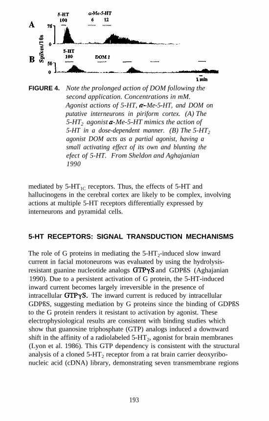
FIGURE 4. Note the prolonged action of DOM following thesecond application. Concentrations in mM.Agonist actions of 5-HT, -Me-5-HT, and DOM onputative interneurons in piriform cortex. (A) The5-HT2 agonist Me-5-HT mimics the action of5-HT in a dose-dependent manner. (B) The 5-HT2
agonist DOM acts as a partial agonist, having asmall activating effect of its own and blunting theefect of 5-HT. From Sheldon and Aghajanian1990
mediated by 5-HT1C receptors. Thus, the effects of 5-HT andhallucinogens in the cerebral cortex are likely to be complex, involvingactions at multiple 5-HT receptors differentially expressed byinterneurons and pyramidal cells.
5-HT RECEPTORS: SIGNAL TRANSDUCTION MECHANISMS
The role of G proteins in mediating the 5-HT2-induced slow inwardcurrent in facial motoneurons was evaluated by using the hydrolysis-resistant guanine nucleotide analogs and GDPßS (Aghajanian1990). Due to a persistent activation of G protein, the 5-HT-inducedinward current becomes largely irreversible in the presence ofintracellular The inward current is reduced by intracellularGDPßS, suggesting mediation by G proteins since the binding of GDPßSto the G protein renders it resistant to activation by agonist. Theseelectrophysiological results are consistent with binding studies whichshow that guanosine triphosphate (GTP) analogs induced a downwardshift in the affinity of a radiolabeled 5-HT2, agonist for brain membranes(Lyon et al. 1986). This GTP dependency is consistent with the structuralanalysis of a cloned 5-HT2 receptor from a rat brain carrier deoxyribo-nucleic acid (cDNA) library, demonstrating seven transmembrane regions
193
typical of G protein coupled receptors (Pritchett et al. 1988). The identityof the G proteins that couple to the 5-HT2 receptor remains to bedetermined.
Serotonin stimulates phosphatidylinositol hydrolysis in the brain through5-HT2 and 5-HT1C receptors (Conn and Sanders-Bush 1986). LSD andDOM act as partial agonists of this effect (Sanders-Bush et al. 1988).Phosphatidylinositol hydrolysis yields at least two major secondmessengers: inositol trisphosphate (IP3) and diacylglycerol. The latteractivates protein kinase C (PKC), thereby potentially affecting manylong-term cellular responses through protein phosphorylation. Theauthor and coworkers have tested the effect of protein kinase inhibitorson the response of facial motoneurons to serotonin (Aghajanian 1990). Inconcentrations that have no effect of their own (100 and 10µM, re-spectively), two protein kinase inhibitors with different mechanisms ofaction, 1-(5-isoquinolylsulfonyl)-2-methylpiperazine (H7), a nonselectiveprotein kinase inhibitor, and sphingosine, a selective PKC inhibitor, bothmarkedly enhance and prolong the excitation of facial motoneuronsinduced by 5-HT. Conversely, phorbol esters that are known to activatePKC reduce the excitatory effect of serotonin. These results suggest thatactivation of phosphatidylinositol turnover, perhaps through PKC-induced receptor phosphorylation, has a negative feedback effect on5-HT-induced excitations in the facial nucleus. These observations areconsistent with studies on 5-HT2 receptor-mediated responses in rat aorta,which indicate that the activation of PKC desensitizes 5-HT2 receptors(Roth et al. 1986).
An interesting implication of the negative feedback model is thepossibility that the partial agonist (Sanders-Bush et al. 1988) or evenantagonist (Pierce and Peroutka 1988) properties of hallucinogens withrespect to 5-HT-stimulated phosphatidylinositol hydrolysis maycontribute to, rather than interfere with, their electrophysiological actions.Thus, the greater enhancement of Ihin facial motoneurons byhallucinogens relative to 5-HT could be explained by a combination oftwo factors: LSD has high efficacy in this transduction system, and bynot markedly activating phosphatidylinositol hydrolysis, LSD does notprovoke a large negative feedback reduction in response.
194
SUMMARY AND CONCLUSIONS
Receptor Specificity
This review has considered the electrophysiological effects ofhallucinogens in three different brain regions: the LC, the facial motornucleus, and the cerebral cortex. The results of those studies, togetherwith findings from earlier studies in the raphe nuclei, make it clear thatthe indoleamine and phenethylamine hallucinogens have commonelectrophysiological actions on 5-HT2 but not 5-HT1A, receptors.Furthermore, in each of these three regions, the effects of thehallucinogens are blocked by spiperone, which has almost a thousandfoldhigher affinity for 5-HT2 than 5-HT1C receptors. Thus, it may beconcluded that the electrophysiological effects of hallucinogens, at leastthose examined to date, are likely to be mediated primarily by 5-HT2
rather than 5-HT1C receptors, although the latter may have a contributoryrole.
Signal Transduction Mechanisms
The net effect of 5-HT2 receptor stimulation is to produce an increase inneuronal excitability through a G protein-coupled mechanism. The ionicmechanism underlying increased excitability involves a reduction inresting potassium conductance and/or enhancement of the hyper-polarizing-activated cationic current I,. In addition, biochemical studiesshow that both 5-HT2 and 5-HT1C receptors are coupled to PKC and IP3
mechanisms through the phosphoinositide signal transduction pathway.Altogether it would appear that 5-HT2 receptors are coupled to at leastthree different transduction systems: potassium channels, cationic Ih
channels, and phosphoinositide hydrolysis. The coupling could bethrough a single G protein or through multiple G proteins and multiple5-HT2 receptor subtypes, one for each transduction pathway. Severalhypothetical models for 5-HT2-receptor/G protein coupling mechanismsare presented in figure 5.
It might be supposed that the electrophysiological effects of 5-HT andhallucinogens acting upon 5-HT2 receptors are mediated through thephosphoinositide pathway. However, it has been found, mainly fromstudies in the facial motor nucleus, that activators of PKC suppress, whileinhibitors of PKC enhance, 5-HT2-induced increases in neuronalexcitability. Thus, the phosphoinositide second messenger system may
195
serve as a negative feedback system with respect to the electro-physiological effects produced by 5-HT2 receptor activation. The factthat hallucinogens are only partial agonists at stimulating phospho-inositide turnover may enhance rather than diminish theirelectrophysiological effects.
Behavioral Correlations
As yet, the interrelationships between electrophysiological and behavioralactions of hallucinogenic drugs have not been examined in detail.Nevertheless, certain intriguing links are emerging. For example, theenhancement of sensory responsivity in LC neurons may contribute to thecharacteristic intensification of certain kinds of sensory experienceproduced by hallucinogens in humans. The increase in motoneuronalexcitability producted by hallucinogens could explain the hyperreflexiainduced by these drugs. Finally, the persistent activation of a sub-population of 5-HT2-expressing interneurons in the cerebral cortex mayunderlie the various cognitive and perceptual distortions produced by thehallucinogenic drugs.
FIGURE 5. (facing page) Hypothetical models for the coupling of 5-HT2
receptors to three different effector mechanisms: decreasedK+ conduc-tance, increased phosphoinositide (PI) turnover,and increased Ih. (A) A single class of 5-HT2 receptorscouples via one type of G protein to the three differenteffectors. (B) A single class of 5-HT2 receptors couples tothree different G proteins (G, G’, and G”), one for eacheffector system. (C) Three 5-HT2 receptor variants (5-HT2,5-HT2’, 5-HT2”) couple separately to the three effectors,each via a different G protein.
196
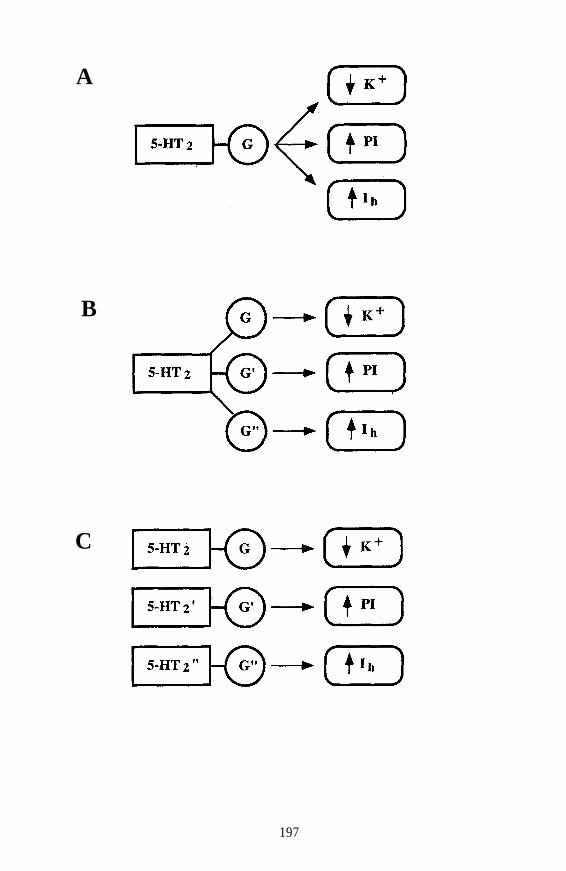
A
B
C
197
ACKNOWLEDGMENTS
This chapter was prepared with support from National Institute of MentalHealth grant MH-17871, the State of Connecticut, and a gift from Bristol-Myers Squibb.
REFERENCES
Aghajanian, G.K. Mescaline and LSD facilitate the activation of locuscoeruleus neurons by peripheral stimuli. Brain Res 186:492-498,1980.
Aghajanian, G.K. Serotonin-induced inward current in rat facialmotoneurons: Evidence for mediation by G proteins but not proteinkinase C. Brain Res 524:171-174, 1990.
Aghajanian, G.K.; Haigler, H.J.; and Bloom, F.E. Lysergic aciddiethylamide and serotonin: Direct actions on serotonin-containingneurons. Life Sci 1:615-622, 1972.
Aghajanian, G.K., and Rasmussen, K. Intracellular studies in the facialnucleus illustrating a simple new method for obtaining viablemotoneurons in adult rat brain slices. Synapse 3:331-338, 1989.
Ashby, C.R., Jr.; Jiang, L.H.; Kasser, R.J.; and Wang, R.Y.Electrophysiological characterization of 5-hydroxytryptamine-2receptors in rat medial prefrontal cortex. J Pharmacol Exp Ther252:171-178, 1989.
Barker, E.L.; Burris, K.D.; and Sanders-Bush, E. Phosphoinositidehydrolysis linked 5-HT2 receptors in fibroblasts from choroid plexus.Brain Res 552:330-332, 1991.
Bobker, D.H., and Williams, J.T. Serotonin augments the cationic currentIh in central neurons. Neuron 2:1535-1540, 1989.
Burris, K.D.; Breeding, M.; and Sanders-Bush, E. (+) Lysergic aciddiethylamide, but not its nonhallucinogenic congeners, is a potentserotonin 5-HT1C receptor agonist. J Pharmacol Exp Ther 258:891-896, 1991.
Conn, P. J., and Sanders-Bush, E. Regulation of serotonin-stimulatedphosphoinositide hydrolysis: Relation to the serotonin 5-HT2 bindingsite. J Neurosci 6:3669-3675, 1986.
Davies, M.G.; Deisz, R.A.; Prince, D.A.; and Peroutka, S.J. Two distincteffects of 5-hydroxytryptamine on single cortical neurons. Brain Res423:347-352, 1987.
Garratt, J.C.; Alreja, M.; and Aghajanian, G.K. LSD has high efficacyrelative to serotonin in enhancing the cationic current Ih: Intracellularstudies in rat facial motoneurons. Synapse 13:123-134, 1993.
198
Glennon, R.A. Do classical hallucinogens act as 5-HT2 agonists orantagonists? Neuropsychopharmacology 3:509-517, 1990.
Glennon, R.A.; Young, R.; and Rosecrans, J.A. Antagonism of the effectsof the hallucinogen DOM and the purported 5-HT agonist quipazineby 5-HT2 antagonists. Eur J Pharmacol 91:189-196, 1983.
Glennon, R.A.; Titeler, M.; and McKennay, J.D. Evidence for 5-HT2involvement in the mechanism of action of hallucinogenic agents. LifeSci 35:2505-2511, 1984.
Gorea, E., and Adrien, J. Serotonergic regulation of noradrenergiccoerulean neurons: Electrophysiological evidence for the involvementof 5-HT2 receptors. Eur J Pharmacol 154:285-291, 1988.
Haigler, H.J., and Aghajanian, G.K. Mescaline and LSD: Direct andindirect effects on serotonin-containing neurons in brain. Eur JPharmacol 21:53-60, 1973.
Hartig, P.R. Molecular biology of 5-HT receptors. Trends Pharmacol Sci10:64-69, 1989.
Heym, J.; Rasmussen, K.; and Jacobs, B.L. Some behavioral effects ofhallucinogens are mediated by a postsynaptic serotonergic action:Evidence from single unit studies in freely moving cats. Eur JPharmacol 101:57-68, 1984.
Hoyer, D. Functional correlates of serotonin 5-HT1 recognition sites.J Receptor Res 8:59-81, 1988.
Lakoski, J.M., and Aghajanian, G.K. Effects of ketanserin on neuronalresponses to serotonin in the prefrontal cortex, lateral geniculate anddorsal raphe nucleus. Neuropharmacology 24:265-273, 1985.
Larkman, P.M.; Penington, N.J.; and Kelly, J.S. Electrophysiology ofadult rat facial motoneurones: The effects of serotonin (5-HT) in anovel in vitro brainstem slice. J Neurosci Meth 28:133-146, 1989.
Lyon, R.A,; Davis, K.H.; and Titeler, M. 3H-DOB (4-bromo-2,5-dimethoxyphenylisopropylamine) labels a guanyl nucleotide-sensitivestate of cortical 5-HT2 receptors. MOl Pharmacol 31:194-199, 1986.
McCall, R.B., and Aghajanian, G.K. Serotonergic facilitation of facialmotoneuron excitation. Brain Res 169:11-27, 1979.
McCall, R.B., and Aghajanian, G.K. Hallucinogens potentiate responsesto serotonin and norepinephrine in the facial motor nucleus. Life Sci26:1149-1156, 1980a.
McCall, R.B., and Aghajanian, G.K. Pharmacological characterization ofserotonin receptors in the facial motor nucleus: A microiontophoreticstudy. Eur J Pharmacol 65:175-183, 1980b.
McCormick, D.A., and Pape, H.-C. Noradrenergic and serotonergicmodulation of a hyperpolarization-activated cation current in thalamicrelay neurones. J Physiol 431:319-342, 1990.
199
Mengod, G.; Nguyen, H.; Lee, H.; Waeber, C.; Lubbert, H.; and Palacios,J.M. The distribution and cellular localization of 5-HT1C receptormRNA in the rodent brain examined by in situ hybridizationhistochemistry. Comparison with receptor binding distribution.Neuroscience 35:577-592, 1990a.
Mengod, G.; Pompeiano, M.; Martinez-Mir, M.I.; and Palacios, J.M.Localization of the mRNA for the 5-HT2 receptor by in situhybridization histochemistry. Correlation with the distribution ofreceptor sites. Brain Res 524:139-143, 1990b .
Mokler, D.J.; Stoudt, K.W.; and Rech, R.H. The 5-HT2 antagonistpirenperone reverses disruption of FR-40 by hallucinogenic drugs.Pharmacol Biochem Behav 22:677-682, 1985.
Nedergaard, S.; Flatman, J.A.; and Engberg, I. Excitation of substantianigra pars compacta neurones by 5-hydroxtrytamine in vitro. NeuroReport 3:329-332, 1991.
North, R.A., and Uchimura, N. 5-Hydroxtryptamine acts at 5-HT2receptors to decrease potassium conductance in rat nucleus accumbensneurones. J Physiol 417:1-12, 1989.
Pazos, A.; Cortes, R.; and Palacios, J.M. Quantitative autoradiographicmapping of serotonin receptors in the rat brain. II. Serotonin-2receptors. Brain Res 346:231-249, 1985.
Pierce, P.A., and Perotka, S.J. Antagonism of 5-hydroxytryptamine2receptor-mediated phosphatidylinositol turnover by d-lysergic aciddiethylamide. J Pharmacol Exp Ther 247:918-925, 1988.
Pierce, P.A., and Peroutka, S.J. Antagonist properties of d-LSD at5-hydroxytryptamine2 receptors. Neuropsychopharmacology 3:503-508, 1990.
Pritchett, D.B.; Bach, A.W.J.; Wozny, M.; Taleb, O.; Dal Toso, R.; Shin,J.C.; and Seeburg, P.H. Structure and functional expression of clonedrat serotonin 5-HT2 receptor. EMBO J 7:4135-4140, 1988.
Rasmussen, K., and Aghajanian, G.K. Effects of hallucinogens onspontaneous and sensory-evoked locus coeruleus unit activity in therat: Reversal by selective 5-HT2 antagonists. Brain Res 385:395-400,1986.
Rasmussen, K., and Aghajanian, G.K. Potency of antipsychotics inreversing the effects of a hallucinogenic drug on locus coeruleusneurons correlates with 5-HT2 binding affinity.Neuropsychopharmacology 1:101-107, 1988.
Rasmussen, K., and Aghajanian, G.K. Serotonin excitation of facialmotoneurons: Receptor subtype characterization. Synapse 5:324-332,1990.
200
Roth, B.L.; Nakaki, T.; Chuang, D.M.; and Costa, E.5-Hydroxytryptamine2 receptors coupled to phospholipase C in rataorta: Modulation of phosphoinositide turnover by phorbol ester.J Pharmacol Exp Ther 238:480-485, 1986.
Sanders-Bush, E., and Breeding, M. Choroid plexus epithelial cells inprimary culture: A model of 5-HT1C receptor activation byhallucinogenic drugs. Psychopharmacology 105:340-346, 1991.
Sanders-Bush, E.; Burris, K.D.; and Knoth, K. Lysergic aciddiethylamide and 2,5-dimethoxy-4-methylamphetamine are partialagonists at serotonin receptors linked to phosphoinositide hydrolysis.J Pharmacol Exp Ther 246:924-928, 1988.
Sheldon, P.W., and Aghajanian, G.K. Excitatory responses to serotonin(5-HT) in neurons of the rat piriform cortex: Evidence for mediationby 5-HT1C receptors pyramidal cells and 5-HT2 receptors ininterneurons. Synapse 9:208-218, 1991.
Sheldon, P.W., and Aghajanian, G.K. Serotonin (5-HT) induces IPSPs inpyramidal layer of rat piriform cortex: Evidence for the involvementof a 5-HT2-activated interneuron. Brain Res 506:62-69, 1990.
Sprouse, J.S., and Aghajanian, G.K. Electrophysiological responses ofserotonergic dorsal raphe neurons to 5-HT1A and 5-HT1B agonists.Synapse 1:3-9, 1987,
Sprouse, J.S., and Aghajanian, G.K. Responses of hippocampalpyramidal cells to putative serotonin 5-HT1A and 5-HT1B agonists: Acomparative study with dorsal raphe neurons. Neuropharmacology27:707-715, 1988.
Takahashi, T., and Berger, A.J. Direct excitation of rat spinalmotoneurones by serotonin. J Physiol 423:63-76, 1990.
Titeler, M.; Lyon, R.A.; and Glennon, R.A. Radioligand bindingevidence implicates the brain 5-HT2 receptor as a site of action forLSD and phenylisopropylamine hallucinogens. Psychopharmacology94:213-216, 1988.
VanderMaelen, C.P., and Aghajanian, G.K. Intracellular studies showingmodulation of facial motoneurone excitability by serotonin. Nature287:346-347, 1980.
VanderMaelen, C.P., and Aghajanian, G.K. Serotonin-induceddepolarization of rat facial motoneurons in vivo: Comparison withamino acid transmitters. Brain Res 239:139-152, 1982.
White, S.R., and Fung, S.J. Serotonin depolarizes cat spinal motoneuronsin situ and decreases motoneuron after hyperpolarizing potentials.Brain Res 502:205-213, 1989.
201

AUTHOR
George K. Aghajanian, M.D.Professor of Psychiatry and PharmacologyDepartments of Psychiatry and PharmacologyYale University School of Medicine and theConnecticut Mental Health Center34 Park StreetNew Haven, CT 06508
202
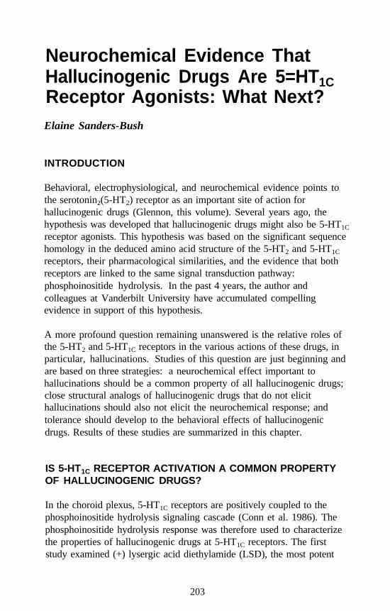
Neurochemical Evidence ThatHallucinogenic Drugs Are 5=HT1CReceptor Agonists: What Next?Elaine Sanders-Bush
INTRODUCTION
Behavioral, electrophysiological, and neurochemical evidence points tothe serotonin2(5-HT2) receptor as an important site of action forhallucinogenic drugs (Glennon, this volume). Several years ago, thehypothesis was developed that hallucinogenic drugs might also be 5-HT1C
receptor agonists. This hypothesis was based on the significant sequencehomology in the deduced amino acid structure of the 5-HT2 and 5-HT1C
receptors, their pharmacological similarities, and the evidence that bothreceptors are linked to the same signal transduction pathway:phosphoinositide hydrolysis. In the past 4 years, the author andcolleagues at Vanderbilt University have accumulated compellingevidence in support of this hypothesis.
A more profound question remaining unanswered is the relative roles ofthe 5-HT2 and 5-HT1C receptors in the various actions of these drugs, inparticular, hallucinations. Studies of this question are just beginning andare based on three strategies: a neurochemical effect important tohallucinations should be a common property of all hallucinogenic drugs;close structural analogs of hallucinogenic drugs that do not elicithallucinations should also not elicit the neurochemical response; andtolerance should develop to the behavioral effects of hallucinogenicdrugs. Results of these studies are summarized in this chapter.
IS 5-HT1C RECEPTOR ACTIVATION A COMMON PROPERTYOF HALLUCINOGENIC DRUGS?
In the choroid plexus, 5-HT1C receptors are positively coupled to thephosphoinositide hydrolysis signaling cascade (Conn et al. 1986). Thephosphoinositide hydrolysis response was therefore used to characterizethe properties of hallucinogenic drugs at 5-HT1C receptors. The firststudy examined (+) lysergic acid diethylamide (LSD), the most potent
203
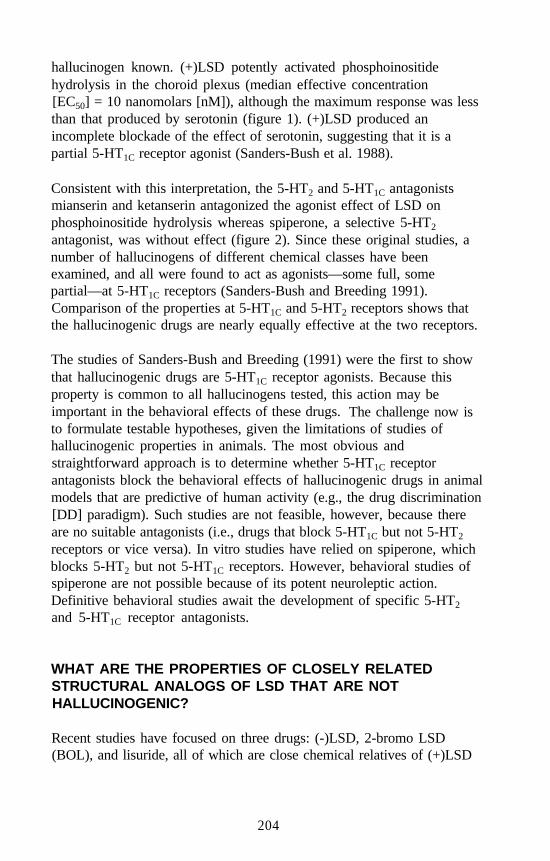
hallucinogen known. (+)LSD potently activated phosphoinositidehydrolysis in the choroid plexus (median effective concentration[EC50] = 10 nanomolars [nM]), although the maximum response was lessthan that produced by serotonin (figure 1). (+)LSD produced anincomplete blockade of the effect of serotonin, suggesting that it is apartial 5-HT1C receptor agonist (Sanders-Bush et al. 1988).
Consistent with this interpretation, the 5-HT2 and 5-HT1C antagonistsmianserin and ketanserin antagonized the agonist effect of LSD onphosphoinositide hydrolysis whereas spiperone, a selective 5-HT2
antagonist, was without effect (figure 2). Since these original studies, anumber of hallucinogens of different chemical classes have beenexamined, and all were found to act as agonists—some full, somepartial—at 5-HT1C receptors (Sanders-Bush and Breeding 1991).Comparison of the properties at 5-HT1C and 5-HT2 receptors shows thatthe hallucinogenic drugs are nearly equally effective at the two receptors.
The studies of Sanders-Bush and Breeding (1991) were the first to showthat hallucinogenic drugs are 5-HT1C receptor agonists. Because thisproperty is common to all hallucinogens tested, this action may beimportant in the behavioral effects of these drugs. The challenge now isto formulate testable hypotheses, given the limitations of studies ofhallucinogenic properties in animals. The most obvious andstraightforward approach is to determine whether 5-HT1C receptorantagonists block the behavioral effects of hallucinogenic drugs in animalmodels that are predictive of human activity (e.g., the drug discrimination[DD] paradigm). Such studies are not feasible, however, because thereare no suitable antagonists (i.e., drugs that block 5-HT1C but not 5-HT2
receptors or vice versa). In vitro studies have relied on spiperone, whichblocks 5-HT2 but not 5-HT1C receptors. However, behavioral studies ofspiperone are not possible because of its potent neuroleptic action.Definitive behavioral studies await the development of specific 5-HT2
and 5-HT1C receptor antagonists.
WHAT ARE THE PROPERTIES OF CLOSELY RELATEDSTRUCTURAL ANALOGS OF LSD THAT ARE NOTHALLUCINOGENIC?
Recent studies have focused on three drugs: (-)LSD, 2-bromo LSD(BOL), and lisuride, all of which are close chemical relatives of (+)LSD
204
FIGURE 1. Partial agonist effect of LSD at 5-HT1C receptors in ratchoroid plexus. [3H]-Inositol-labeled choroid plexiwere incubated for 45 minutes with lithium chloride,pargyline, and increasing concentrations of ( + )LSDwith and without 100 nM serotonin. [3H]-Inositol-monophosphate (IP) formation is expressed aspercentage of the basal radioactivity. The circlesrepresent a dose-response for LSD alone (n=4). Thediamonds show the effect of 100 nM serotonin plusincreasing concentrations of LSD alone (n=4). Themaximum [3H]-IP formation in the presence of 1 µMserotonin was 1363 ± 143% of basal. Mean basal[3H]-IP formation was 729 ± 117 cpm.
SOURCE: Sanders-Bush et al. 1988.
(figure 3) but apparently do not mimic its effects in humans (White1986). The properties of these drugs at both 5-HT2 and 5-HT1C receptorswere examined. (-)LSD and BOL were pure antagonists of thesereceptors, with no evidence of agonist properties (Burris et al. 1991).
The effects of lisuride on the 5-HT2 and 5-HT1C receptors are illustratedin figure 4. As shown, lisuride had no apparent 5-HT1C receptor agonistactivity, and it completely blocked the response to 100 nM serotonin. In
205
FIGURE 2. Antagonism of (+)LSD-stimulated phosphoinositidehydrolysis in rut choroid plexus. Antagonists (1 µM)were added 15 minutes before the addition of 20 nM(+)LSD. Results are expressed as percent of basal,which was 2,751±219 cpm (n=21). Antagonists alonedid not alter basal. *p<0.05, ***p<0.001. Spi =Spiperone; Ket = Ketunserin; Mia = Mianserin.
contrast, lisuride was an agonist at the 5-HT2 receptor, eliciting a strongphosphoinositide hydrolysis signal that was somewhat less than that ofserotonin. Like LSD, lisuride partially blocked the effect of serotoninconsistent with a partial agonist action at 5-HT2 receptors. Therefore,lisuride, a nonhallucinogenic congener of LSD, appears to mimic LSD’sagonist properties at the 5-HT2 receptor. Lisuride does not, however,reproduce the effects of LSD at the 5-HT1C receptor, consistent with thehypothesis that the 5-HT1C receptor may be involved in the hallucino-genic action of LSD. On the other hand, the finding that 5-HT2 receptoractivation is a property shared by (+)LSD and lisuride is not consistentwith a critical role for the 5-HT2 receptor in the production of such auaction.
206
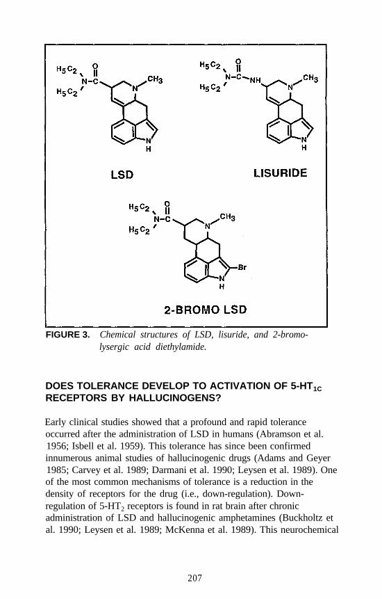
FIGURE 3. Chemical structures of LSD, lisuride, and 2-bromo-lysergic acid diethylamide.
DOES TOLERANCE DEVELOP TO ACTIVATION OF 5-HT1C
RECEPTORS BY HALLUCINOGENS?
Early clinical studies showed that a profound and rapid toleranceoccurred after the administration of LSD in humans (Abramson et al.1956; Isbell et al. 1959). This tolerance has since been confirmedinnumerous animal studies of hallucinogenic drugs (Adams and Geyer1985; Carvey et al. 1989; Darmani et al. 1990; Leysen et al. 1989). Oneof the most common mechanisms of tolerance is a reduction in thedensity of receptors for the drug (i.e., down-regulation). Down-regulation of 5-HT2 receptors is found in rat brain after chronicadministration of LSD and hallucinogenic amphetamines (Buckholtz etal. 1990; Leysen et al. 1989; McKenna et al. 1989). This neurochemical
207
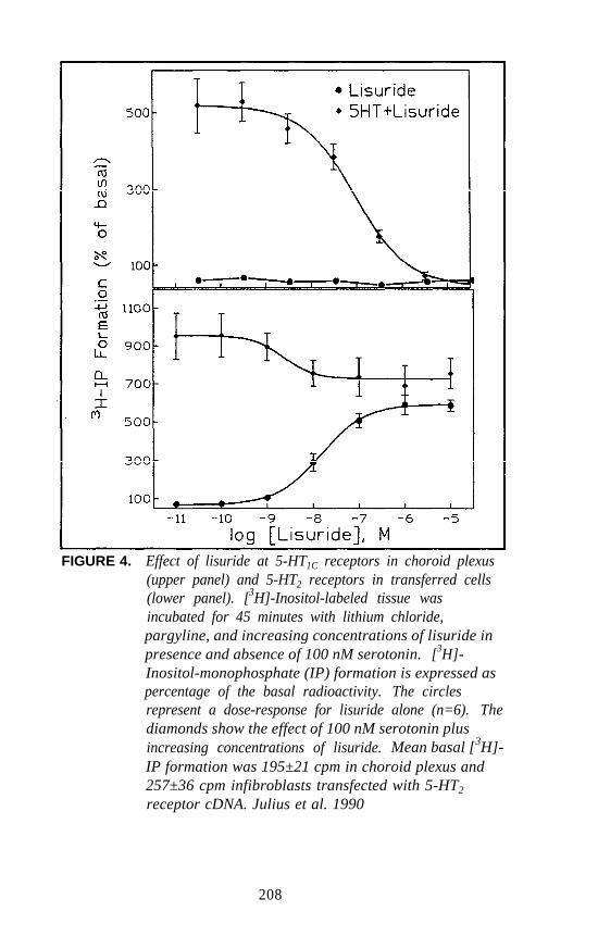
FIGURE 4. Effect of lisuride at 5-HT1C receptors in choroid plexus(upper panel) and 5-HT2 receptors in transferred cells(lower panel). [3H]-Inositol-labeled tissue wasincubated for 45 minutes with lithium chloride,pargyline, and increasing concentrations of lisuride inpresence and absence of 100 nM serotonin. [3H]-Inositol-monophosphate (IP) formation is expressed aspercentage of the basal radioactivity. The circlesrepresent a dose-response for lisuride alone (n=6). Thediamonds show the effect of 100 nM serotonin plusincreasing concentrations of lisuride. Mean basal [3H]-IP formation was 195±21 cpm in choroid plexus and257±36 cpm infibroblasts transfected with 5-HT2
receptor cDNA. Julius et al. 1990
208
response may be involved in the tolerance that occurs to the behavioraleffects of hallucinogens.
Recent studies have begun to explore the question of whether a down-regulation of 5-HT1C, receptors is also produced after chronic treatmentwith hallucinogenic drugs. These studies have been done in choroidplexus epithelial cells that express the 5-HT1C receptor. Chronic exposureto 1 micromolar (µM) of (-)1(4-bromo-2,5-dimethoxyphenyl)-2-aminopropane (DOB) elicited a delayed decrease’in the density of 5-HT1C
receptors (Barker and Sanders-Bush 1993). This loss of receptorspresumably will blunt the response to 5-HT1C, receptor agonists such ashallucinogenic drugs; however, this effect has not yet been demonstrated.A blunted response would be consistent with the interpretation that5-HT1C receptors may be involved in the behavioral effects ofhallucinogenic drugs.
WHAT IS DIFFERENT ABOUT 5-HT2/1 AGONISTS THAT AREHALLUCINOGENIC VERSUS THOSE THAT ARE NOT?
This author advocates the opinion that actions shared by lisuride andLSD, such as 5-HT2 receptor activation, are probably not important inmediating hallucinations. This is not a new idea; for example, it waseloquently defended several years ago by White (1986). An alternativeinterpretation is that although LSD and lisuride, as well as othernonhallucinogenic 5-HT2 agonists (e.g., quipazine), are capable ofeliciting a receptor signal, they do so by different mechanisms (e.g., byinteracting with different functional domains of the receptor). Thispossibility is especially interesting in light of the suggestion that LSD isan allosteric antagonist of the 5-HT2 receptor (Kaumann 1989; Xu andPurdy 1989). Since the antagonist and agonist properties of a partialagonist such as LSD presumably reflect the same binding event, this mayindicate that LSD has unique agonist properties (i.e., it may interact witha domain that is different from the serotonin binding domain).Conversely, lisuride and other nonhallucinogenic 5-HT2/5-HT1C agonistsmay not interact in an allosteric fashion.
Recent investigations have explored the putative allosteric interactions ofLSD in cells transfected with the 5-HT2 receptor (Burris and Sanders-Bush 1992). LSD produced a noncompetitive blockade of 5-HT-mediated phosphoinositide hydrolysis; high concentrations of serotoninwere not able to overcome the LSD blockade. One possible
209
interpretation of these results is that LSD interacts at a site on the 5-HT2
receptor that is different from the site where serotonin binds (i.e., anallosteric site). An alternative interpretation is that LSD binds tightly tothe active site of the receptor in an irreversible or pseudoirreversiblemanner. To investigate the latter possibility, the dissociation rate of LSDwas determined in membranes isolated from cells expressing the cloned5-HT2 receptor carrier deoxyribonucleic acid (cDNA).
In the initial studies of 3H-LSD binding by Bennett and Snyder (1975),3H-LSD dissociated slowly, but since a whole brain was used and LSD isnonspecific, it is not clear which 5-HT receptor was involved. This is nota problem with fibroblasts transfected with the 5-HT2 receptor cDNA,since only one 5-HT receptor subtype is expressed: the 5-HT2 receptor(Julius et al. 1990). In membranes from these cells, 3H-LSD dissociatedslowly, with a half life (t l/2) of 20 minutes. Consistent with this slowoff-rate, LSD caused an apparent loss of receptors in the usual3H-ketanserin binding assay, where the incubation time was 30 minutes.However, this receptor loss was not found when 3H-ketanserin wasincubated with the LSD-pretreated membranes for 60 minutes (Burris andSanders-Bush 1992). Therefore, it is likely that the apparent blunting ofthe phosphoinositide hydrolysis response, as well as the blunting ofserotonin-elicited contractions in smooth muscle (Kaumann 1989; Xu andPurdy 1989), are explained by the fact that LSD dissociates slowly fromthe 5-HT2 receptor. With short incubation times, equilibrium is notreached.
These data suggest that LSD is not an allosteric agonist/antagonist, butrather that it binds tightly to the 5-HT2 receptor and dissociates veryslowly. This property of slow dissociation could be important. It wouldbe interesting to compare the rates of dissociation of hallucinogenicversus nonhallucinogenic agonists from 5-HT2 and 5-HT1C receptors.
CONCLUSION
Hallucinogens of different chemical classes activate the 5-HT1C receptorwhereas nonhallucinogenic congeners interact with the receptor asantagonists. The numerous behavioral studies of hallucinogens thatutilize 5-HT2 receptor antagonists should be reevaluated in light of thisfinding, especially because currently available 5-HT2 receptor antagonistsalso block 5-HT1C receptors (Sanders-Bush and Breeding 1988). Theseneurochemical studies are consistent with a role for the 5-HT1C receptor
210
in the psychoactive effects of hallucinogenic drugs, but they are far fromconclusive. In the future new pharmacological tools that discriminatebetween 5-HT2 and 5-HT1C receptor subtypes may become available andcan be used to explore this possibility in greater detail.
REFERENCES
Abramson, H.A.; Jarvik, M.E.; Govin, M.H.; and Hirsch, M.H. Lysergicacid diethylamide antagonists. II. Development of tolerance in man toLSD-25 by prior administration of l-methyl-d-lysergic aciddiethylamide. J Psychol 51:81-82, 1956.
Adams, L.M., and Geyer, M.A. A proposed animal model forhallucinogens based on patterns of exploration in rats. Behav Neurosci5:881-900, 1985.
Barker, E.L., and Sanders-Bush, E. 5-HT1C receptor density and mRNAlevels in choroid plexus epithelial cells after treatment with mianserinand (-) 1 -(4-bromo-2,5-dimethoxyphenyl)-2-aminopropane. MolPharmacol 44:725-730, 1993.
Bennett, J.P., and Snyder, S.H. Stereospecific binding of D-lysergic aciddiethylamide to brain membranes: Relationship to serotonin receptors.Brain Res 94:523-524, 1975.
Buckholtz, N.S.; Zhou, D.F.; Freedman, D.X.; and Potter, W.Z. Lysergicacid diethylamide (LSD) administration selectively downregulatesserotonin, receptors in rat brain. Neuropsychopharmacolgy 3:137-148, 1990.
Burris, K.D.; Breeding, M.; and Sanders-Bush, E. (+)Lysergic aciddiethylamide, but not its nonhallucinogenic congeners, is a potentserotonin 5-HT1C receptor agonist. J Pharmacol Exp Ther 258:891-896, 1991.
Burris, K.D., and Sanders-Bush, E. Unsurmountable antagonism of brain5-HT2 receptors by (+)lysergic acid diethylamide (LSD) and bromo-LSD. Mol Pharmacol 42:826-830, 1992.
Carvey, P.; Nausieda, P; Weertz, R.; and Klawans, H. LSD and otherrelated hallucinogens elicit myoclonic jumping behavior in the guineapig. Prog Neuropsychopharmacol Biol Psychiatry 13:199-210, 1989.
Conn, P.J.; Sanders-Bush, E.; Hoffman, B.J.; and Hartig, P.R. A uniqueserotonin receptor in choroid plexus is linked to phosphatidylinositolturnover. Proc Nat1 Acad Sci U S A 83:4086-4088, 1986.
211
Darmani, N.A.; Martin, B.R.; and Glennon, R.A. Withdrawal fromchronic treatment with (+/-)-DOI causes supersensitivity to 5-HT2
receptor-induced head-twitch behavior in mice. Eur J Pharmacol186:115-118, 1990.
Isbell, H.; Miner, E.J.; and Logan, C.R. Cross tolerance between2-bromlysergic acid diethylamide and lysergic acid diethylamide.Psychopharmacologia 1:109-116, 1959.
Julius, D.; Huang, K.N.; Livelli, T. J.; Axel, R.; and Jessell; T.M. ‘The5-HT2 receptor defines a family of structurally distinct but functionallyconserved serotonin receptors. Proc Natl Acad Sci U S A 87:923-932,1990.
Kaumann, A.J. The allosteric 5-HT2 receptor system. In: Fozard, J.R., ed.The Peripheral Actions of 5-Hydroxytryptamine. New York: OxfordUniversity Press, 1989. pp. 45-71.
Leysen, J.E.; Janssen, P.F.M.; and Niemegerrs, C.J.E. Rapiddesensitization and down-regulation of 5-HT2 receptors DOMtreatment. Eur J Pharmacol 163:145-149, 1989.
McKenna, D.J.; Nazarali, A.J.; Himeno, A.; and Saavedra, J.M. Chronictreatment with (+/-) DOI, a psychotomimetic 5-HT2 agonist,downregulates 5-HT2 receptors in rat brain. Neuropsycho-pharmacology 2:81-87, 1989.
Sanders-Bush, E., and Breeding, M. Putative selective 5-HT2 antagonistsblock serotonin 5-HT1C receptors in the choroid plexus. J PharmacolExp Ther 247:169-173, 1988.
Sanders-Bush, E., and Breeding, M. Choroid plexus epithelial cells inprimary culture: A model of 5-HT1C receptor activation byhallucinogenic drugs. Psychopharmacology 105:340-346, 1991.
Sanders-Bush, E.; Burris, K.D.; and Knoth, K. Lysergic aciddiethylamide and 2,5-dimethoxy-4-methylamphetamine are partialagonists at serotonin receptors linked to phosphoinositide hydrolysis.J Pharmacol Exp Ther 246:924-928, 1988.
White, F.J. Comparative effects of LSD and lisuride: Clues to specifichallucinogenic drug actions. Pharmacol Biochem Behav 24:365-379,1986.
Xu, Z., and Purdy, R.E. Evidence for allosteric blockade of serotonergicreceptors in rabbit thoracic aorta. J Phurmacol Exp Ther 248:1091-1095, 1989.
212
ACKNOWLEDGMENTS
This chapter was prepared with support from National Institute on DrugAbuse grant DA-01581. Kevin D. Burris, Ph.D., Marsha Breeding, B.S.,and Antoinette Poindexter, B.S. were primarily responsible for theexperimental results presented in this manuscript.
AUTHOR
Elaine Sanders-Bush, Ph.D.Professor of PharmacologyVanderbilt University School of Medicine432 Medical Research BuildingNashville, TN 37232-6600
213
Autoradiographic Approachesto Studying Hallucinogensor Other DrugsNathan M. Appel
INTRODUCTION
The technique of autoradiographic localization of neurotransmitter anddrug binding sites, often referred to as receptor autoradiography, hasbecome a frequently used approach to investigate drug effects in thecentral nervous system (CNS). The technique enables the researcher toreveal the anatomic locus of actions of drugs and can be used to evaluateconsequences of drug treatment. It is a rare issue of Current Contents inwhich the keyword “autoradiography” does not yield relevant citations.
This chapter provides brief descriptions of the theory and practice of invitro and in vivo receptor autoradiography, provides examples of howthis approach has been used to study hallucinogens and other drugs inexperimental animals, describes how the technique of receptorautoradiography has been extended to study hallucinogens in a clinicalsetting using positron emission tomography (PET), and describes a novelautoradiographic approach for assessing fatty acid incorporation that is apromising research and clinical tool for studying the response to andeffects of hallucinogens and other classes of drugs or stimuli.
There are many excellent review articles and books that address thetheoretical basis, practical aspects, and limitations of the receptorautoradiographic technique. For the reader who is interested in a morecomprehensive treatment of the subject, the chapters on receptorautoradiography by Kuhar are an excellent “first read” (Kuhar 1985;Kuhar and Unnerstall 1990).
RECEPTOR AUTORADIOGRAPHY
Receptor autoradiography is essentially a receptor binding assay. Theassay is performed on an intact, instead of homogenized, tissue sample.Thus, the anatomic integrity of the tissue under investigation is preserved.
214
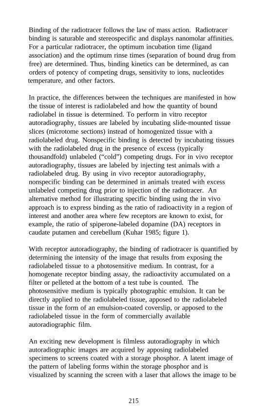
Binding of the radiotracer follows the law of mass action. Radiotracerbinding is saturable and stereospecific and displays nanomolar affinities.For a particular radiotracer, the optimum incubation time (ligandassociation) and the optimum rinse times (separation of bound drug fromfree) are determined. Thus, binding kinetics can be determined, as canorders of potency of competing drugs, sensitivity to ions, nucleotidestemperature, and other factors.
In practice, the differences between the techniques are manifested in howthe tissue of interest is radiolabeled and how the quantity of boundradiolabel in tissue is determined. To perform in vitro receptorautoradiography, tissues are labeled by incubating slide-mounted tissueslices (microtome sections) instead of homogenized tissue with aradiolabeled drug. Nonspecific binding is detected by incubating tissueswith the radiolabeled drug in the presence of excess (typicallythousandfold) unlabeled (“cold”) competing drugs. For in vivo receptorautoradiography, tissues are labeled by injecting test animals with aradiolabeled drug. By using in vivo receptor autoradiography,nonspecific binding can be determined in animals treated with excessunlabeled competing drug prior to injection of the radiotracer. Analternative method for illustrating specific binding using the in vivoapproach is to express binding as the ratio of radioactivity in a region ofinterest and another area where few receptors are known to exist, forexample, the ratio of spiperone-labeled dopamine (DA) receptors incaudate putamen and cerebellum (Kuhar 1985; figure 1).
With receptor autoradiography, the binding of radiotracer is quantified bydetermining the intensity of the image that results from exposing theradiolabeled tissue to a photosensitive medium. In contrast, for ahomogenate receptor binding assay, the radioactivity accumulated on afilter or pelleted at the bottom of a test tube is counted. Thephotosensitive medium is typically photographic emulsion. It can bedirectly applied to the radiolabeled tissue, apposed to the radiolabeledtissue in the form of an emulsion-coated coverslip, or apposed to theradiolabeled tissue in the form of commercially availableautoradiographic film.
An exciting new development is filmless autoradiography in whichautoradiographic images are acquired by apposing radiolabeledspecimens to screens coated with a storage phosphor. A latent image ofthe pattern of labeling forms within the storage phosphor and isvisualized by scanning the screen with a laser that allows the image to be
215
FIGURE 1. Distribution of [125I] DOI-labeled serotonin, (5-HT2)receptors in cerebral cortex. A: Bright-fieldphotomicrograph of a Nissl-stained horizontalsection through parietal cortex. Lamination isannotated with Roman numerals and delineated bythe short black horizontal bars. B: Dark-fieldphotomicrograph of the “coverslip” autoradiograph,generated by the section shown in A (samemagnification), depicting ([125I]DOI) 1-(2,5-dimethoxy-4-[125I]iodophenyl)-2-aminopropane)binding in cerebral cortex. Note that silver grainsare most concentrated in laminae I and IV.Magnification bar (lower right-hand corner)= 200 µm.
SOURCE: Appel et al. 1990a
216
detected electronically using a photomultiplier. The image then is storedon a computer for subsequent analysis (Appel et al. 1991; Kuhar et al.1991). The image pattern (autoradiographic distribution) depicts thespatial distribution of binding sites in the tissue.
Regardless of the photographic medium used, the optical density orintensity of the image is related to the quantity of bound drug and, byextension, of binding sites in a particular region of interest. Thus, theprincipal advantage that receptor autoradiography provides overhomogenate binding assays is increased anatomic resolution. Forexample, using a homogenate binding assay, it can be determined thatbinding of a certain drug is greater in the cerebral cortex than in thecerebellum. Using autoradiography, it can be said with certainty thatbinding is greater in cerebral cortex layer V than layer VI but less than inlayers I or IV (e.g., figure 1). For an exact quantitative assessment ofregional binding, regional optical densities can be measured bydensitometry and compared with the optical density produced bycalibrated radioactive standards coexposed with the experimentalmaterial. Several computer-assisted digital image analysis systems areavailable commercially to facilitate quantification of autoradiographs.
Another advantage of receptor autoradiography over homogenate bindingassays is increased sensitivity. In a homogenate binding assay, the abilityto detect binding is limited if the density of a particular binding site or thespecific activity of a labeled drug is low. Using autoradiography, one canincrease the duration the photosensitive medium is exposed to theradiolabeled tissue to overcome these impediments.
IN VITRO RECEPTOR AUTORADIOGRAPHY TO STUDYPOTENTIAL SITES OF ACTION OF HALLUCINOGENS
The exact criteria that categorize a substance as a hallucinogen remainunresolved (Szára, this volume). However, three classes of compoundsconstitute the majority of what typically are referred to as hallucinogenicdrugs (see chapters by Glennon; Pfaff et al.; and Jacob and Shulgin, thisvolume). They are indolealkylamines (e.g., dimethyltryptamine [DMT]),ergolines (e.g., lysergic acid diethylamide [LSD]), and phenylalkylamines(e.g., 2,5-dimethoxy-4-methylamphetamine [DOM]). They have incommon a high affinity for serotonin2 (5-HIT2) receptors. Drugdiscrimination studies, other behavioral tests, and homogenate receptorbinding assays have related hallucinogenic potency to binding affinity of
217
5-HT2 receptor agonists (Glennon and Rosecrans 1982; Glennon et al.1984; Heym and Jacobs 1987, 1988; Titeler et al. 1988). Moreover, asignificant positive correlation has been demonstrated between 5-HT2
binding affinities of drugs and their human hallucinogenic potencies(Glennon et al. 1984). In consideration of these data, drugs that label5-HT2 receptors would, by extension, label at least some sites in the brainwhere hallucinogenic effects of these drugs may be manifested.
In an early study in which binding sites for a hallucinogen weredemonstrated, autoradiographic distribution of 5-HT binding sites wasdemonstrated using [3H]LSD. However, unlabeled serotonin (5-HT) wasunable to displace [3H]LSD labeling in all areas (Meibach et al., 1980).The recognition of subtypes of 5-HT receptors, as distinguished bydifferential binding of [3H]5-HT, [3H]LSD, and [3H]spiroperidol(spiperone), would point the way toward using more selective ligands tostudy the now apparent different classes of 5-HT receptors (Peroutka andSnyder 1979). Thus, anatomic distribution of 5-HT2 receptors has beenstudied autoradiographically using radioligands such as [3H]spiperone(Altar et al. 1985; De Souza 1986; Palacios et al. 1981; Pazos et al. 1985)and [3H]ketanserin (Fishette et al. 1987; Pazos et al. 1985; Slater andPatel 1983). However, both these radioligands are antagonists:[3H]spiperone potently labels DA type 2 (D2) receptors (Niznik et al.1985; Stefanini et al. 1980), and [3H]ketanserin labels nonserotonergicsites in striatum (Leysen et al. 1987).
The autoradiographic distribution of 5-HT2 receptors also has beenstudied using another radioiodinated ligand, [125I]-LSD (Altar et al. 1986;De Souza 1986; Engel et al. 1984; Nakada et al. 1984). However,because [125I]-LSD binds D2 receptors with nanomolar affinity (Engel etal. 1984; Hartig et al. 1985a), one must perform [125I]-LSDautoradiography for 5-HT2 receptors in the presence of unlabeled D2
ligands to ensure that only 5-HT2 receptors are being visualized. There isanother congener of LSD, N1-methyl-2-[125I]-LSD, that is more selectivefor 5-HT2 receptors than D2 receptors. It also has been used toautoradiographically localize 5-HT2 receptors (Hoffman et al. 1987).
The hallucinogenic potency of drugs appears to be related to their 5-HT2
receptor binding affinities and their being agonists at this binding site(Glennon and Rosecrans 1982; Glennon et al. 1984; Heym and Jacobs1988; Titeler et al. 1988). However, evidence has been presentedsuggesting that 5-HT2 receptors exist in two states-one state having highaffinity for agonists and one state having low affinity for agonists-and
218
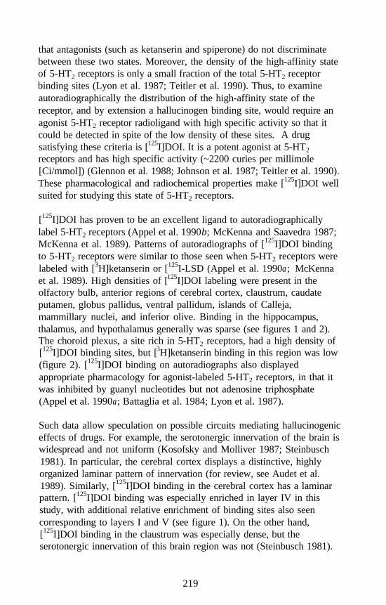
that antagonists (such as ketanserin and spiperone) do not discriminatebetween these two states. Moreover, the density of the high-affinity stateof 5-HT2 receptors is only a small fraction of the total 5-HT2 receptorbinding sites (Lyon et al. 1987; Teitler et al. 1990). Thus, to examineautoradiographically the distribution of the high-affinity state of thereceptor, and by extension a hallucinogen binding site, would require anagonist 5-HT2 receptor radioligand with high specific activity so that itcould be detected in spite of the low density of these sites.satisfying these criteria is [125I]DOI. It is a potent agonist at 5-HT2
A drug
receptors and has high specific activity (~2200 curies per millimole[Ci/mmol]) (Glennon et al. 1988; Johnson et al. 1987; Teitler et al. 1990).These pharmacological and radiochemical properties make [125I]DOI wellsuited for studying this state of 5-HT2 receptors.
[125I]DOI has proven to be an excellent ligand to autoradiographicallylabel 5-HT2 receptors (Appel et al. 1990b; McKenna and Saavedra 1987;McKenna et al. 1989). Patterns of autoradiographs of [125I]DOI bindingto 5-HT2 receptors were similar to those seen when 5-HT2 receptors werelabeled with [3H]ketanserin or [125I-LSD (Appel et al. 1990a; McKennaet al. 1989). High densities of [125I]DOI labeling were present in theolfactory bulb, anterior regions of cerebral cortex, claustrum, caudateputamen, globus pallidus, ventral pallidum, islands of Calleja,mammillary nuclei, and inferior olive. Binding in the hippocampus,thalamus, and hypothalamus generally was sparse (see figures 1 and 2).The choroid plexus, a site rich in 5-HT2 receptors, had a high density of[125I]DOI binding sites, but [3H]ketanserin binding in this region was low(figure 2). [125I]DOI binding on autoradiographs also displayedappropriate pharmacology for agonist-labeled 5-HT2 receptors, in that itwas inhibited by guanyl nucleotides but not adenosine triphosphate(Appel et al. 1990a; Battaglia et al. 1984; Lyon et al. 1987).
Such data allow speculation on possible circuits mediating hallucinogeniceffects of drugs. For example, the serotonergic innervation of the brain iswidespread and not uniform (Kosofsky and Molliver 1987; Steinbusch1981). In particular, the cerebral cortex displays a distinctive, highlyorganized laminar pattern of innervation (for review, see Audet et al.1989). Similarly, [125I]DOI binding in the cerebral cortex has a laminarpattern. [125I]DOI binding was especially enriched in layer IV in thisstudy, with additional relative enrichment of binding sites also seencorresponding to layers I and V (see figure 1). On the other hand,[125I]DOI binding in the claustrum was especially dense, but theserotonergic innervation of this brain region was not (Steinbusch 1981).
219
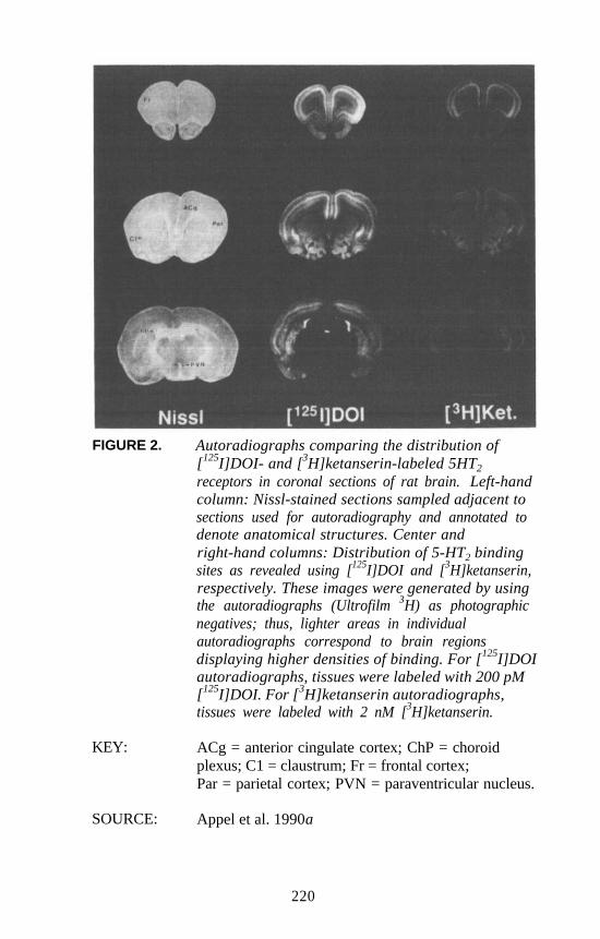
FIGURE 2. Autoradiographs comparing the distribution of[125I]DOI- and [3H]ketanserin-labeled 5HT2
receptors in coronal sections of rat brain. Left-handcolumn: Nissl-stained sections sampled adjacent tosections used for autoradiography and annotated todenote anatomical structures. Center andright-hand columns: Distribution of 5-HT2 bindingsites as revealed using [125I]DOI and [3H]ketanserin,respectively. These images were generated by usingthe autoradiographs (Ultrofilm 3H) as photographicnegatives; thus, lighter areas in individualautoradiographs correspond to brain regionsdisplaying higher densities of binding. For [125I]DOIautoradiographs, tissues were labeled with 200 pM[125I]DOI. For [3H]ketanserin autoradiographs,tissues were labeled with 2 nM [3H]ketanserin.
KEY: ACg = anterior cingulate cortex; ChP = choroidplexus; C1 = claustrum; Fr = frontal cortex;Par = parietal cortex; PVN = paraventricular nucleus.
SOURCE: Appel et al. 1990a
220
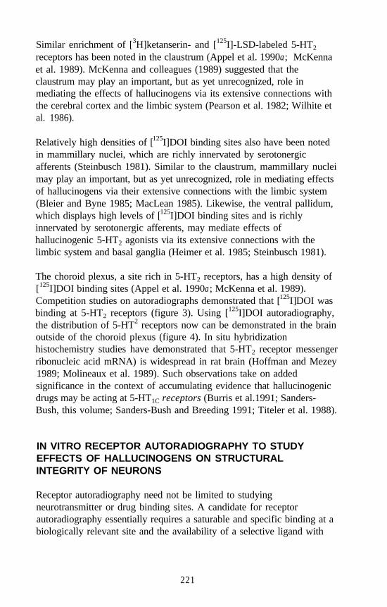
Similar enrichment of [3H]ketanserin- and [125I]-LSD-labeled 5-HT2
receptors has been noted in the claustrum (Appel et al. 1990a; McKennaet al. 1989). McKenna and colleagues (1989) suggested that theclaustrum may play an important, but as yet unrecognized, role inmediating the effects of hallucinogens via its extensive connections withthe cerebral cortex and the limbic system (Pearson et al. 1982; Wilhite etal. 1986).
Relatively high densities of [125I]DOI binding sites also have been notedin mammillary nuclei, which are richly innervated by serotonergicafferents (Steinbusch 1981). Similar to the claustrum, mammillary nucleimay play an important, but as yet unrecognized, role in mediating effectsof hallucinogens via their extensive connections with the limbic system(Bleier and Byne 1985; MacLean 1985). Likewise, the ventral pallidum,which displays high levels of [125I]DOI binding sites and is richlyinnervated by serotonergic afferents, may mediate effects ofhallucinogenic 5-HT2 agonists via its extensive connections with thelimbic system and basal ganglia (Heimer et al. 1985; Steinbusch 1981).
The choroid plexus, a site rich in 5-HT2 receptors, has a high density of[125I]DOI binding sites (Appel et al. 1990a; McKenna et al. 1989).Competition studies on autoradiographs demonstrated that [125I]DOI wasbinding at 5-HT2 receptors (figure 3). Using [125I]DOI autoradiography,the distribution of 5-HT2 receptors now can be demonstrated in the brainoutside of the choroid plexus (figure 4). In situ hybridizationhistochemistry studies have demonstrated that 5-HT2 receptor messengerribonucleic acid mRNA) is widespread in rat brain (Hoffman and Mezey1989; Molineaux et al. 1989). Such observations take on addedsignificance in the context of accumulating evidence that hallucinogenicdrugs may be acting at 5-HT1C receptors (Burris et al.1991; Sanders-Bush, this volume; Sanders-Bush and Breeding 1991; Titeler et al. 1988).
IN VITRO RECEPTOR AUTORADIOGRAPHY TO STUDYEFFECTS OF HALLUCINOGENS ON STRUCTURALINTEGRITY OF NEURONS
Receptor autoradiography need not be limited to studyingneurotransmitter or drug binding sites. A candidate for receptorautoradiography essentially requires a saturable and specific binding at abiologically relevant site and the availability of a selective ligand with
221
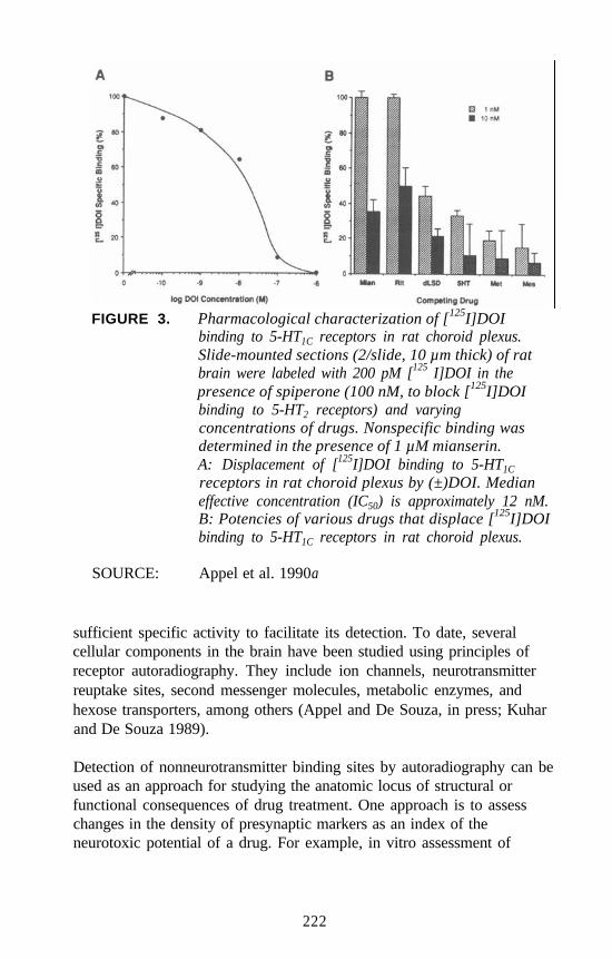
FIGURE 3. Pharmacological characterization of [125I]DOIbinding to 5-HT1C receptors in rat choroid plexus.Slide-mounted sections (2/slide, 10 µm thick) of ratbrain were labeled with 200 pM [125 I]DOI in thepresence of spiperone (100 nM, to block [125I]DOIbinding to 5-HT2 receptors) and varyingconcentrations of drugs. Nonspecific binding wasdetermined in the presence of 1 µM mianserin.A: Displacement of [125I]DOI binding to 5-HT1C
receptors in rat choroid plexus by (±)DOI. Medianeffective concentration (IC50) is approximately 12 nM.B: Potencies of various drugs that displace [125I]DOIbinding to 5-HT1C receptors in rat choroid plexus.
SOURCE: Appel et al. 1990a
sufficient specific activity to facilitate its detection. To date, severalcellular components in the brain have been studied using principles ofreceptor autoradiography. They include ion channels, neurotransmitterreuptake sites, second messenger molecules, metabolic enzymes, andhexose transporters, among others (Appel and De Souza, in press; Kuharand De Souza 1989).
Detection of nonneurotransmitter binding sites by autoradiography can beused as an approach for studying the anatomic locus of structural orfunctional consequences of drug treatment. One approach is to assesschanges in the density of presynaptic markers as an index of theneurotoxic potential of a drug. For example, in vitro assessment of
222
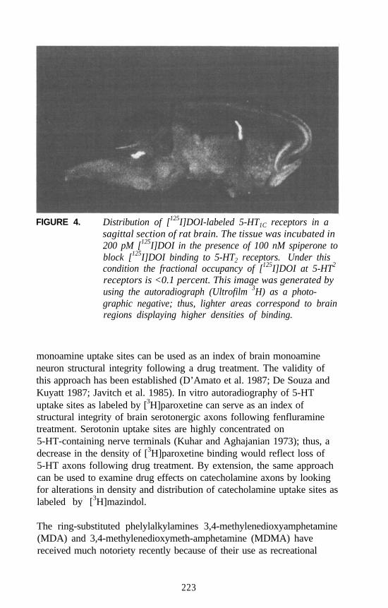
FIGURE 4. Distribution of [125I]DOI-labeled 5-HT1C receptors in asagittal section of rat brain. The tissue was incubated in200 pM [125I]DOI in the presence of 100 nM spiperone toblock [125I]DOI binding to 5-HT2 receptors. Under thiscondition the fractional occupancy of [125I]DOI at 5-HT2
receptors is <0.1 percent. This image was generated byusing the autoradiograph (Ultrofilm 3H) as a photo-graphic negative; thus, lighter areas correspond to brainregions displaying higher densities of binding.
monoamine uptake sites can be used as an index of brain monoamineneuron structural integrity following a drug treatment. The validity ofthis approach has been established (D’Amato et al. 1987; De Souza andKuyatt 1987; Javitch et al. 1985). In vitro autoradiography of 5-HTuptake sites as labeled by [3H]paroxetine can serve as an index ofstructural integrity of brain serotonergic axons following fenfluraminetreatment. Serotonin uptake sites are highly concentrated on5-HT-containing nerve terminals (Kuhar and Aghajanian 1973); thus, adecrease in the density of [3H]paroxetine binding would reflect loss of5-HT axons following drug treatment. By extension, the same approachcan be used to examine drug effects on catecholamine axons by lookingfor alterations in density and distribution of catecholamine uptake sites aslabeled by [3H]mazindol.
The ring-substituted phelylalkylamines 3,4-methylenedioxyamphetamine(MDA) and 3,4-methylenedioxymeth-amphetamine (MDMA) havereceived much notoriety recently because of their use as recreational
223
drugs. (For a comprehensive review, see Asghar and De Souza 1989.)Although these compounds differ in structure by only a single methylgroup, evidence suggests that, whereas MDA is hallucinogenic, MDMAis not (Glennon 1989; Nichols and Oberlender 1989). Evidence has beenpresented that MDA and MDMA are neurotoxic and cause diffuse andprolonged decreases in brain 5-HT markers (Battaglia et al. 1987;Commins et al. 1987; Insel et al. 1989; Ricuarte et al. 1985).
Effects of these compounds on serotonergic neurons have been assessedusing immunohistochemistry (O’Hearn et al. 1988). Widespread loss of5-HT-like immunoreactivity was observed in the cerebral cortex,hippocampus, caudate putamen, hypothalamus, and other forebrain areas.Decreases in immunostaining appeared to be more profound followingMDA treatment than following MDMA treatment. In addition, someremaining 5-HT-like immunoreactive axons displayed morphologyreminiscent of that seen following treatment with the 5-HT neurotoxin5,7-dihydroxytryptamine. Such axons appeared swollen, fragmented, andintensely stained. They suggested also that, in particular, axons derivedfrom the dorsal raphe nucleus, which are morphologically distinct fromaxons derived from the median raphe nucleus (Kosofsky and Molliver1987), were selectively vulnerable to these drugs. Serotonin-likeimmunoreactive cell bodies did not appear to be affected. When treatedtissues were immunostained for tyrosine hydroxylase, a marker ofmonoaminergic neurons, immunostaining appeared unaffected bytreatment with either drug. O’Hearn and colleagues (1988) thusconcluded that MDA, a purported hallucinogen, and MDMA wereexerting neurotoxic effects on dorsal raphe-derived serotonergic axons.
Although these immunohistochemical data strongly supported theconclusion that MDA and MDMA were neurotoxic, a shortfall ofinterpreting immunohistochemical data is that detection techniquesdepend on the presence of the antigen (in this case, 5-HT). Thus, if drugtreatment causes depletion of the antigen of interest, lack of or decreasedimmunostaining could mistakenly be interpreted as an actual loss ofaxons. Furthermore, immunohistochemistry is limited from theperspective that related changes in staining are difficult to assessquantitatively.
To address this question with respect to MDA and MDMA,[3H]paroxetine autoradiography was used to assess the potentialneurotoxic effects of these drugs by examining changes in a presynapticmarker, 5-HT uptake sites (Battaglia et al. 1991; De Souza and Kuyatt
224
1987). Both the hallucinogen MDA and MDMA caused widespreaddecreases in regional [3H]paroxetine binding that correlated with loss of5-HT-like immunoreactivity noted by O’Hearn and coworkers (1988).
Furthermore, regional decreases in [3H]paroxetine binding inMDMA-treated rats were greater 2 weeks after, rather than immediatelyafter, cessation of drug treatment. This suggested that there was acontinuing degenerative process occurring during this time interval(Battaglia et al. 1991).
To confirm the neurochemical specificity of this effect, Battaglia andcolleagues (199 1) performed parallel experiments using [3H]mazindol toassess the structural integrity of catecholaminergic nerve terminals.[3H]Mazindol is a marker for DA and norepinephrine (NE) uptake sites(Javitch et al. 1985). [3H]Mazindol binding was unaffected by eitherMDA or MDMA treatment, confirming the specificity of the neurotoxiceffect of these drugs to 5-HT monoaminergic neurons.
Such autoradiographic assays also have been used to investigate theneurotoxic potential of other drugs structurally derived fromphenylalkylamines. Regional decreases in 5-HT-uptake site density havebeen noted following treatment with the CNS stimulant methamphet-amine using [3H]cyanoimipramine binding as a marker (Kovachich et al.1989). Fenfluramine is a substituted phenylalkylamine derivativestructurally similar to MDA and MDMA. Unlike methamphetamine,MDA, or MDMA, it is devoid of undesirable psychostimulant effects.Racemic fenfluramine and its dextrorotary (D) isomer are usedtherapeutically as anorectics to treat obesity (McTavish and Heel 1992;Rowland and Carlton 1986). Short-term administration of racemicfenfluramine at high doses results in a pattern of 5-HT immunoreactivitysimilar to that seen with MDA, suggesting neurotoxic effects of this drugon 5-HT axons using this treatment protocol (Appel et al. 1989; Molliverand Molliver 1990).
This hypothesis was supported by autoradiographic studies using[3H]paroxetine and [3H]mazindol as markers for 5-HT and catecholamineuptake sites, respectively (Appel et al. 1990b). Decreased paroxetinebinding parallels decreases in 5-HT-like immunostained axons followingan identical fenfluramine treatment protocol. Catechol-aminergic axonsas assessed by [3H]mazindol autoradiography were unaffected. Thesedata suggest that as a class, phenylalkylamine derivatives may haveincreased potential for neurotoxic side effects regardless of their being
225
hallucinogenic and may reinforce the utility of autoradiographicapproaches as a tool for studying such effects of drug treatment.
PRECLINICAL AND CLINICAL APPLICATIONS OF IN VIVORECEPTOR AUTORADIOGRAPHY IN THE STUDYOF HALLUCINOGENS
As discussed earlier, receptor autoradiographic technique lends itself tobeing applied in vivo. The obvious advantage is that assessment can bemade in situ in a living subject. Furthermore, using tracer drugs labeledwith positron-emitting isotopes such as 11C, 18F, 15O, and 13N, dynamicmapping studies can be accomplished in animal or, potentially, humansubjects using PET. In PET, the spatial distribution of positron-emittingisotopes in the brain or any other body region is detected using a circulararray of detectors that surrounds the subject. Precise spatial localizationis realized by accepting as the signal pairs of simultaneously emitted511-kiloelectron volt (keV) gamma rays that are detected 180” apart fromeach other. The gamma rays are the result of positron annihilation thatoccurs when a positron interacts with a negatively charged electron toform a short-lived positronium that is subsequently converted intoelectromagnetic radiation in the form of two photons (gamma rays) (Frost1990; Ter-Pogossian 1985). With high probability, the gamma raysrepresent the product of a single disintegrating positron. An obviousadvantage of PET over postmortem (in vitro) receptor autoradiography isthat it permits measurements several times from the same subject. Thus,dynamic studies during, as well as longitudinal studies following,hallucinogen administration are possible.
Several preconditions limit whether a drug is useful as an in vivo marker.As a minimum, the tracer drug cannot be toxic to the host. Other basicrequirements summarized by Kung (1990) are that the radiotracer:
Be labeled with a short-lived isotope and be quickly prepared(minutes);
Possess high specific activity;
Be able to penetrate the intact blood-brain barrier followingintravenous (IV) injection;
Be relatively resistant to in vivo metabolism;
226
Have a high affinity for the binding site of interest (nM Kd);
Possess a high target-to-nontarget ratio after IV injection; and
Can be modeled to quantitate receptor density based on kineticinformation obtained by imaging.
For the most part, in vivo studies performed with radiotracers inexperimental animals and having the potential for improvingunderstanding of hallucinogens have been focused not so much towardimaging binding sites in that setting as toward determining whether aparticular tracer is of potential clinical use. For this purpose, regionalbinding under different experimental conditions is quantified bysolubilizing dissected regions of the brain and counting boundradioactivity in a scintillation counter. As discussed earlier, ligands thatlabel 5-HT2 binding sites would theoretically label these binding sites inliving brain and, by extension, identify initial sites of action ofhallucinogenic drugs. Drugs tested for this use include derivatives ofspiperone (spiroperidol) and ketanserin (Frost 1990; Kuhar et al. 1986).However, these drugs are 5-HT2 receptor antagonists and do not label5-HT2 receptors exclusively (Leysen et al. 1987; Niznik et al. 1985;Stefanini et al. 1980). A tritiated naphthosultam derivative, RP 62203,also is being used as an in vivo probe for 5-HT2 receptors. This drug isalso a 5-HT2 receptor antagonist but is reported to have greater 5-HT2
receptor selectivity than spiperone or ketanserin (Fajolles et al. 1992).
Derivatives of LSD also have been used as in vivo probes for 5-HT2
receptors. They include [125I]-LSD (Hartig et al. 1985b),N1-methyl-2-[125I]-LSD (Hoffman et al. 1987), and D-(+)-(N1-[11C]methyl)-2-Br-LSD ([11C]MBL) (Lever at al. 1989). [11C]MBL hasbeen used to image 5-HT2 receptors in living baboon and human brainsby PET (Wang et al. 1987). In the baboon brain, specific binding of[11C]MBL to 5-HT2 receptors in vivo was demonstrated by comparingbinding ratios between the frontal cortex and cerebellum in the presenceand absence of ketanserin. In a human subject, widespread uniformlabeling of brain structures not associated with 5-HT2 receptors, such asthe cerebellum, was seen soon after administration of [11C]MBL.However, this most likely reflected blood flow delivery of the tracerbecause persistent labeling was seen subsequently in the frontal cortexand temporal poles, sites endowed with 5-HT2 receptors. Nopharmacological effects of drug administration were noted in any subjectsreceiving [11C]MBL. However, MBL may be an antagonist at 5-HT2
227
receptors (Ginzel and Mayer-Gross 1956). As discussed earlier,theoretical arguments support using 5-HT2 agonists as probes for sites ofaction of hallucinogens rather than anatagonists (Lyon et al. 1987; Teitleret al. 1990). One such compound under investigation is 1-[2’,5’-di-methoxy-4’- (ß-fluoroethyl)phenyl]-2-aminopropane, a phenylalkylamine(Gerdes et al. 1988).
As discussed earlier, presynaptic markers are useful probes to assessstructural integrity of neurons. By using this approach, the regionaldistribution of neurotoxic effects of the hallucinogenic phenylalkylamineMDA and its nonhallucinogenic chemical congener fenfluramine havebeen reported using radiotracers selective for serotonin uptake sites invitro (Appel et al. 1990b; Battaglia et al. 1991). Radiotracers with invivo selectivity for serotonin uptake sites also have been reported,including radiolabeled derivatives of paroxetine, quipazine, imipramine,chlorimipramine, cyanoimipramine, citalopram, fluoxetine, and sertralme.However, all these compounds have limitations that compromise theirusefulness as radiotracers in a clinical setting (Scheffel et al. 1992).
Recently, radiolabeled congeners of another class of compound haveappeared that label cocaine binding sites. For example, RTI-55 is aparasubstituted cocaine analog derived from WIN 35,065-2 that labelsDA and serotonin uptake sites (Boja et al., 1992; Madras et al. 1989;Scheffel et al. 1992). In vivo studies in mice using [125I]RTI-55 haveyielded satisfactory autoradiographs demonstrating regional bindingpatterns that reflect the distribution of serotonin and DA uptake sites(Cline et al. 1992). By labeling 5-HT uptake sites in vivo with[125I]RTI-55, investigators have been able to detect regional changes indensities of 5-HT uptake sites following fenfluramine treatment in rats(Scheffel et al. 1992). Fenfluramine effects on 5-HT uptake sites aresimilar to those seen following MDA treatment (Appel et al. 1990bBattaglia et al. 1991). These data suggest that RTI-55 might be useful ina clinical setting to assess potential long-lasting adverse effects on theCNS in individuals who may have been abusers of phenylalkylaminehallucinogens such as MDA.
228
AUTORADIOGRAPHIC ASSESSMENT OF IN VIVO FATTYACID INCORPORATION TO STUDY HALLUCINOGENS OROTHER DRUGS
In addition to its use in receptor or binding site mapping applications, thein vivo autoradiographic approach has been used to study brainmetabolism. The best-characterized and most well-known example ofthis approach is metabolic mapping by assessing incorporation of[14C]deoxyglucose (2-deoxy-D- [1-14C]glucose). 2-Deoxyglucose istransported bidirectionally from blood to brain by the same hexosetransport protein as glucose. Glucose is the primary metabolic substrateof the brain in the fed state. The functional activity of the brain in vivohas been correlated with regional cerebral glucose utilization and withregional cerebral blood flow. By assessing incorporation of[14C]deoxyglucose into the brain, regional glucose consumption and, byextension, regional cerebral metabolic activity can be estimated (for areview, see Sokoloff 1985). Using this approach, Freo and colleagues(1991) observed decreased incorporation of [14C]deoxyglucose into rathippocampus and prosencephalic regions (cingulate; prefrontal;precentral medial; and motor, auditory, and visual cortices) followinghigh (25 milligrams per kilogram [mg/kg]) intraperitoneal (IP) doses ofDOI. Another in vivo autoradiographic metabolic mapping approachunder development is assessment of local cerebral [14C]leucineincorporation as an index of regional brain protein synthesis (Smith et al.1984; Sokoloff 1985). However, this model may underestimate actualprotein synthesis (Smith et al. 1988).
The Laboratory of Neurosciences of the National Institute on Aging hasdeveloped in vivo autoradiographic methods to quantify local cerebralrates of incorporation of radiolabeled long-chain fatty acids into brainphospholipids (DeGeorge et al. 1989; Rimes et al. 1983; Noronha et al.1990). A partial model underlying this approach has been described(Robinson et al. 1992). The technique is based on infusing radiolabeledfatty acids, such as [3H]palmitic, [14C]arachidonic, or [14C]docosa-hexaenoic acids, into conscious rats. Regional accumulated radioactivityin the brain and integrated plasma fatty acid radioactivity (integrated overthe time course of the experiment) are determined. Currently, data areexpressed as normalized regional activities (k) having units of millilitersper second times grams (ml/sec x g). However, exact quantification of‘fatty acid incorporation will not be possible until the kinetics of proteinbinding of fatty acids and the contributions of different precursor pools
229
and metabolic pathways have been determined (Robinson et al. 1992).Nevertheless, the technique has proven useful in that regional brainchanges in fatty acid incorporation can be correlated with alterations inregional structural integrity and functional activation.
When infused in vivo, [3H]palmitic acid is incorporated into brainmembrane phospholipids, with the majority incorporated intophosphatidylcholine. This allows its use as a marker for synthesis andturnover of this phospholipid and, by extension, as a marker for neuronalstructural integrity (Gnaedinger et al. 1988; Noronha et al. 1990).Increased [14C]palmitate or [3H]palmitate incorporation has beendemonstrated in regions where cell division or proliferation is takingplace, such as in active tumors or in areas of glial reaction and phagocyticinvasion of the brain having extensive cell death secondary to carotidocclusion (Nariai et al. 1991a; Tone et al. 1987). In contrast, regionalbrain [3H]palmitate incorporation is not altered in response to chemicalstimulation of neurotransmitter receptors (DeGeorge et al. 199 1; Freed etal. 1991).
When infused in vivo, [14C]arachidonic acid is incorporated into brainphospholipids primarily as phosphatidylinositol and phosphatidylcholine.Turnover of arachidonate in brain inositol phosphoglycerides, which iscatalyzed by phospholipase C, is a metabolic pathway used for signaltransduction by several neurotransmitters, including alpha, adrenergicreceptors, cholinergic muscarinic receptors, excitatory amino acidreceptors, H1 histaminergic receptors, and 5-HT2 and 54-HT1C serotoninreceptors (Fisher and Agranoff 1987; Sanders-Bush et al. 1990). Thus,incorporation of in vivo administered [14C]arachidonic acid could act asan index of regional functional activation in response to neurotransmitter,drug, or other stimulation whose transduction is coupled to phospha-tidylinositol. [14C]Arachidonic acid incorporation is increased inresponse to muscarinic cholinergic receptor activation with arecoline. Incontrast, [3H]palmitate incorporation is unaffected (DeGeorge et al. 1991;Nariai et al. 1991b). Similarly, visual stimulation to unilateral enucleatedrats resulted in increased [14C]arachidonic acid incorporation in brainstructures afferent to the intact orbit when compared with incorporation inthe brain structures afferent to the enucleated orbit. No differencesbetween sides were noted for [3H]palmitate incorporation (Wakabayashiet al. 1992). These data support the validity of using fatty acid incorpora-tion as indices of structural integrity and functional activation of neurons.
230
The fatty acid incorporation technique also has been applied to studyingeffects on rats treated with hallucinogens. DOI (10 mg/kg IP) resulted inincreased [14C]arachidonic acid incorporation in frontal cortex layer IV,claustrum, medial cortex, and motor cortex. In contrast, [3H]palmitic acidincorporation was unaffected in these brain regions assayed (Freed et al.1991). These brain regions have high densities of 5-HT2 serotoninreceptors (Fishette et al. 1987; Pazos et al. 1985; Slater and Patel 1983)and are strongly labeled by [125I]DOI, which binds both 5-HT2 and5-HT1C, serotonin receptors (Appel et al. 1990a; McKenna et al. 1989).Activation of 5-HT2 and 5-HT1C serotonin receptors results inphosphatidylinositol turnover (Sanders-Bush et al. 1990).
Evidence has been presented that hallucinogens are likely acting at 5-HT2
(Glennon and Rosecrans 1982; Glennon et al. 1984; Heym and Jacobs1987, 1988; Titeler et al. 1988) and possibly 5-HT1C serotonin receptors(Burris et al. 1991; Sanders-Bush, this volume; Sanders-Bush andBreeding 1991; Titeler et al. 1988). The Freed and colleagues (1991)data demonstrate brain regions that appear to be specifically activated inresponse to administration of a hallucinogen. Moreover, these data offerfurther evidence that fatty acid incorporation can be used to studyreceptors linked to brain lipid metabolism. This approach should be apowerful tool for studying functional aspects of hallucinogenadministration.
The fatty acid technique also lends itself to a clinical setting. Fatty acidshave been labeled with 11C and detected using PET (Channing et al.1991). Thus, in the future, it may be possible to study dynamicfunctional activation of human brain in response to hallucinogenadministration to provide a better understanding of how these drugsproduce their psychotropic effects.
SUMMARY
Autoradiography provides a powerful tool whereby an investigator canstudy different aspects of hallucinogens in the laboratory and the clinic.Receptor autoradiography can be performed in vitro to map binding sitesof hallucinogens or to assess potential neurotoxic sequelae ofhallucinogen treatments. Similarly, such studies can be performed invivo to the same end. Receptor autoradiography can be performed in aclinical setting using PET to study acute dynamic binding properties ofhallucinogens in humans or for long-term followup studies. In vivo
231
autoradiography of metabolic markers appears useful in the laboratoryand potentially in the clinic to help researchers understand not onlywhere, but also the manner in which, the brain responds functionally tohallucinogens.
REFERENCES
Altar, C.A.; Boyar, C.; and Marien, M.R. 125I-LSD autoradiographyconfirms the prefrontal localization of caudate-putamen S2 receptors tothe caudal (peripallidal) region. Brain Res 372:130-136, 1986.
Altar, C.A.; Kim, H.; and Marshall, J.F. Computer imaging and analysisof dopamine (D2) and serotonin (S2) binding sites in rat basal gangliaor neocortex labeled by [3H]spiroperidol. J Pharmacol Exp Ther233:527-538, 1985.
Appel, N.M., and De Souza, E.B. Autoradiographic localization ofnonreceptor proteins in brain. In: Stumpf, W.E., and Solomon, H.F.,eds. Autoradiography and Correlative Imaging. New York: AcademicPress, in press.
Appel, N.M.; Contrera, J.F.; and De Souza, E.B. Fenfluramine selectivelyand differentially decreases the density of serotonergic nerve terminalsin rat brain: Evidence from immunocytochemical studies. JPharmacol Exp Ther 249:928-943, 1989.
Appel, N.M.; Mathews, S.A.; Storti, G.M.; and Kuhar, M.J. Storagephosphors as an alternative to photographic emulsion for recordingand quantifying autoradiograms. Soc Neurosci Abstr 17:373, 1991.
Appel, N.M.; Mitchell, W.M.; Garlick, R.K.; Glennon, R.A.; Teitler, M.;and De Souza, E.B. Autoradiographic characterization of [125I]-l-(2,5-dimethoxy-4-iodophenyl)-2aminopropane (125I]DOI) binding to5-HT2 and 5-HT1C receptors in rat brain. J Pharmacol Exp Ther255:843-857, 1990a.
Appel, N.M.; Mitchell, W.M.; Contrera, J.F.; and De Souza, E.B. Effectsof high-dose fenfluramine treatment on monoamine uptake sites in ratbrain: Assessment using quantitative autoradiography. Synapse3:33-44, 1990b.
Asghar, K., and De Souza, E., eds. Pharmacology and Toxicology ofAmphetamine and Related Designer Drugs. National Institute on DrugAbuse Research Monograph No. 94. DHHS Pub. No. (ADM)89-1640.Washington, DC: Supt. of Dots., U.S. Govt. Print. Off., 1989.
Audet, M.A.; Descarries, L.; and Doucet, G. Quantified regional andlaminar distribution of the serotonin innervation in the anterior half ofadult rat cerebral cortex. J Chem Neuroanat 2:29-44, 1989.
232
Battaglia, G.; Shannon, M.; and Titeler, M. Guanyl nucleotide anddivalent cation regulation of cortical S2 serotonin receptors. JNeurochem 43:1213-1219, 1984.
Battaglia, G.; Sharkey, J.; Kuhar, M.J.; and De Souza, E.B.Neuroanatomic specificity and time course of alterations in rat brainserotonergic pathways induced by MDMA (3,4-methylene-dioxymethamphetamine): Assessment using quantitativeautoradiography. Synapse 8:249-260, 1991.
Battaglia, G.; Yeh, S.Y.; O’Hearn, E.; Molliver, M.E.; Kuhar, M.J.; andDe Souza, E.B. 3,4-Methylenedioxymethamphetamine and3,4-methylenedioxyamphetamine destroy serotonin terminals in ratbrain: Quantification of neurodegeneration by measurement of[3H]paroxetine-labeled serotonin uptake sites. J Pharmacol Exp Ther242:911-916, 1987.
Bleier, R., and Byne, W. Septum and hypothalamus. In: Paxinos, G., ed.The Rat Nervous System. Vol. 1. Forebrain and Midbrain. Sydney:Academic Press, 1985. pp. 87-118.
Boja, J.W.; Mitchell, W.M.; Patel, A.; Kopajtic, T.; Carroll, F.I.; Lewin,A.H.; and Kuhar, M.J. High affinity binding of [125I]RTI-55 todopamine and serotonin transporters in rat brain. Synapse 12:27-36,1992.
Burris, K.D.; Breeding, M.; and Sanders-Bush, E. (+)Lysergic aciddiethylamide, but not its nonhallucinogenic congeners, is a potentserotonin 5-HT1C receptor agonist. J Pharmacol Exp Ther258:891-896, 1991.
Channing, M.A.; Freed, L.; Wakabayashi, S.; Carson, R.; Simpson, N.;Dunn, B.B.; and Rapoport, S.I. 1-[11C]Labeled polyhomoallylic fattyacids: Phospholipid metabolic tracers for the brain. Am J Nucl Med13:1093, 1991.
Cline, E.J.; Scheffel, U.; Boja, J.W.; Mitchell, W.M.; Carroll, F.I.;Abraham, P.; Lewin, A.H.; and Kuhar, M.J. In vivo binding of[125I]RTI-55 to dopamine transporters: Pharmacology and regionaldistribution with autoradiography. Synapse 12:37-46,1992.
Commins, D.L.; Vosmer, G.; Viris, R.; Woolverton, W.; Schuster, C.R.;and Seiden, L.S. Biochemical and histological evidence thatmethylenedioxymethamphetamine (MDMA) is toxic to neurons in ratbrain. J Pharmacol Exp Ther 241:338-345, 1987.
D’Amato, R.J.; Largent, B.L.; Snowman, A.M.; and Snyder, S.H.Selective labeling of serotonin uptake sites in rat brain by[3H]citalopram contrasted to labeling of multiple sites by[3H]imipramine. J Pharmacol Exp Ther 242:364-371, 1987.
233
DeGeorge, J. J.; Nariai, T.; Yamazaki, S.; Williams, W.M.; and Rapoport,S.I. Arecoline-stimulated brain incorporation of intravenouslyadministered fatty acids in unanesthetized rats. J Neurochem56:352-355, 1991.
DeGeorge, J.J.; Noronha, J.G.; Bell, J.; Robinson, P.; and Rapoport, S.I.Intravenous injection of [1-14C]arachidonate to examine regional brainlipid metabolism in unanesthetized rats. J Neurosci Res 24:413-423,1989.
De Souza, E.B. Serotonin and dopamine receptors in the rat pituitarygland: Autoradiographic identification, characterization, andlocalization. Endocrinology 119: 1534-1542, 1986.
De Souza, E.B., and Kuyatt, B.L. Autoradiographic localization of3H-paroxetine-labeled serotonin uptake sites in rat brain. Synapse1:488-496, 1987.
Engel, G.; Müller-Schweinitzer, E.; and Palacios, J.M. 2-[125Iodo]LSD, anew ligand for the characterization and localisation of 5-HT2 receptors.Naunyn-Schmiedebergs Arch Pharmacol 325:328-336,1984.
Fajolles, C.; Boireau, A.; Ponchant, M.; and Laduron, P.M. [3H]RP62203, a ligand of choice to label in vivo brain 5-HT2 receptors. Eur JPharmacol 216:53-72, 1992.
Fisher, S.K., and Agranoff, B.W. Receptor activation and inositol lipidhydrolysis in neural tissues. J Neurochem 48:999-1017, 1987.
Fishette, C.T.; Nock, B.; and Renner, K. Effects of 5,7-dihydroxytryptamine on serotonin, and serotonin2 receptorsthroughout the rat central nervous system using quantitativeautoradiography. Brain Res 421:263-279, 1987.
Freed, L.M.; Nariai, T.; and Rapoport, S.I. Effect of 5-HT2 receptoragonist on fatty acid incorporation into brain. Tr Am Soc Neurochem22:189, 1991.
Freo, U.; Soncrant, T.T.; Holloway, H.W.; and Rapoport, S.I. Dose- andtime-dependent effects of l-(2,5-dimethoxy-4-iodophenyl)-2-aminopropane (DOI), a serotonergic 5-HT2 receptor agonist, onlocal cerebral glucose metabolism in awake rats. Brain Res 541:63-69,1991.
Frost, J.J. Imaging the serotonergic system by positron emissiontomography. Ann NY Acad Sci 600:272-280, 1990.
Gerdes, J.M.; Mathis, C.A.; and Shulgin, A.T. Synthesis of1-[2’,5’-dimethoxy-4’ (ß-fluoroethyl)phenyl]-2aminopropane:Studies related to F-labeled serotonin receptor ligands. Tetrahed Lett29:6537-6540, 1988.
234
Ginzel, K.H., and Mayer-Gross, W. Prevention of psychological effectsof d-lysergic acid diethylamide (LSD 25) by its 2-Brom derivative(BOL-148). Nature 178:210, 1956.
Glennon, R.A. Stimulus properties of hallucinogenic phenalkylaminesand related designer drugs: Formulation of structure-activityrelationships. In: Asghar, K., and De Souza, E., eds. Pharmacologyand Toxicology of Amphetamine and Related Designer Drugs.National Institute on Drug Abuse Research Monograph No. 94.DHHS Pub. No. (ADM)89-1640. Washington, DC: Supt. of Docs.,U.S. Govt. Print. Off., 1989. pp. 43-67.
Glennon, R.A., and Rosecrans, F.A. Indolealkylamine and phenalkyl-amine hallucinogens: A brief overview. Neurosci Biobehav Rev6489-497, 1982.
Glennon, R.A.; Seggel, M.R.; Soine, W.H.; Herrick-Davis, K.; Lyon,R.A.; and Titeler, M. [125I]-1-(2,5-dimethoxy-4-iodophenyl)-2-amino-propane: An iodinated radioligand that specifically labels the agonisthigh-affinity state of 5-HT2 serotonin receptors. J Med Chem 31:5-7,1988.
Glennon, R.A.; Titeler, M.; and McKenney, J.D. Evidence for 5-HT2
involvement in the mechanism of action of hallucinogenic agents. LifeSci 35:2505-2511, 1984.
Gnaedinger, J.M.; Miller, J.C.; Latker, C.H.; and Rapoport, S.I. Cerebralmetabolism of plasma [14C]palmitate in awake, adult rat: Subcellularlocalization. Neurochem Res 13:21-29, 1988.
Hartig, P.R.; Evans, M.J.; Krohn, A.M.; Leder, S.A.; Sze, P.C.; andStoffers, D.A. [125I]LSD binding to serotonin and dopamine receptorsin bovine caudate membranes. Neurochem Int 7:699-707, 1985a.
Hartig, P.R.; Scheffel, U.; Frost, J.J.; and Wagner, H.N., Jr. In vivobinding of 125I-LSD to serotonin 5-HT2 receptors in mouse brain. LifeSci 37:657-664, 1985b.
Heimer, L.; Alheid, G.F.; and Zaborszky, L. Basal ganglia. In: Paxinos,G., ed. The Rat Nervous System. Vol. 1. Forebrain and Midbrain.Sydney: Academic Press, 1985. pp. 37-86.
Heym, J., and Jacobs, B.L. Serotonergic mechanisms of hallucinogenicdrug effects. Monogr Neural Sci 13:55-81, 1987.
Heym, J., and Jacobs, B.L. 5-HT2 agonist’activity as a common action ofhallucinogens. In: Rech, R.H., and Gudelsky, G.A., eds. 5-HTAgonists as Psychoactive Drugs. Ann Arbor, MI: NPP Books, 1988.pp. 95-106.
Hoffman, B.J., and Mezey, E. Distribution of serotonin 5-HT1C receptormRNA in adult rat brain. FEBS Lett 247:453-462, 1989.
235
Hoffman, B.J.; Scheffel, U.; Lever, J.R.; Karpa, M.D.; and Hartig, P.R.N1-methyl-2-125I-lysergic acid diethylamide, a preferred ligand for invitro and in vivo characterization of serotonin receptors. J Neurochem48:115-124, 1987.
Insel, T.R.; Battaglia, G.; Johannesson, J.; Marra, S.; and De Souza, E.B.3,4-Methylenedioxymethamphetamine ("Ecstacy") selectivelydestorys brain serotonin terminals in rhesus monkeys. J Pharmacol
Exp Ther 249:713-720, 1989.Javitch, J.A.; Strittmatter, S.M.; and Snyder, S.H. Differential
visualization of dopamine and norepinephrine uptake sites in rat brainusing [3H]mazindol autoradiography. J Neurosci 5: 1513-1521, 1985.
Johnson, M.P.; Hoffman, A.J.; Nichols, D.E.; and Mathis, C.A. Bindingto the serotonin 5-HT2 receptor by the enantiomers of 125I-DOI.Neuropharmacology 26:1803-1806, 1987.
Kimes, A.S.; Sweeney, D.; London, E.D.; and Rapoport, S.I. Palmitateincorporation into different brain regions in the awake rat. Brain Res274:291-301, 1983.
Kosofsky, B.E., and Molliver, M.E. The serotoninergic innervation ofcerebral cortex: Different classes of axon terminals arise from dorsaland median raphe nuclei. Synapse 1:153-168, 1987.
Kovachich, G.B.; Aronson, C.E.; and Brunswick, D.J. Effects of high-dose methamphetamine administration on serotonin uptake sites in ratbrain measured using [3H]cyanoimipramine autoradiography. BrainRes 505:123-129, 1989.
Kuhar, M.J. Receptor localization with the microscope. In: Yamamura,H.I.; Enna, S.J.; and Kuhar, M.J., eds. Neurotransmitter ReceptorBinding. New York: Raven Press, 1985. pp. 153-176.
Kuhar, M.J., and Aghajanian, G.K. Selective accumulation of3H-serotonin by nerve terminals of raphe neurons: An autoradiographystudy. Nature New Biol 241:187-189, 1973.
Kuhar, M.J., and De Souza, E.B. Autoradiographic imaging: Localizationof binding sites other than neurotransmitter receptors. In: Ottoson, D.,and Rostene, W., eds. Visualization of Brain Functions. New York:Stockton Press, 1989. pp. 57-66.
Kuhar, M.J., and Unnerstall, J.R. Receptor autoradiography. In:Yamamura, H.I.; Enna, S.J.; and Kuhar, M.J., eds. Methods inNeurotransmitter Receptor Binding Analysis. New York: Raven Press,1990. pp. 177-218.
Kuhar, M.J.; De Souza, E.B.; and Unnerstall, J.R. Neurotransmitterreceptor mapping by autoradiography and other methods. Annu RevNeurosci 9:27-59, 1986.
236
Kuhar, M.J.; Lloyd, D.G.; Appel, N.; and Kuhar, M.J. Imaging receptorsby autoradiography: Computer-assisted approaches. J ChemNeuroanat 4:319-327, 1991.
Kung, H.F. Radiopharmaceuticals for CNS receptor imaging withSPECT. Int J Radiat Appl Instrum [B] 7:85-92, 1990.
Lever, J.R.; Dannals, R.F.; Wilson, A.A.; Ravert, H.T.; Scheffel, U.;Hoffman, B.J.; Hartig, P.R.; Wong, D.F.; and Wagner, H.F., Jr.Synthesis and in vivo characterization of D-(+)-(N1-[11C]methyl)-2-Br-LSD: A radioligand for positron emission tomographic studies ofserotonin 5-HT2 receptors. Int J Radiat Appl Instrum [B] 16:697-704,1989.
Leysen, J.E.; Eens, A.; Gommeren, W.; Van Gompel, P.; Wynants, J.;and Janssen, P.A.J. Non-serotonergic [3H]ketanserin binding sites instriata; membranes are associated with a dopac release system ondopaminergic nerve endings. Eur J Pharmacol 134:373-375, 1987.
Lyon, R.A.; Davis, K.H.; and Titeler, M. 3H-DOB (4-bromo-2, 5-dimethoxyphenylisopropyl-amine) labels a guanyl nucleotide-sensitivestate of cortical 5-HT2 receptors. J Pharmacol Exp Ther 31:194-199,1987.
MacLean, P.D. Fiber systems of the forebrain, In: Paxinos, G., ed. TheRat Nervous System. Vol. 1. Forebrain and Midbrain. Sydney:Academic Press, 1985. pp. 417-440.
Madras, B.K.; Spealman, R.D.; Fahey, M.A.; Neumeyer, J.L.; Saba, J.K.;and Millus, R.A. Cocaine receptors labeled by [3H]2ß-carbomethoxy-3-(4-fluorophenyl)tropane. Mol Pharmacol 36:518-524, 1989.
McKenna, D.J., and Saavedra, J.M. Autoradiography of LSD and2,5-dimethoxyphenyl-isopropylamine psychomimetics demonstratesregional, specific cross-displacement in the rat brain. Eur J Pharmacol142:313-315, 1987.
McKenna, D. J.; Nazarali, A. J.; Hoffman, A.J.; Nichols, D.E.; Mathis,C.A.; and Saavedra, J.M. Common receptors for hallucinogens in ratbrain: A comparative autoradiographic study using [125I]-LSD and[125I]DOI, a new psychomimetic radioligand. Brain Res 476:45-56,1989.
McTavish, D., and Heel, R.C. Dexfenfluramine: A review of itspharmacological properties and therapeutic potential in obesity. Drugs43:713-733, 1992.
Meibach, R.C.; Maayani, S.; and Green, J.P. Characterization andradioautography of [3H]LSD binding by rat brain slices in vitro: Theeffect of 5-hydroxytryptamine. Eur J Pharmacol 67:371-382, 1980.
237
Molineaux, S.M.; Jessell, T.M.; Axel, R.; and Julius, D. 5-HT receptor isa prominent serotonin receptor subtype in the central nervous system.Proc Natl Acad Sci U S A 86:6793-6797, 1989.
Molliver, D.C., and Molliver, M.E. Anatomic evidence for a neurotoxiceffect of (±)-fenfluramine upon serotonergic projections in the rat.Brain Res 511:165-168, 1990.
Nakada, M.T.; Wieczorek, C.M.; and Rainbow, T.C. Localization andcharacterization by quantitative autoradiography of [125I]LSD bindingsites in rat brain. Neurosci Lett 49:13-18, 1984.
Nariai, T.; DeGeorge, J.J.; Greig, N.H.; and Rapoport, S.I. In vivoincorporation of [9,10-3H]-palmitate into a rat metastatic brain-tumormodel. J Neurosurg 74:643-649, 1991a.
Nariai, T.; DeGeorge, J.J.; Lamour, Y. ; and Rapoport, S.I. In vivo brainincorporation of [1-14C]arachidonate in awake rats, with or withoutcholinergic stimulation, following unilateral lesioning of nucleusbasalis magnocellularis. Bruin Res 559:1-9, 1991b .
Nichols, D.E., and Oberlender, R. Structure-activity relationships ofMDMA-like substances. In: Asghar, K., and De Souza, E., eds.Pharmucology and Toxicology of Amphetamine and Related DesignerDrugs. National Institute on Drug Abuse Research Monograph No.94. DHHS Pub. No. (ADM)89-1640. Washington, DC: Supt. ofDocs., U.S. Govt. Print. Off., 1989. pp. l-29.
Niznik, H.B.; Grigoriadis, D.E.; Pri-Bar, I.; Buchman, O.; and Seeman,P. Dopamine D2 receptors selectively labeled by a benzamineneuroleptic: [3H]-TM-09151-2. Naunyn-Schmiedebergs ArchPharmacol 329:333-343, 1985.
Noronha, J.G.; Bell, J.M.; and Rapoport, S.I. Quantitative brainautoradiography of [9,10-3H]palmitic acid incorporation into brainlipids. J Neurosci Res 26: 196-208, 1990.
O’Hearn, E.; Battaglia, G.; De Souza, E.B.; Kuhar, M.J.; and Molliver,M.E. Methylenedioxyamphetamine (MDA) and methylenedioxy-methamphetamine (MDMA) cause ablation of serotonergic axonterminals in forebrain: Immunohistochemical evidence. J Neurosci8:2788-2803, 1988.
Palacios, J.M.; Niehoff, D.L.; and Kuhar, M.J. [3H]Spiperone bindingsites in brain: Autoradiographic localization of multiple receptors.Brain Res 213:277-289, 1981.
Pazos, A.; Cortés, R.; and Palacios, J.M. Quantitative autoradiographicmapping of serotonin receptors in the rat brain. II. Serotonin-2receptors. Brain Res 346:231-249, 1985.
238
Pearson, R.C.A.; Brodal, P.; Gatter, K.C.; and Powell, T.P.S. Theorganization of the connections between the cortex and the claustrumin the monkey. Bruin Res 234:435-441, 1982.
Peroutka, S.J., and Snyder, S.H. Multiple serotonin receptors: Differentialbinding of [3H]5-hydroxytryptamine, [3H]lysergic acid diethylamideand [3H]spiroperidol. Mol Pharmacol 16:687-699, 1979.
Ricuarte, G.; Bryan, G.; Strauss, L.; Seiden, L.S.; and Schuster, C.R.Hallucinogenic amphetamine selectively destroys brain serotoninnerve terminals. Science 229:153-163, 1985.
Robinson, P.J.; Noronha, J.; DeGeorge, J.J.; Freed, L.M.; Nariai, T.; andRapoport, S.I. A quantitative method for measuring regional in vivofatty acid incorporation into and turnover within brain phospholipids:Review and critical analysis. Brain Res Rev 17:187-214, 1992.
Rowland, N.E., and Carlton, J. Neurobiology of an anorectic drug:Fenfluramine. Prog Neurobiol 27:13-62,1986.
Sanders-Bush, E., and Breeding, M. Choroid plexus epithelial cells inprimary culture: A model of 5-HT1C receptor activation byhallucinogenic drugs. Psychopharmacology 105:340-346, 1991.
Sanders-Bush, E.; Tsutsumi, M.; and Burris, K.D. Serotonin receptorsand phosphatidylinositol turnover. Ann NY Acad Sci 600:224-236,1990.
Scheffel, U.; Dannals, R.F.; Cline, E.J.; Ricuarte, G.A.; Carroll, F.I.;Abraham, P.; Lewin, A.H.; and Kuhar, M.J. [123/125I]RTI-55, an in vivolabel for the serotonin transporter. Synapse 11: 134-139, 1992.
Slater, P., and Patel, S. Autoradiographic distribution of serotonin,receptors in rat brain. Eur J Pharmacol 92:297-298, 1983.
Smith, C.B.; Crane, A.M.; Kadekara, M.; Agranoff, B.W.; and Sokoloff,L. Stimulation of protein synthesis and glucose utilization in thehypoglossal nucleus induced by axotomy. J Neurosci 4:2489-2496,
1984.Smith, C.B.; Deibler, G.E.; Eng, N.; Schmidt, K.; and Sokoloff, L.
Measurement of local cerebral protein synthesis in vivo: Influence ofrecycling of amino acids derived from protein degradation. Proc NatlAcad Sci USA 85:9341-9345, 1988.
Sokoloff, L. Application of quantitative autoradiography to themeasurement of biochemical processes in vivo. In: Reivich, M., andAlavi, A., eds. Positron Emission Tomography. New York: Alan R.Liss, 1985. pp. 1-42.
Stefanini, E.; Marchisio, A.M.; Devoto, P.; Vernaleone, F.; Collu, R.; andSpano, P.F. Sodium-dependent interaction of benzamides withdopamine receptors. Brain Res 198:229-233, 1980.
239
Steinbusch, H.W.M. Distribution of serotonin-immunoreactivity in thecentral nervous system of the rat-cell bodies and terminals.Neuroscience 6:557-618, 1981.
Teitler, M.; Leonhardt, S.; Weisberg, E.L.; and Hoffman, B.J.4-[125I]Iodo-(2,5-dimethoxy)phenylisopropylamine and [3H]ketanserinlabeling of 5-hydroxytryptamine2 (5-HT2) receptors in mammaliancells transfected with a rat 5-HT2 cDNA: Evidence for multiple statesand not multiple 5HT2 receptor subtypes. Mol Pharmacol 38:594-598,1990.
Ter-Pogossian, M.M. Positron emission tomography instrumentation. In:Reivich, M., and Alavi, A., eds. Positron Emission Tomography. NewYork: Alan R. Liss, 1985. pp. 43-61.
Titeler, M.; Lyon, R.A.; and Glennon, R.A. Radioligand bindingevidence implicates the brain 5-HT2 receptor subtype as a site ofaction for LSD and phenylisoproplyamine hallucinogens.Psychopharmacology 94:213-216, 1988.
Tone, 0.; Miller, J.C.; Bell, J.M.; and Rapoport, S.I. Regional cerebralpalmitate incorporation following transient bilateral carotid arteryocclusion in awake gerbils. Stroke 18:1120-1127, 1987.
Wakabayashi, S.; Freed, L.M.; Bell, J.M.; and Rapoport, S.I. Fatty acidincorporation into brain lipids after enucleation in the rat. Tr Am SocNeurochem 23:147, 1992.
Wilhite, B.L.; Teyler, T.J.; and Hendricks, C. Functional relations of therodent claustral-entorhinal-hippocampal system. Brain Res 365:54-60,1986.
Wong, D.F.; Lever, J.R.; Hartig, P.R.; Dannals, R.F.; Villemagne, V.;Hoffman, B.J.; Wilson, A.A.; Ravert, H.T.; Links, J.M.; Scheffel, U.;and Wagner, H.N., Jr. Localization of serotonin 5-HT2 receptors inliving human brain by positron emission tomography usingN1-([11C]-methyl)-2-Br-LSD. Synapse 1:393-398, 1987.
AUTHOR
Nathan M. Appel, Ph.D.BiologistDivision of Research and TestingOffice of Research ResourcesCenter for Drug Evaluation and ResearchFood and Drug Administration8301 Muirkirk RoadLaurel, MD 20708
240
Hallucinogens Acting at 5-HTReceptors: Toward a MechanisticUnderstanding at AtomicResolutionHarel Weinstein, Daqun Zhang, and Juan A. Ballesteros
INTRODUCTION
The cumulative scientific evidence, including new data discussed at theNational Institute on Drug Abuse (NIDA) technical review meeting andpresented elsewhere in this chapter, suggests that entire classes ofcompounds which elicit the complex array of psychotomimetic responsesclassifying them as hallucinogens shark specific actions on 5-hydroxy-tryptamine (5-HT) receptors and bind to 5-HT binding sites (for someadditional recent reviews see Glennon 1990; Glennon et al. 1991;Peroutka 1991; Weinstein and Osman ‘1990a ; Weinstein et al. 1987). Thecorrelation between affinity for 5-HT2-type binding sites and receptorsand the doses required to elicit hallucinogenic responses in humans hasbeen demonstrated repeatedly (Glennon 1984, 1990). Of special interestin this context are similarities and differences in the pharmacologicalprofiles of compounds belonging to the various families of chemicalstructures that yield hallucinogenic agents, that is, tryptamine (TRYP)derivatives (including the hallucinogenic N,N,-dimethyltryptamine),ergolines (including d-lysergic acid diethylamide [d-LSD]), andderivatives of phenylethylamine and phenylisopropylamine (includingmescaline, 1-(2,5-dimethoxy-4-methylphenyl)-2-aminopropane [DOM],and methylenedioxymethamphetamine [MDMA]). These families ofcompounds are active both on the 5-HT1A and 5-HT2 receptor systems(Weinstein and Osman 1990a), but hallucinogenic action has beenconnected with partial agonism on 5-HT2 receptors (Sanders-Bush et al.1988) and more recently on 5-HT1C receptors (Burris et al. 1991).
If one can understand what in the structures of the ligands is most directlyinvolved in receptor activation and what receptor activation involves interms of specific changes in molecular structure resulting from ligandbinding, then one can design drugs to target such receptors and activate orinactivate them, as required. The actions of any other receptor ligands,the hallucinogens among #em, then could be understood and
241
countermanded by such specifically targeted and designed drugs. Inaddition to novel understanding of cellular signaling processes at themolecular level, the practical significance of this insight is thereforeinherent to the potential it offers for targeted molecular design. Suchdesigned compounds should provide new tools for investigation of themechanisms of actions of hallucinogenic compounds and a promise ofnew therapeutic and preventive modalities.
The design of effective therapeutic agents with predetermined actions atspecific receptors requires solid information on the pharmacology of thetarget compounds. A detailed understanding of the processes of drug-receptor interaction and the mechanisms involved in the evolution ofpharmacological responses elicited by ligand-receptor interactions is alsorequired (Jensen et al. 1990). All of these requirements are essentialelements in rational design (Weinstein and Osman 1989a, 1989b, 1990b;Weinstein et al. 1987) and are subjects of the authors’ studies (Weinsteinand Osman 1990a; Weinstein et al. 1988). The resulting insights aresummarized elsewhere; recent developments are reviewed briefly in thischapter. These insights and developments constitute the first steps in anovel understanding of the relationships between ligand structure,receptor structure, and receptor response.
This novel understanding is achieved at an atomic level of resolutionfrom the analysis, modeling, and simulation of molecular mechanisms ofdrug-receptor interaction. Such computational simulations have becomepossible because of new information available on the molecular andstructural biology of 5-HT receptors (Adham et al. 1992; Demchyshyn etal. 1992; Guan et al. 1992; Hen 1992; Julius et al. 1988, 1989; Shih et al.1991) combined with insights from the molecular pharmacology ofhallucinogens and other 5-HT receptor ligands (Mylecharane et al. 1989;Peroutka 1991).
An initial focus of the authors’ work in the molecular and structuralbiology of the 5-HT receptors has been the computational modeling ofthe transmembrane helix bundle of the serotonin receptors of the 5-HT2
subtype. This chapter describes success in the first steps of constructingmacromolecular models of the membranal portions of the receptorsbecause of a redefinition and refinement of structural criteria andfunctional considerations. These models are used to simulatecomputationally the effects of some receptor ligands binding inside thestructure of the model receptor proteins under various conditions in orderto explore the mechanisms of receptor activation.
242
The first aim of these computational simulations is to investigate thestructural correlates of properties that affect the receptor proteinsfunction. The current literature contains ample examples for simulationsof this kind, usually performed on cytosolic proteins and water-solublesystems and leading to targeted experimental probing (e.g., throughmutation studies) (Aqvist and Warshel 1992; Karplus and Petsko 1990;Levitt and Sharon 1988; Pascual-Ahuir et al. 1991; van Gunsteren et al.1992; Weinstein 1986; Weinstein and Mehler 1992; Weinstein andOsman 1989b, 1990b).
The computational exploration of membrane protein systems is morecomplex because of methodological difficulties and uncertainties relatedto the representation of the protein in the membrane environment and thenature of the embedding of the entire system in a surrounding aqueoussolvent (Ahlstrom et al. 1989; Milik et al. 1992; Stigter et al. 1992;Wendoloski et al. 1989; Xing and Scott 1992). Nevertheless, cautiouscomputational studies performed on the transmembrane bundlecomponents of the receptor structures, combined with the experimentalprobing, can be used as described below to define molecular correlates ofpharmacological efficacy. In the context of computational simulations ofreceptor function, these insights can be evaluated further with knownligands exhibiting a range of efficacies at the 5-HT receptor subtypes.
Difficulties in the accurate description of the biophysical mechanismsfurther increase the importance of experimental validation of theinferences emerging from the computational simulations. Such validationdepends on a concomitant probing of all the resulting pharmacologicalmodels and predictions in collaborative efforts, including experimentaldetermination of the pharmacological profiles of compounds acting at the5-HT receptor subtypes. As illustrated by examples from recentpublications, the pharmacological assays used in the authors’collaborative studies have included measurements of responses mediatedby the 5-HT1A, receptor coupledto adenylate cyclase in membranepreparations from rat hippocampus (DeVivo and Maayani 1990), thecontraction of the isolated rabbit aorta mediated by 5-HT2 receptors (Ben-Harari et al. 1991; Christ et al. 1990; Mahle et al. 1990), and the responseto 5-HT1C receptors coupled to the hydrolysis of phosphatidyl inositol inselected areas of the brain (Burris et al. 1991). In addition, thecollaborative experimental studies probe the modulation of agonistbinding in various brain sections by receptor-guanosine triphosphatebinding protein (G-protein) interactions to learn about the relationsbetween receptor selectivity in vitro and the likelihood that the agonists
243
will elicit responses on multiple receptors in the brain (Yocca et al. 1990).The results are analyzed for clues on the chemical determinants requiredfor recognition (binding) and activation (efficacy) for each of the tworeceptors.
The combination of computational simulation studies and experimentalverifications is expected to yield a quantitative relationship between themolecular structures of such ligands and their efficacy in activating thereceptor. Such a mechanistic understanding of structure-functionrelationships will lead to new molecular structures that will be selectivefor 5-HT receptor subtypes and have a predicted degree of activity onthese receptors. The following sections provide a brief overview ofrecent results from such studies.
CONSTRUCTION AND EXPLORATION OF MOLECULARMODELS FOR THE TRANSMEMBRANE PORTIONOF 5-HT RECEPTORS
Identification of an Appropriate Template
The construction of molecular models for the transmembrane region ofthe 5-HT receptor subtypes by homology modeling and energyrefinement requires specific structural templates. The moleculararchitecture of bacteriorhodopsin (BR) and the photosynthetic reactioncenters (PRCs) is commonly regarded as a model for membrane proteins(Rees et al. 1989). In particular, BR has been offered as a structuraltemplate for the three-dimensional (3-D) structure of membrane receptorsthat are functionally coupled to regulatory guanine nucleotide-bindingproteins (G-proteins) (Findlay and Eliopoulos 1990).
More recently, specific molecular models of such G-protein coupledreceptors (GPCRs) were constructed on the basis of the functional andstructural relation of rhodopsin to BR and the sequence homologybetween rhodopsin and the GPCR (Dahl et al. 1991; Hibert et al. 1991).Such models of GPCRs suffer from the apparent lack of any significantdegree of sequence homology between the seven transmembranal helices(TMHs) of BR and the portions in the sequence of the various GPCRsthat are considered their transmembrane domain (TMDs). However,strong arguments have been proposed in favor of the structural relationbetween BR and the opsins and, hence: the GPCR (Lefkowitz 1991).
244
These arguments prompted the authors’ investigation of the possibilitythat the true sequence homology, including any existing similarity in thedistribution of kink-inducing proline (Pro) among the helices, might havebeen obscured by the assumption that the TMHs maintained theirsequential order from BR in the evolution of the mammalian proteins.With a definition of the TMHs in the neurotransmitter GPCRs guided byhydropathicity predictions and additional criteria used to define the spanof each helix, an optimal alignment is achieved for each pair of sequencescalculated with no gaps allowed in the matching (Pardo et al. 1992). Thisproposed alignment reveals considerable homology between the TMHs inBR and those in GPCRs, if the sequential order of the helices is ignored.The findings point to the possibilities of modifying the BR template toaccount for the correct packing of the helices in the tertiary structures ofGPCRs.
This strategy was followed in the construction of a 3-D model of theneurotransmitter GPCR of 5-HT2 on the basis of specific interhelicalinteractions such as those observed in BR and in PRC as well as onadditional criteria. The main advantage of the resulting modifiedtemplate for the modeling of GPCRs based on the nonsequentialhomology to BR is that it is possible to incorporate several additionalcriteria in the construction of the helix bundle at the level of proteinsequence comparisons as well as at the level of helix-helix and helix-membrane interactions. Such considerations (described in the nextsection) should improve the quality of 3-D models of GPCRs andfacilitate inferences on the mechanisms by which these proteins performtheir biological functions.
ANALYSIS AND REFINEMENT OF THE CRITERIAFOR PREDICTING THE STRUCTURE AND RELATIVEORIENTATION OF TRANSMEMBRANE HELICES IN MODELSOF THE NEUROTRANSMlTTER RECEPTOR
The membrane-spanning domain of the serotonin GPCRs is considered toshare at least the general topology of a seven-transmembrane helix bundleobserved in BR. As a result of the significant homologies that werefound (as described above, by allowing for a rearrangement of the helix-bundle template), the strategy for building the model for the 5-HT2
receptor has been to construct hypotheses concerning helix-helixinteractions, their orientations, and their arrangement in bundlessurrounded by lipid based on inferences and observations from the
245
3.5 angstrom (Å) resolution structure of BR (Henderson et al. 1990).Criteria resulting from such considerations are being tested against the2.3 Å resolution Structure of the PRC from Rhodobacter viridis(Deisenhofer and Michael 1989).
These comparisons led to a reevaluation of some of the methods availablefor the prediction and relative orientations of membrane-embedded
helices (Ballesteros and Weinstein 1992a). The authors found thatmethods used in the construction of helical transmembrane domainscould be misleading; e.g., by assuming that the arginine (Arg) and lysine(Lys) residues that appear preferentially at the cytoplasmic end of thetransmembrane helices of BR and PRC must be inside the cell because oftheir polar nature. More careful structural considerations would placeonly the ends of these residues close to the phospholipid headgroups ofthe membrane, with the rest of the residues inside the lipid portion. Thus,the intramembrane helix “gains” 1 to 1.5 turns, and the span is redefinedby 3 to 7 residues (Ballesteros and Weinstein 1992a). Because theorientation of the p-carbon atom positions the Arg-Lys side chains in adirection pointing from the C-terminus toward the N-terminus of thehelix, the inaccuracy in determining the helix span caused by the Arg-Lysresidues affects the helices traversing the membrane from inside tooutside (i.e., N-terminal to C-terminal) more than it affects those orientedinward.
Consequently, the inaccuracy can lead to significant errors in packing thehelices against each other in the transmembrane bundle and thus induceerrors in the construction of models for these structures. Therefore, theseconsiderations will have a significant effect on the results of methodscommonly used in constructing such models, including the criteria basedon hydrophobicity profiles, hydrophobic moments, helix amphiphilicity,and charge neutralization.
Another consideration centers on the structural role of the kinks inducedby the presence of Pro residues in the a-helix. (For some recent reviewsof these structural properties of Pro-containing helices and their possibleconsequences for protein function, see MacArthur and Thornton 1991and Williams and Deber 1991.) Because the region of the Pro-inducedkink does not have a 3.6 amino acids/turn periodicity, one would expect atwisting of the faces before/after the Pro-kink compared with the case of astraight helix. Such an effect would vitiate the profiles for amphiphilicityand hydrophobic moments of Pro-containing helices because the
246
algorithms for the calculation of these properties assume that the helicesare regular and straight.
Yet, such profiles are commonly used for the prediction/modeling oftransmembrane helices without special attention to the effects of Pro-induced kinks. Consequently, the modeling approaches will requiremodification according to the authors’ findings. The proposedrefinements from the considerations presented above overcome some ofthe shortcomings inherent in the use of the above-mentioned methods;they constitute an additional guide for identifying the cytoplasmic end ofa transmembrane helix. Importantly, such criteria can be exploredquantitatively from computational simulations after a specific mechanisticmodel has been proposed.
The authors have recently described such a model for a dynamicactivation mechanism in the function of a transmembrane protein basedon the structural properties induced by the presence of Pro residues in keytransmembrane helices (Ballesteros and Weinstein 1992a,b). The modeltakes advantage of the following properties of the kink region: A Proresidue within a helix disrupts two 1-4 backbone H-bonds: theC=Oi-4...CNi due to the imide functional group replacing the amine andthe C=Oi-3 HNi+1 due to the axial kink. This can generate up to threeputatively H-bond-free reactive sites around the kink region, the C=Osbeing unusually exposed (Woolfson and Williams 1990). Most chargedand polar side chains in transmembrane domains are likely to be involvedin extensive H-bonding because of the low dielectric constant of thetransmembrane domain. Consequently, the three types of reactive sites inthe backbone should be expected to make up a significant percentage ofthe total number of free H-bonding donors or acceptors within thetransmembrane region.
The involvement of such available polar sites in the function of theproteins has been illustrated recently in the structure of the bacterialaspartate receptor where the crystal structure of the binding domainshows the ligand binding site to include the -C=O groups exposed by aPro-induced kink (Milburn et al. 1991) and by the structure of a bacterialCa2+-channel crystallized in nonpolar solvents (Karle et al. 1991).
The putative gating mechanism for a channel formed by thetransmembrane arrangement of a bundle of helical leucine (Leu)-zervamicin peptides that the authors proposed recently (Ballesteros andWeinstein 1992b) is based on the likely dynamic properties of the
247
structure containing Pro-induced kinks in a transmembrane helix. Thesedynamic properties are induced by the Pro-kink which, as shown belowfrom a molecular dynamics simulation of the 5-HT2 receptor model, islikely to be involved in the propagation of the structural response of thereceptor protein to ligand binding. Thus, the dynamic mechanismsupported by the special structure around a Pro site in a helix is likely tobe involved in the relay of information about ligand binding into thetransmembrane structure connected to the effector (i.e., a G-nucleotidebinding regulatory protein). This mechanism requires a low activationenergy as illustrated by the structure and dynamics of the Pro-containinghelix of melittin (Bazzo et al. 1988; Pastore et al. 1989) and thus providesa reasonable mode for the transduction of a signal produced by ligandbinding. Such mechanistic hypotheses for receptor activation, anchoredin the structural and dynamic properties of a 3-D model of the receptorprotein, are amenable to exploration by the methods of theoreticalbiophysics used in the computational simulations described briefly below.
A Three-Dimensional Structural Model of the 5-HT2 Receptor
The authors have constructed a complete 3-D model of the TMD of the5-HT2 receptor at atomic resolution. Preliminary molecular dynamicssimulations also have been carried out on the receptor model and itscomplexes with ligands, including full agonists, partial agonists, andantagonists. The results suggest an activation mechanism of the receptorbased on the structural response of the receptor to the binding of ligands.Both the local effects at the recognition site, which produce the activationtrigger, and the distal effects, which are propagated from the recognitionsite into the receptor region responsible for coupling to the effector, areidentifiable from the analysis of the dynamic behavior of the ligand-receptor complex.
The 5-HT2 receptor model was constructed based on specific criteria fromthe following considerations: ligand, structure-affinity relationshipsdeveloped for pharmacologically well-characterized ligands, structuralfeatures of membrane proteins for which structures are available at atomicresolution (BR and PRC-see above), and results of studies on themolecular biology of GPCRs.
The construction of the 5-HT2 receptor model and the first computationalsimulation of the dynamic behavior of the transmembrane helix bundle inthe presence and absence of ligand bound at the putative recognition siteare described in detail elsewhere (Zhang and Weinstein 1993). The main
248
considerations in the construction of the model and the qualitativefeatures of the results from the computational simulations are outlinedbriefly here.
Ligand Binding Site. As discussed in detail elsewhere (Weinstein andOsman 1990a), the nature of the interaction between ligands andrecognition sites in the various subtypes of 5-HT receptors is expected tobe different, consonant with the differences in ligand selectivity of thereceptor subtypes. The main molecular property related to recognition at5-HT1A receptors was shown to be the directional character of theelectrostatic potential generated by the ligands in the molecular regioncorresponding to the indole in 5-HT. The corresponding recognition sitein the receptor was predicted to have properties of a positively charged(imidazolium) form of the side chain of a histidine residue (Mercier et al.1988, 1989; Weinstein and Osman 1989b, 1990a,b).
From studies of model proteins incorporating the type of residuesproposed for the recognition site in the structural arrangement required tobe selective for 5-HT1A ligands, the mechanism of recognition at the5-HT1A receptor was shown to be electrostatic and conducive to atriggering of the receptor response through the change in the electronicstructure of the imidazolium recognition site when it interacts with anactivating ligand (agonist) (Weinstein and Osman 1990u). This effectwas shown to induce a proton transfer from the ring to a neighboringresidue to which it can be hydrogen bonded in the resting state (Mercieret al. 1988, 1989; Weinstein and Osman 1990b).
However, a different model was proposed for selective recognition at the5-HT2receptors based on structural details of 5-HT-binding peptides(Weinstein and Osman 1990a). The recognition site in the 54-HT2
receptor was thus proposed to consist of two aromatic residues separatedby a hydrophilic residue.
In contrast to the model for 5-HT1A, the recognition was considered to bebased on the interaction of neutral molecules with the stabilizationprovided by dispersion forces (Weinstein and Osman 1990a andreferences therein). These inferences from structure-activity relationships(SAR) and specific calculations of molecular complexes (see Weinsteinand Osman 1990a and references therein) were incorporated in theconstruction of the receptor model.
249
Putative residues of the ligand binding site in the 5-HT2 receptorsequence were determined according to the requirements for matchingrecognition sites in the ligand binding pocket based on specific modelsproposed from SAR studies (Glennon et al. 1991; Weinstein and Osman1990a); the specific sequence of amino acids in the receptor subtype andits relation to BR (Pardo et al. 1992) and the other 5-HT receptors(Adham et al. 1992; Hartig 1989; Hen 1992), and published results ofGPCR structures modification using methods of molecular biology,including point mutations and chimeric receptors of various types (Guanet al. 1992; Shih et al. 1991; Strader et al. 1989). The positions of theputative residues of the ligand binding site were used as constraints inpositioning the TMHs relative to one another.
The ligand binding sites are assumed to be aspartic acid (Asp) 133(TMH3) to interact with the protonated amine group of the ligand; sidechains of phenylalanine (Phe)-218 and Phe-222 (TMH 5) to interact withthe aromatic rings of the ligand; methionine (Met)-313 (TMH 6) to matchthe region of the ligand corresponding to N1, C7 of the indole ring in5-HT; and serine (Ser)-350 (TMH 7) to interact with the regioncorresponding to the 5-OH of 5-HT.
Assembly of the Helix Bundle. With 5-HT as the template of ligands,TMH 3,5, 6, and 7 were positioned so that the residues in the putativeligand binding site could interact with the key groups of 5-HT asmentioned above. These initial positions of the TMHs were refined bythe guideline of maximizing interactions between conserved residues.
The position of TMH 2 is determined further by possible interactionbetween conserved residues, specifically Asp-98 (TMH 2), Ser-140(TMH 3), and asparagine (Asn)-354 (TMH 7). The position of TMH 1 isdetermined by allowing for an interaction between Asn-79 (TMH 1) andhistidine (His)-143 (TMH 3), which also interacts with Asp-98 (TMH 2).Such a positioning of TMH 1 relative to TMH 2 is based on theassumption that conserved residues in a receptor sequence play animportant role in determining the structure and function of the receptorand that their interaction is essential for accomplishing this function.Consequently, the conserved residues, like the polar residues, wereplaced in the interior of the assembled helix bundle to the fullest extentpossible.
Molecular Dynamics Simulations. After the assembly of the sevenTMHs, energy minimization and dynamics simulation were run tooptimize the structure of the model. Average structures were obtained
250
from the simulations, energy was minimized, and then the averagestructures were used as the starting structures for studies on theinteraction between the receptor model and ligands of the receptor.Ligands were roughly docked into the putative ligand binding site withnuclear overhauser effect (NOE)-type constraints to the ligandrecognition sites. Molecular dynamics simulations were applied again toobtain the structure of model/ligand complexes and to study theinteractions between ligands and the model.
All the molecular dynamics simulations on the model were performedwith the CHARMm program, following protocols similar to thosedescribed in other publications (Mehler and Weinstein 1990; Pascual-Ahuir et al. 1991) but with a fixed dielectric permittivity constant of
= 4. The energy-minimized average (more than 20 picoseconds [ps]) ofa final equilibrated structure was taken as a starting structure for studiesof the interaction between the receptor model and various ligands.
The detailed analysis of the ligand-receptor complexes is expected toyield information on the mode of ligand recognition expressed as thephysicochemical nature of the complex between the ligand and therecognition sites as well as on the mechanism by which the response ofthe receptor to ligand binding is triggered and then propagated toward theeffector. The resulting activation mechanism from such a mode of ligandrecognition and binding is based on a structural rearrangementpropagated from the recognition site into the receptor protein structureconnected to the portion that interacts with the effector protein. Theseinferences were explored with the computational simulations of thedynamics of the modeled transmembranal region of the 5-HT2 receptor.
Dynamics of the Receptor Response to Ligand Binding. Theconformational changes of the receptor model induced by the interactionwith ligands were analyzed from the root mean square (rms) deviation ofthe alpha-carbon (C- ) atoms of the complexes with respect to thestarting structures to obtain information on the structural response of thereceptor to the binding of the ligand. The preliminary results indicate thatthe patterns of the distribution of rms are different for the threecomplexes of full agonists, partial agonists, and antagonists, suggestingthat different types of the ligands induce different types or degrees ofconformational changes of the receptor model (Zhang and Weinstein1993). In the results from initial simulations, these conformationalchanges were found to be consistent with experimental results, most
251

significantly by their concentration in the region connecting TMHs5 and 6.
Mutation experiments and the pharmacology of chimera from different’receptor subtypes have indicated that the beginning and ending regions ofthe third intracellular loop are important for GPCRs to interact withG-proteins and that the regions that affect the binding of the activatingligands are the extensions of TMH 5 and TMH 6 in the intracellular side(Strader et al. 1989; Kjelsberg et al. 1992).
Therefore, it is reasonable to expect that the binding of an agonist wouldinduce the most significant conformational changes in these two helicesand that the binding of a partial agonist also would induce conformationalchanges similar to, but less significant than, those induced by an agonist.The binding of an antagonist should not induce conformational changesof this kind. All these expectations were met by the first results from thecomputational simulations (Zhang and Weinstein 1993).
Thus, figure 1 illustrates a comparison between the averaged structuresobtained from a simulation of the receptor model and a simulation of thereceptor with a 5-HT ligand in the recognition site described above. Theinitial position of the 5-HT molecule is indicated in the same shade as thestructure of the receptor without the ligand (light grey), but the moleculardynamics run had been performed in the absence of ligand. Thestructures of the helices and the ligand outlined in solid black representthe result of the simulation of the receptor-ligand complex. It is evidentthat the position of 5-HT in the recognition pocket changes significantlyduring the simulation (compare the position of the initial [gray] structureof 5-HT with that of the final black one).
Although the interaction with Asp-133 is maintained at the same distance,there is considerable rearrangement around the flexible side chain with aresulting strengthening of the interactions indicated in the description ofthe recognition site. However, the overall orientation of the indole ringremains similar to the starting point, and the planes are nearly parallel.This finding is in striking contrast to the results obtained with TRYP as aligand (figure 2), where a “flip” of the indole ring positions the bonddipole of the N-H group in the indole in roughly the same positionrelative to the recognition site that the -OH group occupies in thecomplex with 5-HT (Zhang and Weinstein 1993).
252
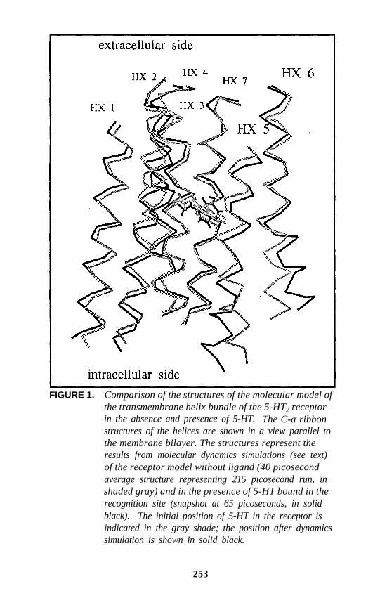
FIGURE 1. Comparison of the structures of the molecular model ofthe transmembrane helix bundle of the 5-HT2 receptorin the absence and presence of 5-HT. The C-a ribbonstructures of the helices are shown in a view parallel tothe membrane bilayer. The structures represent theresults from molecular dynamics simulations (see text)of the receptor model without ligand (40 picosecondaverage structure representing 215 picosecond run, inshaded gray) and in the presence of 5-HT bound in therecognition site (snapshot at 65 picoseconds, in solidblack). The initial position of 5-HT in the receptor isindicated in the gray shade; the position after dynamicssimulation is shown in solid black.
253
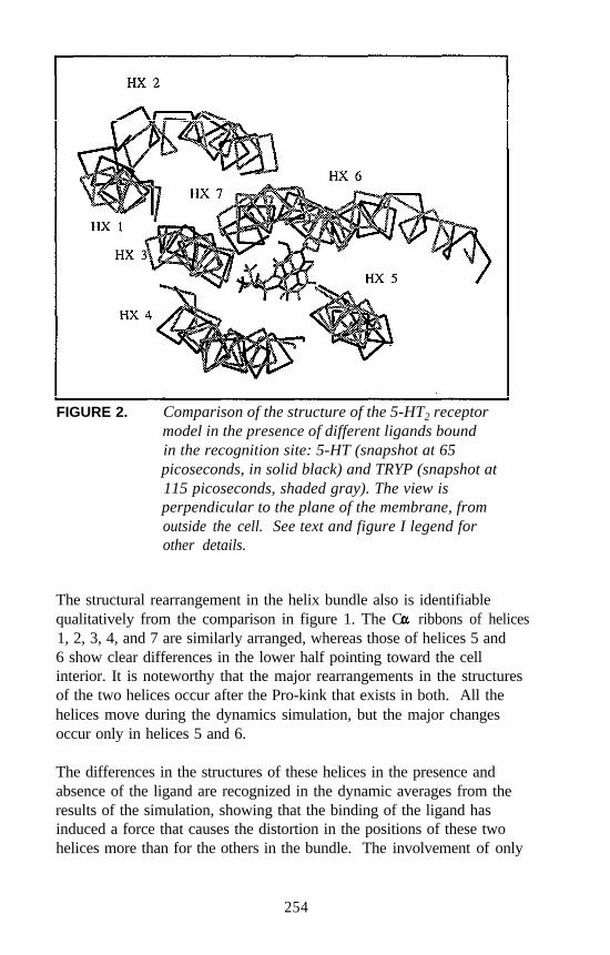
FIGURE 2. Comparison of the structure of the 5-HT2 receptormodel in the presence of different ligands boundin the recognition site: 5-HT (snapshot at 65picoseconds, in solid black) and TRYP (snapshot at115 picoseconds, shaded gray). The view isperpendicular to the plane of the membrane, fromoutside the cell. See text and figure I legend forother details.
The structural rearrangement in the helix bundle also is identifiablequalitatively from the comparison in figure 1. The C ribbons of helices1, 2, 3, 4, and 7 are similarly arranged, whereas those of helices 5 and6 show clear differences in the lower half pointing toward the cellinterior. It is noteworthy that the major rearrangements in the structuresof the two helices occur after the Pro-kink that exists in both. All thehelices move during the dynamics simulation, but the major changesoccur only in helices 5 and 6.
The differences in the structures of these helices in the presence andabsence of the ligand are recognized in the dynamic averages from theresults of the simulation, showing that the binding of the ligand hasinduced a force that causes the distortion in the positions of these twohelices more than for the others in the bundle. The involvement of only
254
helices 5 and 6 at this level of comparative analysis (for more extensivedetails and analyses see Zhang and Weinstein 1993) is significant becausethese helices are the intramembrane ends of the cytoplasmic Loop III thatconstitutes the major structural element in the interaction of the receptorwith the effector system (Kjelsberg et al. 1992; Strader et al. 1989).
As a probe of the validity of this receptor model, it is most gratifying thatthe main structural response to ligand binding seems to be in the regionrequired for transmission of the signal to the effector system. The strongcorrelation between the structure of this region and ligand binding hasbeen demonstrated from mutation studies in the general area of the loop(Strader et al. 1987) and has focused more specifically on one residue inthat region (Kjelsberg et al. 1992).
Further support for the relation between the structural rearrangementcaused by the ligand binding in the recognition site and the mechanism ofreceptor activation toward interaction with the effector is provided by thecorrelation the authors observed between the extent of the rearrangementand the known pharmacological efficacy of the ligand (Zhang andWeinstein 1993). This is illustrated in figure 2, which shows thecomparison between the receptor helix bundle in the presence of the fullagonist 5-HT and the partial agonist TRYP. The view of the helixbundles in figure 2 is perpendicular to the plane of the membrane.Careful inspection reveals that the bundles representing complexes with5-HT (solid black) and TRYP (gray) differ little with the exception ofhelix 6. In fact, helix 6 in the TRYP-bound receptor is not significantlydistorted from its position in the free receptor. Because 5-HT distortshelix 6, the superposition shown in figure 2 identifies a difference in thatregion. On the other hand, helix 5 superimposes well from the dynamicsof the receptors with 5-HT and with TRYP, showing that the distortionproduced by 5-HT (see above) is similar to that induced by the binding ofTRYP. Overall, TRYP induces less distortion in the structure of theLoop III region than 5-HT does, consonant with its lower efficacyindicated by the pharmacological property of partial agonism.
A computational simulation of the receptor complex with the structurallyrelated 5-HT2-receptor antagonist gramine (not shown here) is remarkablein that no distortion in the average structure of the receptor bundle isobserved in the key regions of helices 5 and 6 (Zhang and Weinstein1993). Similar results are being obtained in preliminary studies with the5-HT2 antagonist ketanserin and with the hallucinogenic DOM, whichbehaves in this simulation in accordance with its pharmacological profile
255
as a partial agonist. Therefore, it is clear that a coherent outline of themolecular mechanism of ligand recognition and receptor response toligand binding is emerging from the computational simulations with thepresent model of the transmembrane helix bundle of the 5-HT2 receptor.The qualitative features of this mechanism of receptor function at atomicresolution are in good agreement with the molecular pharmacology of thereceptor.
CONCLUSION
The major work of probing against experimental data the inferencesobtained here for the first time from the simulation of the dynamicresponse of a receptor model to the binding of structurally related ligandsis still ahead. To validate the model and the inferences to produce areliable description of the molecular pharmacology of hallucinogens, it isnecessary to extend these exciting preliminary findings to agonists, partialagonists, and antagonists in different chemical classes and to relate thestructural effects of ligand binding to a reliable mechanistic hypothesis atatomic resolution. Such developments would permit specificphysicochemical testing of the mechanisms and examination in detail ofthe behavior of hallucinogenic compounds in models of this type forother relevant receptor subtypes (e.g., 5-HT1C).
These aims constitute only some of the exciting tasks that can beaddressed by computational simulations with the 3-D receptor model.Such projects offer a comprehensive analysis of fundamental aspects ofmechanisms of drug-receptor interactions and receptor function describedat atomic resolution and also offer arguably the best hope of interferingwith and/or exploiting the properties of hallucinogenic substances basedon rational mechanistic means and specifically designed medications.
REFERENCES
Adham, N.; Romanienko, P.; Hartig, P.; Weinshank, R.L.; and Branchek,T. The rat 5-HT1B receptor is the species homologue of the human5-HT1D beta receptor. Mol Pharmacol 41:1-7, 1992.
Ahlstrom, P.; Teleman, 0.; and Jonsson, B. Interfacial water studied bymolecular dynamics simulations. Chim Scripta 29A:97-101, 1989.
Aqvist, J., and Warshel, A. Computer simulation of the initial protontransfer step in human carbonic anhydrase. J Mol Biol 224:7-14, 1992.
256
Ballesteros, J.A., and Weinstein, H. Analysis and refinement of criteriafor predicting the structure and relative orientations of transmembranalhelical domains. Biophys J 62:107-109, 1992a.
Ballesteros, J.A., and Weinstein, H. The role of Pro/Hyp-kinks indetermining the transmembrane helix length and gating mechanism ofa Leu-zervamicin channel. Biophys J 62:110-111, 1992b.
Bazzo, R.; Tappin, M.J.; Pastore, A.; Harvey, T.S.; Carver, J.A.; andCampbell, I.D. The structure of melittin. Eur J Biochem 173:139-146,1988.
Ben-Hararl, R.R.; Dalton, B.A.; Osman, R.; and Maayani, S. Kineticcharacterization of 5-hydroxytriptamine receptor desensitization inisolated guinea-pig trachea and rabbit aorta. J Pharmacol Exp Ther257:416-424, 1991.
Burris, K.D.; Breeding, M.; and Sanders-Bush, E. (+)Lysergic aciddiethylamide, but not its nonhallucinogenic congeners, is a potentserotonin 5-HT1C receptor agonist. J Pharmacol Exp Ther 258:891-896,199l.
Christ, G.J.; Goldfarb, J.; and Maayani, S. Receptor-mediated mutual-effect amplification elicited by phenylephrine and serotonin in isolatedrabbit aorta. J Pharmacol Exp Ther 252:500-506, 1990.
Dahl, S.G.; Edvardsen, 0.; and Sylte, I. Molecular dynamics of dopamineat the D2 receptor. Proc Natl Acad Sci U S A 88:8111-8115, 1991.
Deisenhofer, J., and Michael, H. The photosynthetic reaction center fromthe purple bacterium Rhodopseudomonas viridis. Science 245:1463-1473, 1989.
Demchyshyn, L.; Sunahara, R.K.; Miller, K.; Teitler, M.; Hoffman, B.J.;Kennedy, J.L.; Seeman, P.; van Tol, H.H.M.; and Niznik, H.B. Ahuman serotonin1D receptor variant (5-HT1D beta) encoded by anintronless gene on chromosome 6. Proc Natl Acad Sci U S A 89:5522-5526, 1992.
DeVivo, M., and Maayani, S. Stimulation and inhibition of adenylylcaclase by distinct 5-hydroxytryptamine receptors. BiochemPharmacol 40:1551-1558, 1990.
Findlay, J., and Eliopoulos, E. Three-dimensional modelling of Gprotein-linked receptors. Trends Pharmacol Sci 11:492-499, 1990.
Glennon, R.A. Evidence for 5-HT2 involvement in the mechanism ofaction of hallucinogenic agents. Life Sci 35:2505-25 11, 1984.
Glennon, R.A. Do classical hallucinogens act as 5-HT2 agonists orantagonists? Neuropsychopharmacology 3:509-517, 1990.
257
Glennon, R.A.; Westkaemper, R.B.; and Bartyzel, P. Medicinal chemistryof serotonergic agents. In: Peroutka, S.J., ed. Serotonin ReceptorSubtypes: Basic and Clinical Aspects. New York: Wiley-Liss, Inc.,1991. pp. 19-64.
Guan, X.-M.; Peroutka, S.J.; and Kobilka, B.K. Identification of a singleamino acid residue responsible for the binding of a class of beta-adrenergic receptor antagonists to 5-HT1A receptors. Mol Pharmacol41:695-698, 1992.
Hartig, P.R. Molecular biology of 5-HT receptors. Trends Pharmacol Sci10:64-69, 1989.
Hen, R. Of mice and flies: Commonalities among 5-HT receptors. TrendsPharmacol Sci 13:160-165, 1992.
Henderson, R.; Baldwin, J.M.; Ceska, T.A.; Zemlin, F.; Beckmann, E.;and Downing, K.H. Model for the structure of Bacteriorhodopsinbased on high-resolution electron cryo-microscopy. J MoZ Biol213:899-929, 1990.
Hibert, M.F.; Trumpp-Kallmeyer, S.; Bruinvels, A.; and Hoflack, J.Three-dimensional models of neurotransmitter G-binding protein-coupled receptors. MOl Pharmacol 40:8-15, 1991.
Jensen, B.; Jorgensen, F.S.; and Kofod, H., eds. Frontiers in DrugResearch: Crystallographic and Computational Methods. AlfredBenzon Symposia 28. Copenhagen: Munksgaard, 1990.
Julius, D.; Livelli, T.J.; Jessell, T.M.; and Axel, R. Ectopic expression ofthe serotonin 1C receptor and the triggering of malignanttransformation. Science 244:1057-1062, 1989.
Julius, D.; MacDermott, A.B.; Axel, R.; and Jessel, T.M. Molecularcharacterization of a functional cDNA encoding the serotonin 1Creceptor. Science 241:558-564, 1988.
Karle, I.L.; Flippen-Anderson, J.L.; Adarwalla, S.; and Balaram, P.Crystal structure of [Leu]zervamicin, a membrane ion channel peptide:Implications for gating mechanisms. Proc Natl Acad Sci U S A88:5307-5311, 1991.
Karplus, M., and Petsko, G.A. Molecular dynamics simulations inbiology. Nature 347:631-639, 1990.
Kjelsberg, M.A.; Cotecchia, S.; Ostrowski, J.; Caron, M.G.; andLefkowitz, R.J. Constitutive activation of the alphal B-adrenergicreceptor by all amino acid substitutions at a single site. J Biol Chem267:1430-1433, 1992.
Lefkowitz, R.J. Variations on a theme. Nature 35 1:353-354, 199 1.Levitt, M., and Sharon, R. Accurate simulation of protein dynamics in
solution. Proc Natl Acad Sci U S A 85:7557-7561, 1988.
258
MacArthur, M.W., and Thornton, J.M. Influence of proline residues onprotein conformation. J Mol Biol 218:397-412, 1991.
Mahle, C.D.; Weiner, H.L.; Yocca, F.D.; and Maayani, S. Destabilizationof hormone/receptor/G-protein (H-R-G) ternary complex is dependenton receptor occupancy but independent of drug efficacy. Neuroscience16:1036, 1990.
Mehler, E.L., and Weinstein, H. Computer-simulation studies ofcalmodulin and troponin C in solution. Experientia 46:A4, 1990.
Mercier, G.A.; Osman, R.; and Weinstein, H. A molecular theoreticalmodel of recognition and activation of a 5-HT receptor. In: Rein, R.,and Golombek, A., eds. Computer Assisted Modeling of Receptor-Ligand Interactions. New York: Alan R. Liss, 1989. pp. 399-410.
Mercier, G.A.; Osman, R.; and Weinstein, H. Role of primary andsecondary protein structure in neurotransmitter activation mechanisms.Protein Eng 2:261-270, 1988.
Milburn, M.V.; Prive, G.G.; Milligan, D.L.; Scott, W.G.; Yeh, J.;Jancarik, J.; Koshland, D.E.; and Kim, S.-H. Three-dimensionalstructure of the ligand binding domain of the bacterial aspartatereceptor with and without a ligand. Science 254:1342-1347, 1991.
Milik, M.; Skolnick, J.; and Kolinski, A. Monte Carlo studies of anidealized model of a lipid-water system. J Phys Chem 96:4015-4022,1992.
Mylecharane, E. J.; Angus, J. A.; de la Lande, I.S.; and Humphrey, P.P.A.,eds. Serotonin: Actions, Receptors, Pathophysiology. London: TheMacmillan Press Ltd., 1989.
Pardo, L.; Ballesteros, J.A.; Osman, R.; and Weinstein, H. On the use ofthe transmembrane domain of bacteriorhodopsin as a template formodeling the three-dimensional structure of guanine nucleotide-binding regulatory protein-coupled receptors. Proc Natl Acad SciU S A 89:4009-4012, 1992.
Pascual-Ahuir, J.-L.; Mehler, E.L.; and Weinstein, H. Calmodulinstructure and function: Implication of arginine residues in thecompaction related to ligand binding. Molec Eng 1:231-247, 1991.
Pastore, A.; Harvey, T.S.; Dempsey, C.E.; and Campbell, I.D. Thedynamic properties of melittin in solution. Eur Biophys J 16:363-367,1989.
Peroutka, S.J., ed. Serotonin Receptor Subtypes: Basic and ClinicalAspects. Vol. 15. New York: Wiley-Liss, 1991.
Rees, D.C.; Komiya, H.; Yeates, T.O.; Allen, J.P.; and Feher, G. Thebacterial photosynthetic reaction center as a model for membraneproteins. Annual Rev Biochem 58:607-633, 1989.
259
Sanders-Bush, E.; Burt-is, K.D.; and Knoth, K. Lysergic aciddiethylamide and 2,5-dimethoxy-4-methylamphetamine are partialagonists at serotonin receptors linked to phosphoinositide hydrolysis.J Pharmacol Exp Ther 246:924-928, 1988.
Shih, J.C.; Yang, W.; Chen, K.; and Gallaher, T. Molecular biology ofserotonin (5-HT) receptors. Pharmacol Biochem Behav 40:1053-1058,1991.
Stigter, D.; Mingins, J.; and Dill, K.A. Phospholipid interactions in modelmembrane systems. Biophys J 61:1616-1629, 1992.
Strader, C.D.; Dixon, R.A.F.; Cheung, A.H.; Candelore, M.R.; Blake,A.D.; and Sigal, I.S. Mutations that uncouple the beta-adrenergicreceptor from Gs and increase agonist affinity. J Biol Chem262:16439-16443, 1987.
Strader, C.D.; Sigal, I.S.; and Dixon, R.A.F. Structural basis ofß-adrenergic receptor function. FASEB J 3:1825-1831, 1989.
van Gunsteren, W.F.; Brunne, R.M.; Mark, A.E.; and van Helden, S.P.Computer simulation of biomolecules: Comparison with experimentaldata. In: Bertran, J., ed. Molecular Aspects of Biotechnology:Computational Models and Theories. Dordrecht, The Netherlands:Kluwer Academic Publishers, 1992. pp. 105- 122.
Weinstein, H., ed. Computational Approaches to Enzyme Structure andFunction. (Enzyme, Vol. 36.) Basel, Switzerland: Karger ScientificPublishers, 1986.
Weinstein, H., and Mehler, E.L. Structural specificity in the engineeringof biological function: Insights from the dynamics of calmodulin. In:Bertran, J., ed. Molecular Aspects of Biotechnology: ComputationalModels and Theories. Dordrecht, The Netherlands: Kluwer AcademicPublishers, 1992. pp. 153-173.
Weinstein, H., and Osman, R. Interaction mechanisms at biologicaltargets: Implications for design of serotonin receptor ligands. In:Richards, W.G., ed. Computer-Aided Molecular Design. London:IBC Technical Services, 1989a. pp. 105-108.
Weinstein, H., and Osman, R. Molecular biophysics of specificity andfunction in enzymes, receptors and calcium binding proteins. In:Beveridge, D.L., and Lavery, R., eds. Theoretical Biochemistry andMolecular Biophysics: A Comprehensive Survey. New York: AdinePress, 1989b. pp. 275-289.
Weinstein, H., and Osman, R. On the structural and mechanistic basis offunction, classification, and ligand design for 5-HT receptors.Neuropsychopharmacology 3:397-409, 1990a.
260
Weinstein, H., and Osman, R. Simulations of ligand-receptor interactionsas guides for design. In: Jensen, B.; Jorgensen, F.S.; and Kofod, H.,eds. Frontiers in Drug Research: Crystallographic andComputational Methods. Alfred Benzon Symposium 28. Copenhagen:Munksgaard, 1990b. pp. 169-182.
Weinstein, H.; Osman, R.; and Mazurek, A.P. Simulations of molecularstereoelectronic mechanisms for the interaction of hallucinogens andindole derivatives at 5-HT receptors. In: Naray-Szabo, G., andKalman, S., eds. Steric Aspects of Biomolecular Interactions. BocaRaton, FL: CRC Press, 1987. pp. 199-210.
Weinstein, H.; Osman, R.; and Mercier, G., Jr. Recognition andactivation of a 5-HT receptor by hallucinogens and indole derivatives.In: Harris, L.S., ed. Problems of Drug Dependence 1988: Proceedingsof the 50th Annual Scientific Meeting, The Committee on Problems ofDrug Dependence, Inc. National Institute on Drug Abuse ResearchMonograph No. 90. DHHS Pub. No. (ADM)89-1605. Washington,DC: Supt. of Docs., U.S. Govt. Print. Off., 1988. pp. 243-255.
Wendoloski, J.J.; Kimatian, S.J.; Schutt, C.E.; and Salemme, F.R.Molecular dynamics simulation of a phospholipid micelle. Science243:636-638, 1989.
Williams, K.A., and Deber, C.M. Proline residues in transmembranehelices: Structural or dynamic role? Biochemistry 30:8919-8923,1991.
Woolfson, D.N., and Williams, D.H. The influence of proline residues onalpha-helical structure. FEBS Lett 277:185-188, 1990.
Xing, J., and Scott, H.L. Monte Carlo studies of a model for lipid-gramicidin A bilayers. Biochim Biophys Acta 1106:227-232, 1992.
Yocca, F.D.; Moon, S.L.; Torrente, J.; Iben, L.; Ryan, E.; and Lamy, R.Region-dependent drug efficacy in brain: Pre- vs. postsynaptic actionsof drugs on the 5-HT1A receptor in rat hippocampus and cortex.FASEB J 4:A810, 1990.
Zhang, D., and Weinstein, H. Signal transduction by 5-HT2 receptor: Amechanistic hypothesis from molecular dynamics simulations of thethree-dimensional model of the receptor complexed to ligands, J MedChem 36:934-938, 1993.
ACKNOWLEDGMENTS
The work was supported in part by National Institute on Drug Abusegrant DA-06620 and Research Scientist Award DA-00060 (to HW) andby a Fulbright/MEC scholarship (to JAB). Computations were performed
261
on the supercomputer systems at the Pittsburgh Supercomputer Center(sponsored by the National Science Foundation), Cornell NationalSupercomputer Facility (sponsored by the National Science Foundationand IBM), Advanced Scientific Computing Laboratory at the FrederickCancer Research Facility of the National Cancer Institute (Laboratory forMathematical Biology), and University Computer Center of the CityUniversity of New York.
AUTHORS
Harel Weinstein, D.Sc.Professor and Chairman
Daqun Zhang, B.Sc.Graduate Student
Juan A. Bahesteros, B.Sc.Graduate Student
Department of Physiology and BiophysicsMount Sinai School of Medicine of the City University of New YorkNew York, NY 10029-6574
262
Molecular Modelingof the Interaction of LSDand Other HallucinogensWith 5-HT2 ReceptorsRichard B. Westkaemper and Richard A. Glennon
INTRODUCTION
Multiple populations of serotonin (5-hydroxytryptamine [5-HT])receptors have been identified, including 5-HT1A, 5-HTlB, 5-HT1c,5-HT1D, 5-HT2, 5-HT3, and 5-HT4 subtypes. The 5-HT2 and possibly the5-HT1C receptors have been implicated as the probable sites of action ofclassical hallucinogenic agents (Glennon 1990). The goal of this chapteris to identify potential modes of interaction of hallucinogens with 5-HT2
receptors,
In general terms, there are two ways to embark on a program designed toelucidate drug-receptor interactions. These have been referred to as theligand-ligand and ligand-receptor approaches (Westkaemper and Glennon1991). In the first approach, indirect information about specific receptorstructural features and their locations in three dimensions is inferredindirectly from the structures of ligands. Typically, common features ofstructurally related ligands are graphically superimposed to illustratecommon and disparate features that aid formulation of hypotheses relatedto specific drug-receptor interaction.
For example, (+)lysergic acid diethylamide (LSD) contains manystructural features in a rigid polycyclic template that are important for5-HT2 receptor binding. Figure 1 shows one way in which the structuresof LSD and the more flexible hallucinogenic agent 1-(4-bromo-2,5-dimethoxyphenyl)-2-aminopropane (DOB) can be related. For additionalexamples of the application of this approach to serotonergic agents, seeGlennon and colleagues (199 1).
263
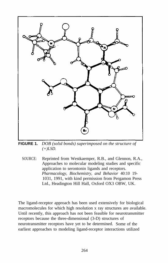
FIGURE 1. DOB (solid bonds) superimposed on the structure of(+)LSD.
SOURCE: Reprinted from Westkaemper, R.B., and Glennon, R.A.,Approaches to molecular modeling studies and specificapplication to serontonin ligands and receptors.Pharmacology, Biochemistry, and Behavior 40:10 19-1031, 1991, with kind permission from Pergamon PressLtd., Headington Hill Hall, Oxford OX3 OBW, UK.
The ligand-receptor approach has been used extensively for biologicalmacromolecules for which high resolution x ray structures are available.Until recently, this approach has not been feasible for neurotransmitterreceptors because the three-dimensional (3-D) structures ofneurotransmitter receptors have yet to be determined. Some of theearliest approaches to modeling ligand-receptor interactions utilized
264
simulations employing fragments that might be present at the ligand-binding site of a receptor.
Kier and Aldrich (1974) investigated ligand-receptor interactions usingsemiempirical molecular orbital methods to calculate the interactionenergies between a drug and a small molecule thought to mimic a portionof the receptor binding site in a process known as receptor mapping usingmodel interaction energy calculations. Later, this paradigm was appliedto hallucinogenic phenethylamines (Di Paolo et al. 1978). In thesestudies, interaction energies between ring-substituted phenethylaminesand methylindole, a presumed tryptophan surrogate, were found tocorrelate with hallucinogenic potency.
Weinstein explored a similar approach by calculating interaction energiesbetween an imidazolium ion as a receptor model and 5-HT (Weinsteinand Osman 1989). Although the implications of such studies areintriguing, the receptor models do not incorporate what is known aboutthe molecular architecture of proteins. Later, more elaborate models,based on known protein structures not related to neurotransmitters, wereused to provide a framework for the incorporation of ligand and receptor-like features (Weinstein and Osman 1990).
The construction of more complete receptor models had not been possiblebecause information pertinent to 3-D structure, such as deduced aminoacid sequence, was not available, Although several 5-HT receptors havenow been cloned and their sequences reported (Fargin et al. 1988;Hamblin and Metcalf 1991; Hartig 1989; Hartig et al. 1990a,b; Huangand Julius 1991; Julius et al. 1988, 1990; Pritchett et al. 1988; Shih andChen 1990; Voigt et al. 1991), their 3-D structures remain unknown.
On the other hand, the 3-D structure of the light-sensitive proton pumpfrom bacteria, bacteriorhodopsin (BRH), has been determined to nearatomic resolution (Henderson et al. 1990; Henderson and Schertler 1990).Although BRH is not a guanosine triphosphate binding protein(G-protein) receptor, it may be possible to deduce the structures ofneurotransmitter receptors from it. BRH consists of seven membrane-spanning -helices linked by extracellular and intracellular loops. TheN-terminus is extracellular, and the C-terminus is located within the cell.The amphiphilic helices are found to form a pore in the membrane, withlipophilic sides oriented toward the membrane and with the morehydrophilic residues located in the central cavity.
265
The mammalian visual pigment rhodopsin, which is G-protein coupled, isfunctionally different from BRH, but both are light- and retinal-dependent (Henderson and Schertler 1990). There is little sequencehomology between the mammalian visual pigments and BRH (Dohlmanet al. 1987). However, evidence suggests that the overall topology for themammalian protein is similar to that of BRH: both consist of sevenmembrane-spanning -helices (see Westkaemper and Glennon 1991).
There is a significant homology between rhodopsin and the G-protein-coupled neurotransmitter receptors and an even greater homologybetween the neurotransmitters. Studies utilizing secondary structure-predicting methods, the analysis of the distribution of hydrophobic spansof amino acids, and the fact that the G-protein receptors have the greatestdegree of sequence homology in the putative helical segments suggestthat the architecture of G-protein neurotransmitter receptors is similar tothat observed for BRH and to that presumed for rhodopsin (Dohlman etal. 1987).
If sequence homology between BRH and serotonin receptors were good,construction of 3-D receptor models could begin by graphically changingthe amino acid side chains of the experimentally determined structure tothose of the receptor of interest. Because there is no unambiguousalignment of the transmembrane portions of the 5-HT receptors and BRHsequences, model building is much more tenuous. Given that the helicalsegments of the serotonin receptors can be identified, placement of thesein three dimensions requires the establishment of helix connectivity,vertical placement of each helix (perpendicular to the membrane),rotational alignment (in the plane of the membrane), and side chaingeometry, none of which can be known with absolute certainty.
Given these ambiguities, abbreviated receptor models in which only asingle helix is represented were chosen for investigation (Westkaemperand Glennon 1991). Docking studies performed with 5-HT ligands andmodels of helix 3 and helix 2 of 5-HT2 (and other) receptors indicatedthat helix 3 is the more probable primary ligand-binding site; this hasbeen recently verified by site-directed mutagenesis (Shih et al., thisvolume). Calculated binding energies for 5-HT ligands and the helix 3model of 5-HT2 receptors correlated qualitatively with experimentallydetermined affinities for a wide range of structural types, indicating thatthe features of helix 3 can account for ligand binding.
266
Despite this success, more complete receptor models are necessary fordetailed understanding of ligand-receptor interaction and, in particular,linkage between agonist binding and second messenger coupling.
Hibert and coworkers (1991) recently described the first seven-helixmodel of a serotonin receptor. A potential sequence alignment betweenthe helical segments of multiple G-protein receptors and BRH wasestablished. The helix connectivity (both in the Hibert model and in themodel presented below) of BRH was preserved; that is, helix 1 of the5-HT receptor corresponds with helix 1 of BRH and so on. Alternativeconnectivity schemes have been proposed (Weinstein et al., this volume).Although not described, it appears that the final receptor configurationwas arrived at by rotational adjustment of helices guided by the principlesthat highly conserved and charged amino acids should be oriented towardthe receptor pore (central to the aggregate of helices). Consideration alsowas given to site-directed mutagenesis data for ß-adrenergic receptors(ß-AR).
The authors have taken a somewhat different approach and haveattempted to identify sequence alignments between the 5-HT receptorsand BRH that could be used to construct serotonin receptor models bymutating amino acid side chains without manual modification of helixdisposition. White and Jacobs (1990) recently have shown that lengthsand centers of helical spans in the photosynthetic reaction center andBRH can be accurately predicted (helical centers within 0.2 amino acidsand helix ends within 1 to 3 residues of the experimentally determinedvalues) using a modified hydrophobicity index, and that the helix turnpotential has a minimum value at helix centers. Figure 2 shows theresults of such an analysis for the 5-HT2 receptor sequence.
The authors have chosen to evaluate the hypothesis that helical centers ofserotonin receptors and BRH (see figure 3 for amino acid sequence ofBRH) are structurally coincident. Once such a coincidence and sequencealignment are established, 3-D models of serotonin receptors can beconstructed, subjected to molecular mechanics geometry optimization,and evaluated for reasonableness with respect to important criteria: helixpacking, disposition of polar and hydrophobic residues, orientation ofhighly conserved residues, and accessibility of side chains known tointeract with ligands.
267
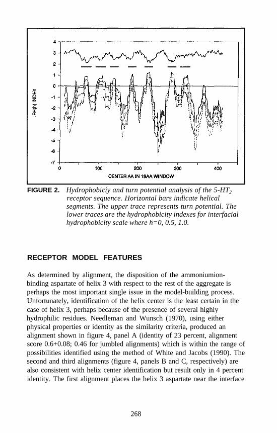
FIGURE 2. Hydrophobiciy and turn potential analysis of the 5-HT2
receptor sequence. Horizontal bars indicate helicalsegments. The upper trace represents turn potential. Thelower traces are the hydrophobicity indexes for interfacialhydrophobicity scale where h=0, 0.5, 1.0.
RECEPTOR MODEL FEATURES
As determined by alignment, the disposition of the ammoniumion-binding aspartate of helix 3 with respect to the rest of the aggregate isperhaps the most important single issue in the model-building process.Unfortunately, identification of the helix center is the least certain in thecase of helix 3, perhaps because of the presence of several highlyhydrophilic residues. Needleman and Wunsch (1970), using eitherphysical properties or identity as the similarity criteria, produced analignment shown in figure 4, panel A (identity of 23 percent, alignmentscore 0.6+0.08; 0.46 for jumbled alignments) which is within the range ofpossibilities identified using the method of White and Jacobs (1990). Thesecond and third alignments (figure 4, panels B and C, respectively) arealso consistent with helix center identification but result only in 4 percentidentity. The first alignment places the helix 3 aspartate near the interface
268
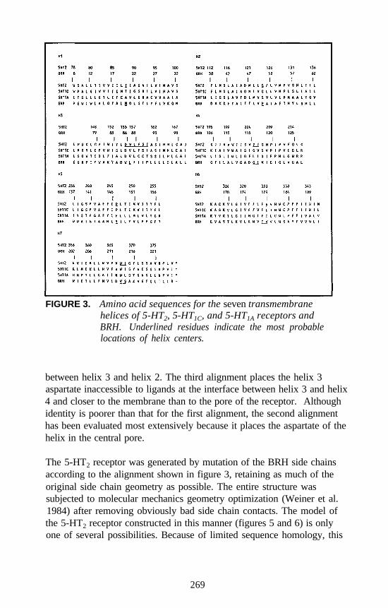
FIGURE 3. Amino acid sequences for the seven transmembranehelices of 5-HT2, 5-HT1C, and 5-HT1A receptors andBRH. Underlined residues indicate the most probablelocations of helix centers.
between helix 3 and helix 2. The third alignment places the helix 3aspartate inaccessible to ligands at the interface between helix 3 and helix4 and closer to the membrane than to the pore of the receptor. Althoughidentity is poorer than that for the first alignment, the second alignmenthas been evaluated most extensively because it places the aspartate of thehelix in the central pore.
The 5-HT2 receptor was generated by mutation of the BRH side chainsaccording to the alignment shown in figure 3, retaining as much of theoriginal side chain geometry as possible. The entire structure wassubjected to molecular mechanics geometry optimization (Weiner et al.1984) after removing obviously bad side chain contacts. The model ofthe 5-HT2 receptor constructed in this manner (figures 5 and 6) is onlyone of several possibilities. Because of limited sequence homology, this
269
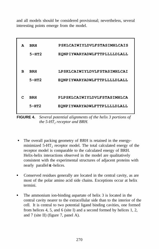
and all models should be considered provisional; nevertheless, severalinteresting points emerge from the model.
A BRH PSKLCAIWIYLDVLFSTASIMHLCAIS
5-HT2 EQNPIYWARYADWLFTTPLLLLDLALL
B BRH LPSKLCAIWIYLDVLFSTASIMHLCAI
5-HT2 EQNPIYWARYADWLFTTPLLLLDLALL
C BRH PLPSKLCAIWIYLDVLFSTASIMHLCA
5-HT2 EQNPIYWARYADWLFTTPLLLLDLALL
FIGURE 4. Several potential alignments of the helix 3 portions ofthe 5-HT2 receptor and BRH.
The overall packing geometry of BRH is retained in the energy-minimized 5-HT2 receptor model. The total calculated energy of thereceptor model is comparable to the calculated energy of BRH.Helix-helix interactions observed in the model are qualitativelyconsistent with the experimental structures of adjacent proteins withnearly parallel -helices.
Conserved residues generally are located in the central cavity, as aremost of the polar amino acid side chains. Exceptions occur at helixtermini.
The ammonium ion-binding aspartate of helix 3 is located in thecentral cavity nearer to the extracellular side than to the interior of thecell. It is central to two potential ligand binding cavities, one formedfrom helices 4, 5, and 6 (site I) and a second formed by helices 1, 2,and 7 (site II) (figure 7, panel A).
270
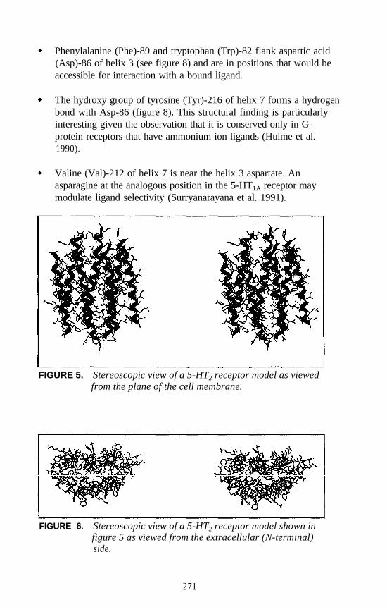
Phenylalanine (Phe)-89 and tryptophan (Trp)-82 flank aspartic acid(Asp)-86 of helix 3 (see figure 8) and are in positions that would beaccessible for interaction with a bound ligand.
The hydroxy group of tyrosine (Tyr)-216 of helix 7 forms a hydrogenbond with Asp-86 (figure 8). This structural finding is particularlyinteresting given the observation that it is conserved only in G-protein receptors that have ammonium ion ligands (Hulme et al.1990).
Valine (Val)-212 of helix 7 is near the helix 3 aspartate. Anasparagine at the analogous position in the 5-HT1A receptor maymodulate ligand selectivity (Surryanarayana et al. 1991).
FIGURE 5. Stereoscopic view of a 5-HT2 receptor model as viewedfrom the plane of the cell membrane.
FIGURE 6. Stereoscopic view of a 5-HT2 receptor model shown infigure 5 as viewed from the extracellular (N-terminal)side.
271
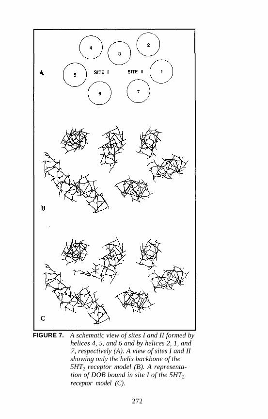
FIGURE 7. A schematic view of sites I and II formed byhelices 4, 5, and 6 and by helices 2, 1, and7, respectively (A). A view of sites I and IIshowing only the helix backbone of the5HT2 receptor model (B). A representa-tion of DOB bound in site I of the 5HT2
receptor model (C).
272
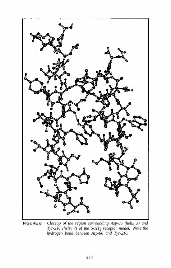
FIGURE 8. Closeup of the region surrounding Asp-86 (helix 3) andTyr-216 (helix 7) of the 5-HT2 receptor model. Note thehydrogen bond between Asp-86 and Tyr-216.
273
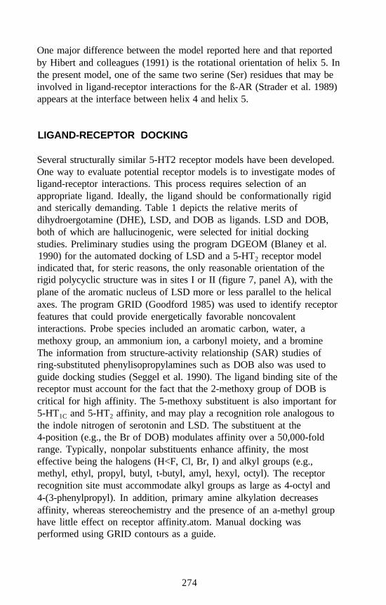
One major difference between the model reported here and that reportedby Hibert and colleagues (1991) is the rotational orientation of helix 5. Inthe present model, one of the same two serine (Ser) residues that may beinvolved in ligand-receptor interactions for the ß-AR (Strader et al. 1989)appears at the interface between helix 4 and helix 5.
LIGAND-RECEPTOR DOCKING
Several structurally similar 5-HT2 receptor models have been developed.One way to evaluate potential receptor models is to investigate modes ofligand-receptor interactions. This process requires selection of anappropriate ligand. Ideally, the ligand should be conformationally rigidand sterically demanding. Table 1 depicts the relative merits ofdihydroergotamine (DHE), LSD, and DOB as ligands. LSD and DOB,both of which are hallucinogenic, were selected for initial dockingstudies. Preliminary studies using the program DGEOM (Blaney et al.1990) for the automated docking of LSD and a 5-HT2 receptor modelindicated that, for steric reasons, the only reasonable orientation of therigid polycyclic structure was in sites I or II (figure 7, panel A), with theplane of the aromatic nucleus of LSD more or less parallel to the helicalaxes. The program GRID (Goodford 1985) was used to identify receptorfeatures that could provide energetically favorable noncovalentinteractions. Probe species included an aromatic carbon, water, amethoxy group, an ammonium ion, a carbonyl moiety, and a bromineThe information from structure-activity relationship (SAR) studies ofring-substituted phenylisopropylamines such as DOB also was used toguide docking studies (Seggel et al. 1990). The ligand binding site of thereceptor must account for the fact that the 2-methoxy group of DOB iscritical for high affinity. The 5-methoxy substituent is also important for5-HT1C and 5-HT2 affinity, and may play a recognition role analogous tothe indole nitrogen of serotonin and LSD. The substituent at the4-position (e.g., the Br of DOB) modulates affinity over a 50,000-foldrange. Typically, nonpolar substituents enhance affinity, the mosteffective being the halogens (H<F, Cl, Br, I) and alkyl groups (e.g.,methyl, ethyl, propyl, butyl, t-butyl, amyl, hexyl, octyl). The receptorrecognition site must accommodate alkyl groups as large as 4-octyl and4-(3-phenylpropyl). In addition, primary amine alkylation decreasesaffinity, whereas stereochemistry and the presence of an a-methyl grouphave little effect on receptor affinity.atom. Manual docking wasperformed using GRID contours as a guide.
274
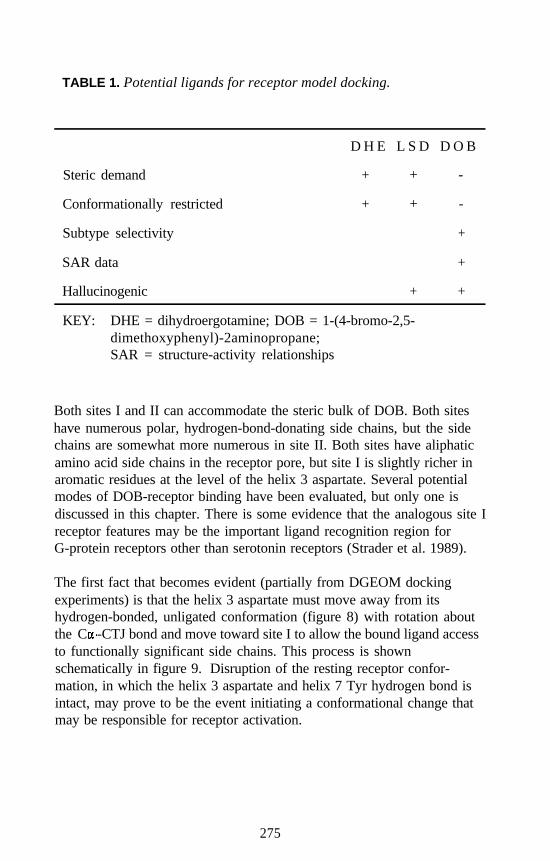
TABLE 1. Potential ligands for receptor model docking.
Steric demand
Conformationally restricted
Subtype selectivity
SAR data
Hallucinogenic
D H E L S D D O B
+ + -
+ + -
+
+
+ +
KEY: DHE = dihydroergotamine; DOB = 1-(4-bromo-2,5-dimethoxyphenyl)-2aminopropane;SAR = structure-activity relationships
Both sites I and II can accommodate the steric bulk of DOB. Both siteshave numerous polar, hydrogen-bond-donating side chains, but the sidechains are somewhat more numerous in site II. Both sites have aliphaticamino acid side chains in the receptor pore, but site I is slightly richer inaromatic residues at the level of the helix 3 aspartate. Several potentialmodes of DOB-receptor binding have been evaluated, but only one isdiscussed in this chapter. There is some evidence that the analogous site Ireceptor features may be the important ligand recognition region forG-protein receptors other than serotonin receptors (Strader et al. 1989).
The first fact that becomes evident (partially from DGEOM dockingexperiments) is that the helix 3 aspartate must move away from itshydrogen-bonded, unligated conformation (figure 8) with rotation aboutthe C -CTJ bond and move toward site I to allow the bound ligand accessto functionally significant side chains. This process is shownschematically in figure 9. Disruption of the resting receptor confor-mation, in which the helix 3 aspartate and helix 7 Tyr hydrogen bond isintact, may prove to be the event initiating a conformational change thatmay be responsible for receptor activation.
275
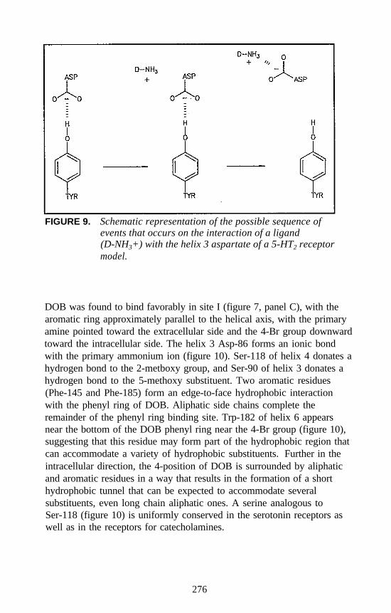
FIGURE 9. Schematic representation of the possible sequence ofevents that occurs on the interaction of a ligand(D-NH3+) with the helix 3 aspartate of a 5-HT2 receptormodel.
DOB was found to bind favorably in site I (figure 7, panel C), with thearomatic ring approximately parallel to the helical axis, with the primaryamine pointed toward the extracellular side and the 4-Br group downwardtoward the intracellular side. The helix 3 Asp-86 forms an ionic bondwith the primary ammonium ion (figure 10). Ser-118 of helix 4 donates ahydrogen bond to the 2-metboxy group, and Ser-90 of helix 3 donates ahydrogen bond to the 5-methoxy substituent. Two aromatic residues(Phe-145 and Phe-185) form an edge-to-face hydrophobic interactionwith the phenyl ring of DOB. Aliphatic side chains complete theremainder of the phenyl ring binding site. Trp-182 of helix 6 appearsnear the bottom of the DOB phenyl ring near the 4-Br group (figure 10),suggesting that this residue may form part of the hydrophobic region thatcan accommodate a variety of hydrophobic substituents. Further in theintracellular direction, the 4-position of DOB is surrounded by aliphaticand aromatic residues in a way that results in the formation of a shorthydrophobic tunnel that can be expected to accommodate severalsubstituents, even long chain aliphatic ones. A serine analogous toSer-118 (figure 10) is uniformly conserved in the serotonin receptors aswell as in the receptors for catecholamines.
276
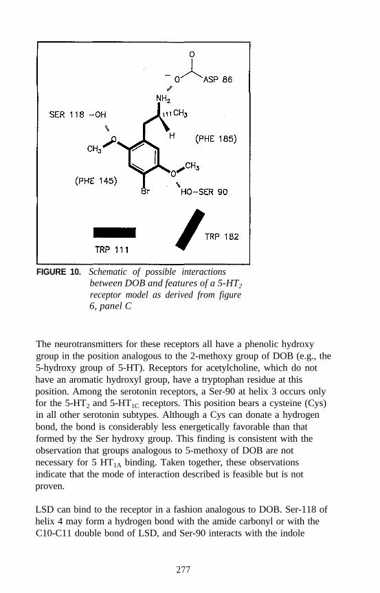
FIGURE 10. Schematic of possible interactionsbetween DOB and features of a 5-HT2
receptor model as derived from figure6, panel C
The neurotransmitters for these receptors all have a phenolic hydroxygroup in the position analogous to the 2-methoxy group of DOB (e.g., the5-hydroxy group of 5-HT). Receptors for acetylcholine, which do nothave an aromatic hydroxyl group, have a tryptophan residue at thisposition. Among the serotonin receptors, a Ser-90 at helix 3 occurs onlyfor the 5-HT2 and 5-HT1C receptors. This position bears a cysteine (Cys)in all other serotonin subtypes. Although a Cys can donate a hydrogenbond, the bond is considerably less energetically favorable than thatformed by the Ser hydroxy group. This finding is consistent with theobservation that groups analogous to 5-methoxy of DOB are notnecessary for 5 HT1A binding. Taken together, these observationsindicate that the mode of interaction described is feasible but is notproven.
LSD can bind to the receptor in a fashion analogous to DOB. Ser-118 ofhelix 4 may form a hydrogen bond with the amide carbonyl or with theC10-C11 double bond of LSD, and Ser-90 interacts with the indole
277
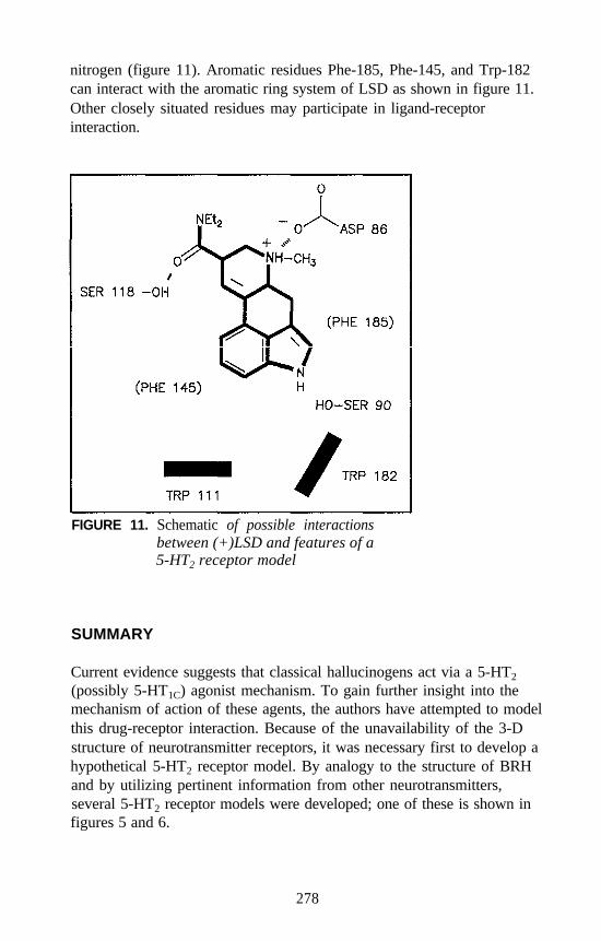
nitrogen (figure 11). Aromatic residues Phe-185, Phe-145, and Trp-182can interact with the aromatic ring system of LSD as shown in figure 11.Other closely situated residues may participate in ligand-receptorinteraction.
FIGURE 11. Schematic of possible interactionsbetween (+)LSD and features of a5-HT2 receptor model
SUMMARY
Current evidence suggests that classical hallucinogens act via a 5-HT2
(possibly 5-HT1C) agonist mechanism. To gain further insight into themechanism of action of these agents, the authors have attempted to modelthis drug-receptor interaction. Because of the unavailability of the 3-Dstructure of neurotransmitter receptors, it was necessary first to develop ahypothetical 5-HT2 receptor model. By analogy to the structure of BRHand by utilizing pertinent information from other neurotransmitters,several 5-HT2 receptor models were developed; one of these is shown infigures 5 and 6.
278
Using this model, the authors attempted to dock the structures of DOBand (+)LSD using DGEOM and GRID to aid this process. Docking alsowas guided by known SAR. (That is, the validity of any model rests withits ability to account for what is known about the binding of hallu-cinogens at 5-HT2 receptors.) Two potential binding domains wereidentified (i.e., site I and site II, see figure 7). Several different modes ofbinding are possible within each of these sites. Figure 7, panel C showsone way in which DOB can bind at site I of the 5-HT2 receptor model. Acloseup view of the specific amino acid residues that may be involved inbinding is shown as a schematic (figure 10). The proposed mode ofinteraction also accounts for the binding of (+)LSD and is consistent withestablished SAR. The proposed mode of interaction between drug andreceptor is neither inflexible nor static. (That is, ligands with slightlydifferent aromatic substitution patterns may still interact in an analogousmanner with the receptor features that have been identified.) Further-more, it must be emphasized that figures 10 and 11 represent onepotential mode of binding of DOB and (+)LSD at site I of a singlemultiple-helix 5-HT2 receptor model. Other modes of binding-indeed,other 5-HT2 receptor models-are possible. Although the present drug-receptor model is certainly reasonable, not all other possible drug-receptor models have been fully evaluated yet.
The objective is to identify the most likely drug-receptor models usingsite-directed mutagenesis. For example, all models utilize the helix 3aspartate as the ammonium ion binding site; thus, this aspartate must playa critical role in binding, and its elimination or replacement should resultin reduced affinity. In collaboration with Dr. Shih, the authors now havedemonstrated that replacement of this aspartate (but not the aspartate ofhelix 2) by asparagine (Asn) results in a dramatic decrease in affinity forvarious 5-HT2 ligands. Similar studies are required to test other potentialbinding features consistent with the different models.
The classical hallucinogens bind at 5-HT1C receptors with affinitiescomparable with the affinities for 5-HT2 receptors. Indeed, there is lessthan a tenfold difference in 5-HT1C versus 5-HT2 affinities for a largeseries of DOB-related analogs (Glennon et al. 1992). Thus, any drug-receptor model developed for 5-HT2 receptors should possess those (orstructurally analogous) features found in 5-HT1C receptors. In contrast,because classical hallucinogens (with the exception of (+)LSD) typicallybind with low affinity at 5-HT1A receptors, structural features common to5-HT2 and 5-HT1A receptors would likely be unable to account forselectivity. One of several possible drug-receptor 5-HT2 models is
279
described here in detail. The model accounts for, and is consistent with,much that is known about classical hallucinogens. Studies are under wayto further validate this (and other) models. These studies, in addition tobeing relevant to the investigation of hallucinogenic agents, go wellbeyond the present limited scope of study and can have a significantinfluence on the understanding of drug-receptor interactions in general.
NOTE
All sequence numbers refer to the BRH numbering system for purposesof comparison and uniformity. The actual numbering system for theamino acid sequence of 5-HT2 receptors is shown in figure 3.
REFERENCES
Blaney, J.M.; Crippen, G.M.; Dearing, A.; and Dixon, J.S. DGEOM.QCPE program number 590. QCPE Bull 10:27-38, 1990.
Di Paolo, T.; Hall, L.H.; and Kier, L.B. Structure-activity studies onhallucinogenic amphetamines using model interaction calculations.J Theor Biol 71:295-309, 1978.
Dohlman, H.G.; Caron, G.C.; and Lefkowitz, R.J. A family of receptorscoupled to guanine nucleotide regulatory proteins. Biochemistry26:2657-2664, 1987.
Fargin, A.; Raymond, J.R.; Lohse, M.J.; Kobilka, B.K.; Caron, M.G.; andLefkowitz, R.J. The genomic clone G-21 which resembles a ß-adrenergic receptor sequence encodes the 5-HT1A receptor. Nature335:358-360, 1988.
Glennon, R.A. Do hallucinogens act as 5-HT2 agonists or antagonists?Neuropsychopharmacology 56:509-517, 1990.
Glennon, R.A.; Westkaemper, R.B.; and Bartyzel, P. Medicinal chemistryof serotonergic agents. In: Peroutka, S.J., ed. Serotonin ReceptorSubtypes: Basic and Clinical Aspects. New York: Wiley-Liss, 1991.pp. 19-64.
Glennon, R.A.; Raghupathi, R.; Bartyzel, P.; Teitler, M.; and Leonhardt,S. Binding of phenylalkylamine derivatives at 5-HT1C and 5-HT2
serotonin receptors: Evidence for a lack of selectivity. J Med Chem35:734-740, 1992.
Goodford, P.J. A computation procedure for determining energeticallyfavorable binding sites on biologically important macromolecules.J Med Chem 28:849-857, 1985.
280
Hamblin, M., and Metcalf, M. Primary structure and characterization of ahuman 5-HT1D receptor. Mol Pharmacol 40:143-148, 1991.
Hartig, P.R. Molecular biology of 5-HT receptors. Trends Pharmacol Sci10:64-69, 1989.
Hartig, P.R.; Hoffman, B.J.; Kaufman, M.J.; and Hirata, F. The 5-HT1C
receptor. Ann N Y Acad Sci 600:149-166, 1990a.Hartig, P.R.; Kao, H.-T.; Macchi, M.; Adham, N.; Zgombic, J.;
Weinshank, R.; and Branchek, T. The molecular biology of serotoninreceptors. Neuropsychopharmacology 3:335-347,1990b.
Henderson, R., and Schertler, G.F.X. The structure of bacteriorhodopsinand its relevance to the visual opsins and other seven-helix G protein-coupled receptors. Philos Trans R Soc Lond [Biol] 326:379-389,1990.
Henderson, R.; Baldwin, J.M.; Ceska, T.A.; Zemlin, F.; Beckmann, E.;and Downing, K.H. Model for the structure of bacteriorhodopsinbased on high-resolution electron cryo-microscopy. J Mol Biol213:899-929, 1990.
Hibert, M.F.; Trumpp-Kallmeyer, S.; Bruinvels, A.; and Hoflack, J.Three-dimensional models of neurotransmitter G-binding protein-coupled receptors. Mol Pharmacol 40:8-15, 1991.
Huang, K.N., and Julius, D. Molecular characterization of three serotoninreceptor subtypes. In: Peroutka, S.J., ed. Serotonin Receptor Subtypes:Basic and Clinical Aspects. New York: Wiley-Liss, 1991. pp. 1-17.
Hulme, E.C.; Birdsall, N.J.M.; and Buckley, N.J. Muscarinic receptorsubtypes. Ann Rev Pharmacol Toxicol 30:633-673,1990.
Julius, D.; MacDermott, A.B.; Axel, R.; and Jessel, T.M. Molecularcharacterization of a functional cDNA encoding the serotonin 1Creceptor. Science 241:558-564, 1988.
Julius, D.; Huang, K.N.; Livelli T.J.; Axel, R.; and Jessel, T.M. The5-HT2 receptor defines a family of structurally distinct but functionallyconserved serotonin receptors. Proc Natl Acad Sci U S A 87:928-932,1990.
Kier, L.B., and Aldrich, H.S. A theoretical study of receptor site modelsfor trimethylammonium group interaction. J Theor Biol 46:529-541,1974.
Needleman, S., and Wunsch, C.J. A general method applicable to thesearch for similarities in the amino acid sequence of two proteins.J Mol Biol 48:443-453, 1970.
Pritchett, D.B.; Bach, A.W.J.; Wozny, M.; Taleb, O.; Dal Toso, R.; Shih,J.C.; and Seeburg, P.H. Structure and functional expression of clonedrat serotonin 5-HT2 receptor. EMBO J 7:4135-4140, 1988.
281
Seggel, M.R.; Yousif, M.Y.; Lyon, R.A.; Titeler, M.; Roth, B.L.; Suba,E.A.; and Glennon, R.A. A structure-affinity study of the binding of4-substituted analogs of 1-(2,5-dimethoxyphenyl)-2-aminopropane at5-HT2 serotonin receptors. J Med Chem 33:1032-1036, 1990.
Shih, J.C., and Chen, K. Molecular studies of 5-HT receptors. Ann N YAcad Sci 600:206-211, 1990.
Strader, C.D.; Candelore, M.R.; Hill, W.S.; Sigal, I.S.; and Dixon, R.A.F.Identification of two serine residues involved in agonist activation ofthe ß-adrenergic receptor. J Biol Chem 264:13572-13578, 1989.
Surryanarayana, S.; Daunt, D.A.; von Zastrow, M.; and Kobilka, B.K. Apoint mutation in the seventh hydrophobic domain of theadrenergic receptor increases its affinity for a family of 13 receptorantagonists. J Biol Chem 266:15488-15492, 1991.
Voigt, M.M.; Laurie, D.J.; Seeburg, P.H.; and Bach A. Molecular cloningand characterization of a rat brain cDNA encoding a 5-hydroxytrypt-amine,, receptor. EMBO J 10:4017-4023, 1991.
Weiner, S.J.; Kollman, P.A.; Case, D.; Singh, U.C.; Alagona, G.; Profeta,S.; and Weiner, P. A new force field for molecular mechanicalsimulation of nucleic acids and proteins. J Am Chem Soc 106:765-784,1984.
Weinstein, H., and Osman, R. Interaction mechanisms at biologicaltargets: Implications for design of serotonin receptor ligands. In:Richards, W.G., ed. Computer-Aided Molecular Design. New York:VCH Verlagsgesellshaft mbH, 1989. pp. 105-118.
Weinstein, H., and Osman, R. On the structural and mechanistic basis offunction, classification, and ligarid design for 5-HT receptors.Neuropsychopharmacology 3:397-407, 1990.
Westkaemper, R.B., and Glennon, R.A. Approaches to molecularmodeling studies and specific application to serotonin ligands andreceptors. Pharmacol Biochem Behav 40:1019-1031, 1991.
White, S.H., and Jacobs, R.E. Observations concerning topology andlocations of helix ends of membrane proteins of known structure.J Membrane Biol 115:145-158, 1990.
282

AUTHORS
Richard B. Westkaemper, Ph.D.Associate Professor
Richard A. Glennon, Ph.D.Professor
Department of Medicinal ChemistryMedical College of VirginiaSchool of PharmacyVirginia Commonwealth UniversityRichmond, VA 23298
283
Structure and Functionof Serotonin 5-HT2 ReceptorsJean C. Shih, Kevin Chen, and Timothy K. Gallaher
INTRODUCTION
One of at least seven known subtypes of serotonin (5-hydroxytryptamine[5-HT2]) receptors is the 5-HT2 receptor. The molecular cloning of thesesubtypes has unequivocally defined receptor subtypes and confirmed theexistence of 5-HT receptor subtypes found by earlier pharmacological,biochemical, and physiological studies. The 5-HT2 receptors are integralmembrane proteins that elicit a cellular response to serotonin inconjunction with a guanosine triphosphate binding protein (G-protein)and an effector enzyme (Shih et al. 1991). The 5-HT2 receptors areknown to activate the inositol triphosphate (IP3)/diacylglycerol secondmessenger system via enzymatic cleavage of polyphosphoinositol byphospholipase C, which ultimately results in increased calcium ion levelsin the cell (Conn and Sanders-Bush 1986; Pritchett et al. 1988).
The 5-HT2 receptors are found in the brain cortical regions and may beinvolved in depression and suicide (Meltzer and Lowy 1987; Sternbach1991). In addition, 5-HT2 receptors are targets for hallucinogenic drugsthat also implicate 5-HT2 receptors as being important in mental health(Glennon, this volume; Pierce and Peroutka 1989; Sadzot et al. 1989).Outside the brain, 5-HT2 receptors are found in platelets and smoothmuscle tissue and play a role in blood pressure and in hypertension(Vanhoutte 1982). The 5-HT2 receptors are of great interest for theirmultifaceted actions in the body and the importance of their function formental and physical health.
This chapter presents the background of the research leading up to theidentification and cloning of 5-HT2 receptors and the current molecularknowledge of the 5-HT2 receptor, with an emphasis on the structure andfunctions of these receptors.
284
BACKGROUND
Many fields of research contributed to the formation of receptor theoryand the identification of 5-HT2 receptors. Studies of hallucinogenic drugsand antipsychotic drugs provided information that led directly to theidentification and cloning of 5-HT2 receptors. Hallucinogenic drugs havebeen used for millennia in human culture, and scientific analysis of theirchemical properties and functions began in this century. In 5-HTreceptor research, lysergic acid diethylamide (LSD) has made aninvaluable contribution. Hofmann first synthesized LSD and discoveredits profound psychological properties (Hofmann 1975). The effects ofLSD were recognized to be related to the effects of mescaline, acompound from the peyote cactus that has been used traditionally byNative Americans in religious ceremonies (Schultes 1938, 1972). LSDand mescaline present two basic structural types of hallucinogenic drugs:indoleamines (LSD) and phenylalkylamines (mescaline). Hofmann laterisolated psilocybin and psilocin, the active ingredients of hallucinogenicmushrooms, and discovered that these compounds also wereindolealkylamines (Schultes and Hofmann 1973). The sharedindolealkylamine structure of hallucinogenic drugs and serotoninsuggested that they acted through identical or similar physicalmechanisms that were unknown at the time.
Concurrent with the early studies of hallucinogenic drugs was thedevelopment of a class of compounds known as antipsychotics. Thesedrugs, exemplified by chlorpromazine and haloperidol, greatly reducedpsychosis in mental patients and also were able to counteract LSD-induced psychosis (Shoichet and Solursh 1969). The observations of thestructure and actions of hallucinogens and the actions of antipsychoticsproduced information indicating that specific functions of the, brain canbe altered by specific drugs. The question then was how the drugs exertthese effects.
Now it is clear that the actions of these drugs are explained by theexistence of specific receptors at the neuronal surface. Early studies ofreceptors used isolated tissue systems such as the guinea pig ileum, wheretwo types of serotonin receptors were identified: the D- and M-typereceptors (Gaddum and Picarelli 1957). Much work also was done usingelectrophysiological methods to measure electrical responses of neuronsto neurotransmitters and drugs such as LSD and chlorpromazine. Thebreakthrough technique of radioligand binding analysis occurred in theearly 1970s and led to detailed pharmacological analyses of receptors and
285
identification of different receptor subtypes based on pharmacologicalspecificities of individual receptors. Peroutka and Snyder (1979) firstidentified 5-HT1 and 5-HT2 receptors using [3H]serotonin, [3H]LSD, and[3H]spiperone as radiolabels. Since that time, at least eight different 5-HTreceptor subtypes have been identified by pharmacological analysis. Theclassification of 5-HT receptors by pharmacological methods now hasbeen confirmed, and the pharmacological definitions are known to havebeen extremely accurate.
MOLECULAR CLONING OF 5-HT RECEPTORS
Today all 5-HT receptors that were identified by radioligand analysishave been cloned. (Table 1 summarizes the cloned G-protein-coupled5-HT receptors and some of their molecular and functional properties.)The 5-HT1C receptor was the first serotonin receptor cloned and identified(Julius et al. 1988). This receptor was cloned by using messengerribonucleic acid (mRNA) extracts in conjunction with the Xenupusoocyte expression system to provide the basis for a functional assay. The5-HT2 receptor was cloned next, using a hybridization approach with aprobe derived from the 5-HT1C receptor (Pritchett et al. 1988).
The 5-HT1A receptor was cloned by screening a genomic deoxyribo-nucleic acid (DNA) library with a probe derived from the ß-adrenergicreceptor (P-AR). The isolated clone was called G-21, and its identity wasunknown; it was later identified as the 5-HT1A receptor (Fargin et al.1988). The list of cloned receptors now includes the 5-HT1D, 5-HT1B,5-HT1E (also cloned as S31), and three 5-HT receptors from Drosophila.Another 5-HT receptor that has been cloned is the 5-HT3 receptor(Maricq et al. 1991). It is not a member of the G-protein-coupledreceptor family but a member of the ligand-gated ion channel receptorfamily.
The gene structures of several of the serotonin receptors have beendetermined, including the 5-HT1A, 5-HT1D, 5-HT2, and Drosophila 5-HTreceptors. Of considerable interest is that 5-HT1A, receptors, 5-HT1D,receptors, and two of the three Drosophila receptors are intronless genes,whereas the 5-HT2 receptor and one of the Drosophila receptors containmultiple introns. These observations raise questions concerning therelationship and evolution of serotonin receptors and G-protein receptorsin general.
286
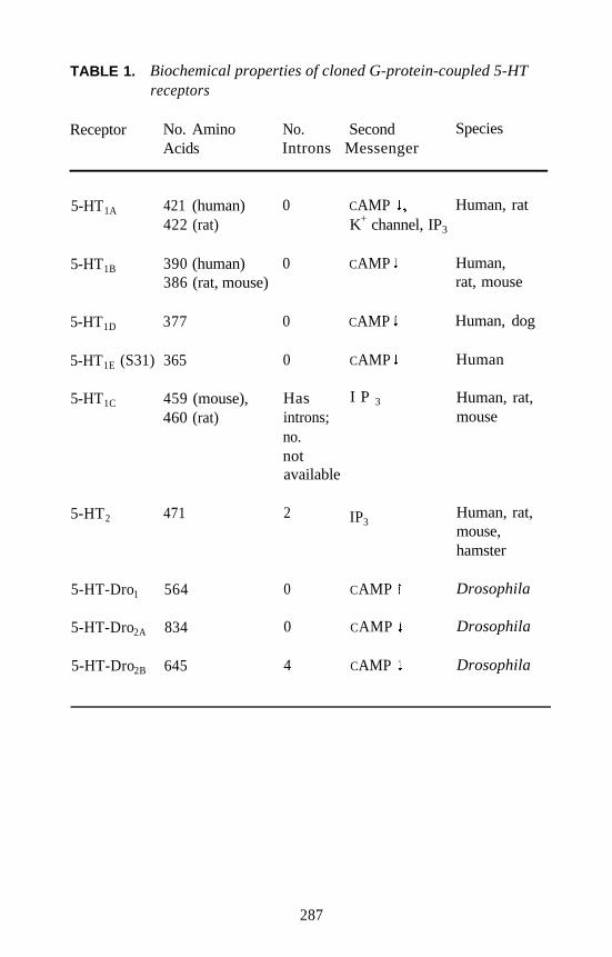
TABLE 1. Biochemical properties of cloned G-protein-coupled 5-HTreceptors
Receptor No. AminoAcids
No. SecondIntrons Messenger
Species
5-HT1A 421 (human)422 (rat)
5-HT1B 390 (human)386 (rat, mouse)
5-HT1D 377
5-HT1E (S31) 365
5-HT1C 459 (mouse),460 (rat)
5-HT2 471
5-HT-Dro1 564
5-HT-Dro2A 834
5-HT-Dro2B 645
0 CAMP Human, ratK+ channel, IP3
0 CAMP Human,rat, mouse
0 CAMP
0 CAMP
Has I P 3
introns;no.notavailable
Human, dog
Human
Human, rat,mouse
2 IP3Human, rat,mouse,hamster
0 CAMP
0 CAMP
4 CAMP
Drosophila
Drosophila
Drosophila
287
Traditionally, classification of receptors has been along lines of sharedligands, for example, the serotonin, acetylcholine, or gammaaminobutyric acid (GABA) receptors. However, molecular cloning ofreceptors has demonstrated clearly that protein structural relations are notreflected in ligand specificities of receptors. For example, 5-HT acts onthe G-protein-coupled receptors and the 5-HT3 receptor coupled with anion channel. The observation that a receptor’s functional andevolutionary relationship to other receptors cannot be determined byshared ligands is reinforced by the demonstration of the intronless natureof some 5-HT receptors.
Further evidence arose from the molecular cloning of the human andmouse 5-HT2 receptor genes (Chen et al. 1992; Yang et al. 1992). The5-HT2 receptor genes contain three exons for the coding region of thereceptor (figure 1). This was the first demonstration of an introncontaining the 5-HT receptor gene: The 5-HT1A, 5-HTB, 5-HTD, and 5-HTE
receptors are intronless. The 5-HT1C, (Yu et al. 1991) and 5-HT2 receptorshave a high degree of sequence homology, and both contain introns. Onthe other hand, 5-HT1A, 5-HT1B, and 5-HT1D, receptors are intronless, as isthe ß-AR. The intronless 5-HT receptors are clearly more closely relatedto the P-AR than to the 5-HT1C, and 5-HT2, receptors, based on sequencehomology and gene structure, but share activating ligand 5-HT with the5-HT1C, and 5-HT2, receptors, not the more closely related P-AR.
Thus, in many cases the use by two receptors of the same activatingligand is seen as a convergent evolution that does not reflect a commonancestral source gene. More anomalies are presented by the structures ofDrosophila 5-HT receptor genes when evolutionary relationships areexamined. Three Drosophila 5-HT receptors have been cloned. Theprimary structures of these genes indicate that they are more closelyrelated to the intronless 5-HT/ß-AR group, but one of these three genesin Drosophila contains introns. The intron organization is different fromthat of the 5-HT2 gene. The questions concerning 5-HT gene structureand functional and evolutionary relationships—when and where intronswere acquired or lost-are open and amenable to further study.
288
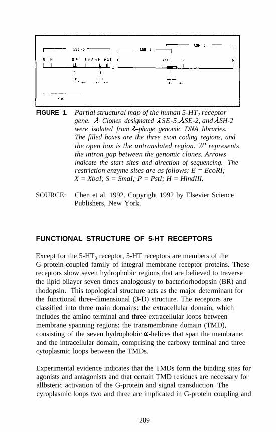
FIGURE 1. Partial structural map of the human 5-HT2 receptorgene. Clones designated SE-5, SE-2, and SH-2were isolated from -phage genomic DNA libraries.The filled boxes are the three exon coding regions, andthe open box is the untranslated region. '//’ representsthe intron gap between the genomic clones. Arrowsindicate the start sites and direction of sequencing. Therestriction enzyme sites are as follows: E = EcoRI;X = XbaI; S = SmaI; P = PstI; H = HindIII.
SOURCE: Chen et al. 1992. Copyright 1992 by Elsevier SciencePublishers, New York.
FUNCTIONAL STRUCTURE OF 5-HT RECEPTORS
Except for the 5-HT3 receptor, 5-HT receptors are members of theG-protein-coupled family of integral membrane receptor proteins. Thesereceptors show seven hydrophobic regions that are believed to traversethe lipid bilayer seven times analogously to bacteriorhodopsin (BR) andrhodopsin. This topological structure acts as the major determinant forthe functional three-dimensional (3-D) structure. The receptors areclassified into three main domains: the extracellular domain, whichincludes the amino terminal and three extracellular loops betweenmembrane spanning regions; the transmembrane domain (TMD),consisting of the seven hydrophobic -helices that span the membrane;and the intracellular domain, comprising the carboxy terminal and threecytoplasmic loops between the TMDs.
Experimental evidence indicates that the TMDs form the binding sites foragonists and antagonists and that certain TMD residues are necessary forallbsteric activation of the G-protein and signal transduction. Thecyroplasmic loops two and three are implicated in G-protein coupling and
289
are necessary for activation of the G-protein and conferring the specificG-protein/receptor coupling. Most of the knowledge of the structure andfunction of the G-protein receptors comes from studies of adrenergic andmuscarinic receptors. The general principles of their structure andfunction mentioned above are expected to be true also for 5-HT receptorsand other G-protein receptors, but as yet little work has been done on the5-HT receptors. The authors’ laboratory has begun an analysis of the rat5-HT2 receptor using site-directed mutagenesis and the known primarystructures of 5-HT2 receptors from different species.
MUTAGENESIS OF ASPARTIC ACID RESIDUES IN THE 5-HT2
RECEPTOR-G-PROTEIN COUPLING
Three aspartic acid (Asp) residues of the rat 5-HT2 receptor were mutatedto asparagine (Asn) residues to determine the role of the negativelycharged aspartate on receptor function. The mutated residues wereAsp- 120, Asp-l 55, and Asp- 172. The effects of the mutations wereanalyzed by using [125I]-LSD for radioligand binding studies to determineagonist and antagonist binding properties and by measuring agonist-stimulated [3H]polyphosphoinositol formation in the mutant and wild-type receptors.
The results indicate that Asp-120 is necessary for the activation of secondmessengers through the G-protein. Mutation of this residue in the secondTMD abolishes 5-HT-stimulated phosphoinositol (PI) turnover. Thismutation also confers a loss of affinity for 5-HT and (±)-1-(2,5-dimeth-oxy-4-iodophenyl)-2-aminopropane (DOI) binding to the receptor,comparable with the loss of affinity seen in wild-type receptors caused bythe presence of guanosine triphosphate (GTP) that induces uncoupling ofthe receptor G-protein complex and a lower affinity state for agonist. Theeffect of GTP on the binding of 5-HT and DOI is greatly reduced in thismutation. Little difference is seen in DOI and 5-HT binding in thepresence of GTP, whereas the wild-type receptor affinity is greatlyreduced because of the presence of GTP. These results are consistentwith Asp- 120 being necessary for G-protein/receptor allostericity wherethe receptor activates the G-protein and the G-protein/receptor complexconfers the high-affinity agonist binding state to the receptor. Whenuncoupled because of GTP, the receptor exhibits lower affinity agonistbinding. The Asp-120 mutation is seen as an uncoupled mutant that isunable to activate second messenger production because of its inability toallosterically modulate the G-protein. This uncoupling may be caused by
290
a loss of allosteric effects conferred by the loss of the negatively chargedaspartate, rather than the loss of a direct interaction between Asp-120 andthe G-protein, because the position of Asp-120 in the lipid bilayer TMDof the protein makes a direct contact unlikely.
The result of this mutation correlates well with mutagenesis of the P-AR,-adrenergic receptor ( AR), and muscarinic receptors where the
cognate Asp in the TMD of these receptors also played the same role.The conserved structure of G-protein receptors where an aspartic acidoccurs in the hydrophobic second TMD suggests that this is an allostericfeature retained by all members of the G-protein-coupled receptor family.
AGONIST-BINDING SITE
Mutagenesis implicates Asp-155 in the third TMD as being necessary forhigh-affinity agonist and antagonist binding. Mutation of this residue toAsn causes a profound loss of binding affinity for the agonists 5-HT andDOI, as determined by competition for [125I]-LSD binding, and alsodecreases antagonist affinity. Phosphoinositide turnover is not abolishedin this mutant, and GTP-sensitive binding is retained. The role of thisnegative amino acid in the hydrophobic third TMD is presumably to actas a counter ion for the amine group of the ligands. As a negative aminoacid, Asp-155 acts as one of the epitopes for ligand binding but not as adeterminant for G-protein coupling.
These observations parallel those seen for the adrenergic receptors andthe muscarinic receptor. The conserved nature of the Asp residue in thethird TMD in receptors for amine-containing ligands (e.g., adrenergic,muscarinic, dopaminergic, and serotonergic receptors) and its absence inreceptors whose ligands do not contain aliphatic amines, support aconserved role in binding in the aliphatic amine receptors such as the5-HT2 receptor.
Another putative binding epitope for 5-HT association with the 5-HT2
receptor is serine (Ser)-239 in the fifth TMD. The difference in primarystructure of the rat and human 5-HT2 receptors has been advantageous inexamining this possibility. The human 5-HT2 receptor and rat receptorare highly homologous in their amino acid structure. Both containSer-239, but one of the three differences in the TMD sequence is seen atposition 242 in the fifth TMD, where the human receptor expresses a Serand the rat receptor expresses an alanine (Ala). The Ser-239 is predicted
291
to act as a hydrogen-bonding site for serotonin’s 5-hydroxyl group inboth rat and human 5-HT2 receptors.
Ser-242 of the human receptor presents another possible hydrogen bonddonor, whereas Ala-242 of the rat receptor does not. The helical structureof the fifth TMD indicates that a hydroxy group at the 4-position of theindole ring could interact with Ser-242 of the human receptor with thesame geometry as 5-HT interacting with Ser-239. Therefore, a4-hydroxyl serotonin analog should bind with higher affinity to thehuman receptor than to the rat receptor.
The hallucinogenic drugs 4-hydroxydimethyltryptamine (psilocin) and5-hydroxydimethyltryptamine (bufotenin), both amino-dimethylatedanalogs of 5-HT, were used to examine this possibility. Psilocin bound tohuman receptors with a fifteenfold higher affinity than to rat receptors,whereas bufotenin bound to both species’ receptors with equal affinity(less than twofold difference in dissociation constants). These resultsindicate the hydoxyl substituents serve as binding epitopes and thatSer-239 associates with 5-hydroxy groups as in serotonin.
The human receptor with the Ser-242 can bind 4-hydroxytryptaminessuch as psilocin with high affinity, but other 5-HT2 receptors that expressAla-242 do not. These results are also consistent with observationsconcerning the adrenergic receptors and their interactions with ligandhydroxy groups. The conservation of hydroxyl-containing amino acids inthe fifth TMD of receptors for agonists, with hydroxyl substituents(e.g., serotonin), but not in those without, also supports this analysis.
TOPOLOGICAL STRUCTURE
The role of Asp-172 at the interface of the third TMD and the secondcytoplasmic loop is not clearly defined by mutagenesis to asparagine.This mutation reduces the binding affinity of agonists and antagonists;decreases the magnitude of GTP-induced binding affinity changes, butdoes not abolish it; and does not abolish 5-HT-stimulated PI production.These results could be interpreted to indicate that Asp-172 may serve asan epitope for the ligand amine groups as suggested for Asp-155.However, the inconsistency of mutagenesis results in other G-proteinreceptors indicates Asp-172 may serve another role in 5-HT2 receptorstructure. When mutated, cognate aspartic acids sometimes abolishsecond messenger stimulation and sometimes do not, depending on the,
292
receptor. Asp-172 and its cognates are found in virtually all G-proteinreceptors, regardless of the structure of their ligand, in a three amino acidmotif of Asp-Arg-Tyr (aspartic acid-arginine-tyrosine [DRY]). Someopsin proteins express the functionally analogous motif of Glu-Arg-Tyr(ERY).
It is possible that this motif serves as a topological determinant for thepositioning of the third TMD in the lipid bilayer. Charged amino acidshave been shown to serve this function in other membrane proteins (Boydand Beckwith 1989). Mutation of Asp-172 would thus result in achanged topography and affect the global 3-D structure of the receptor.This mutation would account for lowered affinities seen for ligandbinding and can account for the loss of G-protein coupling in somereceptors but not others. The global changes induced by this mutationwould slightly change the binding site geometry, conferring the observedlower affinity binding. In some receptors the topological differencewould affect G-protein coupling, whereas in others such as the 5-HT2
receptor the change would not affect such a coupling. Different receptorscouple with different G-proteins, so this interpretation is consistent withthe known properties of G-proteins and the nature of membrane proteintopological determinants.
FUTURE
It is evident that 5-HT2 receptors and other G-proteins are structurallycomplex and that the mutagenesis studies of these receptors and proteinshave just begun to address their structure-function relationship. Manymore studies combining mutagenesis with biophysical investigations canreveal the molecular mechanisms of their function. Currently,mutagenesis yields the most information regarding the structure of thereceptors and provides the most easily accessible, although indirect butpowerful, method to study the structure and function of 5-HT receptors.
Biophysical studies that can provide direct information concerningstructure and function are hampered by the unavailability of purifiedreceptor preparations. For example, fluorescent ligand studies usingpurified reconstituted ß-AR have shown the binding site to reside in thehydrophobic TMD of the receptor, a finding consistent with the results ofmutagenesis. Because of the low percentage of 5-HT2 receptor proteinexpressed in brain tissues, the receptors are difficult to purify in any largequantity. To overcome this problem, alternate expression systems need
293
to be developed in microorganisms such as yeast or bacteria to provideample source materials for purification.
Detailed knowledge of the structure and function of the 5-HT2 receptorobtained by a variety of methods will provide a basis for designing newtherapeutic agents for treating diseases associated with 5-HT2 receptors.
The isolation of the carrier deoxyribonucleic acid (cDNA) and genomicclones of 5-HT2 receptors provides more than just a basis for the study oftheir molecular structure and function. The possibility that polymorph-isms in 5-HT2 receptor genes may contribute to disease states can beexamined. Any polymorphisms in the gene can be examined usingcDNA or gene probes. If a polymorphism is found to be associated witha disease state, then the DNA sequences can provide possible diagnostictools for these states.
The most exciting future project is the regulation of the 5-HT2 receptorgene expression. The promoter region of a gene consists of the corepromoter, enhancer, and repressor. The core promoter is the regioncontrolling the transcription of the gene and is regulated by enhancers orrepressors. These DNA sequences bind specific protein transcriptionfactors to fine-tune gene expression. An example of such geneexpression regulatory factor is SP-1 proteins known to be active in theregulation of monoamine oxidase A gene expression,
Researchers are at an exciting point where they can use molecularbiological techniques to investigate the mechanisms for regulation of the5-HT2 receptor gene. It is now possible to investigate whetherhallucinogenic agents play a role in the gene expression by influencingeither cis element or trans-element.
Moreover, it is interesting to note that there may be tissue-specificelements in the 5-HT2 receptor promoter. Once brain-specific elementsare identified, it would be possible to manipulate the expression of thereceptor only in the brain to better understand the function of brain 5-HT2
receptors and their relation to hallucinogenic effects. This newknowledge will help researchers to design therapeutic agents that arebrain specific. Any mutations in this region may affect the expression ofthe receptor and contribute to diseases. The molecular cloning of the5-HT2 receptor gene allows many possibilities to be examined and cancontribute to the knowledge and treatment of mental disease or otherserotonin-related illnesses.
294
REFERENCES
Boyd, D., and Beckwith, J. Positively charged amino acid residues canact as topogenic determinants in membrane proteins. Proc Natl AcadSci U S A 86:9446-9450, 1989.
Chen, K.; Yang, W.; Grimsby, J.; and Shih, J.C. The human 5-HT2
receptor is encoded by a multiple intron-exon gene. Mol Brain Res14:20-26, 1992.
Conn, P.J., and Sanders-Bush, E. Regulation of serotonin-stimulatedphosphoinositide hydrolysis: Relation to the serotonin 5-HT2 bindingsite. J Neurosci 6:3669-3675, 1986.
Fargin, A.; Raymond, J.R.; Lohse, M.J.; Kobilka, B.K.; Cat-on, M.G.; andLefkowitz, R.J. The genomic clone of G-21 which resembles aß-adrenergic receptor sequence encodes the 5-HT1A receptor. Nature335:358-360, 1988.
Gaddum, J.H., and Picarelli, Z.P. Two kinds of tryptamine receptors. Br JPharmacol Chemother 12:323-328, 1957.
Hofmann, A. Chemistry of LSD. In: Sankar, D., ed. LSD: A Total Study.Westbury, NY: PJD Publications, 1975. pp. 107-139.
Julius, D.; MacDermot, A.B.; Axel, R.; and Jessell, T.M. Molecularcharacterization of a functional cDNA encoding the serotonin 1C
receptor. Science 241:558-564, 1988.Maricq, A.V.; Peterson, A.S.; Brake, A.J.; Myers, R.M.; and Julius, D.
Primary structure and functional expression of the 5-HT3 receptor, aserotonin-gated ion channel. Science 254:432-437, 1991.
Meltzer, H.Y., and Lowy, M.T. The serotonin hypothesis of depression.In: Meltzer, H.Y., ed. Psychopharmacology: The Third Generation ofProgress. New York: Raven Press, 1987. pp. 513-526.
Peroutka, S.J., and Snyder, S.H. Multiple serotonin receptors: Differentialbinding of [3H]5-hydroxytryptamine, [3H]lysergic acid diethylamideand [3H]spiroperidol. Mol Pharmacol 16:687-699, 1979.
Pierce, P.A., and Peroutka, S.J. Hallucinogenic drug interactions withneurotransmitter receptor binding sites in human cortex.Psychopharmacology 97:118-122, 1989.
Pritchett, D.B.; Bach, A.W.J.; Wozny, M.; Taleb, O.; Dal Toso, R.; Shih,J.C.; and Seeberg, P.H. Structure and functional expression of clonedrat serotonin 5-HT2 receptor. EMBO J 7:4135-4140, 1988.
Sadzot, B.; Baraban, J.M.; Glennon, R.A.; Lyon, R.A.; Leonhardt, S.;Jan, C.-R.; and Titeler, M. Hallucinogenic drug interactions at humanbrain 5-HT2 receptors: Implications for treating LSD-inducedhallucinogenesis. Psychopharmacology 98:495-499, 1989.
295
Schultes, R.E. The appeal of Peyote (Lophophora williamsii) as amedicine. Am Anthropologist 40:698-715, 1938.
Schultes, R.E. An overview of hallucinogens in the western hemisphere.In: Furst P., ed. Flesh of the Gods: The Ritual Use of Hallucinogens.New York: Praeger, 1972. pp. 3-54.
Schultes, R.E., and Hofmann, A. The Botany and Chemistry ofHallucinogens. Springfield, IL: Charles C. Thomas, 1973.
Shih, J.C.; Yang, W.; Chen, K., and Gallaher, T. Molecular biology ofserotonin (5-HT) receptors. Pharmacol Biochem Behav 40:1053-1058,1991.
Shoichet, R., and Solursh, L. Treatment of the hallucinogenic drug crisis.Appl Therapeutics 11:283-286, 1969.
Sternbach, H. The serotonin syndrome. Am J Psychiatry 148:705-713,1991.
Vanhoutte, P.M. Does 5-hydroxytryptamine play a role in hypertension?Trends Pharmacol Sci 3:370-373, 1982.
Yang, W.; Chen, K.; Lan, N.C.; Gallaher, T.K.; and Shih, J.C. Genestructure and expression of the mouse 5-HT2 receptor. J Neurosci Res33:196-204, 1992.
Yu, L.; Nguyen, H.; Bloem, L.J.; Kozak, C.A.; Hoffman, B.J.; Snutch,T.P.; Lester, H.A.; Davidson, N.; and Lubbert, H. The mouse 5-HT1C
receptor contains eight hydrophobic domains and is X-linked. MolBrain Res 11:143-149, 1991.
ACKNOWLEDGMENTS
This work was supported by National Institute of Mental Health grantsR01-MH37020, R37-MH39085 (Merit Award), and Research ScientistAward K05-MH00796. The support from the Boyd and Elsie WelinProfessorship is also appreciated.
296
AUTHORS
Jean C. Shih, Ph.D.Boyd and Elsie Welin Professor
Kevin Chen, Ph.D.Research Associate Professor
Timothy K. Gallaher, Ph.D.Research Associate
Department of Molecular Pharmacology and ToxicologyUniversity of Southern California School of Pharmacy1985 Zonal AvenueLos Angeles, CA 90033
297
SummaryRichard A. Glennon
It is perhaps appropriate that the National Institute on Drug Abuse(NIDA) sponsored a technical review on hallucinogenic agents during the500th anniversary of Columbus’ sailing to the New World, because it isreportedly Columbus who wrote the first account of the use ofpsychoactive (presumably tryptamine- or ß-carboline-containing) plantsin this hemisphere (Holmstedt and Lindgren 1967; Wassen 1967).
This meeting was the first technical review devoted solely to classicalhallucinogens and covered the exciting progress being made inresearchers’ understanding of this class of agents. Just before coming tothis meeting, the author reread the monographs from the NIDA technicalreviews of hallucinogenic agents held in 1976 and 1978. An enormousamount of progress has been made on some fronts, whereas more needsto be done on others. The earlier two meetings were primarily concernedwith the identification of new chemical entities and the development ofnovel pharmacological techniques and animal models to investigatehallucinogenic agents. For example, both of those reviews were heldbefore the widespread use of radioligand binding and autoradiography,the discovery of multiple populations of central serotonin (5-HT)receptors, elucidation of second messenger systems, molecular graphics,and receptor cloning. Consequently the present meeting addressed topicsand issues and described technologies that were essentially unknown atthe time of the prior meetings. Serotonin, (5-HT2) receptors wereoriginally identified in 1979 and were first implicated as playing amechanistic role in the actions of hallucinogens in 1983. Much of thefocus of the present meeting was on 5-HT2 receptors.
There has been, and continues to be, dissatisfaction with the termhallucinogen; not all classical hallucinogens produce frankhallucinations. This point was emphasized by the first several speakers,including Szára, Glennon, and Shulgin. Szára presented a historicalperspective of the different phases of research with classicalhallucinogens over the years and concluded with the suggestion that theseagents be referred to as psychoheuristic agents. Many of the attendeesagreed that a new terminology would be useful, but there was noconsensus on any specific term.
298
After an introductory overview on classical hallucinogens by Glennon,Shulgin and Strassman presented papers on the human evaluation,including qualitative and quantitative aspects, of hallucinogens.Shulgin’s presentation was broad in scope, of a review nature, andcovered many examples of the classical hallucinogens. In contrast,Strassman described in depth his recent clinical studies on one specificagent, N,N-dimethyltryptamine (DMT). Both speakers commented on aneed for the availability of more human data; this feeling was shared bymany of the participants.
During the overview, it was mentioned that structure-activityrelationships (SAR) have been formulated for certain subclasses of theclassical hallucinogens but that such information is rather limited withregard to ergoline derivatives. Nichols presented the results of his timelySAR studies on ergolines, with specific emphasis on the nature ofsubstituents at the 6- and 8-positions.
The next two speakers, Geyer and Winter, presented new data andreviewed some of the older results using several animal techniques thathave been employed to study a large number of classical hallucinogensand structurally related agents. It might be noted that these techniqueswere scarcely mentioned during the 1976 and 1978 meetings.
Aghajanian outlined his work on the electrophysiology of hallucinogensand addressed the issue of whether these agents act as agonists,antagonists, or partial agonists. Depending upon what cells areexamined, it appears that hallucinogens behave as agonists or partialagonists; in some cells, there is evidence that certain hallucinogens appearto have a significantly greater intrinsic activity than 5-HT itself. Sanders-Bush also addressed this issue and further described her secondmessenger studies attempting to find differences in the actions of agentsat 5-HT2 and 5-HT1C receptors. Although hallucinogens bind at bothpopulations of receptors, it is not yet known which (or whether both) areinvolved in their mechanism of action. Sanders-Bush showed that(+)LSD is a partial agonist, and the non-hallucinogenic 2-bromo-LSD(BOL) is a pure antagonist both at 5-HT2 and 5-HT1C receptors, whereaslisuride is a pure antagonist at 5-HT1C receptors but a partial agonist at5-HT2 receptors. Lisuride could become a useful tool to further study therole of 5-HT1C versus 5-HT2 receptors.
The locus of action of hallucinogens in the brain is unknown. With therecent availability of radiolabeled hallucinogens and hallucinogen
299
analogs, it should eventually be possible to identify such loci. Appeldescribed his (and others’) work using autoradiography, positronemission tomography (PET) scanning, and other imaging techniques tolabel 5-HT2 and 5-HT1C receptors, and apprised the participants of newtechniques currently being developed.
After discussion about human and animal evaluations and studies at themolecular, receptor, and second messenger level, the concluding sessionwas devoted to investigations at the atomic level. Weinstein andWestkaemper presented their attempts to develop molecular graphicsmodels of 5-HT2 receptors. Both models begin with the known structureof bacteriorhodopsin, but each investigator described a unique approachto the problem. Weinstein presented a model to account for the actions ofagonists, antagonists, and partial agonists, whereas Westkaemperidentified several specific amino acid residues that could be important forligand binding. One such key residue is the aspartate of transmembranehelix III. The models, although hypothetical, provide a framework forfuture investigation. Indeed, both models have been recently revised(Westkaemper and Glennon 1993; Zhang and Weinstein 1993). Shihoutlined her work with the cloning of 5-HT2 receptors and with site-directed mutagenesis. Mutation of the above mentioned aspartate toarginine reduces significantly the affinity of 5-HT2 ligands, supporting arole for this aspartate moiety in a binding interaction, Additional site-directed mutagenesis is required to further test the receptor models.
Again, it should be emphasized that the present meeting was very timely.Certain philosophical issues such as terminology and explicit definitionof the actions of classical hallucinogens remain undefined but, whileimportant, have not detracted from other scientific studies. It is realizedthat animal models have severe limitations but provide useful data; this isparticularly true when human data are hard to come by. Nevertheless,there was a consensus that there is an urgent need for new human testing.There is also a realization that certain agents are enigmatic in that they areroutinely identified as being “active” in certain animal models eventhough human data (scant as they may be in some instances) suggest thatthey are not hallucinogenic. Hopefully, these agents will be used tochallenge other animal models, and, conversely, the agents themselvesmay become the subject of additional investigations.
The identification of 5-HT2 (perhaps 5-HT1C) receptors as possibly beinginvolved in the action of hallucinogens has provided a focal point for newstudies. State-of-the-art techniques and methodologies have been brought
300
to bear to investigate this possibility. There is no question thathallucinogens interact at both populations of receptors (regardless ofwhether this is their site of action); thus, hallucinogens are providing astimulus for the investigation of these, and other, populations ofreceptors.
Legitimate human investigation with classical hallucinogens was severelycurtailed about 25 years ago. During the ensuing period, a significantbody of information has been accrued primarily on the basis of animalstudies. Novel agents have been identified, mechanisms of action havebeen proposed, new animal models have been developed, and means toantagonize the effects of classical hallucinogens have been described.New clinical data are now required to challenge or validate the results ofthese studies.
REFERENCES
Holmstedt, B., and Lindgren, J. Chemical constituents and pharmacologyof South American snuffs. In: Efron, D.H.; Holmstedt, B.; and Kline,N.S., eds. Ethnopharmacological Search for Psychoactive Drugs.National Institutes of Mental Health, Washington, DC: Supt. of Docs.,U.S. Govt. Print. Off., 1967. pp 339-373.
Wassen, S.H. Anthropological survey of the use of South Americansnuffs. In: Efron, D.H.; Holmstedt, B.; and Kline, N.S., eds.Ethnopharmacological Search for Psychoactive Drugs. NationalInstitutes of Mental Health, Washington, DC: U.S. Govt. Print. Off.,1967. pp 233-289.
Westkaemper, R.B., and Glennon, R.A. Molecular graphics models ofmembers of the 5-HT2 subfamily: 5-HT2A, 5-HT2B, and 5-HT2C,receptors. Med Chem Res 3:317-324, 1993.
Zhang, D., and Weinstein,, H. Ligand selectivity and the molecularproperties of the 5-HT2 receptor: Computational simulations reveal amajor role for transmembrane helix 7. Med Chem Res 3:357-369,1993.
301
While limited supplies last, single copies of the monographs may beobtained free of charge from the National Clearinghouse for Alcohol andDrug Information (NCADI). Please also contact NCADI for informationabout availability of coming issues and other publications of the NationalInstitute on Drug Abuse relevant to drug abuse research.
Additional copies may be purchased from the U.S. Government PrintingOffice (GPO) and/or the National Technical Information Service (NTIS)as indicated. NTIS prices are for paper copy; add $3.00 handling chargefor each order. Microfiche copies also are available from NTIS. Pricesfrom either source are subject to change.
Addresses are:
NCADINational Clearinghouse for Alcohol and Drug Information
P.O. Box 2345Rockville, MD 20852
(301) 468-2600(800) 729-6686
GPOSuperintendent of Documents
U.S. Government Printing OfficeP.O. Box 371954
Pittsburgh, PA 15220-7954(202) 738-3238
FAX (202) 512-2233
NTISNational Technical Information Service
U.S. Department of CommerceSpringfield, VA 22161
(703) 487-4650
For information on availability of NIDA Research Monographs from1975-1993 and those not listed, write to NIDA, Community andProfessional Education Branch, Room 10A-39, 5600 Fishers Lane,Rockville, MD 20857.
302
26
42
50
52
53
54
56
61
62
THE BEHAVIORAL ASPECTS OF SMOKING. Norman A.Krasnegor, Ph.D., ed. (Reprint from 1979 Surgeon General’sReport on Smoking and Health.)NCADI#M26 NTIS PB #80-118755/AS (A09) $27.00
THE ANALYSIS OF CANNABINOIDS IN BIOLOGICALFLUIDS. Richard L. Hawks, Ph.D., ed.NCADI #M42 NTIS PB #83-136044/AS (A07) $27.00
COCAINE: PHARMACOLOGY, EFFECTS, ANDTREATMENT OF ABUSE. John Grabowski, Ph.D., ed.NCADI #M50 NTIS PB #85-150381/AS (A07) $27.00
TESTING DRUGS FOR PHYSICAL DEPENDENCEPOTENTIAL AND ABUSE LIABILITY. Joseph V. Brady, Ph.D.,and Scott E. Lukas, Ph.D., eds.NCADI #M52 NTIS PB #85-150373/AS (A08) $27.00
PHARMACOLOGICAL ADJUNCTS IN SMOKINGCESSATION. John Grabowski, Ph.D., and Sharon M. Hall, Ph.D.,eds.NCADI #M53 NTIS PB #89-123186/AS (A07) $27.00
MECHANISMS OF TOLERANCE AND DEPENDENCE.Charles Wm. Sharp, Ph.D., ed.NCADI #M54 NTIS PB #89-103279/AS (A19) $52.00
ETIOLOGY OF DRUG ABUSE: IMPLICATIONS FORPREVENTION. Coryl LaRue Jones, Ph.D., and Robert J. Battjes,D.S.W., eds.NCADI #M56 NTIS PB #89-123160/AS (A13) $36.50
COCAINE USE IN AMERICA: EPIDEMIOLOGIC ANDCLINICAL PERSPECTIVES. Nicholas J. Kozel, M.S., and EdgarH. Adams, MS., eds.NCADI #M61 NTIS PB #89-131866/AS (Al 1) $36.50
NEUROSCIENCE METHODS IN DRUG ABUSE RESEARCH.Roger M. Brown, Ph.D., and David P. Friedman, Ph.D., eds.NCADI #M62 NTIS PB #89-130660/AS (A08) $27.00
303
63
64
65
69
70
72
74
78
79
PREVENTION RESEARCH: DETERRING DRUG ABUSEAMONG CHILDREN AND ADOLESCENTS. Catherine S. Bell,M.S., and Robert J. Battjes, D.S.W., eds.NCADI #M63 NTIS PB #89-103287/AS (A11) $36.50
PHENCYCLIDINE: AN UPDATE. Doris H. Clouet, Ph.D., ed.NCADI #M64 NTIS PB #89-131858/AS (A12) $36.50
WOMEN AND DRUGS: A NEW ERA FOR RESEARCH.Barbara A. Ray, Ph.D., and Monique C. Braude, Ph.D., eds.NCADI #M65 NTIS PB #89-130637/AS (A06) $27.00
OPIOID PEPTIDES: MEDICINAL CHEMISTRY. Rao S.Rapaka, Ph.D.; Gene Barnett, Ph.D.; and Richard L. Hawks, Ph.D.,eds.NCADI #M69 NTIS PB #89-158422/AS (A17) $44.50
OPIOID PEPTIDES: MOLECULAR PHARMACOLOGY,BIOSYNTHESIS, AND ANALYSIS. Rao S. Rapaka, Ph.D., andRichard L. Hawks, Ph.D., eds.NCADI #M70 NTIS PB #89-15843O/AS (A18) $52.00
RELAPSE AND RECOVERY IN DRUG ABUSE. Frank M.Tims, Ph.D., and Carl G. Leukefeld, D.S.W., eds.NCADI #M72 NTIS PB #89-151963/AS (A09) $36.50
NEUROBIOLOGY OF BEHAVIORAL CONTROL IN DRUGABUSE. Stephen I. Szara, M.D., D.Sc., ed.NCADI #M74 NTIS PB #89-151989/AS (A07) $27.00
THE ROLE OF NEUROPLASTICITY IN THE RESPONSE TODRUGS. David P. Friedman, Ph.D., and Doris H. Clouet, Ph.D.,eds.NCADI #M78 NTIS PB #88-245683/AS (A10) $36.50
STRUCTURE-ACTIVITY RELATIONSHIPS OF THECANNABINOIDS. Rao S. Rapaka, Ph.D., and AlexandrosMakriyannis, Ph.D., eds.NCADI #M79 NTIS PB #89-109201/AS (A10) $36.50
304
80 NEEDLE SHARING AMONG INTRAVENOUS DRUGABUSERS: NATIONAL AND INTERNATIONALPERSPECTIVES. Robert J. Battjes, D.S.W., and Roy W. Pickens,Ph.D., eds.NCADI #M80 NTIS PB #88-236138/AS (A09) $36.50
82 OPIOIDS IN THE HIPPOCAMPUS. Jacqueline F. McGinty,Ph.D., and David P. Friedman, Ph.D., eds.NCADI #M82 NTIS PB #88-245691/AS (A06) $27.00
83 HEALTH HAZARDS OF NITRITE INHALANTS. Harry W.Haverkos, M.D., and John A. Dougherty, Ph.D., eds.NCADI #M83 NTIS PB #89-125496/AS (A06) $27.00
84 LEARNING FACTORS IN SUBSTANCE ABUSE. Barbara A.Ray, Ph.D., ed.NCADI #M84 NTIS PB #89-125504/AS (A10) $36.50
85 EPIDEMIOLOGY OF INHALANT ABUSE: AN UPDATE.Raquel A. Crider, Ph.D., and Beatrice A. Rouse, Ph.D., eds.NCADI #M85 NTIS PB #89-123178/AS (A10) $36.50
87 OPIOID PEPTIDES: AN UPDATE. Rao S. Rapaka, Ph.D., andBhola N. Dhawan, M.D., eds.NCADI #M87 NTIS PB #89-158430/AS (A11) $36.50
88 MECHANISMS OF COCAINE ABUSE AND TOXICITY. DorisH. Clouet, Ph.D.; Khursheed Asghar, Ph.D.; and Roger M. Brown,Ph.D., eds.NCADI #M88 NTIS PB #89-125512/AS (A16) $44.50
89 BIOLOGICAL VULNERABILITY TO DRUG ABUSE. Roy W.Pickens, Ph.D., and Date S. Svikis, B.A., eds.NCADI #M89 NTIS PB #89-125520/AS (A09) $27.00
92 TESTING FOR ABUSE LIABILITY OF DRUGS IN HUMANS.Marian W. Fischman, Ph.D., and Nancy K. Mello, Ph.D., eds.NCADI #M92 NTIS PB #90-148933/AS (A17) $44.50
305
94
95
96
97
98
99
100
101
PHARMACOLOGY AND TOXICOLOGY OF AMPHETAMINEAND RELATED DESIGNER DRUGS. Khursheed Asghar, Ph.D.,and Errol De Souza, Ph.D., eds.NCADI #M94 NTIS PB #90-148958/AS (A16) $44.50
PROBLEMS OF DRUG DEPENDENCE, 1989. PROCEEDINGSOF THE 51st ANNUAL SCIENTIFIC MEETING. THECOMMITTEE ON PROBLEMS OF DRUG DEPENDENCE, INC.Louis S. Harris, Ph.D., ed.NCADI #M95 NTIS PB #90-237660/AS (A99) $67.00
DRUGS OF ABUSE: CHEMISTRY, PHARMACOLOGY,IMMUNOLOGY, AND AIDS. Phuong Thi Kim Pham, Ph.D., andKenner Rice, Ph.D., eds.NCADI #M96 NTIS PB #90-237678/AS (A11) $36.50
NEUROBIOLOGY OF DRUG ABUSE: LEARNING ANDMEMORY. Lynda Erinoff, Ph.D., ed.NCADI #M97 NTIS PB #90-237686/AS (A11) $36.50
THE COLLECTION AND INTERPRETATION OF DATAFROM HIDDEN POPULATIONS. Elizabeth Y. Lambert, M.S.,ed.NCADI #M98 NTIS PB #90-237694/AS (A08) $27.00
RESEARCH FINDINGS ON SMOKING OF ABUSEDSUBSTANCES. C. Nora Chiang, Ph.D., and Richard L. Hawks,Ph.D., eds.NCADI #M99 NTIS PB #91-141119 (A09) $27.00
DRUGS IN THE WORKPLACE: RESEARCH ANDEVALUATION DATA. VOL II. Steven W. Gust, Ph.D.;J. Michael Walsh, Ph.D.; Linda B. Thomas, B.S.; and Dennis J.Crouch, M.B.A., eds.NCADI #M 100 GPO Stock #017-024-01458-3 $8.00
RESIDUAL EFFECTS OF ABUSED DRUGS ON BEHAVIOR.John W. Spencer, Ph.D., and John J. Boren, Ph.D., eds.NCADI #M101 NTIS PB #91-172858/AS (A09) $27.00
306
102 ANABOLIC STEROID ABUSE. Geraline C. Lin, Ph.D., andLynda Erinoff, Ph.D., eds.NCADI #M102 NTIS PB #91-172866/AS (A11) $36.50
106 IMPROVING DRUG ABUSE TREATMENT. Roy W. Pickens,Ph.D.; Carl G. Leukefeld, D.S.W.; and Charles R. Schuster, Ph.D.,eds.NCADI #M106 NTIS PB #92-105873(A18) $50.00
107 DRUG ABUSE PREVENTION INTERVENTION RESEARCH:METHODOLOGICAL ISSUES. Carl G. Leukefeld, D.S.W., andWilliam J. Bukoski, Ph.D., eds.NCADI #M107 NTIS PB #92-160985 (A13) $36.50
108 CARDIOVASCULAR TOXICITY OF COCAINE:UNDERLYING MECHANISMS. Pushpa V. Thadani, Ph.D., ed.NCADI #M108 NTIS PB #92-106608 (A11) $36.50
109 LONGITUDINAL STUDIES OF HIV INFECTION ININTRAVENOUS DRUG USERS: METHODOLOGICAL ISSUESIN NATURAL HISTORY RESEARCH. Peter Hartsock, Dr. P.H.,and Sander G. Genser, M.D., M.P.H., eds.NCADI #M109 NTIS PB #92-106616 (A08) $27.00
111 MOLECULAR APPROACHES TO DRUG ABUSE RESEARCH:RECEPTOR CLONING, NEUROTRANSMITTEREXPRESSION, AND MOLECULAR GENETICS: VOLUME I.Theresa N.H. Lee, Ph.D., ed.NCADI #M111 NTIS PB #92-135743 (A10) $36.50
112 EMERGING TECHNOLOGIES AND NEW DIRECTIONS INDRUG ABUSE RESEARCH. Rao S. Rapaka, Ph.D.; AlexandrosMakriyannis, Ph.D.; and Michael J. Kuhar, Ph.D., eds.NCADI #M112 NTIS PB #92-155449 (A15) $44.50
113 ECONOMIC COSTS, COST EFFECTIVENESS, FINANCING,AND COMMUNITY-BASED DRUG TREATMENT. William S.Cartwright, Ph.D., and James M. Kaple, Ph.D., eds.NCADI #M113 NTIS PB #92-155795 (A10) $36.50
307
114 METHODOLOGICAL ISSUES IN CONTROLLED STUDIESON EFFECTS OF PRENATAL EXPOSURE TO DRUG ABUSE.M. Marlyne Kilbey, Ph.D., and Khursheed Asghar, Ph.D., eds.NCADI #M114 NTIS PB #92-146216 (A16) $44.50
115 METHAMPHETAMINE ABUSE: EPIDEMIOLOGIC ISSUESAND IMPLICATIONS. Marissa A. Miller, D.V.M., M.P.H., andNicholas J. Kozel, MS., eds.NCADI #M115 NTIS PB #92-146224/11 (A07) $27.00
116 DRUG DISCRIMINATION: APPLICATIONS TO DRUGABUSE RESEARCH. R.A. Glennon, Ph.D.; Toubjörn U.C. Järbe,Ph.D.; and J. Frankenheim, Ph.D., eds.NCADI #M116 NTIS PB #94-169471 (A20) $52.00
117 METHODOLOGICAL ISSUES IN EPIDEMIOLOGY,PREVENTION, AND TREATMENT RESEARCH ON DRUG-EXPOSED WOMEN AND THEIR CHILDREN. M. MarlyveKilbey, Ph.D., and Kursheed Asghar, Ph.D., eds.
GPO Stock #017-024-01472-g $12.00NCADI #M117 NTIS PB #93-102101/LL (A18) $52.00
118 DRUG ABUSE TREATMENT IN PRISONS AND JAILS. CarlG. Leukefeld, D.S.W., and Frank M. Tims, Ph.D., eds.
GPO Stock #017-024-01473-7 $16.00NCADI #M118 NTIS PB #93-102143/LL (A14) $44.50
120 BIOAVAILABILITY OF DRUGS TO THE BRAIN AND THEBLOOD-BRAIN BARRIER. Jerry Frankenheim, Ph.D., andRoger M. Brown, Ph.D., eds.
NCADI #M120GPO Stock #017-024-01481-8 $10.00
NTIS PB #92-214956/LL (A12) $36.50
121 BUPRENORPHINE: AN ALTERNATIVE TREATMENT FOROPIOID DEPENDENCE. Jack D. Blame, Ph.D., ed.
GPO Stock #117-024-01482-6 $5.00NCADI #M121 NTIS PB #93-129781/LL (A08) $27.00
123 ACUTE COCAINE INTOXICATION: CURRENT METHODSOF TREATMENT. Heinz Sorer, Ph.D., ed.
GPO Stock #017-024-01501-6 $6.50NCADI #M123 NTIS PB #94-15433/LL (A09) $27.00
308
124 NEUROBIOLOGICAL APPROACHES TO BRAIN-BEHAVIORINTERACTION. Roger M. Brown, Ph.D., and Joseph Fracella,Ph.D., eds.
NCADI #M124GPO Stock #017-024-01492-3 $9.00
NTIS PB #93-203834/LL (A12) $36.50
125 ACTIVATION OF IMMEDIATE EARLY GENES BY DRUGSOF ABUSE. Reinhard Grzanna, Ph.D., and Roger M. Brown,Ph.D., eds.
NCADI #M125GPO Stock #017-024-01503-2 $7.50NTIS PB #94-169489 (A12) $36.50
126 MOLECULAR APPROACHES TO DRUG ABUSE RESEARCHVOLUME II: STRUCTURE, FUNCTION, AND EXPRESSION.Theresa N.H. Lee, Ph.D., ed.NCADI #M126 NTIS PB #94-169497 (A08) $27.00
127 PROGRESS AND ISSUES IN CASE MANAGEMENT. RebeccaSager Ashery, D.S.W., ed.NCADI #M127 NTIS PB #94-169505 (A18) $52.00
128 STATISTICAL ISSUES IN CLINICAL TRIALS FORTREATMENT OF OPIATE DEPENDENCE. Ram B. Jain, Ph.D.,ed.NCADI #M128 NTIS PB #93-203826/LL (A09) $27.00
129 INHALANT ABUSE: A VOLATILE RESEARCH AGENDA.Charles W. Sharp, Ph.D.; Fred Beauvais, Ph.D., and RichardSpence, Ph.D., eds.
NCADI #M129GPO Stock #017-024-01496-6 $12.00
NTIS PB #93-183119/LL (A15) $44.50
130 DRUG ABUSE AMONG MINORITY YOUTH: ADVANCES INRESEARCH AND METHODOLOGY. Mario De La Rosa, Ph.D.,and Juan-Luis Recio Adrados, Ph.D., eds.
GPO Stock #017-024-01506-7 $14.00NCADI #M130 NTIS PB #94-169513 (A15) $44.50
309
13 1 IMPACT OF PRESCRIPTION DRUG DIVERSION CONTROLSYSTEMS ON MEDICAL PRACTICE AND PATIENT CARE.James R. Cooper, Ph.D.; Dorynne J. Czechowicz, M.D.; Stephen P.Molinari, J.D., R.Ph.; and Robert C. Peterson, Ph.D., eds.
GPO Stock #017-024-01505-9 $14.00NCADI #M131 NTIS PB #94-169521 (A15) $44.50
132 PROBLEMS OF DRUG DEPENDENCE, 1992: PROCEEDINGSOF THE 54TH ANNUAL SCIENTIFIC MEETING OF THECOLLEGE ON PROBLEMS OF DRUG DEPENDENCE. LouisHarris, Ph.D., ed.
NCADI #M132GPO Stock #017-024-01502-4 $23.00
NTIS PB #94-115508/LL (A99)
133 SIGMA, PCP, AND NMDA RECEPTORS. Errol B. De Souza,Ph.D.: Doris Clouet, Ph.D., and Edythe D. London, Ph.D., eds.NCADI #M133 NTIS PB #94-169539 (A12) $36.50
134 MEDICATIONS DEVELOPMENT: DRUG DISCOVERY,DATABASES, AND COMPUTER-AIDED DRUG DESIGN. RaoS. Rapaka, Ph.D., and Richard L. Hawks, Ph.D., eds.
GPO Stock #017-024-0151 1-3 $11.00NCADI #M134 NTIS PB #94-169547 (A14) $44.50
135 COCAINE TREATMENT: RESEARCH AND CLINICALPERSPECTIVES. Frank M. Tims, Ph.D., and Carl G. Leukefeld,D.S.W., eds.
NCADI #M135GPO Stock #017-024-01520-2 $11.00
NTIS PB #94-1 69554 (A13) $36.50
136 ASSESSING NEUROTOXICITY OF DRUGS OF ABUSE.Lynda Erinoff, Ph.D., ed.
NCADI #M136GPO Stock #017-024-01518-1 $11.00
NTIS PB #94-169562 (A13) $36.50
137 BEHAVIORAL TREATMENTS FOR DRUG ABUSE ANDDEPENDENCE. Lisa Simon Onken, Ph.D.; Jack D. Blame, M.D.,and John J. Boren, Ph.D., eds.
NCADI #M137GPO Stock #017-024-01519-9 $13.00
NTIS PB #94-169570 (A15) $44.50
310
138
139
140
141
142
143
144
145
IMAGING TECHNIQUES IN MEDICATIONSDEVELOPMENT: CLINICAL AND PRECLINICAL ASPECTS.Heinz Sorer, Ph.D., and Rao S. Rapaka, Ph.D., eds.NCADI #M138
SCIENTIFIC METHODS FOR PREVENTION INTERVENTIONRESEARCH. Arturo Cazares, M.D., M.P.H., and Lula A. Beatty,Ph.D., eds.NCADI #M139
PROBLEMS OF DRUG DEPENDENCE, 1993: PROCEEDINGSOF THE 55TH ANNUAL SCIENTIFIC MEETING, THECOLLEGE ON PROBLEMS OF DRUG DEPENDENCE, INC.VOLUME I: PLENARY SESSION SYMPOSIA AND ANNUALREPORTS. Louis S. Harris, Ph.D., ed.NCADI #M140
PROBLEMS OF DRUG DEPENDENCE, 1993: PROCEEDINGSOF THE 55TH ANNUAL SCIENTIFIC MEETING, THECOLLEGE ON PROBLEMS OF DRUG DEPENDENCE, INC.VOLUME II: ABSTRACTS. Louis S. Harris, Ph.D., ed.NCADI #M141
ADVANCES IN DATA ANALYSIS FOR PREVENTIONINTERVENTION RESEARCH. Linda M. Collins, Ph.D., andLarry A. Seitz, Ph.D., eds.
THE CONTEXT OF HIV RISK AMONG DRUG USERS ANDTHEIR SEXUAL PARTNERS. Robert J. Battjes, D.S.W.; ZiliSloboda, Sc.D.; and William C. Grace, Ph.D., eds.
THERAPEUTIC COMMUNITY: ADVANCES IN RESEARCHAND APPLICATION. Frank M. Tims, Ph.D.; George De Leon,Ph. D.; and Nancy Jainchill, Ph.D., eds.
NEUROBIOLOGICAL MODELS FOR EVALUATINGMECHANISMS UNDERLYING COCAINE ADDICTION.Lynda Erinoff, Ph.D., and Roger M. Brown, Ph.D., eds.
U.S. GOVERNMENT PRINTING OFFICE : 1994 /300-973 00004
311

NIH Publication No. 94-3872
Printed 1994