genomes & developmental control differential requirements ...haylab/publication/chen2008.pdf ·...
TRANSCRIPT

Available online at www.sciencedirect.com
Developmental Biology 315 (2008) 535–551www.elsevier.com/developmentalbiology
Genomes & Developmental Control
Differential requirements for the Pax6(5a) genes eyegone and twin ofeyegone during eye development in Drosophila
Jih-Guang Yao a,b,1,2, Bonnie M. Weasner c,2, Lan-Hsin Wang a,d, Chuen-Chuen Jang a,Brandon Weasner c, Chiou-Yang Tang a, Claire L. Salzer c, Chun-Hong Chen e, Bruce Hay e,
Y. Henry Sun a,b,d,⁎, Justin P. Kumar c,⁎
a Institute of Molecular Biology, Academia Sinica, Taipei 11529, Taiwan, ROCb Institute of Genomic Sciences, National Yang-Ming University, Taipei 112, Taiwan, ROC
c Department of Biology, Indiana University, Bloomington, IN 47405, USAd Graduate Institute of Life Sciences, National Defense Medical Center, Taipei, Taiwan, ROC
e Division of Biology, California Institute of Technology, Pasadena, CA 91125, USA
Received for publication 29 August 2007; revised 29 November 2007; accepted 11 December 2007Available online 4 January 2008
Abstract
In eye development the tasks of tissue specification and cell proliferation are regulated, in part, by the Pax6 and Pax6(5a) proteins respectively.In vertebrates, Pax6(5a) is generated as an alternately spliced isoform of Pax6. This stands in contrast to the fruit fly, Drosophila melanogaster,which has two Pax6(5a) homologs that are encoded by the eyegone and twin of eyegone genes. In this report we set out to determine the respectivecontributions that each gene makes to the development of the fly retina. Here we demonstrate that both eyg and toe encode transcriptionalrepressors, are expressed in identical patterns but at significantly different levels. We further show, through a molecular dissection of both proteins,that Eyg makes differential use of several domains when compared to Toe and that the number of repressor domains also differs between the twoPax6(5a) homologs. We predict that these results will have implications for elucidating the functional differences between closely related membersof other Pax subclasses.© 2008 Elsevier Inc. All rights reserved.
Keywords: Pax6(5a); Eyegone; Twin of eyegone; Retina; Eye; Drosophila
Introduction
Pax6 genes play an indispensable role in the development ofwide range of retinal systems. Mutations within Pax6 orthologslead to severe retinal abnormalities in humans, mice and flies(Ton et al., 1991; Hill et al., 1991; Quiring et al., 1994). Incontrast forced expression of Pax6 is sufficient to rewrite the
⁎ Corresponding authors. Y.H. Sun is to be contacted at Institute of MolecularBiology, Academia Sinica, Taipei 11529, Taiwan, ROC.
E-mail addresses: [email protected] (Y.H. Sun),[email protected] (J.P. Kumar).1 Present address: Howard Hughes Medical Institute and Department of
Molecular and Human Genetics, Baylor College of Medicine, Houston, TX77030, USA.2 These authors contributed equally to this work.
0012-1606/$ - see front matter © 2008 Elsevier Inc. All rights reserved.doi:10.1016/j.ydbio.2007.12.037
developmental program of non-retinal tissues thereby producingan ectopically situated eye (Halder et al., 1995a). Furthermore,the universal presence of Pax6 in all seeing animals examined sofar has underscored its importance in eye development andsparked a rethinking of the evolutionary origins of the eye(Halder et al., 1995b; Gehring, 2002, 2005). As a consequencePax6 has turned into one of the best-studied members of thepaired box (Pax) family of transcription factors (Gehring, 1996;Gehring and Ikeo, 1999; Pichaud and Desplan, 2002).
Pax6, like all other Pax proteins, contains a PAIRED DNAbinding domain (PD) which itself is comprised of twofunctionally separable helix–turn–helix motifs, the PAI andthe RED domains (Noll, 1993; Jun and Desplan, 1996). Inaddition Pax6 contains a third nucleic acid recognition motif,the homeodomain (HD). The composition and structure of Pax6provides for considerable flexibility in its interactions with
536 J.-G. Yao et al. / Developmental Biology 315 (2008) 535–551
nucleic acids thereby allowing for the combinatorial use of threefunctionally independent DNA recognition domains. Whilevertebrates have only a single Pax6 gene, the fruit fly, Droso-phila melanogaster, contains two Pax6 orthologs eyeless (ey)and twin of eyeless (toy). Both play central roles in thespecification of the retina (Quiring et al., 1994; Halder et al.,1995a,b; Czerny et al., 1999).
Alternate splicing of vertebrate Pax6 leads to the productionof a second isoform, Pax6(5a). This isoform (1) lacks afunctional PAI domain; (2) binds to DNA through its RED andHD; and (3) has a different PD binding specificity than canonicalPax6 (Walther et al., 1991; Jaworski et al., 1997). In vertebratesPax6 and Pax6(5a) appear to play different roles in retinaldevelopment. Pax6(5a) loss-of-function mutants have differentphenotypes than those of Pax6. Likewise, overexpression ofPax6(5a) induces different developmental defects and patternsof gene expression than Pax6 (Duncan et al., 2000a,b; Chauhanet al., 2002a,b,c; Singh et al., 2002; Haubst et al., 2004).
Pax6(5a) is also found in Drosophila but, unlike vertebrates,does not result from alternate splicing of Pax6 but rather isencoded by two separate genes, eyegone (eyg) and twin ofeyegone (toe). These genes arose from a relatively recentduplication event and together with vertebrate Pax6(5a)represent a novel subclass of Pax genes (Jun et al., 1998;Aldaz et al., 2003). Similar to vertebrates, Drosophila Pax6 andPax6(5a) appear to play different roles in eye development.While ey and toy act primarily in retinal specification, the mainfunction of eyg is to promote cell proliferation (Dominguez etal., 2004; Chao et al., 2004). Each isoform exerts its influenceon development through different transcriptional mechanisms:Ey acts as an activator while Eyg has the unique property ofacting as a dedicated repressor (Punzo et al., 2001, 2004; Yaoand Sun, 2005).
The Pax6 genes in Drosophila do not play completelyredundant roles in development. There are some differences inthe expression patterns of the two genes (Quiring et al., 1994;Czerny et al., 1999; Kammermeier et al., 2001). As a result eyand toy loss and gain-of-function mutants have some significantphenotypic differences (Kammermeier et al., 2001). Even withinthe eye specification hierarchy toy appears to sit atop ey (Czernyet al., 1999; Kronhamn et al., 2002). Interestingly, there are alsodisparities between the abilities of the two genes to direct eyeformation in non-retinal tissues (Halder et al., 1995a; Czerny etal., 1999; C. Salzer and J. Kumar unpublished data). Differencesin the activities of individual DNA recognition domains andprotein–protein interaction motifs account for these manydistinctions (B.M. Weasner and J. Kumar unpublished data).
Since the Drosophila genome encodes two Pax6(5a) geneswe set out to determine if there are disparities between the rolesthat eyg and toe play in eye development. We will show thateyg and toe are expressed in identical patterns in the eye buteyg mRNAs account for the vast majority of Pax6(5a)transcripts. A comparison of the effects that loss of each genehas on eye development demonstrates that eyg and toe aredifferentially required in the retina. We have gone on to showthat while Toe, like Eyg, is a transcriptional repressor, thenumber of repressor domains is different. And finally, we
demonstrate that each Pax(5a) protein makes use of a uniquecombination of domains during normal eye development andextra eye field induction. Together, these results suggest thatalthough eyg and toe arose through a recent duplication event,the two Pax6(5a) proteins likely play non-redundant roles in theeye and exert their influence on retinal development throughdifferential use of combinations of protein domains.
Materials and methods
Fly stocks, reagents and microscopy
The following stocks were used in this study: eyg[1], eyg[22-2], eyg[M3-12],wg[W11]-GAL4, eyg-GAL422-2, tub-GAL4, ey-GAL4, GMR-GAL4, dpp-GAL4,CD-Gal4, UAS-ey, UAS-toy, UAS-so, UAS-optix, UAS-eya, UAS-GFP, wg-lacZ and an additional 220 GAL4 lines from the Bloomington Drosophila StockCenter (details of these stocks are available upon request). The eyg-Gal422-2
(also referred to as EM458) carries a P[GawB] insertion 527 bp upstream of theeyg transcript. It is homozygous viable and has no visible phenotype on its own(Jang et al., 2003). CD-Gal4 drives expression mimicking eyg expression (LHWand YHS, unpublished results). The following antibodies were used in thisstudy: rat anti-ELAV, mouse anti-Eyg (gift of Ntalia Azpiazu), IgG conjugatedCy3. Adult eyes were prepared for scanning and light microscopy as essentiallydescribed in Kumar et al., 1998. Developing imaginal discs, salivary glands andembryos were prepared for light and confocal microscopy as essentiallydescribed in Yao and Sun, 2005 and Jang et al., 2003.
Generation of eyg and toe protein variants
Schematic drawings of Eyg deletion, Toe deletion and Eyg/Toe chimericproteins are diagramed in Fig. 9. An alignment of the Eyg and Toe proteins,along with a demarcation of the individual domains, is provided within theSupplemental Data Section Fig. S1). Our full-length Eyg protein is 525 aminoacids in length and represents the shortest functional isoform. Each proteindomain was originally defined by Jun and Desplan, 1996. The N-terminal (NT)region consists of residues 1–13, the PD domain consists of residues 14–104,the B region contains residues 105–231, the HD domain contains residues 232–291 and the C-terminal (CT) region contains residues 292–525. The N-terminaldeletion (Eyg ΔNT) contains amino acids 14–525, the paired domain deletionconstruct (Eyg ΔNT+PD) contains amino acids 105–525, the B domaindeletion construct (EygΔB) contains amino acids 1–104 fused to residues 232–525, the homeodomain deletion construct (Eyg ΔHD) contains amino acids 1–231 fused to 292–525 and the C-terminal deletion construct (EygΔCT) containsamino acids 1–291. In addition we made two multiple domain deletionconstructs. The combined N and C terminal deletion constructs (EygΔNT+CT)contains amino acids 14–291 and the triple N terminal, B domain and C-terminal deletion construct (Eyg ΔNT+B+CT) contains amino acids 14–104fused to residues 232–291.
Our full-length TOE protein is 640 amino acids in length. The N-terminalregion consists of residues 1–142, the PD domain consists of residues 143–233,the B region consists of 234–383, the HD domain consists of residues 384–443and the C-terminal region consists of residues 444–640. The N-terminal deletion(ToeΔNT) contains amino acids 143–640, the paired domain deletion construct(Toe ΔNT+PD) contains amino acids 234–640, the B domain deletionconstruct (Toe ΔB) contains amino acids 1–233 fused to residues 384–640, thehomeodomain deletion construct (Toe ΔHD) contains amino acids 1–383 fusedto 444–640 and the C-terminal deletion construct (Toe ΔCT) contains aminoacids 1–443. In addition we made two multiple domain deletion constructs. Thecombined N and C terminal deletion constructs (ToeΔNT+CT) contains aminoacids 143–443 and the triple N terminal, B domain and C-terminal deletionconstruct (Toe ΔNT+B+CT) contains amino acids 143–233 fused to residues384–443.
Wemade a series of chimeric proteins in which single or multiple domains ofEyg were replaced with the corresponding domains of Toe. The Eyg/Toe NTchimera contains amino acids 1–142 of TOE fused to residues 14–525 of Eyg,

537J.-G. Yao et al. / Developmental Biology 315 (2008) 535–551
the Eyg/Toe PD chimera was generated by replacing the PD of Eyg with aminoacids 143–233 from Toe, the Eyg/Toe B chimera was generated by replacing theB domain of Eyg with amino acids 234–383 of Toe, the Eyg/Toe HD chimerawas generated by replacing the HD of Eyg with amino acids 384–443 of Toe andthe Eyg/Toe CT chimera contains amino acids 1–291 of Eyg fused to residues444–640 of Toe. The Eyg/Toe NT+CT chimera was generated by replacing theNT and CT regions of Eyg with amino acids 1–142 and 444–640 of Toe whilethe Eyg/Toe PD+HD chimera was generated by replacing the PD and HD ofEyg with amino acids 143–233 and 384–443 of Toe.
With the exception of three constructs (Eyg ΔNT+PD, ΔHD and EygΔCT) each of the remaining deletion and chimeric constructs are new and novellines. The Eyg ΔNT+PD, Eyg ΔHD and Eyg ΔCT constructs, whileindependently generated, are similar to those described in Yao and Sun, 2005.The results reported here, using these lines, are in agreement with those in theearlier report.
Real-time PCR
Total RNA was isolated from 50 third instar larval eye-antennal discs.1 μg of total RNA was reverse transcribed using the First Strand cDNASynthesis Kit (Roche). 1 μl of the RT reaction was amplified using the SYBRGreen PCR Master Mix (Roche Diagnostics, Germany) according to itsmanufacturer's instructions. mRNA was quantified using a LightCycler(Roche Diagnostics, Germany). Thermocycling conditions were 95 °C for10 min and 45 cycles of [95 °C/10 s; 60 °C/5 s; 72 °C/9 s]. Fluorescence wasdetected at the end of the extension phase. Melting curve analyses wereperformed at the end of the amplification to confirm the specificity of theamplified products and lack of primer dimers. The expected lengths ofamplified products were verified in gel electrophoresis and sequenced.Quantified PCR was performed using the eyg-specific primer set (5′-AGGCAAGAGTTCAGGTGTGG-3′ and 5′-CAACGGCTGCTGAGGTG-3′)and toe-specific primer set (5′-GGCCAGGGTGCAGGTT-3′ and 5′TTGCTGGTGCTGTACGGATA-3′), respectively.
Absolute quantification of transcripts
Two stand curves for eyg and toe, respectively, were generated using serialdilutions of a known amount of the corresponding cDNA. Plasmids containingeyg cDNA and toe cDNAwere linearized. The copy number was estimated byoptical density according to the molar mass derived from the plasmid size.Different dilutions were made to obtain 102, 103, 104, 105 and 106 copies in 1 μl.All standards were amplified in duplicates. Target mRNA copy numbers werecalculated based on the standard curves generated by LightCycler softwareVersion 4.05. The same cDNA samples were examined in five independentPCR. When comparing different cDNA samples, the fluorescence intensity wasnormalized to ribosomal gene rp49. The primers for rp49 were 5′-TACAGGCCCAAGATCGTGAA-3′ and 5′-ACGTTGTGC ACCAG-GAACTT-3′.
toe miRNA generation
The 22 nucleotides (nt. 1464 to 1485) of the toe coding sequence(underlined in oligo 1 and 2) were selected as a target sequence. To generatethe toemiRNAi construct, the below four long primers were synthesized. ThePCR product of oligo 1 and 2 was amplified using oligos 3 and 4. The final PCRproduct was digested with EcoRI and NotI and then cloned into pUAST.
[GGCAGCTTACTTAAACTTAATCACAGCCTTTAATGTGAAGCAGC-CATATCCGTACCGCTAAGTTAATATACCATATC] — oligonucleotide 1[AATAATGATGTTAGGCACTTTAGGTACGAAGCAGCCATATCCG-TACAGCTAGATATGGTATATTAACTTAGCGG] — oligonucleotide 2[GGCGAATTCATGTTTAAAGTCCACAACTCATCAAGGAAAAT-GAAAGTCAAAGTTGGCAGCTTACTTAAACTTAATCA] — oligonu-cleotide 3[GGCGCGGCCGCATCCAAAACGGCATGGTTATTCGTGTGC-CAAAAAAAAAAAAAATTAAATAATGATGTTAGGCACTT] — oligo-nucleotide 4
In situ hybridization
Staged embryos and third instar larvae were prepared for in situhybridization (detailed protocols are available upon request). Non-radioactivelabeled antisense RNA probes were used for in situ hybridization (Tautz andPfeifle, 1989). The pBluescript-SKII-lune plasmid was linearized andtranscribed to generate eyg antisense RNA probes (Jun et al., 1998). The ESTclone, pOT2a-toe was similarly transcribed to synthesize the toe antisense RNAprobe. Both probes were denatured and hybridized to either embryos or imaginaldiscs. All hybridizations and washes were done at 65 °C. Specific details of theRNA probes and hybridization conditions are available upon request.
Results
Expression of eyg and toe during development
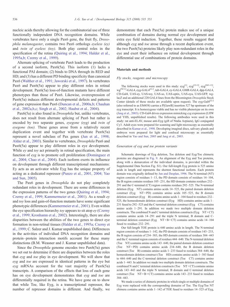
eyg transcripts are distributed within several embryonictissues as well as the leg, wing and eye-antennal imaginal discs(Jones et al., 1998; Jun et al., 1998; Aldaz et al., 2003). Here wehave characterized the expression pattern of toe and compare itto that of eyg (Fig. 1). eyg and toe transcripts are first detectedin stage 9 embryos within the salivary gland precursor (SGP)and a small cluster of cells within the dorsal head (Figs. 1A–C;Jones et al., 1998; Jun et al., 1998). The expression of toetranscripts in the SGP will persist through the rest of embryonicand larval development while eyg expression is terminated inlate stage embryos and reinitiated later (Figs. 1D–I, Figs. 2J, K,Jones et al., 1998). By late stage 10 both transcripts are alsofound in identical patterns within the posterior spiracle (PS) andwithin a cluster of cells at the anterior edge of each thoracic andabdominal segment (Figs. 1D–F, Jones et al., 1998; Jun et al.,1998). Expression of eyg and toe expands to the larval antennalorgan (AO) as well as the leg disc primordia by stage 12 (Figs.1G–I, Jones et al., 1998; Jun et al., 1998). During the latterstages of embryogenesis both eyg and toe transcripts accumu-late in the presumptive eye-antennal imaginal disc (Figs. 1J–L;Jones et al., 1998; Jun et al., 1998). Only two other members ofthe eye specification cascade, ey and toy, share this expressionpattern (Quiring et al., 1994; Czerny et al., 1999). Theremaining members are added sequentially during the larvaldevelopment (Kumar and Moses, 2001). The only discernabledifference between the expression patterns of either Pax6(5a)gene during embryogenesis is found within the SG: eygexpression is eliminated while toe transcriptional levels aremaintained (Figs. 1J–L; Jones et al., 1998).
Within the developing larval eye-antennal discs both eyg andtoe transcripts accumulate in identical patterns. Within theantennal segment both transcripts localize to the medial anddistal segments while in the eye disc expression of both genes isfound anterior to the morphogenetic furrow (Figs. 2A–C,Dominguez et al., 2004). Unlike the similarities found in theembryo, eyg and toe expression is somewhat different from thatof ey and toy. The Pax6 transcripts are expressed broadly aheadof the advancing furrow (Quiring et al., 1994; Czerny et al.,1999). However, eyg and toe expression is restricted to anarrow domain of cells that straddle the dorsal–ventralcompartment boundary and does not extend laterally (Figs.2A–C, Dominguez et al., 2004). This difference in expression is

Fig. 1. eyg and toe are expressed in nearly identical patterns during embryogenesis. Transcriptional profile of eyg and toe during several stages of embryogenesis.Probes are listed at the top of each column. Embryonic stage is denoted at the right of each row. Note that the expression patterns are nearly identical except for thesalivary glands in stage 15 embryos. SGP=salivary gland precursor, PS=posterior spiracle, AO=antennal organ, SG=salivary gland. Anterior is to the left.
538 J.-G. Yao et al. / Developmental Biology 315 (2008) 535–551
likely due to the requirements of eyg (and possibly toe) inNotch mediated control of cell proliferation at the organizingcenter versus the role of ey and toy in tissue specification.Within the developing wing primordium both transcripts areexpressed broadly within the notum and in two discrete regionswithin the presumptive wing (Figs. 2D–F). It is interesting thatone of those areas is particularly susceptible to beingtransformed into retinal tissue in response to forced expressionof ey (Fig. 2F, arrow). Both eyg and toe transcripts are alsofound within identical patterns of the leg primordium (Figs.2G–I) and the anterior duct cells of the salivary gland (Figs. 2J,K; Jones et al., 1998). The results from this and other studies ofeyg and toe expression suggest at first glance that these genesmay play redundant roles within several developing tissuesincluding the compound eye. It is unlikely, however, that these
genes play completely surplus roles (at least in the eye) as eygloss-of-function mutants show near complete loss of retinaltissue and forced expression of toe is insufficient to restore eyedevelopment to these flies (see below).
Quantitative contribution of eyg and toe mRNA transcripts
In order to further examine the contributions of eyg and toeto the development of the eye we used real-time PCR tomeasure the levels of each Pax6(5a) mRNA transcript in theeye-antennal disc (Fig. 3). We measured the combined levels ofeyg and toe in normal eye-antennal imaginal discs andcompared it to discs in which we forcibly expressed amicroRNA that is predicted to target and reduce the levels oftoe mRNA transcripts. This method was employed because a
Fig. 2. eyg and toe are expressed in nearly identical patterns in salivary glands and imaginal discs. Transcriptional profile of eyg and toe during the third larval instar.Probes are listed at the top of each column. Tissue type is denoted at the right of each row. Note that the expression patterns are nearly identical. Arrow in panel I marksarea of the wing disc in which ectopic eye development is supported by the expression of several retinal determination genes including ey.
539J.-G. Yao et al. / Developmental Biology 315 (2008) 535–551
toe specific loss-of-function mutation does not yet exist. Ourresults indicate that the vast majority (approximately 87%) ofPax6(5a) mRNAs are transcribed from the eyg locus (Fig. 3A).A direct comparison of eyg and toe levels in wild type eye-antennal discs confirms the unequal levels of Pax6(5a)expression (Fig. 3B). This relationship is maintained autono-mously in individual eye and antennal discs (Fig. 3C). Finally,we set out to determine if the relative levels of toe are dependentupon eyg expression. In both eyg loss-of-function mutants andforced expression experiments the level of toe remainedconstant suggesting that toe levels are regulated independentlyof eyg (Fig. 3D). It appears that toe transcriptional regulation isalso independent of eyg in several other tissues including the
developing embryo and wing imaginal discs (Aldaz et al., 2003;Jang et al., 2003).
An anti-Pax6(5a) antibody recognizes the PD of Eyg and Toe
An in vivo comparison of the roles played by the two Pax6(5a) genes has been hampered by the lack of availablemolecular markers for the distribution of Eyg and Toe proteins.To overcome this obstacle we generated a polyclonal antibodythat recognizes full-length Eyg (data not shown). Since theantibody recognized a region of the eye disc in which both eygand toe are expressed, and since both proteins shareconsiderable sequence similarity within the DNA binding

Fig. 3. eyg is expressed at a higher level than toe in eye-antennal disc. (A) Relative level of anti-Eyg/Toe immunostaining on third instar eye discs. The signal intensityin wild type (n=12) is taken as 100%. The signal intensity when Toe is efficiently knockdown (eyg-Gal4 driving UAS-toe miR; n=7) is reduced to about 87%. (B–D)Graphs depict the levels of eyg and toe in the developing eye and antenna as assayed by quantitative RT-PCR. White bars are eyg transcript level. Black bars are toetranscript level. (B) RT-PCR from eye-antennal disc from late third instar (left two bars) and second instar (right two bars). (C) When the eye and antenna discs weresurgically separated, the eyg transcript is higher than toe in both eye and antennal discs. (D) Absolute eyg and toe transcript numbers were estimated from eye-antennaldiscs. toe transcript level was not significantly affected when eyg is overexpressed (CD-Gal4 driving UAS-eyg), indicate that toe is not transcriptionally regulated byeyg. eygM3-12 is a null mutation with a deletion beginning 23 bp upstream of eyg and extending 13 kb downstream of eyg transcription unit (Jang et al., 2003).Although the toe transcriptional unit is not affected, it is not clear whether toe expression is affected by the deletion. There is no eye-antennal disc in eygM3-12 toexamine whether toe is affected. So we drove eyg expression by the CD-Gal4 in eygM3-12 to rescue the eye-antennal disc. In these rescued eye disc, toe transcript levelis not significantly different from that in the wild type disc. This result clearly demonstrate that the eygM3-12 mutation affects only eyg.
540 J.-G. Yao et al. / Developmental Biology 315 (2008) 535–551
domains we wanted to determine the specificity of the anti-Eygantibody. We expressed toe along the A/P axis of the wing discusing a dpp-GAL4 driver. Our antibody not only recognizedendogenous Eyg, which is found within the hinge region, but italso recognized the exogenously added Toe protein along theA/P axis (Figs. 4A, B). Thus the antibody we recoveredrecognizes both Eyg and Toe and will be referred to as anti-Pax6(5a).
The DNA binding domains of Eyg and Toe (PD and HD) arelikely epitopes for the anti-Pax6(5a) antibody as they share ahigh degree of sequence similarity between the two proteins.We set out to determine which one of these two domains the
antibody recognizes. We expressed individual domains of thePax6(5a) proteins along the margins of the eye disc using anenhancer of the wingless (wg) gene. The anti-Pax6(5a) antibodywas then used to detect the expressed protein segments. Inaddition to the endogenous Pax6(5a) proteins, the antibody onlydetected the exogenously added Eyg PD (Figs. 4C–F, arrows).The antibody failed to recognize a mutant Eyg PD thusconfirming the specificity of anti-Pax6(5a) to the PD (Fig. 4G).The Pax6(5a) PDs share 96% sequence similarity and theantibody fails to recognize the remaining regions of Toe (Fig.4H; data not shown). A mutated version of the Toe PD is alsonot recognized by the antibody (Fig. 4H). Together, our results

Fig. 4. The Eyg/Toe antibody recognizes the PD of Eyg and Toe. (A, B) Wing discs. (C–I) Eye-antennal discs. Arrowhead in all panels denotes endogenous Pax6(5a)expression in the wing and eye disc. Arrows in panels a and e indicate detection of exogenous Pax6(5a) protein. Genotype of each tissue is indicated in each panel.Note that wgw11 enhancer drives expression in the lateral regions of the eye disc ahead of the morphogenetic furrow. Anterior is to the right.
541J.-G. Yao et al. / Developmental Biology 315 (2008) 535–551
indicate that the anti-Pax6(5a) antibody recognizes the Eyg andToe PDs.
As the anti-Pax6(5a) antibody recognizes both proteins wegenerated tagged proteins in which full-length Eyg and Toe aremarked with the FLAG epitope tag in order to visualize eachprotein individually. When UAS-Eyg[flag] is combined with aneyg-GAL4 driver the distribution of the marked protein can befollowed in the imaginal discs and salivary glands (data notshown). Unfortunately, as a toe-GAL4 line does not yet exist weare unable to specifically follow the distribution of Toe.However, combinatorial use of anti-Pax6(5a) with the Eyg[flag] is sufficient to differentiate between the distributionpatterns of both proteins during normal and forced expressionexperiments.
Differential requirement for eyg and toe in the eye and thorax
The absence of toe loss-of-function mutants has beenanother obstacle to clearly defining the contributions that eachgene makes to retinal development. To complement the study ofexisting eyg loss-of-function mutant phenotypes we made useof a microRNA (miRNA) that targets toe mRNA transcriptthereby reducing Toe protein levels and potentially substituting
for toe loss-of-function mutants. If the miRNA is co-expressedwith FLAG tagged version of either Pax6(5a) protein, only theToe levels are eliminated (Figs. 5A–F). Additionally, onlysalivary gland defects that results from the overexpression ofToe-Flag, are reversed by the miRNA (Figs. 5C, F). Eyg[flag]levels remain unaffected (Figs. 5G–I). Furthermore, expressionof the miRNA in severe eyg mutants eliminates endogenousToe protein from both salivary glands and several imaginal discs(Figs. 5J–O). Note that the images in M–O have beenoverexposed to indicate that Toe protein cannot be detected inthe nucleus. Together, these results indicate that the miRNAselectively targets toe transcripts.
We then set out to determine what contribution, if any, to eyespecification is made by toe. Using an eyg-GAL4 driver weexpressed 2 copies of the toe miRNA in regions of thedeveloping eye that normally express both Pax6(5a) proteins.Interestingly, we did not observe any discernable defectssuggesting that although Toe levels are being eliminated inthe retina, the endogenous levels of Eyg is sufficient to fullysupport eye development. The thorax, which also requires eyg,is similarly unaffected by the expression of the miRNA undercontrol of either the eyg-GAL4 and/or tub-GAL4 (Figs. 6A, E).Since eyg and toe mRNA levels make up 87% and 13% of Pax6

Fig. 5. An miRNA specifically knocked down Toe protein level. (A, D, G) eye-antennal discs. (B, E, H, J, M) wing discs. (C, F, I, K, N) salivary glands. (L, O) legdiscs. Genotypes of each tissue are indicated at the left of each row. Arrows in each panel indicate exogenous Toe or Eyg. Note that panels M–O have beenoverexposed to show that there is Toe protein is down regulated (detected by anti-Flag in panels A–I, by anti-Eyg/Toe in panels J–O). Also note that the morphology ofthe salivary gland is rescued when the toe miRNA is coexpressed with a full-length toe construct (panel F). The RED stain in panels A, D, G is Elav.
542 J.-G. Yao et al. / Developmental Biology 315 (2008) 535–551
(5a) levels respectively we expressed the miRNA in fliesheterozygous for an eyg null mutant. In this situation the eyeremains unaffected but the anterior–central region of the thoraxdoes not develop (a phenotype that is not observed in eygheterozygotes). This is visibly manifested as a severe groovewithin the thorax (Figs. 6B, F). The failure to develop the
anterior–central portions of the thorax is reminiscent of theeffect of severely diminished levels of eyg (Fig. 6G, Aldaz etal., 2003). Interestingly, 50% reductions in Eyg protein levelsare not enough to severely alter the structure of the eye (Fig.6C). However, if eyg and toe levels are simultaneouslycompromised, then development of both the head and

Fig. 6. toe and eyg are differentially required in the developing eye and thorax. (A–D) adult eye and head. (E–G) adult thorax. Genotype of each tissue is indicated atthe top of each column. Arrow in panel D indicates the near complete inhibition of eye and head development. Arrows in panels F and G denote defects in thoraxdevelopment. Anterior is to the left.
543J.-G. Yao et al. / Developmental Biology 315 (2008) 535–551
compound eyes is blocked (Fig. 6D). The thorax is much moresensitive to simultaneous reductions in Eyg and Toe levels thanis the developing eye. Our results also suggest that removal oftoe, on its own, has little to no effect on the development ofeither tissue. This is consistent with the minor contribution thatthe toe locus makes to the overall levels of Pax6(5a) mRNA(Fig. 3). These results suggest that Eyg and Toe proteins aredifferentially required in the eye and thorax.
Consistent with this hypothesis, we have noted 43 differentsituations in which expression of eyg and toe had differingphenotypic consequences (Supplemental Data — Table 1). Forexample, expression of eyg in the wing disc via a vg-GAL4driver has no effect. However, expression of toe within the samedomain leads to increased levels of cell death. Conversely, whileexpression of toe in the embryonic CNS and brain via the c768-GAL4 driver has no effect, expression of eyg leads toembryonic lethality. We have been able to exclude trivialexplanations such as line strength and protein levels as reasonfor these disparities. Instead, our results further the contentionthat the Pax6(5a) proteins are differentially required duringdevelopment. This contention is also supported by differentialeffects on the developing eye and wing in response to theexpression of various Pax6(5a) deletion and chimeric proteins(Fig. 9; Supplemental Data — Table 2).
Toe is transcriptional repressor
Using Eyg-VP16 (transcriptional activator) and Eyg-En(transcriptional repressor) protein fusions, eyg has been shownpreviously to encode a dedicated transcriptional repressor (Yaoand Sun, 2005). Based on the evolutionary relationship betweenboth Pax6(5a) genes, toe is also predicted to encode a
transcriptional repressor. To test this hypothesis we createdtransgenic flies that expressed full-length Toe fused to the VP16activation domain (Toe-VP16) along the A/P axis of severalimaginal discs using a dpp-GAL4 driver. The activity of thistranscriptional-activating form of Toe failed to mimic theactivity of wild type Toe protein in several assays. In fact, incertain instances Toe-VP16 appeared to induce dominant-negative phenotypes. For instance, in contrast to Toe, which caninduce extra eye fields, the expression of the transcriptional-activating form of Toe failed to promote and support eyedevelopment in a forced expression assay (compare Figs. 7A to10B). Second, Toe-VP16 induced the formation of abnormalantennal structures and extra machrochaetes on the thorax,which are likely dominant negative effects (Figs. 7B, C).Another dominant-negative effect is seen when the expressionof Toe-VP16 in eyg hypomorphic mutants leads to the pro-duction of “headless” flies (Figs. 7D, E). These phenotypes arereminiscent of the effects observed when either the toe miRNAor Eyg-VP16 is individually expressed within the same eygmutant background (Fig. 6D, Yao and Sun, 2005). It should benoted that expression of the toe miRNA induced dominantnegative phenotypes, such as the production of headless flies,only when the genetic background was compromised for eygfunction. As these animals are headless and die as pharate adultswe were unable to assay the effects that the toe miRNA had onmacrochaetes numbers and antennal structure. The induction ofdominant negative phenotypes by Toe-VP16 in an otherwisewild type background is likely due to the strong activation ofputative Toe target genes via the VP16 activation domain. Wealso expect that Toe-VP16 would also activate some Eyg targetswhile the toemicroRNAwould only affect levels of Toe mRNAand protein. This might also contribute to the stronger Toe-

Fig. 7. Toe is a transcriptional repressor. (A) Schematic of VP16 fusion assay. (B–E) adult eye, thorax, antenna and head. Genotypes of each tissue are indicated at thetop of each column. Arrows in panels C and D mark defects in thorax and antennal development. Anterior is to the left.
544 J.-G. Yao et al. / Developmental Biology 315 (2008) 535–551
VP16 phenotype. As the activity of Toe-VP16 closely mimicsthat of Eyg-VP16 (Yao and Sun, 2005) our results aresuggestive that Toe, like Eyg, functions as a transcriptionalrepressor.
Further evidence of Toe serving as a repressor comes fromthe partial rescue of the eyg loss-of-function retinal phenotypeby expression of a chimeric protein in which Toe is fused to theEngrailed repressor (Toe-EN, data not shown). It should benoted that expression of Toe-EN failed to induce extra eyefields along the ventral surface of the head. There are twoplausible explanations for these relatively mild effects. First,the Toe-EN construct may not be expressed at high enoughlevels to either fully rescue eyg mutants or induce extra eyefields. We think that this is unlikely as the EN domain that isfused to TOE is a strong transcriptional repressor and highlevels of the fusion protein are not predicted to be required inthis assay. Instead it may be that during normal developmentToe functions as a repressor on some target genes and as anactivator on others. Unlike Eyg, Toe may not function as adedicated repressor. Instead it may have multiple functionswith repression being one of its activities. This could accountfor several of the observed difference in the activities of thePax6(5a) homologs (see below).
Mapping of the toe repressor domains
We set out to determine if the repressor domain(s) map tosimilar locations within Eyg and Toe. It has been previouslydemonstrated that Eyg contains two repressor domains: onemaps to the CT region, the other is located in either the NT orthe B regions of the protein (Yao and Sun, 2005; Figs. 8B–D).Expression of Eyg ΔB anterior to the furrow using an ey-GAL4 has a dominant negative effect on eye development(data not shown). Often times these dominant negative effects
can be attributed to the deletion of either an activation orrepressor domain. Here we show that the repressor domainwithin Toe is not located within the CT tail and may residewithin the NT and B domains. The assay used by Yao and Sunand here is a bipartite system. In one half of the system achimeric protein in which the GAL4 DNA binding domain isfused to either a full-length Pax6(5a) protein or an individualdomain is expressed along the margins of the eye under thecontrol of a wingless (wg) enhancer element. In the secondhalf of the system the same wg enhancer directs the expressionof a GFP reporter. A cluster of UAS sites separates theenhancer element from the reporter (Fig. 8A). In flies lackingthe driver construct, GFP is expressed along the margins of theeye field (Fig. 8B). When a portion of Eyg containing a strongrepressor (Eyg CT) is expressed in the same pattern,expression of the reporter is completely lost (Fig. 8C). If aweak or moderate repressor domain (Eyg ! CT mPDHD) isexpressed then a reduction in reporter activity is observed (Fig.8D). If now we express just the CT of Toe along the marginswe see normal levels of the GFP reporter indicating that thisdomain does not contain any repressor activity (Fig. 8E).However, if the N-terminal portion of Toe containing mutatedPD and HDs is expressed then strong repression of the reporteris seen. The most likely explanation is that a strong repressordomain resides within either the NT or the B domains (Fig.8F). Thus it appears that a major functional difference betweenthe Eyg and Toe proteins is the number and location of therepressor domains.
Molecular dissection of Eyg and Toe during normaldevelopment
As Eyg and Toe are derived from the same ancestral gene, areexpressed in identical patterns and function as transcriptional

Fig. 8. Toe repressor activity resides in its N-terminal portion. (A) Schematic of repressor assay. (B–D) Third instar eye-antennal discs accompanied by schematic ofdriver/responder combinations. Note that wg-GAL4 drives expression in the lateral margins ahead of the morphogenetic furrow. Anterior is to the right.
545J.-G. Yao et al. / Developmental Biology 315 (2008) 535–551
repressors, yet have differential effects on eye and thoraxdevelopment, we conducted a molecular dissection of bothproteins in an effort to understand the biochemical basis thatunderlies these unique functions. These experiments which areaimed at elucidating the differences between two Pax6(5a)proteins (Eyg and Toe) extend those of Yao and Sun, 2005which focused on the functional differences between theactivities of Eyg and Pax6 (Ey). An alignment of the Eyg andToe proteins, along with a demarcation of the individualdomains, is provided within the Supplemental Data Section.The reagents that we generated for our studies include a seriesof protein deletions in which individual or multiple domains ofeither Eyg or Toe were removed. These Pax6(5a) variants
were used to test the functional requirements for each domain.We also generated a series of chimeric Pax6(5a) proteins inwhich single or multiple domains of Eyg were deleted andreplaced with the corresponding regions of Toe. Thesechimeric proteins were used to test the degree to which eachdomain has been functionally conserved. Diagrams of thePax6(5a) deletions and chimeras are depicted in Figs. 9A–C.Each variant was assayed for the ability to rescue an eyg[1]
loss-of-function mutant (Figs. 9 and 10) and to induce extraeye fields within ventral head segments (Figs. 9 and 11). Itshould be noted that for each deletion and chimera we testedmultiple UAS insertion lines and conducted our experiments atseveral temperatures. We did this in an attempt to eliminate the

Fig. 9. Functional assay of domains in Eyg and Toe. (A–C) Schematic of Eyg deletions, Toe deletions and Eyg/Toe chimeric proteins. (D) Summary of Eyg and Toedomain requirements in rescue and overexpression assays. (E) Summary of results from rescue of eyg loss-of-function mutants and extra eye field induction assays.
546 J.-G. Yao et al. / Developmental Biology 315 (2008) 535–551
possibility that our results are affected by expression levels orinsert integrity. We also crossed each construct to severalGAL4 lines to ensure that each deletion or chimeric proteinwas functional.
Both wild type and all variants of Eyg and Toe wereexpressed in eyg[1] homozygous mutant retinas, which containbetween 40 and 50 ommatidia (Fig. 10A). Flies that arehomozygous for eyg null alleles die during embryogenesis andare therefore not appropriate for this particular assay. Expres-sion of wild type Eyg but not Toe is sufficient to return eyg[1]
mutant retinas to near wild type structure suggesting that thesegenes have functionally diverged since the duplication (Figs.9D and 10B, C). These genes are thus unlikely to play redun-dant roles in eye development.
Requirements for the non-DNA-binding domains in normal eyedevelopment
We first proceeded to test the functional requirements of thenon-DNA-binding domain. We started with deleting thesequences that lie upstream of the RED DNA binding domainin both proteins (Eyg ΔNT, Toe ΔNT). Toe ΔNT, but not EygΔNT, restored eye development to eyg[1] mutant retinas (Figs.9D and 10D, E) suggesting that not only is there a functionalrequirement for the NT region of both proteins but also that thisregion may functional distinguish the two Pax6(5a) proteinsfrom each other. Surprisingly, expression of the Eyg/Toe NTchimera is also capable of rescuing eyg[1] (Figs. 9D and 10F).This result indicates that while the NT domain may functionally

Fig. 10. Rescue of eyg1 loss-of-function mutants by expression of Eyg and Toe protein variants. Scanning electron micrographs of adult compound eyes. Genotypes ofeach animal are listed within each panel. All UAS lines were expressed using an ey-GAL4 driver. Anterior is to the right.
547J.-G. Yao et al. / Developmental Biology 315 (2008) 535–551
distinguish one Pax6(5a) from another, their function is stillcontext dependent. Eyg contains two repressor domains, ofwhich one lies within the first 443 amino acids of the protein(Yao and Sun, 2005). This region includes the NT, PD, B andHD as shown in Fig. 9A. Mutational analysis excluded the PDand HD regions thus leaving either the NT or B regions aspossible sites for the repressor activity of Eyg. Our results raisethe possibility that the repressor activity of Eyg resides withinthe NT region.
We then looked at the requirements for the B domain, a stretchof amino acids that lies between the PD and HD DNA bindingmotifs but is yet to be assigned a role in Pax protein function.Forced expression of constructs in which the B regions from Eygand Toe were deleted individually or in combination with theNT and CT regions (Eyg ΔB, Toe ! B, Eyg ΔNT+B+CT, ToeΔNT+B+CT) had a dominant negative effect on eyg[1] flies;the heads were severely deformed, the retinas were not restoredto wild type and the flies died in their pupal cases (Figs. 9D and10J–K, V, X). In contrast, eyg[1] mutant retinas were restoredto near wild type levels when region B of Eyg was replacedwith homologous region from Toe (Eyg/Toe B; Figs. 9D and10L). These results suggest that the B domain is functionally
essential for Pax6(5a) activity and has been functionallyconserved between the two transcription factors. This conclu-sion is supported by the observation, from a related set ofexperiments in which the Pax6 proteins Ey and Toy do notfunction normally in the absence of the B domain. Proteinslacking this domain produce ectopic eyes that are less frequentlyobserved and are significantly smaller in size than thoseproduced by the full-length proteins (B.M. Weasner and J.P.Kumar, unpublished data). There is the possibility, however, thatregion B simply acts as a linker or spacer for the two DNAbinding motifs and that deleting this region from any Pax proteinmay disrupt the normal structural configuration as the RED andHD motifs are brought together. One could interpret the rescueof eyg[1] by Eyg/Toe B as simply the result of restoring thespacing between the DNA binding motifs. We think that is ratherunlikely as a similar chimera in which the B domains of EYandTOY have been interchanged appear to have acquired newactivities and do not simply function as the parental Pax6 protein(B.M. Weasner and J.P. Kumar, unpublished data).
The C-terminal tail (residues 3′ of the HD) of Eyg but notToe contains a transcriptional repressor domain (Yao and Sun,2005; this report). We deleted the CT region in an attempt to

Fig. 11. Induction of extra eye fields by the expression of Eyg and Toe variants. Scanning electron micrographs of adult compound eyes and extra eye fields. Genotypesof each animal are listed within each panel. All UAS lines were expressed using a dpp-GAL4 driver. Yellow arrow in panel A indicates position of extra eye field.Anterior is to the right.
548 J.-G. Yao et al. / Developmental Biology 315 (2008) 535–551
determine if this domain serves to functionally distinguish onePax6(5a) protein from the other. Expression of Eyg ΔCT andToe ΔCT fully restored eye development to eyg[1] mutantretinas (Yao and Sun, 2005; Figs. 9D and 10P, Q). While theseresults suggest that the CT is dispensable for Eyg function, itappears that the CT region is required for Toe activity. In fact,the results also suggest that there is a combinatorial interactionbetween the Toe NT and CT regions as the presence of bothdomains prevents Toe from rescuing the eyg mutants. Removalof either the NT or the CT is sufficient to then allow for rescue.These results are also intriguing as they suggest that therepressor domain within the CT of Eyg is not essential for itsnormal activity. Rather, it seems that the second repressor site,which is located in either the NT or B regions of the protein, ismore essential to Eyg function. There appears, however, to be agenetic interaction between the NT and CT regions of bothproteins. Expression of either Eyg ! NT+CT or Toe ΔNT+CTwas insufficient to support eye development in eyg[1] mutants(Figs. 9D and 10S, T). This is in contrast to the near full rescueof eyg[1] retinas that is observed when either NT or CT regionsof Toe are individually removed (Figs. 9D and 10E, Q). Suchinteractions are also evidenced by the inability of Eyg/Toe NT+CT to rescue eyg[1] flies when expression of chimeras involvingindividual domains (Eyg/Toe NT, Eyg/Toe CT) is sufficient torestore eye development (Figs. 9D and 10F, R, U).
Requirements of the RED and homeobox DNA bindingdomains in normal eye development
We were also interested in determining if functionaldistinctions between Eyg and Toe could be accounted for by
differences in the use and requirements of the RED and HDmotifs. Eye development could be restored to eyg[1] mutantsthrough expression of Pax6(5a) variants that in which the REDdomain was interchanged but not deleted (Yao and Sun, 2005;Figs. 9D and 10G–I). These results suggest that both Eyg andToe exert their influence on transcription through the REDdomain and that these domains have been functionallyconserved. We similarly deleted and substituted the HDs andobserved that expression of EYG ΔHD and Eyg/Toe HDrescued the small eye phenotype of eyg[1] mutants while ToeΔHD failed in this respect (Yao and Sun, 2005; Figs. 9D and10M–O). These results indicate that in contrast to absoluterequirement for the RED domain it appears that the Eyg HD iscompletely dispensable for eye development. As a consequenceEyg primarily uses its RED domain to interact with DNA.There are several precedents for this observation. Several Paxgenes including Drosophila pox meso and mammalian Pax1and Pax9 completely lack the HD (Noll, 1993; Mansouri et al.,1999). Second, during eye development the HD of EY/Pax6 isalso dispensable as an EY protein lacking the HD is sufficientto rescue loss-of-function ey mutants (Punzo et al., 2004).These results do not speak to the requirements of the Toe HDsince the Toe full-length protein also failed to rescue. However,data presented below on the generation of extra eye fieldsindicates that Toe also does not make use of the HD (seebelow).
A large body of evidence indicates that a considerabledegree of flexibility exists for the combinatorial use of DNAbinding motifs by Pax proteins. We attempted to test theextreme limits of this feature by simultaneously replacingboth the RED and HD regions of Eyg with the corresponding
549J.-G. Yao et al. / Developmental Biology 315 (2008) 535–551
domains of Toe. Surprisingly expression of the Eyg Toe PD+HD chimera rescued the structural defects of eyg[1] mutants(Figs. 9D and 10X). This result provides further evidence thatthe contextual framework motifs provided by the remainingnon-DNA binding regions can influence how certaincombinations of DNA binding domains are used duringdevelopment.
Domain requirements for extra eye field induction
We set out to determine if Toe, like Eyg, is only capableof inducing extra eye fields adjacent to the developingendogenous retinal epithelium (as opposed to ectopic eyeformation in other non-retinal tissues). We expressed eachPax6(5a) gene within 219 different developmental patternsand looked for the presence of retinal tissue. In the case ofthe full-length Eyg and Toe proteins, we were only able toinduce extra eye fields adjacent to the normal compound eye(Figs. 11A, B; Jang et al., 2003). We were interested indetermining if the domain requirements for the generation ofextra eye fields are the same as those needed for thepromotion of normal eye development. There is a precedentfor the two processes requiring different protein domains. Theactivity of the CT regions of the SIX family proteins SineOculis and Optix (results of an ancient duplication) is anexample. These regions are not interchangeable duringnormal eye development and in fact are thought to confer,in part, functional specificity upon SIX proteins. However,this is not the case for ectopic eye generation. The CTdomains are in fact interchangeable under these conditions(Weasner et al., 2007). This result suggests that there aredifferent molecular and biochemical requirements for normaland ectopic eye formation.
Of all the deletion constructs only the Toe ΔHD, in whichthe HD has been deleted, is capable of promoting the formationof an extra eye field (Fig. 11C). This implies that each domainof Eyg and all but the HD of Toe are required. This stands incontrast to our rescue assays in which the HD and CT regions ofEyg are dispensable for normal eye formation (Figs. 10M, P).Other differences were observed when the chimeric proteinswere used to induce extra eye fields. In these cases the NT, Band PD domains can be individually substituted. Certaindomain combinations (Eyg/Toe NT+CT and the Eyg/Toe PD+HD) could also be substituted successfully (Figs. 11D–H).Again, these requirements are dissimilar from those needed forEyg and Toe to function properly during normal eyedevelopment. Our rescue assays concluded that all individualdomains and only the PD+HD combination could beexchanged and still rescue eyg[1] mutants (Figs. 10F, I, L, O,R, X). As mentioned earlier, similar differences in domainrequirements during normal eye development and ectopic eyeformation (or extra eye field generation) are observed with othereye specification genes. These apparent disparities may reflectactual differences in the protein–protein interactions that occurbetween eye specification proteins and their cofactors. Such amodel might imply that there is some flexibility in the path toproducing an eye.
Discussion
In eye development the tasks of tissue specification and cellproliferation are regulated, in part, by the Pax6 and Pax6(5a)proteins respectively. In vertebrates, Pax6(5a) is generated asan alternately spliced isoform of Pax6. However, in Droso-phila Pax6(5a) homologs are encoded by the eyegone and twinof eyegone genes. In this report we sought to determine therespective contributions that each gene makes to thespecification of the fly. An initial analysis of transcriptionalpatterns indicates that both Pax6(5a) genes are expressed inidentical patterns within the retina. However, eyg is expressedat a much higher level than toe. Not surprisingly, whilemutations in eyg nearly delete the eye, a reduction in toe viamiRNA treatments has no effects on its own. Simultaneousreductions in both genes, in contrast, result in a “headless”phenotype. Using a set of mini genetic screens and activator/repressor fusion assays we also demonstrated that both proteinsfunction as transcriptional repressors. In total, these character-istics suggest that eyg and toe might play redundant roles induring development.
However, the high level of sequence divergence within thenon-DNA binding domains hints that their functions may onlybe partially redundant. We set out to molecularly dissect bothPax6(5a) proteins and determine what, if any, differences existbetween the activities of each protein. In two experimentalcontexts we were able to demonstrate that such differencesbetween eyg and toe exist. First, a comparison of eyg and toeloss-of-function phenotypes indicated that toe played a greaterrole in the development of the thorax than the eye. Second,forced expression of both full-length proteins throughout thedeveloping fly identified 43 different instances in whichexpression of one Pax6(5a) gene induced a different phenotypethan the other. Taken together, these results hint that the roles ofeyg and toe may be not be completely redundant.
We then set to determine which domain(s) might accountfor the differences seen in loss-of-function mutants and forcedexpression assays. We generated a set of deletion and chimericproteins to dissect the requirement for each domain as well asthe level of functional conservation. We attempted to rescueeyg1 mutants as well as generate extra eye fields with theseprotein variants. Our results indicate that Eyg and Toe makedifferential use of several domains. Many of these differencesmap to the non-DNA binding domains. We have alsodemonstrated that one possible mechanism for this is thatToe has only one repressor domain, while Eyg has two. Ourprediction is that the differences in the non-DNA bindingdomains are the primary determinants of how each Pax6(5a)protein will influence development. It is less likely that thetwo DNA binding domains functionally distinguish oneprotein from another as there is an extremely high level ofsequence conservation within these motifs. Thus our modelfor how Eyg and Toe Function is that both transcriptionfactors bind to similar target genes but can differentiallyinfluence transcription through differing levels of repressoractivity and/or interactions with disparate binding partners(Figs. 12A, B).
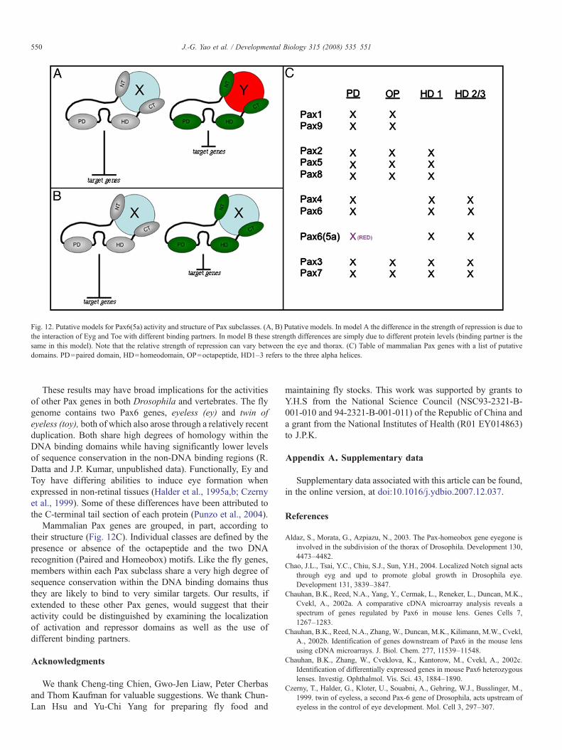
Fig. 12. Putative models for Pax6(5a) activity and structure of Pax subclasses. (A, B) Putative models. In model A the difference in the strength of repression is due tothe interaction of Eyg and Toe with different binding partners. In model B these strength differences are simply due to different protein levels (binding partner is thesame in this model). Note that the relative strength of repression can vary between the eye and thorax. (C) Table of mammalian Pax genes with a list of putativedomains. PD=paired domain, HD=homeodomain, OP=octapeptide, HD1–3 refers to the three alpha helices.
550 J.-G. Yao et al. / Developmental Biology 315 (2008) 535–551
These results may have broad implications for the activitiesof other Pax genes in both Drosophila and vertebrates. The flygenome contains two Pax6 genes, eyeless (ey) and twin ofeyeless (toy), both of which also arose through a relatively recentduplication. Both share high degrees of homology within theDNA binding domains while having significantly lower levelsof sequence conservation in the non-DNA binding regions (R.Datta and J.P. Kumar, unpublished data). Functionally, Ey andToy have differing abilities to induce eye formation whenexpressed in non-retinal tissues (Halder et al., 1995a,b; Czernyet al., 1999). Some of these differences have been attributed tothe C-terminal tail section of each protein (Punzo et al., 2004).
Mammalian Pax genes are grouped, in part, according totheir structure (Fig. 12C). Individual classes are defined by thepresence or absence of the octapeptide and the two DNArecognition (Paired and Homeobox) motifs. Like the fly genes,members within each Pax subclass share a very high degree ofsequence conservation within the DNA binding domains thusthey are likely to bind to very similar targets. Our results, ifextended to these other Pax genes, would suggest that theiractivity could be distinguished by examining the localizationof activation and repressor domains as well as the use ofdifferent binding partners.
Acknowledgments
We thank Cheng-ting Chien, Gwo-Jen Liaw, Peter Cherbasand Thom Kaufman for valuable suggestions. We thank Chun-Lan Hsu and Yu-Chi Yang for preparing fly food and
maintaining fly stocks. This work was supported by grants toY.H.S from the National Science Council (NSC93-2321-B-001-010 and 94-2321-B-001-011) of the Republic of China anda grant from the National Institutes of Health (R01 EY014863)to J.P.K.
Appendix A. Supplementary data
Supplementary data associated with this article can be found,in the online version, at doi:10.1016/j.ydbio.2007.12.037.
References
Aldaz, S., Morata, G., Azpiazu, N., 2003. The Pax-homeobox gene eyegone isinvolved in the subdivision of the thorax of Drosophila. Development 130,4473–4482.
Chao, J.L., Tsai, Y.C., Chiu, S.J., Sun, Y.H., 2004. Localized Notch signal actsthrough eyg and upd to promote global growth in Drosophila eye.Development 131, 3839–3847.
Chauhan, B.K., Reed, N.A., Yang, Y., Cermak, L., Reneker, L., Duncan, M.K.,Cvekl, A., 2002a. A comparative cDNA microarray analysis reveals aspectrum of genes regulated by Pax6 in mouse lens. Genes Cells 7,1267–1283.
Chauhan, B.K., Reed, N.A., Zhang, W., Duncan, M.K., Kilimann, M.W., Cvekl,A., 2002b. Identification of genes downstream of Pax6 in the mouse lensusing cDNA microarrays. J. Biol. Chem. 277, 11539–11548.
Chauhan, B.K., Zhang, W., Cveklova, K., Kantorow, M., Cvekl, A., 2002c.Identification of differentially expressed genes in mouse Pax6 heterozygouslenses. Investig. Ophthalmol. Vis. Sci. 43, 1884–1890.
Czerny, T., Halder, G., Kloter, U., Souabni, A., Gehring, W.J., Busslinger, M.,1999. twin of eyeless, a second Pax-6 gene of Drosophila, acts upstream ofeyeless in the control of eye development. Mol. Cell 3, 297–307.

551J.-G. Yao et al. / Developmental Biology 315 (2008) 535–551
Dominguez, M., Ferres-Marco, D., Gutierrez-Avino, F.J., Speicher, S.A.,Beneyto, M., 2004. Growth and specification of the eye are controlledindependently by Eyegone and Eyeless in Drosophila melanogaster. Nat.Genet. 36, 31–39.
Duncan, M.K., Cvekl, A., Li, X., Piatigorsky, J., 2000a. Truncated forms of Pax-6 disrupt lens morphology in transgenic mice. Investig. Ophthalmol. Vis.Sci. 41, 464–473.
Duncan, M.K., Kozmik, Z., Cveklova, K., Piatigorsky, J., Cvekl, A., 2000b.Overexpression of PAX6(5a) in lens fiber cells results in cataract andupregulation of (alpha)5(beta)1 integrin expression. J. Cell. Sci. 113 (Pt 18),3173–3185.
Gehring, W.J., 1996. The master control gene for morphogenesis and evolutionof the eye. Genes Cells 1, 11–15.
Gehring, W., 2002. The genetic control of eye development and itsimplications for the evolution of the various eye-types. Int. J. Dev. Biol.46, 65–73.
Gehring, W.J., 2005. New perspectives on eye development and the evolution ofeyes and photoreceptors. J. Hered. 96, 171–184.
Gehring, W.J., Ikeo, K., 1999. Pax 6: mastering eye morphogenesis and eyeevolution. Trends Genet. 15, 371–377.
Halder, G., Callaerts, P., Gehring, W.J., 1995a. Induction of ectopic eyes bytarget expression of the eyeless gene in Drosophila. Science 267,1788–1792.
Halder, G., Callaerts, P., Gehring, W.J., 1995b. New perspectives on eyeevolution. Curr. Opin. Genet. Dev. 5, 602–609.
Haubst, N., Berger, J., Radjendirane, V., Graw, J., Favor, J., Saunders, G.F.,Stoykova, A., Gotz, M., 2004. Molecular dissection of Pax6 function: thespecific roles of the paired domain and homeodomain in brain development.Development 131, 6131–6140.
Hill, R.E., Favor, J., Hogan, B.L., Ton, C.C., Saunders, G.F., Hanson, I.M.,Prosser, J., Jordan, T., Hastie, N.D., van Heyningen, V., 1991. Mouse smalleye results from mutations in a paired-like homeobox-containing gene.Nature 354, 522–525.
Jang, C.C., Chao, J.L., Jones, N., Yao, L.C., Bessarab, D.A., Kuo, Y.M., Jun, S.,Desplan, C., Beckendorf, S.K., Sun, Y.H., 2003. Two Pax genes, eye goneand eyeless, act cooperatively in promoting Drosophila eye development.Development 130, 2939–2951.
Jaworski, C., Sperbeck, S., Graham, C., Wistow, G., 1997. Alternative splicingof Pax6 in bovine eye and evolutionary conservation of intron sequences.Biochem. Biophys. Res. Commun. 240, 196–202.
Jones, N.A., Kuo, Y.M., Sun, Y.H., Beckendorf, S.K., 1998. The Drosophila Paxgene eye gone is required for embryonic salivary duct development.Development 125, 4163–4174.
Jun, S., Desplan, C., 1996. Cooperative interactions between paired domain andhomeodomain. Development 122, 2639–2650.
Jun, S., Wallen, R.V., Goriely, A., Kalionis, B., Desplan, C., 1998. Lune/eyegone, a Pax-like protein, uses a partial paired domain and a home-
odomain for DNA recognition. Proc. Natl. Acad. Sci. U. S. A. 95,13720–13725.
Kammermeier, L., Leemans, R., Hirth, F., Flister, S., Wenger, U., Walldorf, U.,Gehring, W.J., Reichert, H., 2001. Differential expression and function ofthe Drosophila Pax6 genes eyeless and twin of eyeless in embryonic centralnervous system development. Mech. Dev. 103, 71–78.
Kronhamn, J., Frei, E., Daube, M., Jiao, R., Shi, Y., Noll, M., Rasmuson-Lestander, A., 2002. Headless flies produced by mutations in the paralogousPax6 genes eyeless and twin of eyeless. Development 129, 1015–1026.
Kumar, J.P., Moses, K., 2001. EGF receptor and Notch signaling act upstream ofEyeless/Pax6 to control eye specification. Cell 104, 687–697.
Kumar, J.P., Tio, M., Hsiung, F., Akopyan, S., Gabay, L., Seger, R., Shilo, B.Z.,Moses, K., 1998. Dissecting the roles of the Drosophila EGF receptor in eyedevelopment and MAP kinase activation. Development 125, 3875–3885.
Mansouri, A., Goudreau, G., Gruss, P., 1999. Pax genes and their role inorganogenesis. Cancer Res. 59, 1709s–1710s discussion 1709s–1710s.
Noll, M., 1993. Evolution and role of Pax genes. Curr. Opin. Genet. Dev. 3,595–605.
Pichaud, F., Desplan, C., 2002. Pax genes and eye organogenesis. Curr. Opin.Genet. Dev. 12, 430–434.
Punzo, C., Kurata, S., Gehring, W.J., 2001. The eyeless homeodomain isdispensable for eye development in Drosophila. Genes Dev. 15, 1716–1723.
Punzo, C., Plaza, S., Seimiya, M., Schnupf, P., Kurata, S., Jaeger, J., Gehring,W.J., 2004. Functional divergence between eyeless and twin of eyeless inDrosophila melanogaster. Development 131, 3943–3953.
Quiring, R., Walldorf, U., Kloter, U., Gehring, W.J., 1994. Homology of theeyeless gene of Drosophila to the Small eye gene in mice and Aniridia inhumans [see comments]. Science 265, 785–789.
Singh, S., Mishra, R., Arango, N.A., Deng, J.M., Behringer, R.R., Saunders,G.F., 2002. Iris hypoplasia in mice that lack the alternatively spliced Pax6(5a) isoform. Proc. Natl. Acad. Sci. U. S. A. 99, 6812–6815.
Tautz, D., Pfeifle, C., 1989. A non-radioactive in situ hybridization method forthe localization of specific RNAs in Drosophila embryos reveals transla-tional control of the segmentation gene hunchback. Chromosoma 98, 81–85.
Ton, C.C., Hirvonen, H., Miwa, H., Weil, M.M., Monaghan, P., Jordan, T., vanHeyningen, V., Hastie, N.D., Meijers-Heijboer, H., Drechsler, M., et al.,1991. Positional cloning and characterization of a paired box- andhomeobox-containing gene from the aniridia region. Cell 67, 1059–1074.
Walther, C., Guenet, J.L., Simon, D., Deutsch, U., Jostes, B., Goulding, M.D.,Plachov, D., Balling, R., Gruss, P., 1991. Pax: a murine multigene family ofpaired box-containing genes. Genomics 11, 424–434.
Weasner, B., Salzer, C., Kumar, J.P., 2007. Sine oculis, a member of the SIXfamily of transcription factors, directs eye formation. Dev. Biol. 303,756–771.
Yao, J.G., Sun, Y.H., 2005. Eyg and Ey Pax proteins act by distincttranscriptional mechanisms in Drosophila development. EMBO J. 24,2602–2612.