genome structure: from dna to chromosome lecture 2 of introduction to molecular biology 生理所...
Post on 21-Dec-2015
217 views
TRANSCRIPT

GENOME STRUCTURE:
From DNA To Chromosome
Lecture 2 of
Introduction to Molecular Biology
生理所 蔡少正

The length of the DNA as an extended molecule would vastly exceed the dimensions of the compartment that contains it; therefore, the DNA (in some cases, the RNA) must be compressed exceedingly tightly to fit into the space available.
Packing Genetic Materials

Highly Organized
DNA Molecule
The length to width ratio of a typical human chromosome is over 10 million to one.
2 meter
10 m

of tightly packed material, but in regions that have become stretched, they can be seen to consist of discrete particles, called nucleosomes. A continuous duplex thread of DNA runs through the series of particles.
Interphase DNA When interphase nuclei are suspended in a low ionic strength solution, they will swell and rupture to release fibers of chromatin. In some regions, the fibers consist
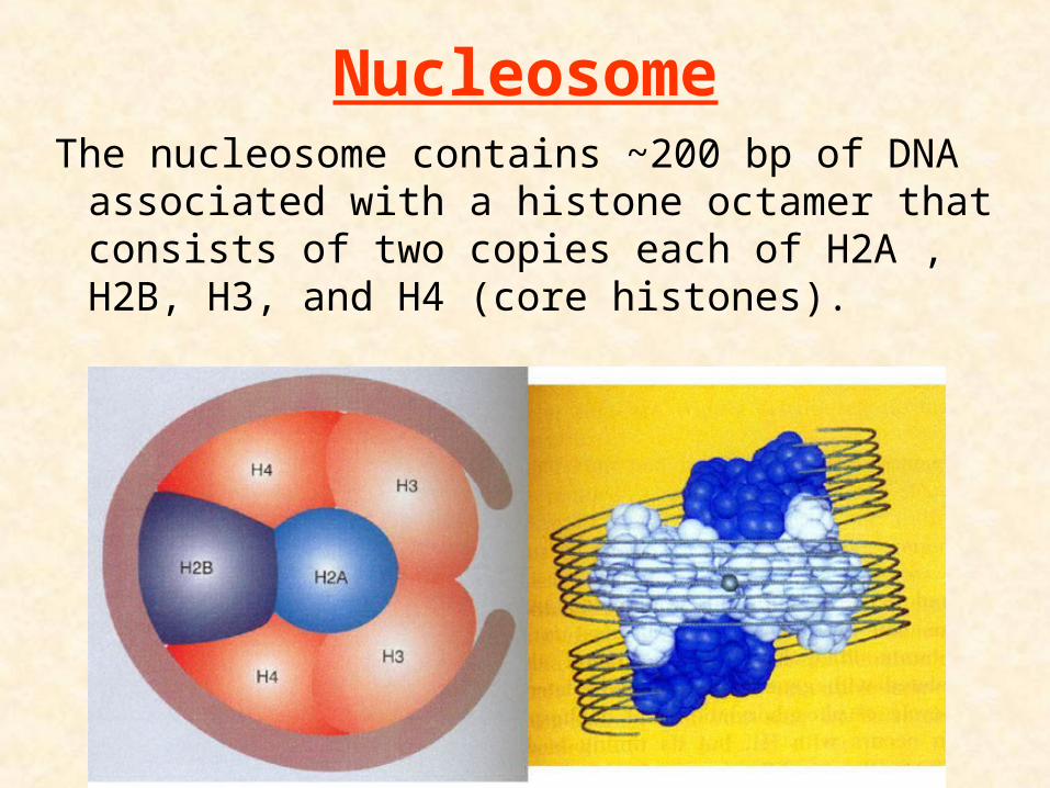
NucleosomeThe nucleosome contains ~200 bp of DNA
associated with a histone octamer that consists of two copies each of H2A , H2B, H3, and H4 (core histones).

Composition of Nucleosome
The number of residues in H1 (or H5) is about 220. Other types of histones are smaller, each containing 100-135 residues.

Core and Linker DNAA nucleosome DNA can be further divided into 2 regions:
1. Core DNA: 146 bp, resistant to digestion by nucleases.
2. linker DNA: from as little as 8 bp to as much as 114 bp per nucleosome, sensitive to nucleases digestion.
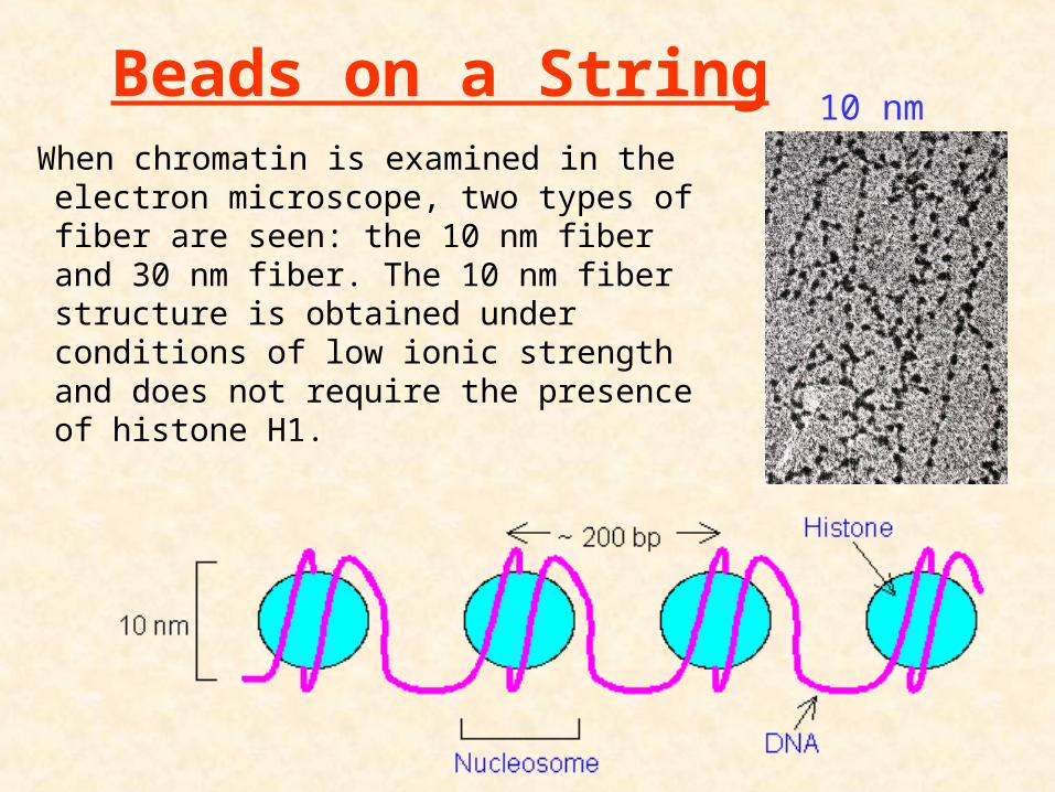
Beads on a StringWhen chromatin is examined in the
electron microscope, two types of fiber are seen: the 10 nm fiber and 30 nm fiber. The 10 nm fiber structure is obtained under conditions of low ionic strength and does not require the presence of histone H1.
10 nm

SolenoidWhen chromatin is condensed, each nucleosome is associated with an H1 (or H5) to form a solenoid structure. H1 and H5 are called linker histones. They are essential in stabilizing the solenoid conformation.
30 nmWhen chromatin is visualized in conditions of greater ionic strength, the 30 nm fiber is obtained.
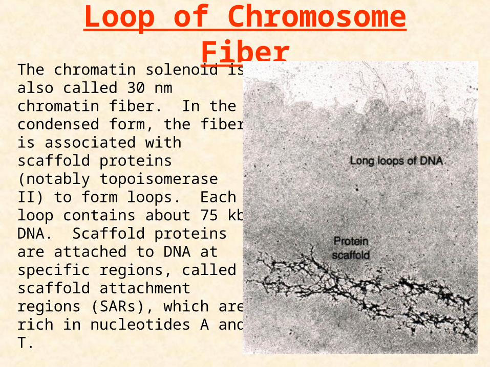
The chromatin solenoid is also called 30 nm chromatin fiber. In the condensed form, the fiber is associated with scaffold proteins (notably topoisomerase II) to form loops. Each loop contains about 75 kb DNA. Scaffold proteins are attached to DNA at specific regions, called scaffold attachment regions (SARs), which are rich in nucleotides A and T.
Loop of Chromosome Fiber
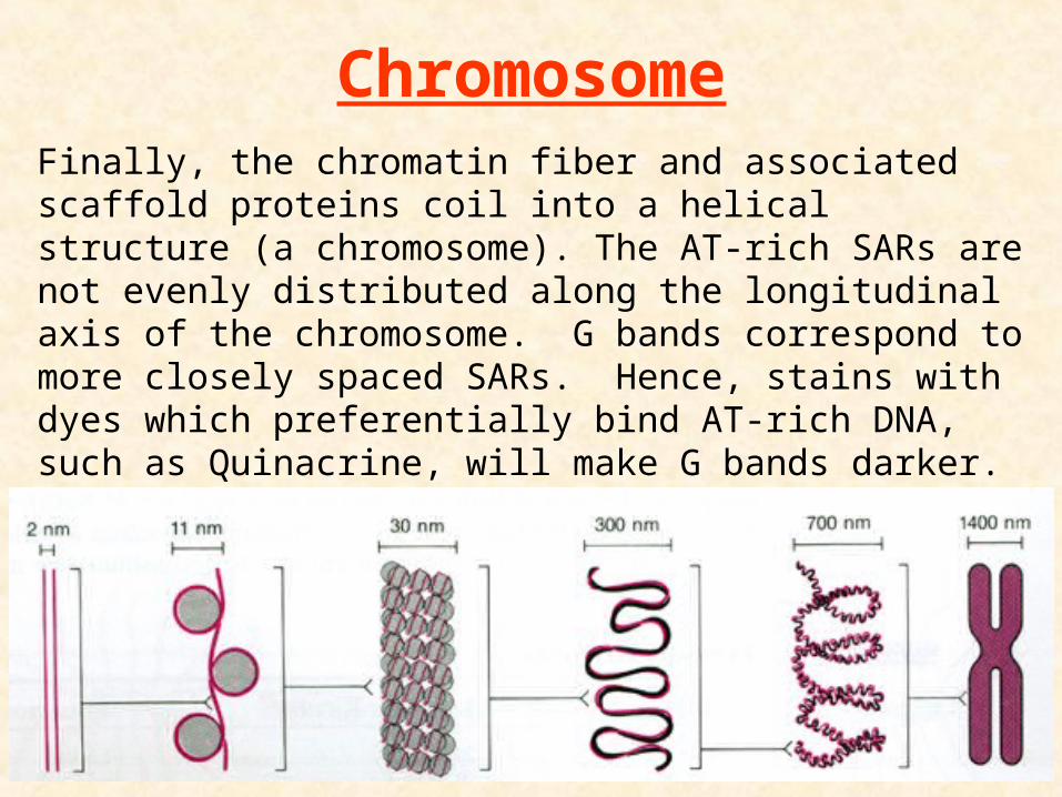
Finally, the chromatin fiber and associated scaffold proteins coil into a helical structure (a chromosome). The AT-rich SARs are not evenly distributed along the longitudinal axis of the chromosome. G bands correspond to more closely spaced SARs. Hence, stains with dyes which preferentially bind AT-rich DNA, such as Quinacrine, will make G bands darker.
Chromosome

Release of DNA from Histone
Histones contain a few lysine (K) residues at the N terminus. Under normal cellular conditions, the R group of lysine is positively charged, which can bind to the negatively charged phosphates in DNA. The positive R group of lysine may be neutralized by acetylation (by histone acetyltransferase, HAT), reducing the binding force between histones and DNA. Such mechanism has been demonstrated to play a major role in the regulation of gene transcription.

1. adding acetyl and methyl groups neutralizes the positive charges on Lys and Arg
2. adding phosphate groups adds negative charges to Ser and Thr residues
3. both actions would reduce the strength of the association between the highly-negative DNA and the highly-positive histones.
Modification of Histones

Nucleosomes are quickly regenerated once the DNA has been replicated
Regenerates of Nucleosomes
Histones need to be removed before replication of DNA can proceed.
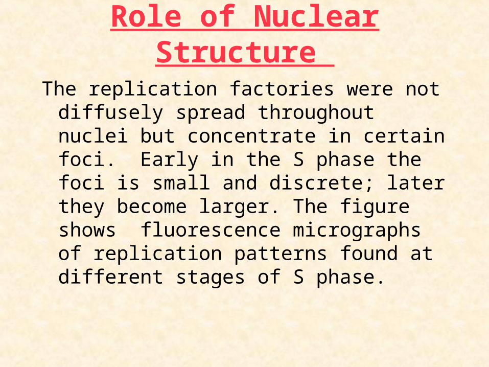
Role of Nuclear Structure
The replication factories were not diffusely spread throughout nuclei but concentrate in certain foci. Early in the S phase the foci is small and discrete; later they become larger. The figure shows fluorescence micrographs of replication patterns found at different stages of S phase.
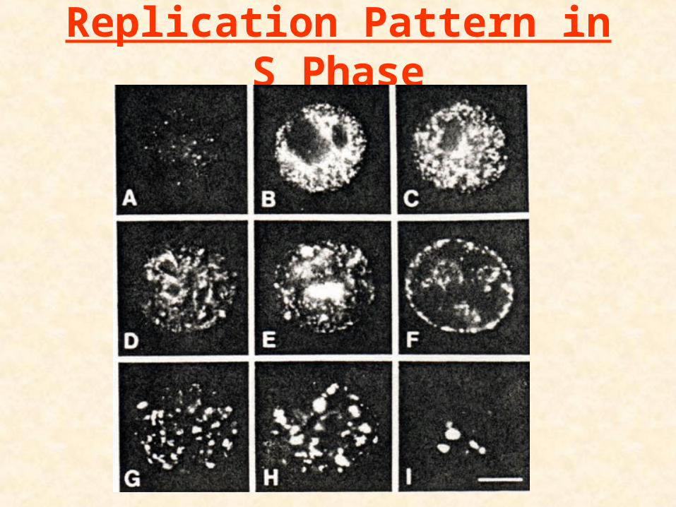
Replication Pattern in S Phase