genetics and kidney disease - nephrology2021.com
TRANSCRIPT

Boston Children’s Hospital
Division of Nephrology
Harvard Medical School
Friedhelm Hildebrandt
Genetics and
Kidney Disease

Friedhelm Hildebrandt, MD▪ Chief, Div. of Nephrology,
Boston Children’s Hospital -
Harvard Medical School
▪ William E. Harmon Professor
of Pediatrics,
Harvard Medical School
o Clinical: Monogenic CKD
o Research: Gene discovery
and dz mechanisms of CKD

Disclosure
Cofounder of Goldfinch-Bio

Steroid-Resistant Nephrotic Syndrome
Focal segmental glomerulosclerosis (FSGS)

Homozygous
Point Mutation
in the Podocin
Gene
A mutation in
1 bp of the
3,300,000,000 bp
of the total genome
is sufficient to
cause FSGS
Patient
Normal

Definition: Monogenic disease(Single-gene disorder)
Definition
• In 1 patient the disease is caused
by mutation of 1 gene only of ~20,000 genes(recessive, e.g. ARPKD; dominant, e.g. ADPKD)
• In different patients different genes may
cause a similar disease:(“genetic locus heterogeneity”, e.g. podocin, nephrin)
• 47 recessive, 8 dominant genes for SRNS

Gene mRNA Protein Phenotype
Monogenic Disease
AAAA
Flow of Genetic Information (Central dogma)
= Etiology
Gene mRNA Protein Disease
defect
AAAA
“Pathogenesis”

Consequences from Disease Gene Identification
Gene mRNA Protein Disease
mutation
AAAA
Molecular
Genetic
Diagnosis
Genotype/
Phenotype
Correlation
= Etiology
Novel Insights
into
Pathogenesis
& Physiology „Personalized
Therapy“
“Pathogenesis”

Genetic causality and predictive power in
monogenic and polygenic diseases
Recessive
monogenic
Dominant
monogenic
Polygenic
Genetic causality Strong Intermediate Weak
Penetrance ~100% Incomplete Weak
Predictive power
of mut. analysis
Almost 100% Strong Weak
Onset Fetus, child Adolescent Adult
Molecular genetic
approach
Mapping, Exon
sequencing, CNV
Mapping, Exon
sequencing, CNV
GWAS“association”
Animal model Very feasible feasible difficult
Frequency ~ 1 in 40,000 ~ 1 in 1,000 ~ 1 in 5

Age related penetrance of
monogenic alleles (mutations)
100 Years50
50%
100%
Cu
m. %
of
1s
tD
ise
ase M
an
ifesta
tio
n
AR, homozygous truncating AR, hom. missense
AD, incomplete
penetrance
AD, “mild” allele
Treatment,
prophy-
laxis
Environ-
mental
risk
10
same gene
AR or AD

Boards questionThe term “monogenic disease” …
A. Is synonymous with “single-gene” disease
and “Mendelian disorder”.
B. Describes autosomal recessive, autosomal
dominant, and X-linked disorders.
C. Describes a genetic “association”.
D. Means that a mutation in a single gene is
sufficient to cause disease in an individual.
E. A, B, and D.

What percentage of
chronic kidney disease
(onset <25 yrs)
is caused by single-gene
mutations?

Diagnostic Groups of CKD <25 yrs
Chronic Kidney Disease (NAPRTCS 2008) Cause
CAKUT (Congenital Anomalies of the
Kidneys & Urinary Tract) 50%
STEROID RES. NEPHROTIC SYNDROME 15%
CHRONIC GLOMERULONEPHRITIS 14%
MPGN, SLE, IgA, Wegeners
CYSTIC KIDNEY DISEASE 6%
ARPKD, ADPKD, Nephronophthisis, MCKD
NEPHROLITHIASIS / NEPHROCALINOSIS 3%
OTHER 12%
TOTAL (n=8,990) 100%

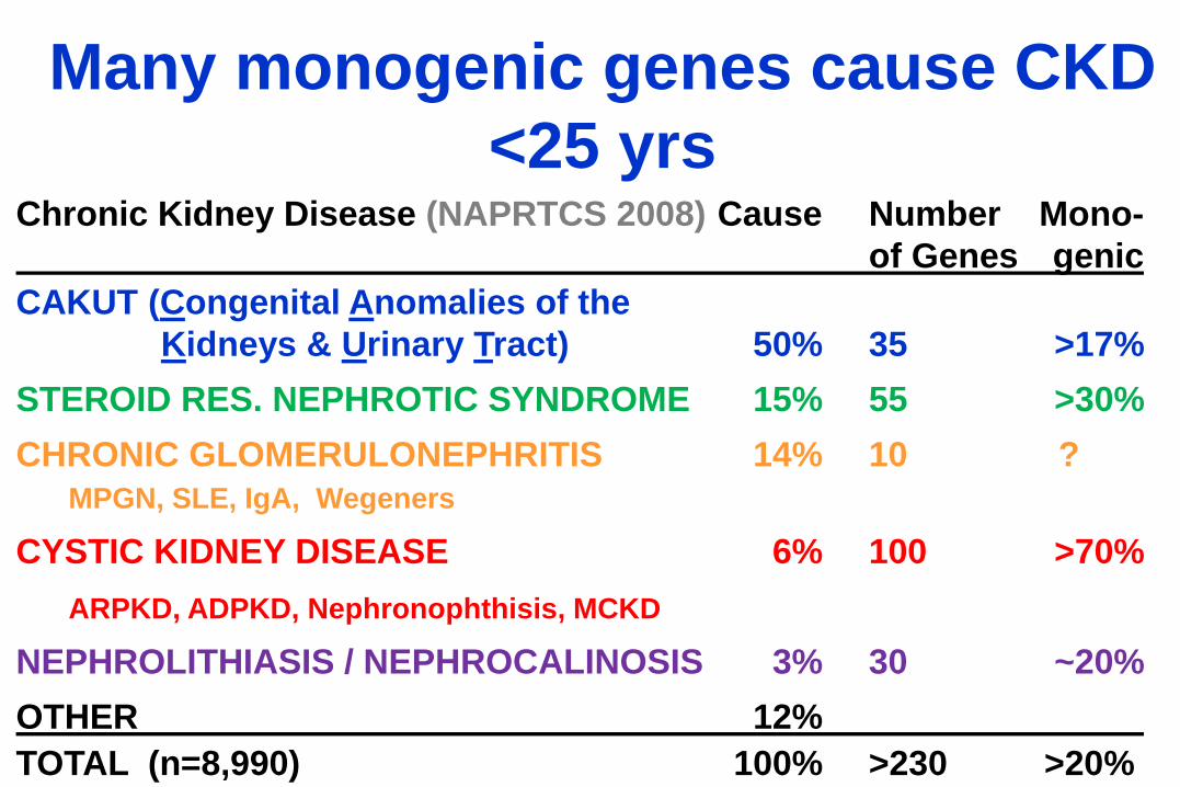
Many monogenic genes cause CKD
<25 yrsChronic Kidney Disease (NAPRTCS 2008) Cause Number
of Genes
CAKUT (Congenital Anomalies of the
Kidneys & Urinary Tract) 50% 35
STEROID RES. NEPHROTIC SYNDROME 15% 55
CHRONIC GLOMERULONEPHRITIS 14% 10
MPGN, SLE, IgA, Wegeners, aHUS
CYSTIC KIDNEY DISEASE 6% 100
ARPKD, ADPKD, Nephronophthisis, MCKD
NEPHROLITHIASIS / NEPHROCALINOSIS 3% 30
OTHER 12%
TOTAL (n=8,990) 100% >230

Many monogenic genes cause CKD
<25 yrsChronic Kidney Disease (NAPRTCS 2008) Cause Number Mono-
of Genes genic
CAKUT (Congenital Anomalies of the
Kidneys & Urinary Tract) 50% 35 >17%
STEROID RES. NEPHROTIC SYNDROME 15% 55 >30%
CHRONIC GLOMERULONEPHRITIS 14% 10 ?
MPGN, SLE, IgA, Wegeners
CYSTIC KIDNEY DISEASE 6% 100 >70%
ARPKD, ADPKD, Nephronophthisis, MCKD
NEPHROLITHIASIS / NEPHROCALINOSIS 3% 30 ~20%
OTHER 12%
TOTAL (n=8,990) 100% >230 >20%

What percentage of
Steroid-Resistant Nephrotic
Syndrome (SRNS) is
caused by single-gene
mutations?
(onset <25 yrs)

Causative Gene N Families % of Total Families
NPHS2 (podocin) 170 10%
NPHS1 (nephrin) 125 7%
WT1 85 5%
PLCE1 35 2%
22 Other Genes 105 6%
Total 520 30%
In 30% of 1,780 worldwide SRNS families
(<25 yr) the causative mutation was
detected (26 genes)
(Sadowski & Lovric JASN 26:1279, 2015) (www.renalgenes.org)

Single-gene causation of SRNS is higher
in early manifestationSingle
Age of Onset [Years]
% o
f F
am
ilie
s W
ith
Dete
cti
on
of
Cau
sati
ve M
uta
tio
n

Causative mutation
found (% cases)

A recessive disease will most likely appear
sporadic (non-familial) in your clinic

Statistically, there need to be 8 sibs
for >1 to be affected (= “familial cases”)

Put Netter here
(Netter; www.georgetown.edu/.../GUE-scopeLibrary1.html)
Gene Identification Moved the Glomerular
Podocyte to Center Stage of SRNS Pathogenesis
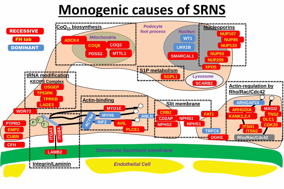
Lysosome
SCARB2
Endothelial Cell
Glomerular basement membrane
WT1
LMX1B
SMARCAL1
Nucleus
Integrin/Laminin
PV
T
LAMB2
PTPRO
Actin-binding
INF2
MYO1E
MYH9
EMP2
ARHGDIA
ARHGAP24
Rho/Rac/Cdc42
Actin-regulation by
Rho/Rac/Cdc42
KANK1,2,4
MitochondriaADCK4
COQ2COQ6
PDSS2 MTTL1
CoQ10 biosynthesis
NPHS2
NPHS1
Slit membrane
CD2AP
NPHS1
CRB2
CFH
NUP205
NUP107
WDR73
XPO5
Nucleoporins
NUP85
NUP93
NUP133
KEOPS Complex
OSGEP
TP53RK
TPRKB
LAGE3
tRNA modification SGPL1
ANLN
AVIL
PLCE1
FAT1
TRPC6
S1P metabolism
TNS2
MAGI2
DLC1
CDK20ITSN1ITSN2
DGKE
Podocyte
foot process
CUBN
RECESSIVE
DOMINANT
FH lab
Monogenic causes of SRNS

Q10 treatment in SRNS & COQ6 mutationP
rote
inu
ria
mg
/m2/h
Weeks after start of Tx
CoQ10 CoQ10

Detection of monogenic
causes of SRNS may
reveal existing
therapeutic options.
(e.g. Coenzyme Q10)

Many monogenic genes cause CKD
<25 yrsChronic Kidney Disease (NAPRTCS 2008) Cause Number Mono-
of Genes genic
CAKUT (Congenital Anomalies of the
Kidneys & Urinary Tract) 50% 35 >17%
STEROID RES. NEPHROTIC SYNDROME 15% 55 >30%
CHRONIC GLOMERULONEPHRITIS 14% 10 ?
MPGN, SLE, IgA, Wegeners
CYSTIC KIDNEY DISEASE 6% 100 >70%
ARPKD, ADPKD, Nephronophthisis, MCKD
NEPHROLITHIASIS / NEPHROCALINOSIS 3% 30 ~20%
OTHER 12%
TOTAL (n=8,990) 100% >230 >20%

Hil
de
bra
nd
t N
EJ
M3
64
:15
33, 2
011
Cystic kidney diseases are “ciliopathies”

(Braun Kidney Internat 89:468, 2016)
Increased renal echogenicity: Causative
mutation detected in ~63% of cases

Whole exome sequencing
may reveal the disease
cause in a difficult
differential diagnosis.(Clinical entry criteria: Cysts and/or
echogenicity on renal US <25 yrs)

SLC41A1
AGXT
SLC4A1
PKHD1
NPHP4
Gee
Kid
ne
yIn
t85:8
80
, 20
14 /
ed
ito
rial
p 7
48

Many monogenic genes cause CKD
<25 yrsChronic Kidney Disease (NAPRTCS 2008) Cause Number Mono-
of Genes genic
CAKUT (Congenital Anomalies of the
Kidneys & Urinary Tract) 50% 35 >17%
STEROID RES. NEPHROTIC SYNDROME 15% 55 >30%
CHRONIC GLOMERULONEPHRITIS 14% 10 ?
MPGN, SLE, IgA, Wegeners
CYSTIC KIDNEY DISEASE 6% 100 >70%
ARPKD, ADPKD, Nephronophthisis, MCKD
NEPHROLITHIASIS / NEPHROCALINOSIS 3% 30 ~20%
OTHER 12%
TOTAL (n=8,990) 100% >230 >20%

15% (11-21%) of nephrolithisasis is
caused by single-gene mutations
(Halbritter JASN 26:543, 2015)
(Braun cJASN 11:664, 2016)
Many monogenic genes cause CKD
<25 yrsChronic Kidney Disease (NAPRTCS 2008) Cause Number Mono-
of Genes genic
CAKUT (Congenital Anomalies of the
Kidneys & Urinary Tract) 50% 35 >17%
STEROID RES. NEPHROTIC SYNDROME 15% 55 >30%
CHRONIC GLOMERULONEPHRITIS 14% 10 ?
MPGN, SLE, IgA, Wegeners
CYSTIC KIDNEY DISEASE 6% 100 >70%
ARPKD, ADPKD, Nephronophthisis, MCKD
NEPHROLITHIASIS / NEPHROCALINOSIS 3% 30 ~20%
OTHER 12%
TOTAL (n=8,990) 100% >230 >20%

• Normal renal function, BP and urinalysis
• No systemic abnormalities related to his CKD
• Additional findings:
–Hyperuricemia (5.6-7.4 mg/dL)
– Slightly low Mg (1.5 mg/dL)
– Autism spectrum disorder
–Significant positive family history as his
father, paternal uncles and paternal
grandmother have similar condition.
Case: A 7-year-old boy with cystic kidney
disease, hyperuricemia and ASD

Renal US - age 7y

• Diagnostic consideration include:
• UMOD mutation
• HNF1B mutation
• HNF1B deletion (isolated/continuous gene del)

Uromodulin (UMOD) mutations may cause
Medullary Cystic Kideny Disease Type 2 (MCKD2)
• The Uromodulin (UMOD) gene encodes the Tamm-Horsfall protein.
• Mutations in UMOD can also lead to two additional clinical entities named:
• (1) familial juvenile hyperuricemic nephropathy (FJHN) (2) glomerulocystic kidney disease (GCKD).
• Although MCKD represents a subtype of CAKUT, among cases of isolated CAKUT it is estimated that UMODmutations are very rare.

Molecular diagnostic analysis
• UMOD sequencing – Negative
• HNF1B mutation - Negative
• HNF1B deletion (isolated/continuous gene del)
– traditionally deletions can not be detected
with Sanger sequencing and we referred the
patient for genetic counseling in order to
perform CNV analysis.
• Nonetheless, sequencing of all HNF1B exons,
showed absence of heterozygosity, suggesting
that the patient has heterozygous deletion.

Maternalcopy
Paternal copy
Heterozygous Homozygous Hemizygous
Absence of Heterozygosity may indicate deletion of
1 allele, consanguinity, or uniparental disomy
(~0.00000025)

HNF1B mutations cause a
a multi-system disorder
• Diabetes/Maturity onset diabetes of the young (MODY 5)
• Pancreatic hypoplasia• Genital malformations• Elevated liver function tests• Hyperuricemia• Hypomagnesemia
(Clissold RL, Nature Rev Neph, 2015; Bockenhauer D, Pediatr Nephrol, 2015)

• More than 150 different HNF1B genetic mutations have been
reported, and so far there is no evidence for genotype-phenotype
correlation, other than the continuous gene deletion.
• About 50% of the genetic abnormalities in HNF1B are
heterozygous deletions of the entire gene. Those large deletions,
cannot be identified using Sanger sequencing or WES and require
copy number variation analysis
• De novo occurrence of HNF1B deletions/mutations can be as high
as 50%. This explains why often there is no family history of
affected individuals.
HNF1B spectrum disorders –
genetic findings
(Clissold RL, Nature Rev Neph, 2015)

Clinical implications of genetic diagnosis for
patients with HNF1B spectrum of disorders
• Provides a definitive diagnosis and allows genetic counseling.
• Allows avoidance of unnecessary diagnostic procedures.
• Early detection and treatment of extra-renal manifestations:
– Monitoring Mg and uric acid levels
– Monitoring for the development of diabetes and reducing
future risk for diabetic (e.g. minimization of prodiabetic drugs,
such as glucocorticoids and tacrolimus)
– Take into consideration for parent donor selection prior to
transplantation.
– Genital abnormalities
(Bockenhauer D, Pediatr Nephrol, 2015)
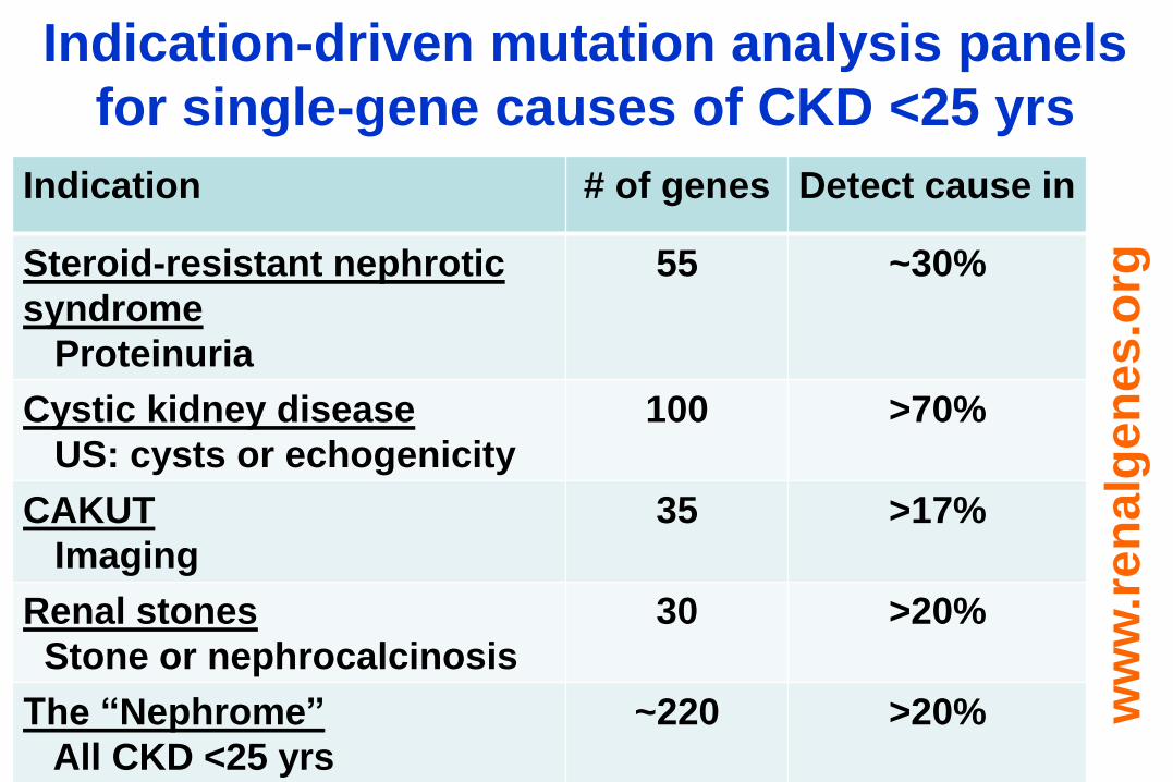
Indication-driven mutation analysis panels
for single-gene causes of CKD <25 yrs
Indication # of genes Detect cause in
Steroid-resistant nephrotic
syndrome
Proteinuria
55 ~30%
Cystic kidney disease
US: cysts or echogenicity
100 >70%
CAKUT
Imaging
35 >17%
Renal stones
Stone or nephrocalcinosis
30 >20%
The “Nephrome”
All CKD <25 yrs
~220 >20% ww
w.r
en
alg
en
es.o
rg

State of the ArtEvery patient with a kidney disease caused by a single-
gene mutation should have a chance at having this
mutation identified (if consenting), because:
… it is now feasible
… provides an unequivocal diagnosis
… may reveal a potential treatment
… allows etiologic classification for therapeutic trials,
and potentially personalized treatment
… provides the missing pieces for
the puzzle of pathogenic pathways
… enables generation of gene-specific animal models
… enables screening for therapeutic molecules

–Halbritter JASN 26:543, 2015
–Braun cJASN 11:664, 2016
–Braun Kidney Internat 89:468, 2016
–Clissold RL Nature Rev Neph 11:102, 2015
–Bockenhauer D Pediatr Nephrol 31:707, 2016
References: