general pharmacology - humsc
TRANSCRIPT


1
GENERAL PHARMACOLOGY
This chapter provides basic knowledge necessary for subsequent study of
individual drugs. Important terms & definitions are presented, together with the
two basic areas of pharmacology; pharmacodynamics & pharmacokinetics.
Pharmacology
It is the science that deals with drugs, their nature, pharmacodynamics,
pharmacokinetics, therapeutic uses, adverse effects, preparations and
administration.
Drug
It is a chemical substance that alters body functions and can be used for
treatment, prevention or diagnosis of disease.
Pharmacokinetics
They are the studies of the Absorption, Distribution, Metabolism and
Excretion of drugs (ADME) and their mathematical relationship, i.e. what
body does to drugs.
Ph.Kinetics
Body Drug Ph.Dynamics
Pharmacodynamics
They are the studies of the biological and therapeutic effects of drugs and
their mechanism of action, i.e. what drugs do to the body.
Pharmacotherapeutics:
Study the selection & use of the drugs for treatment, prevention or
diagnosis of diseases

2
Sources of drugs:
1. Plant: e.g. atropine from leaves of belladonna
2. Animal: insulin from the pancreas of pigs
3. Mineral: MgSO4, iodine
4. Microorganisms: penicillin from the fungus penicillinum
5. Synthetic: in laboratory e.g. aspirin
6. Biotechnology: human insulin by genetic engineering
Drug nomenclature:
1. Chemical name: N-acetyl p-aminophen
2. Generic name: Acetaminophen (paracetamol)
3. Trade name: Panadol- Adol
P H A R M A C O K I N E T I C S
The term pharmacokinetics denotes the quantitative studying of drug
Absorption, Distribution, Metabolism and Excretion (ADME) and their
mathematical relationship.
Target Site
Other Sites
Therapeutic effect
Adverse effect
Fate of the drug in the body

3
ABSORPTION OF DRUGS
Definition: absorption is the passage of drug from the site of administration
to the systemic circulation.
Methods of transport across cell membranes:
1- Passive transport:
a. Simple (lipid) diffusion: the lipid soluble drugs can easily
cross lipid membranes along concentration gradient with no
energy.
b. Aqueous diffusion (filtration): the water soluble drugs can
pass only through water filled pores or channels.
2- Carrier-mediated (facilitated) transport: the drug passes
across cell membrane by specialized carrier molecules (which are
sites for saturation & competition):
a. Facilitated diffusion: as simple diffusion but with aid of
carrier. e.g. glucose uptake
b. Active transport: the drug is carried against concentration
gradient by energy. e.g. Na/K pump
3- Endocytosis (pinocytosis): it occurs in cases of large molecule
by invagination of part of cell membrane and engulfing the drug
molecule. Energy is needed. e.g. absorption of vit.B12 & intrinsic
factor in terminal ileum.

4
Factors affecting drug absorption:
A. Factors related to drug:
1. Molecular size: small molecules are absorbed than large molecules
2. Pharmaceutical preparations
- Dosage form: - solutions are better absorbed than suspensions
- sustained-release preparations are slow in absorption
- Shape & size of particles and rates of disintegration & dissolution:
Rapid with paracetamol and slow with digoxin
- Excipient (filer): Ca++
salts →↓ absorption of tetracyclines
3. Nature: inorganic (small molecules) is better than organic (large molecules)
4. Valency: ferrous (Fe++
) is better absorbed than ferric (Fe+++
)
5. Lipid and water solubility:
- Drug must be water soluble as well as lipid soluble
- More lipid solubility → high lipid/water partition coefficient → better
absorption
6. Ionization: - Ionized (polar or charged) forms are poorly absorbed
- Unionized (non-polar or non-charged) forms are more absorbed
e.g. - Quaternary ammonium compounds→ ionized → poor absorption
- Tertiary amines (physostigmine) → unionized → better absorption
Most drugs are either weak acids or weak bases.
Acidic drugs (HA) release an H+ producing a charged anion (A-):
Weak bases (BH+) can also release an H+ producing the uncharged base
(B):

5
Ionization depend on pH of the medium and pka of the drug (pKa is a
measure of the strength of the interaction of a compound with a proton).
The lower the pKa of a drug, the more acidic is the drug. Conversely,
the higher the pKa, the more basic is the drug.
Relation between pH of the medium and pka of the drug is presented by
(Henderson-Hasselbach equation):
pka = pH + log concentration of protonated
concentration of nonprotonated
® If the drug is weak Acid :
pka = pH + log concentration of Unionized acid
concentration of ionized acid
® lf the drug is weak base:
pKa=pH + log concentration of the ionizd base
concentration of unionized base
pKa of a drug : is the pH at which 50% of the drug molecules exist in
the ionized form & 50% in the unionized form.
Clinical Significance of pKa
1. GIT: knowing site of drug absorption:
Acidic drugs (e.g. Aspirin) become mostly unionized in acidic pH
Basic drugs (e.g.Amphetamine) become mostly unionized in alkaline pH
Streptomycin has a high pKa→ always ionized→ very poor oral
absorption

6
Ion trapping of aspirin: Aspirin (pKa = 3.5) in the empty stomach (pH
= 1.5) → more unionized → more absorbable into gastric cells, but once
entered the cells (pH = 7.4) becomes more ionized → trapped inside
these cells (aspirin trap) → death of the cells inducing “peptic
ulceration”.
2. Kidney: treatment of drug toxicity
In drug poisoning, changing urinary pH → increases drug ionization and
inhibits tubular reabsorption:
Alkalinization of urine is useful in acidic drug poisoning e.g. aspirin.
Acidification of urine is used in basic drug poisoning, e.g. amphetamine.
B. Factors related to patient:
1. Route of administration: IV > Inhalation > IM > SC > Oral > Skin
2. Absorbing surface:
a. Vascularity: Alveoli > skeletal muscle > subcutaneous
b. Surface area: Intestine > Stomach
c. State of health: Diarrhea & malabsorption ↓↓ oral absorption
3. Systemic circulation: Shock & heart failure ↓↓ absorption
4. Specific factors: intrinsic factor for vit.B12
5. Presence of other drugs: - vit.C ↑ absorption of iron
- Activated charcoal ↓↓ oral absorption of most of drugs
- Adrenaline SC → VC → ↓↓ absorption of local anesthetics → longer
duration of action
7
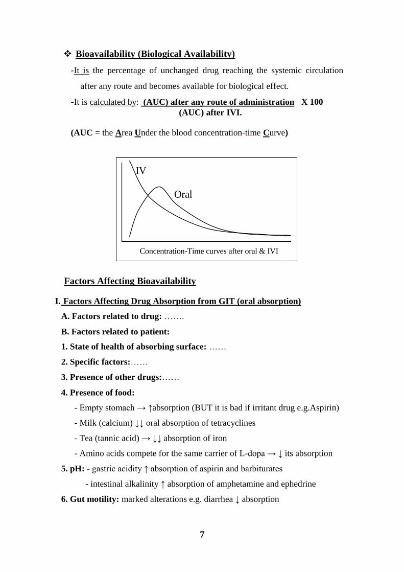
Bioavailability (Biological Availability)
-It is the percentage of unchanged drug reaching the systemic circulation
after any route and becomes available for biological effect.
-It is calculated by: (AUC) after any route of administration X 100
(AUC) after IVI.
(AUC = the Area Under the blood concentration-time Curve)
Factors Affecting Bioavailability
I. Factors Affecting Drug Absorption from GIT (oral absorption)
A. Factors related to drug: …….
B. Factors related to patient:
1. State of health of absorbing surface: ……
2. Specific factors:……
3. Presence of other drugs:……
4. Presence of food:
- Empty stomach → ↑absorption (BUT it is bad if irritant drug e.g.Aspirin)
- Milk (calcium) ↓↓ oral absorption of tetracyclines
- Tea (tannic acid) → ↓↓ absorption of iron
- Amino acids compete for the same carrier of L-dopa → ↓ its absorption
5. pH: - gastric acidity ↑ absorption of aspirin and barbiturates
- intestinal alkalinity ↑ absorption of amphetamine and ephedrine
6. Gut motility: marked alterations e.g. diarrhea ↓ absorption
Concentration-Time curves after oral & IVI
Oral
IV

8
7. Gastric emptying:
a. Metocloperamide → ↑ emptying →
- ↑ absorption of paracetamol (rapid rate of disintegration & dissolution)
- ↓ absorption of digoxin (slow rate of disintegration & dissolution)
b. Atropine → ↓ emptying →the REVERSE effects
II. First-Pass Effect (First-Pass Metabolism; Presystemic Elimination)
It is the metabolism of some drugs in a single passage through the liver,
gut wall or the lungs before reaching the systemic circulation.
A. Hepatic 1ST
pass effect: drugs absorbed from the GIT are carried first
in the portal circulation to the liver. Some drugs are extensively
metabolized in their first-pass e.g. nitroglycerin & propranolol.
Factors reducing Hepatic 1st pass metabolism
Reduction in portal blood flow: portal hypertension, propranolol.
Inhibition of hepatic enzymes: liver failure - enzyme inhibitors; erythromycin.
B. Gut 1ST
pass effect:
Gastric acidity: benzyl penicillin
Digestive enzymes: insulin & pituitary hormones
Mucosal enzyme: L-dopa , alpha-methyldopa
C. Pulmonary metabolism: after aerosol inhalation (nicotine).
How to overcome the First-pass Effect
1. Increase oral dose
2. Other routes: Sublingual - Parenteral - Rectal (to some extent)

9
DISTRIBUTION OF DRUGS
It is the passage of drug through body compartments which are separated by
capillary walls and cell membranes.
Body fluid compartments:
1- intravascular volume = 4 L/70 Kg
2- interstitial volume = 10 L/70 Kg
(Extracellular volume)
3- intracellular volume = 28 L/70 Kg
*Total body fluid = 42 L/70 Kg
Pattern of distribution:
1. Plasma compartment (one compartmental model):
If a drug:
- has a high molecular weight or
- binds strongly to plasma proteins
It is too large to move out through the endothelial slit junctions of the
capillaries and, thus, is effectively trapped within the plasma (vascular)
compartment.
e.g. Heparin , Dextran.
2. Extracellular fluid (two compartmental models):
If a drug has a low molecular weight and is hydrophilic
It can move through the endothelial slit junctions of the capillaries into the
interstitial fluid BUT cannot move across the lipid membranes of cells
e.g. Aminoglycoside antibiotics, Mannitol.
3. Extra & intracellular fluid (multi-compartmental model)
If a drug has a low molecular weight and is lipophilic
It moves into the interstitium through the slit junctions and
also moves through the cell membranes into the intracellular fluid.
Some drugs uniformly distribute throughout whole body water e.g. Ethanol,
sulphonamides.

10
the majority of drugs distribute into several compartments, often binding
cellular components for example, lipids (abundant in adipocytes
and cell membranes), proteins (abundant in plasma and within
cells), or nucleic acids (abundant in the nuclei of cells)
4. Tissue reservoir: Drugs concentrated in certain tissues
Iodine in thyroid & salivary glands
Calcium & tetracyclines in bone & teeth
Chloroquine in liver
Thiopental in fat (Redistribution ??)
Volume of Distribution (Vd)
Definition: the apparent volume of fluid required to accommodate the
entire amount of the drug in the body in the same concentration as that
present in plasma (i.e. when the drug is equally distributed between plasma
and tissues).
The apparent volume of distribution does not describe a real, physical volume, but rather,
reflects the ratio of drug in the extraplasmic spaces relative to the plasma space as it
assumes that the drug distributes uniformly, in a single compartment.
Example: The Vd for digoxin is 6 L/Kg (in adult 70 Kg), this means that:
- Each 1 Kg of tissue takes 6-fold the concentration of digoxin in 1 L of
plasma
- If we want to distribute digoxin equally between all body tissues and
plasma, we need an imaginary volume of plasma= 70 X 6 = 420 L
Vd (L) = Amount of drug in the body
Plasma concentration
( Vd = A/C or Q/C)

11
Importance of Vd
1. It is an estimate of the extent of tissue uptake of drugs:
Small Vd (e.g. frusemide) indicates that tissue uptake is limited.
Large Vd (e.g. digoxin) indicates extensive tissue distribution.
2. In cases of drug toxicity:
Dialysis is not useful for high Vd drugs (most of drug is in the
tissues).
Dialysis is useful for low Vd drugs (most of drug is in the blood).
3. Vd can be used to calculate the loading dose (LD):
[LD = Vd x Css (Steady State plasma Concentration)]
4. Vd can be used to calculate the total amount of drug in the body:
[A = Vd x Cp ]
Factors Affecting Distribution of Drugs:
1) Perfusion: the amount of the drug which is delivered to a particular organ
depends on the blood flow to that organ: ↑ blood flow → ↑ distribution.
2) Diffusion: the ability of the drug to diffuse across the cell membranes is
governed by its lipophilicity, ionization & molecular weight: (as absorption)
3) Binding to plasma proteins (PPs):
Most of drugs when introduced into the body are bound to plasma
proteins (pp) e.g.
- Albumin: - the most important pp
- Acidic & lipophilic drugs bind mainly with it
- Other: globulin, glycoprotein…etc
Drug in blood exists in 2 forms: free form & plasma protein bound
form which exist in equilibrium; when the free form is metabolized
and/or excreted, another part is released from plasma proteins

12
Free fraction Bound fraction
Active
Diffusible
Can be Metabolized
Can be Excreted
Inactive
Nondiffusible
Cannot be metabolized
Cannot be excreted
Act as a reservoir for drug
Significance of Binding to Plasma Proteins
1. The binding of drug to plasma proteins limits its tissue penetration &
decreases its Vd.
2. The bound drug cannot be eliminated → prolongs the t½ of the drug
→ prolongs the effect of drug.
3. Hyboalbuminemia e.g. starvation, malnutrition →↑ free drug →
therapeutic dose changes to toxic dose e.g. phenytoin.
4. Competition for binding sites between drugs → displacement of each
other → clinically-significant drug interactions e.g.
- Aspirin, sulphonamide displace warfarin → bleeding.
- Sulphonamide displaces bilirubin → kernicterus in premature neonates.
{When two drugs with high affinity for albumin are given, they compete for the available
binding sites. The drugs with high affinity for albumin can be divided into two classes:
1. Class I drugs: If the dose of drug is less than the binding capacity of albumin i.e. low
dose/capacity ratio → high bound fraction and small free fraction
2. Class II drugs: If the doses greatly exceed the number of albumin binding sites i.e. high
dose/capacity ratio → high free fraction.
* When a patient taking a Class I drug, such as warfarin, is given a Class II drug, such as a
sulfonamide antibiotic. Sulfonamide displaces warfarin from albumin, leading to a rapid
increase in the concentration of free warfarin in plasma → ↑ therapeutic effects, as well as ↑
toxic effects → bleeding}

13
4) Binding to cell and tissue constituents:
Drugs concentrated in certain tissues (Tissue reservoir).
Passage across barriers:
Passage of Drugs to CNS
1. Lipid-soluble drugs pass freely through BBB, e.g. general anesthetics and other
CNS depressants.
2. 3ry amines can pass while 4ry
NH4+ compounds (ionized) cannot.
3. Some hydrophilic antibiotics e.g. penicillin can pass inflamed BBB only
Passage of Drugs to the Fetus
Many drugs cross placental barrier by simple diffusion (depending on their lipid
solubility & their degree of ionization) and can harm the fetus:
Drugs given in 3rd
to 10th
week of pregnancy → teratogenicity e.g.
thalidomide → phocomelia
Oral anticoagulants → fatal hemorrhage in the newborn.
Oral hypoglycemics (sulfonylureas) → prolonged neonatal hypoglycemia.
Aminoglycosides → 8th
cranial nerve damage.
During labor, Morphine → respiratory depression (asphyxia neonatorum).
Passage of drugs to breast milk
Most of drugs administrated to lactating women are detectable in breast milk.
pH of milk is more acidic (7.0) than that of plasma (7.4) basic drugs
accumulate in milk (ion trapping).
Milk contains more fat than plasma retention of lipid soluble drugs.
Drugs are contraindicated during lactation:
Sedatives, hypnotics and narcotics → CNS depression in baby.
Oral penicillins and purgatives → diarrhea in baby.
Anticancer drugs → decrease growth of baby.
Bromocriptine & sex hormones → suppress lactation.

14
BIOTRANSFORMATION
(Metabolism)
These are: the chemical changes that occur to drugs after absorption until
excretion.
Drug metabolism occurs mainly in the liver, also in other organs, e.g.
intestinal lumen or wall, lung, plasma, skin and kidney.
The aim of drug metabolism is the conversion of the lipophilic drug to a
more polar (hydrophilic, ionized) metabolite which is easily excreted in
urine.
The hydrophilic drugs usually do not undergo metabolism and secreted
unchanged in urine
Types of Biotransformation Reactions
Phase I (Non-Synthetic)
Phase I reactions include: oxidation - reduction - hydrolysis.
The most important reaction is oxidation by cytochrome P450
enzyme system.
Phase I reactions result in unmasking of a polar group (-OH, -SH, or -
NH2) → an ionized metabolite that can be easily excreted.
Phase II (Synthetic)
An endogenous substrate, (e.g. glucuronic acid, glycine, glutathione,
sulfate or acetic acid) is conjugated with the functional group of the
drug or its metabolite → nontoxic highly polar, rapidly eliminated
conjugates.
The most important is conjugated with glucuronic acid.

15
Phase I reactions
A. Oxidation:
The most important is cytochrome P450 oxidases “CYP” (mixed
function oxidases) which are hepatic microsomal enzymes
CYP is further classified by family, subfamily & gene into many isozymes. The name of
each one is designated by the term CYP followed by 3 characters e.g. CYP 2C9:
1. The first Arabic numeral represents the family.
2. The alphabetic letter represents the subfamily.
3.The second Arabic numeral represents the individual gene within the subfamily.
Xanthine oxidase: converts xanthine → uric acid
Monoamine oxidase (MAO): oxidizes catecholamines & serotonin
B. Reduction:
- Nitroreductase → chloramphenicol
- Carbonyl reductase → naloxone
C. Hydrolysis:
It occurs mainly non-microsomal (in plasma and body fluids)
- Cholinestrase → Ach.
- Peptidase → insulin
Consequences of phase I reactions:
The activity of the drug is modified in one of the following ways:
1- Active drugs → inactive drugs (occurs with most drugs).
2- Inactive drugs (prodrugs) → active drugs, e.g. cortisone to cortisol
(hydrocortisone).
3- Active drug → another active one, e.g. codeine to morphine.
4- Active drug → a toxic metabolite e.g. methanol → formaldehyde →
retinotoxic & paracetamol → toxic metabolite (NAPQI) → hepatotoxic
in case of toxicity

16
Phase II reactions
A. Glucuronide conjugation:
It is the most common conjugation reaction
Glucuronide conjugates secreted in bile may be hydrolyzed by intestinal
bacteria and free drug can be reabsorbed again i.e. enterohepatic
circulation → prolong duration of drug action e.g. estrogen (so
contraceptive pills are given once daily)
B. Non-Glucuronide conjugation:
Sulphate formation e.g. steroids
Glycine conjugation e.g. salicylic acid
Glutathione conjugation e.g. ethacrynic acid
Acetyl conjugation (slow & rapid acetylation) e.g. isoniazid
Consequences of phase II reactions:
Mostly result in drug inactivation
Some exceptions can occur e.g. morphine is partially converted into
morphine-6-glucuronide (active metabolite)
o Most of drugs is metabolizes by phase-I followed by phase-II reactions
- Some drugs is metabolizes firstly by phase-II then by phase-I
reactions e.g. isoniazid.
- Some drugs undergo phase-I or phase-II only
o Types of enzymes responsible for biotransformation reactions
Microsomal Enzyme
Cytoplasmic EnzymePlasma Enzyme
e.g. xanthine
oxidase
e.g. Ach
esterase
Non-microsomal Enzyme
e.g. Cyt. P450 oxidase
for oxidation
e.g. glucuronyl transferase
for conjugation

17
Microsomal enzymes Non-microsomal enzymes
Site: in the liver, in microsomes of ER. So,
they are called hepatic microsomal enzymes
Present in liver, GIT, lung, kidney, plasma,
skin: in cytoplasm and mitochondria
Reactions:
Phase-I: Oxidation
Reduction
Hydrolysis (few reactions)
Phase-II: Glucuronic a. conjugation Only
Reactions:
Phase-I: Oxidation
Reduction
Hydrolysis (mostly)
Phase-II: All Conjugations Except Glucuronic
Only
Substrate: lipophilic drugs & bilirubin Lipophilic , hydrophilic drugs (to terminate
action as succinylcholine) & natural body
constituents
Affection by drugs: Inducible Non-inducible
Factors Affecting Biotransformation:
1. Drugs: (Enzyme induction & enzyme inhibition).
Some drugs and environmental substances can induce or inhibit the
microsomal enzyme activity and lead to undesirable drug interactions
Clinical significance of Enzyme Induction:
Drugs stimulating the microsomal enzyme systems ↑ activity
↑ their own metabolism → tolerance e.g. phenobaritone.
↑ metabolism of other drugs metabolized by these enzymes and are
given at same time→ drug interactions e.g.:
- Rifampicin →↑ oral contraceptive metabolism → pregnancy
- Phenytoin →↑ cyclosporine metabolism → transplant rejection
- Rifampicin →↑ warfarin metabolism → therapeutic failure.
↑ metabolism of endogenous substrates e.g. phenobarbitone →
↑ elimination of bilirubin → used in treatment of neonatal jaundice)
↑ metabolism of vitamins e.g. phenytoin → ↑ of vit.D, vit.K, folic acid
→ osteomalacia, bleeding and megaloplastic anemia

18
Enzyme induction is reversible. It occurs over a few days-months and
passes off over 2-3 weeks after withdrawal of the inducer.
Clinical significance of Enzyme Inhibition:
Drugs inhibiting the microsomal enzyme systems ↓ activity
↓ their own metabolism →↑ drug level.
↓ metabolism of other drugs metabolized by these enzymes → drug
interactions e.g.:
- Ciprofloxacin →↓ warfarin metabolism → bleeding
- Cimetidine →↓ carbamazepine metabolism → toxicity
It occurs faster than enzyme induction.
2. Pathological factors which affect hepatic activity e.g. liver failure
starvation, cancer → ↓ activity of HME → need to adjust dose.
3. Pharmacogenetic variations in metabolizing enzymes e.g. slow &
fast acetylators (see pharmacogenetics).
4. Hepatic blood flow: drugs ↓ hepatic blood flow (e.g. propranolol) →
↓drug matabolism
5. Age: ↓ enzymatic activity in extremities of age
Premature babies ↓ conjugate of chloramphenicol → fatal gray
baby syndrome.
Examples of Enzyme Inhibitors
Cimetidine- chloramphenicol - ciprofloxacin- erythromycin - ketocenazol -
♀ (F) estrogen, progesterone, contraceptive pills.
Examples of Enzyme Inducers
Phenytoin & carbamazepine- phenobarbitone – rifampicin -
griseofulvin - ♂ androgen- nicotine- chronic alcohol ingestion.

19
6. Sex: female sex hormones are HME inhibitors → receive lower doses
than male.
7. Drug properties: lipophilicity → hepatic metabolism of drugs.
8. Drug dosage: toxic dose can deplete substances needed for drug
detoxification e.g. paracetamol toxic dose → depletion of GSH→
accumulation of toxic metabolite NAPQI
EXCRETION OF DRUGS
1- The kidney:
It is the most important route of excretion. It occurs through:
1. Glomerular filtration:
For hydrophilic free (non-bound) drugs with M.W. < 500 (i.e. < the
glomerular pores). e.g. mannitol
2. Active tubular secretion: through special transport system (carrier) →
saturable & site for competition.
Acid carrier e.g. for penicillins, probenecid, frusemide, uric acid
- Probenecid →↓ tubular secretion of penicillin→↑ duration of
action of penicillin
- frusemide →↓ tubular secretion of uric acid →hyperuricemia
as an adverse effect.
basic carrier e.g. for digoxin, quinidine.
3. Active tubular reabsorption:
Unionized form of drug (lipophilic) → tubular reabsorption
Factors affecting glomerular filtration
Glomerular filtration rate (GFR)
Plasma protein binding (PPB) prevents filtration

20
Changes in urinary pH: affect excretion of drugs
Alkalinization of urine (Na or K Acetate, Bicarbonate, Citrate) →
↑ renal excretion of weak acid drugs e.g. Aspirin, Barbiturates
Acidification of urine (NH4Cl or Ascorbic acid "vit.C") → ↑ renal
excretion of weak base drugs e.g. amphetamine. ephedrine
2- GIT:
* Saliva: e.g. Morphine, Iodine, Metronidazole → metallic taste
* Stomach: e.g. Morphine→ gastric wash is done in aute morphine
toxicity despite it is administrated by IV route.
* Bile: in active or conjugated form → intestine → EITHER
o Excreted in large intestine→ stool
o Reabsorbed → enterohepatic circulation e.g. Morphine, Rifampicin
o Some antibacterials are excreted in bile in an active form → useful
in: - treatment of cholecystitis & typhoid fever e.g. Ampicillin
- patients with renal impairment (No need for dose adjustment)
* Stool: conjugated metabolites & poorly absorbed orally
3- Lungs: e.g. volatile liquids (inhalant general anesthesia), gases (CO2)
4- Sweat: e.g. Rifampicine → red discoloration of sweat
5- Breast Milk: - Many drugs are excreted in breast milk → can affect baby
- lipid soluble and basic drugs are trapped in breast milk
Significance of lipophilicity
* Lipophilicity → ↑ drug absorption. * Lipophilicity → ↑ Vd.
* Lipophilicity → ↑ hepatic elimination.
* Lipophilicity → ↑ renal reabsorption & ↓ renal excretion.

21
PARAMETERS OF ELIMINATION
1. Systemic clearance (Cls)
Definition
It the volume of a fluid cleared from the drug per unit time.
Cls = Kel X Vd
- Kel → Elimination rate constant = 0.693
t1/2
[(0.693) is the natural logarithm of 2 (i.e. In 2) and gets into the equation because (t1/2)
involves a halving of concentration → -Kel= In(C2/C1) = In (1/2) → Kel = In(2)]
t1/2 t1/2 t1/2
- So, systemic clearance Cl = 0.693 X Vd
t1/2
The systemic clearance is equal to the sum of individual organs clearances
i.e. the clearnce by the liver, kidney, lung, ….etc.
Cls = renal clearance (Clr) + non-renal clearance (Clnr)
Factors affecting drug clearance
1. Blood flow to the clearing organ (directly proportional).
2. Binding of the drug to plasma proteins (inversely proportional).
3. Activity of processes responsible for drug removal as hepatic enzymes,
glomerular filtration rate and secretory processes (directly proportional).
Significance of clearance
1. Calculation of the maintenance dose (MD)
2. Adjustment of the dosing regimen for drugs eliminated by glomerular
filtration e.g. dosing of gentamicin
Clsystemic
= Cllung Cl
liverCl
kidney+ + + . . .

22
2. Plasma (elimination) half life (t½)
Definition
It is the time required to eliminate 50% of drug from plasma.
Calculation:
It depends on: Clearance & Vd
The larger the Vd, the longer the t½ (it takes longer to remove drug from
deep within tissue). The larger the Cl, the shorter the t½
t½ = 0.693 X Vd
Cls
Value of elimination t½
1. It determines the dosage interval ( or Tm).
If = t½ this is an accepted choice to avoid wide fluctuations of
the peak (highest pl.conc. of the drug) and trough (lowest
pl.conc.).
If < t½ more drug accumulation occurs.
If > t½ decrease in drug concentration
occurs between doses.
2. It indicates Tss (time required to attain Css): it is equal
to 5 t½ (after 4 t½ ; > 95% of the Css is attained)
3. It indicates the time needed for complete elimination: occure after 5 t½
4. Drugs having long t½ are given once/day
Conc.
Time
C
1/2 C
t1/2
{

23
Factors affecting elimination t½
1. The state of the eliminating organs i.e. liver & kidney functions
2. The delivery of the drug to the eliminating organs e.g.:
a. Plasma protein binding limits renal filtration and increase t½
b. Drugs with very high Vd may escape from elimination in the tissues
and increase t½
c. Blood flow (decrease renal bl.flow in HF may increase t½)
3. Steady state concentration (Css)
Definition: the steady level of drug in plasma achieved when the rate of
administration equals the rate of elimination.
The rule of (5):
The Cpss is reached after 5 t½
If we change the dose, the new Cpss is reached after 5 t½
If dosing stop, complete elimination of drug occurs after 5 t½

24
4. Loading dose (LD)
Loading dose (LD): the dose given at the onset of therapy to achieve a
rapid increase in plasma drug concentration to reach Cpss without toxicity.
LDIV = Vd X Css (target CP)
LDOral = LDIV
F (fraction of oral bioavailability) - Used for:
1. drugs with Long t1/2 (e.g. amiodaron) or
2. in an Emergency
5. Maintenance dose (MD)
Maintenance dose (MD): the dose needed to keep the plasma drug
concentration constant at Css (the dose needed to compensate the amount
eliminated).
- Dosing rate (rate of administration) = rate of elimination = Cl X Css
- If drug taken by continuous IV infusion:
Infusion rate = CLs X Css
- If drug taken in repetitive doses:
MDIV = CLs X Css X Tm (dosing interval)
MDOral = CLs X Css X Tm
F (fraction of oral bioavailability)

25
6. Kinetic orders
A. First order kinetics B. Zero order (saturation) Kinetics
A constant fraction of drug is
eliminated per unit time.
A constant amount of drug is eliminated
per unit time.
Rate of elimination is proportional to
the concentration of drug
Rate of elimination is constant (limited
capacity of kinetics due to saturation of
involved enzymes and/or carriers
It has a linear elimination kinetics i.e.
plasma concentration can be expected at
any time (using log conc.-time
disappearance curve)
It has a non-linear elimination kinetics i.e.
plasma concentration can NOT be expected
at any time (using log conc.-time
disappearance curve)
Constant t½. t½ is not constant
A steady state concentration (Css) is
reached on repeated dosing after 5 t1/2.
NO Css is reached; repeated dosing
overshooting of drug concentration.
Modest changes in dose are usually
tolerated because when drug conc. ↑→
elimination ↑ by the same ratio.
Modest changes in dose toxicity due
to drug cumulation
Drug metabolites do Not vary with dose. Drug metabolites may vary with dose
Examples: Most drugs. Example: Large dose of Aspirin, Alcohol,
Phenytoin (they follow 1st order kinetics at
small doses)
C ss
Conc.
Time
Conc.
Time

26
PHARMACODYNAMICS
Types of Drug Action:
Local or topical action: drugs act on site of application e.g. ointment or
eye drops.
Systemic or general action: the drug acts after administration and
distribution by circulation to various tissues. e.g. Aspirin
Reflex or remote action: the drug acts locally at one site to produce
reflex action elsewhere. e.g. Ammonia inhalation → irritation of nose →
reflex stimulation of respiration
Mechanism (Mode) of Action of Drugs
Drugs can induce a tissue response, initially through:
I. Body control systems (the regulatory proteins): involving
interactions with:
(1) Receptors (2) Ion channels
(3) Enzymes (4) Carrier molecules
II. Other mechanisms:
(5) Subcellular structures (6) Genetic apparatus
(7) Physical mechanisms (8) Chemical mechanisms
1) Receptor-Mediated Mechanisms
Receptors are specific cellular macromolecules (usually proteins) that
interact with a ligand (binding) to produce a response.
Ligand: any molecule that can combine with the receptor. A ligand that
activates receptor is called agonist. A ligand that blocks the receptor is
called antagonist

27
Types of receptors (signaling mechanisms or signal transduction):
1. Ligand-gated ion channels: (for fast neurotransmitters)
Receptors are ion-selective channels in the plasma membrane.
- Binding of agonist to the extracellular part of receptor →opening
of the channel → alteration in membrane potential or change in
intracellular ion concentration → change in cell activity,
- e.g. GABAA receptors (Cl- channels).
2. G protein-Coupled Receptors (for slow neurotransmitters)
Receptor consists of 7 transmembrane subunits which are linked to
G proteins.
The G protein is a trimer (, and ).
Agonist binding → dissociation of subunit which regulates activity of
several effectors.

28
Types of G Proteins
a. Gs (stimulatory) → increased cAMP → activation of specific proteins.
b. Gi (inhibitory) → decreased cAMP → inhibition of specific proteins.
c. Gq (query) → increased DAG (diacylglycerol) and IP3 (inositol
triphosphate) → increased intracellular Ca++
and activate PKC (protein
kinase C)
- Examples: β-adrenergic receptors linked to Gs protein
α2- adrenergic receptors linked to Gi protein
α1- adrenergic receptors linked to Gq protein
3. Receptors linked to Tyrosine Kinase (RTKs)
The receptor is formed of two domains:
a. An extracellular domain, to which the agonist binds.
b. An intracellular domain, which is a tyrosine kinase enzyme (effector).
c. A transmembrane segment connecting two domains.
- e.g. insulin receptors
4. Intracellular (DNA-linked) receptors (very slow)
The ligand enter the target cell and combine with intracellular receptor
proteins → complex → acts on nuclear DNA → modify transcription of
the nearby gene → modify protein production → changes in the structure
or function of the target tissue.
Examples: receptors for corticosteroids, sex hormones, thyroid
hormones and vitamin D

29
5. Nitric Oxide (NO) Receptors:
NO receptors are protein receptors inside the cell. Binding of NO
receptors → formation of a "second messenger" within the cell.
The most common: NO activates guanylyl cyclase enzyme → cyclic
GMP (cGMP).
NO receptors are activated by many drugs that increase NO level e.g.
nitroglycerine.
Biological response to drug-receptor binding:
Affinity Ka
Efficacy
Drug (D) + Receptor (R) D/R complex Response Kd
- Affinity: ability of drug to bind with the receptor to form D/R complex
- Efficacy: ability of D/R complex to evoke a response.
- Ka is the association constant
- Kd is the dissociation constant
When a drug combines with a receptor, this may lead to:
1- Agonist effect or 2- Antagonist effect or 3- Partial agonist effect

30
1. Agonist effect:
Agonist has 1. Affinity 2. High Efficacy 3. Rapid rate of ass. & diss.
Theories for drug-receptor interaction:
1. Receptor occupation theory: response (efficacy) depends on
number of occupied receptors
- When maximum effect is reached, still some receptors remain free
(spare receptors)
2. Rate theory: response (efficacy) depends on rate of association (Ka)
and rate of dissociation (Kd)
Response will never exceed a certain limit whatever the drug
concentration. This is termed Emax i.e. the maximal response or effect
e.g. - acetylcholine (Ach) activates nicotinic receptors → skeletal
muscle contraction.
- adrenaline activates beta adrenoceptors → increased HR
They are 2 types of drug responses:
1. Graded dose-response: the response increases by increasing the agonist
e.g. increases of heart rate against different doses of adrenaline.
2. Quantal dose-response : the response is all or none e.g. the % of
epileptic patients who are treated by different doses of an antiepileptic
drug

31
2. Antagonist effect:
Antagonist has: 1. Affinity 2. No Efficacy 3. Slow Rate of ass. & diss.
Types of receptor (pharmacological) antagonists:
1- Competitive Antagonist 2- Noncompetitive Antagonist
Antagonist competes with the
agonist for the same recognition
site of the receptor.
Antagonist binds to an allosteric site
i.e. a site away from recognition site
but can affect its binding with agonist
Duration of antagonism depends
on the relative plasma
concentrations of agonist and
antagonist.
Antagonist can be Displaced by
excess agonist (surmountable)
Duration of antagonism depends on
synthesis of new receptors or
antagonist metabolism
Antagonist can Not be Displaced by
agonist (non-surmountable)
Causes parallel shift to the right
in the dose-response curve i.e. No
change in Emax but ↓↓in potency.
Causes downward & non-parallel
shift in the dose-response curve i.e.
↓↓ in Emax , ↓↓ in potency(?!)
Examples: Atropine Non-competitive is divided into:
Reversible:
1-bind reversibly to receptor
2- Duration depends on metabolism
of antagonist
3- usually of short duration
4- e.g. Succinylcholine,
Irreversible:
1-bind covalently to receptor
2- Duration depends on resynthesis of
new receptors.
3- usually of long duration
4- e.g. phenoxybenzamine

32
3. Partial Agonist (Agonist-Antagonist)
In absence of the agonist: it has:
1. Affinity
2. Moderate efficacy (submaximal effect) whatever its concentration.
3. Moderate or slow rate of association & dissociation.
In the presence of the agonist, it acts as an antagonist i.e blocks effect of
agonist.
e.g. Succinylcholine , Nicotine L.D.
2) Drugs acting on ion channels: drugs can modulate ion channels
through:
Voltage-gated ion channels: Local anesthetics (Na+ channels).
ATPase-sensitive ion channels: Oral hypoglycemics (ATPase-sensitive
K+
channels in pancreatic β cells)
Ion channels modulated by G protein-linked receptors (2ry
messenger)
Ligand-gated ion channels (ion channel-linked receptors)
Receptor Cycling or Turnover
Binding of the agonist number of receptors [down regulation]
Binding of the antagonist the number of receptors [up regulation]

33
3) Drugs Acting on Enzymes: drugs can modulate enzyme through:
Activation of enzyme systems.
Inhibition of enzyme:
- Reversible inhibition of enzyme e.g Neostigmine on cholinesterase
- Irreversible inhibition of enzyme e.g OPC on cholinesterase
False substrate for enzyme e.g. α-methyldopa on dopa-decarboxylase
4) Drugs Acting on carrier systems
Drugs may affect carrier systems or transport processes in the
plasmatic membrane. Examples:
- Digitalis inhibit Na+/K
+ ATPase pump in cardiac cell.
- Diuretics affect ions transporters in renal tubules
5) Drugs Acting on Subcellular Structures
Microtubules: Colchicine disrupts microtubules inhibiting mitosis.
6) Drugs Acting on the Genetic Apparatus
Aminoglycosides inhibit bacterial protein synthesis.
Anticancer drugs affect DNA synthesis or function.
7) Drugs Acting Physically:
Demulcents (soothing): bismuth salts coat intestinal mucosa.
Lubricants: liquid paraffin is used in constipation.
Adsorbent: Kaolin in treatment of diarrhea
Activated charcoal in treatment of drug toxicity
8) Drugs Acting Chemically:
a. Neutralization: - Antacids neutralize HCL in peptic ulcer.
- Protamin sulfate (basic, +ve) for toxcicty of heparin (acidic, -ve)
b. Chelation; is the capacity of organic compounds to form complexes
with metals (chelates). The chelate may become more water-soluble and
easily excreted. It is useful in treatment of heavy metal poisoning e.g.
EDTA for lead & calcium) - Deferrioxamine for iron

34
DOSE-RESPONSE RELATIONSHIP
Dose-response curves
The dose-response relationship can be represented graphically by 2 types of
curves: the graded dose-response curve and the quantal (All/None) dose-
response curve:
I. Graded dose-response curve is obtained if the degree of response is depicted
against log the dose e.g. increases of heart rate against the dose.
Parameters that can be obtained from the graded dose-response curve:
1. Maximal Efficacy (Emax): is the maximal effect produced by the drug
(= the maximum value of the dose-response curve)
- Value of knowing the (Emax):
a) Knowing the maximal responding capacity of the organ
b) Differentiation between full agonist and partial agonist
2. Potency of the drug is assessed from 2 parameters:
a. ED50: it is dose that produces 50% of the maximal response (E50). The
lower the ED50 the more potent the drug is.
- Value of knowing the (ED50):
a) Calculation of drug potency
b) Comparing potencies of multiple drugs in one animal

35
b. Steepness (Slope) of the middle portion of the curve: meams sharpness of
the response i.e. minimal change of the dose may lead to dramatic
response
- Value of knowing the slope of the curve:
a) Comparing potencies of multiple drugs: the steeper the curve (the
higher the slope) the more potent the drug is.
b) A drug having a steep curve may have multible actions e.g. effects on
heart, brain, blood vessels; all decrease blood pressure
II. Quantal (All/None) dose-response curve: is obtained if the percentage of
patients who respond to the drug is depicted against log the dose e.g.
the % of epileptic patients who are treated by different doses of an
antiepileptic drug

36
Parameters that can be obtained from the All/None curve:
1. ED50: It is the dose that cures 50% of cases (E50). It is used for comparison
between drugs e.g. drug with a lower ED50 → more potent than that with a
higher ED50.
2. LD50: The dose that kills 50% of animals. lower LD50 → more toxic. The
dose used should not exceed 10% of the estimated LD50.
3. Therapeutic index (TI):
It is the ratio between LD50 & ED50 → TI = LD50/ED50.
The higher TI ratio ( i.e. the LD50 is much higher than the ED50)
the safer the drug.
4. Safety index (SI):
It is the ratio between LD1 & ED99 → SI = LD1/ED99.
LD1: the lowest toxic dose – ED99: the highest therapeutic dose
The higher SI ratio the safer the drug .
Potency versus Efficacy
Potency: it is the effect of drug in relation to
dose.
- Potent drug means that the drug can give
certain E50 by a small dose, but this does
not necessarily mean that it can give high
Emax by increasing its dose.
Efficacy: it is the ability of the drug to give certain Emax
- Efficacious drug means that the drug can give high Emax by increasing its dose
Clinically: Efficacy is more important than potency (why??)
Drugs with narrow therapeutic index:
Aminoglycosides, anticoagulants, antiepileptics, lithium, quinidine, theophylline.

37
DRUG INTERACTIONS
Drug interactions occur when one drug modifies the action of another drug in
the body.
Clinically important drug interactions occur with:
1. Drugs that possess:
a. A steep dose-response curve.
b. Low therapeutic index (TI).
c. Enzyme inducing or inhibiting properties.
d. Zero-order (saturable) kinetics.
2. Patients:
a. Receiving multiple drugs.
b. Impaired liver or kidney function.
c. At extremes of age
Types of Drug Interactions
Pharmaceutical: incompatibilities occur outside the body
Pharmacokinetic: involving absorp., distrib., metab. & excretion.
Pharmacodynamic: interactions at the sites of action or nearby.
I. Pharmaceutical Interactions
This usually occurs as a result of physical or chemical reactions between
two drugs.
A. During pharmaceutical formulation, e.g.
Calcium lactate as a diluent with tetracyclines → insoluble chelate
B. Mixing of drugs with intravenous infusions:
affects the pH, clarity of the solution or causes formation of crystals or
produces a gas → inactivation of one or more components e.g.
- Hydrocortisone should not be mixed with heparin.
- Calcium chloride should not be mixed with tetracyclines or NaHCO3
Important Examples
Oral anticoagulants.
Oral hypoglycemics
Cardiac glycosides.
Antiepileptics.

38
C. Simple mixing of drugs before administration:
- Thiopentone (alkaline) + succinylcholine (acidic) →
neutralization.
- Protamine (basic) + heparin (acidic) →inactivation
II. Pharmacokinetic Interactions
A. Interactions at site of absorption
Tetracyclines chelate metals → ↓ absorption of Ca2+
, Mg2+
& Al3+
containing antacids.
Drugs that alter GI motility influence the rate and extent of absorption of
other drugs (e.g., atropine & metocloperamide).
Drugs affect epithelial structure of the gut wall
- Colchicine →↓ absorption of Vitamin B12
- Phenytoin →↓absorption of folic acid
B. Competition for plasma protein binding sites
A drug with higher affinity class-II will displace another drug class-I
increasing its free concentration and hence its effect.
Sulfonamides displaces warfarin → bleeding.
Sulfonamides & Salicylates displace bilirubin → hyperbilirubinemia
→ kernicterus in the newborn.
C. Interactions involving metabolism
1. Enzyme Induction by enzyme inducers →
↑ Metabolism of drugs given simultaneously → ↓ level→ ↓effect
- Rifampicin→↑ metabolism of oral contraceptives → pregnancy.
- Phenytoin→↑ metabolism of vitamin D → osteomalacia.
2. Enzyme inhibition by enzyme inhibitors→
↓ Metabolism of drugs given simultaneously→ ↑ level → ↑ effect
- Ciprofloxacin inhibits metabolism of warfarin→ bleeding

39
D. Interactions at Site of Excretion
Alkalinization of urine→↑ionization of acidic drugs ( aspirin)→↓tubular
reabsorption→↑ excretion (useful in treatment of toxicity)
Acidfication of urine →↑ionization of basic drugs
(amphetamines)→↓tubular reabsorption →↑ excretion (useful in treatment
of toxicity.
Probenecid competes with penicillin for renal tubular excretion (acid
carrier transport) → ↓ excretion → prolongs its action.
III. Pharmacodynamic Interactions
A. Enhancement Interactions
1- Addition or summation:the combined effects of two drugs are equal to the
sum of their individual effects (i.e. 1 + 1 = 2) e.g. histamine and ACH on B.P.
2- Synergism:the combined effects of two drugs are greater than the sum of
their individual effects (i.e. 1 + 1 = 3)e.g. sulphonamide and trimethoprim.
3- Potentiation: one drug lacks the specific effect but can potentiate the effect
of another drug (i.e. 0 + 1 = 2) e.g. barbiturates has no analgesic effect but it
can potentiate the analgesic effect of aspirin.
B. Antagonistic Interactions:
one drug abolish the effect of the other (i.e. 1 + 1 = 0).
1. Chemical: One drug reacts chemically with another to form an inactive
compound e.g.
- protamine (basic) and heparin (acidic).
2. Physiological: Antagonism by action at different receptor sites i.e.
2 drugs + 2 receptors → 2 opposing actions e.g.
- Adrenaline (2-receptors → Br.dilat.) # histamine (H1-receptors →
Br.constriction) on bronchioles.

40
3. Pharmacological:
1- Competitive antagonism: e.g. atropine , -Blockers or
-blockers with their agonists.
2- Non competitive antagonism:
Reversible:
- Succinylcholine (depolarizing) muscle relaxant
Irreversible:
- Phenoxybenzamine on -receptors.
Beneficial Drug Interactions By combining drugs having different mechanisms of action OR
drugs that correct undesirable reactions of each other.
Examples: multiple drug therapy for treatment of hypertension
- Vasodilators → ↓ peripheral resistance (VD) → ↓ afterload (therapeutic)
→ reflex tachycardia (side effect)
- -Blockers → ↓ HR, contractility → ↓ C.O.P (therapeutic)
→ bradycardia (side effect)
Vasodilators & -Blockers may combined together in treatment of
hypertension.

41
Routes of Administration
Advantages Disadvantages Dosage form
Enteral 1) Oral Most convenient, Safe,
Economical , Easy *Not suitable for: Unconscious, Uncooperative, excessive Vomiting or Diarrhea, Emergencies, Irritant drugs, drugs destroyed by (gastric acidity, enzymes , 1st pass effect)
*Solid: powder, effervescent granules, tablet(simple, sugar-coated, enteric coated, sustained release; SR), capsule(hard, soft, SR) *Liquid: syrup, suspension, solution, emulsion- spirit, elixir, extract
2) Rectal Escape 1st pass effect, useful if oral is unsuitable:……
*Solid: suppository *Liquid: enema (evacuant, retention)
3) Sublingual *Escape 1st pass effect, acidity, enzymes *Rapid absorption
*Pellet, linguate, spray *(buccal route for local effect: lozenge, wash, paint, gargle)

42
Parenteral 1) Intravenous (IV) Rapid onset, 100%
bioavailab., suitable for emergency and large volume drugs
*Most dangerous *transmission of diseases e.g. AIDS *If allergy…> anaphylactic Shock *Pyrogenic reaction *Not suitable for oily preparation, irritant drugs
Ampule (single dose), Vial (multiple doses), bottle
2) Intramuscular (IM) Suitable for mild irritant drugs, oily preparation
Unsuitable for large volume, Ampule, vial
3) Subcutaneous (SC) *Water solution or fine suspension *SC implant: small rods
Others
Inhalation Excellent absorption due to rich blood supply and alveoli …>large surface area, porous, thin
Gases, solution (nebulizer), Fine powder(spinhaler), vapours of volatile liquids
Topical
1) Local effect For skin, nose, eye
Ointment, cream, lotion, spray, drops Drops, ointment
2) Transdermal: TDS (Transdermal Delivery System)
Prolonged effect and avoid 1st pass effect e.g. nitroglycerin, estrogen
Patch, ointment, cream