gaba neuronal deletion of shank3 exons 14–16 in mice ... · induces social and locomotor ... 40 c...
TRANSCRIPT

ORIGINAL RESEARCHpublished: 09 October 2018
doi: 10.3389/fncel.2018.00341
GABA Neuronal Deletion of Shank3Exons 14–16 in Mice SuppressesStriatal Excitatory Synaptic Input andInduces Social and LocomotorAbnormalitiesTaesun Yoo1†, Heejin Cho1†, Jiseok Lee2†, Haram Park1, Ye-Eun Yoo1, Esther Yang3,Jin Yong Kim3, Hyun Kim3 and Eunjoon Kim1,2*
1Department of Biological Sciences, Korea Advanced Institute for Science and Technology (KAIST), Daejeon, South Korea,2Center for Synaptic Brain Dysfunctions, Institute for Basic Science (IBS), Daejeon, South Korea, 3Department of Anatomyand Division of Brain Korea 21, Biomedical Science, College of Medicie, Korea University, Seoul, South Korea
Edited by:Alessandro Tozzi,
University of Perugia, Italy
Reviewed by:Maria Passafaro,
Università degli Studi di Milano, ItalyRichard J. Weinberg,
University of North Carolina at ChapelHill, United States
*Correspondence:Eunjoon Kim
†These authors have contributedequally to this work
Received: 01 August 2018Accepted: 14 September 2018Published: 09 October 2018
Citation:Yoo T, Cho H, Lee J, Park H, Yoo Y-E,
Yang E, Kim JY, Kim H and Kim E(2018) GABA Neuronal Deletion of
Shank3 Exons 14–16 in MiceSuppresses Striatal Excitatory
Synaptic Input and Induces Socialand Locomotor Abnormalities.Front. Cell. Neurosci. 12:341.
doi: 10.3389/fncel.2018.00341
Shank3 is an excitatory postsynaptic scaffolding protein implicated in multiple braindisorders, including autism spectrum disorders (ASD) and Phelan-McDermid syndrome(PMS). Although previous neurobiological studies on Shank3 and Shank3-mutant micehave revealed diverse roles of Shank3 in the regulation of synaptic, neuronal and brainfunctions, whether Shank3 expression in specific cell types distinctly contributes tomouse phenotypes remains largely unclear. In the present study, we generated twoShank3-mutant mouse lines (exons 14–16) carrying global and GABA neuron-specificdeletions and characterized their electrophysiological and behavioral phenotypes. Thesemouse lines show similar decreases in excitatory synaptic input onto dorsolateral striatalneurons. In addition, the abnormal social and locomotor behaviors observed in globalShank3-mutant mice are strongly mimicked by GABA neuron-specific Shank3-mutantmice, whereas the repetitive and anxiety-like behaviors are only partially mimicked. Theseresults suggest that GABAergic Shank3 (exons 14–16) deletion has strong influences onstriatal excitatory synaptic transmission and social and locomotor behaviors in mice.
Keywords: autism, Phelan-McDermid syndrome, Shank3, striatum, social interaction, repetitive behavior
INTRODUCTION
Shank represents a family of postsynaptic scaffolding proteins with three known members:Shank1/ProSAP3, Shank2/ProSAP1 and Shank3/ProSAP2 (Sheng and Kim, 2000; Sheng and Sala,2001; Boeckers et al., 2002; Sheng and Hoogenraad, 2007; Grabrucker et al., 2011; Sheng and Kim,2011; Jiang and Ehlers, 2013; Sala et al., 2015; Monteiro and Feng, 2017; Mossa et al., 2017). Shankproteins interact with many other synaptic proteins and are known to regulate excitatory synapseassembly as well as excitatory synaptic transmission and plasticity.
Mutations of SHANK3 (Boeckers et al., 1999; Lim et al., 1999; Naisbitt et al., 1999; Tuet al., 1999) have been implicated in diverse brain disorders, including autism spectrumdisorders (ASD), neurological and psychiatric symptoms of Phelan-McDermid syndrome(PMS), schizophrenia, intellectual disability and mania (Phelan et al., 1993; Bonaglia et al.,2001; Wilson et al., 2003; Durand et al., 2007; Moessner et al., 2007; Gauthier et al.,2010; Bonaglia et al., 2011; Hamdan et al., 2011; Leblond et al., 2012; Boccuto et al., 2013;
Frontiers in Cellular Neuroscience | www.frontiersin.org 1 October 2018 | Volume 12 | Article 341

Yoo et al. Effects of GABAergic Neurons-Specific Shank3 Deletion
Han et al., 2013; Guilmatre et al., 2014; Leblond et al., 2014;Cochoy et al., 2015; De Rubeis et al., 2018).
A number of Shank3-mutantmouse lines have been generatedand characterized in an effort to understand the in vivo functionsof Shank3 and identify important mechanisms underlyingShank3-related brain disorders (Bozdagi et al., 2010; Peca et al.,2011; Wang et al., 2011; Schmeisser et al., 2012; Yang et al., 2012;Han et al., 2013; Kouser et al., 2013; Lee et al., 2015; Speed et al.,2015; Jaramillo et al., 2016; Mei et al., 2016; Wang et al., 2016;Zhou et al., 2016; Jaramillo et al., 2017; Vicidomini et al., 2017;Bey et al., 2018; Qin et al., 2018).
Given that Shank3 is an important component of excitatorysynapses (Boeckers et al., 1999; Lim et al., 1999; Naisbitt et al.,1999; Tu et al., 1999), and that the imbalance of excitationand inhibition (E/I) at synaptic and neuronal levels has beenimplicated in ASD (Yizhar et al., 2011; Nelson and Valakh, 2015;Lee E. et al., 2017), Shank3 dysfunctions may have significantinfluences on E/I imbalances associated with ASD. Importantly,however, because Shank3 is expressed in both excitatory andinhibitory neurons (Han et al., 2013), the consequences ofShank3 mutations in mixed neuronal populations are not easyto predict and should be assessed by direct cell type-specificShank3 deletion in vivo for better understanding of relatedbrain regions, cell types, and neural circuits. In furthersupport of the importance of Shank3 expression in GABAergicneurons, Shank3 is highly expressed in the striatum (Pecaet al., 2011), a brain region enriched with GABAergic neuronsand known to be associated with various brain functions aswell as neurological and psychiatric disorders (Balleine et al.,2007; Kreitzer and Malenka, 2008; Grueter et al., 2012; Báez-Mendoza and Schultz, 2013). In addition, GABAergic neuronsin the striatum have dendritic spines where Shank3 may playimportant roles in the regulation of spinogenesis and axospinoussynapse functions (Harris and Weinberg, 2012; O’Rourke et al.,2012).
To this end, we attempted a GABAneuron-specific deletion ofShank3 exons 14–16, which encodes the PDZ domain known tointeract with many synaptic proteins, including GKAP/SAPAP(Kim and Sheng, 2004; Sheng and Kim, 2011), using theViaat-Cre mouse line that drives Cre recombinase expressionin widespread GABAergic neurons (Chao et al., 2010). Theelectrophysiological and behavioral phenotypes of these micewere compared with those from mice carrying a global Shank3deletion (exons 14–16). We found that GABA neuron-specificShank3 deletion induces a strong reduction in excitatory synapticinput onto dorsolateral striatal neurons and abnormal social andlocomotor behaviors, while having moderate effects on repetitiveand anxiety-like behaviors.
MATERIALS AND METHODS
AnimalsMice carrying a deletion of exons 14–16 of the Shank3gene flanked by LoxP sites were designed and generated byBiocytogen. The EGFP+ Neo cassette was eliminated by crossingthese mice with protamine-Flp mic. EGFP+ Neo cassette-deletedShank3flox/+ mice were crossed with protamine-Cre mice, and
the resulting mice were then crossed with wild-type (WT) miceto introduce the Shank3∆14–16 allele. Experimental Shank3∆14–16
global knockout mice were obtained by heterozygous mating(Shank3∆14–16/+
× Shank3∆14–16/+). To generate Shank3∆14–16
cell type-specific conditional knockout (cKO) mice in whichShank3 is knocked out in Viaat (vesicular inhibitory aminoacid transporter)-expressing GABAergic neurons (Viaat-Cre;Shank3fl/fl mice), homozygous Shank3flox/flox female micewere crossed with double-heterozygous Viaat-Cre;Shank3flox/+male mice. The control group for the cKO mouse wasCre-negative Shank3flox/flox littermates. Viaat-Cre, protamine-Flp and protamine-Cre mouse lines used in this study weremaintained in a C57BL/6J genetic background for more thanfive generations, a breeding strategy that allowed us to compareall global and Viaat-Cre mouse line in the same pure C57BL/6Jbackground. All mice were bred and maintained at the mousefacility of Korea Advanced Institute of Science and Technology(KAIST) according to Animal Research Requirements ofKAIST, and all experimental procedures were approved bythe Committee of Animal Research at KAIST (KA2016-30). Allanimals were fed ad libitum and housed under the 12 h light/darkcycle (light phase during 1:00 am to 1:00 pm). Polymerase chainreaction (PCR) genotyping of conventional knockout mice wasperformed using the following primers: for WT allele (276 bp):5’-GGG TTC CTA TGA CAG CCT CA-3’ and 5’-TTC TGCAGG ATA GCC ACC TT-3’; for deletion (del) allele (1,159 bp):5’-GGG TTC CTA TGA CAG CCT CA-3’ and 5’-AGC TCAGCC GTC ATG GAC-3’. Genotypes of Viaat-Cre;Shank3fl/flmice were determined by PCR using the following primers: forfloxed (478 bp) or WT allele (276 bp): 5’-GGG TTC CTA TGACAG CCT CA-3’ and 5’-TTC TGC AGG ATA GCC ACC TT-3’;for Viaat-Cre allele (272 bp): 5’-GTG TTG CCG CGC CAT CTGC-3’ and 5’-CAC CAT TGC CCC TGT TTC ACT ATC-3’. Onlymale mice were used for behavioral and electrophysiologicalexperiments. Both male and female were used for biochemicalexperiments.
Fluorescent in situ Hybridization (FISH)In brief, frozen sections (14µm thick) were cut coronally throughthe cortex and striatum formation. Sections were thaw-mountedonto Superfrost Plus Microscope Slides (Fisher Scientific #12-550-15). The sections were fixed in 4% formaldehyde for 10 min,dehydrated in increasing concentrations of ethanol for 5 min,and finally air-dried. Tissues were then pretreated for proteasedigestion for 10 min at room temperature. Probe hybridizationand amplification were performed at 40◦C using HybEZhybridization oven (Advanced Cell Diagnostics, Hayward, CA,USA). The probes used in this study were three syntheticoligonucleotides complementary to the nucleotide (nt) sequence1488–2346 of Mm-Shank3, nt 62–3113 of Mm-Gad1-C3, nt552–1506 of Mm-Gad2-C2, nt 464–1415 of Mm-Slc17a7/Vglut1-C2, and nt 1986–2998 of Mm-Slc17a6/Vglut2-C3 (AdvancedCell Diagnostics, Hayward, CA, USA). The labeled probes wereconjugated to Alexa Fluor 488, Atto 550, and Atto 647. Thesections were hybridized with the labeled probe mixture at40◦C for 2 h per slide. Unbound hybridization probes wereremoved by washing the sections three times with 1× wash
Frontiers in Cellular Neuroscience | www.frontiersin.org 2 October 2018 | Volume 12 | Article 341
Yoo et al. Effects of GABAergic Neurons-Specific Shank3 Deletion
buffer at room temperature for 2 min. Following steps for signalamplification included incubations at 40◦C with Amplifier 1-FLfor 30 min, with Amplifier 2-FL for 15 min, with Amplifier3-FL for 30 min and with Amplifier 4 Alt B-FL for 15 min.Each amplifier solution was removed by washing with 1×wash buffer at room temperature for 2 min. The slides wereviewed, analyzed and photographed using TCS SP8 Dichroic/CS(Leica), and the ImageJ program (NIH) was used to analyze theimages.
Brain LysatesBrains from Shank3∆14–16 mice and their WT littermates(13 weeks; male), and those from Viaat-Cre;Shank3fl/fl miceand their WT littermates (12 weeks; female), were extractedand dissected on ice into cortex, thalamus, striatum andhippocampus, followed by homogenization with ice-coldhomogenization buffer (0.32 M sucrose, 10 mM HEPES, pH 7.4,2 mM EDTA, pH 8.0, 2 mM EGTA, pH8.0, protease inhibitors,phosphatase inhibitors). Total lysates were prepared by boilingwith β-mercaptoethanol directly after homogenization.
Western BlotTotal brain lysates separated in electrophoresis and transferredto a nitrocellulose membrane were incubated with primaryantibodies to Shank3 (#2036 guinea pig polyclonal antibodiesraised against aa 1289–1318 of the mouse Shank3 protein, 1:500;Lee et al., 2015) and α-tubulin (Sigma T5168; 1:1,000) at 4◦Covernight. Fluorescent secondary antibody signals were detectedusing Odysseyr Fc Dual Mode Imaging System.
Rat Neuron Culture, Immunocytochemistryand ImagingPrimary hippocampal neuronal cultures were prepared fromSprague-Dawley rats at E18 as described previously (Goslin andBanker, 1991). Dissociated neurons were plated in coverslipscoated with poly-L-lysine and laminin, and grown in neurobasalmedia supplemented with B27 (Invitrogen), 0.5 mM glutamax(Invitrogen) and 12.5 µM glutamate (plating media) in a 10%CO2 incubator. After, this plating media and maintained mediawere replaced with feeding media (same as plating media onlyexcept for glutamate) every week. For immunocytochemistry,cultured neurons (at days in vitro or DIV 15) were fixedwith 1% paraformaldehyde/1% sucrose (5 min) and methanol(5 min), permeabilized with 0.1% gelatin, 0.3% Triton X-100,450 mM NaCl in phosphate buffered saline (PBS), andimmunostained with primary antibodies against Shank3 (SantaCruz H-160, 1:200) and GAD67 (Abcam ab26116, 1:200),and FITC-, and Alexa594-conjugated secondary antibodies(Jackson ImmunoResearch). The images were acquired using aconfocal microscope (LSM780, Carl Zeiss) with a ×63 objectivelens. The Z-stacked images were converted to maximalprojection.
ElectrophysiologyMice at P28–35 (for dorsolateral striatum mEPSC and mIPSC)were anesthetized with diethyl ether. Mouse brain sections(300 µm) were sectioned in ice-cold dissection buffer containing
(in mM) 212 sucrose, 25 NaHCO3, 10 D-glucose, 2 Na-pyruvate,1.25 ascorbic acid, 1.25 NaH2PO4, 5 KCl, 3.5 MgSO4 and0.5 CaCl2 bubbled with 95% O2 and 5% CO2 gases using LeicaVT 1,200 vibratome. The slices were recovered for 30 minand maintained in artificial cerebrospinal fluid (ACSF) at32◦C (in mM: 124 NaCl, 25 NaHCO3, 10 Glucose, 2.5 KCl,1 NaH2PO4, 2.5 CaCl2, 1.3 MgSO4 oxygenated with 95%O2 and 5% CO2 gases). All recordings were performed afterrecovery for additional 30 min at room temperature. Duringall recordings, brain slices were maintained in a submerge-typerecording chamber perfused with 26.5–28◦C ACSF (2 mlmin−1). Recording and stimulus glass pipettes from borosilicateglass capillaries (Harvard Apparatus) were pulled using anelectrode puller (Narishige). All electric responses were amplifiedand filtered at 2 kHz (Multiclamp 700B, Molecular Devices)and then digitized at 10 kHz (Digidata 1550, MolecularDevices). For whole-cell patch recordings in the dorsolateralstriatum, a recording pipette (2.5–3.5 M�) was filled withthe internal solution (in mM: 100 CsMeSO4, 10 TEA-Cl,8 NaCl, 10 HEPES, 5 QX-314-Cl, 2 Mg-ATP, 0.3 Na-GTP and10 EGTA for mEPSCs; 115 CsCl, 10 EGTA, 8 NaCl, 10 TEACl,10 HEPES, 4 Mg-ATP, 0.3 Na-GTP, 5 QX-314 for mIPSCs)adjusted to pH 7.35 and 285 mOsm. To measure mEPSCsand mIPSCs, dorsolateral striatal MSN neurons were voltage-clamped at −70 mV. For mEPSCs and mIPSCs, picrotoxin(60 µM) and NBQX (10 µM) + APV (50 µM) were addedto ACSF with TTX (1 µM), respectively. Responses wererecorded for 2 min after maintaining stable baseline for5 min. MSNs in the dorsal striatum were identified by thesoma size (8–12 µm) and basic membrane properties (cellcapacitance >100 pF and input resistance >160 MOhm, asreported previously (Cepeda et al., 1998, 2008; Gertler et al.,2008).
Behavioral AssaysBefore behavioral experiments, all mice were handled for 10 minper day for 3 days. All behavioral assays were proceeded after30 min habituation in a dark booth. All tested mice were2–7 months male mice. The order of behavioral tests wasdesigned in a way to minimize stress in animals. The behavioraltests for global Shank3∆14–16 and Viaat-Cre;Shank3∆14–16 micewere performed in the orders described in SupplementaryTable S1.
Three-Chamber TestSocial approach was measured using the three-chambered test(Moy et al., 2004; Nadler et al., 2004; Silverman et al., 2010).The apparatus is a white acrylic box (60 × 40 × 20 cm)divided into three chambers. The illumination condition was∼10 lux for global Shank3∆14–16 mice and 70–80 lux forViaat-Cre;Shank3∆14–16 mice. We used a dim light condition(∼10 lux) for global Shank3∆14–16 mice because a brighter lightcondition (∼70–80 lux) did not yield optimal results in WTmice. Both left and right side chambers contained a cage inthe upper or lower corner for an object or a stranger mouse.Experimental mice were isolated in a single cage for 3 daysprior to the test, whereas unfamiliar stranger mice (129S1/SvlmJ
Frontiers in Cellular Neuroscience | www.frontiersin.org 3 October 2018 | Volume 12 | Article 341

Yoo et al. Effects of GABAergic Neurons-Specific Shank3 Deletion
strain) were group-housed (5–7 mice/cage). All stranger micewere age-matched males and were habituated to a corner cageduring the previous day (30 min). The test consisted of threephases: empty-empty (habituation), stranger1-object (S1-O) andstranger1-stranger2 (S1-S2). In the first (habituation) phase, atest mouse was placed in the center area of the three-chamberedapparatus, and allowed to freely explore the whole apparatusfor 10 min. The mouse was then gently guided to the centerchamber while an inanimate blue cylindrical object (O) and aWTstranger mouse, termed stranger 1 (S1), were placed in the twocorner cages. The positions of object (O) and S1 were alternatedbetween tests to prevent side preference. In the S1-O phase,the test mouse was allowed to explore the stranger mouse orthe object freely for 10 min. Before the third S1-S2 phase, thesubject mouse was again gently guided to the center chamberwhile the object was replaced with a new WT stranger mouse,termed stranger 2 (S2). The subject mouse again was allowedto freely explore all three chambers and interact with bothstranger mice for 10 min. The duration of sniffing, definedas positioning of the nose of the test mouse within 2.5 cmfrom a cage, was measured using Ethovision XT10 (Noldus)software.
Direct Social Interaction TestEach individual mouse spent 10 min in a gray box(30 × 30 × 30 cm; ∼25–30 lux) for two consecutive daysfor habituation. On day 3, pairs of mice of the same genotype(originally housed separately) were placed in the test boxfor 10 min. All mice were isolated for 3 days prior to theexperimental day. Time spent in nose-to-nose interaction,following, and total interaction were measured manually ina blinded manner. Nose-to-nose interaction was defined assniffing the head part of the other mouse. Following includedthe behavior of a mouse following the other mouse as wellas nose-to-tail sniffing. Total interaction included nose-to-nose interaction, following, body contact, allo-grooming andmounting.
Courtship Ultrasonic VocalizationAdult subject male mice were isolated in their home cage for3 days before the test, whereas age-matched intruder female micewere group-housed (6–7 mice/cage). We did not measure femaleestrous cycles, assuming that group housing may synchronizethe cycles. Basal ultrasonic vocalizations (USVs) of an isolatedmale mouse in its home cage under a light condition of∼60 lux in a soundproof chamber were recorded for 5 minin the absence of a female intruder. Next, a randomly chosenstranger C57BL/6J female mouse was introduced into the cage,and female-induced courtship USVs were recorded for 5 minduring free interaction between males and females. AvisoftSASLab Pro software was used to automatically analyze thenumber of USV calls, latency to first call, and total durationof calls from recorded USV files. Signals were filtered from1 Hz to 100 kHz and digitized with a sampling frequencyof 250 kHz, 16 bits per sample (Avisoft UltraSoundGate116H). To generate spectrograms, the following parameterswere used (FFT length: 256, frame size: 100, window: FlatTop,
overlap: 75%), resulting in a frequency resolution of 977 Hzand a temporal resolution of 0.256 msec. Frequencies lowerthan 25 kHz were filtered out to reduce background whitenoises.
Repetitive Behavior and Self-GroomingTestEach mouse was placed in a fresh home cage (∼60–70 lux) withbedding and recorded for 20 min. The last 10 min was analyzedmanually to measure times spent in self-grooming and diggingbehavior. Self-grooming behavior was defined as stroking orscratching of its body or face, or licking its body parts. Diggingwas defined as the behavior of scattering bedding using itshead and forelimbs. To further analyze self-grooming behavior,mice were placed in an empty home cage without beddingand were recorded for 20 min. Time spent in self-groomingbehavior was counted manually during the last 10 min in a blindmanner.
Laboras Test (Long-Term Monitoring)Each mouse was placed in a single cage and recorded for96 consecutive hours from the start of the night cycle.Illumination condition during light-on periods was ∼60 lux.Basal activities (locomotion, climbing, rearing, grooming, eatingand drinking) were recorded and automatically analyzed bythe Laboratory Animal Behavior Observation Registrationand Analysis System (LABORAS, Metris). Laboras resultswere not validated by own manual analyses, given theavailability of previous validation results (Van de Weerdet al., 2001; Quinn et al., 2003, 2006; Dere et al., 2015).Mouse movements during the whole 4-day period were usedfor quantification, except for other behaviors, for whichmovements during light-off periods were used for more clearresults.
Open-Field TestMice were put in the center of a white acrylic box(40 × 40 × 40 cm), and their locomotion was recorded witha video camera for 1 h. The illumination of the open field was90–100 lux. The recorded video was analyzed using EthovisionXT10 software (Noldus). The center zone was defined as an areawith 4× 4 squares when the whole-field was 6× 6 squares.
Elevated Plus-Maze TestThe maze was elevated to a height of 75 cm from the floor, withtwo open arms (30 × 6 cm, ∼180 lux) and two closed arms(30 × 6 cm, ∼20 lux). Mice were introduced onto the center ofthe apparatus with their head toward the open arms and allowedto freely explore the environment for 8 min. Amounts of timespent in open or closed arms and number of transitions weremeasured by Ethovision XT10 software (Noldus).
Light-Dark TestThe light-dark apparatus was divided into light and darkchambers (21 × 29 × 20 cm, 700 lux, light chamber;21× 13× 20 cm,∼5 lux, dark chamber) separated by an entrancein the middle wall (5 × 8 cm). Mice were introduced in the light
Frontiers in Cellular Neuroscience | www.frontiersin.org 4 October 2018 | Volume 12 | Article 341

Yoo et al. Effects of GABAergic Neurons-Specific Shank3 Deletion
chamber with their head toward the opposite side of the darkchamber and allowed to freely explore the apparatus for 10 min.Amounts of time spent in light and dark chambers and number oftransitions were analyzed by Ethovision XT10 software (Noldus).
Statistical AnalysisStatistical analyses were performed using GraphPad Prism5 software. Details of statistical analyses and results arepresented in Supplementary Table S2. The normality ofthe data distribution was determined using the D’Agostinoand Pearson omnibus normality test, followed by Student’st-test (in the case of normal distribution) and Mann-Whitney U test (in the case of non-normal distribution).If, sample is dependent each other, paired t-test (in thecase of normal distribution), and Wilcoxon signed rank test(in the case of non-normal distribution). Repeated-measuresof two-way ANOVA and subsequent Bonferroni post hocmultiple comparison tests, performed only when there aresignificant interactions, were used for the time-varying analysisof open-field test and Laboras test. If a single value makes thedata distribution as non-normal and is detected as significantoutlier (∗P < 0.05) under the Grubb’s test, we removed thedata as outliers. One sample t-test was used for the analysisof western blot data. The statistical significance of valuesare indicated in the figure panels as follows: ∗P < 0.05,∗∗P < 0.01, ∗∗∗P < 0.001, nd, not detectable and ns, notsignificant.
RESULTS
Expression of Shank3 in BothGlutamatergic and GABAergic NeuronsTo explore the contributions of Shank3 expression in excitatoryand inhibitory neurons to synaptic functions and behaviorsin mice, we first tested whether Shank3 is expressed inglutamatergic and GABAergic neurons using fluorescencein situ hybridization (FISH). Shank3 in situ signals werepresent in Vglut1- and Vglut2-positive glutamatergic neuronsin brain regions including the medial prefrontal cortex (mPFC;Figures 1A,B), indicative of Shank3 expression in glutamatergicexcitatory neurons. Shank3 signals were also present inGad1- and Gad2-positive GABAergic neurons in brain regionsincluding the mPFC and the dorsolateral region of the striatum(Figures 1C,D). These results suggest that Shank3 mRNAis expressed in both glutamatergic and GABAergic neurons.Shank3 mRNA signals outside of DAPI-labeled nuclei orneighboring cell body regions may represent dendritic (ratherthan somatic) Shank3 mRNA, as previously reported (Epsteinet al., 2014).
To further characterize Shank3 expression in GABAergicneurons, we immunostained for Shank3 protein in GABAergicneurons in cultured rat hippocampal neurons. Shank3 signalswere detected in dendrites of both GAD67 (encoded byGad1)-positive GABAergic neurons and GAD67-negativecells (Figure 1E). In addition, punctate Shank3 signals wereobserved at shaft excitatory synapses on dendrites of GAD67-
positive GABAergic neurons (Figure 1F). These results,together with the previously reported positive expression ofEGFP-tagged Shank3 in GAD-6–positive GABAergic neurons(Han et al., 2013), suggest that Shank3 is expressed in bothglutamatergic and GABAergic neurons.
Generation and Characterization of GlobalShank3∆14–16 and Viaat-Cre;Shank3∆14–16
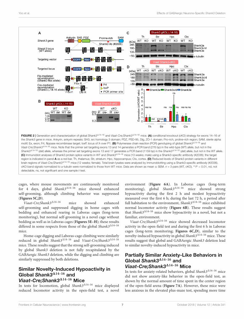
MiceTo analyze the effects of cell type-specific Shank3 deletion, wefirst generated a new mouse line harboring a cassette containingexons 14–16 of the Shank3 gene flanked by flox sequences, andthen crossed these mice with protamine-Flp and protamine-Cremice to producemice in which Shank3 exons 14–16 were globallyand homozygously deleted (Shank3∆14–16 mice; Figure 2A).PCR confirmed the genotype of these mice (Figure 2B), andimmunoblot analyses revealed that the two main splice variantsof the Shank3 protein (Shank3a and Shank3c/d) were absent inseveral brain regions (Figure 2C), a result expected based onprevious studies on the alternative splicing of Shank3 (Lim et al.,1999; Maunakea et al., 2010;Waga et al., 2014;Wang et al., 2014).
We next generated mice carrying Shank3∆14–16 deletionrestricted to GABAergic neurons by crossing Shank3fl/fl micewith Viaat-Cre mouse lines, which drives gene expressionglobally in GABAergic neurons by the solute carrier family32 (GABA vesicular transporter) member 1 (Slc32a1 orViaat/vesicular inhibitory amino acid transporter) promoter(Chao et al., 2010; Kim et al., 2018). Viaat-Cre;Shank3∆14–16
mice, genotyped by PCR (Figure 2B), showed a strong reductionin Shank3a in the striatum (Figure 2D), a brain region enrichedwith GABAergic neurons. Notably, the hippocampus displayeda strong tendency for an increase in Shank3 expression, likelyreflecting compensatory changes in the mutant pyramidalneurons caused by the Shank3 deletion in GABAergic neuronsin the hippocampus or other brain regions.
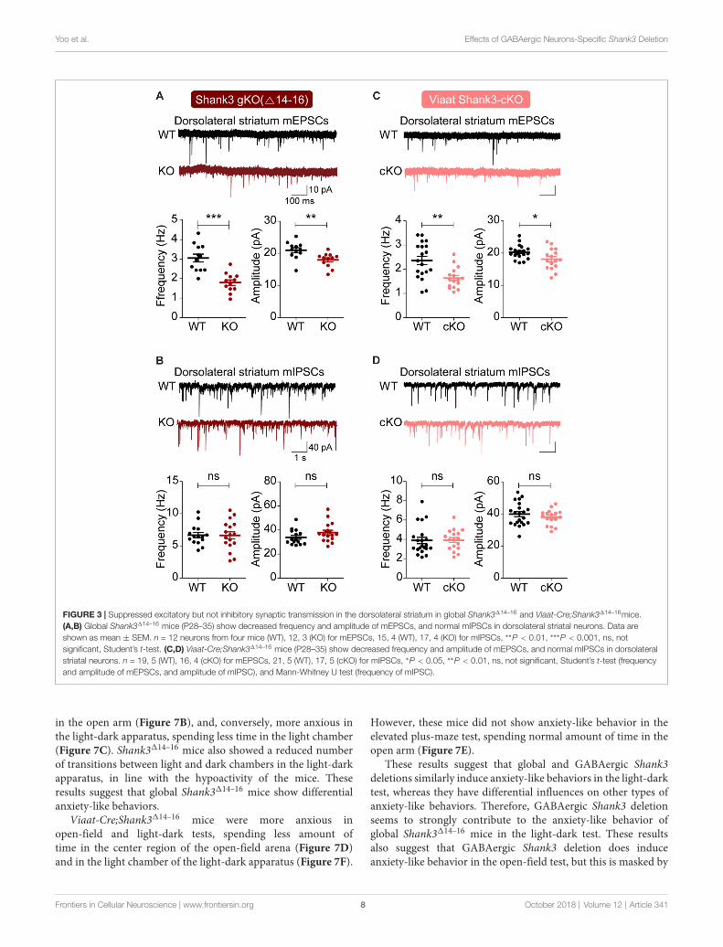
Suppressed Excitatory SynapticTransmission in the Dorsolateral Striatumin Global Shank3∆14–16 andViaat-Cre;Shank3∆14–16 MiceWe first measured excitatory and inhibitory synaptictransmission in the dorsal striatum, a region with enrichedwith GABAergic neurons and implicated in the development ofabnormal behaviors in Shank3-mutant mice (Peca et al., 2011;Peixoto et al., 2016). Both the frequency and amplitudeof mEPSCs were substantially decreased in dorsolateralstriatal neurons in global Shank3∆14–16 mice, with frequencyexhibiting a larger decrease; in contrast, mIPSCs were normal(Figures 3A,B).
Similar changes were observed in dorsolateral striatalneurons in Viaat-Cre;Shank3∆14–16 mice: mEPSC frequencyand amplitude were decreased, whereas mIPSCs were normal(Figures 3C,D). Collectively, these results suggest that globaland GABAergic Shank3 deletions similarly suppress excitatorysynaptic transmission in dorsolateral striatal neurons withoutaffecting inhibitory synaptic transmission.
Frontiers in Cellular Neuroscience | www.frontiersin.org 5 October 2018 | Volume 12 | Article 341

Yoo et al. Effects of GABAergic Neurons-Specific Shank3 Deletion
FIGURE 1 | Expression of Shank3 in both glutamatergic and GABAergic neurons. (A,B) Detection of Shank3 mRNA in Vglut1/2 mRNA-positive glutamatergicneurons in the prelimbic region of the medial prefrontal cortex (mPFC) in mice (P56) by double-immunofluorescence in situ hybridization. Note that Vglut2 mRNAsignals in the mPFC were weaker than those of Vglut1, and that, in the dorsolateral striatum, Vglut1/2 mRNA signals are very weak or absent. Scale bar, 0.5 mm, 20µm. (C,D) Detection of Shank3 mRNA in Gad1/2-positive GABAergic neurons in the prelimbic region of the mPFC and the dorsolateral region of the striatum of mice(P56) by double-immunofluorescence in situ hybridization. Scale bar, 0.5 mm, 20 µm. (E,F) Detection of Shank3 proteins in GAD67-positive GABAergic neurons incultured rat hippocampal neurons at 15 days in vitro (DIV 15), as shown by double immunofluorescence staining for Shank3 and GAD67 (encoded by Gad1). Scalebar, 50 µm, 20 µm, 50 µm, 5 µm.
Enhanced Direct Social Interaction andSuppressed Social Communication inGlobal Shank3∆14–16 andViaat-Cre;Shank3∆14–16 MiceTo test the impact of global and GABA neuron-specific deletionsof Shank3 exons 14–16 on behaviors in mice, we subjectedShank3∆14–16 and Viaat-Cre;Shank3∆14–16 mice to a battery ofbehavioral tests. Shank3∆14–16 mice displayed normal socialapproach behavior in the three-chamber test, but increased socialinteraction in the direct social interaction test (Figures 4A,B).These mice also showed suppressed USVs upon encounter witha novel female stranger (courtship USVs; Figure 4C).
Viaat-Cre;Shank3∆14–16 mice showed normal social approachbehavior in the three-chamber test but enhanced direct social
interaction and suppressed courtship USVs (Figures 4D–F),similar to the behaviors observed in global Shank3∆14–16 mice.These results suggest that social interaction phenotypes inducedby global Shank3∆14–16 deletion is largely recapitulated in micewith Shank3 exons 14–16 deletion restricted to GABAergicneurons.
Strongly Altered Repetitive Behaviors inGlobal Shank3∆14–16 Are Partially Mimickedby Viaat-Cre;Shank3∆14–16 MiceIn tests for repetitive behaviors, global Shank3∆14–16 micedisplayed enhanced self-grooming in a test cage without bedding(Figure 5A), and exhibited enhanced self-grooming but reduceddigging in home cages with bedding (Figure 5B). In Laboras
Frontiers in Cellular Neuroscience | www.frontiersin.org 6 October 2018 | Volume 12 | Article 341
Yoo et al. Effects of GABAergic Neurons-Specific Shank3 Deletion
FIGURE 2 | Generation and characterization of global Shank3∆14–16 and Viaat-Cre;Shank3∆14–16 mice. (A) conditional knockout (cKO) strategy for exons 14–16 ofthe Shank3 gene in mice. Ankyrin, ankyrin repeats; SH3, src homology 3 domain; PDZ, PSD-95, Dlg, ZO-1 domain; Pro-rich, proline-rich region; SAM, sterile alphamotif; Ex, exon; Frt, flippase recombinase target; loxP, locus of X-over P1. (B) Polymerase chain reaction (PCR) genotyping of global Shank3∆14–16 andViaat-Cre;Shank3∆14–16 mice. Note that the primer set targeting exons 13 and 14 generates a PCR band (276 bp) in the wild-type (WT) allele, but not in theShank3∆14–16 (del) allele, whereas the primer set targeting exons 13 and 17 generates a PCR band (1159 bp) in the Shank3∆14–16 (del) allele, but not in the WT allele.(C) Immunoblot analyses of Shank3 protein splice variants in WT and Shank3∆14–16 mice (13 weeks; male) using a Shank3-specific antibody (#2036); the targetregion is indicated in panel A as a red bar. Th, thalamus; Str, striatum; Hpc, hippocampus; Ctx, cortex. (D) Reduced levels of Shank3 protein variants in differentbrain regions of Viaat-Cre;Shank3∆14–16 mice (12 weeks; female). Total brain lysates were analyzed by immunoblotting using a Shank3-specific antibody (#2036).cKO band signals normalized to α-tubulin were normalized to those from WT mice. Data are shown as mean ± SEM. n = 3 pairs (WT, cKO), ∗∗P < 0.01, nd, notdetectable, ns, not significant and one sample t-test.
cages, where mouse movements are continuously monitoredfor 4 days, global Shank3∆14–16 mice showed enhancedself-grooming, although climbing behavior was suppressed(Figures 5C,D).
Viaat-Cre;Shank3∆14–16 mice showed enhancedself-grooming and suppressed digging in home cages withbedding and enhanced rearing in Laboras cages (long-termmonitoring), but normal self-grooming in a novel cage withoutbedding as well as in Laboras cages (Figures 5E–H); these resultsdiffered in some respects from those of the global Shank3∆14–16
mice.Home-cage digging and Laboras-cage climbing were similarly
reduced in global Shank3∆14–16 and Viaat-Cre;Shank3∆14–16
mice. These results suggest that the strong self-grooming inducedby global Shank3 deletion is not fully recapitulated by theGABAergic Shank3 deletion, while the digging and climbing aresimilarly suppressed by both deletions.
Similar Novelty-Induced Hypoactivity inGlobal Shank3∆14–16 andViaat-Cre;Shank3∆14–16 MiceIn tests for locomotion, global Shank3∆14–16 mice displayedreduced locomotor activity in the open-field test, a novel
environment (Figure 6A). In Laboras cages (long-termmonitoring), global Shank3∆14–16 mice showed stronghypoactivity during the first 2 h and modest hypoactivitymeasured over the first 6 h; during the last 72 h, a period afterfull habitation to the environment, Shank3∆14–16 mice exhibitednormal locomotor activity (Figure 6B). These results suggestthat Shank3∆14–16 mice show hypoactivity in a novel, but not afamiliar, environment.
Viaat-Cre;Shank3∆14–16 mice showed decreased locomotoractivity in the open-field test and during the first 6 h in Laborascages (long-term monitoring; Figures 6C,D), similar to thenovelty-induced hypoactivity in global Shank3∆14–16 mice. Theseresults suggest that global and GABAergic Shank3 deletion leadto similar novelty-induced hypoactivity in mice.
Partially Similar Anxiety-Like Behaviors inGlobal Shank3∆14–16 andViaat-Cre;Shank3∆14–16 MiceIn tests for anxiety-related behaviors, global Shank3∆14–16 micedid not show anxiety-like behavior in the open-field test, asshown by the normal amount of time spent in the center regionof the open-field arena (Figure 7A). However, these mice wereless anxious in the elevated plus-maze test, spending more time
Frontiers in Cellular Neuroscience | www.frontiersin.org 7 October 2018 | Volume 12 | Article 341
Yoo et al. Effects of GABAergic Neurons-Specific Shank3 Deletion
FIGURE 3 | Suppressed excitatory but not inhibitory synaptic transmission in the dorsolateral striatum in global Shank3∆14–16 and Viaat-Cre;Shank3∆14–16mice.(A,B) Global Shank3∆14–16 mice (P28–35) show decreased frequency and amplitude of mEPSCs, and normal mIPSCs in dorsolateral striatal neurons. Data areshown as mean ± SEM. n = 12 neurons from four mice (WT), 12, 3 (KO) for mEPSCs, 15, 4 (WT), 17, 4 (KO) for mIPSCs, ∗∗P < 0.01, ∗∗∗P < 0.001, ns, notsignificant, Student’s t-test. (C,D) Viaat-Cre;Shank3∆14–16 mice (P28–35) show decreased frequency and amplitude of mEPSCs, and normal mIPSCs in dorsolateralstriatal neurons. n = 19, 5 (WT), 16, 4 (cKO) for mEPSCs, 21, 5 (WT), 17, 5 (cKO) for mIPSCs, ∗P < 0.05, ∗∗P < 0.01, ns, not significant, Student’s t-test (frequencyand amplitude of mEPSCs, and amplitude of mIPSC), and Mann-Whitney U test (frequency of mIPSC).
in the open arm (Figure 7B), and, conversely, more anxious inthe light-dark apparatus, spending less time in the light chamber(Figure 7C). Shank3∆14–16 mice also showed a reduced numberof transitions between light and dark chambers in the light-darkapparatus, in line with the hypoactivity of the mice. Theseresults suggest that global Shank3∆14–16 mice show differentialanxiety-like behaviors.
Viaat-Cre;Shank3∆14–16 mice were more anxious inopen-field and light-dark tests, spending less amount oftime in the center region of the open-field arena (Figure 7D)and in the light chamber of the light-dark apparatus (Figure 7F).
However, these mice did not show anxiety-like behavior in theelevated plus-maze test, spending normal amount of time in theopen arm (Figure 7E).
These results suggest that global and GABAergic Shank3deletions similarly induce anxiety-like behaviors in the light-darktest, whereas they have differential influences on other types ofanxiety-like behaviors. Therefore, GABAergic Shank3 deletionseems to strongly contribute to the anxiety-like behavior ofglobal Shank3∆14–16 mice in the light-dark test. These resultsalso suggest that GABAergic Shank3 deletion does induceanxiety-like behavior in the open-field test, but this is masked by
Frontiers in Cellular Neuroscience | www.frontiersin.org 8 October 2018 | Volume 12 | Article 341

Yoo et al. Effects of GABAergic Neurons-Specific Shank3 Deletion
FIGURE 4 | Enhanced direct social interaction and suppressed social communication in global Shank3∆14–16 and Viaat-Cre;Shank3∆14–16 mice. (A) Normal socialapproach and social novelty recognition in Shank3∆14–16 mice (14–21 weeks) in the three-chamber test, as shown by time spent sniffing. S1, stranger; O, object;S2, novel stranger. Data are shown as mean ± SEM. n = 28 (WT) and 22 (KO), ∗∗∗P < 0.001, paired t-test. Details on the order of behavioral tests performed onShank3∆14–16 mice and conditional Shank3∆14–16 mouse lines (see below) are described in Supplementary Table S1. (B) Enhanced social interaction inShank3∆14–16 mice (14–20 weeks) in the direct social interaction test, as shown by nose-to-nose interaction, following and total interaction, the latter of whichadditionally includes allo-grooming and body contacts. Mean ± SEM. n = 20 (WT) and 16 (KO), ∗P < 0.05, ∗∗P < 0.01, ∗∗∗P < 0.001, Student’s t-test.(C) Suppressed ultrasonic vocalizations (USVs) in Shank3∆14–16 mice (18–24 weeks), upon encounter with a novel female stranger. n = 21 (WT) and 19 (KO),∗P < 0.05, Student’s t-test. (D–F) Viaat-Cre;Shank3∆14–16 mice (10–13 weeks for D, 15–28 weeks for E, and 11–21 weeks for F) show normal social approach in thethree-chamber test (D) enhanced direct social interaction (E) and suppressed courtship USVs (F). n = 19 mice (WT), 13 (cKO) for three-chamber, 11 (WT), 8 (cKO)for direct social interaction, and 19 (WT), 15 (cKO) for USV, ∗∗P < 0.01, ∗∗∗P < 0.001, ns, not significant, paired t-test (three-chamber test), Mann-Whitney U test(nose to nose time and following time of direct social interaction test), and Student’s t-test (total interaction time of direct social interaction test and adult USV test).
global Shank3 deletion. In contrast, GABAergic Shank3 deletionseems to have minimal impacts on the anxiety-like behaviorin the elevated plus-maze test, suggesting that non-GABAergicShank3 deletions are more important for the anxiolytic-likebehaivor of global Shank3∆14–16 mice in the elevated plus-maze.
DISCUSSION
In this study, we investigated the impacts of global and GABAneuron-specific deletion of Shank3 exons 14–16 on synaptictransmission and behaviors in mice. Global Shank3∆14–16
mice display decreased excitatory input onto dorsolateralstriatal neurons and strong abnormalities in social, repetitive,locomotor and anxiety-like behaviors. The electrophysiologicaland behavioral (social and locomotor) phenotypes observedin global Shank3∆14–16 mice are strongly mimicked by Viaat-Cre;Shank3∆14–16 mice, although the repetitive and anxiety-likebehavioral deficits in global Shank3∆14–16 mice are partially
mimicked by Viaat-Cre;Shank3∆14–16 mice (summarized inTable 1).
The result that both the frequency and amplitude of mEPSCsare reduced in global Shank3∆14–16 mice (Figure 3) furtherstrengthens the notion that Shank3 is important for thedevelopment and function of excitatory synapses in the dorsalstriatum. Similar decreases in the frequency and amplitude ofmEPSCs in the dorsal striatum have been observed in the Shank3mouse line lacking exons 13–16 (Shank3B−/− mice; Peca et al.,2011; Mei et al., 2016; Wang et al., 2017).
A more important finding from our study is that bothmouse lines (global and Viaat-Cre) show similar decreases inthe frequency and amplitude of mEPSCs in dorsolateral striatalneurons (Figure 3). This suggests that the suppressed excitatoryinput onto dorsolateral striatal neurons in these mouse linesare likely to be induced by the deletion of Shank3 in striatalGABAergic neurons in a cell autonomous manner.
In support of this possibility, our FISH data indicate thatShank3mRNAs are expressed in Gad1/2-positive neurons in the
Frontiers in Cellular Neuroscience | www.frontiersin.org 9 October 2018 | Volume 12 | Article 341

Yoo et al. Effects of GABAergic Neurons-Specific Shank3 Deletion
FIGURE 5 | Strongly altered repetitive behaviors in global Shank3∆14–16 mice are partially mimicked by Viaat-Cre;Shank3∆14–16 mice. (A) Enhanced self-grooming inShank3∆14–16 mice (11–14 weeks) in a novel cage without bedding. n = 22 (WT) and 21 (KO), ∗∗∗P < 0.001, Student’s t-test. (B) Enhanced self-groomingand suppressed digging in Shank3∆14–16 mice (12–14 weeks) in home cages with bedding. n = 21 (WT) and 20 (KO), ∗∗∗P < 0.001, Mann-Whitney U test.(C) Enhanced self-grooming in Shank3∆14–16 mice (9–14 weeks) in Laboras cages, in which mouse movements are monitored for 4 days. n = 20 (WT) and 21 (KO),∗P < 0.05, ∗∗P < 0.01, ∗∗∗P < 0.001, repeated measures two-way ANOVA (genotype p value = 0.0149 and genotype × time p value < 0.0001). (D) Enhancedself-grooming and suppressed climbing in Shank3∆14–16 mice (9–14 weeks) in Laboras cages (long-term monitoring). These graphs are a summary of all types ofmovements in Laboras cages for 4 days, including self-grooming. n = 20 (WT) and 21 (KO), ∗P < 0.05, ∗∗∗P < 0.001, ns, not significant, Student’s t-test.(E–H) Viaat-Cre;Shank3∆14–16 mice (11–15 weeks for E, 10–26 weeks (cohort 3: 10–20 weeks, cohort 4: 23–26 weeks) for F, 12–14 weeks for G,H) show enhancedself-grooming and suppressed digging in home cages with bedding (F) but normal self-grooming in a novel cage without bedding (E) and normal self-grooming butenhanced rearing and suppressed climbing in Laboras cages (long-term monitoring; G,H). n = 18 mice (WT), 14 (cKO) for w/o bedding, 14 (WT), 18 (cKO) forw/bedding, and 15 (WT), 13 (cKO) for Laboras, ∗P < 0.05, ∗∗P < 0.01, ∗∗∗P < 0.001, repeated measures two-way ANOVA (for Laboras; genotype p value = 0.79),Student’s t-test (for self-grooming test, repetitive behavior test, and Laboras (climbing, rearing, grooming and drinking)), and Mann-Whitney U test (for Laboras(eating)).
dorsal striatum in addition to mPFC (Figure 1). In addition, theimmunostaining result indicates that Shank3-positive punctatestructures are observed on the dendrites of GAD67-positiveGABAergic neurons in cultured hippocampal neurons. Giventhat Shank3 is an important component of the postsynapticdensity at excitatory synapses (Sheng and Kim, 2000; Shengand Sala, 2001; Boeckers et al., 2002; Sheng and Hoogenraad,2007; Grabrucker et al., 2011; Sheng and Kim, 2011; Jiangand Ehlers, 2013; Sala et al., 2015; Monteiro and Feng, 2017;Mossa et al., 2017), the lack of Shank3 in dorsolateral striatalneurons may suppress normal development and maturationof the postsynaptic density, dendritic spines, and excitatorysynapses. In addition, previous studies have reported a strongdecrease in dendritic spine density in dorsal striatal neuronsin Shank3B−/− mice (Peca et al., 2011), further suggestingthat the decreased mEPSC frequency may be a consequence ofpostsynaptic changes.
More recently, however, additional analyses of excitatorysynaptic inputs onto D1 and D2 medium spiny neurons(MSNs) in the dorsal striatum of Shank3B−/− mice haverevealed that D2 MSNs show reductions in both presynaptic
release and spine density (Wang et al., 2017), suggestingthat both pre- and postsynaptic factors may be involved.In addition, a previous study on Shank3B−/− mice showedthat early abnormal excitability in pyramidal neurons in thesomatosensory cortex driven by the limited inhibitory inputfrom neighboring GABAergic neurons induces precociousdevelopment of excitatory synapses on dorsomedial striatalneurons that leads to a decrease in the mEPSC frequency atlater stages (Peixoto et al., 2016). It is therefore possible thatthe decreased mEPSC frequency in dorsolateral striatal neuronsin global Shank3∆14–16 and Viaat-Cre;Shank3∆14–16 mice mayrepresent the consequences of the primary changes occurring incortical GABAergic neurons.
Behaviorally, global Shank3∆14–16 mice display altered socialand repetitive behaviors, including suppressed courtship USVsand enhanced self-grooming (Figures 4, 5). Thesemice also showhypoactivity and altered anxiety-like behaviors (Figures 6, 7).Given that Shank3∆14–16 mice lack the PDZ domain-containingShank3 variants, Shank3a and Shank3c/d, but retain Shank3e,our mouse line is likely to display behavioral phenotypes similarto those observed in the Shank3B–/– mouse line, which globally
Frontiers in Cellular Neuroscience | www.frontiersin.org 10 October 2018 | Volume 12 | Article 341

Yoo et al. Effects of GABAergic Neurons-Specific Shank3 Deletion
FIGURE 6 | Novelty-induced hypoactivity in global Shank3∆14–16 and Viaat-Cre;Shank3∆14–16 mice. (A) Suppressed locomotor activity, as measured by distancemoved in Shank3∆14–16 mice (11–13 weeks) in the open-field test. Data are shown as mean ± SEM. n = 23 (WT) and 22 (KO), ∗∗P < 0.01, ∗∗∗P < 0.001, repeatedmeasures two-way ANOVA (genotype p value < 0.0001; genotype × time interaction p value = 0.0109) and Student’s t-test. (B) Shank3∆14–16 mice (9–14 weeks)show suppressed locomotor activity during an early period (first 2 and 6 h), but not in the later habituated period (last 72 h), in Laboras cages (long-term monitoring).n = 20 (WT) and 21 (KO), ∗P < 0.05, ∗∗∗P < 0.001, repeated measures two-way ANOVA (genotype p value = 0.3711; genotype × time interaction p value < 0.0001)and Student’s t-test. (C,D) Viaat-Cre;Shank3∆14–16 mice (8–9 weeks for C and 12–14 weeks for D) show hypoactivity in the open-field test (C) and during the first6 h but not the last 68 h in Laboras cages (long-term monitoring; D). n = 18 (WT), 15 (cKO) for open-field, 15 (WT), 13 (cKO) for Laboras, ∗P < 0.05, ∗∗∗P < 0.001,ns, not significant, repeated measures two-way ANOVA (for the left panels in open-field and Laboras results; genotype p values = 0.0006 and 0.7414,time × genotype p values = 0.0454 and 0.0002, respectively), Student’s t-test.
lacks exons 13–16 encoding the PDZ domain (Peca et al., 2011).Indeed, Shank3∆14–16 and Shank3B−/− mice show largely similarbehaviors, including suppressed courtship USVs, hypoactivity,and anxiety-like behavior (elevated zero maze and light-darktest), although Shank3B−/− mice additionally show suppressedsocial approach (Peca et al., 2011; Dhamne et al., 2017). AnotherShank3-mutant mice similar to ours is the one lacking exon13, encoding the PDZ domain (Shank3E13 mice; Jaramillo et al.,2017). These mice show enhanced self-grooming and socialinteraction deficits, but normal locomotion and anxiety-relatedbehavior; the partial similarity to our behavioral phenotypesis likely attributable to the different exon targeting strategy(insertion of a stop codon in front of exon 13) in Shank3E13 mice.
Although the behavioral phenotypes of global Shank3∆14–16
mice are strong in multiple domains (social, repetitive,locomotor, and anxiety-like), the following points need to befurther discussed. First, global Shank3∆14–16 mice show enhanceddirect social interaction, which was unexpected and is at variancewith the normal three-chamber social approach observed inthese mice. Notably, a previous study on Shank3∆4–22 mice hasreported a similar increase in direct social interaction whereShank3∆4–22 mice display frequently attempted but unsuccessfulsocial interactions with a stranger C3H mouse, a differentstrain, that does not reciprocate and terminate the socialinteraction attempted by the subject mouse (Wang et al., 2016),suggesting that Shank3∆4–22 mice have normal social interest
but struggle with persisting social failures. We could not testwhether this is the case for our mice because we used genotype-matched (WT-WT or KO-KO) mouse pairs where monitoringof non-reciprocated social interaction is difficult because of thesame coat color and the confusion over retraction vs. rejection.However, our results suggest that social interest is normal inglobal Shank3∆14–16 mice, which is different from the significantsocial interaction deficits observed inmany other Shank3-mutantmouse lines (Jiang and Ehlers, 2013; Monteiro and Feng, 2017).We propose that the difference in the specific Shank3 exonsdeleted in each mouse lines might explain the discrepancy. Forinstance, the exons deleted in our Shank3mice (exons 14–16) aredistinct from those deleted in Shank3B−/− mice (exon 13–16;Peca et al., 2011). In support of this possibility, a very smalldifference in the exons deleted in Shank2-mutant mice (i.e.,exons 6 and 7 vs. exon 7) has been shown to cause strongdifferences in molecular, synaptic and behavioral phenotypes(Schmeisser et al., 2012; Won et al., 2012; Lim et al., 2017;Wegener et al., 2018).
Another notable result is that global Shank3∆14–16 micedisplay anxiolytic-like behavior in the elevated plus-mazewhereas they show anxiety-like behavior in the light-darkapparatus and normal anxiety-like behavior in the center regionof open-field arena. This could be due to the different anxiogeniccomponents in these tests (Belzung and Griebel, 2001; Carolaet al., 2002; Carobrez and Bertoglio, 2005), as exemplified by
Frontiers in Cellular Neuroscience | www.frontiersin.org 11 October 2018 | Volume 12 | Article 341

Yoo et al. Effects of GABAergic Neurons-Specific Shank3 Deletion
FIGURE 7 | Partially similar anxiety-like behaviors in global Shank3∆14–16 and Viaat-Cre;Shank3∆14–16 mice. (A) Normal anxiety-like behavior in Shank3∆14–16 mice(11–13 weeks), as measured by the time spent in the center region of the open field arena in the open-field test. Note that locomotor activity results from theopen-field test are described in Figure 6A. Data are shown as mean ± SEM. n = 23 (WT) and 22 (KO), ns, not significant, Student’s t-test. (B) Anxiolytic-likebehavior in Shank3∆14–16 mice (13–21 weeks) in the elevated plus-maze test. n = 22 (WT) and 20 (KO), ∗∗P < 0.01, ∗∗∗P < 0.001, paired t-test (for the left panels),and Student’s t-test (for the right panel). (C) Anxiety-like behavior in Shank3∆14–16 mice (14–21 weeks) in the light-dark test. Note that these mice are also hypoactivein this test, as shown by the number of transitions. n = 23 (WT) and 20 (KO), ∗P < 0.05, ∗∗P < 0.01, Student’s t-test. (D–F) Viaat-Cre;Shank3∆14–16 mice (8–9 weeksfor D, 8–10 weeks for E, and 11–20 weeks for F) spend a reduced amount of time in the center region of the open-field area (D; locomotor activity results aredescribed in Figure 6C), a normal amount of time in the open arm of the elevated plus-maze (E) and a decreased amount of time in the light chamber of thelight-dark apparatus (F, left) and show decreased number of transitions (F, right). n = 18 (WT), 15 (cKO) for open-field, 19 (WT), 15 (cKO) for elevated plus-maze, and19 (WT), 15 (cKO) for light-dark, ∗∗P < 0.01, ∗∗∗P < 0.001, Student’s t-test (for open-field test, right panel of elevated-plus maze test, and number of transition oflight-dark test), paired t-test (for the left panel of elevated plus-maze test), and Mann-Whitney U test (for time in light chamber of light-dark test).
the differential responses of nine different mouse strains to theelevated plus-maze and light-dark tests (Griebel et al., 2000).Notably, Shank3B–/– mice (exons 13–16) also display differentialanxiety-like behaviors in these assays, being partly similar toour results; normal anxiety-like behavior in elevated plus-maze,anxiety-like behavior in zero maze, light-dark apparatus andopen-field center (Peca et al., 2011; Dhamne et al., 2017).
Our results indicate that GABAergic neurons contributeto some of the abnormal behaviors observed in globalShank3∆14–16 mice. Specifically, the enhanced direct socialinteraction, suppressed courtship USVs, and novelty-inducedhypoactivity observed in global Shank3∆14–16 mice were alsoobserved in Viaat-Cre;Shank3∆14–16 mice. In contrast, the strongself-grooming behavior observed in global Shank3∆14–16 micewere only partially mimicked by Viaat-Cre;Shank3∆14–16 mice.In anxiety-like behaviors, only the light-dark test results weresimilar in global Shank3∆14–16 andViaat-Cre;Shank3∆14–16 mice.Therefore, GABA neuronal Shank3 deletion seems to be moreimportant for social and locomotor behaviors than repetitive andanxiety-like behaviors.
A recent study reported the effects of a deletion of Shank3exons 4–22 restricted to Nex-positive glutamatergic neurons in
the cortex, hippocampus and amygdala (Nex-Shank3 cKO mice)and Dlx5/6-positive GABAergic neurons in the striatum (Dlx5/6-Shank3 cKO mice; Bey et al., 2018). Neither Nex-Shank3 norDlx5/6-Shank3 cKO mice exhibit social approach deficits, resultssimilar to the normal social approach behavior reported by thesame group using mice with a global Shank3∆4–22 mice (Wanget al., 2016). This phenotype is also similar to the normal socialapproach behavior observed in our global Shank3∆14–16 andViaat-Cre;Shank3∆14–16 mice.
The suppressed courtship USV and hypoactivity phenotypesobserved in global Shank3∆4–22 mice (Wang et al., 2016)were not recapitulated in either Nex-Shank3 or Dlx5/6-Shank3 cKO mice (Bey et al., 2018). These results aredifferent from our findings that both global Shank3∆14–16 andViaat-Cre;Shank3∆14–16 mice show suppressed courtship USVand hypoactivity. Furthermore, the enhanced self-groomingobserved in global Shank3∆4–22 mice (Wang et al., 2016)was observed in Nex-Shank3 cKO mice, but not in Dlx5/6-Shank3 cKO mice (Bey et al., 2018). These results are slightlydifferent from our finding that the enhanced self-grooming inglobal Shank3∆14–16 mice was partially recapitulated in Viaat-Cre;Shank3∆14–16mice.
Frontiers in Cellular Neuroscience | www.frontiersin.org 12 October 2018 | Volume 12 | Article 341

Yoo et al. Effects of GABAergic Neurons-Specific Shank3 Deletion
TABLE 1 | Summary of electrophysiological and behavioral phenotypes of global Shank3∆14–16 and Viaat-Cre;Shank3∆14–16 mice.
Shank3 exons 14-16 global KO Shank3 exons 14-16 Viaat cKO
Electrophysiology Brain region Measurement Frequency Amplitude Frequency AmplitudeDorsolateral striatum mEPSC ↓ ↓ ↓ ↓
mIPSC - - - -
Behavior Behavioral domain Behavioral test Shank3 exons 14-16 global KO Shank3 exons 14-16 Viaat cKOSocial interaction 3-chamber - -
Direct Total interaction ↑ Total interaction ↑interaction Nose-to-nose ↑ Nose-to-nose, -
Following ↑ Following, -
Social communication Adult USV (courtship) ↓ ↓
Repetitive behavior Laboras Self-grooming ↑ Rearing ↑Climbing ↓ Climbing ↓
Self-grooming (w/o bedding) ↑ -Repetitive behavior Self-grooming ↑ Self-grooming ↑(with bedding) Digging ↓ Digging ↓
Locomotor activity Laboras (first 6 h) ↓ ↓
Open-field ↓ ↓
Anxiety-like behavior Open field (center time) - ↓
Elevated plus-maze (time in open arms) ↑ -Light/dark box (time in light chamber) ↓ ↓
This table summarizes only the increases or decreases of various electrophysiological and behavioral phenotypes in a given mouse line relative to wild-type (WT)/controlmice, but is not intended to compare the phenotypic severities across different mouse lines. -, no significant change; up and down arrows, increases and decreases.
These results indicate that two different global Shank3deletions (exons 14–16 and 4–22) in mice lead to remarkablysimilar behavioral phenotypes in mice in social, repetitive,locomotor and anxiety-like behavioral domains, but that thesesimilarities are minimized by two different cKOs restricted toGABAergic neurons (Dlx5/6 and Viaat). These discrepanciescould be attributable to differences in the specific exons ofShank3 deleted and/or specific characteristics of Dlx5/6-Cre vs.Viaat-Cremice (Oh et al., 2005; Goebbels et al., 2006; Chao et al.,2010). For instance, Dlx5/6-Cre primarily targets GABAergicneurons in the striatum (Monory et al., 2006), whereas Viaat-Cretargets the majority of GABAergic neurons in the brain (Chaoet al., 2010). In addition, it could be subtle differences in mousehousing conditions or experimental details.
It remains unclear how GABA neuronal deletion of Shank3(exons 14–16) leads to the above mentioned diverse behavioralabnormalities. However, functional defects in the striatum havebeen strongly implicated in abnormal phenotypes in variousShank3-mutant mouse lines (Peca et al., 2011; Schmeisser et al.,2012; Filice et al., 2016; Jaramillo et al., 2016; Mei et al.,2016; Peixoto et al., 2016; Sarowar et al., 2016; Zhou et al.,2016; Jaramillo et al., 2017; Lee Y. et al., 2017; Reim et al.,2017; Vicidomini et al., 2017; Wang et al., 2017; Bey et al.,2018). In addition, a recent study has shown that chemogeneticstimulation of D2, but not D1, MSN activity by DREADD-hM3Dq for the activation of the striatopallidal pathway canrescue self-grooming in Shank3B−/− mice (Wang et al., 2017).Therefore, the suppressed excitatory synaptic transmission indorsolateral striatal neurons in global Shank3∆14–16 and Viaat-Cre;Shank3∆14–16 mice might have contributed to the behavioralabnormalities observed in our mouse lines, including enhancedself-grooming. However, the substantial difference between thestrong self-grooming in global Shank3∆14–16 mice and theweak self-grooming in Viaat-Cre;Shank3∆14–16 mice suggests
that GABAergic Shank3 deletion only partially contributeto the self-grooming phenotype. However, care should betaken in the interpretation because Viaat-mediated GABAergicShank3 deletion can affect multiple types of GABAergicneurons.
In conclusion, our results suggest that the deletion of Shank3exons 14–16 restricted to GABAergic neurons in mice inducesphenotypes that are similar to those induced by global Shank3deletion. These include strongly suppressed excitatory synapticonto dorsolateral striatal neurons and strongly altered socialand locomotor behaviors but modestly altered repetitive andanxiety-like behaviors.
AUTHOR CONTRIBUTIONS
TY, JL and HC performed behavioral experiments. TY andJL performed immunoblot experiments. TY, HP and Y-EYperformed electrophysiological experiments. EY and JKperformed in situ hybridization experiments. TY performedhippocampal neuron culture and immunocytochemicalexperiments. HK and EK designed research and wrote themanuscript.
FUNDING
This study was supported by the Institute for Basic Science (IBS-R002-D1 to EK).
SUPPLEMENTARY MATERIAL
The Supplementary Material for this article can be foundonline at: https://www.frontiersin.org/articles/10.3389/fncel.2018.00341/full#supplementary-material
Frontiers in Cellular Neuroscience | www.frontiersin.org 13 October 2018 | Volume 12 | Article 341

Yoo et al. Effects of GABAergic Neurons-Specific Shank3 Deletion
REFERENCES
Báez-Mendoza, R., and Schultz, W. (2013). The role of the striatumin social behavior. Front. Neurosci. 7:233. doi: 10.3389/fnins.2013.00233
Balleine, B. W., Delgado, M. R., and Hikosaka, O. (2007). The role of thedorsal striatum in reward and decision-making. J. Neurosci. 27, 8161–8165.doi: 10.1523/JNEUROSCI.1554-07.2007
Belzung, C., and Griebel, G. (2001). Measuring normal and pathologicalanxiety-like behaviour in mice: a review. Behav. Brain Res. 125, 141–149.doi: 10.1016/s0166-4328(01)00291-1
Bey, A. L., Wang, X., Yan, H., Kim, N., Passman, R. L., Yang, Y., et al. (2018).Brain region-specific disruption of Shank3 in mice reveals a dissociation forcortical and striatal circuits in autism-related behaviors. Transl. Psychiatry 8:94.doi: 10.1038/s41398-018-0142-6
Boccuto, L., Lauri, M., Sarasua, S. M., Skinner, C. D., Buccella, D., Dwivedi, A.,et al. (2013). Prevalence of SHANK3 variants in patients with differentsubtypes of autism spectrum disorders. Eur. J. Hum. Genet. 21, 310–316.doi: 10.1038/ejhg.2012.175
Boeckers, T. M., Bockmann, J., Kreutz, M. R., and Gundelfinger, E. D. (2002).ProSAP/Shank proteins—a family of higher order organizing molecules of thepostsynaptic density with an emerging role in human neurological disease.J. Neurochem. 81, 903–910. doi: 10.1046/j.1471-4159.2002.00931.x
Boeckers, T. M., Winter, C., Smalla, K. H., Kreutz, M. R., Bockmann, J.,Seidenbecher, C., et al. (1999). Proline-rich synapse-associated proteinsProSAP1 and ProSAP2 interact with synaptic proteins of the SAPAP/GKAPfamily. Biochem. Biophys. Res. Commun. 264, 247–252. doi: 10.1006/bbrc.1999.1489
Bonaglia, M. C., Giorda, R., Beri, S., De Agostini, C., Novara, F., Fichera, M.,et al. (2011). Molecular mechanisms generating and stabilizing terminal22Q13 deletions in 44 subjects with Phelan/McDermid syndrome. PLoS Genet.7:e1002173. doi: 10.1371/journal.pgen.1002173
Bonaglia, M. C., Giorda, R., Borgatti, R., Felisari, G., Gagliardi, C., Selicorni, A.,et al. (2001). Disruption of the ProSAP2 gene in a t(12;22)(q24.1;Q13.3)is associated with the 22Q13.3 deletion syndrome. Am. J. Hum. Genet. 69,261–268. doi: 10.1086/321293
Bozdagi, O., Sakurai, T., Papapetrou, D., Wang, X., Dickstein, D. L., Takahashi, N.,et al. (2010). Haploinsufficiency of the autism-associated Shank3 gene leads todeficits in synaptic function, social interaction, and social communication.Mol.Autism 1:15. doi: 10.1186/2040-2392-1-15
Carobrez, A. P., and Bertoglio, L. J. (2005). Ethological and temporal analysesof anxiety-like behavior: the elevated plus-maze model 20 years on. Neurosci.Biobehav. Rev. 29, 1193–1205. doi: 10.1016/j.neubiorev.2005.04.017
Carola, V., D’Olimpio, F., Brunamonti, E., Mangia, F., and Renzi, P. (2002).Evaluation of the elevated plus-maze and open-field tests for the assessmentof anxiety-related behaviour in inbred mice. Behav. Brain Res. 134, 49–57.doi: 10.1016/s0166-4328(01)00452-1
Cepeda, C., André, V. M., Yamazaki, I., Wu, N., Kleiman-Weiner, M., andLevine, M. S. (2008). Differential electrophysiological properties of dopamineD1 and D2 receptor-containing striatal medium-sized spiny neurons. Eur.J. Neurosci. 27, 671–682. doi: 10.1111/j.1460-9568.2008.06038.x
Cepeda, C., Colwell, C. S., Itri, J. N., Chandler, S. H., and Levine, M. S.(1998). Dopaminergic modulation of NMDA-induced whole cell currentsin neostriatal neurons in slices: contribution of calcium conductances.J. Neurophysiol. 79, 82–94. doi: 10.1152/jn.1998.79.1.82
Chao, H. T., Chen, H., Samaco, R. C., Xue, M., Chahrour, M., Yoo, J., et al. (2010).Dysfunction in GABA signalling mediates autism-like stereotypies and Rettsyndrome phenotypes. Nature 468, 263–269. doi: 10.1038/nature09582
Cochoy, D. M., Kolevzon, A., Kajiwara, Y., Schoen, M., Pascual-Lucas, M.,Lurie, S., et al. (2015). Phenotypic and functional analysis of SHANK3 stopmutations identified in individuals with ASD and/or ID. Mol. Autism 6:23.doi: 10.1186/s13229-015-0020-5
De Rubeis, S., Siper, P. M., Durkin, A., Weissman, J., Muratet, F., Halpern, D.,et al. (2018). Delineation of the genetic and clinical spectrum of Phelan-McDermid syndrome caused by SHANK3 point mutations. Mol. Autism 9:31.doi: 10.1186/s13229-018-0205-9
Dere, E., Winkler, D., Ritter, C., Ronnenberg, A., Poggi, G., Patzig, J., et al. (2015).Gpm6b deficiency impairs sensorimotor gating and modulates the behavioral
response to a 5-HT2A/C receptor agonist. Behav. Brain Res. 277, 254–263.doi: 10.1016/j.bbr.2014.04.021
Dhamne, S. C., Silverman, J. L., Super, C. E., Lammers, S. H. T., Hameed, M. Q.,Modi, M. E., et al. (2017). Replicable in vivo physiological and behavioralphenotypes of the Shank3B null mutant mouse model of autism. Mol. Autism8:26. doi: 10.1186/s13229-017-0142-z
Durand, C. M., Betancur, C., Boeckers, T. M., Bockmann, J., Chaste, P.,Fauchereau, F., et al. (2007). Mutations in the gene encoding the synapticscaffolding protein SHANK3 are associated with autism spectrum disorders.Nat. Genet. 39, 25–27. doi: 10.1038/ng1933
Epstein, I., Tushev, G., Will, T. J., Vlatkovic, I., Cajigas, I. J., and Schuman, E. M.(2014). Alternative polyadenylation and differential expression of ShankmRNAs in the synaptic neuropil. Philos. Trans. R. Soc. Lond. B Biol. Sci.369:20130137. doi: 10.1098/rstb.2013.0137
Filice, F., Vörckel, K. J., Sungur, A. O., Wöhr, M., and Schwaller, B. (2016).Reduction in parvalbumin expression not loss of the parvalbumin-expressingGABA interneuron subpopulation in genetic parvalbumin and shank mousemodels of autism.Mol. Brain 9:10. doi: 10.1186/s13041-016-0192-8
Gauthier, J., Champagne, N., Lafrenière, R. G., Xiong, L., Spiegelman, D.,Brustein, E., et al. (2010). De novomutations in the gene encoding the synapticscaffolding protein SHANK3 in patients ascertained for schizophrenia. Proc.Natl. Acad. Sci. U S A 107, 7863–7868. doi: 10.1073/pnas.0906232107
Gertler, T. S., Chan, C. S., and Surmeier, D. J. (2008). Dichotomousanatomical properties of adult striatal medium spiny neurons. J. Neurosci. 28,10814–10824. doi: 10.1523/JNEUROSCI.2660-08.2008
Goebbels, S., Bormuth, I., Bode, U., Hermanson, O., Schwab, M. H., andNave, K. A. (2006). Genetic targeting of principal neurons in neocortex andhippocampus of NEX-Cre mice. Genesis 44, 611–621. doi: 10.1002/dvg.20256
Goslin, K., and Banker, G. (1991). ‘‘Rat hippocampal neurons in low-densityculture,’’ in Culturing Nerve Cells, eds G. Banker and K. Goslin (Cambridge,MA: The MIT Press), 337–370.
Grabrucker, A. M., Schmeisser, M. J., Schoen, M., and Boeckers, T. M. (2011).Postsynaptic ProSAP/Shank scaffolds in the cross-hair of synaptopathies.Trends Cell Biol. 21, 594–603. doi: 10.1016/j.tcb.2011.07.003
Griebel, G., Belzung, C., Perrault, G., and Sanger, D. J. (2000). Differencesin anxiety-related behaviours and in sensitivity to diazepam in inbredand outbred strains of mice. Psychopharmacology 148, 164–170.doi: 10.1007/s002130050038
Grueter, B. A., Rothwell, P. E., and Malenka, R. C. (2012). Integrating synapticplasticity and striatal circuit function in addiction. Curr. Opin. Neurobiol. 22,545–551. doi: 10.1016/j.conb.2011.09.009
Guilmatre, A., Huguet, G., Delorme, R., and Bourgeron, T. (2014). The emergingrole of SHANK genes in neuropsychiatric disorders. Dev. Neurobiol. 74,113–122. doi: 10.1002/dneu.22128
Hamdan, F. F., Gauthier, J., Araki, Y., Lin, D. T., Yoshizawa, Y., Higashi, K.,et al. (2011). Excess of de novo deleterious mutations in genes associated withglutamatergic systems in nonsyndromic intellectual disability. Am. J. Hum.Genet. 88, 306–316. doi: 10.1016/j.ajhg.2011.02.001
Han, K., Holder, J. L. Jr., Schaaf, C. P., Lu, H., Chen, H., Kang, H., et al.(2013). SHANK3 overexpression causes manic-like behaviour with uniquepharmacogenetic properties. Nature 503, 72–77. doi: 10.1038/nature12630
Harris, K. M., and Weinberg, R. J. (2012). Ultrastructure of synapsesin the mammalian brain. Cold Spring Harb. Perspect. Biol. 4:a005587.doi: 10.1101/cshperspect.a005587
Jaramillo, T. C., Speed, H. E., Xuan, Z., Reimers, J. M., Escamilla, C. O.,Weaver, T. P., et al. (2017). Novel Shank3 mutant exhibits behaviors with facevalidity for autism and altered striatal and hippocampal function. Autism Res.10, 42–65. doi: 10.1002/aur.1664
Jaramillo, T. C., Speed, H. E., Xuan, Z., Reimers, J. M., Liu, S., andPowell, C.M. (2016). Altered striatal synaptic function and abnormal behaviourin Shank3 Exon4–9 deletion mouse model of autism. Autism Res. 9, 350–375.doi: 10.1002/aur.1529
Jiang, Y. H., and Ehlers,M. D. (2013).Modeling autism by SHANK genemutationsin mice. Neuron 78, 8–27. doi: 10.1016/j.neuron.2013.03.016
Kim, R., Kim, J., Chung, C., Ha, S., Lee, S., Lee, E., et al. (2018). Cell-type-specific shank2 deletion in mice leads to differential synaptic and behavioralphenotypes. J. Neurosci. 38, 4076–4092. doi: 10.1523/JNEUROSCI.2684-17.2018
Frontiers in Cellular Neuroscience | www.frontiersin.org 14 October 2018 | Volume 12 | Article 341

Yoo et al. Effects of GABAergic Neurons-Specific Shank3 Deletion
Kim, E., and Sheng, M. (2004). PDZ domain proteins of synapses. Nat. Rev.Neurosci. 5, 771–781. doi: 10.1038/nrn1517
Kouser, M., Speed, H. E., Dewey, C. M., Reimers, J. M., Widman, A. J., Gupta, N.,et al. (2013). Loss of predominant Shank3 isoforms results in hippocampus-dependent impairments in behavior and synaptic transmission. J. Neurosci. 33,18448–18468. doi: 10.1523/JNEUROSCI.3017-13.2013
Kreitzer, A. C., and Malenka, R. C. (2008). Striatal plasticity and basal gangliacircuit function. Neuron 60, 543–554. doi: 10.1016/j.neuron.2008.11.005
Leblond, C. S., Heinrich, J., Delorme, R., Proepper, C., Betancur, C., Huguet, G.,et al. (2012). Genetic and functional analyses of SHANK2 mutations suggesta multiple hit model of autism spectrum disorders. PLoS Genet. 8:e1002521.doi: 10.1371/journal.pgen.1002521
Leblond, C. S., Nava, C., Polge, A., Gauthier, J., Huguet, G., Lumbroso, S., et al.(2014). Meta-analysis of SHANK mutations in autism spectrum disorders:a gradient of severity in cognitive impairments. PLoS Genet. 10:e1004580.doi: 10.1371/journal.pgen.1004580
Lee, J., Chung, C., Ha, S., Lee, D., Kim, D. Y., Kim, H., et al. (2015). Shank3-mutant mice lacking exon 9 show altered excitation/inhibition balance,enhanced rearing and spatial memory deficit. Front. Cell. Neurosci. 9:94.doi: 10.3389/fncel.2015.00094
Lee, Y., Kim, S. G., Lee, B., Zhang, Y., Kim, Y., Kim, S., et al. (2017). Striataltranscriptome and interactome analysis of Shank3-overexpressing mice revealsthe connectivity between Shank3 andmTORC1 signaling. Front. Mol. Neurosci.10:201. doi: 10.3389/fnmol.2017.00201
Lee, E., Lee, J., and Kim, E. (2017). Excitation/inhibition imbalance inanimal models of autism spectrum disorders. Biol. Psychiatry 81, 838–847.doi: 10.1016/j.biopsych.2016.05.011
Lim, C. S., Kim, H., Yu, N. K., Kang, S. J., Kim, T., Ko, H. G., et al.(2017). Enhancing inhibitory synaptic function reverses spatial memory deficitsin Shank2 mutant mice. Neuropharmacology 112, 104–112. doi: 10.1016/j.neuropharm.2016.08.016
Lim, S., Naisbitt, S., Yoon, J., Hwang, J. I., Suh, P. G., Sheng, M., et al. (1999).Characterization of the Shank family of synaptic proteins. Multiple genes,alternative splicing and differential expression in brain and development.J. Biol. Chem. 274, 29510–29518. doi: 10.1074/jbc.274.41.29510
Maunakea, A. K., Nagarajan, R. P., Bilenky, M., Ballinger, T. J., D’Souza, C.,Fouse, S. D., et al. (2010). Conserved role of intragenic DNAmethylation in regulating alternative promoters. Nature 466, 253–257.doi: 10.1038/nature09165
Mei, Y., Monteiro, P., Zhou, Y., Kim, J. A., Gao, X., Fu, Z., et al. (2016). Adultrestoration of Shank3 expression rescues selective autistic-like phenotypes.Nature 530, 481–484. doi: 10.1038/nature16971
Moessner, R., Marshall, C. R., Sutcliffe, J. S., Skaug, J., Pinto, D., Vincent, J., et al.(2007). Contribution of SHANK3mutations to autism spectrum disorder. Am.J. Hum. Genet. 81, 1289–1297. doi: 10.1086/522590
Monory, K., Massa, F., Egertová, M., Eder, M., Blaudzun, H., Westenbroek, R.,et al. (2006). The endocannabinoid system controls key epileptogenic circuitsin the hippocampus. Neuron 51, 455–466. doi: 10.1016/j.neuron.2006.07.006
Monteiro, P., and Feng, G. (2017). SHANK proteins: roles at the synapse and inautism spectrum disorder. Nat. Rev. Neurosci. 18, 147–157. doi: 10.1038/nrn.2016.183
Mossa, A., Giona, F., Pagano, J., Sala, C., and Verpelli, C. (2017). SHANK genesin autism: defining therapeutic targets. Prog. Neuropsychopharmacol. Biol.Psychiatry 84, 416–423. doi: 10.1016/j.pnpbp.2017.11.019
Moy, S. S., Nadler, J. J., Perez, A., Barbaro, R. P., Johns, J. M., Magnuson, T. R.,et al. (2004). Sociability and preference for social novelty in five inbred strains:an approach to assess autistic-like behavior in mice. Genes Brain Behav. 3,287–302. doi: 10.1111/j.1601-1848.2004.00076.x
Nadler, J. J., Moy, S. S., Dold, G., Trang, D., Simmons, N., Perez, A., et al. (2004).Automated apparatus for quantitation of social approach behaviors in mice.Genes Brain Behav. 3, 303–314. doi: 10.1111/j.1601-183x.2004.00071.x
Naisbitt, S., Kim, E., Tu, J. C., Xiao, B., Sala, C., Valtschanoff, J., et al. (1999).Shank, a novel family of postsynaptic density proteins that binds to theNMDA receptor/PSD-95/GKAP complex and cortactin. Neuron 23, 569–582.doi: 10.1016/s0896-6273(00)80809-0
Nelson, S. B., and Valakh, V. (2015). Excitatory/inhibitory balance and circuithomeostasis in autism spectrum disorders. Neuron 87, 684–698. doi: 10.1016/j.neuron.2015.07.033
Oh, W.-J., Noggle, S. A., Maddox, D. M., and Condie, B. G. (2005). Themouse vesicular inhibitory amino acid transporter gene: expression duringembryogenesis, analysis of its core promoter in neural stem cells and areconsideration of its alternate splicing. Gene 351, 39–49. doi: 10.1016/j.gene.2005.01.009
O’Rourke, N. A., Weiler, N. C., Micheva, K. D., and Smith, S. J. (2012). Deepmolecular diversity of mammalian synapses: why it matters and how tomeasure it. Nat. Rev. Neurosci. 13, 365–379. doi: 10.1038/nrn3170
Peca, J., Feliciano, C., Ting, J. T., Wang, W., Wells, M. F., Venkatraman, T. N.,et al. (2011). Shank3 mutant mice display autistic-like behaviours and striataldysfunction. Nature 472, 437–442. doi: 10.1038/nature09965
Peixoto, R. T., Wang, W., Croney, D. M., Kozorovitskiy, Y., and Sabatini, B. L.(2016). Early hyperactivity and precocious maturation of corticostriatalcircuits in Shank3B-/- mice. Nat. Neurosci. 19, 716–724. doi: 10.1038/nn.4260
Phelan, K., Rogers, R. C., and Boccuto, L. (1993). ‘‘Phelan-McDermid syndrome,’’in GeneReviews((R)), eds M. P. Adam, H. H. Ardinger, R. A. Pagon,S. E. Wallace, L. J. H. Bean, K. Stephens and A. Amemiya (Seattle, WA:University of Washington), 1993–2018.
Qin, L., Ma, K., Wang, Z.-J., Hu, Z., Matas, E., Wei, J., et al. (2018). Social deficitsin Shank3-deficient mouse models of autism are rescued by histone deacetylase(HDAC) inhibition. Nat. Neurosci. 21, 564–575. doi: 10.1038/s41593-018-0110-8
Quinn, L. P., Stean, T. O., Chapman, H., Brown, M., Vidgeon-Hart, M., Upton, N.,et al. (2006). Further validation of LABORAS using various dopaminergicmanipulations in mice including MPTP-induced nigro-striatal degeneration.J. Neurosci. Methods 156, 218–227. doi: 10.1016/j.jneumeth.2006.03.013
Quinn, L. P., Stean, T. O., Trail, B., Duxon, M. S., Stratton, S. C., Billinton, A., et al.(2003). LABORASTM: initial pharmacological validation of a system allowingcontinuous monitoring of laboratory rodent behaviour. J. Neurosci. Methods130, 83–92. doi: 10.1016/s0165-0270(03)00227-9
Reim, D., Distler, U., Halbedl, S., Verpelli, C., Sala, C., Bockmann, J., et al. (2017).Proteomic analysis of post-synaptic density fractions from Shank3mutant micereveals brain region specific changes relevant to autism spectrum disorder.Front. Mol. Neurosci. 10:26. doi: 10.3389/fnmol.2017.00026
Sala, C., Vicidomini, C., Bigi, I., Mossa, A., and Verpelli, C. (2015). Shank synapticscaffold proteins: keys to understanding the pathogenesis of autism and othersynaptic disorders. J. Neurochem. 135, 849–858. doi: 10.1111/jnc.13232
Sarowar, T., Chhabra, R., Vilella, A., Boeckers, T. M., Zoli, M., andGrabrucker, A. M. (2016). Activity and circadian rhythm influence synapticShank3 protein levels in mice. J. Neurochem. 138, 887–895. doi: 10.1111/jnc.13709
Schmeisser, M. J., Ey, E., Wegener, S., Bockmann, J., Stempel, A. V., Kuebler, A.,et al. (2012). Autistic-like behaviours and hyperactivity in mice lackingProSAP1/Shank2. Nature 486, 256–260. doi: 10.1038/nature11015
Sheng, M., and Hoogenraad, C. C. (2007). The postsynaptic architecture ofexcitatory synapses: a more quantitative view. Annu. Rev. Biochem. 76,823–847. doi: 10.1146/annurev.biochem.76.060805.160029
Sheng, M., and Kim, E. (2000). The Shank family of scaffold proteins. J. Cell Sci.113, 1851–1856.
Sheng, M., and Kim, E. (2011). The postsynaptic organization of synapses. ColdSpring Harb. Perspect. Biol. 3:a005678. doi: 10.1101/cshperspect.a005678
Sheng, M., and Sala, C. (2001). PDZ domains and the organizationof supramolecular complexes. Annu. Rev. Neurosci. 24, 1–29.doi: 10.1146/annurev.neuro.24.1.1
Silverman, J. L., Yang, M., Lord, C., and Crawley, J. N. (2010). Behaviouralphenotyping assays for mouse models of autism. Nat. Rev. Neurosci. 11,490–502. doi: 10.1038/nrn2851
Speed, H. E., Kouser, M., Xuan, Z., Reimers, J. M., Ochoa, C. F., Gupta, N.,et al. (2015). Autism-associated insertion mutation (InsG) of Shank3 exon21 causes impaired synaptic transmission and behavioral deficits. J. Neurosci.35, 9648–9665. doi: 10.1523/JNEUROSCI.3125-14.2015
Tu, J. C., Xiao, B., Naisbitt, S., Yuan, J. P., Petralia, R. S., Brakeman, P., et al.(1999). Coupling ofmGluR/Homer and PSD-95 complexes by the Shank familyof postsynaptic density proteins. Neuron 23, 583–592. doi: 10.1016/s0896-6273(00)80810-7
Van deWeerd, H. A., Bulthuis, R. J., Bergman, A. F., Schlingmann, F., Tolboom, J.,Van Loo, P. L., et al. (2001). Validation of a new system for the automatic
Frontiers in Cellular Neuroscience | www.frontiersin.org 15 October 2018 | Volume 12 | Article 341

Yoo et al. Effects of GABAergic Neurons-Specific Shank3 Deletion
registration of behaviour in mice and rats. Behav. Processes 53, 11–20.doi: 10.1016/s0376-6357(00)00135-2
Vicidomini, C., Ponzoni, L., Lim, D., Schmeisser, M. J., Reim, D., Morello, N.,et al. (2017). Pharmacological enhancement of mGlu5 receptors rescuesbehavioral deficits in SHANK3 knock-out mice. Mol. Psychiatry 22, 689–702.doi: 10.1038/mp.2016.30
Waga, C., Asano, H., Sanagi, T., Suzuki, E., Nakamura, Y., Tsuchiya, A., et al.(2014). Identification of two novel Shank3 transcripts in the developing mouseneocortex. J. Neurochem. 128, 280–293. doi: 10.1111/jnc.12505
Wang, X., Bey, A. L., Katz, B. M., Badea, A., Kim, N., David, L. K., et al.(2016). Altered mGluR5-Homer scaffolds and corticostriatal connectivity ina Shank3 complete knockout model of autism. Nat. Commun. 7:11459.doi: 10.1038/ncomms11459
Wang, W., Li, C., Chen, Q., Van der Goes, M. S., Hawrot, J., Yao, A. Y., et al.(2017). Striatopallidal dysfunction underlies repetitive behavior in Shank3-deficient model of autism. J. Clin. Invest. 127, 1978–1990. doi: 10.1172/jci87997
Wang, X., McCoy, P. A., Rodriguiz, R. M., Pan, Y., Je, H. S., Roberts, A. C.,et al. (2011). Synaptic dysfunction and abnormal behaviors in micelacking major isoforms of Shank3. Hum. Mol. Genet. 20, 3093–3108.doi: 10.1093/hmg/ddr212
Wang, X., Xu, Q., Bey, A. L., Lee, Y., and Jiang, Y. H. (2014). Transcriptionaland functional complexity of Shank3 provides a molecular frameworkto understand the phenotypic heterogeneity of SHANK3 causing autismand Shank3 mutant mice. Mol. Autism 5:30. doi: 10.1186/2040-2392-5-30
Wegener, S., Buschler, A., Stempel, A. V., Kang, S. J., Lim, C. S., Kaang, B. K.,et al. (2018). Defective synapse maturation and enhanced synaptic plasticity inShank2 δex7-/- mice. eNeuro 5:ENEURO.0398-17.2018. doi: 10.1523/ENEURO.0398-17.2018
Wilson, H. L., Wong, A. C., Shaw, S. R., Tse,W.-Y., Stapleton, G. A., Phelan, M. C.,et al. (2003). Molecular characterisation of the 22Q13 deletion syndrome
supports the role of haploinsufficiency of SHANK3/PROSAP2 in the majorneurological symptoms. J. Med. Genet. 40, 575–584. doi: 10.1136/jmg.40.8.575
Won, H., Lee, H. R., Gee, H. Y., Mah, W., Kim, J. I., Lee, J., et al. (2012). Autistic-like social behaviour in Shank2-mutant mice improved by restoring NMDAreceptor function. Nature 486, 261–265. doi: 10.1038/nature11208
Yang, M., Bozdagi, O., Scattoni, M. L., Wöhr, M., Roullet, F. I., Katz, A. M.,et al. (2012). Reduced excitatory neurotransmission and mild autism-relevantphenotypes in adolescent Shank3 null mutant mice. J. Neurosci. 32, 6525–6541.doi: 10.1523/JNEUROSCI.6107-11.2012
Yizhar, O., Fenno, L. E., Prigge, M., Schneider, F., Davidson, T. J.,O’Shea, D. J., et al. (2011). Neocortical excitation/inhibition balancein information processing and social dysfunction. Nature 477, 171–178.doi: 10.1038/nature10360
Zhou, Y., Kaiser, T., Monteiro, P., Zhang, X., Van der Goes, M. S., Wang, D., et al.(2016). Mice with Shank3 mutations associated with ASD and schizophreniadisplay both shared and distinct defects. Neuron 89, 147–162. doi: 10.1016/j.neuron.2015.11.023
Conflict of Interest Statement: The authors declare that the research wasconducted in the absence of any commercial or financial relationships that couldbe construed as a potential conflict of interest.
The reviewer RW declared a past co-authorship with one of the authors EK to thehandling Editor.
Copyright © 2018 Yoo, Cho, Lee, Park, Yoo, Yang, Kim, Kim and Kim. This is anopen-access article distributed under the terms of the Creative Commons AttributionLicense (CC BY). The use, distribution or reproduction in other forums is permitted,provided the original author(s) and the copyright owner(s) are credited and that theoriginal publication in this journal is cited, in accordance with accepted academicpractice. No use, distribution or reproduction is permitted which does not complywith these terms.
Frontiers in Cellular Neuroscience | www.frontiersin.org 16 October 2018 | Volume 12 | Article 341