fundamentals of protein structure - computer sciencepredrag/classes/2006springi690/class2.pdf ·...
TRANSCRIPT

Fundamentals of Protein Structure
I690/B680: Structural Bioinformatics
January 11, 2006

The Logic of Biological Phenomena
Facts• molecules are lifeless
• in appropriate complexity and number, molecules compose living things
• the organisms consist of cells
• the cells consist of (bio)molecules that must conform to the physical and chemical principles that govern all matter
What makes living things distinct?• they can grow• they can move• they can replicate themselves• they can respond to stimuli• they can perform metabolism
http://www.enchantedlearning.com/subjects/animals/cell/anatomy.GIF

More Formally
• living organisms are complex and highly organized– organism → cell → subcellular structures (organelles) → polymeric
molecules (macromolecules) → building blocks (e.g. sugars, amino acids)– 3-D structure of a molecule aka its conformation
• biological structures serve functional purposes– biological purpose can be given for each component
• living systems are actively involved in energy transformations– extract energy from the environment (ultimate source is the sun)– organisms capture sun’s energy (from photosynthesis of metabolism of food)– the living state is characterized by the flow of energy through the organism– energy is spent to maintain a steady-state (which is very dynamic)
• living systems can self-replicate– e.g. simple division by bacteria, sexual reproduction by plants or animals– it is high-fidelity reproduction, but imperfect (enables evolution)

Biomolecules• Some facts
– hydrogen (H), oxygen (O), carbon (C), and nitrogen (N) constitute >99% of human body
– most of the H and O occur as H2O
• Why are H, O, C, N so suitable to chemistry of life?– they can form covalent bonds by electron-pair sharing!– also, H, C, N, and O are among the lightest elements of the periodic
table capable of forming covalent bonds ⇒ they form the strongest bonds
– phosphorus (P) and sulfur (S) are also important in biomolecules
• So, what are biomolecules?– all biomolecules contain carbon (a very versatile atom)– necessary for the existence of all known forms of life– C can share 4 electrons (can form 4 bonds); N has 3, O has 2 and H has
1 unpaired electron(s)

How are Biomolecules Produced?• major precursors for the formation of biomolecules are water,
carbon dioxide, and 3 inorganic nitrogen compounds (amonium NH4
+; nitrate NO3-; dinitrogen N2).
• metabolic processes transform these inorganic precursors into biomolecules.
precursors
metabolites
building blocks
macromolecules
supramolecules
... .

Precursors → Cell
Biochemistry by Reginald H. Garrett and Charles M. Grisham

Weak Forces and Interactions
• Van der Waals forces• result of induced electrical interactions between closely approaching atoms or
molecules as their negatively-charged electron clouds fluctuate instantaneously in time; may be attractive – between positively charged nuclei and the electrons of nearby atoms- dipole-dipole interactions, dipole induced dipole interactions- or repulsive when two atoms approach too close to one another
• hydrogen bonds• form between hydrogen atom covalently bonded to an electronegative atom (O,
N) and a second electronegative atom that serves as the hydrogen bond acceptor; stronger than Van der Waals forces; highly directional
• ionic interactions• result of attractive forces between oppositely charged polar functions, such as
negative carboxyl groups and positive amino groups• hydrophobic interactions
• exist due to the strong tendency of water to exclude non-polar groups or molecules; water molecules prefer the stronger interactions that they share with one another, compared with their interaction with non-polar interactions

Weak Forces and Interactions
946Covalent bond (N2)
402Covalent bond (O2)
2.5The average kinetic energy of molecules at 25°C
<40Hydrophobic interactions
20Ionic interactions
12-30Hydrogen bonds
0.4-4.0Van der Waals interactions
Strength (kJ/mol)Type of bonding
A commonly -used example of a polar compound is water (H2O).
The electrons of water's hydrogen atoms are strongly attracted to
the oxygen atom, and are actually closer to oyxgen's nucleus than to the hy drogen nuclei; thus, water has a relativ ely strong negative
charge in the middle (red shade), and a positiv e charge at the ends (blue shade). (source: Wikipedia)
1. weak forces are non-covalent bonds2. they are 1-3 orders of magnitude weaker
than covalent bonds3. they are several times greater than
dissociating tendency due to thermal motion of molecules (at 25°C)
Biochemistry by Reginald H. Garrett and Charles M. Grishamwww.biology.arizona.edu

Why are Weak Forces Important?
1. They are reversible! Rigid molecules would not facilitate cellular activities.
2. Biomolecular recognition is performed via interplay of complementary molecules. So, biological function is achieved through mechanisms based on structural complementarity and weak chemical interactions.
3. If a sufficient number of weak bonds can be formed (as in macromolecules complementary in structure to one another) larger structures assemble spontaneously.
4. A consequence: weak forces restrict organisms to a narrow range of environmental conditions (temperature, ionic strength, relative acidity).
5. The loss of structural order is called denaturation. It is accompanied by loss of function.

Protein
• biomolecule, macromolecule– more than 50% of the dry
weight of cells → proteins
• polymer of amino acids connected into linear chains
• strings of symbols
• machinery of life– play central role in the
structure and function of cells
– regulate and execute many biological functions
a) amino acid b) peptide bond
Introduction to Protein Structure by Branden and Tooze

Zwitterion
Jan Feng, Protein Structure

• they are planar, nearly• they are very strong
Peptide Bonds
a) Pauling’s theoretical model (1951)
b) experimentally determined bond lengthsIntroducti on to Protein Structure by Branden and T ooze

Amino Acids

Amino Acids

Amino Acids

Amino Acids
Aliphatic side chains: A, V, L, I
Hydroxyl-containing residues: S, T
Acidic residues: D, E
Amide-containing residues: N, Q
Sulfur-containing residues: C, M
Basic residues: K, R
Aromatic residues: F, Y, W
Other: P, H, G
P H GIntroduction to Protein Structure by Branden and Tooze
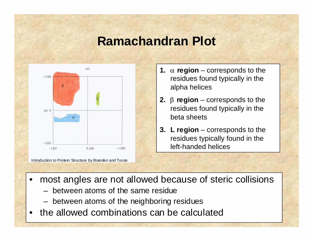
Ramachandran Plot
• most angles are not allowed because of steric collisions– between atoms of the same residue– between atoms of the neighboring residues
• the allowed combinations can be calculated
1. α region – corresponds to the residues found typically in the alpha helices
2. β region – corresponds to the residues found typically in the beta sheets
3. L region – corresponds to the residues typically found in the left-handed helices
Introduction to Protein Structure by Branden and Tooze

Ramachandran Plot
a) observed Ramachandran angles for all residues except glycineb) observed Ramachandran angles for glycine
Introduction to Protein Structure by Branden and Tooze

Ramachandran Angles
• Almost completely describe overall fold of a protein
• Why?– Bond lengths are approximately fixed– Bond angles are approximately fixed– Only Ramachandran (torsion, dihedral) angles are variable– Only 2 variables per amino acid (for the backbone)
Michael S. Chapman

Peptide Bond Revisited
NC
O
CHCH
H
trans
NC
O
CHH
CH
cis
• Usually trans– with ω ≈ 180° ± 6° rms°
• Occasionally cis– with ω ≈ 0°:– ~ 1/4 of prolines– very infrequently glycines– almost never other amino acids
Michael S. Chapman

Levels of Protein Structure
Native state (conformation) – conformation at which protein shows its activity
Introduction to Protein Structure by Branden and Tooze

Helical Structure
a) idealized diagramb) the same as a) but with approximate positions for main-chain atomsc) schematic diagram of an alpha helixd) a ball and stick model
Introduction to Protein Structure by Branden and Tooze

Helical Structure

Other Helical Conformations
• 310 helix• found in proteins when a regular helix is distorted by the presence of
unfavorable residues, near turn regions or in short helices• hydrogen bonds between i and i+3 (instead of i+4)• 3 residues per turn and 10 backbone atoms between donor and
acceptor atom• tighter and narrower
• π helix• more loosely coiled• hydrogen bonds between i and i+5 (instead of i+4)• 4.4 residues per turn• can be very long
• Poly(Pro) helices• all cis with 3.3 residues per turn, right-handed (type I)• all trans with 3 residues per turn, left-handed (type II)
• Poly(Gly) chains• type I: beta conformation• type II: similar to poly(Pro) helix with 3 residues per turn

Polyproline II Helices (PPII)
• shorter than regular helices (4-5 residues)• longer (physically) than regular helices (rise per
turn is twice that of regular helices – 9.3A)• seem to be stabilized by main chain – water
hydrogen bonds• found mostly on protein surface• preference for hydrophilic residues and proline• Gln, Ser, Arg and Ala are found in PPII regions,
Gly is rare• involved in protein-protein interactions• important roles in signal transduction,
transcription, cell motility, etc.• contain sequence motifs, e.g. PXXP• dominant element of secondary structure in
unfolded proteins (Horng and Raines, Prot. Sci. 2006)

Anti-parallel β-sheet
Introduction to Protein Structure by Branden and Tooze

Anti-parallel β-sheet
Typical length: 5-10 residues
Introduction to Protein Structure by Branden and Tooze
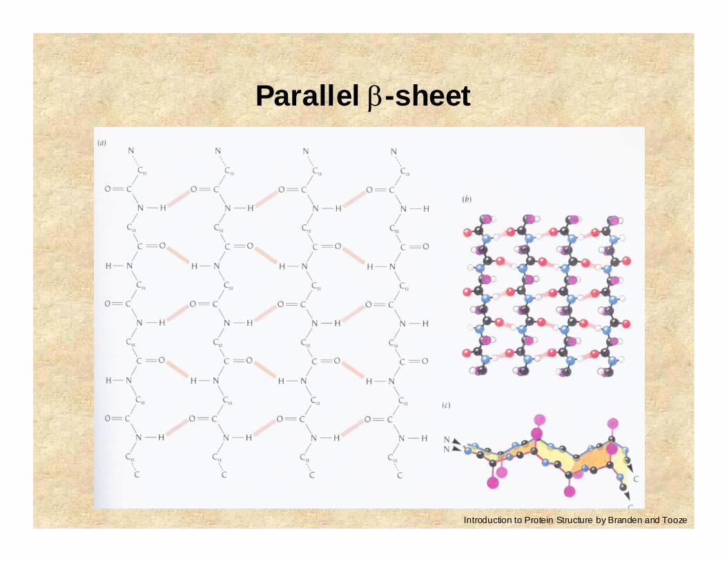
Parallel β-sheet
Introduction to Protein Structure by Branden and Tooze

Mixed β-sheet
Only about 20% of sheets are of mixed type.
Almost all sheets have twisted strands with fixed handedness (right-handed twist)

(Hairpin) Loop
a) histogram showing the frequency of hairpin loops of different lengths in 62 proteins
b) the two most frequently occurring two-residue hairpin loopsIntroduction to Protein Structure by Branden and Tooze

Super Secondary Structure
• helix-loop-helix motif with Ca2+
atom attached
calmodulin
Helix-loop-helix motif
Introduction to Protein Structure by Branden and Tooze

Domains
• hydrophobic residues tend to be on the inside
• polar residues tend to be on the outside of proteins
a) four helix bundle – red cylinders are helices while green parts are loops
b) projection from above
Four Helix Bundle
Introduction to Protein Structure by Branden and Tooze

Disulphide Bonds (Bridges)
• covalent bond between two cysteins (i.e. their sulfur atoms)
• require oxidative environment
• present in extracellular proteins
• stabilize proteins• “create” so-called long-
range interactions
Introduction to Protein Structure by Branden and Tooze

Levinthal’s Calculation
• Cyrus Levinthal 1968
• Q: do proteins explore all possible conformations before they adopt a specific 3-D structure?
• A: let’s consider a simplified problem– each residue can adopt one of the three discrete groups from the
Ramachandran plot (alpha, beta, loop)– a switch between conformations can be done in 10-12 seconds– then, a protein with 150 residues would need to explore 3150
possible states, which is 1071
– at the rate of 10-12 a protein would need ~1050 years
• we know that protein folds between 0.1s and 1000s
Protein Folding Problem
• How do proteins fold into a specific 3-D structure?• How does the primary structure of a protein determine its
secondary and tertiary structure?-----
• there are two conditions a protein needs to meet– there must be a single, stable, folded conformation
(thermodynamic condition)– a protein must fold on an appropriate time scale (kinetic
condition)• thus, only a small amount of conformational space is explored• also, there must exist a specific folding pathway• the paradox how proteins quickly fold into specific 3-D
conformations is called a protein folding problem