functionalcoupling of ca2+ channels and ryanodine ... · ofca2+ channels andryanodine receptors...
TRANSCRIPT

Proc. Natl. Acad. Sci. USAVol. 92, pp. 121-125, January 1995Physiology
Functional coupling of Ca2+ channels and ryanodine receptors incardiac myocytesJAMES S. K. SHAW*, LARs CLEEMANNt, AND MARTIN MORADtt
*Division of Pulmonary and Critical Care Medicine, The Johns Hopkins University Medical School, Baltimore, MD 21205; and tDepartment of Pharmacology,Georgetown University Medical Center, Washington, DC 20007
Communicated by Robert E. Forster II, University of Pennsylvania, Philadelphia, PA, August 26, 1994 (received for review March 4, 1994)
ABSTRACT In skeletal muscle, dihydropyridine receptorsare functionally coupled to ryanodine receptors of the sarcoplas-mic reticulum in triadic or diadic junctional complexes. Incardiac muscle direct physical or functional couplings have notbeen demonstrated. We have tested the hypothesis of fimctionalcoupling of L-type Ca2+ channels and ryanodine receptors in ratcardiac myocytes by comparing the efficacies of Ca2+ in trig-gering Ca2+ release when the ion enters the cell via the Ca2+channels or the Na+/Ca2+ exchanger. Ca2+ transported throughthe Ca2+ channels was 20-160 times more effective than Ca2+influx via the Na+/Ca2+ exchanger in gating Ca2+ release fromthe sarcoplasmic reticulum, suggesting privileged communica-tion between Ca2+ channels and ryanodine receptors. In supportof this hypothesis we found that Ca2+ channels were inactivatedby Ca2+ release from the sarcoplasmic reticulum, even thoughthe myoplasmic Ca2+ concentrations were buffered with 10 mMEGTA. The data thus suggest privileged cross signaling betweenthe dihydropyridine and ryanodine receptors such that Ca2+ fluxthrough either the Ca2+ channel or the ryanodine receptor altersthe gating kinetics of the other channel.
In skeletal muscle, ultrastructural arid biophysical evidencesuggests that the dihydropyridine (DHP) receptors are phys-ically coupled to the ryanodine receptors of the sarcoplasmicreticulum (SR) (1-4). The intramembrane charge movementassociated with the activation of the DHP receptor, rather thanthe influx of Ca2+, is thought to open the Ca2+ release channels(5). In cardiac myocytes, communication between Ca2+ chan-nels and ryanodine receptors and release of Ca2+ from the SRappear to be mediated by the Ca2+-induced Ca2+ releasemechanism requiring influx of Ca2+ through the Ca2+ chan-nels (6-11). For Ca2+-induced Ca2+ release to produce agraded response, it is postulated that either larger Ca2+releases must inactivate the Ca2+ release channel (7) or thereleased Ca2+ must dissipate rapidly from the vicinity of theryanodine receptor during random and Ca2+-independentclosed intervals of the release channel (35). While the firstmechanism requires Ca2+ to accumulate in the microenviron-ment of the ryanodine receptor [a theory supported by Ca2+release in skinned myocytes (7), but not in intact myocytes(12)], the latter emphasizes the intracellular microarchitectureand spatial relationships of sarcolemmal Ca2+ channel and SRrelease channels.
In the present study we test the,hypothesis of functional couplingof Ca2+ channels and ryanodine receptors by evaluating the effi-cacies of Ca2+ entry via different molecular routes in activatingCa2+ release and determine the extent of cross signaling betweenthe Ca2+ channels and ryanodine receptors. This type of couplingrepresents an alternative to the direct physical coupling which wasproposed (13), but later rejected (14), on the basis of the effect ofryanodine on the Ca2+ current (ICa) in the absence and presence ofCa2+ buffers. We present evidence for discrete, Ca2+-mediated
The publication costs of this article were defrayed in part by page chargepayment. This article must therefore be hereby marked "advertisement" inaccordance with 18 U.S.C. §1734 solely to indicate this fact.
cross signaling between the ryanodine and DBP receptors in ratventricular myocytes, independent of bulk cytosolic Ca2+ concen-trations.
MATERIALS AND METHODSCell Isolation. Ventricular myocytes were enzymatically
isolated from male Wistar rats (150-250 g) as described (15).In brief, rats were anesthetized (sodium pentobarbital, 30mg/kg), and the excised hearts were perfused retrogradelythrough the aorta, first for 5 min with Ca2+-free Tyrode'ssolution and then with Ca2+-free Tyrode's solution containingcollagenase (1.4 mg/ml; type A; Boehringer Mannheim) andprotease (0.28 mg/ml; type XIV; Sigma), Myocytes were thendispersed in 0.2 mM Ca2+ Tyrode's solution and stored atroom temperature for 1 hr before use.Measurements of Membrane Currents and Intracellular
Ca2+ Transients. Myocytes were voltage clamped in thewhole-cell clamp configuration. Membrane currents wererecorded with a Dagan Instruments (Minneapolis) amplifier.Intracellular Ca2+ transients were simultaneously monitoredwith Ca2+-sensitive fluorescent dye (0.2 mM fura-2 or 0.1 mMindo-1) introduced into the myocytes through the patch pi-pettes (6, 16). The myocyte under examination was monitoredcontinuously with a CCD television camera (Dage-MTI,Michigan City, IN) using red light. Ca2+ concentrations werecalculated with the equation of Grynkiewicz et at (17) byassuming that the Kd values of fura-2 and indo-1 were 224 and213 nM, respectively (17, 18). Indo-1 fluorescence was cali-brated as described (16).
Solutions. The modified Tyrode's solution contained 142 mMNaCl, 2mM CaCl2, 1 mM MgCl2, 10mM Hepes, 10mM glucose,and 0.02 mM tetrodotoxin (TTX) at pH 7.4. The standardinternal solution contained 110 mM CsCl, 10 mM NaCl, 5 mMMgATP, 10 mM Hepes, 20 mM Et4NCl, and 0.01 mM cAMP atpH 7.2. In the experiments in which we studied possible inacti-vation ofICa by SR Ca2+ release, 0.2 mM cAMP was added to theinternal solution to optimize ICa and Ca2+ uptake. The highconcentrations ofcAMPwere not essential for these experiments,because inactivation of ICa by Ca2+ release could be consistentlyseen in myocytes where no cAMP was included in the patchpipette. In some experiments a solution containing 98 mM CsCl,10 mM Hepes, 20 mM Et4NCl, 10 mM EGTA, 5 mM CaCl2, 5mM MgATP, and 0.2 mM cAMP at pH 7.2 was used to bufferthe internal Ca2+ activity to about 150 nM (10 mM EGTA ishalf-saturated with 5 mM Ca2+, and the Kd is about 150 nM).Experiments were performed at room temperature, about 25°C.
Experimental Protocols and Data Analysis. After the myo-cytes were dialyzed for -5 min, Ca2+ release induced by ICawas elicited by depolarizing pulses from -80 mV to -40, -20,or 0 mV in the presence of 0.02 mM TTX to inhibit Na+
Abbreviations: DHP, dihydropyridine; SR, sarcoplasmic reticulum;TTX, tetrodotoxin.tTo whom reprint requests should be addressed at: Department ofPharmacology, Georgetown University Medical Center, 3900 Res-ervoir Road NW, Washington, DC 20007.
121

Proc. NatL Acad ScL USA 92 (1995)
current. ICa was measured as the peak inward current after leaksubtraction, by assuming that the background current had alinear current-voltage (I-V) relation with reversal potential at0 mV. The Ca2+ charge entering via Ca2+ channels wasmeasured by integrating ICa. Release mediated by the Na+/Ca2+ exchanger was activated by pulses to +80 mV, at whichpotential Ca2+ influx via the exchanger was favored. INa/Ca wasquantified by blocking the exchanger with 5 mM Ni2+. Thecontribution of Cl- current to the measured currents wasminimal, since cAMP-dependent and Ca2+-activated Cl- cur-rents are not expressed in rat ventricular myocytes (19, 20), andthe current traces were leak subtracted. The total chargecarried by Ca2+ (QCa) was quantified by integrating ICa or-24INa/Ca (assuming in the latter case a stoichiometry of 3 Na+to 1 Ca2+). The integration covered a period extending fromthe beginning of depolarization either to the onset (Qonset) orto the peak (Qpeak) of Ca2+ release, yielding values considered,respectively, as the quantity of Ca2+ required to activate orsustain Ca2+ release. The rate of Ca2+ release (R) vs. Qonset wasapproximated by a least-squares fit to the Hill equation,R/Rmax = 1/[1+ (Km/Qonset)N], where Rmax is the maximal rateof release, Km is the half-maximum point, and N is the Hillcoefficient.The inactivation of current through the Ca2+ channel (car-
ried by Ca2+ or Ba2+) and its relation to Ca2+ release wereprobed by use of 180-ms clamp pulses to 0 mV before andfollowing depletion of the SR with 5 mM caffeine or 10 AMryanodine. Time constants for inactivation of ICa were ob-tained by the least-squares fit of two exponentials with theCLAMPFIT program (PCLAMP software; Axon Instruments,Foster City, CA). All external solutions bathing the myocyteswere exchanged rapidly (<200 ms) with a concentration clampsystem (9) for 5-10 s to prevent alteration of cytosolic Na+ andCa2+ concentrations. Data are mean ± SEM.
RESULTSEfficacies ofICa and Na+/Ca2+ Exchange in Triggering SR
Ca2+ Release. To compare the efficacy of Ca2' entry via Ca2+channels and Na+/Ca2+ exchanger in triggering Ca2+ release,we measured the Ca2+ charge required to initiate release whentransported either by the Ca2+ channel or by the Na+/Ca2+exchanger. Fig. 1A shows the Ca2+ currents, intracellular Ca2+transients, and QCa elicited by depolarizing pulses to -40, -20,and 0 mV. At 0 mV, the fully activated Ca2+ current triggeredCa2+ releases which were both larger and faster than thoseactivated by the smaller Ca2+ currents at -40 and -20 mV.Similarly, a larger Qonset was measured at 0 mV than at -40and -20 mV, consistent with the notion that the activity of theCa2+ release channels is proportional to the activating Ca2+concentration as measured in single-channel studies of theryanodine receptor (21-23).
Fig. 1B illustrates the time course and the magnitude ofCa2+ release triggered by activation of the Na+/Ca2+ ex-changer at +80 mV. The exchanger-dependent Ca2+ releasewas characterized by a slow rise in intracellular Ca2+, reaching128 ± 13 nM after 501 ± 46 ms (Table 1), followed by a rapidrise in the intracellular Ca2+ activity. The rapid componentwas abolished by caffeine (24), which depletes SR Ca2+ stores,whereas both the slow and fast components of the Ca2+ releaseat +80 mV were suppressed when 5 mM Ni2+ was added toinhibit Na+/Ca2+ exchange (Fig. 1B; refs. 24 and 25). Whenthe Qonset values of the releases mediated by ICa and theexchanger are compared, it can be seen that the Ca2+ chargerequired to activate Ca2+ release was significantly smallerwhen Ca2+ entered the myocyte via the Ca2+ channels at anyof the three potentials tested.Table 1 quantifies the total transmembrane influx of Ca2+
at the onset (Qonset) and at the peak (Qpeak) of Ca2+ transientfor Ca2+ releases elicited by ICa and Na+/Ca2+ exchange. TheQonset ofIca-activated Ca2+ release at 0, -20, and -40 mV was
-4OmV -20mV Omv B| +80mV
0~~~~~~~~~~~~~~~~~~~~~~~~~~~~~~~ 0 + Nil+
-1 H.P=-7OmVl300,
Qca
)C(P) Qnset Qonset: IQpeak Qonst :QpX* \ 0
0.1 s
0.5 s
FIG. 1. Comparisons of membrane currents, Ca2+ transients, andCa2+ charge (Qca) during the activation of Ca2+ channels andNa+/Ca2+ exchanger. (A) Ca2+ channels were activated by 180-msdepolarizing pulses from a holding potential (H.P.) of -70mV to a testpotential of -40, -20, and 0mV in the presence of 0.02mM TTX. QCawas the integral of the Ca2+ current, Qonset was the QCa at the onsetof the Ca2+ release, and Qpeak was the QCa at the peak of Ca2+ release,estimated by the intersection of the maximum slope of rise in Ca2+transient with the peak intracellular Ca2+ concentration ([Ca2+]1). (B)Ca2+ influx through the Na+/Ca2+ exchanger was activated by a 1-sdepolarizing pulse from a holding potential of -80 mV to +80 mV.Qca was 2 times the integral of the Ni2+-sensitive current, assuming 3Na+/1 Ca2+ stoichiometry. The traces shown in A and B wereobtained from the same cell.
6.0 ± 1.1, 4.0 + 0.9, and 0.8 + 0.2 pC, respectively. These valueswere 20 (at 0 mV) to 160 (at -40 mV) times smaller than Qonsetof the exchanger-mediated Ca2+ release at +80 mV (131 ± 29pC). Since the rate of Ca2+ release triggered by the exchanger(1.4 ± 0.6 ,M/s) was equivalent at best to the lowest rate ofIca-activated release (3.1 ± 0.9 ,tM/s at -40 mV), Ca2+ entryvia the Ca2+ channel was at least 160 times more effective intriggering Ca2+ release from the SR. The high efficacy of ICain activating Ca2+ release suggests a privileged pathway ofCa2+ transport from Ca2+ channels to ryanodine receptors.
Plotting the normalized rate of Ca2+ release activated by ICaat different test potentials versus the quantity of Ca2+ chargetransported prior to the onset of release (Qonset) gives asigmoidal relationship (Fig. 2) similar to the open-probabilitycurves of single SR release channels activated by differentCa2+ concentrations (21, 22). The rate of Ca2+ release pla-teaus at Qonset of 80-100 nC/,F, suggesting that the local Ca2+concentrations were sufficiently high to cause maximal acti-vation of ryanodine receptors. The curve in Fig. 2 may becalibrated in terms of the local Ca2+ concentration at theactivation site if it is assumed that the concentration of Ca2+,for comparable rates of release, may be obtained from thefura-2 measurements of intracellular Ca2+ just prior to theexchanger-induced release. This is based on the assumptionthat during the 0.5-s interval leading up to the exchanger-induced release, the Ca2+ activity at the activation site maycome into equilibrium with bulk cytoplasmic Ca2 . From thesigmoidal curve (or with some approximation from the mea-surements at -40 mV; Table 1), it is estimated that a Qonset Of4.6 + 0.8 nC/,uF, when entering the myocyte through the Ca2+channel, activates a Ca2+ release with the same normalizedrate of release (5.3 ± 1.7 s-1) as that induced by the Na+/Ca2+exchanger when intracellular Ca2+ reaches a threshold of 128nM. If Qonset for the ICa-induced release were proportional toCa2+ concentration in the immediate vicinity of the ryanodine
122 Physiology: Sham et aL

Proc NatL Acad Sci USA 92 (1995) 123
Table 1. Ca2+ charge carried by ICa and INa/Ca at onset and peak of Ca2+ transient
Peak Ca2+ Rrelease,Calcium transporter Vm, mV I, nA Tonset, ms Qonset, pC Qpeak, pC release, nM ZM/s
Ca2+ channel -40 -0.13 ± 0.05 13.4 ± 3.0 0.8 ± 0.2 4.4 ± 1.1 218.5 ± 41.2 3.1 ± 0.9-20 -0.75 ± 0.20 10.3 ± 0.9 4.0 ± 0.9 10.4 ± 1.8 276.3 ± 31.9 10.0 ± 2.0
0 -1.24 ± 0.26 8.6 ± 0.8 6.0 ± 1.1 16.1 ± 2.7 285.7 ± 33.4 11.3 ± 1.1Na+/Ca2+ exchanger +80 0.23 ± 0.03 501.1 ± 45.7 131 ± 29 184 ± 48 322.5 ± 41.3 1.4 ± 0.6
The ICa-dependent Ca2+ release was elicited by 180-ms depolarizing pulses to -40, -20, and 0 mV, whereas the Na+/Ca2+ exchange-dependentCa2+ release was elicited by a 1-s depolarizing pulse to +80 mV. The holding potential was -70 or -80 mV. Tonset was the time from the beginningof depolarization to the onset of Ca2+ release. Qonet and Qpeak were the amounts of Ca2+ charge carried through the transporters at the onsetand the peak of Ca2+ release, respectively. The rate of release, Rrelease, was the maximum slope of rise of Ca2+ transients. Data are expressed inmean ± SEM (n = 6). Cell capacitance = 123 + 14 pF.
receptor, the Ca2+ concentration for maximal activation ofryanodine receptors would be about 2.3 ,.LM [128 nM x(85/4.6)] and Km of about 386 nM.
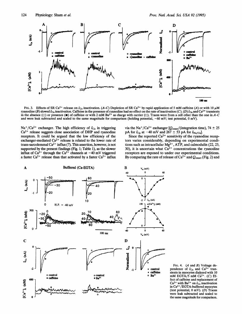
Inactivation ofCa2+ Channel by Release ofCa2+ from the SR.Because of the dependence of the inactivation of Ca2+ channel onintracellular Caj2+ (26-28), the kinetics of inactivation of Ca2+channel may well be the best natural probe for examining thedegree of accessibility of the Ca2+ channel to the ryanodinereceptor (29). When Ica triggers Ca2+ release, the Ca2+ channelshows biexponential inactivation [ = 6.9 + 0.5 and 38.3 ± 1.8 ms(n = 12)]. Following the depletion of SR Ca2+ stores -by rapidapplication of 5 mM caffeine, Ica inactivated significantly slower(Fig. 3A) [ = 16.4 + 0.6 and 51.3 ± 2.5 ms (n = 10)]. The rateof inactivation of Ica [T= 6.8 + 2.3 and 38.2 ± 2.5 ms (n = 5)]was also significantly reduced when ryanodine was used to
I.-
I
60
40
20
0
'(38 iuM)
4.61 (128 nM) 10 100
Normalized Q. (uC/IF)
FIG. 2. The relationship of the rate of Ca2+ release (Rrelease) vs.Qonset at different test potentials. Ica-dependent Ca2+ releases wereactivated by 180-ms depolarizing pulses to -40, -30, -20, -10, and0 mV in six different cells. Rrelease was the maximum slope of rise ofCa2+ transient and was normalized with respect to the maximum Ca2+release at 0 mV. Qonset was normalized with respect to cell capacitance.Data are expressed as mean ± SEM (n = 5). The sigmoidal curve
represents the least-squares fit of the Hill equation, with Rmax = 55 ±
4 s-1, Km = 14 + 2 nC/kLF, and N = 2.1 ± 0.5. Arrows indicate thereference point for the Na+/Ca2+-exchange-dependent Ca2+ release,where an intracellular Ca2+ concentration of 128 ± 13 nM triggeredCa2+ releases with normalized Rrelease of 5.3 ± 1.7 s-1. If we assume
that the cytoplasmic Ca2+ concentration equaled the Ca2+ concen-
tration around the ryanodine receptors, as Ca2+ influx via theNa+/Ca2+ exchanger is slow, 128 nM Ca2+ activates the ryanodinereceptors to the same extent as a Ca2+ influx of 4.6 ± 0.8 nC/,uF viaCa2+ channels. If Qonset is proportional to Ca2+ concentration in thejunctional space, the Km and maximum Ca2+ concentration forryanodine receptor activation in vivo were 386 nM and 5-10 jiM,respectively, and a maximum Qonst of 85 nC/pF at 0 mV was
equivalent to a cytoplasmic Ca2+ concentration of 2.3 ,uM.
suppress Ca2' release [T = 14.9 ± 2.3 and 53.4 ± 4.0 ms (n = 5)]from the SR (Fig. 3B). Further, 5 mM caffeine in the presence ofryanodine did not produce additional slowing of inactivation [ =16.9 ± 3.4 and 55.2 ± 5.5 ms (n = 3); Fig. 3C]. Thus, the reductionin the rate of inactivation ofIca was primarily due to the depletionofSR Ca2+ stores and not to other independent effects ofcaffeineor ryanodine on the inactivation of Ica. Complete suppression ofCa2+-induced inactivation of the Ca2+ channel by the use of Ba2+as charge carrier yielded a slower, monoexponentially decayingcurrent through the Ca2+ channel [T = 72.8 ± 2.4 ms (n = 5)]whose kinetics were not significantly altered by caffeine orryanodine. These results indicate that both SR Ca2+ release andIca contribute to the inactivation of the Ca2+ channel.The effect of release of Ca2+ from the SR on the inactivation
of ICa might result either from an overall increase in the cyto-plasmic Ca2+ concentration or from a local increase in Ca2+, ifthe two channels were closely associated. To distinguish betweenthese two possibilities, myocytes were dialyzed with 10 mMEGTA and 5 mM Ca2+ to buffer the intracellular Ca2+ to about150 nM and to prevent large changes in bulk myoplasmic Ca2+concentration yet allow the SR to load adequately. In these highlyCa2+-buffered myocytes, the rise in cytosolic Ca2+ concentrationevoked by activation of Ica was greatly reduced and the Ca2+transients were very brief, lasting 50-100 ms (Fig. 4A), and thevoltage dependence of Ca2+ release was bell shaped (Fig. 4B).The buffering of intracellular Ca2+ by 10 mM EGTA, however,did not by itself significantly slow the rate of inactivation ofICa [T= 4.9 ± 0.3 and 48.9 ± 3.4 ms (n = 9)] compared with controlmyocytes. Nevertheless, the rate of inactivation of ICa continuedto slow upon depletion of the Ca2+ content of the SR by caffeine[I = 15.9 + 0.6 and 58.9 ± 4.3 ms (n = 9); Fig. 4C], indicating thatthe buffering of cytosolic Ca2+ does not prevent the Ca2+released from the SR from reaching the inactivation site of theDHP receptor. Similarly, replacement of Ca2+ with Ba2+ [T =62.3 ± 4 ms (n = 8); Fig. 4C] reduced the rate of inactivation ofka as effectively as in the unbuffered myocytes (Fig. 3), suggest-ing that Ca2+ ions entering through the Ca2+ channel are notbound by the Ca2+ buffer before reaching the inactivation site.These results suggest that the inactivation of Ica during Ca2+release from the SR does not require an overall increase incytoplasmic Ca2+, but rather a local increase in Ca2+ in thevicinity of the ryanodine receptor.
DISCUSSIONThe major conclusion of this study is that L-type Ca2+ channelsare in close functional association with ryanodine receptors, sothat the flux of Ca2+ through either channel modifies theactivity of the other channel. This cross signaling between theDHP and ryanodine receptors does not seem to include theNa+/Ca2+ exchanger.
Quantitative comparison of the rates of release of Ca2+induced by Ica and INa/Ca suggests that Ica activated a fast Ca2+release compared with the slower and somewhat delayed Ca2+release activated by the exchanger (Fig. 1). For an equivalent rateof Ca2+ release, Ca2+ entering the myocyte through the Ca2+channel was 160 times more efficient than Ca2+ entering via the
Physiology: Sham et al.
Proc Natt Acad Sci USA 92 (1995)
A C
-1o ryanodine. ryanodie + caffene
D
I
.,
+-as
0
FIG. 3. Effects of SR Ca2+ release on Ica inactivation. (A-C) Depletion of SR Ca2+ by rapid application of 5 mM caffeine (A) or with 10 A,Mryanodine (B) slowed Ica inactivation. Caffeine in the presence of ryanodine had no effect on the rate of inactivation (C). (D) Ica and Ca2+ transientsin the absence (0) or presence (0) of caffeine or with 2 mM Ba2+ as charge with carrier (o). Traces were from a cell other than the one inA-Cand were leak subtracted and scaled to the same magnitude for comparison (holding potential, -60 mV; test potential, 0 mV).
Na+/Ca2+ exchanger. The high efficiency of Ica in triggeringCa2+ release suggests close association of DIHP and ryanodinereceptors. It could be argued that the low efficiency of theexchanger-mediated Ca2+ release is related to the lower rate oftrans-sarcolemmal Ca2+ influx (7). This assertion, however, is notsupported by the present findings (Fig. 1; Table 1), as the slowerinflux of Ca2+ through the Ca2+ channels at -40 mV triggereda faster Ca2+ release than that activated by a faster Ca2+ influx
A Buffered (Ca-EGTA)
via the Na+/Ca2+ exchanger [Qonset/(integration time), 74 ± 25pA for Ica at -40 mV and 267 ± 53 pA for INa/ca].
Since the reported Ca2+ sensitivity of the ryanodine recep-tors varies considerably, depending on experimental condi-tions such as intracellular Mg2+, ATP, and calmodulin (22, 23,30), it is uncertain what Ca2+ concentrations the ryanodinereceptors are exposed to under our experimental conditions.By comparing the rate of release of Ca2+ and Qonset (Fig. 2) and
B-60
V. (mV)0 60
60
40
20
100 H.P. - -60 mV
30]100 ms
C
o control* Ba2+
o
v, (mV)
D
o
FIG. 4. (A and B) Voltage de-pendence of Ica and Ca2+ tran-sients in myocytes dialyzed with 10mM EGTA/5 mM Ca2+. (C) Ef-fect of caffeine and replacement ofCa2+ with Ba2+ on Ica inactivationin Ca2+/EGTA-buffered myocytes(test potential, 0 mV). (D) Traceswere leak subtracted and scaled tothe same magnitude for comparison.
100 m
0
-2-
400 -
. o6. O
o control* caffeine
o control* caffeineo Ba2+
124 Pyilg:Sa ta
Proc. NatL Acad Sci USA 92 (1995) 125
assuming that the rise in Ca2+ concentration is uniformlydistributed in the myoplasm during Ca2+ influx via the ex-changer, we estimate a Km of 386 nM with maximal activationoccurring at about 3 ,uM (Fig. 2). These values are remarkablysimilar to the values reported in skinned myocytes, whereactivation of release had a threshold at 100-160 nM and wasmaximal at 3 ,IM (7, 31). Similarly, in SR vesicles andsingle-channel studies, Km and maximal activation were foundto range, respectively, from 300 to 700 nM and from 2 to 20,uM, depending on the mode of activation (steady state vs.transient; refs. 22, 23, and 30).The results showing significant inactivation of ICa by the
release of Ca2+ from the SR in highly Ca2+-buffered myocytesprovide further support for close association of Ca2+ channelsand ryanodine receptors. It was somewhat surprising that therelease of Ca2+ from the SR inactivated the Ca2+ channels toa similar extent as Ca2+ passing through the sarcolemmal Ca2+channels (Fig. 3). Theoretical considerations indicate that thelocal [Ca2+] at the inner mouth of the Ca2+ channel during thepeak of ICa could reach as high as 0.5 mM (32). Although theCa2+ inactivation site of the Ca2+ channel may reside at somedistance from the direct permeation path (26), the Ca2+concentration in the vicinity of the channel (e.g., about 30 ,uMat 5 nm from the pore) (32) should still be well above thephysiological range of global cytoplasmic Ca2+ concentration.The observed inactivation of the Ca2+ channel by the releasedCa2+ therefore suggests that the inactivation site of the Ca2+channel is within the Ca2+-accessible domain of the ryanodinereceptors. This consideration places the Ca2+ channels andryanodine receptors within perhaps 10-20 nm of each other (32).This scheme is further supported by finding that effective buff-ering of the global cytoplasmic Ca2+ concentration with 10 mMEGTA had no significant effect on the Ca2+-dependent inacti-vation of Ca2+ channels caused either by ICa or by Ca2+ releasefrom the SR (Fig. 4). Most likely both the proximity of theryanodine receptor and the slow kinetics of EGTA (33) contrib-ute to the persistence of Ca2+-dependent inactivation of ICa inmyocytes with high content of EGTA. These interactions providea negative feedback mechanism capable of controlling Ca2+influx and Ca2+ release from the SR on a millisecond time scale.The requirements of high Ca2+ and fast response time for suchfunctional interactions tend to undermine "common pool" mod-els, which consider changes of Ca2+ concentration in the myo-plasm as a whole (34), and favor "local control" models, whichconsider dynamic changes of Ca2+ in well-defined spaces or"domains" (32, 35, 36) as controlling the Ca2+ release from theSR. In this model, Ca2+ release from SR is controlled by theunitary current rather than the macrosopic ICa (37). This couldexplain why Ca2+ release is activated by Ca2+-induced Ca2+release (6, 7, 9), even though the voltage dependence of Ca2+transients may depart from that of ICa (11, 38).
Junctional complexes as the site for close coupling between Ca2+channels and ryanodine receptors is consistent with the finding thatthese receptors are concentrated in the transverse tubules andterminal cisternae, respectively (39-41). Since ligand binding stud-ies show that the number of ryanodine receptors is 3-9 times higherthan that of L-type Ca2+ channels (42,43), one Ca2+ channel maybe coupled to a cluster of ryanodine receptors as proposed in the"cluster bomb" model (35) or even to a larger number of "sleepy"ryanodine receptors. Irrespective of the exact stoichiometry, thefunctional coupling of the two types of channel is essential forefficient signaling of Ca2+ release in rat cardiac myocytes becauseonly cCa, and not the Na+/Ca2+ exchanger, triggers the fast, gradedCa2+ releases (24, 44). It is likely that a somewhat different spatialrelation may exist between Na+/Ca2+ exchangers and ryanodinereceptors in guinea pigventricular myocytes, inwhich exchanger hasbeen implicated in fast Ca2+ release (45, 46).
This work was supported by National Institutes of Health GrantRO1-HL16152.
1. Block, B. A., Imagawa, T., Campbell, K P. & Franzini-Arm-strong, C. (1988) J. Cell Biol. 107, 2587-2600.
2. Rios, E. & Brum, G. (1987) Nature (London) 325, 717-720.3. Tanabe, T., Beam, K G., Powell, J. A. & Numa, S. (1988) Nature
(London) 336, 134-139.4. Tanabe, T., Beam, K. G., Adams, B. A., Niidome, T. & Numa, S.
(1990) Nature (London) 346, 567-569.5. Schneider, M. F. & Chandler, W. K. (1973) Nature (London) 242,
244-246.6. Cleemann, L. & Morad, M. (1991) J. Physiol. (London) 432,
283-312.7. Fabiato, A. (1985) J. Gen. Physiol. 85, 247-289.8. Morad, M. & Cleemann, L. (1987) J. Mol. Cell. Cardiol. 19,
527-553.9. Nabauer, M., Callewaert, G., Cleemann, L. & Morad, M. (1989)
Science 244, 800-803.10. Beuckelmann, D. J. & Wier, W. G. (1988) J. Physiol. (London)
405, 233-255.11. Cannell, M. B., Berlin, J. R. & Lederer, W. J. (1987) Science 238,
1419-1423.12. Nabauer, M. & Morad, M. (1990)Am J. Physiol. 258, C189-C193.13. Cohen, N. M. & Lederer, W. J. (1988) J. Physiol. (London) 406,
115-146.14. Balke, C. & Wier, W. G. (1991) Circ. Res. 68, 897-902.15. Mitra, R. & Morad, M. (1985)Am J. Physiol. 249, H1056-H1060.16. Callewaert, G., Lipp, P., Pott, L. & Carmeliet, E. (1991) Cell
Calcium 12, 269-277.17. Grynkiewicz, G., Poenie, M. & Tsien, R. Y. (1985) J. Biol. Chem.
260, 3440-3450.18. Benham, C. D. (1989) J. Physiol. (London) 415, 143-158.19. Callewaert, G., Cleemann, L. & Morad, M. (1989)Am. J. Physiol.
257, C147-C152.20. Dukes, I. D., Cleemann, L. & Morad, M. (1990) J. Pharmacol.
Exp. Ther. 254, 560-569.21. Gyorke, S. & Fill, M. (1993) Science 260, 807-809.22. Rousseau, E. & Meissner, G. (1989) Am. J. Physiol. 256, H328-
H333.23. Smith, J. S., Rousseau, E. & Meissner, G. (1989) Circ. Res. 64,
352-359.24. Sham, J. S. K., Cleemann, L. & Morad, M. (1992) Science 255,
850-853.25. Kimura, J., Miyamae, S. & Noma, A. (1987) J. Physiol. (London)
384, 199-222.26. Imredy, J. P. & Yue, D. T. (1992) Neuron 9, 197-207.27. Kokubun, S. & Irisawa, H. (1984) Jpn. J. Physiol. 34, 599-611.28. Morad, M., Davis, N. W., Kaplan, J. H. & Lux, H. D. (1988)
Science 241, 842-844.29. Gyorke, S. & Palade, P. (1993)Am J. Physiol. 33, C1505-C1512.30. Meissner, G. & Henderson, J. S. (1987) J. Biol. Chem. 262,
3065-3073.31. Fabiato, A. (1982) Fed. Proc. Fed. Am. Soc. Exp. Biol. 41,
2238-2244.32. Simon, S. M. & Llinas, R. R. (1985) Biophys. J. 48, 485-498.33. Stern, M. D. (1992) Cell Calcium 13, 183-192.34. Earm, Y. E. & Noble, D. (1990) Proc. R. Soc. London B 240,
83-96.35. Stern, M. D. (1992) Biophys. J. 63, 497-517.36. Chad, J. E. & Eckert, R. (1984) Biophys. J. 45, 993-999.37. Wier, W. G., Egan, T. M., Lopez-Lopez, J. R. & Balke, C. W.
(1994) J. Physiol. (London) 474, 463-471.38. DuBell, W. J. & Houser, S. R. (1989) Am. J. Physiol. 257,
H746-H759.39. Brandt, N. & Bassett, A. (1986) Arch. Biochem. Biophys. 244,
872-875.40. Inui, M., Saito, A. & Fleischer, S. (1987) J. Biol. Chem. 262,
15637-15642.41. Inui, M., Wang, S., Saito, A. & Fleischer, S. (1988) J. Biol. Chem.
263, 10843-10850.42. Bers, D. M. & Stiffel, V. M. (1993) Am. J. Physiol. 264, C1587-
C1593.43. Wibo, M., Bravo, G. & Godfraind, T. (1991) Circ. Res. 68,
662-673.44. Bouchard, R. A., Clark, R. B. & Clark, W. R. (1993) J. Physiol.
(London) 472, 391-413.45. Leblanc, N. & Hume, J. R. (1990) Science 248, 372-376.46. Kohmoto, O., Levi, A. J. & Bridge, J. H. B. (1994) Circ. Res. 74,
550-554.
Physiology: Sham et al.