epac and phospholipase c regulate ca2+ release in the ... · pathway in the heart does not...
TRANSCRIPT

1
EPAC AND PHOSPHOLIPASE Cε REGULATE CA2+
RELEASE IN THE HEART BY
ACTIVATION OF PROTEIN KINASE Cε AND CALCIUM-CALMODULIN-KINASEII
Emily A. Oestreich*1, Sundeep Malik
*1, Sanjeewa A. Goonasekera
*, Burns C. Blaxall
*,����,
Grant G. Kelley¶, Robert T. Dirksen
* and Alan V. Smrcka
*
*Department of Pharmacology and Physiology and �Department of Medicine, University of
Rochester School of Medicine and Dentistry, Rochester, NY, 14642. ¶Departments of
Pharmacology and Medicine, State University of New York Upstate Medical Center, Syracuse,
NY 14620. 1These authors contributed equally to this work.
Running head: Epac/PLCε enhancement of CICR
Address correspondence to Alan V. Smrcka, Ph.D., 601 Elmwood Avenue, Box 711 Rochester
NY, 14642, FAX 585-273-2652; E-mail; [email protected]
Recently, we identified a novel
signaling pathway involving Epac, Rap,
and PLCεεεε that plays a critical role in
maximal {beta}-adrenergic receptor (ββββAR)
stimulation of Ca2+-induced-Ca
2+ release
(CICR) in cardiac myocytes. Here we
demonstrate that PLCεεεε phosphatidyl-
inositol-4,5-bisphosphate (PIP2) hydrolytic
activity and PLCεεεε-stimulated Rap1 GEF
activity are both required for PLCεεεε-
mediated enhancement of sarcoplasmic
reticulum (SR) Ca2+ release and that PLCεεεε
significantly enhances Rap activation in
response to ββββAR stimulation in the heart.
Downstream of PLCεεεε hydrolytic activity,
pharmacological inhibition of PKC
significantly inhibited both ββββAR- and Epac-
stimulated increases in CICR in PLCεεεε+/+
myocytes but had no effect in PLCεεεε-/-
myocytes. ββββAR and Epac activation caused
membrane translocation of PKCεεεε in
PLCεεεε+/+ but not PLCεεεε
-/- myocytes and
siRNA-mediated PKCε ε ε ε knockdown
significantly inhibited both ββββAR and Epac
mediated CICR enhancement. Further
downstream, the Ca2+/calmodulin-
dependent protein kinase II (CamKII)
inhibitor, KN93, inhibited ββββAR- and Epac-
mediated CICR in PLCεεεε+/+ but not PLCεεεε
-/-
myocytes. Epac activation increased
CamKII Thr286 phosphorylation and
enhanced phosphorylation at CamKII
phosphorylation sites on the ryanodine
receptor (Ryr2) (Ser2815) and
phospholamban (PLB) (Thr17) in a PKC-
dependent manner. Perforated patch
clamp experiments revealed that basal and
ββββAR stimulated peak L-type current
density are similar in PLCεεεε+/+ and PLCεεεε
-/-
myocytes suggesting that control of SR Ca2+
release, rather than Ca2+ influx through L-
type Ca2+ channels, is the target of
regulation of a novel signal transduction
pathway involving sequential activation of
Epac, PLCεεεε, PKCεεεε and CamKII
downstream of ββββAR-activation. These
results provide a detailed characterization
of the signaling mechanism underlying
PLCεεεε-dependent regulation of cardiac
CICR downstream of the ββββ-adrenergic
receptor.
Phospholipase C (PLC) mediated
hydrolysis of phosphatidyl-inositol (4,5) bis-
phosphate (PIP2) results in inositol-
triphosphate (IP3) mediated Ca2+
release from
intracellular stores and diacylglycerol-
mediated activation of protein kinase C. This
ubiquitous signaling pathway plays an integral
role in regulating many physiological
processes, including those of the
cardiovascular system. PLCε is a recently-
identified bi-functional PLC isoform that
possesses both PIP2 hydrolytic and Rap
guanine nucleotide exchange factor (GEF)
http://www.jbc.org/cgi/doi/10.1074/jbc.M806994200The latest version is at JBC Papers in Press. Published on October 27, 2008 as Manuscript M806994200
Copyright 2008 by The American Society for Biochemistry and Molecular Biology, Inc.
by guest on August 17, 2019
http://ww
w.jbc.org/
Dow
nloaded from

2
activity (1-4). The activity of PLCε is
uniquely regulated by direct binding of small
G-proteins including Ras, Rap, and Rho (5;6).
PLCε activity is also stimulated by the
heterotrimeric G-protein subunits Gαs, Gβγ
and Gα12/13 (5;7;8) but direct binding of these
subunits to PLCε has not been demonstrated.
In primary astrocytes isolated from PLCε+/+
and PLCε-/-
mice, multiple G protein-
dependent upstream signals rely critically on
PLCε-dependent generation of IP3 and DAG
(9).
We recently discovered a surprising
role for PLCε regulation downstream of the
β-adrenergic receptor (βAR) in cardiac
myocytes (10). Compared to normal mice,
PLCε-/-
mice exhibit reduced left ventricular
developed pressure in response to strong βAR
stimulation (10). This deficit results from a
decrease in isoproterenol (Iso)-dependent
stimulation of electrically-evoked Ca2+
release
from the sarcoplasmic reticulum (SR) in
single ventricular cardiac myocytes. βAR-
stimulation increases cardiac Ca2+
release in a
cAMP/protein kinase A (PKA)-dependent
mechanism through phosphorylation of
multiple targets of the cardiac excitability and
Ca2+
handling machinery (11). Recently, we
identified a PKA-independent, PLCε-
mediated pathway that contributes to maximal
Iso-dependent enhancement of Ca2+
-induced-
Ca2+
release (CICR) in cardiac myocytes (12)
and explains the decreased βAR function in
PLCε-/-
mice. This novel pathway requires
cAMP-dependent activation of the RapGEF,
Epac (13), which subsequently stimulates
Rap-dependent activation of PLCε.
We previously demonstrated that a
novel Epac/PLCε pathway contributes to
increased CICR during βAR signaling in the
heart. Here, we establish a mechanistic link
between PLCε activity and CICR by showing
that Epac/Rap/PLCε-mediated enhancement
of CICR in the heart requires both PLCε-PIP2
hydrolytic and PLCε-RapGEF activities and
that downstream of PLCε, both PKCε and
CamKII are required for Epac-dependent
enhancement of Ca2+
release. In addition,
voltage clamp experiments reveal that Iso-
dependent activation of the Epac/PLCε
pathway in the heart does not significantly
alter Ca2+
influx through L-type Ca channels
indicating that Ca2+
release from the
sarcoplasmic reticulum is the ultimate target
of this pathway.
Experimental Procedures
Isolation of Cardiac Myocytes - Adult
ventricular cardiomyocytes (AVM) were
isolated from male, 4 to 6 month-old wild type
or PLCε-/-
mice (C57/B6 background) as
previously described (10). Briefly, mice were
anaesthetized with ketamine (100 mg/kg body
weight) and xylazine (5 mg/kg body weight)
by IP injection. Hearts were excised and
digested by Langendorff perfusion using
either Liberase Blendzyme I (Roche) or a
mixture of Collagenase A and D (Roche).
Cells were plated on laminin (BD
Biosciences)-coated coverslips or 35mm
tissue culture dishes in minimum essential
medium (MEM) supplemented with 2 mM L-
glutamine, 2.5% fetal bovine serum
(Hyclone), 1% penicillin/streptomycin, and
2.5 µM blebbistatin (Sigma) to prevent
contractile activity.
Transduction of AVM with Adenovirus -
Adenoviruses were prepared using the AdEasy
system (Stratagene) with the mCMV promoter
used to drive expression of YFP, PLCε WT,
PLCεH1460L, PLCε∆CDC25, or
PLCεK2150E. For wild type and domain
mutant PLCε adenoviruses, a second mCMV
promoter was used to drive the expression of
YFP. Adult AVM were isolated and adhered
to laminin-coated coverslips for 2 hours pre-
infection. Plating media was removed and
replaced with fresh media containing 300
by guest on August 17, 2019
http://ww
w.jbc.org/
Dow
nloaded from

3
m.o.i. of either YFP control, wild type PLCε,
or PLCε domain mutant adenovirus. After 2
hours, the virus was removed and fresh media
was added to the cells. The appearance of
YFP fluorescence was used to determine the
percentage of cells transduced at 24 hours
post-infection. PLC message was measured
by semiquantitative PCR and protein was
detected by western blotting.
PCR For detection of PLCε mRNA in PLCε
-/-
AVMs transduced with either wild type or
domain mutant PLCε adenovirus constructs,
total RNA was isolated using the RNAeasy
mini kit
(Qiagen, Inc., Valencia, CA)
following manufacturer recommendations.
The Superscript III RT-PCR kit (Invitrogen)
was used with 100 ng of total RNA template
for RT-PCR reactions
with mouse PLCε
primers 5'-ACCCTGCGGTAAATGTTCTG-
3' and 5'-ATGTGAATTCCGTGCTACCC-3'
to yield a 300-bp product. GAPDH primers 5'-
CAACGGGAAGCCCATCACCAT-3' and 5'-
CCTTGGCAGCACCAGTGGATGC-3'
yielding a 350-bp product was used as control.
RT was performed for 30 min at 42 °C
followed by incubation at 94 °C for 2 min.
The PCR parameters were denaturation at 94
°C for 30 s, annealing at 45 °C for 45 s, and
extension at 72 °C for 30 sec. The number of
PCR cycles was 30 for GAPDH and 35 for
PLCε.
Electrically-evoked Ca2+ Transients -
Electrically–evoked Ca2+
transients were
measured as previously described (10)For
each experiment, data were collected in the
absence of agonist for 5-15 cells in order to
determine naïve Ca2+
transient amplitude. 1
µM Isoproterenol and 10 µM cpTOME were
prepared in control Ringer solution (145 mM
NaCl, 5 mM KCl, 2 mM CaCl2, 1 mM MgCl2
and 10 mM Hepes, pH=7.4) and locally
perfused for 20 seconds followed by 60
seconds of electrical stimulation (20ms, 8V,
0.5-1 Hz, 60s) in the continued presence of
agonist.
Pharmacological Inhibition of PKC, IP3
Receptors and CamKII – To determine the
effect of PKC inhibition on electrically-
evoked Ca2+
transient amplitude, cells were
pre-treated for 5 minutes with 1 µM bis-
indolylmaleimide-1 (BIM) (Calbiochem), a
broad specificity PKC inhibitor, followed by
constant perfusion of BIM in the presence of 1
µM Iso or 10 µM 8-4-(chlorophenylthio)-2'-
O-methyladenosine-3',5'-monophosphate
(cpTOME). For IP3 receptor inhibition, cells
were pretreated with 20 µM 2-
aminoethoxydiphenyl
borate (2-APB) for 5
minutes followed by constant perfusion of 2-
APB in the presence of 1 µM Iso. For
CamKII inhibition, cells were pretreated with
1 µM KN93 (or inactive KN92) for 30
minutes followed by constant perfusion of
KN93 in the presence of 1 µM Iso or 10 µM
cpTOME.
PKC Translocation AVM were isolated as
described and plated at a density of 50,000
cells/60mm tissue culture dish in serum free
MEM culture media supplemented with 2 mM
L-glutamine, 1% penicillin/streptomycin and
2.5 µM blebbistatin. After 2 hours, the media
was changed to remove dead cells and debris.
Cells were treated with 1 µM Iso for 30
seconds or 10 µM cpTOME for 3 minutes in a
37ºC incubator. Following treatment, dishes
were placed immediately on ice and media
and agonist were removed. Cells were
washed two times with ice-cold PBS
supplemented with protease inhibitors. Cells
were scraped into a lysis buffer (50 mM
HEPES pH 8.0, 3 mM MgCl2, 100 µM EDTA,
100 mM NaCl, 50 µM NaVO4, and protease
inhibitors) and probe sonicated. Samples were
then centrifuged at 100,000g at 4ºC for 15
minutes to pellet the membrane fraction. The
membrane pellet was washed two times with
ice cold lysis buffer, and then re-suspended in
sample buffer (125 mM Tris-HCl pH 6.8, 20%
by guest on August 17, 2019
http://ww
w.jbc.org/
Dow
nloaded from

4
glycerol, 4% SDS, 10% β-mercaptoethanol
(1.42 M), and 0.25% bromophenol blue).
Samples were resolved by 10% SDS-PAGE
and western blotted for specific PKC
isoforms. PKCε and PKCα antibodies (Santa
Cruz) were used at a 1:1000 dilution. HRP-
conjugated anti-rabbit IgG (BioRad) was used
at 1:10000.
PKC siRNA PKCε-specific and CY3-labelled
negative control siRNAs (Ambion) were
reconstituted at 100 pM. Wild type AVM
were isolated as before and media was
changed prior to transfection. For each
siRNA, 600 pmol was added to 600 µL of
OPTIMEM. In a separate tube, 6 µL of
Lipofectamine 2000 was added to 600 µL of
OPTIMEM. After 5 minutes, siRNA and
Lipofectamine tubes were mixed and
incubated at room temperature for 20 minutes.
The 200 pmol siRNA mixture was then added
to each 35mM dish of AVM. Efficiency of
transfection was determined by fluorescence
microscopy of AVM transfected with CY3-
labelled negative control siRNA. Cells
transfected with PKCε-specific siRNA or
negative control siRNA were harvested in
sample buffer at 24, 36, or 48 hours post-
transfection. Knockdown of PKCε protein
was determined by quantitative western
blotting.
Western Blotting Phosphorylation Analysis
Phospho-Thr286 CamKII (1:1000) and total
CamKII specific (1:1000) antibodies were
from Santa Cruz, Phospho-Thr17 PLB
(1:6000), Phospho-Ser16 PLB (1:6000) and
total PLB (1:6000) antibodies were from
Badrilla. Phospho-Ser2815 Ryr2 antibody
(1:2000) was kindly provided by Xander
Wehrens, Baylor College of Medicine and
total Ryr2 antibody (1:2000) was from
Affinity Bioreagents.
Perforated Patch Clamp Briefly, individual
AVM adhered to laminin-coated dishes were
preloaded with 5 µM fluo-4 AM for 30
minutes at 37oC in a Ringer solution.
Myocytes were then washed 2X with an
external Ca2+
current recording solution
containing: 140 mM TEACl, 2 mM MgCl2,
1.8 mM CaCl2, 0.005 mM blebbistatin, 10
mM glucose, and 10 mM Hepes, pH=7.4.
Patch clamp electrodes had a pipette
resistance of 1-2MOhms when backfilled with
internal solution containing: 135 mM CsCl, 1
mM MgCl2, and 10 mM Hepes, pH=7.2, Ca2+
currents and transients were elicited using
200ms test pulses from -50 mV to +70 mV in
10 mV increments delivered at 10s intervals
(0.1 Hz). Peak L-type Ca2+
current magnitude
was normalized to total cell capacitance
(pA/pF), plotted as a function of membrane
potential (Vm), and fitted according to:
I = Gmax*(Vm-Vrev)/(1+exp[(VG1/2-Vm)/kG])
where Gmax is the maximal L-channel
conductance, Vm is the test potential, VG1/2 is
the voltage of half-maximal activation of
Gmax, Vrev is the extrapolated reversal
potential, and kG is a slope factor. The
kinetics of Ca2+
current inactivation was
described by fitting the inactivation phase to
the following single exponential function:
I(t) = A[exp(−t/τinact)] + C
where I(t) is the current at time t after the
depolarization, A is the amplitude of the
inactivating component of current, τinact is the
time constant of inactivation, and C represents
the steady-state non-inactivating component
of current. Ca2+
transients recorded during
each test pulse were expressed as ∆F/F, where
F represents baseline fluorescence and ∆F
represents the fluorescence change from
baseline.
by guest on August 17, 2019
http://ww
w.jbc.org/
Dow
nloaded from

5
Rap and Ras Activation – Hearts were excised
from 4-6 month old PLCε+/+
or PLCε-/-
mice,
cannulated through the aorta, perfused in the
presence or absence of 1 µΜ isoproterenol for
10 min, and snap frozen in liquid nitrogen.
Heart lysates were prepared by polytron in a
buffer containing 50 mM Tris-HCl, pH 7.4,
500 mM NaCl, 2.5 mM MgCl2, 1% NP-40 in
10% glycerol, and protease inhibitors. 1 mg
of heart lysate was incubated with GST-
tagged fusion protein corresponding to amino
acids 788-884 of human RalGDS-Rap-GTP
binding domain or 1-149 of human Raf-1-
Ras GTP binding domain bound to glutathione
agarose in heart lysis buffer. Following
incubation, beads were harvested by
centrifugation, the supernatant removed, and
beads were extensively washed with lysis
buffer. After washing, beads were pelleted by
centrifugation, resuspended in 4X SDS-
sample buffer, resolved on a 15%
polyacrylamide gel and transferred to
nitrocellulose for western blotting.
Statistics - Data are given as mean ± standard
error (SE). Statistical significance was
determined using unpaired Student’s t-test and
a one-way analysis of variance (ANOVA) for
multiple comparisons. Differences were
considered statistically significant at P < 0.05.
Results To determine the relative roles of
PLCε-RapGEF and PLC activities in the
regulation of cardiac Ca2+
handling, we
transduced freshly isolated PLCε-/-
myocytes
with adenoviruses directing expression of
either wild type PLCε or mutants of PLCε
previously shown to eliminate either PLC
hydrolytic activity (PLCεH1460L)(3),
RapGEF activity (PLCε∆CDC25), or Rap
(and other small GTPases) binding to the RA2
domain (PLCεK2150E)(1) (Fig. 1A). Based
on PCR analysis of PLCε transcripts, all
constructs were expressed to similar levels 24
hours after transduction (Fig. 1B). Western
blots of extracts from AVM infected with the
PLCε mutant viruses indicate that the
mutations do not affect expression of PLCε
(Fig. 1C). Electrically-evoked Ca2+
transients
in transduced myocytes were then assessed in
the presence or absence of either the βAR
agonist isoproterenol (1 µM) or the direct
Epac activator cpTOME (10 µM). As
previously observed (12), isoproterenol-
dependent enhancement of electrically-evoked
Ca2+
release was significantly increased 24
hours after transduction of wild type PLCε in
PLCε-/-
AVM (Fig. 1D). Wild type PLCε
expression also restored the 2-fold increase in
evoked Ca2+
transient amplitude in response to
Epac activation with cpTOME (Fig. 1E). In
contrast, PLCε-/-
AVM transduced with either
PLCεH1460L or PLCεK2150E failed to
respond to cpTOME and showed no increase
in isoproterenol responsiveness compared to
YFP control (Fig. 1C-E) (Supplemental Figure
1). These results confirm that direct
stimulation of PLCε hydrolytic activity by
binding of a Ras family GTPase (likely Rap1)
to the PLCε RA2 domain is required for
maximal βAR-mediated increases in
electrically-evoked SR Ca2+
release. PLCε-/-
AVM transduced with PLCε∆CDC25, the
RapGEF deficient PLCε mutant, failed to
exhibit increased responsiveness to either
isoproterenol or cpTOME (Fig. 1D,E)
(Supplemental Figure 1), indicating that
PLCε-RapGEF activity is also required for the
proper execution of the Epac/PLCε pathway
during βAR-mediated regulation of SR Ca2+
release. We have previously shown that the
Rap GEF deletion, PLCε∆CDC25, does not
significantly affect intrinsic PIP2 hydrolysis
activity(9).
The inability of a RapGEF deficient
mutant of PLCε to rescue cpTOME and
maximal isoproterenol-stimulated enhance-
ment of CICR in PLCε-/-
AVM suggests that
Rap activation downstream of βAR
stimulation is at least partially dependent on
by guest on August 17, 2019
http://ww
w.jbc.org/
Dow
nloaded from

6
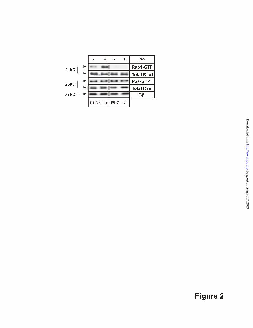
PLCε. Lysates prepared from control and Iso-
perfused hearts of PLCε-/-
or PLCε+/+
mice
were analyzed for activated Rap (RapGTP) by
pulldown with GST-RalGDS and activated
Ras (RasGTP) by pulldown with GST-Raf-1-
RBD. A significant increase in active Rap
over basal levels was observed in lysates from
Iso-treated PLCε+/+
myocytes (Fig. 2, left). In
lysates prepared from PLCε-/-
mice, detectable
Rap activation was not observed under either
basal or Iso-treated conditions (Fig. 2, right).
On the other hand, similar levels of Ras
activation were observed under basal and Iso-
treated conditions in hearts from both PLCε+/+
and PLCε-/-
mice. Total Rap and Ras levels
were identical between PLCε-/-
and wild type
heart lysates. Together, these results are
consistent with previous in vitro findings that
PLCε acts specifically as a GEF for Rap, but
not Ras (2).
PLC-mediated hydrolysis of PIP2
results in generation of IP3 and diacylglycerol
(DAG) and the subsequent activation of Ca2+
release through IP3 receptors in the
sarcoplasmic reticulum and/or PKC activation,
respectively. A definitive role for IP3-
mediated Ca2+
release in EC coupling in
cardiac myocytes has not been identified
despite intensive investigation (14). Type 2
IP3 receptors are the predominant IP3R
isoform present in the heart. Analysis of type
2 IP3R knockout mice indicates that the type 2
IP3 receptor is not required for βAR
enhancement of Ca2+
release in atrial cardiac
myocytes, but is important for endothelin
dependent regulation of Ca2+
release (15). To
assess the potential role of IP3 in our cells, we
compared electrically-evoked Ca2+
release in
control, Iso-, and cpTOME-treated wild type
AVM after treatment with the IP3 receptor
inhibitor, 2-APB (20 µM). 2-APB treatment
did not alter either the Iso (Fig. 3A) or
cpTOME (data not shown) responsiveness,
supporting conclusions of previous studies
that IP3 receptors do not directly contribute to
Iso-dependent enhancement of CICR.
To test for PKC involvement in the
βAR and Epac responses, electrically-evoked
Ca2+
transients in the presence or absence of
isoproterenol were compared in AVM
pretreated with 1 µM bis-indolylmaleimide-1
(BIM), a broad-specificity PKC inhibitor.
BIM treatment significantly inhibited
isoproterenol-stimulated enhancement of SR-
Ca2+
release in wild type AVM (Fig. 3B, left).
In addition, BIM treatment abolished the
increase in electrically-evoked Ca2+
transient
amplitude observed following direct activation
of Epac with cpTOME (Fig. 3C). However,
BIM treatment did not alter Iso-stimulation of
electrically-evoked Ca2+
transients in PLCε-/-
myocytes (Fig. 3B, right) or baseline evoked
transients (data not shown). These results
indicate that the effects of BIM are specific to
the PLCε-dependent pathway downstream of
βAR stimulation and implicate PKC activation
in this pathway.
There are eleven distinct isoforms of
PKC, four of which are consistently detected
in cardiac myocytes: α, βII, δ, and ε (16). To
determine if a specific isoform of PKC is
activated downstream of PLCε, we monitored
translocation of specific PKC isoforms to the
particulate fraction following treatment of
freshly isolated PLCε+/+
and PLCε-/-
cardiac
myocytes with either isoproterenol or
cpTOME. Western blot analysis of the
particulate fraction of wild type cardiac
myocytes revealed a specific increase in PKCε
in the membrane fraction in response to both
isoproterenol and cpTOME treatment relative
to non-treated control (Fig. 4A). In contrast,
PKCα did not translocate to the membrane
fraction in response to isoproterenol. In
PLCε-/-
AVM, neither isoproterenol nor
cpTOME triggered translocation of PKCε to
the membrane (if anything a small decrease in
PKCε at the membrane was observed), placing
by guest on August 17, 2019
http://ww
w.jbc.org/
Dow
nloaded from

7
PKCε downstream of the βAR/Epac/Rap/-
PLCε pathway.
To further test the role of PKCε
downstream of the Epac/Rap/PLCε pathway,
wild type AVM were transfected with either
PKCε-specific siRNA or a CY3-labelled
negative control siRNA. Transfection
efficiency was nearly one-hundred percent
(Supplemental Figure 2) and western blot
analysis revealed that PKCε (but not PKCα)
protein levels were knocked down by at least
95% at 36 hours post-transfection (Fig. 4B).
PKCε protein levels were not substantially
decreased in cells transfected with the
negative control siRNA at all time points
monitored (24, 36, and 48 hours).
Electrically-evoked Ca2+
transients were
monitored 36 hours after transfection of wild
type AVM with either PKCε or negative
control siRNAs. Baseline electrically-evoked
Ca2+
transients were not different between
control and PKCε siRNA-treated AVM. On
the other hand, peak Ca2+
transient amplitude
in the presence of isoproterenol was
significantly inhibited and cpTOME responses
abolished (Fig. 4C) in AVM treated with
PKCε siRNA. These data are consistent with
results obtained following pharmacological
PKC inhibition with BIM (Fig. 3B, C) and
Iso/cpTOME-dependent PKCε membrane
translocation (Fig. 4A), indicating that PKC
acts downstream of PLCε and specifically
implicates PKCε as the relevant PKC isoform
involved.
A recent report demonstrated that
CamKII is activated following Epac
stimulation with cpTOME in rat cardiac
myocytes, however the mechanism for
CamKII activation by Epac was not
determined (17). PKC has also been shown to
activate CamKII in rat ventricular myocytes
(18) and CamKII is directly phosphorylated at
Thr286 by PKC in vitro (19). Therefore, we
determined if CamKII activation is required
for Iso- and Epac-dependent enhancement of
CICR, and if it is downstream of PKC. Iso-
and cpTOME-induced enhancement of
electrically-evoked Ca2+
transients in wild
type AVM were determined in the absence
and presence of KN93, a specific CamKII
inhibitor. KN93, but not the control
compound KN92, attenuated Iso-induced
enhancement of evoked release (Fig. 5A) and
completely blocked cpTOME-induced
enhancement (Fig. 5B). Co-treatment with
BIM and KN93 did not further diminish the
response to isoproterenol relative to treatment
with either compound alone (data not shown).
Additionally, KN93 had no effect on the Iso
response in PLCε-/-
AVM (Fig. 5A) or on
baseline evoked transients (data not shown),
supporting specific involvement of CamKII in
the PLCε dependent pathway.
To determine if CamKII activation was
dependent on PKC, wild type cardiac
myocytes were treated with either
isoproterenol or cpTOME alone or in the
presence of BIM and CamKII phosphorylation
at Thr286 was measured by western blotting
(Fig. 5C). Both isoproterenol and cpTOME
treatment increased CamKII phosphorylation
at Thr286 relative to non-treated control. The
cpTOME-dependent increase was blocked in
the presence of BIM and the Iso-dependent
increase was partially blocked by BIM. These
data support the conclusion that the
Epac/PLCε pathway can control CamKII
activation in a PKCε-dependent, Ca2+
-
independent manner. That the Iso-dependent
increase in phosphorylation was only partially
blocked suggests that there are multiple
mechanisms for CamKII activation
downstream of Iso, one of which includes the
Ca2+
-independent Epac and PKC pathway, but
may also result from changes in Ca2+
that
occur with Iso-dependent regulation of PKA.
CamKII phosphorylates numerous
Ca2+
-handling proteins, including the L-type
by guest on August 17, 2019
http://ww
w.jbc.org/
Dow
nloaded from

8
Ca2+
channel, Ryr2 and PLB, involved in
precisely controlling dynamic changes in
intracellular calcium levels during the cardiac
cycle (20). CamKII-dependent modulation of
RyR function by phosphorylation at Ser2815
has been implicated as a means for positive
regulation of SR-Ca2+
release downstream of
βAR stimulation. CamKII also
phosphorylates PLB at Thr17 to stimulate SR
Ca2+
reuptake to increase content available for
release. To determine if Epac/PLCε-
stimulation results in a PKC-dependent,
CamKII-mediated, phosphorylation of Ryr2
and/or PLB, AVM were treated with cpTOME
in the presence or absence of the PKC
inhibitor BIM. Phosphorylation at the
CamKII specific sites was measured by
western blotting (Fig. 5D). cpTOME
treatment significantly enhanced
phosphorylation of Ryr2 at Ser2815 and PLB
at Thr17 in wild type AVM. These increases
were ablated in AVM pretreated with BIM.
These data identify at least two effector targets
(Ryr2 and PLB) of the Epac/PLCε pathway
that could be involved in regulating the
magnitude of CICR.
To examine the relationship between
Ca2+
influx and release during EC coupling we
conducted voltage clamp experiments to
determine if the Epac/PLCε pathway alters the
properties of depolarization-induced L-type
Ca+ channel function and RyR2-mediated
Ca2+
release. Perforated patch clamp
experiments in fluo-4-loaded AVM were
conducted to simultaneously compare the
voltage dependence, magnitude, and kinetics
of L-type Ca2+
currents and global
intracellular Ca2+
transients in wild type and
PLCε-/-
myocytes before and after βAR
activation (Fig. 6 and Supplemental Figure 3).
L-type Ca2+
currents and intracellular Ca2+
transients were elicited by 200 ms test pulses
from -50 to +70mV at 10mV increments. No
differences in the magnitude or kinetics of L-
type Ca2+
currents (Fig. 6A) or global
intracellular Ca2+
transients (Fig. 6C) were
observed between PLCε-/-
and PLCε+/+
AVM
under basal conditions (closed symbols)
(Table 1). Application of 1 µM Iso (open
symbols) caused a similar 1.5-2 fold increase
in peak L-type Ca2+
current density (Fig. 6A)
and maximum channel conductance (Fig. 6B
and Table 1) in both PLCε-/-
and PLCε+/+
AVM. However, peak Iso-stimulated Ca2+
transient amplitude was significantly
attenuated in PLCε-/-
cardiac myocytes (Fig.
6C and D and Table 1), consistent with results
observed in intact myocytes (Fig. 3B and 5A).
In addition, the kinetics of Ca2+
current
inactivation was significantly slower in
myocytes from PLCε-/-
mice (Supplemental
Figure 3). These data indicate that alterations
in action potential or L-type channel activity
are not necessary for PLCε-dependent
regulation of CICR.
Discussion
PLCε is unique among PLC enzymes
in that it possesses both phospholipase C and
RapGEF activities. Physiological roles for
both catalytic functions of PLCε are beginning
to emerge (9;10;21;22). Here we demonstrate
that both PLCε hydrolytic and RapGEF
activities are required for maximal βAR-
mediated (and Epac-dependent) enhancement
of electrically-evoked Ca2+
release in the
heart. We hypothesize that the PLCε-
RapGEF activity ensures sufficient Rap
activation to maintain PLCε hydrolytic
activity, and that PLCε is required for
sustained Rap activation in the heart. Epac
stimulation by cAMP may initiate a low level
of Rap activation that (undetectable in the
PLCε-/-
myocytes), in turn, stimulates PLCε to
significantly amplify Rap activation that feeds
forward to further stimulate PLCε and
subsequent regulation of CICR. This model is
supported by previous studies in transfected
cells demonstrating that PLCε potentiates its
own activation by RapGTP (2) and in primary
by guest on August 17, 2019
http://ww
w.jbc.org/
Dow
nloaded from

9
astrocytes where PLCε RapGEF activity is
required for sustained Rap activation and
downstream ERK signaling (9). In addition, it
is important to note that Rap-GTP generated
from PLCε may also regulate other enzymes
in the heart such as ERK5 where Rap-
mediated inhibition protects against the
development of hypertrophy (23). This would
be consistent with our findings that PLCε-/-
mice exhibit increased susceptibility to stress
induced hypertrophy (10) and that PLCε
RapGEF activity modulates ERK signaling in
astrocytes(9).
Downstream of PLCε activity, we
demonstrate that diacylglycerol-mediated
PKCε activation is required for maximal
βAR-dependent enhancement of CICR (Fig.
3). Several reports have suggested that PKC
activity modulates CICR in cardiac myocytes.
Treatment with norepinephrine or phorbol
myristic acid causes PKCε translocation to
cross-striated t-tubular regions of cardiac
myocytes upon activation, strategically
placing this enzyme in position to
phosphorylate proteins involved in Ca2+
handling (24). PKCδ and PKCε have been
shown to mediate positive inotropy that is
dependent on subcellular localization (25).
Our data are consistent with a positive
ionotropic effect of PKCε and indicate that
PLCε is an upstream regulator of PKCε in the
heart. siRNA knockdown of PKCε strongly
suppressed cpTOME-dependent increases in
Ca2+
transient amplitudes suggesting that
PKCε is the major isoform of PKC involved
in Epac/PLCε-dependent responses. On the
other hand, the inhibition of Iso-dependent
responses by PKCε siRNA appeared less than
with observed with BIM treatment (Fig. 3B
vs. 4C). One possibility is that another PKC
isoform such as PKCδ is involved in the Iso-
dependent response or that a more complete
inhibition of PKCε that might be achievable
with BIM may be required to fully inhibit the
Iso-response.
We also identified PKC-dependent
CamKII regulation as an essential downstream
component of the Epac/PLCε pathway in
AVM. βAR- and cpTOME enhancement of
electrically-evoked Ca2+
release was
suppressed by PKC inhibition with BIM (Fig.
3B, C), PKCε knockdown (Fig. 4C), and
CamKII inhibition with KN93 (Fig. 5) in
PLCε+/+
AVM. On the other hand, neither
PKC inhibition, CamKII inhibition, nor the
combination, had any significant effect on the
already reduced Iso-dependent regulation of
evoked release in PLCε-/-
AVM, indicating
that both PKCε and CamKII are downstream
from PLCε activation. We also show that
phosphorylation of CamKII at Thr286 is
increased by cpTOME in a PKC-dependent
manner. These data are consistent with a
previous report demonstrating that activation
of Epac stimulates CamKII Thr286
phosphorylation (17) but extend this result to
show that Epac-dependent CamKII activation
relies on PLCε-dependent PKCε activity.
While a mechanism for PKC-dependent
activation of CamKII has not been clearly
delineated, CamKII Thr 286 has been shown
to be directly phosphorylated by PKC in vitro
(19). Thr286 autophosphorylation results in
tight binding of calmodulin such that Ca2+
is
no longer required for activation. If CamKII
Thr286 is directly phosphorylated by PKC it
should also result in Ca2+
-independent
regulation. Physiological evidence for PKC-
dependent regulation of CamKII is sparse but
two previous studies have shown that α-
adrenergic receptor stimulation facilitates
PKC-mediated activation of CamKII (18;26).
PKCε may not directly phosphorylate CamKII
in cardiac myocytes in response to Epac/PLCε
activation but we clearly demonstrate that
PKC activation is upstream of CamKII
Thr286 phosphorylation in this pathway. It is
also possible that PKCε itself phosphorylates
by guest on August 17, 2019
http://ww
w.jbc.org/
Dow
nloaded from
10
components of the Ca2+
handling machinery,
but our data indicate that any such activity
alone is insufficient since CamKII inhibition
with KN93 completely blocks cpTOME
dependent enhancement of Ca2+
transient
amplitudes (Fig. 5B). This indicates that PKC
activation by this pathway is not sufficient to
cause increases in Ca2+
transients.
Nevertheless, it remains formally possible that
local Ca2+
release, dependent on PKC
activation, could lead to CamKII
autophosphorylation. We propose that in
cardiac myocytes, linear activation of Epac-
PLCε-PKCε-CamKII mediates a component
of Iso-dependent regulation of evoked Ca2+
release in cardiac myocytes.
Potential targets downstream of this
pathway include the L-type Ca2+
channel,
Ryr2 and phospholamban. PLCε ablation did
not markedly affect isoproterenol-stimulated
increases in L-type Ca2+
channel current
density in perforated patch clamp experiments
(Fig. 6A, B) and L-type Ca2+
channel activity
was not significantly altered by 20 µM
cpTOME (data not shown). In the same cells
Iso-stimulated enhancement of depolarization-
induced Ca2+
transients was significantly
attenuated (Fig. 6C, D). The fact that Ca2+
release elicited by a uniform voltage clamp
pulse is reduced in PLCε-/-
myocytes, suggests
that changes in action potential waveform are
not likely responsible for the reduction in Iso
responsiveness. Together, these data indicate
that the Epac/PLCε/PKCε/CamKII pathway
contributes to enhanced release of Ca2+
from
the SR during βAR-stimulation that is not
dependent on changes in Ca2+
influx. Two
proteins that control release of Ca2+
from the
SR, Ryr2 and PLB, are phosphorylated at
CamKII-specific sites in response to Epac
stimulation, supporting this idea. The
observed increase in Ca2+
release in response
to a uniform Ca2+
influx could arise from a
combination of both an increase in Ryr2
sensitivity to activation by Ca2+
influx(27;28)
and an increase in Ca2+
reuptake and load.
The observed increase in Ca2+
release we
report here differs from results of Pereira et
al.(17) who report that Epac activation
decreases evoked Ca2+
release due to CamKII
dependent SR store depletion. Apparent
discrepancies between our results and Pereira
et al. could be due to differences in protocol or
species used, but the overall conclusions that
Epac activation results in CamKII activation
and Ryr2(S2815) phosphorylation are in
agreement.
Roles of PKC and CamKII in both
normal and pathological cardiac function have
been steadily emerging over the last several
years, but our understanding of the
physiological mechanisms that control
activation of these pathways have lagged
behind. Here, we have outlined a novel,
PLCε/PKCε/CamKII-dependent regulatory
mechanism for regulating cardiac CICR in
adult ventricular cardiac myocytes. Previous
studies have implicated CamKII-dependent
phosphorylation of RyR2 as a means for
regulating SR-Ca2+
release downstream of
Epac or βAR stimulation (17;28). We extend
these findings by identifying key mechanistic
links between Epac activation and regulation
of CICR such that the majority of the
components of the pathway are now defined.
This pathway is clearly important for cardiac
function because mice lacking PLCε exhibit a
significantly impaired ability to respond to
βAR-stimulation. This impairment is
manifested by both decreased cardiac function
in response to isoproterenol administration in
whole animals (10) and decreased βAR-
dependent enhancement of CICR in AVM
isolated from PLCε-/-
mice (12). PLCε-/-
mice
also show an increased sensitivity to stress
induced cardiac hypertrophy (10) and it
remains to be determined how components of
the pathway could contribute to this
pathology.
by guest on August 17, 2019
http://ww
w.jbc.org/
Dow
nloaded from

11
References
1. Kelley, G. G., Reks, S. E., Ondrako, J. M., and Smrcka, A. V. (2001) EMBO J. 20, 743-754
2. Jin, T. G., Satoh, T., Liao, Y., Song, C., Gao, X., Kariya, K., Hu, C. D., and Kataoka, T.
(2001) J.Biol.Chem. 276, 30301-30307
3. Lopez, I., Mak, E. C., Ding, J., Hamm, H. E., and Lomasney, J. W. (2001) J.Biol.Chem.
276, 2758-2765
4. Shibatohge, M., Kariya, K., Liao, Y., Hu, C. D., Watari, Y., Goshima, M., Shima, F., and
Kataoka, T. (1998) J.Biol.Chem. 273, 6218-6222
5. Kelley, G. G., Reks, S. E., and Smrcka, A. V. (2004) Biochem.J. 378, 129-139
6. Wing, M. R., Snyder, J. T., Sondek, J., and Harden, T. K. (2003) J Biol.Chem. 278, 41253-
41258
7. Wing, M. R., Houston, D., Kelley, G. G., Der, C. J., Siderovski, D. P., and Harden, T. K.
(2001) J.Biol.Chem. 276, 48257-48261
8. Schmidt, M., Evellin, S., Weernink, P. A., von Dorp, F., Rehmann, H., Lomasney, J. W.,
and Jakobs, K. H. (2001) Nat Cell Biol. 3, 1020-1024
9. Citro, S., Malik, S., Oestreich, E. A., Radeff-Huang, J., Kelley, G. G., Smrcka, A. V., and
Brown, J. H. (2007) Proc.Natl.Acad.Sci U.S.A. 104, 15543-15548
10. Wang, H., Oestreich, E. A., Maekawa, N., Bullard, T. A., Vikstrom, K. L., Dirksen, R. T.,
Kelley, G. G., Blaxall, B. C., and Smrcka, A. V. (2005) Circ Res 97, 1305-1313
11. Bers, D. M. (2002) Nature 415, 198-205
12. Oestreich, E. A., Wang, H., Malik, S., Kaproth-Joslin, K. A., Blaxall, B. C., Kelley, G. G.,
Dirksen, R. T., and Smrcka, A. V. (2007) J Biol.Chem. 282, 5488-5495
13. Bos, J. L. (2003) Nat Rev Mol Cell Biol 4, 733-738
14. Woodcock, E. A. and Matkovich, S. J. (2005) Pharmacology & Therapeutics 107, 240-251
15. Li, X., Zima, A. V., Sheikh, F., Blatter, L. A., and Chen, J. (2005) Circ Res 96, 1274-1281
16. Dorn, G. W., II and Force, T. (2005) J.Clin.Invest. 115, 527-537
17. Pereira, L., Metrich, M., Fernandez-Velasco, M., Lucas, A., Leroy, J., Perrier, R., Morel,
E., Fischmeister, R., Richard, S., Benitah, J. P., Lezoualc'h, F., and Gomez, A. M. (2007) J
Physiol. 583, 685-694
18. O-Uchi, J., Komukai, K., Kusakari, Y., Obata, T., Hongo, K., Sasaki, H., and Kurihara, S.
(2005) Proc.Natl.Acad.Sci U.S.A. 102, 9400-9405
19. Waxham, M. N. and Aronowski, J. (1993) Biochemistry. 32, 2923-2930
20. Maier, L. S. and Bers, D. M. (2007) Cardiovascular Research 73, 631-640
21. Ikuta, S., Edamatsu, H., Li, M., Hu, L., and Kataoka, T. (2008) Cancer Res 68, 64-72
22. Tadano, M., Edamatsu, H., Minamisawa, S., Yokoyama, U., Ishikawa, Y., Suzuki, N.,
Saito, H., Wu, D., Masago-Toda, M., Yamawaki-Kataoka, Y., Setsu, T., Terashima, T.,
Maeda, S., Satoh, T., and Kataoka, T. (2005) Mol.Cell.Biol. 25, 2191-2199
23. Dodge-Kafka, K. L., Soughayer, J., Pare, G. C., Carlisle Michel, J. J., Langeberg, L. K.,
Kapiloff, M. S., and Scott, J. D. (2005) Nature 437, 574-578
24. Disatnik, M. H., Buraggi, G., and Mochly-Rosen, D. (1994) Experimental Cell Research
210, 287-297
25. Kang, M. and Walker, J. W. (2005) J.Mol.Cell.Cardiol. 38, 753-764
26. O-Uchi, J., Sasaki, H., Morimoto, S., Kusakari, Y., Shinji, H., Obata, T., Hongo, K.,
Komukai, K., and Kurihara, S. (2008) Circ Res 102, 1378-1388
by guest on August 17, 2019
http://ww
w.jbc.org/
Dow
nloaded from
12
27. Currie, S., Loughrey, C. M., Craig, M. A., and Smith, G. L. (2004) Biochem.J. 377, 357-
366
28. Wehrens, X. H. T., Lehnart, S. E., Reiken, S. R., and Marks, A. R. (2004) Circ Res 94, e61-
e70
by guest on August 17, 2019
http://ww
w.jbc.org/
Dow
nloaded from

13
Footnotes:
Abbreviations: PLC, Phosphoinositide-specific phospholipase C; PIP2, Phosphatidylinositol 4,5
bisphosphate; IP3, inositol 1,4,5 trisphosphate; GEF, guanine nucleotide exchange factor; βAR,
β-adrenergic receptor; AVM, adult ventricular myocytes; PKA, protein kinase A; PKC, protein
kinase C; CICR, calcium-induced calcium release; SR, sarcoplasmic reticulum; PLB,
phospholamban; Ryr, ryanodine receptor; BIM, bisindolylmaleimide-1; cpTOME, 8-4-
(chlorophenylthio)-2'-O-methyladenosine-3',5'-monophosphate.
Acknowledgements: This work was supported by NIH Grant GMR01053536 to AVS, NIH
AR044657 to RTD, AHA Scientist Development Grant 045343T to BCB and NIH DK56294 to
G.G.K. We would like to thank Joan Heller-Brown for critical review of this manuscript.
by guest on August 17, 2019
http://ww
w.jbc.org/
Dow
nloaded from
14
Figure Legends
Figure 1. Epac-mediated enhancement of CICR requires both PLCεεεε hydrolytic and
RapGEF activities. (A), Domain structure of PLCε (CDC25 GEF – Ras family small GTPase
guanine nucleotide exchange factor domain, PH – pleckstrin homology, EF – EF-hand Ca2+
-
binding domain, X and Y – PIP2 hydrolysis catalytic domain, C2 – Ca2+
dependent lipid-binding
domain, RA1 and RA2 – Ras association domains). ∆CDC25(677-772) – GEF deletion mutant,
no RapGEF activity, H1460L – catalytic domain point mutation, lacks PIP2 hydrolysis activity,
K2150E – RA2 domain point mutation – eliminates stimulation of PLC activity by Ras and
Rap1. (B) PLCε-/-
cardiac myocytes were transduced with YFP, PLCε wild type, or PLCε
domain mutant adenoviruses. 24 hours post-transduction, equal expression of PLCε mRNA was
demonstrated by RT-PCR (lower). (C) Cardiac myocytes were transduced with wt and mutant
PLCε viruses and protein expression was measured after 48h by Western blotting (D, E),
Average (±S.E.) peak Ca2+
transient amplitude (∆405/485) for naïve PLCε-/-
myocytes and 24
hours post-transduction with either YFP control, PLCε wild type or PLCε domain mutant
adenovirus (300 m.o.i.) with or without 1µM isoproterenol (D) or 10µM cpTOME (E).
Figure 2. PLCεεεε is required for ββββAR-mediated Rap activation in cardiac myocytes. Hearts
from PLCε+/+
or PLCε-/-
mice were cannulated through the aorta and perfused with or without
1µM isoproterenol for 10 min. 1mg of heart lysate was incubated with either GST-tagged
RalGDS-RBD for assaying activated Rap or GST-tagged Raf1-RBD for activated Ras. Gβ1 was
used as a gel loading control. Data are representative of 3 experiments showing similar results.
Figure 3. Pharmacological inhibition of PKC attenuates ββββAR enhancement of CICR. (A),
IP3R inhibition with 2-APB (20 µM, 5 min pre-treatment) does not inhibit βAR-dependent
increases in Ca2+
transient amplitude (∆405/485). Ca2+
transients were measured in the absence
and presence of 1µM isoproterenol. Data are pooled from 10-15 cells per treatment condition.
Results are average (±S.E.). (B and C), PKC inhibition (1µM bis-indolylmaleimide (BIM), 5
minute pre-treatment) significantly blunts (B) isoproterenol - and (C ) cpTOME -induced
increases in Ca2+
-transient amplitude in PLCε+/+
, but not PLCε-/-
cardiac myocytes. BIM did not
affect naïve Ca2+
transient amplitude. Data were pooled from 20-40 cells for each treatment
condition from n = 3 PLCε+/+
and n = 2 PLCε-/-
mice. Results are average (±S.E.); ***, p<0.001; #, p<0.001 for PLCε
-/- response compared to PLCε
+/+; ns is not significant; one-way ANOVA,
Bonferroni post test.
Figure 4. PLCεεεε-dependent enhancement of CICR requires specific activation of PKCεεεε. . . .
(A), left - PKCε translocates to the membrane fraction following treatment with 1µM
isoproterenol (30 sec) or 10µM cpTOME (3 min) in PLCε+/+
but not PLCε-/-
cardiac myocytes.
PKCα does not translocate to the membrane in response to βAR stimulation. PMA treatment
(500nM, 10 min.) was used as a positive control for PKC translocation. 3 µg of cardiac myocyte
membrane fractions was analyzed for PKC isoform translocation. Gβ subunit was used as a
loading control. right - Densitometric quantitation of PKCε membrane translocation from cells
isolated from 5 PLCε+/+
and 3 PLCε-/-
mice. Data are represented as a percentage of maximal
translocation evoked by PMA treatment. **, p<0.01, ***p<0.001, ns – not significant as
by guest on August 17, 2019
http://ww
w.jbc.org/
Dow
nloaded from

15
compared to nontreated PLCε+/+
cells. One-way ANOVA, Bonferroni post-test. (B), left -
PLCε+/+
cardiac myocytes were transfected with PKCε-specific siRNA or a CY3-labelled
negative control siRNA. PKCε protein levels are nearly completely knocked down in cardiac
myocytes transfected with PKCε-specific siRNA relative to negative control siRNA at 36 hours
post-transfection. Lower left – PKCα protein levels are not significantly affected by PKCε
siRNA 36 hours post-transfection. Right - Densitometric quantitation of PKCε protein expression
from myocytes transfected with either PKCε siRNA or Cy3-labeled negative control siRNA
pooled from three separate experiments (C), Knockdown of PKCε significantly decreases
isoproterenol-induced enhancement of CICR and completely eliminates cpTOME responsiveness
in PLCε+/+
cardiac myocytes. Data are pooled Ca2+
transient amplitudes (∆404/485) from 20-40
cells per condition, n = 3 mice. Results are average (±S.E.); ***, p<0.001, one way ANOVA,
Bonferroni post-test.
Figure 5. PKC-dependent activation of CamKII is required for PLCεεεε-mediated
enhancement of CICR CamKII inhibition (1µM KN-93, 30 minute pre-treatment)
significantly blunts (A) Iso- and (B) cpTOME (10µM) enhancement of electrically-evoked Ca2+
transient amplitude in AVM from PLCε+/+
, but not PLCε-/-
mice. Pretreatment with KN92, an
inactive analogue of KN-93, does not affect Iso responsiveness. Data were pooled from 5-15
cells for each treatment condition from 3 PLCε+/+
and 3 PLCε-/-
mice. Results are average
(±S.E.); ***, p<0.001; ns – not significant; one-way ANOVA, Bonferroni post test. (C)
Phosphorylation of CamKII at Thr286 was measured by Western blotting of extracts of AVM
treated with Iso (1µM), cpTOME (10µM) and BIM (1µM) as indicated. (n=5 animals). right
panel pooled data from densitometric quantitation. Results are average (±S.E.); *, p<0.05; one-
way ANOVA, Bonferroni post test. (D) Phosphorylation of Ryr2 S2815 and PLB T17 was
measured by Western blotting of extracts of AVM treated with cpTOME (10µM) or BIM (1µM)
as indicated (n=3 animals each). right panel pooled data from densitometric quantitation and
normalized relative to untreated cells. Results are average (±S.E.); **, p<0.01, ***, p<0.001;
one-way ANOVA, Bonferroni post test.
Figure 6. PLCεεεε ablation significantly reduces isoproterenol stimulation of depolarization-
induced intracellular calcium transients without altering L-type Ca2+ current density.
Perforated patch clamp experiments were used to simultaneously monitor depolarization-induced
L-type Ca2+
currents (A, B) and intracellular Ca2+
transients (C, D) in the absence (closed
symbols) and presence (open symbols) of βAR-stimulation with 1µM isoproterenol in AVM
from PLCε+/+
(circles) and PLCε-/-
(squares) mice. A, Average (±SE) voltage dependence of L-
type Ca2+
current density. B, Average (±SE) fold stimulation of maximal L-type Ca2+
conductance in AVM from 5 PLCε-/-
and 5 PLCε+/+
mice. C. Average (±SE) voltage dependence
of intracellular Ca2+
transients. D, Average (±SE) fold stimulation of peak intracellular Ca2+
transient (measured at -30 mV) in AVM from 5 PLCε-/-
and 5 PLCε+/+
mice. * p<0.05, t test.
by guest on August 17, 2019
http://ww
w.jbc.org/
Dow
nloaded from

16
Table 1. Parameters of IV and (∆∆∆∆F/F)max Data
Gmax
(nS/nF)
kv (mV)
VG1/2
(mV)
Vrev
(mV) (∆∆∆∆F/F)max
PLCεεεε+/+ Naïve 108±14 7.4±0.4 -14.3±0.4 62.9±1.1 0.15±0.04
(n=5 mice)
PLCεεεε+/+ Iso 197±14* 6.1±1.4 -18.9±1.7 65.0±0.9 0.45±0.04*
,#
(n=5 mice)
PLCεεεε-/- Naïve 103±14 6.6±0.3 -16.0±2.4 64.7±1.6 0.15±0.03
(n=5 mice)
PLCεεεε-/- Iso 162±18** 6.0±0.2 -20.1±2.0 66.2±1.9 0.27±0.04**
(n=5 mice)
Table 1. Values represent mean (±SE) for n number of mice (from a total of 32 PLC+/+
and 27 PLC-/-
myocytes). Parameters for the voltage dependence of Ca2+
current (I-V) was
obtained by fitting myocytes within each group separately to the appropriate equation (I-V,
Eq. 1) as described in Methods. (∆F/F)max values are the mean (±SE) values obtained at a
test potential of -10 mV. Gmax, maximal L-channel conductance; Vrev, reversal potential;
VG1/2, potential at which G is half-maximal, respectively; kG slope factor for I-V. *p<0.01
compared to PLCε+/+
naive. **p<0.05 compared to PLCε-/-
naive. #p<0.05 compared to
PLCε-/-
Iso. by guest on August 17, 2019
http://ww
w.jbc.org/
Dow
nloaded from

X YCDC25 RA1 RA2PH EF C2
K2150EH1460L∆CDC25 (677-772)
WT ∆CDC25 H1460L K2150EYFP WT ∆CDC25 H1460L K2150E
PLCε
GAPDH
PLCε
B
A
ED
C
Figure 1
by guest on August 17, 2019
http://ww
w.jbc.org/
Dow
nloaded from


PKCεεεε
Gββββ
NT
ISO
PMA
PKCαααα
Gββββ
PLCεεεε+/+
NT
ISO
cpTOME
PMA
NT
ISO
cpTOME
PMA
PKCεεεε
Gββββ
t(hr)
PKCεεεεsiRNA Control siRNA
0 024 2436 48 36 48
PKCαααα
Gββββ
PKCεεεεsiRNA
t(hr)0 36
A
B
C
Figure 4
PLCε+/+ PLCε-/-
by guest on August 17, 2019
http://ww
w.jbc.org/
Dow
nloaded from

cpTOME
BIM
ISO
+ +
+
+ +
+
pCamKII
CamKII
C
A B
D
Figure 5
0.00
0.25
0.50
0.75
cpTOME
ISO
BIM
+ +
+
+
+
+
*
*
Band Density
(pCamKII/loading ctrl)
P-RyR2(2815)
RyR2
cpTOME
BIM
+ +
+ +
P-PLB(17)
PLB
ctrl bim cpt cpt + bim0
50
100
150
200**
Ryr S2815P (%ctrl)
ctrl bim cpt cpt + bim0
50
100
150
200
250
300
350
400 ***
PLB T17P (% ctrl)
by guest on August 17, 2019
http://ww
w.jbc.org/
Dow
nloaded from


Kelley, Robert T. Dirksen and Alan V. SmrckaEmily A. Oestreich, Sundeep Malik, Sanjeewa A. Goonasekera, Burns C. Blaxall, Grant G.
calcium-calmodulin-kinaseIIC<IMG SRC="/math/epsilon.gif" ALIGN="BASELINE" ALT="epsilon "> and
+ release in the heart by activation of protein kinase2ALT="epsilon "> regulate CaEPAC and phospholipase C<IMG SRC="/math/epsilon.gif" ALIGN="BASELINE"
published online October 27, 2008J. Biol. Chem.
10.1074/jbc.M806994200Access the most updated version of this article at doi:
Alerts:
When a correction for this article is posted•
When this article is cited•
to choose from all of JBC's e-mail alertsClick here
Supplemental material:
http://www.jbc.org/content/suppl/2008/10/28/M806994200.DC1
by guest on August 17, 2019
http://ww
w.jbc.org/
Dow
nloaded from