functional operons in secondary metabolic gene clusters in ... · functional operons in secondary...
TRANSCRIPT
Functional Operons in Secondary Metabolic Gene Clusters in Glarealozoyensis (Fungi, Ascomycota, Leotiomycetes)
Qun Yue,a,b Li Chen,a,b,c Yan Li,b Gerald F. Bills,b Xinyu Zhang,a Meichun Xiang,a Shaojie Li,a Yongsheng Che,d Chengshu Wang,e
Xuemei Niu,b,f Zhiqiang An,b Xingzhong Liua
State Key Laboratory of Mycology, Institute of Microbiology, Chinese Academy of Sciences, Beijing, Chinaa; Texas Therapeutics Institute, the Brown Foundation Institute ofMolecular Medicine, University of Texas Health Science Center at Houston, Houston, Texas, USAb; University of Chinese Academy of Sciences, Beijing, Chinac; State KeyLaboratory of Toxicology & Medical Countermeasures, Beijing Institute of Pharmacology & Toxicology, Beijing, Chinad; Key Laboratory of Insect Developmental andEvolutionary Biology, Institute of Plant Physiology and Ecology, Shanghai Institutes for Biological Sciences, Chinese Academy of Sciences, Shanghai, Chinae; Laboratory forConservation and Utilization of Bio-Resources and Key Laboratory for Microbial Resources of the Ministry of Education, Yunnan University, Kunming, Yunnan, Chinaf
Q.Y., L.C., and Y.L. contributed equally to this work.
ABSTRACT Operons are multigene transcriptional units which occur mostly in prokaryotes but rarely in eukaryotes. Protein-coding operons have not been reported in the Fungi even though they represent a very diverse kingdom of organisms. Here, wereport a functional operon involved in the secondary metabolism of the fungus Glarea lozoyensis belonging to Leotiomycetes(Ascomycota). Two contiguous genes, glpks3 and glnrps7, encoding polyketide synthase and nonribosomal peptide synthetase,respectively, are cotranscribed into one dicistronic mRNA under the control of the same promoter, and the mRNA is then trans-lated into two individual proteins, GLPKS3 and GLNRPS7. Heterologous expression in Aspergillus nidulans shows that theGLPKS3-GLNRPS7 enzyme complex catalyzes the biosynthesis of a novel pyrrolidinedione-containing compound, xenolozoy-enone (compound 1), which indicates the operon is functional. Although it is structurally similar to prokaryotic operons, theglpks3-glnrps7 operon locus has a monophylogenic origin from fungi rather than having been horizontally transferred from pro-karyotes. Moreover, two additional operons, glpks28-glnrps8 and glpks29-glnrps9, were verified at the transcriptional level in thesame fungus. This is the first report of protein-coding operons in a member of the Fungi.
IMPORTANCE Operons are multigene transcriptional units which occur mostly in prokaryotes but rarely in eukaryotes. Threeoperon-like gene structures for secondary metabolism that were discovered in the filamentous fungus Glarea lozoyensis are thefirst examples of protein-coding operons identified in a member of the Fungi. Among them, the glpks3-glnrps7 operon is respon-sible for the biosynthesis of xenolozoyenone, which is a novel tetramic acid-containing compound. Although structurally similarto prokaryotic operons, the glpks3-glnrps7 operon locus did not result from horizontal gene transfer from prokaryotes. In addi-tion, operonlike structures have been predicted in silico to be common in other fungi. The common occurrence and operonlikestructure in fungi provide evolutionary insight and essential data for eukaryotic gene transcription.
Received 24 April 2015 Accepted 14 May 2015 Published 16 June 2015
Citation Yue Q, Chen L, Li Y, Bills GF, Zhang X, Xiang M, Li S, Che Y, Wang C, Niu X, An Z, Liu X. 2015. Functional operons in secondary metabolic gene clusters in Glarealozoyensis (Fungi, Ascomycota, Leotiomycetes). mBio 6(3):e00703-15. doi:10.1128/mBio.00703-15.
Editor B. Gillian Turgeon, Cornell University
Copyright © 2015 Yue et al. This is an open-access article distributed under the terms of the Creative Commons Attribution-Noncommercial-ShareAlike 3.0 Unported license,which permits unrestricted noncommercial use, distribution, and reproduction in any medium, provided the original author and source are credited.
Address correspondence to Zhiqiang An, [email protected], and Xingzhong Liu, [email protected].
Genes that cooperate in a particular biological process oftenrequire coordinated regulation of their expression (1). In
prokaryotic genomes, many functionally coupled genes aregrouped together within an operon that allows the genes to becotranscribed under the control of a single regulatory element,which results in a single polycistronic mRNA. In contrast, eukary-otic genes are usually transcribed individually into monocistronicmRNA. Although operons were previously thought to exist solelyin prokaryotes, clustered protein-coding genes with coexpressionbehave as operons in some eukaryotes (2), and rRNA genes areoften grouped in operons. Except for the operons transcribed intorRNAs that do not encode proteins, the other eukaryotic operonsreported so far are divided into two broad types. The first type ofeukaryotic operon is transcribed to produce polycistronic tran-scripts that are subsequently converted into mature monocis-
tronic mRNAs. This type of operon was first reported in the nem-atode Caenorhabditis elegans and subsequently in othernematodes and some early-diverging chordates (3–5). A globalanalysis of the C. elegans genome showed that about 15% of allC. elegans genes are in operons (6). The second type of eukaryoticoperon is transcribed as dicistronic mRNAs that are translated asin prokaryotic operons. Examples of the latter type have beenfound in plants, flies, and mammals, encoding �-glutamyl kinaseand �-glutamyl phosphate reductase, sugar receptors, and a can-didate gene for a role in the imprinted Prader-Willi syndrome,respectively (7–9). However, operons with protein-coding geneshave not been previously reported in the Fungi even though theyrepresent a kingdom with more than 100,000 described speciesand potentially more than a million awaiting discovery.
The filamentous fungus Glarea lozoyensis (ATCC 20868) pro-
RESEARCH ARTICLE crossmark
May/June 2015 Volume 6 Issue 3 e00703-15 ® mbio.asm.org 1
on April 24, 2019 by guest
http://mbio.asm
.org/D
ownloaded from
duces pneumocandin B0, which is the starting molecule for theproduction of the antifungal drug caspofungin acetate (10). Toidentify the gene cluster, comprised of a nonribosomal peptidesynthetase (NRPS) and a polyketide synthase (PKS), that is re-quired for the biosynthesis of pneumocandin B0, we sequencedthe genome of G. lozoyensis and annotated the gene structures forsecondary metabolite biosynthesis (10). Among six PKS andNRPS gene clusters, five are predicted to be involved in the bio-synthesis of hybrid polyketide-nonribosomal peptide moleculescontaining 1 amino acid residue. cDNA analysis revealed thatthree of these five polyketide synthase-nonribosomal peptide syn-thetase (PKS-NRPS) hybrid genes are transcribed as operons. Af-ter analysis at the protein and metabolite levels, one of them, con-taining the genes glpks3 and glnrps7, is confirmed to be transcribedas an operon, and the two resultant proteins catalyze the biosyn-thesis of a novel secondary metabolite.
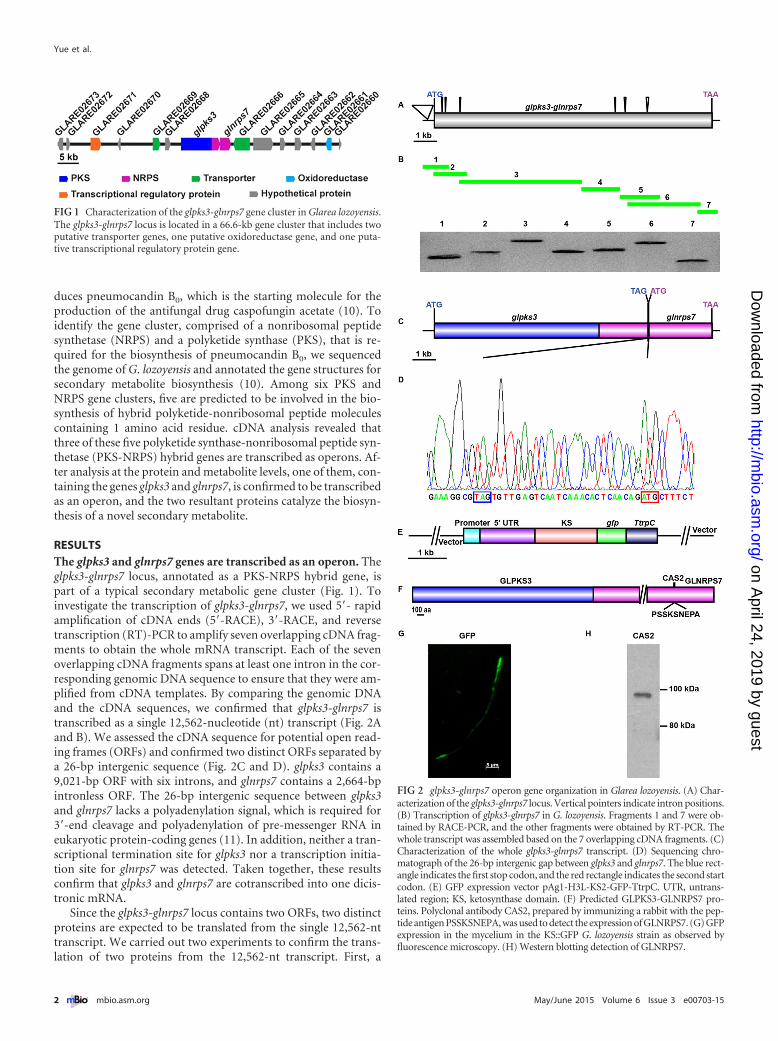
RESULTSThe glpks3 and glnrps7 genes are transcribed as an operon. Theglpks3-glnrps7 locus, annotated as a PKS-NRPS hybrid gene, ispart of a typical secondary metabolic gene cluster (Fig. 1). Toinvestigate the transcription of glpks3-glnrps7, we used 5=- rapidamplification of cDNA ends (5=-RACE), 3=-RACE, and reversetranscription (RT)-PCR to amplify seven overlapping cDNA frag-ments to obtain the whole mRNA transcript. Each of the sevenoverlapping cDNA fragments spans at least one intron in the cor-responding genomic DNA sequence to ensure that they were am-plified from cDNA templates. By comparing the genomic DNAand the cDNA sequences, we confirmed that glpks3-glnrps7 istranscribed as a single 12,562-nucleotide (nt) transcript (Fig. 2Aand B). We assessed the cDNA sequence for potential open read-ing frames (ORFs) and confirmed two distinct ORFs separated bya 26-bp intergenic sequence (Fig. 2C and D). glpks3 contains a9,021-bp ORF with six introns, and glnrps7 contains a 2,664-bpintronless ORF. The 26-bp intergenic sequence between glpks3and glnrps7 lacks a polyadenylation signal, which is required for3=-end cleavage and polyadenylation of pre-messenger RNA ineukaryotic protein-coding genes (11). In addition, neither a tran-scriptional termination site for glpks3 nor a transcription initia-tion site for glnrps7 was detected. Taken together, these resultsconfirm that glpks3 and glnrps7 are cotranscribed into one dicis-tronic mRNA.
Since the glpks3-glnrps7 locus contains two ORFs, two distinctproteins are expected to be translated from the single 12,562-nttranscript. We carried out two experiments to confirm the trans-lation of two proteins from the 12,562-nt transcript. First, a
FIG 1 Characterization of the glpks3-glnrps7 gene cluster in Glarea lozoyensis.The glpks3-glnrps7 locus is located in a 66.6-kb gene cluster that includes twoputative transporter genes, one putative oxidoreductase gene, and one puta-tive transcriptional regulatory protein gene.
FIG 2 glpks3-glnrps7 operon gene organization in Glarea lozoyensis. (A) Char-acterization of the glpks3-glnrps7 locus. Vertical pointers indicate intron positions.(B) Transcription of glpks3-glnrps7 in G. lozoyensis. Fragments 1 and 7 were ob-tained by RACE-PCR, and the other fragments were obtained by RT-PCR. Thewhole transcript was assembled based on the 7 overlapping cDNA fragments. (C)Characterization of the whole glpks3-glnrps7 transcript. (D) Sequencing chro-matograph of the 26-bp intergenic gap between glpks3 and glnrps7. The blue rect-angle indicates the first stop codon, and the red rectangle indicates the second startcodon. (E) GFP expression vector pAg1-H3L-KS2-GFP-TtrpC. UTR, untrans-lated region; KS, ketosynthase domain. (F) Predicted GLPKS3-GLNRPS7 pro-teins. Polyclonal antibody CAS2, prepared by immunizing a rabbit with the pep-tide antigen PSSKSNEPA, was used to detect the expression of GLNRPS7. (G) GFPexpression in the mycelium in the KS::GFP G. lozoyensis strain as observed byfluorescence microscopy. (H) Western blotting detection of GLNRPS7.
Yue et al.
2 ® mbio.asm.org May/June 2015 Volume 6 Issue 3 e00703-15
on April 24, 2019 by guest
http://mbio.asm
.org/D
ownloaded from
genomic DNA fragment spanning the promoter sequence and theregion annotated to encode the ketosynthase domain (KS) of theglpks3 gene was fused downstream with a green fluorescent pro-tein (GFP) reporter gene (Fig. 2E). The reporter plasmid was in-troduced into the G. lozoyensis wild-type strain by Agrobacteriumtumefaciens-mediated transformation. A GFP signal was detectedin the transformants (Fig. 2G), indicating that the transcript wastranslated. Second, we attempted to detect the predicted GL-NRPS7 protein, containing 887 amino acids and with a molecularmass of about 98 kDa, by Western blotting. A rabbit polyclonalantibody (CAS2) was generated against a peptide containing res-idues 361 to 369 of GLNRPS7 (Fig. 2F). A protein band with amolecular mass close to that of the predicted GLNRPS7 protein of98 kDa was detected in the mycelial homogenate of the G. lozoy-ensis wild-type strain by Western blotting using the CAS2 anti-body (Fig. 2H). These results demonstrated that the single glpks3-glnrps7 transcript is translated into two distinct proteins.
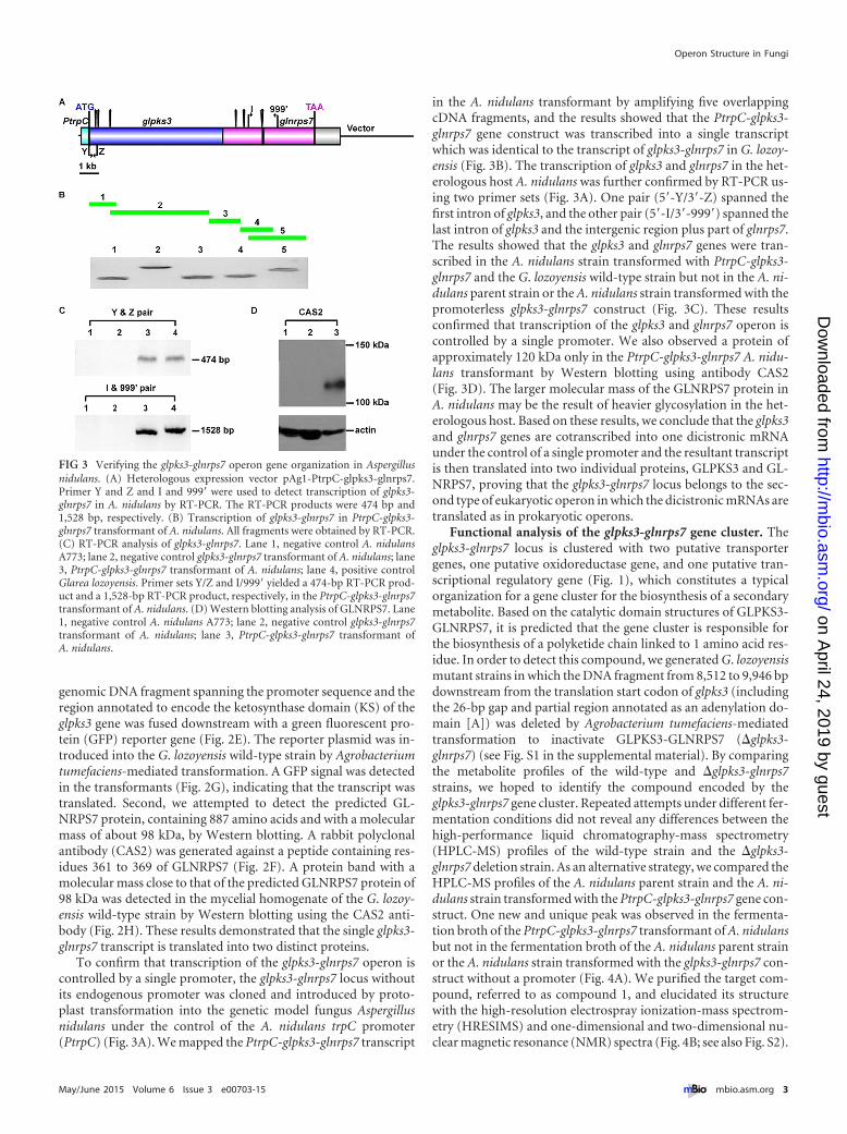
To confirm that transcription of the glpks3-glnrps7 operon iscontrolled by a single promoter, the glpks3-glnrps7 locus withoutits endogenous promoter was cloned and introduced by proto-plast transformation into the genetic model fungus Aspergillusnidulans under the control of the A. nidulans trpC promoter(PtrpC) (Fig. 3A). We mapped the PtrpC-glpks3-glnrps7 transcript
in the A. nidulans transformant by amplifying five overlappingcDNA fragments, and the results showed that the PtrpC-glpks3-glnrps7 gene construct was transcribed into a single transcriptwhich was identical to the transcript of glpks3-glnrps7 in G. lozoy-ensis (Fig. 3B). The transcription of glpks3 and glnrps7 in the het-erologous host A. nidulans was further confirmed by RT-PCR us-ing two primer sets (Fig. 3A). One pair (5=-Y/3=-Z) spanned thefirst intron of glpks3, and the other pair (5=-I/3=-999=) spanned thelast intron of glpks3 and the intergenic region plus part of glnrps7.The results showed that the glpks3 and glnrps7 genes were tran-scribed in the A. nidulans strain transformed with PtrpC-glpks3-glnrps7 and the G. lozoyensis wild-type strain but not in the A. ni-dulans parent strain or the A. nidulans strain transformed with thepromoterless glpks3-glnrps7 construct (Fig. 3C). These resultsconfirmed that transcription of the glpks3 and glnrps7 operon iscontrolled by a single promoter. We also observed a protein ofapproximately 120 kDa only in the PtrpC-glpks3-glnrps7 A. nidu-lans transformant by Western blotting using antibody CAS2(Fig. 3D). The larger molecular mass of the GLNRPS7 protein inA. nidulans may be the result of heavier glycosylation in the het-erologous host. Based on these results, we conclude that the glpks3and glnrps7 genes are cotranscribed into one dicistronic mRNAunder the control of a single promoter and the resultant transcriptis then translated into two individual proteins, GLPKS3 and GL-NRPS7, proving that the glpks3-glnrps7 locus belongs to the sec-ond type of eukaryotic operon in which the dicistronic mRNAs aretranslated as in prokaryotic operons.
Functional analysis of the glpks3-glnrps7 gene cluster. Theglpks3-glnrps7 locus is clustered with two putative transportergenes, one putative oxidoreductase gene, and one putative tran-scriptional regulatory gene (Fig. 1), which constitutes a typicalorganization for a gene cluster for the biosynthesis of a secondarymetabolite. Based on the catalytic domain structures of GLPKS3-GLNRPS7, it is predicted that the gene cluster is responsible forthe biosynthesis of a polyketide chain linked to 1 amino acid res-idue. In order to detect this compound, we generated G. lozoyensismutant strains in which the DNA fragment from 8,512 to 9,946 bpdownstream from the translation start codon of glpks3 (includingthe 26-bp gap and partial region annotated as an adenylation do-main [A]) was deleted by Agrobacterium tumefaciens-mediatedtransformation to inactivate GLPKS3-GLNRPS7 (�glpks3-glnrps7) (see Fig. S1 in the supplemental material). By comparingthe metabolite profiles of the wild-type and �glpks3-glnrps7strains, we hoped to identify the compound encoded by theglpks3-glnrps7 gene cluster. Repeated attempts under different fer-mentation conditions did not reveal any differences between thehigh-performance liquid chromatography-mass spectrometry(HPLC-MS) profiles of the wild-type strain and the �glpks3-glnrps7 deletion strain. As an alternative strategy, we compared theHPLC-MS profiles of the A. nidulans parent strain and the A. ni-dulans strain transformed with the PtrpC-glpks3-glnrps7 gene con-struct. One new and unique peak was observed in the fermenta-tion broth of the PtrpC-glpks3-glnrps7 transformant of A. nidulansbut not in the fermentation broth of the A. nidulans parent strainor the A. nidulans strain transformed with the glpks3-glnrps7 con-struct without a promoter (Fig. 4A). We purified the target com-pound, referred to as compound 1, and elucidated its structurewith the high-resolution electrospray ionization-mass spectrom-etry (HRESIMS) and one-dimensional and two-dimensional nu-clear magnetic resonance (NMR) spectra (Fig. 4B; see also Fig. S2).
FIG 3 Verifying the glpks3-glnrps7 operon gene organization in Aspergillusnidulans. (A) Heterologous expression vector pAg1-PtrpC-glpks3-glnrps7.Primer Y and Z and I and 999= were used to detect transcription of glpks3-glnrps7 in A. nidulans by RT-PCR. The RT-PCR products were 474 bp and1,528 bp, respectively. (B) Transcription of glpks3-glnrps7 in PtrpC-glpks3-glnrps7 transformant of A. nidulans. All fragments were obtained by RT-PCR.(C) RT-PCR analysis of glpks3-glnrps7. Lane 1, negative control A. nidulansA773; lane 2, negative control glpks3-glnrps7 transformant of A. nidulans; lane3, PtrpC-glpks3-glnrps7 transformant of A. nidulans; lane 4, positive controlGlarea lozoyensis. Primer sets Y/Z and I/999= yielded a 474-bp RT-PCR prod-uct and a 1,528-bp RT-PCR product, respectively, in the PtrpC-glpks3-glnrps7transformant of A. nidulans. (D) Western blotting analysis of GLNRPS7. Lane1, negative control A. nidulans A773; lane 2, negative control glpks3-glnrps7transformant of A. nidulans; lane 3, PtrpC-glpks3-glnrps7 transformant ofA. nidulans.
Operon Structure in Fungi
May/June 2015 Volume 6 Issue 3 e00703-15 ® mbio.asm.org 3
on April 24, 2019 by guest
http://mbio.asm
.org/D
ownloaded from
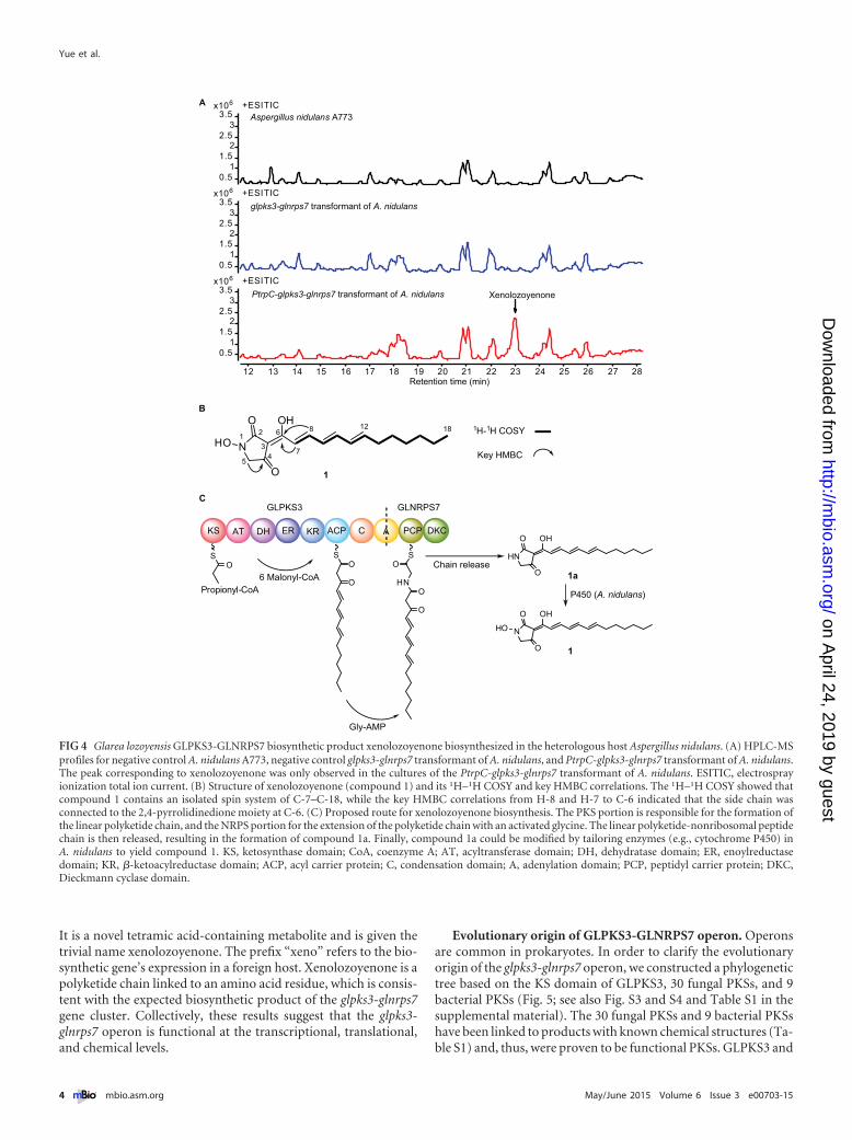
It is a novel tetramic acid-containing metabolite and is given thetrivial name xenolozoyenone. The prefix “xeno” refers to the bio-synthetic gene’s expression in a foreign host. Xenolozoyenone is apolyketide chain linked to an amino acid residue, which is consis-tent with the expected biosynthetic product of the glpks3-glnrps7gene cluster. Collectively, these results suggest that the glpks3-glnrps7 operon is functional at the transcriptional, translational,and chemical levels.
Evolutionary origin of GLPKS3-GLNRPS7 operon. Operonsare common in prokaryotes. In order to clarify the evolutionaryorigin of the glpks3-glnrps7 operon, we constructed a phylogenetictree based on the KS domain of GLPKS3, 30 fungal PKSs, and 9bacterial PKSs (Fig. 5; see also Fig. S3 and S4 and Table S1 in thesupplemental material). The 30 fungal PKSs and 9 bacterial PKSshave been linked to products with known chemical structures (Ta-ble S1) and, thus, were proven to be functional PKSs. GLPKS3 and
6x10
0.51
1.52
2.53
3.5+ESI TIC
6x10
0.51
1.52
2.53
3.5+ESI TIC
6x10
0.51
1.52
2.53
3.5+ESI TIC
12 13 14 15 16 17 18 19 20 21 22 23 24 25 26 27 28Retention time (min)
Xenolozoyenone
Aspergillus nidulans A773
glpks3-glnrps7 transformant of A. nidulans
PtrpC-glpks3-glnrps7 transformant of A. nidulans
A
OH
N
O
O
HO1 2
34
5
612 18 1H-1H COSY
Key HMBC
8
7
1
KS AT DH ER KR ACP C A PCP DKC
SO
SO
O
S
HN
O
O
O
6 Malonyl-CoA
Gly-AMP
Propionyl-CoA
OH
N
O
O
P450 (A. nidulans)
GLPKS3 GLNRPS7
1
B
C
HO
OH
HN
O
O 1aChain release
FIG 4 Glarea lozoyensis GLPKS3-GLNRPS7 biosynthetic product xenolozoyenone biosynthesized in the heterologous host Aspergillus nidulans. (A) HPLC-MSprofiles for negative control A. nidulans A773, negative control glpks3-glnrps7 transformant of A. nidulans, and PtrpC-glpks3-glnrps7 transformant of A. nidulans.The peak corresponding to xenolozoyenone was only observed in the cultures of the PtrpC-glpks3-glnrps7 transformant of A. nidulans. ESITIC, electrosprayionization total ion current. (B) Structure of xenolozoyenone (compound 1) and its 1H–1H COSY and key HMBC correlations. The 1H–1H COSY showed thatcompound 1 contains an isolated spin system of C-7–C-18, while the key HMBC correlations from H-8 and H-7 to C-6 indicated that the side chain wasconnected to the 2,4-pyrrolidinedione moiety at C-6. (C) Proposed route for xenolozoyenone biosynthesis. The PKS portion is responsible for the formation ofthe linear polyketide chain, and the NRPS portion for the extension of the polyketide chain with an activated glycine. The linear polyketide-nonribosomal peptidechain is then released, resulting in the formation of compound 1a. Finally, compound 1a could be modified by tailoring enzymes (e.g., cytochrome P450) inA. nidulans to yield compound 1. KS, ketosynthase domain; CoA, coenzyme A; AT, acyltransferase domain; DH, dehydratase domain; ER, enoylreductasedomain; KR, �-ketoacylreductase domain; ACP, acyl carrier protein; C, condensation domain; A, adenylation domain; PCP, peptidyl carrier protein; DKC,Dieckmann cyclase domain.
Yue et al.
4 ® mbio.asm.org May/June 2015 Volume 6 Issue 3 e00703-15
on April 24, 2019 by guest
http://mbio.asm
.org/D
ownloaded from
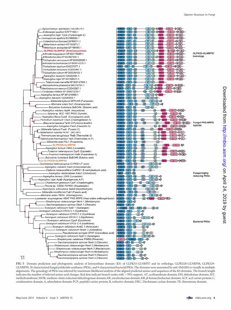
FIG 5 Domain prediction and phylogenetic analysis of ketosynthase domain (KS) of GLPKS3-GLNRPS7 and its orthologs, GLPKS28-GLNRPS8, GLPKS29-GLNRPS9, 30 characterized fungal polyketide synthases (PKSs), and 9 characterized bacterial PKSs. The domains were annotated by anti-SMASH or visually in multiplealignments. The genealogy of PKSs was inferred by maximum-likelihood analysis of the aligned predicted amino acid sequences of the KS domains. The branch lengthindicates the number of inferred amino acid changes. Red dots indicate branch nodes with �70% support. AT, acyltransferase domain; DH, dehydratase domain; MT,methyltransferase; MDR, medium-chain reductase/dehydrogenase domain; ER, enoylreductase domain; KR, �-ketoacylreductase domain; ACP, acyl carrier protein; C,condensation domain; A, adenylation domain; PCP, peptidyl carrier protein; R, reductive domain; DKC, Dieckmann cyclase domain; TE, thioesterase domain.
Operon Structure in Fungi
May/June 2015 Volume 6 Issue 3 e00703-15 ® mbio.asm.org 5
on April 24, 2019 by guest
http://mbio.asm
.org/D
ownloaded from

the 30 fungal PKSs were grouped together (bootstrap value of92%), and the clade was distantly related to and distinct from thebacterial PKSs. Thus, the glpks3-glnrps7 locus and its fungal or-thologs appear to have a monophyletic origin among fungal PKSs,and the glpks3-glnrps7 locus did not result from horizontal genetransfer from prokaryotes. Among the fungal PKSs, GLPKS3 andits fungal orthologs were grouped in a monophyletic clade with ahigh bootstrap value (100%). In this clade, 26 proteins were from13 genera belonging to four classes of Pezizomycotina, which sug-gests that these kinds of enzymes are widespread and possiblyserve a basic function in these fungi.
Two other G. lozoyensis gene loci transcribed as operons. Inaddition to glpks3-glnrps7, the G. lozoyensis genome revealed fourmore genes (glpks26-nrps, glpks27-nrps, glpks28-nrps, and glpks29-nrps) that are predicted to be responsible for the biosynthesis ofpolyketide linked to 1 amino acid residue (10). To find out if theoccurrence of operons is common among these secondary meta-bolic genes in G. lozoyensis, we determined the cDNAs of the otherfour PKS-NRPS hybrid genes. Whole transcripts assembled withoverlapping cDNA fragments obtained by RACE-PCR and RT-PCR revealed that glpks28-nrps and glpks29-nrps possess gene or-ganization patterns similar to that of the glpks3-glnrps7 locus(Fig. 6). Because not every cDNA fragment spans an intron, oneprimer set, PF5-1 and Ourer5 (located in the third intron ofglpks29-nrps), was used as a genomic DNA contamination con-trol. No PCR product was amplified by this primer set with thetemplate cDNA, indicating there was no genomic DNA contami-nation in the RNA samples used for cDNA amplification. Theglpks28-nrps locus was transcribed as a dicistronic transcript withan intergenic sequence of 443 nt, while the glpks29-nrps locus alsowas transcribed as a dicistronic transcript with an intergenic se-quence of 179 nt. No polyadenylation signal sequences or pro-moter elements were detected in the intergenic region. Moreover,no transcriptional termination site for the upstream gene or tran-scription initiation site for the downstream gene were detected inthe intergenic regions. These results demonstrated that theglpks28-nrps and glpks29-nrps loci are organized as operons. Basedon these results, we renamed these two loci as glpks28-glnrps8 andglpks29-glnrps9. glpks28 contained a 966-bp ORF, and glnrps8 con-tained a 10,822-bp ORF. glpks29 had a 9,212-bp ORF, and glnrps9had a 3,439-bp ORF. Among the four genes, glpks28 contains nointrons, while glnrps8, glpks29, and glnrps9 contain multiple in-trons. These two loci are organized as typical secondary metabolicgene clusters which are predicted to be responsible for the biosyn-thesis of compounds consisting of a polyketide chain linked to anamino acid residue.
DISCUSSION
rRNA operons are widespread in eukaryotes, including the Fungi.However, protein-coding operons have not been reported in theFungi. A dicistronic mRNA of CRG1 and PUT1 was reported inthe filamentous fungus Cercospora nicotianae, but these two genesare controlled by separate promoters (12). Therefore, this locusdoes not fulfill the definition of an operon (2). We demonstratedexperimentally in this study that three of the five PKS-NRPS lociin G. lozoyensis are operons. To our knowledge, the three operonsin G. lozoyensis are the first examples of eukaryotic protein-codingoperons identified in a member of the fungal kingdom.
The majority of known eukaryotic operons belong to the firsttype of eukaryotic operons, in which the polycistronic transcripts
are processed into mature monocistronic mRNAs (2). Althoughthe mechanism for transcript maturation in C. elegans operons isdifferent from that in the Drosophila melanogaster CheB42a/llzoperon, the polyadenylation signal in the intergenic region is cru-cial for the 3=-end formation in all of these operons (6, 13). Nopolyadenylation signal was found in the intergenic regions of theglpks3-glnrps7, glpks28-glnrps8, and glpks29-glnrps9 operons, sug-gesting that the operons in G. lozoyensis, like the stonedA/stonedBoperon in D. melanogaster (14), belong to the second type of eu-karyotic operons, in which the dicistronic mRNAs are translatedas in prokaryotic operons.
It has been suggested that some of the second type of eukary-otic operons had originated from bacteria via horizontal genetransfer (8). However, phylogenetic analysis of the glpks3-glnrps7,glpks28-glnrps8, and glpks29-glnrps9 operons showed that they donot resemble prokaryotic genes (Fig. 5). Not only do the threeoperons contain multiple introns, but they also have GC content(46 to 50%) consistent with that the resident genome (46%) (10).These results suggest that these operons were not products of hor-izontal gene transfer from bacteria. We also constructed phyloge-netic trees for the KS and A domains of GLPKS3-GLNRPS7 andtheir orthologs (see Fig. S5 in the supplemental material). Theproteins containing both KS and A orthologs formed distinctclades with GLPKS3-GLNRPS7 in the two trees. However, the Adomain tree appears to be in conflict with the KS tree and estab-lished ascomycete phylogeny, while the KS tree does not signifi-cantly conflict with the established phylogeny. These results sug-gest that these proteins may have descended from a commonancestor, but the NRPS module and the PKS module may haveevolved at different evolutionary rates.
Characterization of the three operon gene structures in theG. lozoyensis genome suggests that operon structures might becommon in the Fungi. Based on the prerequisites for an operon,such as two genes that are transcribed in the same direction with apredicted upstream promoter and an intergenic region of�700 bp that lacks predicted promoter sequences and polyade-nylation signal sequences (13), we predicted the potential operonstructures of three fungal genomes in silico. Genome and tran-scriptome data analysis revealed operonlike gene structures in3.8%, 1.6%, and 3.7% of A. nidulans, Glomerella graminicola, andDrechslerella stenobrocha genes, respectively (see Table S2 in thesupplemental material). If validated experimentally, this analysissuggests that operonlike gene structures may occur widely in theFungi.
Xenolozoyenone, the novel secondary metabolite of GLPKS3-GLNRPS7 expressed in A. nidulans, is a typical fungal PKS-NRPShybrid product. It belongs to the tetramic acid class of metabolites,such as ravenic acid (15), militarinone C (16), and pretenellin A(17), which are common in fungi. Its structure is similar tothose of other tetramic acids but not identical due to having adifferent polyketide side chain and substituents on the 2,4-pyrrolidinedione ring. Moreover, the fungal hybrid polyketide-nonribosomal peptide molecules are generally assembled by aPKS-NRPS hybrid protein (18), including pyranonigrin E, bio-synthesized by PynA in Aspergillus niger, which is one of theGLPKS3 KS domain orthologs showing a domain organizationsimilar to that of GLPKS3-GLNPRS7 (19). However, GLPKS3-GLNRPS7 is an enzyme complex of two proteins, GLPKS3 andGLNRPS7. The PKS portion is responsible for the formation ofthe linear polyketide chain, and the NRPS portion for the exten-
Yue et al.
6 ® mbio.asm.org May/June 2015 Volume 6 Issue 3 e00703-15
on April 24, 2019 by guest
http://mbio.asm
.org/D
ownloaded from
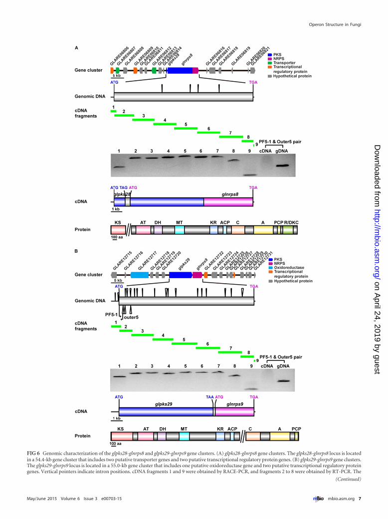
FIG 6 Genomic characterization of the glpks28-glnrps8 and glpks29-glnrps9 gene clusters. (A) glpks28-glnrps8 gene clusters. The glpks28-glnrps8 locus is locatedin a 54.4-kb gene cluster that includes two putative transporter genes and two putative transcriptional regulatory protein genes. (B) glpks29-glnrps9 gene clusters.The glpks29-glnrps9 locus is located in a 55.0-kb gene cluster that includes one putative oxidoreductase gene and two putative transcriptional regulatory proteingenes. Vertical pointers indicate intron positions. cDNA fragments 1 and 9 were obtained by RACE-PCR, and fragments 2 to 8 were obtained by RT-PCR. The
(Continued)
Operon Structure in Fungi
May/June 2015 Volume 6 Issue 3 e00703-15 ® mbio.asm.org 7
on April 24, 2019 by guest
http://mbio.asm
.org/D
ownloaded from
sion of the polyketide chain with an activated glycine. The linearpolyketide-nonribosomal peptide chain undergoes a Dieckmanncyclization reaction that results in the formation of compound 1a(17). Finally, compound 1a could be modified by tailoring en-zymes (e.g., cytochrome P450) of A. nidulans to yield compound 1(Fig. 4C). As the NRPS portion of GLPKS3-GLNRPS7 is located intwo individual proteins, GLPKS3 (containing a condensation do-main [C] and a partial A domain proximal to the C terminus) andGLNRPS7 (containing the complementary partial A domain, apeptidyl carrier protein [PCP], and a Dieckmann cyclase domain[DKC]), we presume that GLPKS3 and GLNRPS7 are translatedin a 1:1 ratio to assemble the optimal number of functional en-zyme complexes, and the operon genomic organization helpsmaintain the stoichiometric ratio (20).
The identification of operons in the Fungi not only reveals thecomplexity of gene transcription and regulation in eukaryotic or-ganisms but also facilitates future studies concerning the originand evolution of fungal genes that encode antibiotics and othersecondary metabolites. These findings may have important prac-tical implications in the construction of synthetic gene clusters forantibiotic discovery.
MATERIALS AND METHODSDNA and RNA procedures. Fungal genomic DNA was extracted as de-scribed previously (10). Plasmids and PCR products were purified usingthe E.Z.N.A. gel extraction kit (Omega Bio-Tek, Norcross, GA). Phusionhigh-fidelity DNA polymerase (New England Biolabs, Ipswich, Massa-chusetts) and EasyTaq DNA polymerase (TransGen Biotech, Beijing,China) were used for PCRs. Sequences were determined by SinoGenoMaxCo., Ltd. (Beijing, China). Restriction endonucleases and DNA-modifying enzymes were from New England Biolabs (Ipswich, Massachu-setts).
RNA was extracted with the Qiagen RNeasy minikit, and carryoverDNA was removed by DNase I digestion (Qiagen, Valencia, CA). cDNAfragments were synthesized using the TransScript II first-strand cDNAsynthesis supermix (TransGen Biotech, Beijing, China). Introns wereidentified by comparing genomic and cDNA sequences. The 5= ends of themRNA and the poly(A) attachment sites were mapped by 5=- and 3=-RACE–PCR (FirstChoice RLM-RACE kit; Ambion, Austin, Texas).
All primers used in the study are listed in Table S3 in the supplementalmaterial.
Genetic manipulation of glpks3-glnrps7 in G. lozoyensis. To detectthe expression of GLPKS3, a genomic fragment extending from the pro-moter to the region annotated as the KS domain of glpks3, the GFP re-porter gene, and the A. nidulans trpC terminator were amplified and in-serted into pAg1-H3 to give the expression vector pAg1-H3L-KS2-TtrpC(see Text S1 and Fig. S6 in the supplemental material).
To disrupt glpks3, the 3= regions of glpks3 and glnrps7 were amplifiedand inserted into pAg1-H3 to give pAg1-H3-glpks3-glnrps7, containingthe gene replacement cassette (see Fig. S1 in the supplemental material).
Conidia of G. lozoyensis were prepared and transformed as previouslydescribed (10). After isolation of single spores from the transformants, thepositive transformants of �glpks3-glnrps7 were determined by PCR usingtwo primer pairs (GLPKS3-F2/999= and GLPKS3-F1/T). Transformantspositive for KS::GFP were determined by PCR using two primer pairs(GFP-F/GFP-R and GFP-F/N).
The wild-type strain of G. lozoyensis, the KS::GFP transformant, and
the �glpks3-glnrps7 mutant were grown in LYCP-5 medium to prepareseed cultures (10). The seed cultures were then inoculated into H mediumand grown for 14 days (21). For the wild-type strain of G. lozoyensis, theculture was divided into two parts. One part was examined by RT-PCRusing two primer pairs (Y/Z and I/999=) and Western blotting using theCAS2 antibody. The other part and the culture of the �glpks3-glnrps7strain were analyzed by HPLC-MS following the same procedures used forA. nidulans. For the KS::GFP transformant, mycelia were collected andobserved by fluorescence microscopy.
Heterologous expression of glpks3-glnrps7 in A. nidulans. Theglpks3-glnrps7 gene was amplified from genomic DNA of G. lozoyensis,and the constitutive A. nidulans trpC promoter was amplified frompAg1-H3 (22). After restriction enzyme digestion and ligation, glpks3-glnrps7 under the control of the constitutive A. nidulans trpC promoterwas constructed and inserted into pAg1-H3 to give the expression vectorpAg1-PtrpC-glpks3-glnrps7 and control vector pAg1-glpks3-glnrps7 (seeText S1 and Fig. S6 in the supplemental material). Protoplast preparationand transformation were performed as previously described (23). Onemicrogram each of pRG3-AMA1 and SbfI-digested pAg1-PtrpC-glpks3-glnrps7 or pAg1-glpks3-glnrps7 were cotransformed into A. nidulansA773 (pyrG89 wA3 pyroA4 veA1). Positive glpks3-glnrps7-containingtransformants were identified by PCR using five primer sets (Pc800F/P22=, A/B, U/F, and M/D for PtrpC-glpks3-glnrps7 transformants andK/D for glpks3-glnrps7 transformants) and DNA sequencing.
The positive transformants and parent strain of A. nidulans were in-cubated on MAG with appropriate supplements (23). Conidia were har-vested and adjusted to 108 conidia ml�1. Amounts of about 2 ml of sporesuspension were inoculated into MMV (1% glucose, 0.001% thiamine,nitrate salts, trace elements, and vitamins, pH 6.5) with appropriate sup-plements (23). The cultures were shaken at 200 rpm in the dark for 7 days.RNA preparations from the cultures were examined for GLNRPS7transcription and translation by RT-PCR using two primer pairs (Y/Z,and I/999=) for all strains of A. nidulans, five primer pairs (Y/P30R-2,GLPKS3-F7/U-R, U/GLPKS3-F2-R, GLPKS3-F2/GLNRPS7-F-R, and I/GLNRPS7-R1) for PtrpC-glpks3-glnrps7 transformants, and proteinpreparations were examined by Western blotting using the polyclonalantibody CAS2.
Protein analysis. Protein extraction, quantification, and Westernblotting were performed as previously described (24). In order to detectGLNRPS7, we prepared a polyclonal antibody, CAS2, by immunizing arabbit (Epitomics, Hangzhou, Zhejiang, China) with the peptide antigenPSSKSNEPA, which was predicted as an immune epitope against the pre-dicted GLNRPS7 protein by the PeptideStructure program of the Univer-sity of Wisconsin Genetics Computer Group Sequence Analysis SoftwarePackage. The IgG titer against the PSSKSNEPA peptide antigen reached64,000. The polyclonal antibody CAS2 was purified by protein A columnas described previously (25).
Fermentation and extraction of A. nidulans and transformants forHPLC-MS analysis. The transformants and parent strain of A. nidulanswere cultured on MAG with appropriate supplements (23). Fifteen pieces(0.5 by 0.5 by 0.5 cm3) of agar cultures were inoculated and incubated in100 ml H medium with appropriate supplements at 30°C and 220 rpm for7 days. The cultures were extracted with 100 ml of methyl ethyl ketone(MEK), and the organic phase was evaporated to dryness and redissolvedin methanol (MeOH) to 30 mg·ml�1.
NMR and MS methods. All 1H, 13C, and two-dimensional (1H–1Hcorrelation spectroscopy [COSY], heteronuclear single quantum coher-ence [HSQC] spectroscopy, and heteronuclear multiple-bond correlation[HMBC] spectroscopy) NMR spectra were acquired on a Bruker 600- or
Figure Legend Continued
whole transcript was assembled based on the overlapping cDNA fragments. Primer set PF5-1 and Outer5 was designed to amplify a 404-bp sequence fromgenomic DNA templates but not from cDNA templates. KS, ketosynthase domain; AT, acyltransferase domain; DH, dehydratase domain; MT, methyltransferase;KR, �-ketoacylreductase domain; ACP, acyl carrier protein; C, condensation domain; A, adenylation domain; PCP, peptidyl carrier protein; R, reductive domain;DKC, Dieckmann cyclase domain.
Yue et al.
8 ® mbio.asm.org May/June 2015 Volume 6 Issue 3 e00703-15
on April 24, 2019 by guest
http://mbio.asm
.org/D
ownloaded from
500-MHz spectrometer equipped with a 5-mm triple-resonance cryo-probe at 298 K. Residual solvent signals were used as references (acetone-d6, �H 2.05/�C 29.8, 206.0, and CDCl3, �H 7.26/�C 77.0). HPLC-MS spec-tra were obtained on an Agilent 6120 quadrupole mass spectrometer usinga positive ESI source. Amounts of 10 �l of the test samples were injectedfor HPLC-MS analysis (Agilent Zorbax Eclipse plus C18 reverse-phasecolumn, 5 �m, 4.6 by 150 mm, 10% to 90% CH3CN in H2O with 01%formic acid for 30 min, 1 ml·min�1). HRESIMS data were obtained usingthe Agilent 6520 quadrupole time of flight (Q-TOF) LC-MS instrumentequipped with an electrospray ionization (ESI) source.
Isolation of xenolozoyenone. The scaled-up fermentation culture(PtrpC-glpks3-glnrps7 transformant of A. nidulans in 2.0 liters of H me-dium) was extracted repeatedly with MEK (3 times per 2.0 liters), and theorganic solvent was vacuum evaporated to dryness to obtain the crudeextract (1.2 g), which was fractionated by using a Sephadex LH-20 columnwith 1:1 CH2Cl2-MeOH as eluents. The resulting subfractions were com-bined and further purified by semipreparative reverse-phase HPLC (Agi-lent Zorbax SB-C18 column, 5 �m, 9.4 by 250 mm, 65% CH3CN in H2Owith 01% formic acid for 25 min, 2 ml·min�1) to obtain xenolozoyenone(compound 1) as follows: 4.5 mg, tR 17.25 min (MeOH); UV (MeOH)�max (log �) 217 (4.46), 256 (4.10), 390 (4.86) nm; 1H NMR (acetone-d6,600 MHz) � 7.73 (1H, brs, OH-1 or OH-6), 7.50 (1H, dd, J � 15.0,11.4 Hz, H-8), 7.12 (1H, d, J � 15.0 Hz, H-7), 6.82 (1H, dd, J � 15.0,10.8 Hz, H-10), 6.50 (1H, dd, J � 15.0, 11.4 Hz, H-9), 6.32 (1H, dd, J �15.0, 10.8 Hz, H-11), 6.10 (1H, dt, J � 15.0, 7.2 Hz, H-12), 3.81 (2H, m,H2-5), 2.19 (2H, m, H2-13), 1.44 (2H, m, H2-14), 1.31 (6H, m, H2-15,H2-16, and H2-17), 0.89 (3H, t, J � 6.6, H3-18); 13C NMR (CDCl3,125 MHz) � 193.4 (C-4), 176.1 (C-2), 175.0 (C-6), 146.3 (C-8), 144.6(C-10), 143.4 (C-12), 130.2 (C-11), 128.8 (C-9), 120.1 (C-7), 99.7 (C-3),62.9 (C-5), 33.2 (C-13), 31.4 (C-15), 29.7 (C-17), 28.6 (C-14), 22.5 (C-16), 14.0 (C-18); HRESIMS m/z [M�H]� 306.1708 (calculated forC17H24NO4, 306.1700).
Phylogenetic analysis of PKSs and PKS-NRPSs. To test whether theglpks3-glnrps7 locus is a product of horizontal gene transfer from bacteria,we built a phylogenetic tree based on the KS domain of GLPKS3-GLNRPS7, its orthologs in other fungi (coverage, 95%, and identity,64%), GLPKS28-GLNRPS8, GLPKS29-GLNRPS9, 30 characterizedfungal PKSs, and 9 characterized bacterial PKSs (see Table S1 in the sup-plemental material). All domains were identified by using the programanti-SMASH (26) or manually in multiple alignments. The amino acidsequences of the KS domains were aligned with MUSCLE and analyzedphylogenetically with MEGA 6.0 by the maximum-likelihood (ML) algo-rithm using an LG�F�G�I model selected by Prot-test and a 1,000-replication bootstrap test (27, 28). The R/DKC domains were also alignedwith MUSCLE. To test whether the glpks3-glnrps7 locus might be of singleor multiple evolutionary origins, we constructed two phylogenetic trees,one based on the KS domain of GLPKS3, all orthologs in other fungi(coverage, 95%, and identity, 55%), and 3 characterized fungal PKS-NRPSs and the other based on the A domain of GLPKS3, all orthologs inother fungi (coverage, 95%, and identity, 40%), and 3 characterizedfungal PKS-NRPSs. The KS and A domains were identified by the pro-gram anti-SMASH (26) or manually in multiple alignments. The aminoacid sequences of these domains were aligned with MUSCLE and analyzedwith RAxML BlackBox by the ML method under a WAG (Whelan AndGoldman) model with or without a constraint from the reference fungaltaxonomy (27, 29). The A domain tree was also analyzed with RAxMLBlackBox by the ML method under the WAG model with a constraintfrom the KS domain tree.
Alternative hypotheses based on the tree topologies under the nullhypothesis that all topologies are equally good explanations of the datawere tested with the Shimodaira-Hasegawa test (30), weightedShimodaira-Hasegawa test, and approximately unbiased test (31), as im-plemented in TREEFINDER (32).
Bioinformatics. Putative promoter sequences in the intergenic regionof glpks3-glnrps7 and 1 kb upstream from the genes in other gene pairs
were investigated with Promoter 2.0 Prediction Server (33). The poly-adenylation signals (AATAAA and ATTAAA) in the intergenic regionof glpks3-glnrps7 and other gene pairs were identified by using thenucleotide sequence pattern search tool Fuzznuc in EMBOSS (http://emboss.bioinformatics.nl/cgi-bin/emboss/fuzznuc). To identify po-tential polycistrons, genome and transcriptome data of A. nidulans (Gen-Bank accession numbers PRJNA13961 and PRJNA182228),G. graminicola (GenBank accession numbers PRJNA37879 andPRJNA151285), and D. stenobrocha (GenBank accession numbersPRJNA67941 and PRJNA236481) were downloaded from NCBI (http://www.ncbi.nlm.nih.gov/). The genomic loci were annotated by Inter-ProScan, KEGG, FunCat, and UniProt-GO (34–37) to rule out the possi-bility of pseudogenes. The RNA-Seq data were assembled as describedpreviously and then mapped and aligned to the genome (38, 39).
The mRNA sequences of glpks3-glnrps7, glpks28-glnrps8, and glpks29-glnrps9 have been deposited at GenBank under the accession numbersKM603664, KM603665, and KM603666, respectively.
SUPPLEMENTAL MATERIALSupplemental material for this article may be found at http://mbio.asm.org/lookup/suppl/doi:10.1128/mBio.00703-15/-/DCSupplemental.
Text S1, DOC file, 0.04 MB.Figure S1, PDF file, 0.1 MB.Figure S2, PDF file, 0.2 MB.Figure S3, PDF file, 0.2 MB.Figure S4, PDF file, 0.3 MB.Figure S5, PDF file, 0.2 MB.Figure S6, PDF file, 0.5 MB.Table S1, DOC file, 0.2 MB.Table S2, DOC file, 0.03 MB.Table S3, DOC file, 0.1 MB.
ACKNOWLEDGMENTS
This work was supported by National Natural Science Foundation ofChina (NSFC) grants 31328001 to Z.A. and 31200055 to Q.Y., WelchFoundation grant AU00024 to Z.A., a University of Texas System StarAward to Z.A., and a grant from the University of Texas Health ScienceCenter at Houston faculty startup fund to G.F.B.
REFERENCES1. Field B, Fiston-Lavier AS, Kemen A, Geisler K, Quesneville H, Osbourn
AE. 2011. Formation of plant metabolic gene clusters within dynamicchromosomal regions. Proc Natl Acad Sci U S A 108:16116 –16121. http://dx.doi.org/10.1073/pnas.1109273108.
2. Blumenthal T. 2004. Operons in eukaryotes. Brief Funct Genomic Pro-teomic 3:199 –211. http://dx.doi.org/10.1093/bfgp/3.3.199.
3. Spieth J, Brooke G, Kuersten S, Lea K, Blumenthal T. 1993. Operons inC. elegans: polycistronic mRNA precursors are processed by trans-splicingof SL2 to downstream coding regions. Cell 73:521–532. http://dx.doi.org/10.1016/0092-8674(93)90139-H.
4. Evans D, Zorio D, MacMorris M, Winter CE, Lea K, Blumenthal T.1997. Operons and SL2 trans-splicing exist in nematodes outside the ge-nus Caenorhabditis. Proc Natl Acad Sci U S A 94:9751–9756. http://dx.doi.org/10.1073/pnas.94.18.9751.
5. Satou Y, Hamaguchi M, Takeuchi K, Hastings KE, Satoh N. 2006.Genomic overview of mRNA 5=-leader trans-splicing in the ascidian Cionaintestinalis. Nucleic Acids Res 34:3378 –3388. http://dx.doi.org/10.1093/nar/gkl418.
6. Blumenthal T, Evans D, Link CD, Guffanti A, Lawson D, Thierry-MiegJ, Thierry-Mieg D, Chiu WL, Duke K, Kiraly M, Kim SK. 2002. A globalanalysis of Caenorhabditis elegans operons. Nature 417:851– 854. http://dx.doi.org/10.1038/nature00831.
7. Gray TA, Saitoh S, Nicholls RD. 1999. An imprinted, mammalian bicis-tronic transcript encodes two independent proteins. Proc Natl Acad Sci US A 96:5616 –5621. http://dx.doi.org/10.1073/pnas.96.10.5616.
8. García-Ríos M, Fujita T, LaRosa PC, Locy RD, Clithero JM, BressanRA, Csonka LN. 1997. Cloning of a polycistronic cDNA from tomatoencoding �-glutamyl kinase and �-glutamyl phosphate reductase. Proc
Operon Structure in Fungi
May/June 2015 Volume 6 Issue 3 e00703-15 ® mbio.asm.org 9
on April 24, 2019 by guest
http://mbio.asm
.org/D
ownloaded from
Natl Acad Sci U S A 94:8249 – 8254. http://dx.doi.org/10.1073/pnas.94.15.8249.
9. Slone J, Daniels J, Amrein H. 2007. Sugar receptors in Drosophila. CurrBiol 17:1809 –1816. http://dx.doi.org/10.1016/j.cub.2007.09.027.
10. Chen L, Yue Q, Zhang X, Xiang M, Wang C, Li S, Che Y, Ortiz-LópezFJ, Bills GF, Liu X, An Z. 2013. Genomics-driven discovery of the pneu-mocandin biosynthetic gene cluster in the fungus Glarea lozoyensis. BMCGenomics 14:339. http://dx.doi.org/10.1186/1471-2164-14-339.
11. Proudfoot NJ. 2011. Ending the message: poly(A) signals then and now.Genes Dev 25:1770 –1782. http://dx.doi.org/10.1101/gad.17268411.
12. Chung KR, Daub ME, Ehrenshaft M. 2003. Expression of the cercosporintoxin resistance gene (CRG1) as a dicistronic mRNA in the filamentousfungus Cercospora nicotianae. Curr Genet 43:415– 424. http://dx.doi.org/10.1007/s00294-003-0414-3.
13. Ben-Shahar Y, Nannapaneni K, Casavant TL, Scheetz TE, Welsh MJ.2007. Eukaryotic operon-like transcription of functionally related genes inDrosophila. Proc Natl Acad Sci U S A 104:222–227. http://dx.doi.org/10.1073/pnas.0609683104.
14. Andrews J, Smith M, Merakovsky J, Coulson M, Hannan F, Kelly LE.1996. The stoned locus of Drosophila melanogaster produces a dicistronictranscript and encodes two distinct polypeptides. Genetics 143:1699 –1711.
15. Michael AP, Grace EJ, Kotiw M, Barrow RA. 2002. Ravenic acid, a newtetramic acid isolated from a cultured microfungus, Penicillium sp. J NatProd 65:1360 –1362. http://dx.doi.org/10.1021/np0200358.
16. Schmidt K, Riese U, Li Z, Hamburger M. 2003. Novel tetramic acids andpyridone alkaloids, militarinones B, C, and D, from the insect pathogenicfungus Paecilomyces militaris. J Nat Prod 66:378 –383. http://dx.doi.org/10.1021/np020430y.
17. Halo LM, Marshall JW, Yakasai AA, Song Z, Butts CP, Crump MP,Heneghan M, Bailey AM, Simpson TJ, Lazarus CM, Cox RJ. 2008.Authentic heterologous expression of the tenellin iterative polyketide syn-thase nonribosomal peptide synthetase requires coexpression with anenoyl reductase. Chembiochem 9:585–594. http://dx.doi.org/10.1002/cbic.200700390.
18. Boettger D, Hertweck C. 2013. Molecular diversity sculpted by fungalPKS-NRPS hybrids. Chembiochem 14:28 – 42. http://dx.doi.org/10.1002/cbic.201200624.
19. Awakawa T, Yang XL, Wakimoto T, Abe I. 2013. Pyranonigrin E: aPKS-NRPS hybrid metabolite from Aspergillus niger identified by genomemining. Chembiochem 14:2095–2099. http://dx.doi.org/10.1002/cbic.201300430.
20. Bratlie MS, Johansen J, Drabløs F. 2010. Relationship between operonpreference and functional properties of persistent genes in bacterial ge-nomes. BMC Genomics 11:71. http://dx.doi.org/10.1186/1471-2164-11-71.
21. Tkacz JS, Giacobbe RA, Monaghan RL. 1993. Improvement in the titer ofechinocandin-type antibiotics: a magnesium-limited medium supportingthe biphasic production of pneumocandins A0 and B0. J Ind Microbiol11:95–103. http://dx.doi.org/10.1007/BF01583681.
22. Zhang A, Lu P, Dahl-Roshak AM, Paress PS, Kennedy S, Tkacz JS, AnZ. 2003. Efficient disruption of a polyketide synthase gene (pks1) requiredfor melanin synthesis through Agrobacterium-mediated transformation ofGlarea lozoyensis. Mol Genet Genomics 268:645– 655. http://dx.doi.org/10.1007/s00438-002-0780-4.
23. Li S, Du L, Yuen G, Harris SD. 2006. Distinct ceramide synthasesregulate polarized growth in the filamentous fungus Aspergillus nidulans.Mol Biol Cell 17:1218 –1227. http://dx.doi.org/10.1091/mbc.E05-06-0533.
24. Garceau NY, Liu Y, Loros JJ, Dunlap JC. 1997. Alternative initiation oftranslation and time-specific phosphorylation yield multiple forms of theessential clock protein FREQUENCY. Cell 89:469 – 476. http://dx.doi.org/10.1016/S0092-8674(00)80227-5.
25. Yu Y, Lee P, Ke Y, Zhang Y, Yu Q, Lee J, Li M, Song J, Chen J, Dai J,Do Couto FJ, An Z, Zhu W, Yu GL. 2010. A humanized anti-VEGFrabbit monoclonal antibody inhibits angiogenesis and blocks tumorgrowth in xenograft models. PLoS One 5:e9072. http://dx.doi.org/10.1371/journal.pone.0009072.
26. Medema MH, Blin K, Cimermancic P, de Jager V, Zakrzewski P,Fischbach MA, Weber T, Takano E, Breitling R. 2011. antiSMASH: rapididentification, annotation and analysis of secondary metabolite biosyn-thesis gene clusters in bacterial and fungal genome sequences. NucleicAcids Res 39:W339 –W346. http://dx.doi.org/10.1093/nar/gkr466.
27. Tamura K, Stecher G, Peterson D, Filipski A, Kumar S. 2013. MEGA6:molecular evolutionary genetics analysis version 6.0. Mol Biol Evol 30:2725–2729. http://dx.doi.org/10.1093/molbev/mst197.
28. Abascal F, Zardoya R, Posada D. 2005. ProtTest: selection of best-fitmodels of protein evolution. Bioinformatics 21:2104 –2105. http://dx.doi.org/10.1093/bioinformatics/bti263.
29. Stamatakis A, Hoover P, Rougemont J. 2008. A rapid bootstrap algo-rithm for the RAxML web servers. Syst Biol 57:758 –771. http://dx.doi.org/10.1080/10635150802429642.
30. Shimodaira H, Hasegawa M. 1999. Multiple comparisons of log-likelihoods with applications to phylogenetic inference. Mol Biol Evol 16:1114–1116. http://dx.doi.org/10.1093/oxfordjournals.molbev.a026201.
31. Shimodaira H. 2002. An approximately unbiased test of phylogenetic treeselection. Syst Biol 51:492–508. http://dx.doi .org/10.1080/10635150290069913.
32. Jobb G, von Haeseler A, Strimmer K. 2004. TREEFINDER: a powerfulgraphical analysis environment for molecular phylogenetics. BMC EvolBiol 4:18. http://dx.doi.org/10.1186/1471-2148-4-18.
33. Knudsen S. 1999. Promoter2.0: for the recognition of PolII promotersequences. Bioinformatics 15:356 –361.
34. Quevillon E, Silventoinen V, Pillai S, Harte N, Mulder N, Apweiler R,Lopez R. 2005. InterProScan: protein domains identifier. Nucleic AcidsRes 33:W116 –W120. http://dx.doi.org/10.1093/nar/gki442.
35. Kanehisa M, Goto S, Sato Y, Furumichi M, Tanabe M. 2012. KEGG forintegration and interpretation of large-scale molecular data sets. NucleicAcids Res 40:D109 –D114. http://dx.doi.org/10.1093/nar/gkr988.
36. Ruepp A, Zollner A, Maier D, Albermann K, Hani J, Mokrejs M, TetkoI, Güldener U, Mannhaupt G, Münsterkötter M, Mewes HW. 2004. TheFunCat, a functional annotation scheme for systematic classification ofproteins from whole genomes. Nucleic Acids Res 32:5539 –5545. http://dx.doi.org/10.1093/nar/gkh894.
37. Dimmer EC, Huntley RP, Alam-Faruque Y, Sawford T, O’Donovan C,Martin MJ, Bely B, Browne P, Mun Chan W, Eberhardt R, Gardner M,Laiho K, Legge D, Magrane M, Pichler K, Poggioli D, Sehra H, Auchin-closs A, Axelsen K, Blatter MC, Boutet E, Braconi-Quintaje S, Breuza L,Bridge A, Coudert E, Estreicher A, Famiglietti L, Ferro-Rojas S, Feuer-mann M, Gos A, Gruaz-Gumowski N, Hinz U, Hulo C, James J,Jimenez S, Jungo F, Keller G, Lemercier P, Lieberherr D, Masson P,Moinat M, Pedruzzi I, Poux S, Rivoire C, Roechert B, Schneider M,Stutz A, Sundaram S, Tognolli M, Bougueleret L. 2012. The UniProt-GOannotation database in 2011. Nucleic Acids Res 40:D565–D570. http://dx.doi.org/10.1093/nar/gkr1048.
38. Grabherr MG, Haas BJ, Yassour M, Levin JZ, Thompson DA, Amit I,Adiconis X, Fan L, Raychowdhury R, Zeng Q, Chen Z, Mauceli E,Hacohen N, Gnirke A, Rhind N, di Palma F, Birren BW, Nusbaum C,Lindblad-Toh K, Friedman N, Regev A. 2011. Full-length transcriptomeassembly from RNA-Seq data without a reference genome. Nat Biotechnol29:644 – 652. http://dx.doi.org/10.1038/nbt.1883.
39. Wu TD, Watanabe CK. 2005. GMAP: a genomic mapping and alignmentprogram for mRNA and EST sequences. Bioinformatics 21:1859 –1875.http://dx.doi.org/10.1093/bioinformatics/bti310.
Yue et al.
10 ® mbio.asm.org May/June 2015 Volume 6 Issue 3 e00703-15
on April 24, 2019 by guest
http://mbio.asm
.org/D
ownloaded from