functional medicine university’s functional … medicine university’s functional diagnostic...
TRANSCRIPT

Functional Medicine University’s Functional Diagnostic Medicine
Training Program
Module 7 * FMDT 563A
Physiology of the Thyroid Gland
By Wayne L. Sodano, D.C., D.A.B.C.I., & Ron Grisanti, D.C., D.A.B.C.O., M.S. http://www.FunctionalMedicineUniversity.com
Limits of Liability & Disclaimer of Warranty
We have designed this book to provide information in regard to the subject matter covered. It is made available with the understanding that the authors are not liable for the misconceptions or misuse of information provided. The purpose of this book is to educate. It is not meant to be a comprehensive source for the topic covered, and is not intended as a substitute for medical diagnosis or treatment, or intended as a substitute for medical counseling. Information contained in this book should not be construed as a claim or representation that any treatment, process or interpretation mentioned constitutes a cure, palliative, or ameliorative. The information covered is intended to supplement the practitioner’s knowledge of their patient. It should be considered as adjunctive and support to other diagnostic medical procedures. This material contains elements protected under International and Federal Copyright laws and treaties. Any unauthorized reprint or use of this material is prohibited.
Functional Medicine University; Functional Diagnostic Medicine Training Program/Insider’s Guide
Module 7: FDMT 563A: Physiology of the Thyroid Gland Copyright © 2010 Functional Medicine University, All Rights Reserved

Functional Medicine University’s
Functional Diagnostic Medicine Training Program
Module 7: FDMT 563A: Physiology of the Thyroid Gland
By Wayne L. Sodano, D.C., D.A.B.C.I., & Ron Grisanti, D.C., D.A.B.C.O., M.S.
http://www.FunctionalMedicineUniversity.com
1
Contents
Signs and Symptoms of Hyperthyroid and Hypothyroid 2
Manifestations of Hyperthyroidism 2
Manifestations of Hypothyroidism 3
Posterior Approach of Thyroid Palpation 3
Physiologic Effects of Thyroid Hormones 3
Thyroid Hormones 4
Synthesis and Secretion of Thyroid Hormones 5
The Sodium-Iodide Symporter 7
Chemistry of Thyroid Hormones 8
T4 8
T3 8
Reverse T3 8
Transport of Thyroid Hormones 9
Thyroid-Binding Globulin 9
Interfering Factors of Thyroid-Binding Globulin (TBG) 10
Effect of Thyroid Hormone 10
Thyroid Hormone Receptors and Nuclear Transcription 11
Control of Thyroid Hormone Synthesis and Secretion 12
Feedback effect of Thyroid Hormone 13
HPA – HPT Axes 14
Peripheral Conversion of T4 to T3 15
Selenium 16
Selenoproteins 16
Selenium & The Thyroid Gland 17
Clinical Association of Selenium 17
Selenium toxicity (Selenosis) 17
Assessment of Selenium Status 18
Selenium - reference ranges 18
Selenium Repletion 18
Summary 19
References 19

Functional Medicine University’s
Functional Diagnostic Medicine Training Program
Module 7: FDMT 563A: Physiology of the Thyroid Gland
By Wayne L. Sodano, D.C., D.A.B.C.I., & Ron Grisanti, D.C., D.A.B.C.O., M.S.
http://www.FunctionalMedicineUniversity.com
2
The spectrum of the clinical presentation of thyroid dysfunction ranges from subclinical asymptomatic to an
overt thyroid storm. Therefore, it is important to recognize signs and symptoms, as well as the physical
examination presentation.
Reprinted with permission: Laboratory Evaluations for Integrative and Functional Medicine, 2nd ed, Richard S. Lord, J. Alexander Bralley
Manifestations of Hyperthyroidism
General Fatigue, heat intolerance, sweating, weight loss,
Dermatological Pruritus, warm moist skin
Ophthalmologic Ophthalmopathy (eyelid lag or retraction, exophthalmoses) of Graves’ disease
Neck Goiter
Pulmonary Dyspnea, tachypnea, signs of pulmonary hypertension
Cardiac Palpitations, tachycardia, atrial fibrillation, high-output cardiac state
Gastrointestinal Increased stool frequency
Genitourinary Menstrual disorders, infertility
Neuromuscular Muscular weakness, fine tremor, hyperreflexia
Psychiatric Anxiety, nervousness

Functional Medicine University’s
Functional Diagnostic Medicine Training Program
Module 7: FDMT 563A: Physiology of the Thyroid Gland
By Wayne L. Sodano, D.C., D.A.B.C.I., & Ron Grisanti, D.C., D.A.B.C.O., M.S.
http://www.FunctionalMedicineUniversity.com
3
Manifestations of Hypothyroidism
General Fatigue, weight gain, anemia, cold intolerance
Dermatologic Dry course skin, brittle hair, hair loss, non-pitting edema
Ears, Eyes, Throat Hearing loss, hoarse voice, periorbital edema, facial puffiness
Neck Goiter
Pulmonary Dyspnea, pleural effusions, hypoventilation, sleep apnea
Cardiac Bradycardia, congestive heart failure, pericardial effusions
Gastrointestinal Anorexia, constipation
Genitourinary Menstrual disorders, decreased libido, impotence, infertility
Neuromuscular Muscle weakness, delayed ankle jerk relaxation phase
Psychiatric Depression, psychomotor retardation, coma
Anatomically, the thyroid gland is located just below the larynx on both sides of the trachea. The physical
examination should consist of both inspection and palpation. A normal thyroid in the adult is estimated to be 10
to 20 grams in weight. The gland should be inspected anteriorly and laterally. Cross-lighting increases shadows
and can improve the detection of masses. Palpation can be performed from the anterior or posterior approach.
Posterior Approach of Thyroid Palpation
1. The patient is examined in the seated or standing position
2. Standing behind the patient, attempt to locate the thyroid isthmus by palpating between the cricoid
cartilage and the suprasternal notch
3. Move your hands laterally to try to feel under the SCMs for the fullness of the thyroid
4. Have the patient swallow a sip of water as you palpate, feeling for the upward movement of the thyroid
gland.
Note: Normally, the gland should be barely palpable.
Physiologic Effects of Thyroid Hormones
It is likely that all cells in the body are targets for thyroid hormones. Thyroid hormones have profound effects
on many physiologic processes, such as development, growth, and metabolism. Thyroid hormones stimulate
diverse metabolic activities in most tissues, leading to an increase in basal metabolic rate. One consequence of
this activity is to increase body heat production, which seems to result, at least in part, from increased oxygen
consumption and rates of ATP hydrolysis.

Functional Medicine University’s
Functional Diagnostic Medicine Training Program
Module 7: FDMT 563A: Physiology of the Thyroid Gland
By Wayne L. Sodano, D.C., D.A.B.C.I., & Ron Grisanti, D.C., D.A.B.C.O., M.S.
http://www.FunctionalMedicineUniversity.com
4
Lipid metabolism: Increased thyroid hormone levels stimulate fat mobilization, leading to increased
concentrations of fatty acids in plasma. They also enhance oxidation of fatty acids in many tissues.
Finally, plasma concentrations of cholesterol and triglycerides are inversely correlated with thyroid
hormone levels – one diagnostic indication of hypothyroidism is increased blood cholesterol
concentration.
Carbohydrate metabolism: Thyroid hormones stimulate almost all aspects of carbohydrate metabolism,
including enhancement of insulin-dependent entry of glucose into cells and increased gluconeogenesis
and glycogenolysis to generate free glucose.
Growth: Thyroid hormones are clearly necessary for normal growth in children, as evidenced by the growth-
retardation observed in thyroid deficiency. The growth-promoting effect of thyroid hormones is intimately
intertwined with that of growth hormone, a clear indication that complex physiologic processes like growth
depend upon multiple endocrine controls.
Development: Of critical importance in mammals is the fact that normal levels of thyroid hormone are essential
to the development of the fetal and neonatal brain.
Cardiovascular system: Thyroid hormones increase heart rate, cardiac contractility and cardiac output.
They also promote vasodilation, which leads to enhanced blood flow to many organs.
Central nervous system: Both decreased and increased concentrations of thyroid hormones lead to
alterations in mental state. Too little thyroid hormone will tend to cause sluggishness, while too much
induces anxiety and nervousness.
Reproductive system: Normal reproductive behavior and physiology is dependent on having essentially
normal levels of thyroid hormone. Hypothyroidism, in particular, is commonly associated with
infertility.
Thyroid Hormones
The thyroid secretes three hormones:
1. Thyroxine (T4)
2. Triiodothyronine (T3)
3. Calcitonin - this hormone is involved with calcium metabolism
Functional Medicine University’s
Functional Diagnostic Medicine Training Program
Module 7: FDMT 563A: Physiology of the Thyroid Gland
By Wayne L. Sodano, D.C., D.A.B.C.I., & Ron Grisanti, D.C., D.A.B.C.O., M.S.
http://www.FunctionalMedicineUniversity.com
5
Synthesis and Secretion of Thyroid Hormones
Thyroid hormones are synthesized by mechanisms fundamentally different from what is seen in other endocrine
systems. Thyroid follicles serve as both factory and warehouse for production of thyroid hormones. About 93%
of the secretion of thyroid hormones from the thyroid gland is T4, with about 7% being T3. It’s important to
keep in mind that most of T4 is converted to T3 in the periphery. The functions of these two hormones are
qualitatively the same but differ in the rapidity and intensity of action. Triiodothyronine is about four times as
potent as thyroxine, but it is present in the blood in much smaller quantities and persists for a much shorter time
than does thyroxine.2
The synthesis occurs in three major steps.
1. Accumulation of raw materials
Tyrosines are provided from a large glycoprotein scaffold called thyroglobulin, which is
synthesized by thyroid epithelial cells and secreted into the lumen of the follicle-colloid which is
essentially a pool of thyroglobulin.
Iodine, or more accurately iodide (I¯), is avidly taken up from blood by thyroid epithelial cells,
which have on their outer plasma membrane a sodium-iodine symporter or ‘iodine trap’. Once
inside the cell, iodide is transported into the lumen of the follicle along with thyroglobulin.
2. Fabrication or synthesis of the hormones on a backbone or scaffold of precursor
3. Release of the free hormones from the scaffold and secretion into blood
Fabrication of thyroid hormones is conducted by the enzyme thyroid peroxidase, an integral membrane protein
present in the apical (colloid-facing) plasma membrane of thyroid epithelial cells. Thyroid peroxidase catalyzes
two sequential reactions:
Iodination of tyrosines on thyroglobulin (also known as ‘organification of iodide’)
Synthesis of thyroxine or triiodothyronine from two iodotyrosines

Functional Medicine University’s
Functional Diagnostic Medicine Training Program
Module 7: FDMT 563A: Physiology of the Thyroid Gland
By Wayne L. Sodano, D.C., D.A.B.C.I., & Ron Grisanti, D.C., D.A.B.C.O., M.S.
http://www.FunctionalMedicineUniversity.com
6
©2010AllRightsReservedCSDesigns
Through the action of thyroid peroxidase, thyroid hormones accumulate in colloid on the surface of thyroid
epithelial cells. Remember that hormone is still tied up in molecules of thyroglobulin – the remaining task is to
liberate it from the scaffold and secrete free hormone into blood.

Functional Medicine University’s
Functional Diagnostic Medicine Training Program
Module 7: FDMT 563A: Physiology of the Thyroid Gland
By Wayne L. Sodano, D.C., D.A.B.C.I., & Ron Grisanti, D.C., D.A.B.C.O., M.S.
http://www.FunctionalMedicineUniversity.com
7
Thyroid hormones are excised from their thyroglobulin scaffold by digestion in lysosomes of thyroid epithelial
cells. This final act in thyroid hormone synthesis proceeds in the following steps:
Thyroid epithelial cells ingest colloid by endocytosis from their apical borders- colloid contains
thyroglobulin decorated with thyroid hormone.
Colloid-laden endosomes fuse with lysosomes, which contain hydrolytic enzymes that digest
thyroglobulin, thereby liberating free thyroid hormones.
Finally, free thyroid hormones diffuse out of lysosomes, through the basal plasma membrane of the cell,
and into blood where they quickly bind to carrier proteins for transport to target cells.
The Sodium-Iodide Symporter
The ability of the thyroid gland to transport and concentrate iodine from blood is absolutely necessary for the
synthesis of thyroid hormones. The key player in this process is the sodium-iodine symporter, an integral
membrane protein that resides in the basolateral membrane of thyroid epithelial cells.
Considering critical role of iodine trapping in the thyroid function, it is not surprising that abnormalities in
expression or function of the symporter can lead to thyroid disease. Two such situations have been identified in
humans.
Inactivating mutations in the symporter gene result in congenital hypothyroidism. In several patients
with this disorder, specific missense mutations in the symporter mRNA have been characterized.
Autoantibodies to the symporter protein adversely affect iodide transport. A substantial number of
patients with autoimmune (Hashimoto’s) thyroiditis have anti-symporter antibodies, and application of
these antibodies to cultured cells expressing the symporter inhibits iodide uptake.
The sodium-iodide symporter cannot distinguish between normal and radioactive iodide, thus providing a useful
exploit for diagnosis and treatment of certain thyroid disease. Small amounts of radioactive iodine injected into
patients are rapidly concentrated in the thyroid, providing a means to image the thyroid for detection of tumors
and other abnormalities. Administration of higher doses of radioiodine is widely used for treatment of
hyperthyroidism and some types of thyroid cancer; in this case the radioactivity is concentrated rather precisely
in the tissue requiring destruction. As functional medicine practitioners, we are interested in healing the gland
by addressing all aspects of immune dysfunction.

Functional Medicine University’s
Functional Diagnostic Medicine Training Program
Module 7: FDMT 563A: Physiology of the Thyroid Gland
By Wayne L. Sodano, D.C., D.A.B.C.I., & Ron Grisanti, D.C., D.A.B.C.O., M.S.
http://www.FunctionalMedicineUniversity.com
8
Chemistry of Thyroid Hormones
T4
Is synthesized from the amino acid tyrosine and iodine, is produced in the thyroid in response to TSH,
and is converted into either T3 or Reverse T3 (rT3) in the peripheral tissue.
Is either stored in the follicle of gland or released from thyroid into the blood stream primarily bound to
thyroid binding globulin.
Deficiencies of zinc, copper, and vitamins A, B2, B3, B6, and C will cause a decrease in production of
T4 by the follicles of the thyroid gland.
Only about 0.03 – 0.05% of circulating T4 is in a free form. The rest is bound to thyroid binding
globulin, albumin, and thyroid-binding prealbumin.
Is either converted to T3 or rT3, or eliminated via conjugation, deamination or decarboxylation in the
liver. It is estimated that about 70% of T4 produced in the thyroid is eventually deiodinated in peripheral
tissues into either T3 or rT3 via the deiodinase enzyme that cleaves an iodine molecule from the
quaternary form.
Total T4 reflects the total amount of T4 present in the blood i.e. amount bound to thyroid binding
globulin and free levels.
T3
Is considered the most metabolically active thyroid hormone, is 4-5 times more metabolically active that
T4, and its systemic effects and half-life are shorter. Although some is produced in the thyroid,
approximately 80-85% is produced outside the thyroid, primarily by conversion of T4 in the liver and
kidneys. Within the liver and kidney, the enzyme responsible for the peripheral conversion of T4 is a
selenium dependent enzyme called 5’-deiodinase.
Similar to T4, the majority of T3 is in a protein bound form. Total T3 reflects the total amount of T3
present in the blood i.e. amount bound to protein and free levels. Free T3 represents approximately 8-
10% of circulating T3. Free T3 is more available for tissue receptors and provides a more accurate
measurement for thyroid assessment in the presence of factors that affects binding proteins.
Reverse T3
T4 can also be converted into a molecule of reverse T3 (rT3) in the peripheral tissues. 95% of rT3 is
produced from peripheral conversion of T4. The enzyme responsible for this conversion is 5-deiodinase
and is not believed to be dependent on selenium.

Functional Medicine University’s
Functional Diagnostic Medicine Training Program
Module 7: FDMT 563A: Physiology of the Thyroid Gland
By Wayne L. Sodano, D.C., D.A.B.C.I., & Ron Grisanti, D.C., D.A.B.C.O., M.S.
http://www.FunctionalMedicineUniversity.com
9
Reverse T3 (con’t)
Under normal conditions, 45-50% of the daily production of T4 is transformed into rT3. Reverse T3 can
be seen as a sort of ‘blocker molecule’ that fits in and occupies the T3 receptors in the target cells.
The production of rT3 is also subject to a range of environmental, lifestyle, and physiological influences.
Adrenal stress levels have a major influence on the production of rT3.
Transport of Thyroid Hormones
Thyroid hormones are poorly soluble in water, and most of the T3 and T4 circulating in blood is bound to
carrier proteins. After leaving the thyroid gland, over 99 percent of T4 and T3 are immediately combined with
several proteins in the plasma. The principle carrier of thyroid hormones is thyroid-binding globulin, a
glycoprotein synthesized in the liver. Two other carriers of importance are transthyrein and albumin. Carrier
proteins allow maintenance of a stable pool of thyroid hormones from which the active, free hormones are
released for uptake by target cells.
Thyroid-Binding Globulin
TBG is the major thyroid hormone protein carrier. It is primarily produced in the liver and is affected by liver
dysfunction. Laboratory testing is used in the evaluation of patients who have abnormal total T4 and T3 levels.
When performed concurrently with a T4/T3 test, the T4 and T3 levels can be more easily interpreted.
Test Explanation
Assays of T4 and T3 are a measure of total T4/T3 levels. That is, they are a measure of bound and unbound
thyroid hormones. Most of these hormones are bound to TBG. The unbound or ‘free T4/T3’ is the metabolically
active hormone. Certain illnesses are associated with elevated or decreased TBG levels. With increased TBG
levels, more T4 and T3 are bound to that protein. Less free, metabolically active T4/T3 is available. TSH is
stimulated to produce higher levels of T4 and T3 to compensate. T4 and T3 levels increase but do not cause
hyperthyroidism because the increase is merely a compensation for the increased TBG. When total T4 is
elevated, one must ascertain whether that elevation is due to an elevation in TBG or a real elevation in T4 alone
associated with hyperthyroidism.
The most common causes of elevated TBG are pregnancy, hormone replacement therapy, or use of oral
contraceptives. Elevated TBG is also present in some cases of porphyria and in infectious hepatitis. Decreased
TBG is commonly associated with other causes of hypoproteinemia (e.g., nephrotic syndrome, gastrointestinal
(GI) malabsorption, malnutrition).

Functional Medicine University’s
Functional Diagnostic Medicine Training Program
Module 7: FDMT 563A: Physiology of the Thyroid Gland
By Wayne L. Sodano, D.C., D.A.B.C.I., & Ron Grisanti, D.C., D.A.B.C.O., M.S.
http://www.FunctionalMedicineUniversity.com
10
Interfering Factors of Thyroid-Binding Globulin (TBG)
Drugs that increase TBG include:
Estrogens
Methadone
Tamoxifen
Oral contraceptives
Drugs that decrease TBG include:
Steroids
Androgens
Danazol
Phenytoin (Dilantin)
Propanolol
Effect of Thyroid Hormone
The main effect of thyroid hormone is to activate nuclear transcription, which in turn, causes protein synthesis.
Thyroid hormone also causes other metabolic functions.
Increases cellular metabolic activity by increasing the number and activity of mitochondria, as well as
increasing active transport of the cell membranes.
Promote growth and development
Stimulates carbohydrate metabolism
Stimulates fat metabolism
Has an effect on plasma and liver fats
Increases basal metabolic rate
Decreases body weight
Has an effect on the cardiovascular system – increase of blood flow, cardiac output, heart rate, heart
strength
Increases respiration
Increases gastrointestinal motility
Excitatory effect on the central nervous system
Increases the rate of secretion of most all other endocrine glands
Has an effect on sexual function

Functional Medicine University’s
Functional Diagnostic Medicine Training Program
Module 7: FDMT 563A: Physiology of the Thyroid Gland
By Wayne L. Sodano, D.C., D.A.B.C.I., & Ron Grisanti, D.C., D.A.B.C.O., M.S.
http://www.FunctionalMedicineUniversity.com
11
Thyroid Hormone Receptors and Nuclear Transcription
The steroid and thyroid hormone superfamily of receptors are proteins that are bi-functional, capable of binding
hormone as well as directly activating gene transcription. Because these receptors bind ligand intracellularly
and then interact with DNA directly they are more commonly called the nuclear receptors.
Unlike peptide hormone receptors, that spans the plasma membrane and bind ligand outside the cell, steroid
hormone receptors are found in the cytosol and the nucleus. The steroid hormone receptors belong to the steroid
and thyroid hormone receptor super-family of proteins, that includes not only the receptors for steroid
hormones (androgen receptor, AR; progesterone receptor PR; estrogen receptor, ER), but also for thyroid
hormone (TR), vitamin D (VDR), retinoic acid (RAR), mineralocorticoids (MR), and glucocorticoids (GR).
Thyroid receptors are either attached to the DNA genetic strands or located in proximity to them. The thyroid
hormone receptor usually forms a heterodimer ( a protein made of two different subunits) with retinoid X
receptor (RXR) at specific thyroid receptor response elements on the DNA. Thyroid hormone enters the target
cell by facilitative diffusion. Thyroid hormone interacts with a heterodimer receptor (thyroid hormone receptor
and retinoid X receptor). The combination of the heterodimer and thyroid hormone activates the thyroid
hormone response gene.
Functional Medicine University’s
Functional Diagnostic Medicine Training Program
Module 7: FDMT 563A: Physiology of the Thyroid Gland
By Wayne L. Sodano, D.C., D.A.B.C.I., & Ron Grisanti, D.C., D.A.B.C.O., M.S.
http://www.FunctionalMedicineUniversity.com
12
Retinoic acid is a metabolite of vitamin A binds to several heterodimers, one of which is retinoid X receptor.
Retinoids influence cell growth and differentiation through retinoid receptors, retinoic acid and retinoid X
receptor. It is interesting to note that clinical and in vitro studies suggest that some patients with advanced
thyroid cancer may respond to therapy with retinoic acid. Research has indicated that vitamin A, as well as
Vitamin D, can regulate gene expression, due to the fact that they share similar pathways with thyroid hormone.
Elevated homocysteine can inhibit retinoic acid synthesis, which in turn can affect receptor activity.
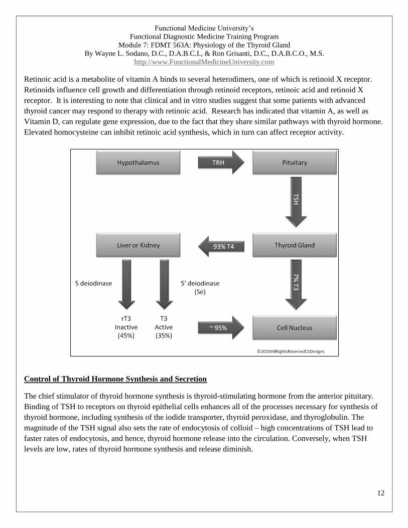
Control of Thyroid Hormone Synthesis and Secretion
The chief stimulator of thyroid hormone synthesis is thyroid-stimulating hormone from the anterior pituitary.
Binding of TSH to receptors on thyroid epithelial cells enhances all of the processes necessary for synthesis of
thyroid hormone, including synthesis of the iodide transporter, thyroid peroxidase, and thyroglobulin. The
magnitude of the TSH signal also sets the rate of endocytosis of colloid – high concentrations of TSH lead to
faster rates of endocytosis, and hence, thyroid hormone release into the circulation. Conversely, when TSH
levels are low, rates of thyroid hormone synthesis and release diminish.

Functional Medicine University’s
Functional Diagnostic Medicine Training Program
Module 7: FDMT 563A: Physiology of the Thyroid Gland
By Wayne L. Sodano, D.C., D.A.B.C.I., & Ron Grisanti, D.C., D.A.B.C.O., M.S.
http://www.FunctionalMedicineUniversity.com
13
Feedback effect of Thyroid Hormone
As thyroid hormone increases in the body fluids, a decrease in secretion of TSH from the anterior pituitary
ensues. Increased thyroid hormone seems to mainly affect the anterior pituitary’s secretion of TSH.

Functional Medicine University’s
Functional Diagnostic Medicine Training Program
Module 7: FDMT 563A: Physiology of the Thyroid Gland
By Wayne L. Sodano, D.C., D.A.B.C.I., & Ron Grisanti, D.C., D.A.B.C.O., M.S.
http://www.FunctionalMedicineUniversity.com
14

Functional Medicine University’s
Functional Diagnostic Medicine Training Program
Module 7: FDMT 563A: Physiology of the Thyroid Gland
By Wayne L. Sodano, D.C., D.A.B.C.I., & Ron Grisanti, D.C., D.A.B.C.O., M.S.
http://www.FunctionalMedicineUniversity.com
15
Peripheral Conversion of T4 to T3
Physiological responses to circulating T4 depend upon its peripheral conversion to T3 by enzymes that require
selenium. Patients with normal circulating of TSH and T4 who display clinical hypothyroid symptoms may be
selenium depleted. The enzyme responsible for the conversion of T4 to T3 is called 5’-deiodinase.

Functional Medicine University’s
Functional Diagnostic Medicine Training Program
Module 7: FDMT 563A: Physiology of the Thyroid Gland
By Wayne L. Sodano, D.C., D.A.B.C.I., & Ron Grisanti, D.C., D.A.B.C.O., M.S.
http://www.FunctionalMedicineUniversity.com
16
Selenium
Is an essential nutrient for 5’ – deiodinase activity, the enzyme involved in the peripheral conversion of
T4 and T3.
Also necessary to degrade rT3
Adequacy Assessment:
RBC, whole blood, hair, serum, urine (TSH, TT4, TT3)
Optimal forms: Mixed selenocompounds including: selenocysteine, selenomethionine, se-
methylselenocysteine
Clinical indications of deficiency:
Compromised immunity, male & female reproductive health, cardiovascular health, inflammation
regulation in asthma and thyroid hormone metabolism.
Low selenium levels are associated with diminished deiodination of T4 and T3.
Consider deficiency if Total T3 is reduced and/or T3 is reduced with a normal TSH and T4 level
Food Sources: garlic, onions, broccoli, brazil nuts, brewer’s yeast
Occupies a unique position regarding biochemical and physiological mechanisms
Selenium can replace the sulfur atom in cysteine, creating a selenocysteine residue.
Selenocysteine (selenoprotein) is the 21st acid recognized as part of the universal genetic code.
Selenoproteins
Glutathione peroxidase (GPx)
Reduction of ROS (hydrogen peroxide)
Protection from oxidation stress
Thioredoxin reductase (TR)
Reduces Vitamin C, lipoic acid, coenzyme Q10, Vitamin K
A selenium-deficient state may lead to uncontrolled release of metals, especially copper and cadmium,
contributing to toxicity.

Functional Medicine University’s
Functional Diagnostic Medicine Training Program
Module 7: FDMT 563A: Physiology of the Thyroid Gland
By Wayne L. Sodano, D.C., D.A.B.C.I., & Ron Grisanti, D.C., D.A.B.C.O., M.S.
http://www.FunctionalMedicineUniversity.com
17
Selenium & The Thyroid Gland
Normal functioning thyroid gland is dependent upon the selenoprotein iodothyronine deiodinase (ID).
ID converts T4 (thyroxin) to the metabolically active T3 (triiodothyronine).
Supplementation with selenium in patients with autoimmune thyroiditis has been shown to significantly
reduce antibody production and oxidative stress.
Clinical Association of Selenium
Selenium deficiency affects most physiological systems
Endocrine
Immunological
Gastrointestinal
Musculoskeletal
Key nutrient in cancer prevention and treatment
Individuals with elevated body burden of toxic elements have greater difficulty maintaining sufficient
selenium levels.
Selenium toxicity (Selenosis)
Rare (usually by contaminated soil)
Signs & Symptoms
Garlic breath
Thick, brittle fingernails
Dry brittle hair
Red swollen hands & feet
Numbness, convulsions, paralysis

Functional Medicine University’s
Functional Diagnostic Medicine Training Program
Module 7: FDMT 563A: Physiology of the Thyroid Gland
By Wayne L. Sodano, D.C., D.A.B.C.I., & Ron Grisanti, D.C., D.A.B.C.O., M.S.
http://www.FunctionalMedicineUniversity.com
18
Assessment of Selenium Status
• Narrow therapeutic window. Evaluation is essential for determining the need for therapy.
• Functional marker
Serum T4 elevated with depressed T3
(peripheral conversion of T4 to T3 is a selenium dependant enzyme called 5’deiodinase. Iodine is removed from
the 5’ site on the first Tyrosyl residual)
Selenium - reference ranges
Serum
95-165 ng/mL
40% higher in whole blood
Critical values: >500 ng/mL
Urinary – 24 hr
Selenometabolites are excreted in the urine
e.g. monomethylselenium & selenosugars
Reference ranges: 15-150 ug/L
RBC selenium
If low selenium – selenium deficiency
If normal – may not represent normal levels (may still be deficient)
Hair – selenium
Hair concentrations correlate with those of blood
May accurately reflect status
Rule out exogenous selenium (shampoos)
Selenium Repletion
Best absorbed form is selenomethionine
Se-methylselenocysteine is less toxic
RDA 55 ug/day
Repletion dose 60-100 ug/day

Functional Medicine University’s
Functional Diagnostic Medicine Training Program
Module 7: FDMT 563A: Physiology of the Thyroid Gland
By Wayne L. Sodano, D.C., D.A.B.C.I., & Ron Grisanti, D.C., D.A.B.C.O., M.S.
http://www.FunctionalMedicineUniversity.com
19
Summary
A basic and advanced understanding of thyroid hormone synthesis, secretion and effect is imperative to
implementing an effective treatment plan. A later lesson in this module will provide a detailed functional
medicine treatment approach to managing thyroid dysfunction.
References
1. Laboratory Evaluations for Integrative and Functional Medicine, 2nd
ed., Richard S. Lord, J. Alexander
Bralley
2. Mosby’s Manual of Diagnostic and Laboratory Tests, 3rd
ed., Pagana and Pagana
3. www.the medical biochemistrypage.org; Dr. Michael King
4. Textbook of Medical Physiology; 11th
ed., Guyton & Hall
5. www.vivo.colostate.edu
6. Retinoic Acid and Retinoid Receptors are Differentially Expressed in Thyroid Cancer and Thyroid
Carcinoma Cell Lines and Predict Response to Treatment with Retinoids, Bryan R. Haugen, Lori Lee
Larson, Umarani Pugazhenthi, William R. Hays, Joshua P. Klopper, Cynthia A. Kramer, Vibha Sharma,
The Journal of Clinical Endocrinology & Metabolism, 89(1):272-280, doi: 10.1210/jc.2003-030770
7. Homocysteine Inhibits Retinoic Acid Synthesis: A Mechanism for Homocysteine-Induced Congenital
Defects, Limpach A, Dalton M., Miles R., Gadson P., U.S. National Library of Medicine, National Institutes
of Health, Exp Cell Res. 2000 Oct 10:260(1): 166-74; 2001 May 15; 266(1): 201