functional cyclic amp response element plays crucial … · afunctional...
TRANSCRIPT
A Functional Cyclic AMPResponse Element Plays a Crucial Role inNeuroendocrine Cell Type-specific Expression of the Secretory GranuleProtein Chromogranin AHongjiang Wu, Sushil K. Mahata, Manjula Mahata, Nicholas J. G. Webster, Robert J. Parmer, and Daniel T. O'ConnorThe Department of Medicine and Center for Molecular Genetics, University of California, San Diego; and the Department of VeteransAffairs Medical Center, San Diego, California 92161
Abstract
Chromogranin A, a soluble acidic protein, is a ubiquitouscomponent of secretory vesicles throughout the neuroendo-crine system. Wereported previously the cloning and initialcharacterization of the mouse chromogranin A gene pro-moter, which showed that the promoter contains both posi-tive and negative domains and that a proximal promoterspanning nucleotides -147 to +42 bp relative to the tran-scriptional start site is sufficient for neuroendocrine celltype-specific expression. The current study was undertakento identify the particular elements within this proximal pro-moter that control tissue-specific expression. Wefound thatdeletion or point mutations in the potential cAMPresponseelement (CRE) site at -68 bp virtually abolished promoteractivity specifically in neuroendocrine (PC12 chromaffin orAtT20 corticotrope) cells, with little effect on activity incontrol (NIH3T3 fibroblast) cells; thus, the CREbox is nec-essary for neuroendocrine cell type-specific activity of thechromogranin A promoter. Furthermore, the effect of theCRE site is enhanced in the context of intact (wild-type)promoter sequences between -147 and -100 bp. DNase Ifootprint analysis showed that these regions (including theCREbox) bind nuclear proteins present in both neuroendo-crine (AtT20) and control (NIH3T3) cells. In AtT20 cells,electrophoretic mobility shift assays and factor-specific anti-body supershifts showed that an oligonucleotide containingthe chromogranin A CREsite formed a single, homogeneousprotein-DNA complex containing the CRE-binding proteinCREB. However, in control NIH3T3 cells we found evidencefor an additional immunologically unrelated protein in thiscomplex. A single copy of this oligonucleotide was able toconfer neuroendocrine-specific expression to a heterologous(thymidine kinase) promoter, albeit with less fold selectivitythan the full proximal chromogranin A promoter. Hence,the CREsite was partially sufficient to explain the neuroen-docrine cell type specificity of the promoter. The functionalactivity of the CREsite was confirmed through studies ofthe endogenous chromogranin A gene. Northern mRNAanalysis showed that expression of the endogenous chro-mogranin A gene was stimulated seven- to eightfold bycAMPin PC12 cells, whereas no induction occurred in the
Address correspondence to Dr. Daniel T. O'Connor, Department ofMedicine (911 1H), University of California, San Diego, 3350 La JollaVillage Drive, San Diego, CA 92161-9111H. Phone: 619-552-8585x7373, FAX: 619-552-8585x7372; E-mail: [email protected]
Received for publication 29 November 1994 and accepted in revisedfor 23 March 1995.
The Journal of Clinical Investigation, Inc.Volume 96, July 1995, 568-578
NIH3T3 cells. Similar cAMP induction was obtained withthe transfected chromogranin A promoter in PC12 cells,and abolition of the CREsite (by deletion or point mutation)eliminated the induction. Thus, the CREsite in the chro-mogranin A proximal promoter is functional and plays acrucial, indeed indispensable, role in neuroendocrine-spe-cific expression of the gene. These results also provide in-sight into transcriptional mechanisms governing acquisitionof the neuroendocrine secretory phenotype. (J. Clin. Invest.1995. 96:568-578.) Key words: Chromogranin A * PC12-AtT20 * cyclic AMP* promoter
Introduction
The chromogranins/secretogranins are a family of soluble,acidic proteins present as the major constituents in secretoryvesicle cores of virtually all neuroendocrine tissues [1-3]. Themost widely expressed of this protein family is the 48-kD chro-mogranin A (4).
Wepreviously isolated a functional transcriptional promoterfrom the mouse chromogranin A gene and characterized itsexpression in neuroendocrine versus control cells (5). Our re-sults suggested that the 5' flanking region of the chromograninA gene contains multiple functional transcriptional regulatoryelements, both positive and negative, which behave largely aspromoter rather than enhancer domains (5). Using deletionmutagenesis, we localized a proximal promoter (which con-ferred neuroendocrine-specific expression) to a region spanningnucleotides -147 to +42 bp relative to the transcriptional startsite. Further deletion to -77 bp retained neuroendocrine-spe-cific expression, whereas deletion to -61 bp abolished celltype-specific promoter activity. A comparison of the mousechromogranin A proximal promoter region sequence with therat (5), bovine (6), and human (7) chromogranin A promotersshowed high conservation of overall promoter structure. Eachspecies' promoter contains a cAMP/response element (CRE)'consensus sequence (10) upstream of a TATA box. Two othermembers of this family of proteins, chromogranin B and se-cretogranin II (also known as chromogranin C), contain similarCREand TATA homologies in their proximal gene promoters(8, 9).
However, the proximal chromogranin A promoter containsnumerous structural matches to potential cis elements other thanthe CREbox (10), and even the functional importance of theCREhomology in this promoter region remains unexplored. Inthe current study, we analyzed the proximal chromogranin Apromoter (-147 to -48 bp) by scanning mutagenesis at high
1. Abbreviations used: CMV, cytomegalovirus; CRE, cAMPresponsiveelement; CREB, CRE-binding protein; CREM,CREmodulator protein;DBH, dopamine ,/-hydroxylase; LUC, luciferase; TK, thymidine kinase.
568 Wuet al.

PanlPTFlBbox
E box GAGA
rGAGA11
H4TF-2histSpl I I
c---
KBF1 Ap2 H4TF-2hist Tstl1 ' Homeodomain
, GA Box' ,' SP1 CRE''~~~~~~~~~~~~ -- I~~~~~~~~~~~~~~~~~~~~~~~~~~~~~~~~~~~~~~~~~~~~~~~~~~-
7 GAGACA-GCTGATGGAGAAGCTGGAGGTGrGGGGG-CGGGACCCCGAAGGmnxiGuAADGGGCGCGGGGGGGCGGTCCTATIGACGTAATTTCCTGGGTGTGTGCGCGCG....
-147 -100 -77 -43
AGGA CA&ATGGAGAAGCTGGAGGTGGGGGGCGGGACCCCGAAGGTGGGAAAGGGCGCGGGGGGGCGGTCCTATGACGTAATTTCCTGGGTGTGTGCGCGCG....GAGACAGCTGtaeqGAGAAGCTGGAGGTGGGGGGCGGGACCCCGAAGGTGGGGAAAGGGCGCGGGGGGGCGGTCCTATGACGTAATTTCCTGGGTGTGTGCGCGCG....
-100 -77 -43
pBRAn
pXP1
A . LuciferaseAn
Insert
pXP100
M2
M3
M4
M6M7
M8
M9
M12
M13
M14
M15
M16
M17
M19
M21
M22
TGGGGAAAGGGCGCGGGGGGGCGGTCCTATGACGTAATTTCCTGGGTGTGTGCGCGCG....TGGttcAGGGCGCGGGGGGGCGGTCCTATGACGTAATTTCCTGGGTGTGTGCGCGCG....TGgGAgGCGCGGGGGGGCGGTCCTATAACGTAATTTCCTGGGTGTGTGCGCGCG....TGGGGAAAGG^gGOGGGGGGGCGGTCCTATGACGTAATTTCCTGGGTGTGTGCGCGCG....TGGGGAAAGGGCGC=QMGGCGGTCCTATGACGTAATTTCC G....
TGGGGAAAGGGCGCGGGGaGAMGTCCTATGACGTAATTTCCTGGGTGTGTGCGCGCG....T'GGGAAAGGGCGCGOCGGG~ttTATGACGTAATTTCC GGTGTGTGCGCGCG....
TGGGGAAAGGGCGCGGGGGGGCGGTCgWaGACGTAATTTCCTGGGTGTGTGCGCGCG....TGGGGAAAGGGCGCGGGGGGGCGGTCCTATGA-GTAATTTCCTGGGTGTG73CGCGCG....TGGGGAAAGGGCGCGGGGGGGCGGTCCTASataATTIC GCG ....
TGGGGAAAGGGCGCGGGGGGGCGGTCCTATGACGT£ATTTCCTGGGTGTGTGCGCGCG....TGGGGAAAGGGCGCGGGGGGGCGGTCCTATGACGaccTTCCTGGGTGTGTGCGCGCG....TGGGGAAAGGGCGCGGGGGGGCGGTCCTATGACGTAAgMCCTGGGTGTGTGCGCGCG....TGGGAAAGGGCGCGGGGGGGCGGTCCTATGACGTAATTrAgoGGGTGTGTGCGCGCG....500GAAAGGGCGCGGGGGGGCGGTCCTATGACGTAATTTCCjtctaGTGTGCGCGCG....TGOGGAAAGGGCGCGGGGGGGCGGTCCTATGACGTAATTTCCTGGGAGGatCGCGCG....
TGGGGAAAGGGCGCGGGGGGGCGGTCCTATGACGTAATTTCCTGGGgaTGCGCGCG....-77 -43
pXP77 GTCCTAMACGTAATTTCCTGGTGTGTGCGCGCG....M41 GTCCTATGA-GTAATTTCCTGGGTGTGTGCGCGCG....
Figure 1. Dense scanning mutagenesis of the proximal chromogranin A promoter. The site-directed mutations (created as described in Methods),indicated by bold and underlined lowercase letters, were inserted into the promoterless luciferase reporter vector pXPl. The shaded boxes representthe regions of the mouse chromogranin A promoter protected from DNase I digestion by AtT20 corticotrope nuclear proteins (5). Known transcriptionfactor-binding site sequence homologies are indicated by the legend directly above the sequence.
density, to determine which element(s) in the promoter governs
neuroendocrine activity of the gene. The results suggest thatthe CREsite in the chromogranin A proximal promoter is func-tional and plays a crucial role in tissue-specific expression.
Methods
Reagents. Most reagents were purchased from Sigma Chemical Co. (St.Louis, MO). The cAMP-dependent protein kinase (protein kinase A)inhibitor KT5720 (11) was from Calbiochem-Novabiochem Corp. (LaJolla, CA). Purified recombinant rat CRE-binding protein (CREB) (12)and anti-rat CREBpolyclonal rabbit antibody (12) (Raised against a
synthetic peptide encompassing amino acids 121-159 [the "kinase-inducible domain" ]) were gifts from Marc Montminy and Paul Brindle(Salk Institute, La Jolla, CA).
Promoter-reporter plasmid constructions and mutagenesis. Pro-moter fragment positions are numbered relative to the major transcrip-tional initiation ("cap") site (5) as + 1. For example, pXPl 133 contains1,133 bp of the mouse chromogranin A promoter (5' flanking region)fused to a luciferase (LUC) reporter in the promoterless luciferasereporter vector pXPl (13). Construction of chromogranin A promoter-luciferase reporter plasmids pXPl 133, pXP426, pXPl81, pXP147,pXPlOO, pXP77, and pXP61 was described previously (5); pXPlOO isa new construct generated using a similar method. To test putativeneuroendocrine-specific regulatory elements in the proximal region ofthe promoter, a dense series of scanning mutants between -100 and-49 bp (Fig. 1) was prepared by PCR amplification on pXPlOO as
template, using different mutated 5' (upstream) oligonucleotide primersand a common 3' (downstream) primer. The 5' primer terminated in
an XhoI restriction site, whereas the 3' primer terminated in a HindIIsite +42 bp downstream of the cap site. The amplified fragments thuscontained the region from -100 to +42 bp, flanked by XhoI and HindHIsites. The mutant fragments were subcloned into the XhoI and HindIIsites of promoterless luciferase reporter vector pXPl (13), generatingmutants of the series M2-M22. Mutant sequences were confirmed bydideoxy sequencing (14). A similar series of mutants (M32-M36) was
constructed between -147 and -100 bp (see Figs. 1 and 5), usingpXP147 as template. The M41 mutation, similar to M12, was createdin pXP77, which contains the region -77 to +42 bp of the chromograninA promoter.
A single copy of a double-stranded oligonucleotide DNAfragment(-76 to -59 bp), which contains the chromogranin A CREsite ("origi-nal CRE"), was inserted into the polylinker immediately upstream ofthe heterologous herpes simplex virus thymidine kinase (TK) promoterin the luciferase reporter vector pTK-LUC (5). Similar constructionswere made containing the M12mutation ("mutated CRE") and a singlenucleotide change to create a consensus CREsite ("perfect CRE").
Cell culture and transfections. Neuroendocrine cell lines used were
the rat adrenal medullary (pheochromocytoma) chromaffin cell linePC12 (15) and the mouse anterior pituitary corticotrope cell line AtT20( 16). The control (nonendocrine) cell line used was the mouse fibroblastline NIH3T3. Cell lines were grown in DMEwith supplements of FBSand horse serum as described previously (5).
Supercoiled plasmid DNAfor transfection was grown in Escherichiacoli strain DH-5 and purified on columns (Qiagen Inc., Chatsworth,CA). Cells (50-70% confluent) were transfected with 2-4 jsg of plas-mid DNAper 6 cm plate, using the lipofection method (Lipofectin or
Lipofectamine; GIBCO-BRL, Bethesda, MD). 48 h after transfection,cell extracts were prepared and assayed for protein (17), luciferase
Role of Cyclic AMPResponse Element in Chromogranin A Expression 569
pXPt14
M32M33
M34M35M36
GAGACAGCTGA7GS=AGCTGGAGG7WC..GGGACCCCGAAGG7WGGAAAGGGCGCGGGGGGGCGGTCCTATGACGTAATTTCCTGGGTGTGTGCGCGCG..GAGACAGCTIGATGGAGAAGCTGGAGGTGGG.tG-tC.tGGACCCCGAAGGTGGGGAAAGGGCGCGGGGWGCGGTCCTATGACGTAATTTCCTGGGTGTGTGCGCGCG..GAGACAGCTIGATGGAGAAGCTGGAGGTGOGGGGCGGGACCCCUMGTGGGGAAAGGGCGCGGGGGGGCGGTCCTATGACGTAATTTCCTGGGTGTGTGCGCGCG..
250 .
200 -
0
1
IUS
150 .
100.
so0
* PO2El AtT20o 3T3
I
pBAn
pXP1
An ciferaseAnO .0 N
, ' CgA \,- promoter Insert n pXP1SI
-147-100
-77-61 +42
pXPl 81 pXP147 pXP100 pXP77 pXP61
CgApromoter deletion mutants
activity (18), and 3-galactosidase activity (17). To control for differ-ences in transfection efficiency among cell lines and plasmids, co-trans-fections were performed with 0.4-0.8 jig of CMV-lacZ (Clontech, PaloAlto, CA), encoding ,3-galactosidase controlled by the cytomegalovirus(CMV) promoter. Luciferase results were normalized to ,6-galactosidaseactivity.
To test functional activation of the CRE in the promoter, thetransfected cells were cultured for 4 or 24 h after transfection in thepresence of 1 mMdibutyryl cAMP and/or 0.1 and 0.5 iM proteinkinase A-specific inhibitor KT5720 before harvesting.
In vitro binding studies. Double-stranded oligonucleotides spanningthe CREregion -76 to -59 bp were synthesized, labeled by filling in5' overhangs with [32p] dCTP and the Klenow fragment of E. coli DNApolymerase I, and used in electrophoretic mobility shift assays withnuclear extracts from AtT20 or NIH3T3 cells. Nuclear extracts wereprepared as described ( 19). Labeled fragments ( I05 dpm, - 0.15 pmol)were incubated with nuclear extracts (2 jg of protein) in the presenceof poly(dl - dC) (0.2 jg/ml) as nonspecific competitor. Bound and freeDNAwere separated by electrophoresis through 5% acrylamide/0.4xTBE gels (19), which were dried and subjected to autoradiography at-70°C. In some experiments, the binding of pure (recombinant) CREBwas used as a positive control, and a polyclonal antipeptide antibodyraised against a synthetic peptide containing amino acids 121-159 (thekinase-inducible domain) of rat CREB(12) was used to supershift theprotein-DNA complex. DNase I footprinting was performed as de-scribed previously (5).
Northern blot analysis of mRNA. PC12 and NIH3T3 cells weretreated with dibutyryl cAMP (1 mM) for 1, 4, 6, and 24 h, with orwithout the addition to the protein kinase A-specific inhibitor KT4720(l0-' or 5 x 10⁻⁷ M) or cycloheximide (5 jg/ml). Treatment ofthese cells with 5 gmg/ml cycloheximide decreases incorporation of[35S]methionine into TCA-precipitable protein by > 95% (20). TotalRNA was isolated by acid guanidinium thiocyanate-phenol-chloro-form extraction (RNAzolB; TelTest, Friendswood, TX). RNAs (10 jig)were size fractionated on denaturing 1% agarose-formaldehyde gels,transferred to nitrocellulose membranes, and fixed with ultraviolet irradi-ation (StrataLinker; Stratagene, La Jolla, CA). The blots were prehy-bridized, hybridized, and washed essentially as described previously(17). Hybridization was performed separately with either a 32P-labeledmouse chromogranin A cDNA probe (21), or a rat cyclophilin("housekeeping") cDNA probe (22). Expression of chromogranin AmRNAwas quantified using a StrataScan 7000 densitometer (Stra-tagene) and normalized to cyclophilin gene expression.
Data presentation. Transfection experiments were repeated threetimes, with three plates per condition per experiment. Results are shownon bar graphs as the mean value for separate transfections ± 1 SD.
pXP147pXP100pXP77pXP61
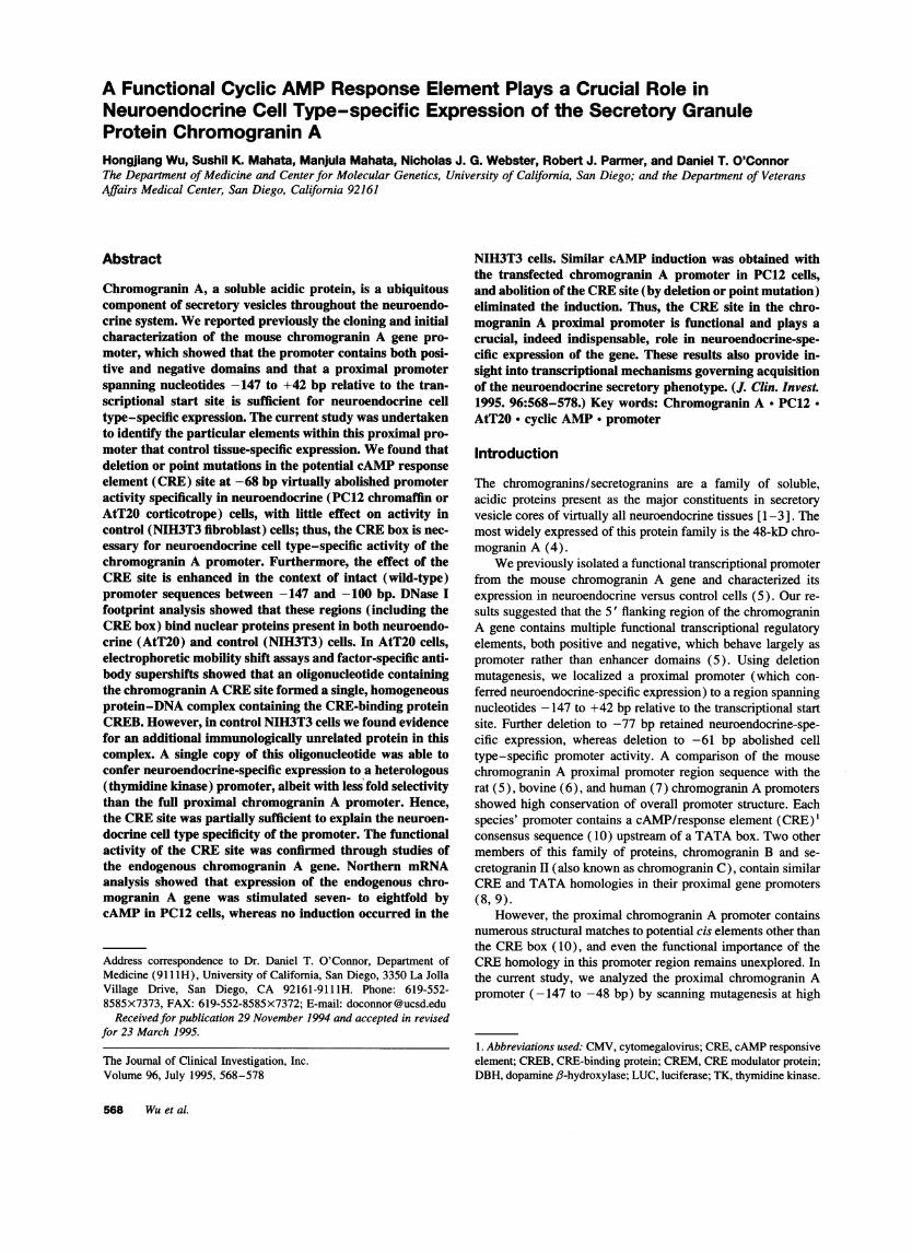
Figure 2. 5' deletion mutants of the chro-mogranin A proximal promoter. Promoterdeletions were performed as described inMethods and inserted into the promoterlessluciferase reporter vector pXPl. The pro-moter deletion/luciferase reporter constructswere transfected into neuroendocrine (PC12chromaffin or AtT20 corticotrope) or control(NIH3T3 fibroblast) cells, along with thetransfection control efficiency plasmidCMV-lacZ. The results are expressed as ra-tios of luciferase//l-galactosidase activities.The pXP100 data amplify the previously re-ported (5) series of deletion mutants.
Results
Site-directed, dense scanning mutation analysis of the chro-mogranin A proximal promoter. Wedemonstrated previouslythat the region from -147 to +42 bp relative to the transcrip-tional initiation (cap) site was sufficient to direct neuroendo-crine-specific expression of the chromogranin A promoter (5).High levels of expression after transfection were observed inthe neuroendocrine cell lines PC12 (rat adrenal medullary chro-maffin cell), AtT20 (mouse anterior pituitary corticotrope),and GH3 (rat anterior pituitary somatolactotrope). By contrast,tranfected NIH3T3 fibroblast and COS monkey kidney cells,the nonendocrine cell lines, showed low levels of luciferaseactivity. To define the promoter elements responsible for neuro-
endocrine expression, additional 5' promoter deletions were
constructed and transiently transfected into NIH3T3, AtT20,and PC12 cells as described previously (Fig. 1). In AtT20 andPC12 cells, deletion from -147 to -100 or -77 bp diminishedreporter expression by - 50%, but had no effect in NIH3T3cells (Fig. 2). Further deletion to -61 bp eliminated all celltype specificity.
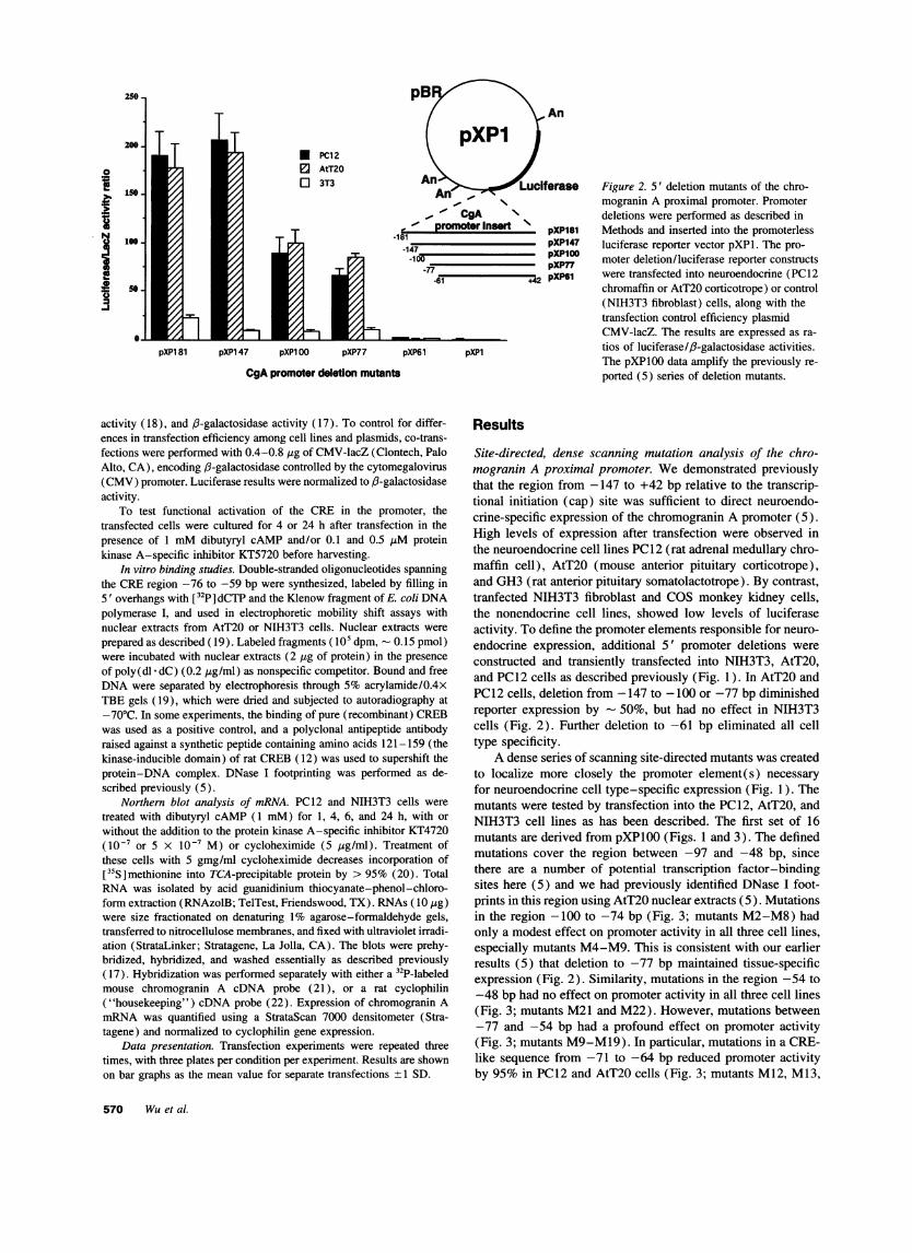
A dense series of scanning site-directed mutants was createdto localize more closely the promoter element(s) necessary
for neuroendocrine cell type-specific expression (Fig. 1). Themutants were tested by transfection into the PC12, AtT20, andNIH3T3 cell lines as has been described. The first set of 16mutants are derived from pXP100 (Figs. 1 and 3). The definedmutations cover the region between -97 and -48 bp, sincethere are a number of potential transcription factor-bindingsites here (5) and we had previously identified DNase I foot-prints in this region using AtT20 nuclear extracts (5). Mutationsin the region -100 to -74 bp (Fig. 3; mutants M2-M8) hadonly a modest effect on promoter activity in all three cell lines,especially mutants M4-M9. This is consistent with our earlierresults (5) that deletion to -77 bp maintained tissue-specificexpression (Fig. 2). Similarity, mutations in the region -54 to-48 bp had no effect on promoter activity in all three cell lines(Fig. 3; mutants M21 and M22). However, mutations between-77 and -54 bp had a profound effect on promoter activity(Fig. 3; mutants M9-M19). In particular, mutations in a CRE-like sequence from -71 to -64 bp reduced promoter activityby 95% in PC12 and AtT20 cells (Fig. 3; mutants M12, M13,
570 Wuet al.
KBF1 Ap2 H4TF-2hist TstlI '~~ E r1' Homeodomain
GA Box ' Sl:)l ' CRE-100 -77
pXP100 TGGGGAAAGGGCGCGG
pttcgAM12,GcAgttq
M3GtatgG
M4MtcMa
GGGGGCGGTCCTATGACGTAATTTCCTGGGTGTGTGCG(
ptc AcatcaccN *TagcaciM7 M13 M17
*Ggttag!G CGTcAT% CatctaG,M8 M14 M19
CgacaG, ,GaccaT, GaGatacCMO MI15 21
GacG, mA-GXAga ,GcatcT,6 M12 M16 M22
-43
;CGCG....
-0
o
Sb
V-IE
pXP100 M2 M3 M4 M6 M7 M8 M9 M12 M13 M14 M15 M16 M17 M19 M21 M22pXP100 scanning mutants
and M15). These mutations were less severe in NIH3T3 cells,but still caused 30-60% declines in promoter activity. Mutationof the chromogranin A CRE-like sequence to a consensus CREsite (mutant M14) caused a 50-80% increase in promoter activ-ity over the corresponding wild-type sequence (in pXPlO0) inall three cell types, underscoring the importance of this sequence(Fig. 3).
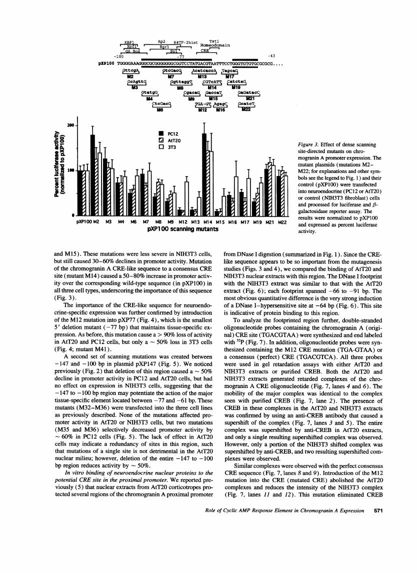
The importance of the CRE-like sequence for neuroendo-crine-specific expression was further confirmed by introductionof the M12 mutation into pXP77 (Fig. 4), which is the smallest5' deletion mutant (-77 bp) that maintains tissue-specific ex-pression. As before, this mutation cause a > 90% loss of activityin AtT20 and PC12 cells, but only a - 50% loss in 3T3 cells(Fig. 4; mutant M41).
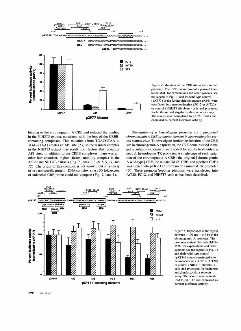
A second set of scanning mutations was created between-147 and -100 bp in plasmid pXP147 (Fig. 5). Wenoticedpreviously (Fig. 2) that deletion of this region caused a - 50%decline in promoter activity in PC12 and AtT20 cells, but hadno effect on expression in NIH3T3 cells, suggesting that the-147 to -100 bp region may potentiate the action of the majortissue-specific element located between -77 and -61 bp. Thesemutants (M32-M36) were transfected into the three cell linesas previously described. None of the mutations affected pro-moter activity in AtT20 or NIH3T3 cells, but two mutations(M35 and M36) selectively decreased promoter activity by
- 60% in PC12 cells (Fig. 5). The lack of effect in AtT20cells may indicate a redundancy of sites in this region, suchthat mutations of a single site is not detrimental in the AtT20nuclear milieu; however, deletion of the entire -147 to -100bp region reduces activity by - 50%.
In vitro binding of neuroendocrine nuclear proteins to thepotential CREsite in the proximal promoter. Wereported pre-viously (5) that nuclear extracts from AtT20 corticotropes pro-tected several regions of the chromogranin A proximal promoter
Figure 3. Effect of dense scanningsite-directed mutants on chro-mogranin A promoter expression. Themutant plasmids (mutations M2-M22; for explanations and other sym-bols see the legend to Fig. 1 ) and theircontrol (pXPlOO) were transfectedinto neuroendocrine (PC12 or AtT20)or control (NIH3T3 fibroblast) cellsand processed for luciferase and 3-galactosidase reporter assay. Theresults were normalized to pXP1OOand expressed as percent luciferaseactivity.
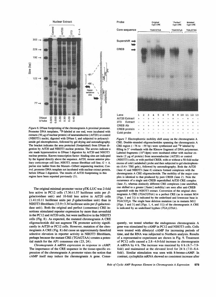
from DNase I digestion (summarized in Fig. 1). Since the CRE-like sequence appears to be so important from the mutagenesisstudies (Figs. 3 and 4), we compared the binding of AtT20 andNIH3T3 nuclear extracts with this region. The DNase I footprintwith the NIH3T3 extract was similar to that with the AtT20extract (Fig. 6); each footprint spanned -66 to -91 bp. Themost obvious quantitative difference is the very strong inductionof a DNase I-hypersensitive site at -64 bp (Fig. 6). This siteis indicative of protein binding to this region.
To analyze the footprinted region further, double-strandedoligonucleotide probes containing the chromogranin A (origi-nal) CREsite (TGACGTAA)were synthesized and end labeledwith 32p (Fig. 7). In addition, oligonucleotide probes were syn-thesized containing the M12 CREmutation (TGA-GTAA) ora consensus (perfect) CRE (TGACGTCA). All three probeswere used in gel retardation assays with either AtT20 andNIH3T3 extracts or purified CREB. Both the AtT20 andNIH3T3 extracts generated retarded complexes of the chro-mogranin A CREoligonucleotide (Fig. 7, lanes 4 and 6). Themobility of the major complex was identical to the complexseen with purified CREB (Fig. 7, lane 2). The presence ofCREBin these complexes in the AtT20 and NIH3T3 extractswas confirmed by using an anti-CREB antibody that caused asupershift of the complex (Fig. 7, lanes 3 and 5). The entirecomplex was supershifted by anti-CREB in AtT20 extracts,and only a single resulting supershifted complex was observed.However, only a portion of the NIH3T3 shifted complex wassupershifted by anti-CREB, and two resulting supershifted com-plexes were observed.
Similar complexes were observed with the perfect consensusCREsequence (Fig. 7, lanes 8 and 9). Introduction of the M12mutation into the CRE (mutated CRE) abolished the AtT20complexes and reduces the intensity of the NIH3T3 complex(Fig. 7, lanes 11 and 12). This mutation eliminated CREB
Role of Cyclic AMPResponse Element in Chromogranin A Expression 571
KBF1 Ap2 H4TF-2hist TstlXI f1' Egr ' HomeodomainGA Box! 1D CRE
-100~~~~~~~~~~~~~~~~~~~~~~~~~~~~~~-100 -77 -61
gP100o TGGGGAAAG.GGCGCGGGGGGCGGTCCTATGACGTAATTTCCI
pXP77
341
pXP61 TCCTGGGTGTGTGCGCGCG....
120-
3 0Lo
acf2
oa C
100 -
80 .
60-
40-
20-
0-
LL'
pXP77
* PC12E2 AtT20o 3T3
M41
pXP77 Mutant
binding to the chromogranin A CREand reduced the bindingin the NIH3T3 extract, consistent with the loss of the CREB-containing complexes. This mutation (from TGACGTAAtoTGA-GTAA) creates an API site (23) so the residual complexin the NIH3T3 extract may result from factors that recognizeAPI sites. in addition to the CREBcomplexes, there was an-other less abundant, higher (faster) mobility complex in theAtT20 and NIH3T3 extracts (Fig. 7, lanes 1, 3-6, 8, 9, 11, and12). The origin of this complex is not known, but it is likelyto be a nonspecific protein-DNA complex, sine a 50-fold excessof unlabeled CREprobe could not compete (Fig. 7, lane 1).
Figure 4. Mutation of the CREsite in the minimalpromoter. The CREmutant promoter plasmid (mu-tation M41; for explanations and other symbols, seethe legend to Fig. 1) and its wild-type control(pXP77) or the further deletion mutant pXP61 weretransfected into neuroendocrine (PC12 or AtT20)or control (NIH3T3 fibroblast) cells and processed
pXP61 for luciferase and 6-galactosidase reporter assay.The results were normalized to pXP77 results andexpressed as percent luciferase activity.
Stimulation of a heterologous promoter by a functionalchromogranin A CREpromoter element in neuroendocrine ver-sus control cells. To investigate further the function of the CREsite in chromogranin A expression, the CREdomains used in thegel retardation experiment were tested for ability to stimulate aneutral, heterologous TK promoter. A single copy of each varia-tion of the chromogranin A CRE(the original [chromograninA wild-type] CRE, the mutant [M12] CRE, and a perfect CRE)was cloned into pTK-LUC upstream of a minimal TK promoter(5). These promoter/reporter plasmids were transfected intoAtT20, PC12, and NIH3T3 cells as has been described.
PanlPTFlBbox
GAGA
-147
pXP147 GAGACAGCTGATGGAGAAGC1
AgtcCacA GctgtA
M32 Gt&CG M34
15 M33
1o0
xc s
5.C
KB1 Ap2 H4TF-2hist TstiH4TF-2hist . . Homeodomain
spi -77 -59
-100 -77 -59
TGGAGGTGGGGGGCGGGACCCCGAAGGTGGGAAAGGGCGCGGGGGGGCGGTCCTATGACGTAATTTCC...
GtGtCtG CtcgaGI a I
M35 M36 * PC12
E3 AtT20o 3T3
Figure 5. Importance of the regionbetween -100 and -147 bp in thechromogranin A promoter. Thepromoter mutant plasmids (M32-M36; for explanations and othersymbols see the legend to Fig. 1)and their wild-type control(pXP147) were transfected intoneuroendocrine (PC12 or AtT20)or control (NIH3T3 fibroblast)cells and processed for luciferaseand /3-galactosidase reporterassay. The results were normal-ized to pXP147 and expressed aspercent luciferase activity.
572 Wuet al.
-43
ITGGGTGTGTGCGCGCG.
pXP147 M32 M33 M34 M35 M36
pXP1 47 scanning mutants
GTCCTATGACGTAATTTCCTGGGTGTGTGCGCGCG...
Nuclear Extract
j iI :0 <_ __ __M
Probe
Core sequence
COACRE
TGACGTAA
CgAjCE CACRE
TGACGTfA TGDcGTAAI I I
-
NMn
303 -_
226 .+
192 -+
/
122 -4
77_-
I
DNas 0 ° ° ° °U/tube 0 o o o o o
c
Figure 6. DNase footprinting of the chromogranin A proximal promoter.Promoter DNAtemplates, 32P-labeled at one end, were incubated withextracts (50 jig of nuclear protein) of neuroendocrine (AtT20) or control(NIH3T3) nuclei, digested with DNase I, and subjected to polyacryl-amide gel electrophoresis, followed by gel drying and autoradiography.The bracket indicates the area protected (footprinted) from DNase di-gestion by AtT20 and NIH3T3 nuclear proteins. The arrows indicate asite made hypersensitive to DNase I digestion by AtT20 and NIH3T3nuclear proteins. Known transcription factor-binding sites are indicatedby the legend directly above the sequence. AtT2O, mouse anterior pitu-itary corticotrope cell line; NIH3T3, mouse fibroblast cell line; G + A,purine size ladder from the Maxam-Gilbert sequencing reaction; Con-trol, promoter DNAtemplate not incubated with nuclear extract protein,before DNase I digestion. The results of AtT20 footprinting in thisregion have been reported previously (5).
The original minimal-promoter vector pTK-LUC was 2-foldless active in PC12 cells (7.36±1.57 luciferase units per ,3-galactosidase unit) and 10-fold less active in AtT20 cells(1.41±0.11 luciferase units per ,3-galactosidase unit) than inNIH3T3 fibroblasts ( 13.9±3.34 luciferase units per /-galactosi-dase unit). Both the original and perfect (consensus) CREin-sertions stimulated reporter expression by more than sevenfoldin the PC12 and AtT20 cells, but were ineffective in the NIH3T3cells (Fig. 8). As expected, the mutated chromogranin A CREoligonucleotide did not augment TK promoter activity signifi-cantly in AtT20 or PC12 cells. However, mutation of the chro-mogranin A CRE(Fig. 8) did cause an approximately threefoldselective elevation in reporter activity in NIH3T3 fibroblasts,perhaps because the mutant CRE(TGAGTAA) creates a poten-tial match for the APl consensus site (23, 24).
Chromogranin A mRNAexpression in response to cAMP.The importance of the CREsequence in the tissue-specific ex-pression of the chromogranin A promoter raises the notion thatcAMP itself may induce the chromogranin A gene. Conse-
Supershift _
CREB _
LaneAtT20 Extract3T3 ExtractCREBAbCREBproteinCold probe
21 2 3 4 5 6 7 8 9 2.0 11 12 13+ _ + + _ _ - + _ _ + _-
_ _ + + - _ + _ _ + _
-_ + _____
Figure 7. Electrophoretic mobility shift assay on the chromogranin ACRE. Double-stranded oligonucleotides spanning the chromogranin ACREregion (-76 to -59 bp) were synthesized and 32P labeled byfilling in 5' overhands with the Klenow fragment of DNApolymerase.Labeled fragments (105/dpm) were incubated either with nuclear ex-tracts (2 jig of protein) from neuroendocrine (AtT20) or control(NIH3T3) cells, or with purified CREB, with or without a 50-fold molarexcess of cold (unlabeled) probe and then subjected to gel electrophore-sis (0.4X TBE gels), followed by autoradiography. Both the AtT20(lane 4) and NIH3T3 (lane 6) extracts formed complexes with thechromogranin A CREoligonucleotide. The mobility of the major com-plex is identical to that produced by pure CREB(lane 2). Note theoccurrence of a single anti-CREB supershifted AtT20 CREcomplex(lane 3), whereas distinctly different CREcomplexes (one unshifted,one shifted to a greater [ faster] mobility) are seen after anti-CREBsupershift with the NIH3T3 extract. Conversion of the original chro-mogranin A CRE(TGACGTAA) to a perfect CRE(as in mutant M14[Figs. 1 and 3]) is indicated by the underlined and lowercase base inTGACGTcA. The single base deletion mutation (as in mutants M12[Figs. 1 and 3] and [Figs. 1, 4, and 11]) of the chromogranin A CREis indicated by an underlined hyphen (TGA-GTAA).
quently, we tested whether the endogenous chromogranin Agene was stimulated by cAMPin PC12 and NIH3T3 cells. Cellswere treated with dibutyryl cAMP for increasing periods oftime, and the RNAwas subjected to Northern analysis. Resultsof a representative experiment are shown in Fig. 9. Treatmentof PC12 cells caused a 2.8-4.0-fold increase in chromograninA mRNAby 4 h. The increase was maximal by 6 h (6.7-7.9-fold) and maintained at the elevated level for 24 h (7.0-8.4-fold). Similar stimulation was seen with 8-bromo-cAMP. Incontrast, cyclophilin mRNAshowed no consistent increase after
Role of Cyclic AMPResponse Element in Chromogranin A Expression 573

46 10
2^0S- 8
6
9 ct 4
a 2
0.L
Core CREsequence:
* PC120 AtT20o 3T3
TK Original CgA-CRE-TK Mutated CgA-CRE-TK
none TGACGTAA TGA:GTAA
Synthetic CRE-TK luciferase repor
cAMP (Figs. 9 and 10). Chromogranin A mRNAlevels were
undetectable in NIH3T3 cells, with or without cAMP(Fig. 9).Weinvestigated next the effect of protein synthesis (transla-
tion) inhibition or protein kinase A inhibition on the inductionof chromogranin A gene by cAMP in PC12 cells (Fig. 10).Cycloheximide (5 jig/ml) was unable to prevent the 2.7-4.0-fold induction at 4 h of cAMP. At later time points, cyclohexi-mide decreased basal chromogranin A mRNAlevels by 61-70% and reduced the cAMPinduction by 62-81%. This indi-cates that new protein synthesis is required to account for thefull magnitude of sustained elevation in chromogranin AmRNA, consistent with the normal turnover of cAMP-respon-sive transcription factors, such as CREB itself (12, 13). Theprotein kinase A inhibitor KT5720 inhibited the cAMPinduc-tion, in an inhibitor dose-dependent fashion, confirming therole of protein kinase A (Fig 10). (11).
Clearly, prolonged (24 h) 5 pg/ml cycloheximide dimin-ishes overall cell growth and proliferation. However, our con-
clusions are drawn from Northern blot lanes (Fig. 10), in whichconstant amounts (10 jtg) of total RNAwere loaded per lane,and the data were normalized to values of chromogranin AmRNA/cyclophilin (housekeeping, control) mRNAsignals.
cAMPstimulation of the transfected chromogranin A pro-
moter. Since cAMP induced the endogenous chromogranin Agene in PC12 cells, we tested whether the transfected chro-
PC12 cells 3T3 cells
Chromogranin A (CgA) *
Cyclophilin (Cph) *
db-cAMP treatment(1 mM), time (hours)
Relative CgA expression(normalized to Cph)
- 1h 4h 6h 24h - 1h 4h 6h 24h
j 1Q.32.8 7.9 8.4 0 0 O 0 0
Figure 8. Stimulation of a heterologous TKpromoter by the chromogranin A CREsite.A single copy of a double-stranded synthetic
or gchromogranin A CREfragment and its mu-t/" tants were cloned into the enhancerless pro-
moter/luciferase reporter vector pTK-LUC,immediately upstream (5') of the TK pro-moter. These plasmids were transfected intoneuroendocrine (PC12 or AtT20) or control(NIH3T3 fibroblast) cells and processed forluciferase reporter and protein assay. The re-sults were normalized to the activity of pTK-LUC and expressed as relative luciferase ac-tivity. Conversion of the original chro-mogranin A CRE(TGACGTAA) to a perfectCRE(as in mutant M14 [Figs. 1 and 3]) asindicated by the underlined and lowercase
;"nPertect"CgA-CRE-TK base in TGACGTcA. The single base deletionTGACGTcA mutation (as in mutants M12 [Figs. 1 and 3]
and M4 [Figs. 1, 4, and 11]) of the chro-
ters mogranin A CREis indicated by an under-ters lined hyphen (TGA-GTAA).
mogranin A promoter/luciferase reporter plasmids were alsoinducible. Transfected cells were treated with dibutyryl cAMPfor 4-24 h before harvesting. All of the deletion constructionsfrom -1.1 kb to - 77 bp were induced by a 24-h cAMPstimula-tion (Fig. 11 pXP1 133, pXP426, pXP147, pXPlOO, andpXP77). Slightly lower inductions (three- to fivefold) wereobtained at 4 h with all the deletion mutants. Deletion to -61bp, as well as introduction of the single base pair CREdeletion(M41) in the shorter promoter fragment pXP77, eliminatedthe induction, consistent with removal of the cAMP-responsivesequence (Fig. 11; pXP61 and mutant M41 ). The involvementof protein kinase A in induction of the chromogranin A pro-moter by cAMP was confirmed using the protein kinase Ainhibitor KT5720. KT5720 ( 10-7 M) inhibited cAMPinductionof either pXP100 or pXP77 by 40% (data not shown).
Discussion
Deletion analysis and site-directed mutagenesis of the proximalchromogranin A promoter (-147 to +42 bp) have demon-strated that a CRE-like sequence is crucial for neuroendocrine-specific promoter activity (Figs. 2, 3, and 4). This CRE se-quence is also able to mediate an 8-fold induction of thetransfected chromogranin A promoter by cAMP (Fig. 11),which is similar in magnitude to the 7.0-8.4-fold induction of
Figure 9. Northern blot analysis of chro-mogranin A and cyclophilin mRNAlevels.Neuroendocrine (PCl2) and control(NIH3T3) cells were exposed to dibutyryl
18S cAMP (I mM) for 1, 4, 6, or 24 h. Total. rRNA RNA(10 jig) from these cells was size frac-
tionated on a denaturing agarose gel and se-quentially hybridized with specific cDNAprobes for chromogranin A and cyclophilin.The mobility of the 18S ribosomal RNAisindicated on the right. Relative chromograninA mRNAlevels were normalized to cycloph-ilin mRNAand expressed as fold inductionby cAMP. A representative experiment isshown.
574 Wuet al.

Treatment time
Chromogranin A (CgA) *
Cyclophilin (Cph)
4hr 6hr 24 hr
o
j.I
24hr
0*0
Relative CgA expression(normalized to Cph) 1.0 2.7 1.1 3.3 1.0 U. 1.3 9.2 1.0 fl& 0.4 0.7 1.0 82 fi. 1.3
db-cAMP (1 mM) _-+-_ . _ ,.-_'.
Cycloheximide (5jig/ml) - - + +
PKA Inhibitor (KT5720) - - lO0nM 500nM
Figure 10. Time course of chromogranin A mRNAinduction by cAMPand the role of new protein synthesis and protein kinase A in the induction.Neuroendocrine PC12 chromaffin cells were exposed to dibutyryl cAMP (1 mM) for 4, 6, or 24 h, either alone or in combination with either theprotein synthesis (translation) inhibitor cycloheximide (5 Hg/ml) or the protein kinase A inhibitor KT5720 (10-' or 5 x 10-i M). ChromograninA and cyclophilin mRNAlevels were determined, normalized, and expressed as described in the legend to Fig. 9. A representative experiment isshown.
the endogenous chromogranin A gene by cAMP (Fig. 9; 24-hstimulation). Similarly, the chromogranin A gene is inducibleby the adenylyl cyclase activator forskolin in bovine chromaffincells (6), though the forskolin response in PC12 cells may beblunted (25).
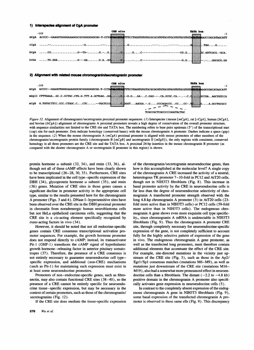
A comparison of mouse chromogranin A's proximal pro-moter region with the rat, bovine, and human promoters (Fig.12) shows high conservation of the overall promoter structure,including the TATA and CREhomologies (5-7). The mouse,rat, bovine, and human promoters all contain a CREbox 18-42 bp upstream of the TATA box (Fig. 12). The bovine (6)and human (7) chromogranin A genes contain perfect consensus(10) CREs (TGACGTCA), similar to that in the M14 mutation(Figs. 1 and 3) generated for this study. The mouse (5) and
I'5aCZ 0.
rat (5) genes differ from the consensus at 1 nucleotide (TGAC-GTAA; difference underlined).
This conservation of the CRE site and TATA box is alsoseen in two other members of the family of chromogranin/secretogranin proteins, chromogranin B and secretogranin II(chromogranin C) (8, 9) (Fig. 12). The chromogranin/secreto-granin promoters show very little homology outside of theTATA and CREsequences.
CRE sites have been identified in many neuroendocrine-specific gene promoters. cAMPstimulates the activity of a num-ber of neuroendocrine-specific genes, including chromograninB (26, 27) and secretogranin H (26-28), tyrosine hydroxylase(29), phenylethanolamine-N-methyltransferase (30), dopa-mine P-hydroxylase (DBH) (31, 34), anterior pituitary glyco-
Figure 11. Induction of activity oftransfected chromogranin A promoter/re-porter plasmids by cAMP. Chromogranin Apromoter progressive 5' deletion mutant/lu-ciferase reporter plasmids (pXPl 133,pXP426, pXP147, pXPlOO, pXP77, orpXP61), or a CREbox site-directed mutantplasmid (mutant M41 [Figs. 1 and ]; pro-moter 5' length to -77 bp; original TGACG-TAA CREaltered to TGA-GTAA) was
Z iA EREmutant transfected into neuroendocrine (PC12) or_ ];S; # t Xf~r/ Icontrol (NIH3T3M) cells. The transfected cellsAd 2 , < . , < I Srs r o 1were treated with either vehicle (control) or'511<j11L] t z s IIL 81 JILL 1T SPLLdibutyryl cAMP (1 mM) for 4 or 24 h, andi2 processed for luciferase reporter and protein
0 assay. The results are expressed as fold in-pXP1 133 pXP426 pXP147 pXP1 00 pXP77 M41 pXP61 duction by cAMPof luciferase activity over
CgA promoter-luciferase reporter plasmid the respective control. In this experiment,since each cAMPstimulation had its own
control (the same promoter fragment, unstimulated by cAMP), the ratios were computed simply as stimulated/unstimulated. Hence, no CMV-lacZinternal control was required. This circumvents the potential pitfall of cAMPresponsiveness of the usual transfection efficiency control plasmid(CMV-lacZ).
Role of Cyclic AMPResponse Element in Chromogranin A Expression 575
1) Interspe-aC alignment of CgApromoter
mCgA-100
CRD alte
ACCCC--GAAGGTGGGGAAAGGGCGCGGGGGGGCGG-T-CCI
rCgA ......-......-A ......---
bCgA .....CG ..... G--TA- .......-. .G
hCGA ..... TG.GGA..... G---AA ..... G.A .0G
....G....C-----------------------CG.G..
.........C------------------------G.G..
boz-3
UhAGCCGGCATAGCATT
j .TAG....G......
3C.GGTCGCG.-GCA.
.....GGCGCG.GG
2) Allgnment with related mouse chromogrannlsecrotogranin promr
rCCTGGGTGTGTCGCGCGTGToGCGTGCGTGTGCGGIP
mSgII CTTTGAAA..GC.C.CCTGC.CTG.G.TTT.A.GCTGAG..G0
mCgB G.TGTGCTTCC.CCC.CTGGC.C CTC..--TGCTCCi
-G.G...AA-..C.GAG---.CA.GCGC.CA.-..-.C.
3GT.. .AGCGA.-.G....CCCAGACCC..CC... CC-.
_ T_.CG
TCGCCGCTCGCCCCCGGGTACTGC
-3AGCCGGCATAGCATT
CCGA..AGCTGTCCG
.... A.GCCTGCGCC
Figure 12. Alignment of chromogranin/secretogranin proximal promoter sequences. (1) Interspecies (mouse [mCgA], rat [rCgA], human [hCgA],and bovine [bCgA]) alignment of chromogranin A proximal promoters reveals a high degree of conservation of the overall promoter structure,with sequence similarities not limited to the CREsite and TATA box. The numbering refers to base pairs upstream (5') of the transcriptional start(cap) site for each promoter. Dots indicate homology (conserved bases) with the mouse chromogranin A promoter. Dashes indicate a space (gap)in the sequence. (2) When the mouse chromogranin A (mCgA) proximal promoter is aligned with mouse promoters of other members of thechromogranin/secretogranin protein family (chromogranin B [mCgB] and secretogranin II [mSgII]), the only regions with consistent, conservedhomology in all three promoters are the CREsite and the TATA box. A proximal 24-bp insertion in the mouse chromogranin B promoter (ascompared with the shorter chromogranin A or secretogranin II promoter in this region) is shown.
protein hormone a subunit (32, 34), and renin (33, 36), al-though not all of these cAMPeffects have been clearly shownto be transcriptional (26-28, 30, 33). Furthermore, CREsiteshave been implicated in the cell type-specific expression of theDBH (34), glycoprotein hormone a subunit (35), and renin(36) genes. Mutation of CRE sites in those genes causes a
significant decline in promoter activity in the appropriate celltype, similar to the results presented here for the chromograninA promoter (Figs. 3 and 4). DNase I-hypersensitive sites havebeen observed over the CREsite in the DBHproximal promoterin chromatin from noradrenergic human neuroblastoma cellsbut not HeLa epithelioid carcinoma cells, suggesting that theCRE site is a cis-acting element specifically recognized bytrans-acting factors in vivo (34).
However, it should be noted that not all endocrine-specificgenes contain CRE consensus transcriptional activation pro-moter sequences. For example, the growth hormone promoterdoes not respond directly to cAMP; instead, its transactivatorPit-i (GHF-1) transduces the cAMP signal of hypothalamicgrowth hormone-releasing factor in anterior pituitary somato-tropes (37). Therefore, the presence of a CRE consensus isnot entirely necessary to guarantee neuroendocrine cell type-specific expression, and additional (non-CRE) mechanisms(such as Pit-i) for maintaining such expression must exist inat least some neuroendocrine promoters.
Promoters of non-endocrine-specific genes, such as fibro-nectin, may also contain functional CREsites (38-40), so thepresence of a CREcannot be entirely specific for neuroendo-crine tissue-specific expression, but may be necessary in thecontext of certain promoters, such as those of the chromogranin/secretogranins (Fig. 12).
If the CREsite does mediate the tissue-specific expression
of the chromogranin/secretogranin neuroendocrine genes, thenhow is this accomplished at the molecular level? A single copy
of the chromogranin A CREincreased the activity of a neutral,heterologous TK promoter 7-10-fold in PC12 and AtT20 cells,though not in NIH3T3 fibroblasts (Fig. 8). This increase inbasal promoter activity by the CRE in neuroendocrine cells isfar less than the degree of neuroendocrine selectivity of chro-mogranin A transfected promoter strength observed with thelong 4.8-kp chromogranin A promoter (5) in AtT20 cells (23-fold more active than in NIH3T3 cells) or PC12 cells (59-foldmore active than in NIH3T3 cells). The endogenous chro-mogranin A gene shows even more exquisite cell type specific-ity,, since chromogranin A mRNAis undetectable in NIH3T3fibroblasts (Fig. 9). Thus the chromogranin A promoter CREsite, through completely necessary for neuroendocrine-specificexpression of the gene, is not completely sufficient to accountfully for the highly selective pattern of expression of the genein vivo. The endogenous chromogranin A gene promoter, as
well as the transfected long promoters, must therefore containadditional elements that accentuate the effect of the CREsite.For example, site-directed mutations in the vicinity just up-stream of the CRE site (Fig. 3), such as those in the Ap2/Egrl/Spl consensus matches (mutations M6-M9), as well as
mutations just downstream of the CREsite (mutations M16-M19), also had a somewhat more pronounced effect in neuroen-
docrine cells than a fibroblasts. The distant (-2.2 to -4.8 kb)positive domain in the chromogranin A promoter also specifi-cally activates gene expression in neuroendocrine cells (5).
In contrast to the completely absent expression of the endog-enous chromogranin A gene in NIH3T3 fibroblasts (Fig. 9),some basal expression of the transfected chromogranin A pro-moter is observed in these same ells (Fig. 9). This discrepancy
576 Wuet al.
-100mCgA
is commonly observed in transient transfection analyses of pro-moter strength; many studies have reported higher basal pro-moter activity after acute introduction of plasmids, as comparedwith table transfection and expression of a plasmid integratedinto cellular genomic DNA. Differences in expression of thesame promoter sequences in the two circumstances may bereferable to the relative lack of nucleosomal or other chromatinstructure in acutely transfected, still episomal plasmids(41, 43).
Although many transcription factors can recognize a CREin vitro, the best characterized protein that recognizes this se-quence in vivo is the CRE-binding protein CREB (23, 43).Electrophoretic mobility shift assays, combined with supershiftassays using an antibody to CREB, demonstrated that CREBispresent in nuclei from both AtT20 and NIH3T3 cells and canbind to the chromogranin A CRE(Fig. 7).
If CREB is present in both endocrine and nonendocrinecells and can bind to the CRE in each, how then is tissuespecificity of expression achieved? Modulation of the activityof CREBon the chromogranin A promoter in NIH3T3 fibro-blasts could occur by several possible mechanisms (23, 43-47), including changes in CREBphosphorylation, competitionfor CREsby repressor factors or different transactivators, bind-ing of CREBby a repressive adaptor molecule, heterodimeriza-tion of CREBwith another bZIP family protein, or relative lackof a CREBcoactivator (48). The polyclonal antibody used inour supershift assay (Fig. 7) was raised against a syntheticpeptide spanning amino acids 121-159 of rat CREB, a regioncontaining the domain that is phosphorylated by protein kinaseA (Ser-133) during CREBactivation (12, 43). This antibodymay recognize other related proteins with partial sequence ho-mology in this kinase-inducible domain, such as the CREmodu-lator protein, CREM(23, 43, 44).
Based on the anti-CREB supershift data (Fig. 7), CREBappears to be at least one component of the AtT20 CREcom-plex. A complex of similar mobility is observed in NIH3T3 cellextracts; however, when anti-CREB antibody supershifts theNIH3T3 complex, it is possible to distinguish two complexesnot seen in the anti-CREB-supershifted AtT20 extract: oneNIH3T3 CREcomplex is not supershifted by the antibody andtherefore does not contain the kinase-inducible domain epitope;another NIH3T3 CREcomplex is supershifted, but to a greater(faster) mobility than the AtT20 complex. The presence ofCREB in two distinct supershifted NIH3T3 CRE complexesraises the possibility that CREBis forming heterodimers withother factors.
An attractive possibility is that such heterodimeric NIH3T3CRE complexes may inhibit stimulation by CREB (23, 43).CREBis a widely distributed transcription factor belonging tothe bZIP superfamily (23). Proteins of this family bind to DNAas dimers, and dimerization is mediated by the leucine-zippermotif (23). CREBhas been shown not only to homodimerize,but also to heterodimerize with bZIP family members (23) suchas ATFI and CREM; CREBalso binds to the CREB-bindingprotein, which may bridge CREBto the transcriptional initiationcomplex (47). The relative ratios of various CREB bindingpartners may have important consequences for transcriptionalstimulation through the CREmotif (23, 43, 45). The CREMfamily of proteins can function in both a positive and negativesense, depending on alternative splicing and alternative pro-moter usage (45). CREMfamily members are present in AtT20and PC12 cells but not in NIH3T3 cells (44) and so are unlikelyto be components of the additional CRE complexes (Fig. 7)
found in NIH3T3 cells. Heterodimers of CREBwith ATF1 orCREB-binding protein still function in a positive sense, sincethey can mediate a cAMPresponse (23, 43). Perhaps there areother as yet unidentified CREBheterodimers in the NIH3T3nuclear milieu, which function to negate CREB's stimulatoryaction on the CREbox in such cells. This possibility may ex-plain why there is no induction of the endogenous chromograninA gene after cAMP in NIH3T3 cells (Fig. 9), in spite of thepresence of CREB in these cells (23, 43, 46). It should benoted that other groups have observed cAMP stimulation oftransfected gene expression in NIH3T3 cells, but only on artifi-cial promoters containing multiple CREsites and never on sin-gle CREs (49, 50).
An NIH3T3 complex with the CRE oligonucleotide per-sisted even when the chromogranin A CREsequence (TGACG-TAA) was mutated to TGA-GTAA (Fig. 7, lane 12), thoughthe complex no longer occurred in AtT20 cells (Fig. 7, lane11). It should be noted that the TGA-GTAAmutation (alsoused in mutants M12 [Figs. 1 and 3 ] and M41 [Figs. 1 and 4]as well as in the inactive TK transfer experiment [Fig. 8]) notonly inactivates the response of this element to cAMP (Fig.11), but also converts this element into a consensus match(TGANTMA; IUPAC code) for an Apl site (24).
In conclusion, the CREsite in the chromogranin A proximalpromoter plays a crucial, indeed indispensable role in tissue-specific expression of the chromogranin A gene. In our model,CREBbinds to the CREin both neuroendocrine and fibroblastcells, but its activity may be inhibited in the fibroblast cells byother factors that can also recognize this element. IN neuroendo-crine cells, CREBbinding alone causes an elevation in the basaltranscription of the chromogranin A promoter, which can befurther increased by cAMP treatment or the presence of addi-tional tissue-specific elements further upstream.
Acknowledgments
Weappreciate the gift of reagents (including CREBand anti-CREBantiserum) and the suggestions from Paul Brindle and Marc Montminyof the Salk Institute (San Diego, CA).
This study was supported by the Department of Veterans Affairs,the National Institutes of Health (RO1-HL46366 and T32-DK07671),and the American Heart Association.
References
1. O'Connor, D. T. 1983. Chromogranin: widespread immunoreactivity inpolypeptide hormone producing tissues and in serum. Regul. Pept. 6:263-280.
2. O'Connor, D. T., D. G. Burton, and L. J. Deftos. 1983. Chromogranin A:immunohistology reveals its universal occurrence in normal polypeptide hormoneproducing endocrine glands. Life Sci. 33:1657-1663.
3. Huttner, W. B., H. H. Gerdes, and P. Rosa. 1991. Chromogranins/secreto-granins-widespread constituents of the secretory granule matrix in endocrinecells and neurons. In Markers for Neural and Endocrine Cells. M. Gratzl and K.Langley, editors. VCHVerlagsgesellschaft, Weinheim, Germany. 93-131.
4. Weiler, R., R. Fischer-Colbrie, K. W. Schmid, H. Feichtinger, G. Bussolati,L. Grimelius, K. Krisch, H. Kerl, D. T. O'Connor, and H. Winkler. 1988. Immuno-logical studies on the occurrence and properties of chromogranin A and B andsecretogranin H in endocrine tumors. Am. J. Surg. Pathol. 12:877-884.
5. Wu, H. J., D. J. Rozansky, N. L. G. Webster, and D. T. O'Connor. 1994.Cell type-specific gene expression in the neuroendocrine system: a neuroendo-crine-specific regulatory element in the promoter of chromogranin A, a ubiquitoussecretory granule core protein. J. Clin. Invest. 94:118-129.
6. lacangelo, A. L., M. Grimes, and L. E. Eiden. 1991. The bovine chro-mogranin A gene: structure basis for hormone regulation and generation of biolog-ically active peptides. Mol. Endocrinol. 5:1651-1660.
7. Mouland, A. J., S. Bevan, J. H. White, and G. N. Hendy. 1994. Humanchromogranin A gene: molecular cloning, structural analysis, and neuroendocrinecell-specific expression. J. Biol. Chem. 269:6918-6926.
Role of Cyclic AMPResponse Element in Chromogranin A Expression 577
8. Pohl, T. M., E. Phillips, K. Song, H.-H. Gerdes, W. B. Huttner, and U.Ruther. 1990. The organisation of the mouse chromogranin B (secretogranin I)gene. FEBS (Fed. Eur. Biochem. Soc.) Lett. 262:219-224.
9. Schimmel, A., 0. Braunling, U. Ruther, W. B. Huttner, and H.-H. Gerdes.1992. The organisation of the mouse secretogranin II gene. FEBS (Fed. Eur.Biochem. Soc.) Lett. 314:375-380.
10. Montminy, M. R., K. A. Sevarino, J. A. Wagner, G. Mandel, and R. H.Goodman. 1986. Identification of a cyclic-AMP-responsive element within therat somatostatin gene. Proc. Natl. Acad. Sci. USA. 83:6682-6686.
11. Kase, H., K. Iwahashi, S. Nakanishi, Y. Matsuda, K. Yamada, M. Taka-hashi, C. Murakata, A. Sato, and M. Kaneko. 1987. K-252 compounds, novel andpotent inhibitors of protein kinase C and cyclic nucleotide-dependent proteinkinases. Biochem. Biophys. Res. Commun. 142:436-440.
12. Yamamoto, K. K., G. A. Gonzalez, P. Menzel, J. Rivier, and M. R.Montminy. 1990. Characterization of a bipartite activator domain in transcriptionfactor CREB. Cell. 60:61-617.
13. Nordeen, S. K. 1988. Luciferase reporter gene vectors for analysis ofpromoters and enhancers. BioTechniques. 6:454-456.
14. Sanger, F., S. Nicklen, and A. R. Coulson. 1977. DNAsequencing withchain-terminating inhibitors. Proc. Natl. Acad. Sci. USA. 74:5463-5467.
15. Greene, L. A., and A. S. Tischler. 1976. Establishment of a noradrenergicclonal line of rat adrenal pheochromocytoma cells which respond to nerve growthfactor. Proc. Natl. Acad. Sci. USA. 73:2424-2428.
16. Dickerson, I. M., and R. E. Mains. 1990. Cell-type specific posttransla-tional processing of peptides by different pituitary cell lines. Endocrinology.127:133-140.
17. Sambrook, J., E. F. Fritsch, and T. Maniatis. 1989. Molecular Cloning:A Laboratory Manual. Second edition. Cold Spring Harbor Laboratory, ColdSpring Harbor, NY.
18. DeWet, J. R., K. V. Wood, M. DeLuca, D. R. Helinski, and S. Subramani.1987. Firefly luciferase gene: structure and expression in mammalian cells. Mol.Cell. Biol. 7:725-737.
19. Ausubel, F. M., R. Brent, R. E. Kingston, D. D. Moore, J. G. Seidman,J. A. Smith, and K. Struhl, editors. 1987-1993. Current Protocols in MolecularBiology. Greene and John Wiley & Sons, Inc., New York.
20. Rozansky, D. J., H. J. Wu, K. Tang, R. J. Parmer, and D. T. O'Connor.1994. Glucocorticoid activation of chromogranin A gene expression. Identificationand characterization of a novel glucocorticoid response element. J. Clin. Invest.94:2357-2368.
21. Wu, H. J., D. J. Rozansky, R. J. Parmer, B. M. Gill, and D. T. O'Connor.1991. Structure and function of the chromogranin A gene: clues to evolution andtissue-specific expression. J. Biol. Chem. 266:13130-13134.
22. Hasel, K. W., and J. G. Sutcliffe. 1990. Nucleotide sequence of a cDNAcoding for mouse cyclophilin. Nucleic Acids Res. 18:4019.
23. Hurst, H. C. 1994. Transcription factors 1: bZIP proteins. Protein Profile.1;123-152.
24. Lee, W., P. Mitchell, and R. Tjian. 1987. Purified transcription factor AP-1 interacts with TPA-inducible enhancer elements. Cell. 49:741-752.
25. Laslop, A., C. Tschernitz, and C. Eiter. 1994. Biosynthesis of proteins oflarge dense-core vesicles in rat PC12 cells: regulation by forskolin and phorbolester. Neuroscience. 59:477-485.
26. Thompson, M. E., W. E. Zimmer, L. B. Wear, L. A. MacMillan, W. J.Thompson, W. B. Huttner, H. Hidaka, and J. G. Scammell. 1992. Differentialregulation of chromogranin B/secretogranin I and secretogranin II by forskolinin PC12 cells. Brain Res. Mol. Brain Res. 12:195-202.
27. Thompson, M. E., D. L. Valentine, S. J. Strada, J. A., Wagner, and J. G.Scammell. 1994. Transcriptional regulation of secretogranin H and chromograninB by cyclic AMPin a rat pheochromocytoma cell line. Mol. Pharmacol. 46:880-889.
28. Fischer-Colbrie, R., J. Gutierrez, C. M. Hsu, A. Iacangelo, and L. E.Eiden. 1990. Sequence analysis, tissue distribution and regulation by cell depolar-ization, and second messengers of bovine secretogranin II (chromogranin C)mRNA. J. Biol. Chem. 265:9208-9213.
29. Kim, K.-S., C. Tinti, B. Song, J. F. Cubells, and T. H. Joh. 1994. CyclicAMP-dependent protein kinase regulates basal and cyclic AMP-stimulated but
not phorbol ester-stimulated transcription of the tyrosine hydroxylase gene. J.Neurochem. 63:834-842.
30. Wan, D. C., P. D. Marley, and B. G. Livett. 1991. Coordinate and differen-tial regulation of proenkephalin A and PNMTmRNAexpression in culturedbovine adrenal chromaffin cells: responses to cAMPelevation and phorbol esters.Brain Res. Mol. Brain Res. 9:135-142.
31. McMahon, A., and E. L. Sabban. 1992. Regulation of expression ofdopamine beta-hydroxylase in PC12 cell by glucocorticoids and cyclic AMPanalogues. J. Neurochem. 59:2040-2047.
32. Hausebkeder, D. J., M. Yasin, and J. C. Marshall. 1992. Enhanced effec-tiveness of pulsatile 3', 5 '-cyclic adenosine monophosphate in stimulating prolac-tin and alpha-subunit gene expression. Endocrinology. 131:3027-3033.
33. Chen, M., J. Schnermann, A. M. Smart, F. C. Brosius, P. D. Killen, andJ. P. Briggs. 1993. Cyclic AMPselectivity increases renin mRNAstability incultured juxtaglomerular granular cells. J. Biol. Chem. 268:24138-24144.
34. Ishiguro, H., K. T. Kim, T. H. Johs, and K. S. Kim. 1993. Neuron-specificexpression of the human dopamine beta-hydroxylase gene requires both thecAMP-response element and a silencer region. J. Biol. Chem. 268:17987-17994.
35. Steger, D. J., J. H. Hecht, and P. L. Mellon. 1994. GATA-binding proteinsregulate the human gonadotropin alpha-subunit gene in the placenta and pituitarygland. Mol. Cell. Biol. 14:5592-5602.
36. Horiuchi, M., N. Nakamura, S. S. Tang, G. Barrett, and V. J. Dzau. 1991.Molecular mechanism of tissue-specific regulation of mouse renin gene expressionby cAMP. Identification of an inhibitory protein that binds nuclear transcriptionalfactor. J. Biol. Chem. 266:16247-16254.
37. Thiell, L. E., and M. Karin. 1993. Transcriptional control of GHexpressionand anterior pituitary development. Endocr. Rev. 14:670-689.
38. Muro, A. F., V. A. Bernath, and A. R. Kornblihtt. 1992. Interaction ofthe -170 cyclic AMPresponse element with the adjacent CCAATbox in thehuman fibronectin gene promoter. J. Biol. Chem. 267:12767-12774.
39. Bowlus, C. L., J. J. McQuillan, and D. C. Dean. 1991. Characterizationof three different elements in the 5 '-flanking region of the fibronectin gene whichmediate a transcriptional response to cAMP. J. Biol. Chem. 226:1122-1127.
40. Dean, D. C., J. J. McQuillan, and S. Weintraub. 1990. Serum stimulationof fibronectin gene expression appears to result from rapid serum-induced bindingof nuclear proteins to cAMPresponse element. J. Biol. Chem. 265:3522-3527.
41. Laybourn, P. J., and J. T. Kadonaga. 1991. Role of nucleosomal coresand histone HI in regulation of transcription by RNApolymerase II. Science(Wash. DC). 254:238-245.
42. Smith, C. L., T. K. Archer, G. Hamlin-Green, and G. L. Hager. 1993.Newly expressed progesterone receptor cannot activate stable, replicated mousemammarytumour virus templates but acquires transactivation potential upon con-tinuous expression. Proc. Natl. Acad. Sci. USA. 90:11202-11206.
43. Brindle, P. K., and M. R. Montminy. 1992. The CREBfamily of transcrip-tion activators. Curr. Opin. Gene Dev. 2:199-204.
44. Brindle, P. K., L. Steve, and M. Montminy. 1993. Protein-kinase-A-dependent activator in transcription factor CREB reveals new role for CREMrepressors. Nature (Lond.). 364:821-824.
45. de Groot, R. P., L. M. Ballou, and P. Sassone-Corsi. 1994. Positiveregulation of the cAMP-responsive activator CREMby the p70 S6 kinase: analternative route to nitrogen-induced gene expression. Cell. 79:81-91.
46. Molina, C. A., N. S. Foulkes, E. Lalli, and P. Sassone-Corsi. 1993. Induc-ibility and negative autoregulation of CREM: an alternative promoter directs theexpression of ICER, an early response repressor. Cell. 75:875-886.
47. Chrivia, J. C., R. P. S. Kwok, N. Lamb, M. Hagiwara, M. R. Montminy,and R. H. Goodman. 1993. Phosphorylated CREBbinds specifically to the nuclearprotein CBP. Nature (Lond.). 365:855-859.
48. Nordheim, A. 1994. Transcription factors. CREB takes CBP to tango.Nature (Lond.). 370:177-178.
49. Alberts, A. S., J. Arias, M. Hagiwara, M. R. Montminy, and J. R. Fera-misco. 1994. Recombinant cyclic AMPresponse element binding protein (CREB)phosphorylated on Ser-133 is transcriptionally active upon its introduction intofibroblast nuclei. J. Biol. Chem. 269:7623-7630.
50. Alberts, A. S., M. Montminy, S. Shenolikar, and J. R. Feramisco. 1994.Expression of a peptide inhibitor of protein phosphatase 1 increases phosphoryla-tion and activity of CREBin NIH 3T3 fibroblasts. Mol. Cell. Biol. 14:4398-4407.
578 Wu et al.