fu n ctio n a l n e u ro a n a to m y o f re m o te e p iso d ic, se m a n tic bla...
TRANSCRIPT

J. Anat.
(2005)
207
, pp35–66
© Anatomical Society of Great Britain and Ireland 2005
Blackwell Publishing, Ltd.
Functional neuroanatomy of remote episodic, semantic and spatial memory: a unified account based on multiple trace theory
Morris Moscovitch,
1,2
R. Shayna Rosenbaum,
2
Asaf Gilboa,
2
Donna Rose Addis,
1,3
Robyn Westmacott,
3
Cheryl Grady,
1,2
Mary Pat McAndrews,
1,3
Brian Levine,
1,2
Sandra Black,
1,2,4
Gordon Winocur
1,2,5
and Lynn Nadel
6
1
University of Toronto, Canada
2
Baycrest Centre for Geriatric Care, Toronto, Canada
3
Toronto Western Hospital, Department of Neuropsychology, 399 Bathurst St., Toronto ON, M5T 2S8, Canada
4
Sunnybrook and Women’s Hospital, Department of Neurology, 2075 Bayview Avenue, Toronto, Ontario M4N 3M5, Canada
5
Trent University, Department of Psychology, 1600 West Bank Drive, Peterborough, Ontario, K9J 7B8, Canada
6
University of Arizona, Department of Psychology, Moll & Cherry St., AZ 85721, Tucson, USA
Abstract
We review lesion and neuroimaging evidence on the role of the hippocampus, and other structures, in retention
and retrieval of recent and remote memories. We examine episodic, semantic and spatial memory, and show that
important distinctions exist among different types of these memories and the structures that mediate them. We
argue that retention and retrieval of detailed, vivid autobiographical memories depend on the hippocampal system
no matter how long ago they were acquired. Semantic memories, on the other hand, benefit from hippocampal
contribution for some time before they can be retrieved independently of the hippocampus. Even semantic memories,
however, can have episodic elements associated with them that continue to depend on the hippocampus. Likewise,
we distinguish between experientially detailed spatial memories (akin to episodic memory) and more schematic
memories (akin to semantic memory) that are sufficient for navigation but not for re-experiencing the environment
in which they were acquired. Like their episodic and semantic counterparts, the former type of spatial memory is
dependent on the hippocampus no matter how long ago it was acquired, whereas the latter can survive inde-
pendently of the hippocampus and is represented in extra-hippocampal structures. In short, the evidence reviewed
suggests strongly that the function of the hippocampus (and possibly that of related limbic structures) is to help
encode, retain, and retrieve
experiences
, no matter how long ago the events comprising the experience occurred,
and no matter whether the memories are episodic or spatial. We conclude that the evidence favours a multiple
trace theory (MTT) of memory over two other models: (1) traditional consolidation models which posit that the
hippocampus is a time-limited memory structure for all forms of memory; and (2) versions of cognitive map theory
which posit that the hippocampus is needed for representing all forms of allocentric space in memory.
Key words
hippocampus; medial temporal lobes; episodic memory; spatial memory; semantic memory; consolida-
tion; multiple trace theory.
‘The fixing of an impression depends on a physiological
process. It takes time for an impression to become so
fixed that it can be reproduced after a long interval; for
it to become part of the permanent store of memory
considerable time may be necessary. This we may sup-
pose is not merely a process of making a permanent
Correspondence
Morris Moscovitch, Department of Psychology, University of Toronto and Rotman Research Institute – Baycrest Centre, Toronto, Ontario, Canada M5S 3G3. T: 416-978-7815; F: 416-978-4811; E: [email protected]
Accepted for publication
March 15, 2005
A unified account based on multiple trace theory, M. Moscovitch et al.
© Anatomical Society of Great Britain and Ireland 2005
36
impression upon the nerve cells, but also a process of
association, of organization of the new impressions
with the old ones.’ Burnham (1904), p. 128.
Though writing in 1904, Burnham could already drawon a substantial literature on the nature of memorythat had accumulated during what Rozin (1976) called‘The Golden Age’ of memory research at the turn ofthe 20th century. It was then that the scientific studyof memory began. Many of the problems confrontingmemory researchers were identified and programs ofresearch to address those problems were outlined andinitiated. The term ‘consolidation’ was introduced atthat time by Müller & Pilzecker (1900) to describe atime-dependent process needed to assimilate an expe-rience and store it permanently as a memory which isrelatively immune to disruption. Integrating what wasknown at that time, Burnham identified two processesimplicated in consolidation: (1) a physiological or bio-chemical process needed for formation and storage ofa memory trace or
engram
(Semon, 1922, cited inSchacter et al. 1978); and (2) a psychological processneeded to assimilate the newly acquired memory intoan already existing body of knowledge, and to allow it,in turn, to influence what will be learned subsequently.Elucidating these processes remains at the heart ofresearch on memory and consolidation, and will be the
focus of this paper. In particular, we wish to examinethree types of memory – autobiographical, semanticand spatial – and investigate what studies of remotememory can tell us about the neural substrates mediat-ing them, and how they may be modified with time. Indoing so, we hope to unify episodic, semantic andspatial memory in a single framework that accountsfor similarities and differences between them.
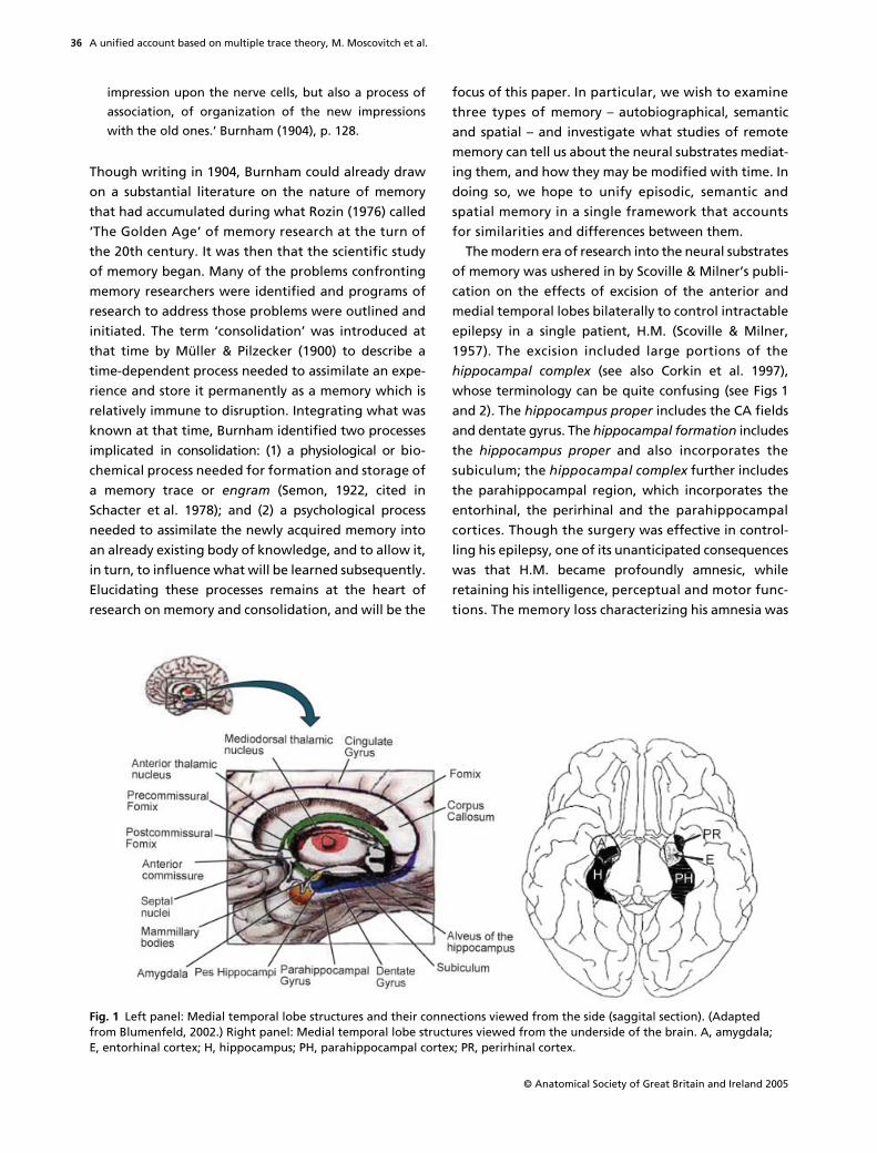
The modern era of research into the neural substratesof memory was ushered in by Scoville & Milner’s publi-cation on the effects of excision of the anterior andmedial temporal lobes bilaterally to control intractableepilepsy in a single patient, H.M. (Scoville & Milner,1957). The excision included large portions of the
hippocampal complex
(see also Corkin et al. 1997),whose terminology can be quite confusing (see Figs 1and 2). The
hippocampus proper
includes the CA fieldsand dentate gyrus. The
hippocampal formation
includesthe
hippocampus proper
and also incorporates thesubiculum; the
hippocampal complex
further includesthe parahippocampal region, which incorporates theentorhinal, the perirhinal and the parahippocampalcortices. Though the surgery was effective in control-ling his epilepsy, one of its unanticipated consequenceswas that H.M. became profoundly amnesic, whileretaining his intelligence, perceptual and motor func-tions. The memory loss characterizing his amnesia was
Fig. 1 Left panel: Medial temporal lobe structures and their connections viewed from the side (saggital section). (Adapted from Blumenfeld, 2002.) Right panel: Medial temporal lobe structures viewed from the underside of the brain. A, amygdala; E, entorhinal cortex; H, hippocampus; PH, parahippocampal cortex; PR, perirhinal cortex.
A unified account based on multiple trace theory, M. Moscovitch et al.
© Anatomical Society of Great Britain and Ireland 2005
37
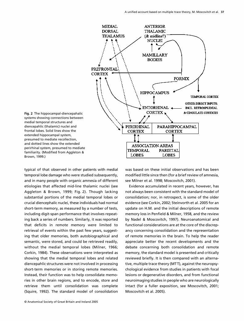
typical of that observed in other patients with medialtemporal lobe damage who were studied subsequently,and in many people with organic amnesia of differentetiologies that affected mid-line thalamic nuclei (seeAggleton & Brown, 1999; Fig. 2). Though lackingsubstantial portions of the medial temporal lobes orcrucial diencephalic nuclei, these individuals had normalshort-term memory, as measured by a number of tests,including digit-span performance that involves repeat-ing back a series of numbers. Similarly, it was reportedthat deficits in remote memory were limited toretrieval of events within the past few years, suggest-ing that older memories, both autobiographical andsemantic, were stored, and could be retrieved readily,without the medial temporal lobes (Milner, 1966;Corkin, 1984). These observations were interpreted asshowing that the medial temporal lobes and relateddiencepahlic structures were not involved in processingshort-term memories or in storing remote memories.Instead, their function was to help consolidate memo-ries in other brain regions, and to encode, store andretrieve them until consolidation was complete(Squire, 1992). The standard model of consolidation
was based on these initial observations and has beenmodified little since then (for a brief review of amnesia,see Milner et al. 1998; Moscovitch, 2001).
Evidence accumulated in recent years, however, hasnot always been consistent with the standard model ofconsolidation; nor, in retrospect, is some of the olderevidence (see Corkin, 2002; Steinvorth et al. 2005 for anupdate on H.M. and the initial descriptions of remotememory loss in Penfield & Milner, 1958, and the reviewby Nadel & Moscovitch, 1997). Neuroanatomical andfunctional considerations are at the core of the discrep-ancy concerning consolidation and the representationof remote memories in the brain. To help the readerappreciate better the recent developments and thedebate concerning both consolidation and remotememory, the standard model is presented and criticallyreviewed briefly. It is then compared with an alterna-tive, multiple trace theory (MTT), against the neuropsy-chological evidence from studies in patients with focallesions or degenerative disorders, and from functionalneuroimaging studies in people who are neurologicallyintact (for a fuller exposition, see Moscovitch, 2001;Moscovitch et al. 2005).
Fig. 2 The hippocampal-diencepahalic systems showing connections between medial temporal structures and diencepahlic (thalamic) nuclei and frontal lobes. Solid lines show the extended hippocampal system, presumed to mediate recollection, and dotted lines show the extended perirhinal system, presumed to mediate familiarity. (Modified from Aggleton & Brown, 1999.)

A unified account based on multiple trace theory, M. Moscovitch et al.
© Anatomical Society of Great Britain and Ireland 2005
38
The standard model of consolidation
According to the standard model (Squire & Alvarez, 1995;Dudai, 2004; McGaugh, 2000), memory consolidationbegins when information, registered initially in theneocortex, is integrated by the hippocampal complex/medial temporal lobes (HC/MTL) and related structuresin the diencephalon to form a memory trace that con-sists of an ensemble of bound hippocampal complex–neocortical neurons (Moscovitch, 1995, 2000). Thisinitial binding into a memory trace involves short-termprocesses, the first of which may be completed withinseconds and the last of which may be completed withinminutes or, at most, days. We refer to the latter as
rapid or synaptic consolidation
or
cohesion
(Moscovitch,1995; Dudai, 2004). The existence of rapid consolida-tion is not in dispute. Indeed, much has been learnedabout its cellular and neurochemical (molecular) basis,which seem to be similar across species and acrossdifferent memory systems in the same species. Excellent,brief reviews of this topic can be found in Kandel (2001)and McGaugh (2000, 2004) and will not be consideredfurther in this paper.
A process of
prolonged
or
system consolidation
(Burnham, 1904; Dudai, 2004; Frankland & Bontempi,2005) is then believed to occur, which may last formonths and even decades. According to the standardmodel, during this process, the HC/MTL and relatedstructures are needed for storage and recovery of thememory trace, but their contribution diminishes asconsolidation proceeds, until the neocortex (and possiblyother extra-hippocampal structures) alone is capable ofsustaining the permanent memory trace and mediatingits retrieval. Thus, the HC/MTL and related structures areconsidered by the standard model to be temporarymemory systems, needed to store and retrieve memoriesonly until prolonged consolidation is complete. The timeit takes for consolidation to be complete corresponds tothe temporal extent of retrograde amnesia followinglesions of the HC/MTL and diencephalon, other insults(concussions, closed head injuries or electrical currents),or the administration of pharmacological agents thatdisrupt memory permanently.
In contrast to rapid consolidation, not only are we farfrom understanding the mechanisms mediating pro-longed consolidation, which includes the psychologicalprocesses that Burnham and others (Squire et al. 1984)emphasized but, as we shall see, the very existenceof this process is in doubt, at least for some types of
memory. In the 1960s, the outlines of the central debateconcerning the validity of the standard model alreadywere crystallized clearly in work with amnesic patients(Warrington & Weiskrantz, 1970; Warrington & Sanders,1971). The debate continues to centre on the followingfour questions. (1) What types of memory are implicated?(2) Which neuroanatomical structures in the medialtemporal lobes and diencephalon are involved? (3) Whatis the extent and duration of retrograde amnesia, and,by implication, of consolidation, and how is it affectedby lesion location and memory type? (4) What otherstructures outside the medial temporal lobe anddiencephalon contribute to retention and retrieval ofremote memory? The first two questions presupposethe existence of different memory systems with differentrules of operation and different neuroanatomicalsubstrates. We will deal with the first two questions inturn, and the third and fourth will be addressed in thecourse of discussing the first two.
Memory types and their neuroanatomical substrates
Explicit and implicit memory
One of the major discoveries of memory researchers inthe latter part of the 20th century, arguably a second‘Golden Age’ of memory research, is that memory is notunitary but consists of various types, each influenced bydifferent variables, governed by different principles,possibly concerned with different materials, and eachmediated by different neural structures and mechanismsthat form distinguishable, and dissociable, systems(see Milner et al. 1968; Warrington & Weiskrantz, 1970;Milner, 1974; O’Keefe & Nadel, 1978; Weiskrantz, 1980;Cermak, 1982; Moscovitch, 1992, 2001; Schacter & Tulving,1994; Tulving & Craik, 2000). Two broad classes of memorywere identified:
explicit memory
, which refers to con-scious recollection of experiences and facts (sometimescalled
declarative memory
; Cohen & Squire, 1980; Squire,1992); and
implicit or nondeclarative memory
(Schacter,1987), which is memory without awareness that isrevealed by the effects of prior experience on behav-iour without the individual consciously retrieving thememory or even being aware of having it. Examples ofimplicit memory are: perceiving a picture or a face morequickly after it was seen, though the person may deny thatthe face or word was familiar (perceptual priming); learn-ing a repeated, complex motor sequence, even though

A unified account based on multiple trace theory, M. Moscovitch et al.
© Anatomical Society of Great Britain and Ireland 2005
39
the individual may not be aware of the sequence or thatit was repeated (procedural memory); or learning to formconditioned responses, though the individual may not beaware of the stimuli controlling the response (condition-ing; see Moscovitch et al. 1993; Roediger & McDermott1993).
Whereas the HC/MTL is crucial for explicit memory,it is not needed for implicit memory. Many, if not all,types of implicit memory can be acquired, retained andretrieved normally even by people who are profoundlyamnesic as a result of HC/MTL or diencephalic damage.It is believed that implicit memory is mediated by theneural structures involved in acquiring information, suchas the posterior neocortex for perception of objects, facesand words such and the basal ganglia for execution ofmotor sequences (for reviews, see Tulving & Schacter,1990; Moscovitch et al. 1993; Schacter & Buckner, 1998;Wiggs & Martin, 1998; Schacter & Badgaiyan, 2001;Schacter et al. 2004). Because we know little aboutprolonged consolidation effects in implicit memory, ourdiscussion will be restricted to explicit memory.
Explicit memory: functional distinctions between episodic memory, familiarity and semantic memory
Explicit memory is itself divisible into two types: episodicand semantic (Tulving, 1972).
Episodic memory
refersto memory for particular autobiographical episodesor specific events in the life of the individual, whichincludes information about both the content of theexperience and the spatial and temporal context in whichit occurred. Having such a memory entails a detailed re-experiencing of the initial event, effectively allowingone to travel mentally back in time (Tulving, 1985). Instudies of anterograde memory, episodic memory isassessed by tests of
recollection
, which refers to repre-sentation of past experiences and includes not only thecontent of those experiences but also their spatial-temporal context, all of which, Tulving (1985) proposed,depends on autonoetic consciousness (consciousnesswith the self in it). Building on Tulving’s distinction,Moscovitch (1995, 2000) emphasized that episodic memoryalso includes the
conscious experience
accompanyingthe episode. Put succinctly, episodic memory refers tomemory of the
experience
of the event, of whichconscious awareness is a part.
Semantic memory
refers to the noncontextualcontent of experience, or knowledge about the worldacquired during experience, which contributes to the
formation and long-term representation of concepts,categories, facts, word meanings, and so on. It evenincludes knowledge about ourselves (where we wereborn, where we lived, who our friends were, whatschools we attended, what jobs we held), what somehave called
personal semantics
(Cermak & O’Connor,1983; Kopelman et al. 1989) to distinguish this memoryfrom that for autobiographical episodes (for a discussionand examples of different tests used to assess differenttypes of memory, see Kopelman et al. 1989; Moscovitchet al. 1999; Fujii et al. 2000; Nadel & Moscovitch, 2001).Unlike episodic, autobiographical memory, semanticmemory is associated only with noetic consciousness,which is consciousness of knowledge without a senseof self and experience accompanying it.
A third type of memory,
familiarity
, shares attributeswith episodic and semantic memory. It refers to memoryof stimuli, rather than of events, which one recognizesas familiar but for which one has no recollection of thecontext in which the stimuli occurred, as happens whenone encounters a person who is familiar without beingable to place the person. Like semantic memory, famili-arity is associated with noetic consciousness. FollowingTulving’s proposal, which itself builds on earlier workby Atkinson & Juola (1973) and Mandler (1980) on testsof anterograde memory, familiarity is assessed byrecognition devoid of recollection. Since Tulving’sinitial proposal, a number of procedures have beenused to distinguish recollection from familiarity in testsof anterograde memory, each having its own virtuesand liabilities, but all share the assumption that differ-ent processes and, possibly representations, distinguishthe two types of memory (for review and critique, seeYonelinas, 2002 and Rotello et al. 2004).
Neuroanatomical substrates associated with different types of explicit memory
Developments concerning the functional properties oftypes of anterograde memory were accompanied byadvances in our appreciation of the neural substratesmediating them. After reviewing the effects of lesionsto different parts of the hippocampal complex, and thethalamic structures with which they are associated,Aggleton & Brown (1999), building on earlier proposalsby Eichenbaum et al. (1994), proposed a division intotwo systems. One system, consisting of the hippocam-pus and its connections to the mammillary bodies andanterior thalamic nuclei (the extended hippocampal
A unified account based on multiple trace theory, M. Moscovitch et al.
© Anatomical Society of Great Britain and Ireland 2005
40
system), is presumed to mediate recollection relying onrelational information, including the temporal–spatialcontext of the memory (see Figs 1 and 2). Damage to thissystem causes deficits in spatial memory and in memoryfor relational information that typifies memory for auto-biographical episodes, but spares recognition based onlyon familiarity (Aggleton et al. 2000; Holdstock et al. 2002a;Mayes et al. 2003, 2004; D. Moscovitch & McAndrews, 2002;Yonelinas, 2002; Yonelinas et al. 2002).
The other system, consisting of the perirhinal cortexand its connections to the dorsomedial nucleus of thethalamus (the extended perirhinal system), is necessaryfor item recognition based on familiarity which does notrequire access to spatial-temporal context (see Fig. 2).Damage to this system will impair recognition even ofsingle items (Aggleton et al. 2000). The parahippocampalcortex seems to be necessary for forming memories ofplaces (Epstein & Kanwisher, 1998; Epstein et al. 1999,2003) or of associating objects with particular locations(Owen et al. 1996a,b), and may provide the allocentric,spatial framework for recollection (O’Keefe & Nadel,1978; Nadel & Moscovitch, 1997; Burgess et al. 2001, 2002;Rosenbaum et al. 2004a).
Semantic memory, on the other hand, does not dependon medial temporal and diencephalic structures, thoughit may benefit from them at acquisition; rather, semanticmemory is mediated by a network of posterior and anteriorneocortical structures, depending on the particularattributes of the memory. Typically, the structuresmediating semantic memory include the lateral andanterior temporal cortex, and ventro-lateral prefrontalcortex, usually on the left (for reviews, see Grahamet al. 1999; Tranel et al. 1997; Martin & Chao, 2001;Thompson-Schill, 2003).
Functional distinctions and neuroanatomical substrates of remote memory
Of all of these types of memory, only the investigationof semantic memory seems to be concerned with infor-mation acquired outside the laboratory. Semantic memoryrefers to one’s general knowledge, which is presumedto be entrenched before laboratory investigation of itbegins. There is general agreement that semantic memorycan survive HC/MTL damage (see Moscovitch et al. 2005),though it initially may benefit from the contribution ofthose structures. What is disputed is whether episodicmemory, either autobiographical or only familiarity-based,is dependent permanently or only temporarily (until
consolidation is complete) on the HC/MTL. Put anotherway, do the structures implicated in recollection andfamiliarity for anterograde memory retain theirfunction for remote memory, or do they relinquishtheir support once consolidation is complete, with the
very same
memories becoming represented only inneocortex?
Spatial memory: distinguishing between detailed (episodic) and schematic (semantic) spatial memory
Spatial memory sometimes is considered in the contextof episodic memory (O’Keefe & Nadel, 1978; Burgesset al. 2002), but rarely in the context of semantic mem-ory, because it seems to be orthogonal to this category.For the purposes of this paper, however, we will dividespatial memory into categories analogous to those inexplicit memory, to see what can be gained from sucha classification. We propose that it is useful to distinguishbetween those spatial memories that consist of
detailedperceptual–spatial representations
of experiencedenvironments (corresponding to episodic, autobiographicalmemory) and those that consist of
schematic represen-tations of the topography
(corresponding to semanticmemory). The detailed representations consist not onlyof allocentric and egocentric information about routesand maps of the environment and of the location of majorlandmarks, that is the topography of the environment,but also of the appearance of the elements of which theenvironment is comprised, such as houses, parks, lawns,fields, hills, and so on. Such detailed representationseffectively consist of vividly remembered routes andscenes which would allow one to re-experience theenvironment as one takes a mental walk through it, aspatial analogue of mental time travel, and to recog-nize its various attributes on formal tests. By contrast,schematic, topographical representations or cognitivemaps are sufficient to allow one to navigate in theenvironment and recognize only those features whichprovide salient cues for navigation, without supportinga rich representation and re-experiencing of the envi-ronment, or even recognition of elements not crucialfor navigation (see Rosenbaum et al. 2000, 2001 for earlierversions of this view, and Rosenbaum et al. 2004a, 2005).For ease of exposition, we refer to the perceptually orexperientially detailed representations as
detailed orvivid spatial memories
, which captures the experientialcomponent in addition to details, and to the schematicrepresentations as
schematic spatial memories
.

A unified account based on multiple trace theory, M. Moscovitch et al.
© Anatomical Society of Great Britain and Ireland 2005
41
There is general agreement that the HC/MTL isneeded for acquisition of allocentric spatial memory,whether detailed or schematic (Burgess et al. 2002),but there has been little research on whether the HC/MTL is needed for retention and retrieval of remotespatial memories of either type. The evidence wereview suggests, as with autobiographical memory,that schematic (semantic) spatial memories can survivedamage to HC/MTL, but that perceptually detailed(episodic) spatial memories cannot.
The MTL, neocortex and predictions based on the standard model: retrograde amnesia is equivalent across all memory types
According to the standard model of consolidation, nodistinction is drawn with respect to consolidationamong different types of explicit memory, be theyspatial or nonspatial, episodic or semantic, recollectiveor familiar (Squire & Zola, 1998; Squire et al. 2004) – allare dependent on the HC/MTL for the duration of theconsolidation period, after which they can be retainedand retrieved independently of it. Thus, damage to theHC/MTL and diencephalon leads to a graded, tempo-rally limited retrograde amnesia for both episodic andsemantic memory, whether autobiographical or spa-tial. Memories acquired most recently are most severelyaffected, with more remote memories being retainednormally, having been fully consolidated beforethe insult (see Squire, 1992; Squire & Alvarez, 1995). Thedata, however, do not consistently support the model.
Problems with the standard model: retrograde amnesia for episodic (including spatial) memory is prolonged and for semantic (including spatial) memory is relatively preserved
The standard model of consolidation had been challengedby Warrington and colleagues, who showed that retro-grade amnesia can be severe and of long duration fol-lowing HC/MTL lesions (Warrington & Sanders, 1971;Warrington & McCarthy, 1988; Warrington, 1996).Kinsbourne & Wood (1975) suggested that retrogradeamnesia is a deficit only of episodic (autobiographical)memory, which affects recent and remote memoryequally. Although few endorsed their ideas at the time (seeMoscovitch, 1982; Nadel & Moscovitch, 1997; Fujii et al.2000; Nadel et al. 2000, 2003), recent reviews revitalizedthem, and argued that the evidence favoured them.
Nadel & Moscovitch identified a number of problemswith the standard model, both with respect to thetypes of memory that are affected, and with the dura-tion and extent of retrograde amnesia. They notedthat retrograde amnesia varied with memory type,decreasing in severity and extent from the autobio-graphical to the semantic. Retrograde amnesia for epi-sodic memory, the details of autobiographical events,after large medial temporal (or diencepahlic) lesionscan extend for decades, or even a lifetime, far longerthan it would be biologically plausible for a consolida-tion process to last. Retrograde amnesia for semanticmemory, however, is less extensive and often is tempo-rally graded as is the case for memory of public eventsand personalities, and even more so for vocabulary andfor facts about the world and oneself (personal seman-tics; Fujii et al. 2000; see also Kapur, 1999 for an exten-sive review of retrograde amnesia). Thus, contrary tothe standard model, nonspatial semantic and episodicmemories are affected differently by lesions producingretrograde amnesia (see Warrington, 1996). As we seebelow (see section on
Spatial Memory
, pp. 54–59), thesame is true of spatial memory.
Multiple trace theory: an alternative to the standard model
To account for this evidence, Nadel & Moscovitch(1997) proposed a multiple trace theory (MTT) ofmemory. According to this theory, the hippocampalcomplex (and possibly the diencephalon) rapidly andobligatorily encodes all information that is attended(consciously apprehended; Moscovitch & Umiltà, 1990;Moscovitch, 1992) and binds the neocortical (andother) neurons that represent that experience intoa memory trace. This information is sparsely encodedin a distributed network of hippocampal complexneurons that act as a pointer, or index, to the neuronsrepresenting the attended information (Teyler &DiScenna, 1986). A memory trace of an episode, there-fore, consists of a bound ensemble of neocortical andHC/MTL (and possibly diencephalic) neurons whichrepresent a memory of the consciously experiencedevent. As noted earlier, formation and consolidationof these traces, or cohesion (Moscovitch, 1995), is rela-tively rapid, lasting on the order of seconds or at mostdays.
In this model, there is no prolonged consolidationprocess, as the standard model asserts, that slowly

A unified account based on multiple trace theory, M. Moscovitch et al.
© Anatomical Society of Great Britain and Ireland 2005
42
strengthens the neocortical component of the memorytrace, so that with time the trace becomes independentof the HC/MTL. Instead, each time an old memory isretrieved, a new hippocampally mediated trace iscreated so that old memories are represented by moreor stronger HC/MTL–neocortical traces than are newones and, therefore, are less susceptible to disruptionfrom brain damage than are more recent memories.Because the memory trace for autobiographical epi-sodes is distributed in the HC, the extent and severity ofretrograde amnesia, and perhaps the slope of the gra-dient, are related to the extent and location of damageto the extended hippocampal system.
Whereas each autobiographical memory trace isunique, the creation of multiple, related traces facilit-ates the extraction of the neocortically mediatedinformation common among them, and which isshared with other episodes. This information is thenintegrated with pre-existing knowledge to formsemantic memories that can exist independently ofthe HC/MTL. Thus, knowledge about the world, aboutpeople and events acquired in the context of a specificepisode is separated from the episode and ultimatelystored independently of it. This process of increased
semanticization
with experience and retrieval overtime may give the impression of prolonged consolida-tion of the original trace (see section on
SemanticMemory
, pp. 51–54). Without a well-functioninghippocampal system, acquisition of semantic memory isslow and effortful, at least in adulthood. It is possible,however, that in childhood, structures mediatingsemantic memory, such as those involved in languagelearning, can form new representations easily withoutbenefit of the hippocampus (Vargha-Khadem et al.1997).
With respect to spatial memory, the issue is whetherthe hippocampus is crucial for retention and retrievalof allocentric, spatial information, no matter how longago it was acquired. According to cognitive maptheory, the hippocampus is crucial, and it provides thespatial context of autobiographical memory. Anotherview, less consistent with the initial formulation ofcognitive map theory, but quite compatible withMTT, is that only detailed, episodic spatial information,directly linked to the re-experiencing of an event, ismediated by the hippocampus. Generic allocentricspatial information necessary for navigation, what wehave termed schematic (semantic) spatial memory, ismediated initially by the hippocampus, but like other
forms of semantic memory, can exist independently ofit once the memory has been assimilated.
Differences between MTT and the standard model: neuroanatomical and functional considerations
Episodic (nonspatial) memory and the role of the HC/MTL and related structures
Defenders of the standard model argue that lesionlocation and size, as much as type and test of memory,need to be taken into account in considering the extentand severity of retrograde amnesia. The initial studieson retrograde amnesia (Scoville & Milner, 1957; Penfield& Milner, 1958; Milner, 1966; Mair et al. 1979) implicatedthe HC/MTL and diencephalon (see Figs 1 and 2). Thefocus of attention, however, shifted quickly to thehippocampal formation, and then to the hippocampusitself (Squire, 1975, 1992; Squire et al. 1984). The cur-rent position is that temporally limited memory appliesonly to the hippocampus and that permanent memo-ries are consolidated either in the adjacent regions of theHC/MTL or in the lateral temporal neocortex (Rempel-Clower et al. 1996; Reed & Squire, 1998; Squire & Zola,1998; Bayley et al. 2003; Manns et al. 2003).
The evidence, reviewed by Nadel, Moscovitch andtheir colleagues (Nadel & Moscovitch, 1997; Fujii et al.2000; Nadel et al. 2003), favoured the older idea thatthe entire HC/MTL region is implicated in retentionand retrieval of remote memory. They noted that theextent and severity of retrograde amnesia dependedon the size of the medial temporal lesion: the largerthe lesion, the greater the loss. As importantly, Nadel& Moscovitch distinguished among different types ofexplicit memory, with episodic, autobiographical mem-ory being the most severely affected, and semanticmemory, the least. Given the multifaceted nature ofepisodic memory in general, and autobiographicalepisodes in particular, Nadel & Moscovitch (1997, 1998)suggested that each of the various regions of themedial temporal lobe may contribute its own informa-tion to the complete, detailed memory of an event,although they left the precise formulation vague. Aswe have learned more about the separate functions ofmedial temporal regions (Aggleton & Brown, 1999), itmay make sense to consider the possibility that each ofthem is involved in retention and retrieval of thoseaspects of an event which they specifically process
A unified account based on multiple trace theory, M. Moscovitch et al.
© Anatomical Society of Great Britain and Ireland 2005
43
(Gilboa, 2004a; Gilboa et al. 2005 submitted). Thus, forremote memory, as for anterograde memory, recollectionof autobiographical episodes, the full re-experiencingof an event, will always depend on the hippocampus.Recognition of objects based on familiarity, andgeneric personal memories, can survive hippocampaldamage, but not damage to perirhinal cortex, whereasrecognition of aspects of places will be impairedfollowing parahippocampal gyrus lesions, and ofemotions, following amygdala lesions. In addition, inrecollecting an autobiographical episode, damage tothese structures should lead to loss of the informationmediated by these extra-hippocampal structures.
Semantic (nonspatial) memory: neocortical–hippocampal interactions
Semantic memory
, on the other hand, depends on neo-cortical structures that are sufficient to form domain-specific and semantic representations based onregularities extracted from repeated experiences withwords, objects, people and environments (Westmacottet al. 2001; Rosenbaum et al. 2004a). This applies evento autobiographical episodes one recollects repeat-edly, thereby creating a gist of each episode whichlacks the details that makes rich re-experiencing possi-ble. The MTL system may aid in the initial formation ofthese neocortical representations (Nadel & Moscovitch,1997), but, once formed, they can exist on their own(Fujii et al. 2000). Because autobiographical memoryconsists of a hierarchical structure which includesgeneric semantic information, as well as event-specificinformation (for more detail, see Conway & Fthenaki,2000; Conway & Pleydell-Pearce, 2000), damage toneocortex may impair the semantic aspects of auto-biographical memory, leaving the perceptual, event-specific information intact, just as is the case foranterograde memory (see Graham et al. 1999, 2000).In these respects, MTT and traditional consolidationtheory are in agreement. Where they diverge is withregard to the autobiographical residue of semanticmemory. Just as autobiographical memories havesemantic components, so purportedly semantic memo-ries may have autobiographical components associatedwith them which can influence performance on seman-tic tests. For example, we may not only know informa-tion about a famous person, such as that John Kennedywas an American President who was assassinated, orthat Princess Diana was a member of the British Royal
family who was killed in a car accident, but we alsohave autobiographical memories associated withthem, such as where we were and how we felt whenwe heard of their deaths (Westmacott & Moscovitch,2003). When making ostensible semantic decisionsabout these people, such as fame judgments, or evenreading their names, the episodic component influencesperformance. According to traditional consolidationtheory, these autobiographical components of semanticmemory, like the semantic components themselves,are mediated by neocortex once consolidation is com-plete. MTT, on the other hand, maintains that theseautobiographical components continue to rely on thehippocampus for retention and retrieval.
Allocentric spatial memory: is the hippocampus always necessary?
MTT and the standard model also make different pre-dictions regarding spatial memory. According to MTT,but not consolidation theory, re-experiencing detailed,event-specific spatial aspects of an autobiographicalepisode, or spatial-perceptual features contained withinan environment that are incidental to navigating in it,requires the hippocampus no matter how long ago thememory was acquired. Consolidation theory predictsthat all forms of allocentric spatial information, crucialfor navigation in familiar environments, can be retainedif the hippocampal lesion occurred after the informationhad been assimilated. As noted above, MTT can addressthis issue in two ways. First, it is possible that semanticallocentric information ultimately can be representedoutside the hippocampus. Alternatively, if cognitivemap theory is correct, all allocentric, spatial informationwill be lost no matter how long ago it was acquired,irrespective of whether or not it enables re-experiencing.
New evidence on neuroanatomical substrates of remote memory: comparison between MTT and the standard consolidation model
Autobiographical memory
Evidence from people with focal lesions and dementia
Since we last reviewed the literature in 1998 (Fujii et al.2000), the evidence that has accumulated continues tofavour MTT over the consolidation model, though itstill is not conclusive (see Moscovitch et al. 2005). Using
A unified account based on multiple trace theory, M. Moscovitch et al.
© Anatomical Society of Great Britain and Ireland 2005
44
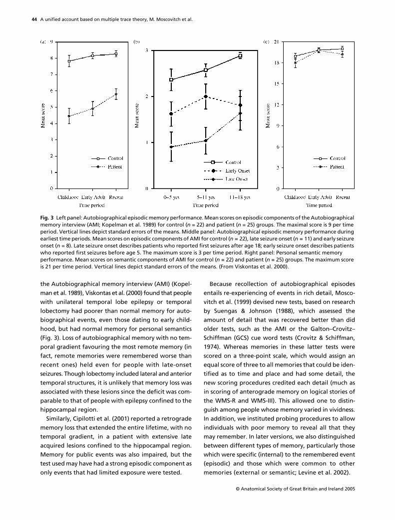
the Autobiographical memory interview (AMI) (Kopel-man et al. 1989), Viskontas et al. (2000) found that peoplewith unilateral temporal lobe epilepsy or temporallobectomy had poorer than normal memory for auto-biographical events, even those dating to early child-hood, but had normal memory for personal semantics(Fig. 3). Loss of autobiographical memory with no tem-poral gradient favouring the most remote memory (infact, remote memories were remembered worse thanrecent ones) held even for people with late-onsetseizures. Though lobectomy included lateral and anteriortemporal structures, it is unlikely that memory loss wasassociated with these lesions since the deficit was com-parable to that of people with epilepsy confined to thehippocampal region.
Similarly, Cipilotti et al. (2001) reported a retrogradememory loss that extended the entire lifetime, with notemporal gradient, in a patient with extensive lateacquired lesions confined to the hippocampal region.Memory for public events was also impaired, but thetest used may have had a strong episodic component asonly events that had limited exposure were tested.
Because recollection of autobiographical episodesentails re-experiencing of events in rich detail, Mosco-vitch et al. (1999) devised new tests, based on researchby Suengas & Johnson (1988), which assessed theamount of detail that was recovered better than didolder tests, such as the AMI or the Galton–Crovitz–Schiffman (GCS) cue word tests (Crovitz & Schiffman,1974). Whereas memories in these latter tests werescored on a three-point scale, which would assign anequal score of three to all memories that could be iden-tified as to time and place and had some detail, thenew scoring procedures credited each detail (much asin scoring of anterograde memory on logical stories ofthe WMS-R and WMS-III). This allowed one to distin-guish among people whose memory varied in vividness.In addition, we instituted probing procedures to allowindividuals with poor memory to reveal all that theymay remember. In later versions, we also distinguishedbetween different types of memory, particularly thosewhich were specific (internal) to the remembered event(episodic) and those which were common to othermemories (external or semantic; Levine et al. 2002).
Fig. 3 Left panel: Autobiographical episodic memory performance. Mean scores on episodic components of the Autobiographical memory interview (AMI; Kopelman et al. 1989) for control (n = 22) and patient (n = 25) groups. The maximal score is 9 per time period. Vertical lines depict standard errors of the means. Middle panel: Autobiographical episodic memory performance during earliest time periods. Mean scores on episodic components of AMI for control (n = 22), late seizure onset (n = 11) and early seizure onset (n = 8). Late seizure onset describes patients who reported first seizures after age 18; early seizure onset describes patients who reported first seizures before age 5. The maximum score is 3 per time period. Right panel: Personal semantic memory performance. Mean scores on semantic components of AMI for control (n = 22) and patient (n = 25) groups. The maximum score is 21 per time period. Vertical lines depict standard errors of the means. (From Viskontas et al. 2000).

A unified account based on multiple trace theory, M. Moscovitch et al.
© Anatomical Society of Great Britain and Ireland 2005
45
Using the new scoring technique, Moscovitch et al.(1999) found that amnesic people whose memory ofremote events fell in the normal range according to thethree-point scoring system, recalled far fewer detailsaccording to the new scoring system. Admittedly,the people they tested did not have lesions confinedto the hippocampal region, so that extra-hippocampallesions may have contributed to their extensive retro-grade amnesia.
Steinvorth et al. (2005) applied the new scoringmethod of Levine et al. (2002) to Scoville & Milner’spatient, H.M., and to W.D., both of whom had lesionsthat were much more circumscribed and largely con-fined to the MTL. They, too, like the patients of Mosco-vitch et al., showed a retrograde amnesia for internal,episodic components of autobiographical events acrosstheir entire life, without a temporal gradient. As a con-trol, they showed that on comparable tests of semanticmemory for public events, their performance was muchbetter, and showed the typical gradient, with the moreremote memories being relatively spared.
Likewise, Gilboa et al. (submitted) applied similartechniques to study memory in A.D.F., an amnesicpatient with small, bilateral lesions to the fornix, anoutput pathway of the hippocampus, and a smalllesion in the left, basal forebrain region, which typic-ally does not lead to extensive, lasting, memory loss.Like H.M., the patient’s remote, autobiographicalmemory loss extended across his entire life withoutevidence of a temporal gradient, but personal semanticand generic memories were spared. This pattern re-sembled anterograde memory loss in patients withbilateral fornix lesions (Aggleton et al. 2000), who showeddeficits in recollection but not familiarity. Importantly,A.D.F. also showed anterograde deficits in recollectionand preserved familiarity, suggesting a common func-tion for the hippocampus across anterograde andretrograde memory.
Mindful that our procedure depends on recall whichmay be biased by probing, and may involve reconstruc-tion of details to which some patients may be moreprone than others (Kapur et al. 2002), Gilboa et al.(submitted) devised recognition tests based on reportsfrom people close to the patients. The tests focusedeither on episodic details or on generic, semantic infor-mation relevant to the event. A.D.F.’s performance onrecognition resembled his performance on recall testsof autobiographical memory: he displayed poor per-formance on recognition of episodic details and
preserved recognition of semantic ones. By contrast, K.C.,another patient with bilateral hippocampal lesions (seeRosenbaum et al. 2005) but with damage to other partsof the MTL and neocortex, performed also poorly onthe semantic component. This corresponds to an exten-sive and ungraded retrograde amnesia for autobio-graphical episodic and semantic details in K.C. on themeasure of Levine et al. (2002) (Rosenbaum et al.2004b). Rosenbaum et al. also investigated whetherthe recovery of autobiographical details continues torely on the hippocampus, or whether these memoriesdepend on visual imagery or strategic retrieval ofdetails, mediated by visual extrastriate (medial occipital)or prefrontal cortex, which also are damaged in K.C.However, performance was normal on visual imagerytesting, and autobiographical memory did not benefitfrom a retrieval support manipulation, contrary towhat would be expected if respective medial occipitalor frontal lesions were responsible.
Together these studies present a very strong case infavour of MTT over the standard consolidation model.The only recent evidence favouring the standard modelis supplied by Bayley et al. (2003), who report thatamnesic patients with lesions confined to the MTL haveintact, detailed autobiographical memories from thefirst third of their lives. They used the GCS cuing tech-nique to elicit the memories, and probing was minimal,but they scored the memories using the procedure ofLevine et al. (2002). The number of details they reportfor their controls (who do not differ from their amnesicpeople), however, is far lower than that reported byour controls: approximately 20 internal details to our50. In fact, their controls score even lower than ouramnesic people. Their findings suggest that the mem-ories they elicited may not have been truly vivid evenin their controls, a fact that is not surprising consider-ing that they were asked to recall 36 memories fromthe first third of their lives to single word cues.
One criticism invoked by proponents of the standardmodel is that evidence from many single case studies issuspect because lesions often extend beyond the MTLto include lateral neocortex. Although we reviewedcases with extensive retrograde amnesia with lesionsconfined to MTL (see Fujii et al. 2000; Moscovitch et al.2005; this paper), another approach is to conductgroup studies to see if the extent of autobiographicaland semantic memory loss correlates with the age ofthe memory, as the standard model predicts, or withthe size of MTL lesion, as MTT predicts. The evidence

A unified account based on multiple trace theory, M. Moscovitch et al.
© Anatomical Society of Great Britain and Ireland 2005
46
suggests that the location of extra-MTL cortical damageand subcortical damage also needs to be taken intoaccount.
The relationship between the size of MTL lesions, lossof autobiographical memory and personal semantics,and age of memory was examined by Gilboa et al.(2005) in a group of people with mild to moderateAlzheimer’s disease (AD) and by Kopelman et al. (2003)in people with focal lesions of various etiologies. UsingMRI volumetry of 28 structures comprising the entirebrain, and the multivariate analysis method of partialleast squares (PLS), Gilboa et al. found a strong correla-tion between retrograde autobiographical memory, asmeasured by AMI, and the amount of remaining tissuein bilateral MTL, more on the right than the left, and inanterior lateral temporal cortex. The age of the mem-ory had no effect on the correlation, and neither didpersonal semantics whose loss was correlated withanother set of structures, such as anterior temporal,lateral temporal and prefrontal cortex but not MTL.Kopelman et al. also found extensive retrograde amne-sia uncorrelated with age of memory, but also no cor-relation with MTL lesions in a group of patients withfocal MTL lesions, though they did report a significantcorrelation with MTL lesion size in a group that alsohad diencephalic lesions.
Although neither study supports the standardmodel’s prediction that age of memory is a determin-ing factor of memory loss, it is not clear why the corre-lation with size of MTL lesion was not consistentlyfound. To account for the differences between the twostudies, Gilboa et al. (2005) suggested that combiningdifferent etiologies of focal lesion patients in theKopelman et al. (2003) study may have obscured anassociation between MTL intactness as reflected byvolume and memory functioning. For example, anoxiaprimarily affects the CA fields, and damage may or maynot encroach on the subiculum. This would lead torelatively small volume loss, but the potential for largememory deficits. Indeed, this is precisely the wayKopelman et al. (2003) interpret the impressive 0.8correlation between hippocampal volume and eventmemory in their diencephalic group. On the other hand,encephalitis causes extensive losses to MTL corticalregions and thus may lead to overall larger volume loss.However, depending on the extent and precise struc-tures affected, memory loss may vary considerably.
Investigating remote memory in people with seman-tic dementia (SD) is also informative because neural
degeneration associated with SD affects primarily theanterior and lateral temporal cortex, typically on theleft, leaving the MTL relatively spared (Mummery et al.2000). If remote autobiographical memories are repre-sented in neocortex, as the standard model predicts,then patients with SD should show impaired memoryfor remote events, but preserved memories for recentones, a pattern opposite to that which the standardmodel predicts for amnesia. Using variations of theAMI and GCS procedures, this is exactly what Graham& Hodges (1997) reported. Westmacott et al. (2001),however, argued that this pattern is observed onlybecause patients with SD do not have the verbal meansnecessary to comprehend the instructions, use theinformation as cues to retrieval, or express themselvesadequately. Given nonverbal cues, such as familyphotos of particular events, and the opportunity tocommunicate by gestures, intonation, and so on,Westmacott et al.
!
s SD patient showed that remoteautobiographical memory was relatively preserved, afinding corroborated by Moss et al. (2003), Ivanoiuet al. (2004) and Piolino et al. (2003). One of thepatients of Graham et al., however, continued to beimpaired in retrieving all memories except those fromthe last two years, even when tested using the methodsof Westmacott et al. (Graham et al. 2003a).
The source of the discrepancy among the studiesremains unknown. A likely possibility is that the extentand locus of degeneration differs among patients, butwhether the differences lie in MTL, anterior and lateraltemporal lobes, or even pre-frontal cortex (PFC), hasyet to be determined (Nestor et al. 2002). Correlatingsize of brain structure with memory in SD, as Gilboaet al. (2005) did in AD patients, may help resolve thedebate.
Though the evidence generally supports MTT, anumber of issues remain unresolved and await futurestudies that pay equal attention to lesion size and loca-tion, and to the methods used to elicit memories andscore them. Because patients with circumscribedlesions restricted only to some parts of the HC/MTL arerare, and often there is disagreement about the purityand extent of their lesions, and because group studiesare not immune to these problems, many investigatorshave turned to functional neuroimaging studies ofhealthy people to address some of these issues. Thoughnot without problems of its own (see Maguire, 2001a),functional neuroimaging studies can provide valuableinformation about the functional neuroanatomy of
A unified account based on multiple trace theory, M. Moscovitch et al.
© Anatomical Society of Great Britain and Ireland 2005
47
remote memory, and appears to converge on conclu-sions obtained from patient studies.
Evidence from functional neuroimaging
The standard model and MTT make different predic-tions about hippocampal activation during retrieval ofrecent and remote autobiographical memories of spe-cific events. According to the standard model, hippoc-ampal activation should be evident for recent but notfor remote memories, whereas the reverse shouldoccur for neocortical activation. By contrast, MTT pre-dicts equivalent HPC activation for recent and remotememories as long as they are vivid and detailed. Usingevent-related functional magnetic resonance imaging(efMRI) designs, Ryan et al. (2001) presented cuesduring scanning which were derived from prescaninterviews about autobiographical events. They foundbilateral hippocampal activation associated with re-experiencing those events in the scanner. Similarly,Maguire and her collaborators (Maguire, 2001a;Maguire et al. 2001; Maguire & Frith, 2003), foundmostly left-sided activation while participants maderecognition judgments to statements referring to auto-biographical events in comparison to control condi-tions which included statements about general personalevents or public events, all based on prescan inter-views. In one study, Maguire et al. (2001a) directlytested whether hippocampal activation was modu-lated by the age of the memories, and found noevidence for this claim of the standard consolidationtheory. In all the other studies, hippocampal activationwas equivalent for recent and remote memories,thereby favouring MTT over the standard model.Piolino et al. (2004) reported similar findings and con-clusions using positron emission tomography (PET; cf.Conway et al. 1999 for similar PET results).
There were two possible confounds in these studies.One concerned item selection. Because all the eventsstudied were based on prescan interviews with theparticipants, it is difficult to know whether truly remotememories were retrieved in the scanner, or only memo-ries of the interviews. If the latter were the case, similarhippocampal activation would have been observed tostatements regarding personal semantics and publicevents, which was not the case (Maguire, 2001a). Tocontrol for this possible confound further, Ryan et al.(2001) also used items selected by a close relativeor friend, whereas Maguire et al. (2001) tested the
developmental hippocampal amnesic, Jon, for the fewevents he could recollect from his remote past, eventhough he had no memory for the prescan interview. Inboth cases, greater hippocampal activation – that didnot vary with time – was found for autobiographicalthan for other events.
The second possible confound is that hippocampalactivation that is observed reflects
re-encoding
ofmemories as they are retrieved in the scanner, ratherthan activation associated with the initial retrievalitself. Sensitive to this criticism, Gilboa et al. (2004) hada person close to the participant select family photoswhich the participant had not viewed recently andwhich were shown only in the scanner. The photoswere from four or five time periods dating from earlychildhood (at least 20 years ago) to the last sixmonths. To control for the effect of re-encoding, theparticipant also was presented with photos from astranger’s family album which were matched as muchas possible in style and content to the participant’sown photos.
On viewing ‘self’ photos in the scanner, the partici-pant had to re-experience the depicted event in asmuch detail as possible; in viewing the ‘other’ photo,the participant had to imagine in equivalent detail ascenario concerning the event depicted in the unfamiliarphoto. If re-encoding were a factor, no difference inhippocampal activation should be observed betweenthe ‘self’ and ‘other’ conditions, since both types ofmaterial are being encoded, but only one requiresretrieval of old memories. Gilboa et al. (2004) foundthat activation was greater for old, ‘self’ memories thanfor novel, imagined ‘other’ material in a number ofregions, including the left hippocampal complex (seealso Maguire et al. 2001a; Addis et al. 2004a), therebyarguing against the re-encoding interpretation.
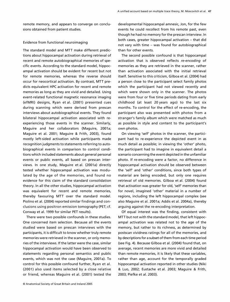
Of equal interest was the finding, consistent withMTT but not with the standard model, that left hippoc-ampal activation was related not to the age of thememory, but rather to its richness, as determined bypostscan vividness ratings for all of the memories, andby descriptions for a subset of them from each time period(see Fig. 4). Because Gilboa et al. (2004) found that, onaverage, recent memories are more vivid and detailedthan remote memories, it is likely that these variables,rather than age, account for the temporally gradedhippocampal activation reported in other studies (Niki& Luo, 2002; Eustache et al. 2003; Maguire & Frith,2003; Piefke et al. 2003).
A unified account based on multiple trace theory, M. Moscovitch et al.
© Anatomical Society of Great Britain and Ireland 2005
48
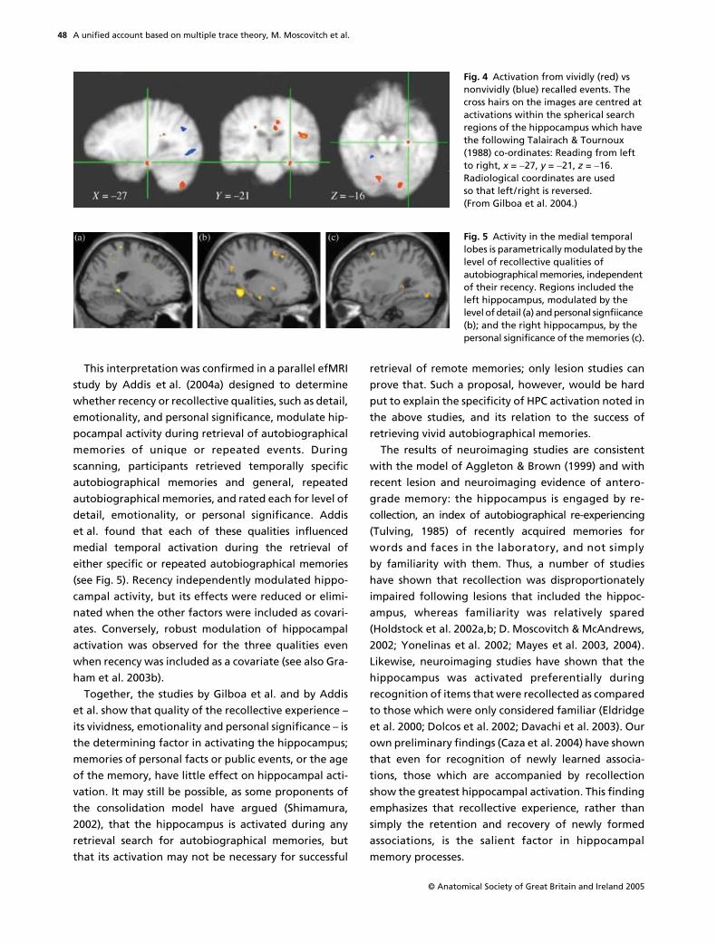
This interpretation was confirmed in a parallel efMRIstudy by Addis et al. (2004a) designed to determinewhether recency or recollective qualities, such as detail,emotionality, and personal significance, modulate hip-pocampal activity during retrieval of autobiographicalmemories of unique or repeated events. Duringscanning, participants retrieved temporally specificautobiographical memories and general, repeatedautobiographical memories, and rated each for level ofdetail, emotionality, or personal significance. Addiset al. found that each of these qualities influencedmedial temporal activation during the retrieval ofeither specific or repeated autobiographical memories(see Fig. 5). Recency independently modulated hippo-campal activity, but its effects were reduced or elimi-nated when the other factors were included as covari-ates. Conversely, robust modulation of hippocampalactivation was observed for the three qualities evenwhen recency was included as a covariate (see also Gra-ham et al. 2003b).
Together, the studies by Gilboa et al. and by Addiset al. show that quality of the recollective experience –its vividness, emotionality and personal significance – isthe determining factor in activating the hippocampus;memories of personal facts or public events, or the ageof the memory, have little effect on hippocampal acti-vation. It may still be possible, as some proponents ofthe consolidation model have argued (Shimamura,2002), that the hippocampus is activated during anyretrieval search for autobiographical memories, butthat its activation may not be necessary for successful
retrieval of remote memories; only lesion studies canprove that. Such a proposal, however, would be hardput to explain the specificity of HPC activation noted inthe above studies, and its relation to the success ofretrieving vivid autobiographical memories.
The results of neuroimaging studies are consistentwith the model of Aggleton & Brown (1999) and withrecent lesion and neuroimaging evidence of antero-grade memory: the hippocampus is engaged by re-collection, an index of autobiographical re-experiencing(Tulving, 1985) of recently acquired memories forwords and faces in the laboratory, and not simplyby familiarity with them. Thus, a number of studieshave shown that recollection was disproportionatelyimpaired following lesions that included the hippoc-ampus, whereas familiarity was relatively spared(Holdstock et al. 2002a,b; D. Moscovitch & McAndrews,2002; Yonelinas et al. 2002; Mayes et al. 2003, 2004).Likewise, neuroimaging studies have shown that thehippocampus was activated preferentially duringrecognition of items that were recollected as comparedto those which were only considered familiar (Eldridgeet al. 2000; Dolcos et al. 2002; Davachi et al. 2003). Ourown preliminary findings (Caza et al. 2004) have shownthat even for recognition of newly learned associa-tions, those which are accompanied by recollectionshow the greatest hippocampal activation. This findingemphasizes that recollective experience, rather thansimply the retention and recovery of newly formedassociations, is the salient factor in hippocampalmemory processes.
Fig. 4 Activation from vividly (red) vs nonvividly (blue) recalled events. The cross hairs on the images are centred at activations within the spherical search regions of the hippocampus which have the following Talairach & Tournoux (1988) co-ordinates: Reading from left to right, x = "27, y = "21, z = "16. Radiological coordinates are used so that left/right is reversed. (From Gilboa et al. 2004.)
Fig. 5 Activity in the medial temporal lobes is parametrically modulated by the level of recollective qualities of autobiographical memories, independent of their recency. Regions included the left hippocampus, modulated by the level of detail (a) and personal signfiicance (b); and the right hippocampus, by the personal significance of the memories (c).

A unified account based on multiple trace theory, M. Moscovitch et al.
© Anatomical Society of Great Britain and Ireland 2005
49
Gilboa et al. (2004) also found that foci of activationin the hippocampus were distributed differently forrecent and remote memories, with the former clus-tered in the anterior region of the hippocampus andthe latter distributed along its rostro–caudal axis (seeFig. 6). It is not yet clear why this pattern should occur.If each retrieval leads to the formation of new traceswithin the MTL, as MTT predicts, then remote memo-ries should be more widely distributed than recentmemories in MTL, and may survive minimal damage tothe MTL.
Other possible interpretations of this pattern of acti-vation are that remote memories, particularly thosedating to childhood and adolescence, may be encodeddifferently from more recent, adult memories, or thatrecent memories may retain their emotional strengthmore than remote ones. With respect to the latter pos-sibility, Dolcos et al. (2002) reported that emotionalmemories activated the anterior hippocampus morethan nonemotional ones, although in their study emo-tionality interacted with recollection, and all theirmemories were recent by our standards.
Extra-hippocampal contributions to autobiographical memory: lesions and neuroimaging
Although the focus has been on the hippocampus andrelated MTL structures, other brain regions are impli-cated in retrieval of autobiographical memory, some ofwhich display a pattern of activation sensitive to the
age of the memory. As we noted earlier, there is amemory retrieval network reported by Maguire andothers (Conway et al. 1999; Maguire & Mummery,1999; Maguire, 2001a; Maguire et al. 2001; Ryan et al.2001; Piefke et al. 2003; Addis et al. 2004a,b) that con-sists primarily of left-lateralized structures when mem-ory for all types of remote events (autobiographicalevents and facts, public events, general knowledge) arecompared with nonmemory control tasks (see Maguire,2001a). In addition to the left hippocampus, theregions included in the network are the left medialfrontal cortex, left temporal pole, left antero-lateralmiddle temporal gyrus, left parahippocampal cortex,retrosplenial/posterior cingulate cortex, precuneus(medial occipital), left temporoparietal junction, righttemporal pole, right posterior cerebellum and thethalamus. Because the specific contribution of theseregions to each type of memory was not examinedsystematically (but see Addis et al. 2004a,b), we can onlyspeculate about their function based on informationgained from other studies. Only some of the regionswill be considered.
Prefrontal cortex
When Maguire (Maguire, Henson et al. 2001a) com-pared autobiographical events to any of the othertypes of memory, only the left hippocampus andmedial frontal cortex were activated more during auto-biographical memory retrieval, a finding consistentwith the observation of Gilboa (2004b) that the latter
Fig. 6 Schematic renderings of remote and recent activations. Each point corresponds to a statistically significant activation from within the left hippocampus in either remote (top; n = 18) or recent (bottom; n = 16) conditions. Red and black squares represent activations at a significance level of P < 0.001 and P < 0.01 uncorrected, respectively. Activations are shown on a single sagittal plane taken from the Talairach & Tournoux (1988) atlas (25 mm lateral to the midline). Overlapping activations were offset slightly in the recent condition. Differences in the lateral displacement of the activations from the midline (along the x-axis of the Talairach atlas) are not represented in the figure. The lateral and vertical dimensions did not show any obvious systematic variability and therefore are not considered as a part of the overall pattern of interest. (From Gilboa et al. 2004.)
A unified account based on multiple trace theory, M. Moscovitch et al.
© Anatomical Society of Great Britain and Ireland 2005
50
region is preferentially activated during retrieval ofautobiographical memory in comparison to other typesof episodic memory. We know that damage to thisregion is associated with temporally extensive retro-grade amnesia and confabulation (Gilboa & Mosco-vitch, 2002), suggesting that it may be a crucial areafor automatically monitoring the retrieved memories(Moscovitch & Winocur, 2002). In addition, this region ofprefrontal cortex also is activated during self-reference,which is a component of autobiographical memory(Craik et al. 1999).
In another event-related fMRI study, Maguire, Hensonet al. (2001) found a region in right ventrolateral pre-frontal cortex that was modulated by the age of theautobiographical memory, showing increased activationto the more recent memories. Activation of this regionhas been associated with specification of retrieval cues inmany neuroimaging studies of episodic memory (Fletcher& Henson, 2001). Maguire et al. (2001) speculate thatactivation in ventrolateral cortex reflects the degree ofintegration of the memory trace with the contextualinformation that can provide retrieval cues. The morerecent the memory, the more likely it is to be contextuallyrich and the more active the integration. Whatever thetrue underlying cause of the activation, the age-relatedpattern is opposite the one predicted by the standardmodel for neocortex (but see Maguire & Frith, 2003).
The pattern of activation observed in prefrontalcortex is consistent with its role as a
working-with-memory
structure that is involved in strategic aspectsof retrieval such as establishing a retrieval mode andgoals, initiating and guiding search, and monitoringand verifying the memories that have been retrieved(Moscovitch, 1992; Moscovitch & Winocur, 2002).Gilboa’s review of the literature (Gilboa, 2004b),however, suggests that some subregions in prefrontalcortex, particularly parts of the frontal pole andventromedial aspects, may be activated preferentiallyduring retrieval of autobiographical as compared toother types of memory. Lesions to the lateral andpolar aspects of prefrontal cortex are associated withdeficits in autobiographical memory retrieval, but aswould be expected of these general-purpose, execu-tive structures, they operate equally on recent andremote memories (Kopelman et al. 1999, 2003).Greater activation may be evident for those memoriesthat are difficult to retrieve and, consequently, greaterloss of such memories may occur following damage tothose structures.
Medial occipital and inferotemporal cortex
In Gilboa et al. (2004), retrieval of context-rich memo-ries was associated with activity in lingual, fusiform andprecuneus gyri independently of their age (see alsoRyan et al. 2001). The precuneus often is implicated inimagery and spatial processing in the context of episodicmemory. Bilateral activation in the lingual and fusiformgyri (BA 19/37) may be related to the complex visual stimuli(photos) and the extended period for re-experiencingused in their study, which likely induced more sensoryperceptual memory-related activation than the verbalmaterials used in previous studies. Similar results arefound, however, even in studies not using photos asmemory cues. Addis et al. (2004b), who used memorytitles as cues, found that specific autobiographicalmemories, rated as more detailed than repeated auto-biographical events, were differentially associatedwith activity in the left precuneus, left superior parietallobule and right cuneus (but see Graham et al. 2003b).
Neuroimaging studies showing activation of poste-rior neocortex and inferotemporal regions duringretrieval of autobiographical memory are consistentwith reports by Ogden (1993) and others that damageto these structures, which is associated with visual,object agnosia and loss of imagery, can also produce aprofound, temporally extensive retrograde amnesiafor autobiographical events (see reviews by Rubin &Greenberg, 1998 and Greenberg & Rubin, 2003).Because re-experiencing autobiographical episodes isaccompanied more by visual imagery than by any otherkind of perceptual information, it has been speculatedthat damage to these structures destroys the represen-tations forming the crucial portion of the content ofautobiographical memories.
Retrosplenial/posterior cingulate
Activation in this region is consistently reported inimaging studies of autobiographical memory andoften is greater than that in any other region (Maguire,2001a,b). Damage to this region is known to causesevere amnesia (Bowers et al. 1988; Heilman et al.1990), but likely of limited duration. In Gilboa et al.(2004), direct comparison of events that were re-experienced and those that were not yielded activationin the precuneus/posterior cingulate, but not the retro-splenial cortex proper (Vogt et al. 2001), suggestingthat these regions should not be treated as part of thesame functional system. As we noted, the precuneushas been labelled the ‘mind’s eye’ (Fletcher et al. 1995)

A unified account based on multiple trace theory, M. Moscovitch et al.
© Anatomical Society of Great Britain and Ireland 2005
51
and its involvement in imagery and episodic memory iswell-established (Cabeza & Nyberg, 2000). The poste-rior cingulate is involved in topokinetic and topograph-ical processing, including retrieval of spatial contextand assessment of retrieved spatial representations(Burgess et al. 2001; Maguire, 2001b; Vogt et al. 2001;Rosenbaum et al. 2004a), and thus may be performinga similar function in the visuospatial domain.
The retrosplenial cortex (a region within the bank ofthe callosal sulcus; Vogt et al. 2001) was significantlymore active in Gilboa et al.’s study for recent memoriesthan for remote memories, even in the comparisonthat included only the remote memories that werevividly re-experienced. These findings are consistent withthe hypothesis that this structure is needed to activate,integrate, and construct generic visual representationsin posterior neocortex (Conway & Pleydell-Pearce,2000), which may be more plentiful for recent than forremote memories. Highly detailed generic and seman-tic information may support or provide a frameworkfor the construction of specific episodic memories(Burgess & Shallice, 1996). Such a function contrastswith that of the posterior cingulate/precuneus, whichis apparently directly associated with retrieval of vividspecific memories, displaying a similar pattern of activa-tion as the hippocampus.
Overview and summary
Taken together, the evidence from the lesion andneuroimaging studies favours MTT in that the HC/MTLis needed for the retrieval of autobiographical memoriesindependent of their age. The evidence, however, isnot conclusive. The temporal gradient observed insome of the lesion studies may be related either tostronger and more distributed traces in the hippo-campus, making them more resilient than recentmemories to partial damage of the hippocampus, or tothe greater
semanticization
of remote, as comparedto recent, memories, making the former less depend-ent on the hippocampus. The neuroimaging evidenceis consistent with both of these alternatives. Remotememories have been found to be more dispersed acrossthe rostrocaudal extent of the hippocampus thanrecent memories which are clustered in its anterior por-tion. In addition, hippocampal activation is related tothe recollective qualities of autobiographical memory,such as details, vividness, emotion, and personal signi-ficance rather than the age of the memory per se. Because
these factors can covary with age, one may erroneouslyinterpret the changes in hippocampal activation withage as suggesting that its involvement in autobio-graphical memory is temporally limited. On thecontrary, when all factors are taken into account, ithas been shown that the age of the memory hasno influence on hippocampal activation. Thus, thestudies support MTT over the standard model.
Nonetheless, MTT, as initially formulated (Nadel &Moscovitch, 1997), would need to be modified toaccount for some of the more recent findings. Accord-ing to MTT’s initial formulation, both the severity andextent of retrograde amnesia for episodic memoryshould be related to the size of the MTL lesions. How-ever, the more recent evidence reported since 1998,and based on more sensitive methods of assessingepisodic memory, suggests that severity of retrogradeamnesia is related more closely to MTL damage or atrophythan is temporal extent of the amnesia. If confirmed,such findings would necessitate a reconsideration ofthe role that multiple traces play in the representationof remote memories in the hippocampus. In addition,the model will have to be modified to account for thedifferent contribution of the various regions of MTL todifferent aspects of remote memory, just as theories ofanterograde memory have.
Regardless of their relevance to MTT and consolida-tion theory, neuroimaging studies have shown that thehippocampus is at the hub of a network of neocorticaland limbic structures whose neurons it binds into amemory trace. The extra-hippocampal structures –usually posterior neocortical, and lateral and anteriortemporal, structures – that contribute to the memorytrace are activated to the extent that the informationthey carry is implicated in that particular memory trace.During retrieval, the prefrontal cortex acts as a working-with-memory structure that initiates and guides retrievalfrom the HC/MTL and monitors its output. Evidencefrom neuroimaging and lesion studies suggest that somestructures in posterior neocortex, such as retrosplenial,inferotemporal and extrastriate cortex, and in prefrontalcortex, such as its ventromedial and polar aspects, havea privileged role in retrieval of remote autobiographicalmemories.
Semantic memory
In contrast to the discrepancy among studies and thecontroversy surrounding episodic memory, there is general
A unified account based on multiple trace theory, M. Moscovitch et al.
© Anatomical Society of Great Britain and Ireland 2005
52
agreement regarding semantic memory. According toboth the standard model and MTT, remote memoriesof the gist of events, of personal semantics, of publicknowledge of people and events, and of vocabulary,are not dependent on the continuing function of theHC/MTL. [Looked at more broadly, semantic memoryencompasses all the general knowledge one has aboutthe world, and as such, likely implicates a large net-work of structures in the neocortex, including the pre-frontal cortex, inferior and superior temporal cortex,the junction between occipital, temporal, and parietalcortex, and possible regions of premotor cortex.Because we cannot deal with all these in this paper, werefer the reader to excellent reviews by Martin & Chao(2001), Wagner et al. (2001), Thompson-Schill (2003),and McClelland & Rogers (2003).] Instead, the HC/MTLis needed only temporarily, until the memory is repre-sented permanently in neocortical structures special-ized in processing the acquired information andcapable of being modified while doing so. At firstglance, semantic memory therefore behaves in a man-ner consistent both with the standard model and withMTT. Examined more carefully, differences betweenthe two views appear even here.
Lesion studies
The evidence from lesions studies bears out predictionsfrom both models. Damage restricted to the hippocam-pal complex, or the fornix (Gilboa et al. submitted), leadsto a temporally graded retrograde amnesia for words,faces and names of famous people, public events andeven personal semantic knowledge, that typically doesnot extend for longer than 10 years (Manns et al. 2003;Moscovitch et al. 2005; but see Sanders & Warrington,1971; Cipilotti et al. 2001), after which performance isnormal and believed to be dependent only on neocortex.In addition, the size of the MTL lesions is not correlatedwith performance on tests of personal semantics inpeople with focal lesions (Kopelman et al. 2003) or withdementia of the Alzheimer type (Gilboa et al. 2005).
Consistent with this interpretation, it is only damageto extra-hippocampal structures in the lateral andanterior temporal lobes, particularly on the left, thatleads to much more severe and temporally extensiveloss of remote memories for facts, events and people.Loss of semantic memory, including vocabulary andpersonal semantics, is associated with damage to pos-terior neocortical structures, particularly the lateral
aspects of the temporal lobe, while loss of names andknowledge of famous people, with damage to the lefttemporal pole (Eslinger, 1998). Such loss is evident in manypatients with dementia and neocortical degeneration,such as people with AD, where the severity and temporalextent of the deficit increases with disease progression(Westmacott et al. 2004a; Gilboa et al. 2005).
Semantic memory loss is most revealing in peoplewith SD whose medial temporal lobes are relativelyspared (Graham & Hodges, 1997; Snowden et al. 1994,1996). They show a reverse temporal gradient, or stepfunction, with recent semantic memories being pre-served and remote ones impaired. Though the reversetemporal pattern has been observed in some patientsfor both episodic and semantic memory (Graham,1999), in other patients, it has been noted only forsemantic memory, with episodic, autobiographicalmemories being spared, presumably because they aredependent on the medial temporal lobes (Westmacottet al. 2001; Westmacott & Moscovitch, 2002; Moss et al.2003). What accounts for the individual differences inthe patterns of preservation and loss among SDpatients has yet to be determined (see Graham et al.2000, 2003a; Westmacott et al. 2001; Moss et al. 2003).
The MTT, unlike the standard model, posits that thehippocampus plays only a supporting role, but not acrucial one, in forming semantic memories, at leastearly in development when conceptual knowledge isacquired quickly and in large amounts. Recent evi-dence from studies of children whose hippocampi weredamaged at birth or shortly thereafter would seem tosupport this view. Vargha-Khadem and her collabora-tors (Vargha-Khadem et al. 1997; Gadian et al. 2000)found that these children acquired sufficient generalknowledge (semantic memories) to complete highschool even though their memory for autobiographicalepisodes was impaired (but see Squire & Zola, 1998 forcounter-arguments). Similar sparing of semantic, com-pared to autobiographical, memory in adults has alsobeen reported in adults with hippocampal lesions(Kitchener et al. 1998; Van der Linden et al. 2001; West-macott & Moscovitch, 2001, 2002; O’Kane et al. 2004;Skotko et al. 2004; but see Kitchener & Squire, 2000).
Proponents of the standard model, however, attributethe relative sparing of semantic memory, and its morerapid consolidation, to repetition or number of presen-tations (Squire & Zola, 1998). Episodic memories arebased on events that occur only once, whereas semanticmemories are based on repeated presentations of
A unified account based on multiple trace theory, M. Moscovitch et al.
© Anatomical Society of Great Britain and Ireland 2005
53
the relevant information. Semantic memories, there-fore, can be retained better and consolidated soonerthan episodic memories, and, therefore, are moreresilient to the effects of hippocampal lesions at bothacquisition, retention and retrieval. Whether it isthese factors alone, or also differences in the natureof semantic and episodic representations as MTT asserts,remains to be determined.
Neuroimaging studies
The neuroimaging evidence is less consistent and basedon few studies. Some report no hippocampal activa-tion; other report hippocampal activation without agradient for personal semantics, knowledge of publicevents (Maguire et al. 2001) and famous faces (Kapuret al. 1995; Leveroni et al. 2000; Douville et al. 2005)still other report temporally graded activation inentorhinal cortex to famous faces (Haist et al. 2001).The source of the discrepancy is difficult to determine.One possibility is that some semantic memories may beassociated with an episodic component which influ-ences the type of activation that is observed. We discussthis possibility in the next section.
Alternative views of preservation of remote semantic memory: the standard model and MTT
Although the standard model and MTT make similarpredictions regarding temporal gradients and neuralrepresentations for semantic memory, their explana-tions of the underlying causes are different. Accordingto the standard model, the semantic memory that isheld temporarily in the HC/MTL is identical to the mem-ory that is stored permanently in neocortex. Indeed,some believe that prolonged consolidation effects atransfer of the same memory from one location toanother (see Kandel, 2001, p. 1038) or strengthens theneocortical traces which initially were bound togetherby the hippocampus, a process that may be supple-mented by repetition of the semantic information.MTT, on the other hand, asserts that informationrelated to semantic memory, whose representationis dependent temporarily on the HC/MTL, differs inimportant ways from the semantic information repre-sented permanently in neocortex. The former retainsits episodic flavour, such that the semantic content istied to the spatio-temporal (autobiographical) contextin which it was acquired. The latter is stripped of its
episodic context and retains only the semantic core. Bythe MTT view, prolonged consolidation refers to theestablishment of a semantic trace in neocortex that cansurvive on its own, but it does not entail the loss of therelated episodic trace mediated by the HC/MTL, nor isit identical to it (see McClelland et al. 1995 for a com-putational model that argues for the necessity of twodistinct memory systems and how the episodic contri-butes to the semantic, and Murre et al. 2001 for reviewof models of retrograde amnesia).
According to MTT, the two types of semantic memo-ries can coexist, so that a person can have both an epi-sodic and semantic representation of the same event,object or fact, one dependent only on neocortex, andone also relying on the HC/MTL. The two can becomefunctionally dissociable following brain damage to thestructures that mediate each type. The evidence frompeople with SD and medial temporal lobe amnesia sup-ports this interpretation. Having lost the neocortical,semantic representation, people with SD rely on medialtemporal representations to identify objects, people,places and facts. Thus, they will recognize an object orperson that has autobiographical significance for them(their own vase or kettle, the names of people withwhom they’ve interacted or about whom they have acherished memory) but not the same type of objectswhich are not their own (e.g. another person’s vase orkettle; Snowden et al. 1994, 1996; Graham et al. 1999)or people who are equally famous but not linked to anautobiographical memory. Conversely, amnesic peoplewith medial temporal damage will recognize objectsand individuals regardless of their autobiographicalsignificance, but not be able to conjure an autobio-graphical event related to them (Westmacott et al.2001; Westmacott & Moscovitch, 2003; Westmacottet al. 2004b). It should be noted, however, that thedetailed episodic trace may also fade in normal people,as most such memories do, unless they are rehearsed,leaving only the general, semantic memory behind.
The separation of the two sources of semantic mem-ory is illustrated best in the studies of Westmacott et al.(2004a,b) on episodic and semantic memory for famousnames. In a first experiment, a large sample of peoplerated names according to familiarity, knowledge, andautobiographical significance. For example, two people,say Queen Elizabeth II and Princess Diana, could beequally familiar, but one of them evokes an autobio-graphical memory (we remember when we heard ofPrincess Diana’s fatal car crash), and the other does not.

A unified account based on multiple trace theory, M. Moscovitch et al.
© Anatomical Society of Great Britain and Ireland 2005
54
After collecting such data from a large sample of people,two sets of 25 famous names were created, matchedfor familiarity and knowledge, but the names in oneset were high in autobiographical significance and thenames in the other set were low. Because the sets werechosen to reflect the high agreement in the generalpopulation about the names, they could then be usedin subsequent studies on a different group of people.
In this second study, new participants were shownthese names and asked to judge whether they werefamous, or to read them as quickly as possible. Theiraccuracy and latency to do so was measured. Althoughthese tasks ostensibly were concerned with semanticmemory, performance was strongly influenced byautobiographical significance: latencies were muchshorter to names high in autobiographical significancethan to those low in autobiographical significance.What is interesting is that the advantage for the auto-biographically significant names was eliminated inpeople with focal MTL lesions and in people with ADwith MTL degeneration. By contrast, in two peoplewith SD, who presumably had neocortical degenera-tion but whose MTL was relatively spared, the advan-tage for names with autobiographical significance wasrelatively preserved, and possibly enhanced, consistentwith the idea that their semantic memory is impaired,and what knowledge they have of the world is basedprimarily on their episodic memory (Snowden et al.1996; Snowden & Neary, 2002).
These studies suggest that the picture regardingsemantic memory, when examined closely, may be ascomplex as that for episodic, autobiographical mem-ory. Tests of either type of memory are not pure, butrather consist of both episodic and semantic elementswhich need to be distinguished from each other, andseparated, if headway is to be made in understandingthe neural substrates that mediate them.
This point is highlighted in the study of Addis et al.(2004b) of memory for repeated autobiographicalevents, such as holiday dinners or vacations at a familyhome, or single episodes, such as a vacation to afriend’s home and a birthday dinner at a restaurant.Memories of repeated events have a generic structureto them which makes them similar to personal seman-tic knowledge, yet they carry autobiographical signi-ficance and detailed perceptual information, whichmakes them akin to autobiographical memories. Notsurprisingly, Addis et al. noted activation for repeatedevents in structures associated with semantic memory,
such as the lateral inferotemporal and prefrontalcortices, as well in the hippocampus and other MTL regions,which are associated with autobiographical memory.
Overview and summary
Taken together, the results from the lesion andneuroimaging studies speak to a fundamental distinctionbetween remote memory for episodic and semanticinformation. Whereas detailed memory for autobio-graphical episodes is dependent on the medial temporallobes for as long as the memory exists, memory forsemantic information benefits from the MTL (hippo-campus and peri-hippocampal cortex) for only a limitedtime, and can be acquired, slowly and with difficulty,without it. Thus, unlike episodic memory, semanticmemory for public events, people, vocabulary and evenfacts about oneself (personal semantics) shows only atemporally limited retrograde amnesia, lasting about10 years, following HC/MTL lesions in humans. Semanticmemory is mediated by a network of neocotical structures,including lateral and anterior temporal lobes, and inferiorfrontal cortex, usually on the left.
These findings are consistent with MTT and thestandard model. In comparison to the standard model,however, MTT argues that the ‘semantic’ memory thatbenefits from hippocampal involvement during the ini-tial period is not identical to the one that is retainedafter that initial vulnerable period has passed.
Whatappears to be memory consolidation really is memorytransformation
, from a semantic memory embedded ina rich context to one in which the context has been lost,or one that becomes impoverished so that only thesemantic core remains. Evidence from humans – andrats (Rosenbaum et al. 2001) – supports this idea, andalso demonstrates that the two types of ‘semantic’memories can coexist and contribute to performance innormal people and rats.
Spatial memory
Lesion studies
An important distinction in studies of spatial memory isbetween routes and maps. ‘Route knowledge describesthe information that encodes a sequential record ofsteps that lead from a starting point, through landmarks,and finally to a destination’ (Aguirre et al. 1996; p.1614). Route learning is based initially on coordinates
A unified account based on multiple trace theory, M. Moscovitch et al.
© Anatomical Society of Great Britain and Ireland 2005
55
in an egocentric frame of reference, coupling land-marks to direction with reference to the self (e.g. ‘turnleft at the store’; Byrne, 1982). Thus, spatial represen-tation in terms of routes is inflexible in that changes inlandmarks or detours lead to disorientation. Maps, onthe other hand, represent space in terms of allocentriccoordinates that preserve the spatial relations of land-marks to one another. As a result, maps are flexiblerepresentations that do not depend on any single land-mark or route to navigate from one place to another(cf. O’Keefe & Nadel, 1978).
Determining whether retrograde amnesia for differ-ent types of spatial memory is temporally graded ortemporally extensive is of great relevance to theoriesof spatial memory. According to a long-standing spatialtheory of hippocampal function, the hippocampus isconsidered crucial for maintaining and operating oncognitive maps necessary for navigation, whether themaps are old or newly formed (O’Keefe & Nadel, 1978;see Burgess et al. 2002 for an updated version). Spatialmemory and navigation therefore should not survivehippocampal damage regardless of whether the mem-ories are recent or remote.
Consistent with cognitive map theory, the role of thehippocampus in acquiring spatial memory in humans andin nonhuman organisms is beyond dispute (Eichenbaum& Cohen, 2001; Burgess et al. 2002), though the inter-pretation of the evidence has been vigorously debatedfor years (Eichenbaum, 2001; Eichenbaum & Cohen, 2001).What is less clear is whether the hippocampus is neededfor memory and navigation of well-learned environ-ments with which the individual had been familiar foryears. There are numerous cases in the neurological lit-erature of patients with deficits in spatial memory andnavigation of familiar environments, but the lesionsassociated with such deficits rarely, if ever, implicatethe hippocampus. In fact, we know of no reported caseof impaired navigation in people with damage restrictedto the medial temporal lobe that does not include theparahippocampal gyrus, though it is possible that suchcases exist but have not been reported. Instead, theareas most implicated are the parietal lobe, the para-hippocampal gyrus, and the posterior cingulate or ret-rosplenial cortex (Aguirre & D’Esposito, 1999).
Two recent studies examined systematically theremote spatial memory of two patients, K.C. and E.P., whohave extensive bilateral damage to the hippocampusand related MTL structure (Teng & Squire, 1999;Rosenbaum et al. 2000, 2001; but see Corkin, 2002).
Though the patients were incapable of new spatiallearning when tested on either artificial table-top ornatural, large scale environments (a new neighbour-hood or building), they performed well on a widerange of spatial-topographical measures based on alarge-scale, natural environment learned long ago,even if it had not been experienced in decades (E.P.). Inthe case of K.C., he could navigate unaided in his oldneighbourhood. Their intact performance on a varietyof tests of spatial memory, in line with the previousneurological literature, suggests that structures otherthan the hippocampus mediate performance even onthose tests that are allocentric. For both patients, thetests included (see Rosenbaum et al. 2000, appendix 1)(1) sketch mapping, (2) vector mapping, and (3) blocked-route problem-solving, the latter two being consideredparadigmatic tests of cognitive map representation. Inaddition, K.C. performed normally on tests of (4) distancejudgments, (5) proximity judgments, (6) sequencinglandmarks along routes, and (7) recognizing gross fea-tures on world maps (i.e. continents, countries, oceans).He was severely impaired, however, in recognizing andidentifying all but the major neighbourhood land-marks and in locating smaller features on world maps(i.e. cities). On the basis of these findings, Rosenbaumet al. (2000) concluded that what is retained afterhippocampal damage is a skeletal or schematic spatialrepresentation of the neighbourhood which is adequatefor navigation but not for detailed re-experiencing ofthe environment in all its richness. A similar pattern ofresults was found by Gilboa et al. (in preparation) in apatient with bilateral lesions of the fornix. As we notedearlier, these schematic cognitive maps may be consid-ered spatial analogues of semantic memory, with thedetailed, perceptual-spatial representations beinganalogous to autobiographical or episodic memory.Consistent with this idea, the schematic spatial repre-sentations of familiar environments seem not to bedependent on the hippocampus, but perceptuallydetailed representations are.
These findings suggest that schematic representationsof topography needed for navigation are dependenton extra-hippocampal structures that have been iden-tified previously in people with topographic amnesiaand agnosia and deficits in navigation (for reviews, seeDe Renzi, 1982; Farrell, 1996; Barrash, 1998; Aguirre &D’Esposito, 1999). These include the posterior parietallobe, the parahippocampal gyrus, and the retrosple-nial /posterior cingulate cortex, each of which performs
A unified account based on multiple trace theory, M. Moscovitch et al.
© Anatomical Society of Great Britain and Ireland 2005
56
a particular function. The posterior parietal lobe is neces-sary for representing spatial information in terms ofegocentric coordinates that allow for accurate reachingtoward objects, movement with respect to landmarksin the environment (Farrell, 1996; Milner & Goodale,1995) and imagining scenes from an egocentric per-spective (Bisiach et al. 1993; Burgess et al. 2001, 2002).The retrosplenial and/or posterior cingulate cortex, onthe other hand, is believed to code information aboutheadings in allocentric space (Takahashi et al. 1997;Maguire, 2001b). A region in the inferior temporallobe, around the anterior part of the lingual sulcus andposterior part of the parahippocampal gyrus, is crucialfor identification of salient landmarks, such as buildings(Whiteley & Warrington, 1978; Incisa della Rocchettaet al. 1996; Aguirre & D’Esposito, 1999; Epstein et al.1999, 2003) or encoding new landmarks as indicatedby activation of this area when new houses are viewed(e.g. Haxby et al. 2000). Damage restricted to theparahippocampal gyrus impairs acquisition of newlandmarks, but has little effect on remote memory forfamiliar landmarks (Habib & Sirigu, 1987). One view ofthe function of the parahippocampal gyrus is that it iscrucial for coding visual–topographic information(Epstein & Kanwisher, 1998) and the other is that it iscrucial for co-ordinating information that constitutesallocentric cognitive maps (Aguirre & D’Esposito, 1999;Epstein et al. 1999, 2003; Burgess et al. 2002). Conse-quently, by either view, individuals with right para-hippocampal lesions are disoriented in new locationsbut not in familiar ones, and have difficulty learninglocations based on new, allocentric spatial configura-tions (Bohbot et al. 1998, 2004; Iaria et al. 2003). Theperirhinal cortex, another region in inferotemporalcortex, may be needed to code the surface features ortexture of the landmarks and scenes (Murray & Bussey,1999, 2001; Murray & Richmond, 2001; Burgess et al.2002; Lee et al. 2005). Many of these structures havereciprocal anatomical connections with each other andwith the hippocampus (Cammalleri et al. 1996; VanHoesen et al. 1993; Suzuki & Amaral, 1994; Rockland &Van Hoesen, 1999; Lavenex et al. 2002), forming a spa-tial network.
Rosenbaum et al. (2005a) illustrate these points wellin a recent study of spatial memory in a taxi-driverwith AD and extensive bilateral HC/MTL and para-hippocampal degeneration, but relatively preservedtemporal neocortex. His performance was compared tothat of a former taxi-driver whose encephalitis left him
with left temporal neocortical damage but less exten-sive hippocampal atrophy, and to eight age-matchedcontrols. The tests were identical to those administeredto KC in his home neighbourhood. In this case, how-ever, the area investigated was a 5 km2 section ofdowntown Toronto which contained many of the city’smajor landmarks. Except for sketch mapping, whichproved too complicated for this large area, the remain-ing tests became part of a regular battery called theToronto Public Places Test (TPPT). All the participantswere familiar with the area, though they had visited itrarely if ever in the last decade. The taxi-driver with ADperformed as well or better than all the other parti-cipants on every test, except for recognition of majorlandmarks from their pictures or imagery of their visualappearance from their names, though he had no diffi-culty in describing their function or spatial location.Subsequent tests showed that he had equal difficultyin visual recognition of world landmarks, such as theEiffel Tower or Taj Mahal, but not of famous faces. Theseresults point to a landmark agnosia, which in his casewe believe is associated with degeneration of the pap-rahippocampal gyrus in a region posterior to the para-hippocampal place area (Epstein & Kanwisher, 1998)which is preserved in the other patients. Importantly,mental, spatial navigation, including that based onallocentric information, was spared in AD, althoughvolumetric analysis confirmed that atrophy claimed over50% of the hippocampus and inferior temporal cortex.
In each of these studies, it is possible that the remaininghippocampal tissue mediates the preserved performanceon allocentric tests of spatial navigation. We believethis is unlikely for two reasons: (1) comparable lesionsproduce deficits, sometimes quite severe ones, onanterograde tests of spatial memory, and on retrogradetests of autobiographical memory; and (2) in Teng &Squire (1999), the patient’s lesion encompasses almostthe entire MTL leaving little, if any, viable hippocampaltissue. These reasons notwithstanding, we conductedfunctional neuroimaging studies to determine moredecisively whether the hippocampus was implicated inperformance on tests of remote spatial memory.
Neuroimaging studies
There are only a handful of functional neuroimagingstudies on remote spatial memory. Maguire et al. (1997)tested the ability of experienced London taxi-drivers tofind new routes from one location to another when
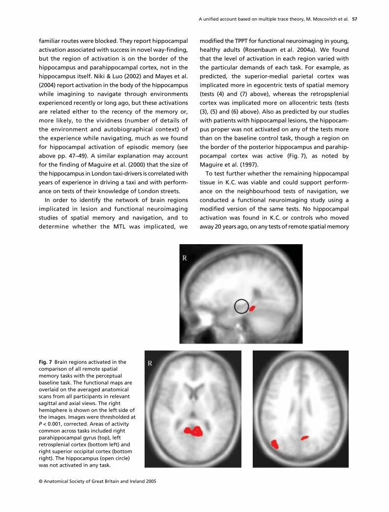
A unified account based on multiple trace theory, M. Moscovitch et al.
© Anatomical Society of Great Britain and Ireland 2005
57
familiar routes were blocked. They report hippocampalactivation associated with success in novel way-finding,but the region of activation is on the border of thehippocampus and parahippocampal cortex, not in thehippocampus itself. Niki & Luo (2002) and Mayes et al.(2004) report activation in the body of the hippocampuswhile imagining to navigate through environmentsexperienced recently or long ago, but these activationsare related either to the recency of the memory or,more likely, to the vividness (number of details ofthe environment and autobiographical context) ofthe experience while navigating, much as we foundfor hippocampal activation of episodic memory (seeabove pp. 47–49). A similar explanation may accountfor the finding of Maguire et al. (2000) that the size ofthe hippocampus in London taxi-drivers is correlated withyears of experience in driving a taxi and with perform-ance on tests of their knowledge of London streets.
In order to identify the network of brain regionsimplicated in lesion and functional neuroimagingstudies of spatial memory and navigation, and todetermine whether the MTL was implicated, we
modified the TPPT for functional neuroimaging in young,healthy adults (Rosenbaum et al. 2004a). We foundthat the level of activation in each region varied withthe particular demands of each task. For example, aspredicted, the superior-medial parietal cortex wasimplicated more in egocentric tests of spatial memory(tests (4) and (7) above), whereas the retropsplenialcortex was implicated more on allocentric tests (tests(3), (5) and (6) above). Also as predicted by our studieswith patients with hippocampal lesions, the hippocam-pus proper was not activated on any of the tests morethan on the baseline control task, though a region onthe border of the posterior hippocampus and parahip-pocampal cortex was active (Fig. 7), as noted byMaguire et al. (1997).
To test further whether the remaining hippocampaltissue in K.C. was viable and could support perform-ance on the neighbourhood tests of navigation, weconducted a functional neuroimaging study using amodified version of the same tests. No hippocampalactivation was found in K.C. or controls who movedaway 20 years ago, on any tests of remote spatial memory
Fig. 7 Brain regions activated in the comparison of all remote spatial memory tasks with the perceptual baseline task. The functional maps are overlaid on the averaged anatomical scans from all participants in relevant sagittal and axial views. The right hemisphere is shown on the left side of the images. Images were thresholded at P < 0.001, corrected. Areas of activity common across tasks included right parahippocampal gyrus (top), left retrosplenial cortex (bottom left) and right superior occipital cortex (bottom right). The hippocampus (open circle) was not activated in any task.

A unified account based on multiple trace theory, M. Moscovitch et al.
© Anatomical Society of Great Britain and Ireland 2005
58
except when K.C. viewed familiar landmarks which hedid not recognize, and considered novel. The latterfinding is important because it shows that the remain-ing tissue is viable, and is active when K.C. perceivessomething to be unfamiliar (novel), but not when he isnavigating mentally through his environment (Rosen-baum et al. 2005b). As with the TPPT tests, the sameextra-hippocampal areas were activated, the amountand pattern of activation being determined by thedemands of each particular test (Fig. 8).
The results of our studies on remote spatial memory,like those of Burgess, Maguire and their collaborators(Burgess, 2002; Burgess et al. 2002; Hartley et al. 2003)on anterograde spatial memory, suggest that remotespatial and episodic memory have much in common,though we differ in our conception of the role of thehippocampus. Whereas they believe the hippocampusis implicated in all forms of allocentric spatial represen-tations, our results suggest that at least for pre-morbidly familiar environments, some allocentricrepresentations exist independently of it. Our studiessuggest that an episodic-semantic distinction may be asusefully applied to spatial memory as to nonspatialmemory, and thus provides a unified framework forconceptualizing hippocampal–neocortical interactions.In this framework, detailed perceptual–spatial repre-sentations of environments (corresponding to episodic,autobiographical memory) are hippocampally depend-ent and distinguished from schematic or coarse, butallocentric, representations of the topography (corre-sponding to semantic memory) that can exist inde-pendently of the hippocampus. The hippocampal codeserves as a pointer or index to regions of neocortex(e.g. parahippocampal and perirhinal cortex) wherethese different kinds of information are stored andbinds them into detailed representations that allow
one to re-experience the environment as one takes amental walk through it (see Fig. 9). A similar proposalhas been advanced by Burgess et al. (2002) regarding theepisodic component, but not about the semantic one.Schematic, allocentric topographical representations,however, are sufficient to allow one to navigate in theenvironment and recognize those features which providesalient cues for navigation, without supporting a richrepresentation needed to re-experience the environment,or recognize noncritical elements of it (see Rosenbaumet al. 2000, 2001; for earlier versions of this view).
Summary and theoretical implications
Overall, findings from both lesion and functionalneuroimaging studies provide compelling evidence thatthe hippocampus is not needed for retention andretrieval of some remote spatial memories, even thosecapturing some allocentric information, for the pur-poses of navigation. These observations, therefore, posea challenge to the original formulation of cognitivemap theory, which proposed that the hippocampus iscrucial for representing all forms of allocentric spatialinformation necessary for navigation, no matter howlong ago that information was acquired. In agreementwith cognitive map theory, however, the hippocampusis required for the retention of detailed spatial memo-ries of the environment, such as the appearance oflocal elements that can support a rich, re-experiencingof the environment. Some supportive evidence hasbeen presented from lesion (Rosenbaum et al. 2000)and neuroimaging studies (Niki & Luo, 2002; Mayes et al.2004; Rosenbaum et al. submitted), but more researchis needed to confirm these findings and to test alternativeinterpretations of them. Overall, however, the findingson spatial memory are consistent with MTT’s postulate
Fig. 8 Structural scan of K.C.’s brain showing small amount of remaining hippocampal tissue that was unactivated (left); for comparison see right parahippocampal activation in parahippocampal cortex during remote spatial memory tasks (right).
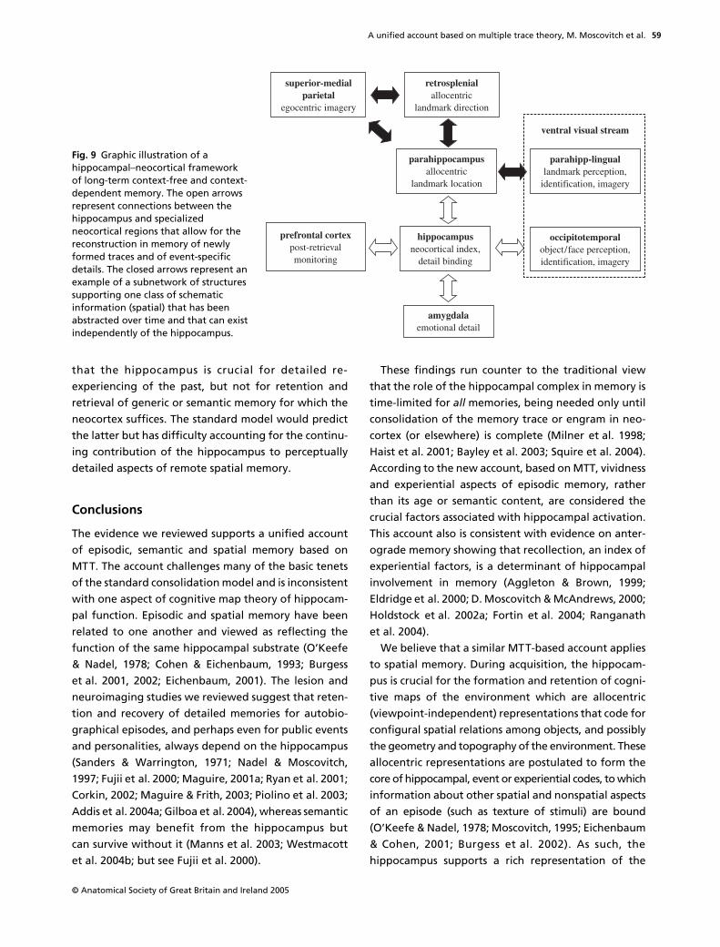
A unified account based on multiple trace theory, M. Moscovitch et al.
© Anatomical Society of Great Britain and Ireland 2005
59
that the hippocampus is crucial for detailed re-experiencing of the past, but not for retention andretrieval of generic or semantic memory for which theneocortex suffices. The standard model would predictthe latter but has difficulty accounting for the continu-ing contribution of the hippocampus to perceptuallydetailed aspects of remote spatial memory.
Conclusions
The evidence we reviewed supports a unified accountof episodic, semantic and spatial memory based onMTT. The account challenges many of the basic tenetsof the standard consolidation model and is inconsistentwith one aspect of cognitive map theory of hippocam-pal function. Episodic and spatial memory have beenrelated to one another and viewed as reflecting thefunction of the same hippocampal substrate (O’Keefe& Nadel, 1978; Cohen & Eichenbaum, 1993; Burgesset al. 2001, 2002; Eichenbaum, 2001). The lesion andneuroimaging studies we reviewed suggest that reten-tion and recovery of detailed memories for autobio-graphical episodes, and perhaps even for public eventsand personalities, always depend on the hippocampus(Sanders & Warrington, 1971; Nadel & Moscovitch,1997; Fujii et al. 2000; Maguire, 2001a; Ryan et al. 2001;Corkin, 2002; Maguire & Frith, 2003; Piolino et al. 2003;Addis et al. 2004a; Gilboa et al. 2004), whereas semanticmemories may benefit from the hippocampus butcan survive without it (Manns et al. 2003; Westmacottet al. 2004b; but see Fujii et al. 2000).
These findings run counter to the traditional viewthat the role of the hippocampal complex in memory istime-limited for all memories, being needed only untilconsolidation of the memory trace or engram in neo-cortex (or elsewhere) is complete (Milner et al. 1998;Haist et al. 2001; Bayley et al. 2003; Squire et al. 2004).According to the new account, based on MTT, vividnessand experiential aspects of episodic memory, ratherthan its age or semantic content, are considered thecrucial factors associated with hippocampal activation.This account also is consistent with evidence on anter-ograde memory showing that recollection, an index ofexperiential factors, is a determinant of hippocampalinvolvement in memory (Aggleton & Brown, 1999;Eldridge et al. 2000; D. Moscovitch & McAndrews, 2000;Holdstock et al. 2002a; Fortin et al. 2004; Ranganathet al. 2004).
We believe that a similar MTT-based account appliesto spatial memory. During acquisition, the hippocam-pus is crucial for the formation and retention of cogni-tive maps of the environment which are allocentric(viewpoint-independent) representations that code forconfigural spatial relations among objects, and possiblythe geometry and topography of the environment. Theseallocentric representations are postulated to form thecore of hippocampal, event or experiential codes, to whichinformation about other spatial and nonspatial aspectsof an episode (such as texture of stimuli) are bound(O’Keefe & Nadel, 1978; Moscovitch, 1995; Eichenbaum& Cohen, 2001; Burgess et al. 2002). As such, thehippocampus supports a rich representation of the
Fig. 9 Graphic illustration of a hippocampal–neocortical framework of long-term context-free and context-dependent memory. The open arrows represent connections between the hippocampus and specialized neocortical regions that allow for the reconstruction in memory of newly formed traces and of event-specific details. The closed arrows represent an example of a subnetwork of structures supporting one class of schematic information (spatial) that has been abstracted over time and that can exist independently of the hippocampus.

A unified account based on multiple trace theory, M. Moscovitch et al.
© Anatomical Society of Great Britain and Ireland 2005
60
environment which is the spatial analogue of vivid, epi-sodic memory, and may be an important aspect of it.Studies of anterograde memory generally support thisview, and suggest that subregions of the MTL, as wellas other regions of neocortex, are likely to contributedifferentially to different aspects of spatial memory(Murray & Mishkin, 1998; Burgess et al. 2002; Rosen-baum et al. 2004a, submitted), just as they do for epi-sodic memory. With repeated experience in navigatingthe environment, however, neocortical structuresabstract the statistical regularities from the episodic,spatial representations, just as they do for other typesof memory, and are capable of storing schematic, allo-centric representations that can be used for navigation.
The results we reviewed support this interpretationby showing that people with hippocampal lesions per-form well on a variety of navigation tests in a familiarenvironment, including tests of allocentric spatialmemory, and by showing that the hippocampus is notactivated in normal people on any of those tests of afamiliar environment. Consistent with this observation,we have recently shown that rats reared for threemonths in a complex environment continue to navig-ate normally in it even after sustaining large hippo-campal lesions (Wincour et al. 2005).
We also reviewed some suggestive, though not con-clusive, evidence that a rich re-experiencing of theenvironment will continue to depend on the hippoc-ampus no matter how old the spatial memory is, just asdoes re-experiencing autobiographical episodes.
Although new evidence suggests modifications toMTT will be required, this theory continues to providethe best, and most unified, account of the evidence onremote episodic, semantic, and spatial memory. It alsopoints the way to developing a parsimonious and unifiedaccount of the function of the MTL, and related struc-tures, in representing recent and remote memories.
References
Addis DR, Moscovitch M, Crawley AP, McAndrews MP (2004a)Recollective qualities modulate hippocampal activationduring autobiographical memory retrieval. Hippocampus14, 752–762.
Addis DR, McIntosh AR, Moscovitch M, Crawley AP, McAndrewsMP (2004b) Characterising the spatial and temporal featuresof autobiographical memory retrieval networks: a partialleast squares approach. NeuroImage 23, 1460–1471.
Aggleton JP, Brown MW (1999) Episodic memory, amnesia,and the hippocampal-anterior thalamic axis. Behav. Brain.Sci. 22, 425–489.
Aggleton JP, McMackin D, Carpenter K, Hornak J, Kapur N,Halpin S, et al. (2000) Differential cognitive effects of colloidcysts in the third ventricle that spare or compromise thefornix. Brain 123, 800–815.
Aguirre GK, D’Esposito M (1999) Topographical disorienta-tion: a synthesis and taxonomy. Brain 122, 1613–1628.
Aguirre GK, Detre JA, Also DC, D’Esposito M (1996) The para-hippocampus subserves topographical learning in man.Cereb Cortex 6, 823–829.
Atkinson RC, Juola JF (1973) Factors influencing speed andaccuracy of word recognition. In: Attention and Perform-ance: IV (ed. Kornblum S). New York, USA: Academic Press.
Barrash J (1998) A historical review of topographical disorien-tation and its neuroanatomical correlates. J. Clin. Exp.Neuropsychol. 20, 807–827.
Bayley PJ, Hopkins RO, Squire LR (2003) Successful recollectionof remote autobiographical memories by amnesic patientswith medial temporal lobe lesions. Neuron 38, 135–144.
Bisiach E, Brouchon M, Poncet M, Rusconi ML (1993) Unilateralneglect in route description. Neuropyschologia 31, 1255–1262.
Blumenfeld H (2002) Neuroanatomy Through Clinical Cases.Sunderland, MA, USA: Sinauer Associates, Inc.
Bohbot VD, Iaria G, Petrides M (2004) Hippocampal functionand spatial memory: evidence from functional neuroimag-ing in healthy participants and performance of patientswith medial temporal lobe resections. Neuropsychology 18,418–425.
Bohbot VD, Kalina M, Stepankova K, Spackova N, Petrides M,Nadel L (1998) Spatial memory deficits in patients withlesions to the right hippocampus and to the right parahip-pocampal cortex. Neuropsychologia 36, 1217–1238.
Bowers D, Verfaellie M, Valenstein E, Heilman KM (1988)Impaired acquisition of temporal information in retrosple-nial amnesia. Brain. Cogn. 8, 47–66.
Burgess N (2002) The hippocampus, space, and viewpoints inepisodic memory. Q. J. Exp. Psychol. A. 55, 1057–1080.
Burgess N, Becker S, King JA, O’Keefe J (2001) Memory forevents and their spatial context: Models and experiments.Philos. Trans. R. Soc. Lond. B. Biol. Sci. 356, 1493–1503.
Burgess N, Maguire EA, O’Keefe J (2002) The human hippo-campus and spatial and episodic memory. Neuron 35, 625–641.
Burgess PW, Shallice T (1996) Confabulation and the controlof recollection. Memory 4, 359–411.
Burnham WH (1904) Retroactive amnesia: Illustrative casesand a tentative explanation. Am. J. Psychol. 14, 382–396.
Byrne RW (1982) Geographical knowledge and orientation.In: Normality and Pathology in Cognitive Functions (ed. EllisAW), pp. 239–264. London, UK: Academic Press.
Cabeza R, Nyberg L (2000) Imaging cognition II: An empiricalreview of 275 PET and fMRI studies. J. Cogn. Neurosci. 12, 1–47.
Cammalleri R, Gangitano M, D’Amelio M, Raieli V, Raimondo D,Camarda R (1996) Transient topographical amnesia andcingulate cortex damage: a case report. Neuropsychologia34, 321–326.
Caza N, McAndrews MP, Hoque T, Ziegler M, Crawley M,Moscovitch M (2004) Neural basis for remembering andknowing in associative memory. Paper Presented at a Con-ference on Cognitive Neuroscience, Montreal, July, 2004.
A unified account based on multiple trace theory, M. Moscovitch et al.
© Anatomical Society of Great Britain and Ireland 2005
61
Cermak LS (1982) Human Memory and Amnesia. Hillsdale, NJ,USA: Lawrence Erlbaum Associates.
Cermak LS, O’Connor M (1983) The anterograde and retro-grade retrieval ability of a patient with amnesia due toencephalitis. Neuropsychologia 21, 213–234.
Cipilotti L, Shallice T, Chan D, Fox N, Scahill R, Harrison G,Stevens J, Rudge P (2001) Long-term retrograde amnesia: Thecrucial role of the hippocampus. Neuropsychologia 39,151–172.
Cohen NJ, Eichenbaum H (1993) Memory, Amnesia, and theHippocampal System. Cambridge, MA, USA: MIT Press.
Cohen NJ, Squire LR (1980) Preserved learning and retentionof pattern-analyzing skill in amnesia: dissociation of know-ing how and knowing that. Science 210, 207–210.
Conway MA, Fthenaki AK (2000) Disruption and loss of auto-biographical memory. In: Handbook of Neuropsychology:Memory (ed. Cermak LS), pp. 257–288. Amsterdam, theNetherlands: Elsevier.
Conway MA, Pleydell-Pearce CW (2000) The construction ofautobiographical memories in the self-memory system.Psychol. Rev. 107, 261–288.
Conway MA, Turk DJ, Miller SL, Logan J, Nebes RD, Meltzer CC,Becker JT (1999) A positron emission tomography (PET)study of autobiographical memory retrieval. Memory 5–6,679–702.
Corkin S (1984) Lasting consequences of bilateral medial tem-poral lobectomy: Clinical course and experimental findingsin H.M. Semin. Neurol. 4, 252–262.
Corkin S (2002) What’s new with the amnesic patient H.M.?Nat. Rev. Neurosci. 3, 153–160.
Corkin S, Amaral DG, Gonzalez G, Johnson KA, Hyman BT(1997) H.M.’s medial temporal lobe lesion: Findings frommagnetic resonance imaging. J. Neurosci. 17, 3964–3979.
Craik FIM, Moroz TM, Moscovitch M, Stuss DT, Winocur G,Tulving E, et al. (1999) In search of the self, a positron emis-sion tomography study. Psychol. Sci. 10, 26–34.
Crovitz HF, Schiffman H (1974) Frequency of episodic memo-ries as a function of their age. Bull. Psychon. Soc. 4, 519–521.
Davachi L, Mitchell JP, Wagner AD (2003) Multiple routes tomemory: Distinct medial temporal lobe processes build itemand source memories. Proc. Natl. Acad. Sci. USA 100, 2157–2162.
De Renzi E (1982) Disorders of Space Exploration and Cogni-tion. Chichester, UK: John Wiley.
Dolcos F, LaBar KS, Cabeza R (2002) Interaction between theamygdala and the medial temporal lobe memory systempredicts better memory for emotional events. Neuron 42,855–863.
Douville K, Woodard JL, Seidenberg M, Miller SK, Leveroni CL,Nielson KA, et al. (2005) Medial temporal lobe activity forrecognition of recent and remote famous names: an event-related fMRI study. Neuropsychologia 43, 693–703.
Dudai Y (2004) The neurobiology of consolidations, or, howstable is the engram? Annu. Rev. Psychol. 55, 51–86.
Eichenbaum H (2001) The hippocampus and declarative mem-ory: Cognitive mechanisms and neural codes. Behav. BrainRes. 127, 199–207.
Eichenbaum H, Cohen NJ (2001) From Conditioning to Con-sciousness: Memory Systems of the Brain. London, UK:Oxford University Press.
Eichenbaum H, Otto T, Cohen NJ (1994) Two functional com-ponents of the hippocampal memory system. Behav. BrainSci. 17, 449–518.
Eldridge LL, Knowlton BJ, Furmanski CS, Bookheimer SY,Engel SA (2000) Remembering episodes: a selective role forthe hippocampus during retrieval. Nat. Neurosci. 3, 1149–1152.
Epstein R, Graham KS, Downing PE (2003) Viewpoint-specificscene representations in human parahippocampal cortex.Neuron 37, 865–876.
Epstein R, Harris A, Stanley D, Kanwisher N (1999) Theparahippocampal place area: Recognition, navigation, orencoding? Neuron 23, 115–125.
Epstein R, Kanwisher N (1998) A cortical representation of thelocal visual environment. Nature 392, 598–601.
Eslinger PJ (1998) Autobiographical memory after temporallobe lesios. Neurocase 4, 481–495.
Eustache F, Piolino P, Giffard B, Viader F, De La Sayette V,Baron JC, Desgranges B (2003) In the course of time: a PETstudy of the cerebral substrates of autobiographical amnesiain Alzheimer’s disease. Brain 127, 1549–1560.
Farrell MJ (1996) Topographical disorientation. Neurocase 2,509–520.
Fletcher PC, Frith CD, Baker SC, Shallice T, Frackowiak RS,Dolan RJ (1995) The mind’s eye – precuneus activation inmemory-related imagery. NeuroImage 2, 195–200.
Fletcher PC, Henson RN (2001) Frontal lobes and humanmemory: insights from functional neuroimaging. Brain 124,849–881.
Fortin NJ, Wright SP, Eichenbaum H (2004) Recollection-likememory retrieval in rats is dependent on the hippocampus.Nature 431, 188–191.
Frankland PW, Bontempi B (2005) The organization of recentand remote memories. Nat. Rev. Neurosci. 6, 119–130.
Fujii T, Moscovitch M, Nadel L (2000) Consolidation, retro-grade amnesia, and the temporal lobe. In: The Handbook ofNeuropsychology, 2nd edn, Vol. 4 (eds Boller F, Grafman J,section ed. Cermak LS), pp. 223–250. Amsterdam, the Nether-lands: Elsevier.
Gadian DG, Aicardi J, Watkins KE, Porter DA, Mishkin M,Vargha-Khadem F (2000) Developmental amnesia associatedwith early hypoxic-ischaemic injury. Brain 123, 499–507.
Gilboa A (2004a) Neuropsychology of Remote and RecentAutobiographical Memory. Unpublished doctoral Thesis,University of Toronto, Toronto, Canada.
Gilboa A (2004) Autobiographical and episodic memory-oneand the same! Evidence from prefrontal activation in neuro-imaging studies. Neuropsychologia 47, 1336–1349.
Gilboa A, Moscovitch M (2002) The cognitive neuroscience ofconfabulation: A review and a model. In: Handbook ofMemory Disorders, 2nd edn (eds Baddeley AD, KopelmanMD, Wilson BA), pp. 315–342. London, UK: Wiley.
Gilboa A, Rosenbaum RS, Westmacott R, Winocur G, Mosco-vitch M (2005a) Recognition of remote episodic details isspecifically compromised following lesions to the fornicesand left basal forebrain, (In press.)
Gilboa A, Ramirez J, Köhler S, Westmacott R, Black SE,Moscovitch M (2005) Retrieval of autobiographical memory inAlzheimer’s disease: Relation to volumes of medial tempo-ral lobe and other structures. Hippocampus 15, 535–550.

A unified account based on multiple trace theory, M. Moscovitch et al.
© Anatomical Society of Great Britain and Ireland 2005
62
Gilboa A, Winocur G, Grady CL, Hevenor SJ, Moscovitch M(2004) Remembering our past: Functional neuroanatomy ofrecollection of recent and very remote personal events.Cereb. Cortex. 14, 1214–1225.
Graham KS (1999) Semantic dementia: a challenge to themultiple-trace theory? Trends. Cogn. Sci. 3, 85–87.
Graham KS, Hodges JR (1997) Differentiating the roles ofthe hippocampal system and the neocortex in long-termmemory storage. Neuropsychology 11, 77–89.
Graham KS, Kropelnicki A, Goldman WP, Hodges JR (2003a)Two further investigations of autobiographical memory insemantic dementia. Cortex 39, 729–750.
Graham KS, Lee ACH, Brett M, Patterson K (2003b) The neuralbasis of autobiographical and semantic memory: Newevidence from three PET studies. Cogn. Affect. Behav.Neurosci. 3, 234–254.
Graham KS, Patterson K, Hodges JR (1999) Episodic memory:New insights from the study of semantic dementia. Curr.Opin. Neurobiol. 9, 245–250.
Graham KS, Simons JS, Pratt KH, Patterson K, Hodges JR(2000) Insights from semantic dementia on the relationshipbetween episodic and semantic memory. Neuropsychologia38, 313–324.
Greenberg DL, Rubin DC (2003) The neuropsychology of auto-biographical memory. Cortex 39, 687–728.
Habib M, Sirigu A (1987) Pure topographical disorientation: adefinition and anatomical basis. Cortex 23, 73–85.
Haist F, Bowden Gore J, Mao H (2001) Consolidation of humanmemory over decades revealed by functional magnetic res-onance imaging. Nat. Neurosci. 4, 1139–1145.
Hartley T, Maguire EA, Spiers HJ, Burgess N (2003) The well-wornroute and the path less traveled: Distinct neural bases of routefollowing and wayfinding in humans. Neuron 37, 877–888.
Haxby JV, Petit L, Ungerleider LG, Courtney SM (2000) Distin-guishing the functional roles of multiple regions in distributedneural systems for visual working memory. NeuroImage 11,380–391.
Heilman KM, Bowers D, Watson RT, Day A, Valenstein E,Hammond E, et al. (1990) Frontal hypermetabolism andthalamic hypometabolism in a patient with abnormal orientingand retrosplenial amnesia. Neuropsychologia 28, 161–169.
Holdstock JS, Mayes AR, Roberts N, Cezayirli E, Isaac CL,O’Reilly RC, Norman KA (2002a) Under what conditions isrecognition spared relative to recall after selective hippoc-ampal damage? Hippocampus 12, 341–351.
Holdstock JS, Mayes AR, Isaac CL, Roberts JN (2002b) Differ-ential involvement of the hippocampus and temporal corti-ces in rapid and slow learning of new semantic information.Neuropsychologia 40, 748–768.
Iaria G, Petrides M, Dagher A, Pike B, Bohbot VD (2003)Cognitive strategies dependent on the hippocampus andcaudate nucleus in human navigation: variability and changewith practice. J. Neurosci. 23, 5945–5952.
Incisa della Rocchetta A, Cipolotti L, Warrington EK (1996)Topographical disorientation: selective impairment oflocomotor space? Cortex 32, 727–735.
Ivanoiu A, Cooper JM, Shanks JM, Venneri A (2004) Retrievalof episodic and semantic autobiographical memories inearly Alzheimer’s Disease and semantic dementia. Cortex40, 173–175.
Kandel ER (2001) The molecular biology of memory storage:a dialogue between genes and synapses. Science 294,1030–1038.
Kapur N (1999) Syndromes of retrograde amnesia: a concep-tual and empirical analysis. Psychol. Bull. 125, 800–825.
Kapur N, Friston KJ, Young A, Frith CD, Frackowiak RS (1995)Activation of human hippocampal formation during memoryfor faces: a PET study. Cortex 31, 99–108.
Kapur N, Thompson P, Evans J, Abbott P (2002) Can epilepsy-related phenomena unblock some of the secrets of long-termmemory? Frontiers of Human Memory (eds Yamadori A,Kawashima R., Fujii T, Suzuki K), pp. 137–150. Sendai, Japan:Tohoku University Press.
Kinsbourne M, Wood F (1975) Short-term memory processesand the amnesic syndrome. In: Short-Term Memory (edsDeutsch D, Deutsch AJ), pp. 258–291. New York, USA:Academic Press.
Kitchener EG, Hodges JR, McCarthy R (1998) Acquisition ofpost-morbid vocabulary and semantic facts in the absenceof episodic memory. Brain 121, 1313–1327.
Kitchener EG, Squire LR (2000) Impaired verbal category learn-ing in amnesia. Behav. Neurosci. 114, 907–911.
Kopelman MD, Lasserson D, Kingsley DR, Bello F, Rush C,Stanhope N, et al. (2003) Retrograde amnesia and thevolume of critical brain structures. Hippocampus 13, 879–891.
Kopelman MD, Stanhope N, Kingsley D (1999) Retrogradeamnesia in patients with diencephalic, temporal lobe orfrontal lesions. Neuropsychologia 37, 939–958.
Kopelman MD, Wilson BA, Baddeley AD (1989) The autobio-graphical memory interview: a new assessment of autobio-graphical and personal semantic memory in amnesic patients.J. Clin. Exp. Neuropsychol. 5, 724–744.
Lavenex P, Suzuki WA, Amaral DG (2002) Perirhinal and para-hippocampal cortices of the macaque monkey: projectionsto the neocortex. J. Comp. Neurol. 447, 394–420.
Lee CH, Bussey TJ, Murray EA, Saksida LM, Epstein RA, Kapur N,et al. (2005) Perceptual deficits in amnesia: challenging themedial temporal lobe ‘mnemonic’ view. Neuropsychologia43, 1–11.
Leveroni CL, Seidenberg M, Mayer AR, Mead LA, Binder JR,Rao SM (2000) Neural systems underlying the recognitionof familiar and newly learned faces. J.Neurosci. 20, 878–886.
Levine B, Svoboda E, Hay JF, Winocur G, Moscovitch M (2002)Aging and autobiographical memory: Dissociating episodicfrom semantic retrieval. Psychol. Aging 17, 677–689.
Maguire EA (2001a) Neuroimaging studies of autobiographicalevent memory. Philos. Trans. R. Soc. Lond. B. Biol. Sci. 356,1441–1451.
Maguire EA (2001b) The retrosplenial contribution to humannavigation: a review of lesion and neuroimaging findings.Scand. J. Psychol. 42, 225–238.
Maguire EA, Frackowiak RS, Frith CD (1997) Recalling routesaround London: activation of the right hippocampus in taxidrivers. J. Neurosci. 17, 7103–7110.
Maguire EA, Frith CD (2003) Lateral asymmetry in the hippo-campal response to the remoteness of autobiographicalmemories. J. Neurosci. 23, 5302–5307.
Maguire EA, Henson RN, Nummerg CJ, Frith CD (2001) Activityin prefrontal cortex, not hippocampus, varies parametrically
A unified account based on multiple trace theory, M. Moscovitch et al.
© Anatomical Society of Great Britain and Ireland 2005
63
with the increasing remoteness of memory. Neuroreport12, 441–444.
Maguire EA, Mummery CJ (1999) Differential modulation of acommon memory retrieval network revealed by positronemission tomography. Hippocampus 9, 54–61.
Maguire EA, Gadian DG, Johnsrude IS, Good CD, Ashburner J,Frackowiak RS, Frith CD (2000) Navigation-related struc-tural change in the hippocampi of taxi drivers. Proc. Natl.Acad. Sci. USA 97, 4398–4403.
Maguire EA, Vargha-Khadem F, Mishkin M (2001) The effectsof bilateral hippocampal damage on fMRI regional activa-tions and interactions during memory retrieval. Brain 124,1156–1170.
Mair WG, Warrington EK, Weiskrantz L (1979) Memory disorderin Korsakoff’s psychosis: a neuropathological and neuropsy-chological investigation of two cases. Brain 102, 749–783.
Mandler G (1980) Recognizing the judgement of previousoccurrence. Psychol. Rev. 87, 252–271.
Manns JR, Hopkins RO, Squire LR (2003) Semantic memoryand the human hippocampus. Neuron 38, 127–133.
Martin A, Chao L (2001) Semantic memory and the brain:structure and processes. Curr. Opin. Neurobiol. 11, 194–201.
Mayes AR, Holdstock JS, Isaac CL, Montaldi D, Grigor J,Gummer A, et al. (2004) Associative recognition in a patientwith selective hippocampal lesions and relatively normalitem recognition. Hippoccampus 14, 763–784.
Mayes AR, Isaac CL, Holdstock JS, Cariga P, Gummer A,Roberts N (2003) Long-term amnesia: a review and detailedillustrative case study. Cortex 39, 567–603.
McClelland JL, McNaughton BL, O’Reilly RC (1995) Why thereare complementary learning systems in the hippocampusand neocortex: Insights from the successes and failures ofconnectionist models of learning and memory. Psychol. Rev.102, 419–457.
McClelland JL, Rogers TT (2003) The parallel distributedprocessing approach to semantic memory. Nat. Rev. Neuro-sci. 4, 310–322.
McGaugh JL (2000) Memory – a century of consolidation.Science 287, 248–251.
McGaugh JL (2004) The amygdala modulates the consolidationof memories of emotionally arousing experiences. Annu. Rev.Neurosci. 27, 1–28.
Milner B (1966) Amnesia following operation on the temporallobe. In: Amnesia (eds Whitty CWM, Zangwill OL), pp. 109–133. London, UK: Butterworth.
Milner B (1974) Functional recovery after lesions of the nervoussystem. 3. Developmental processes in neural plasticity.Sparing of language functions after early unilateral braindamage. Neurosci. Res. Program Bull. 12, 213–217.
Milner B, Corkin S, Teuber HL (1968) Further analysis of thehippocampal amnesic syndrome: 14-year follow-up of H.M.Neuropsychologia 6, 215–234.
Milner AD, Goodale MA (1995) The Visual Brain in Action.Oxford, UK: Oxford University Press.
Milner B, Squire LR, Kandel ER (1998) Cognitive neuroscienceand the study of memory. Neuron 20, 445–468.
Moscovitch M (2001) Memory consolidation. In: Encyclopediaof Cognitive Science (ed. Nadel L). New York, USA: NaturePublishing Group.
Moscovitch M (1982) Multiple dissociation of function in the
amnesic syndrome. In: Human Memory and Amnesia (ed.Cermak LS), pp. 337–370. Hillsdale, NJ, USA: LawrenceErlbaum Associates.
Moscovitch M (1992) Memory and working with memory: acomponent process model based on modules and centralsystems. J. Cogn. Neurosci. 4, 257–267.
Moscovitch M (1995) Recovered consciousness: a hypothesisconcerning modularity and episodic memory. J. Clin. Exper.Neuropsychol. 17, 276–291.
Moscovitch M (2000) Theories of memory and consciousness.In: The Oxford Handbook of Memory (eds Tulving E, Craik FIM),pp. 609–625. Oxford, UK: Oxford University Press.
Moscovitch DA, McAndrews MP (2002) Material-specificdeficits in ‘remembering’ in patients with unilateral temporallobe epilepsy and excisions. Neuropsychologia 40, 1335–1342.
Moscovitch M, Umiltà C (1990) Modularity and neuropsychology:Modules and central processes in attention and memory. In:Modular Deficits in Alzheimer’s Disease (ed. Schwartz MF),pp. 1–59. Cambridge, MA, USA: MIT Press/Bradford.
Moscovitch M, Vriezen E, Goshen-Gottstein Y (1993) Implicittests of memory in patients with focal lesions or degenera-tive brain disorders. In: The Handbook of Neuropsychology,Vol. 8 (eds Boller F, Spinnler H), pp. 133–173. Amsterdam,the Netherlands: Elsevier.
Moscovitch M, Westmacott R, Gilboa A, Addis DR, Rosen-baum RS, Viskontas I, et al. (2005) Hippocampal complexcontributions to retention and retrieval of recent andremote episodic and semantic memories: Evidence frombehavioural and neuroimaging studies of normal and brain-damaged people. In: Dynamic Cognitive Processes (eds OhtaN, MacLeod CM, Uttl B), pp. 333–380. Tokyo, Japan:Springer-Verlag.
Moscovitch M, Winocur G (2002) The frontal cortex and work-ing with memory. In: The Frontal Lobes (eds Stuss DT, KnightRT), pp. 188–209. Oxford, UK: Oxford University Press.
Moscovitch M, Yaschyshyn T, Ziegler M, Nadel L (1999)Remote episodic memory and amnesia: Was Endel Tulvingright all along? Memory, Consciousness and the Brain: theTallinn Conference (ed. Tulving E), pp. 331–345. New York,USA: The Psychology Press.
Moss HE, Kopelman MD, Cappelletti M, de Mornay Davies P,Jaldow E (2003) Lost for words or loss of memories? Auto-biographical memory in semantic dementia. Cogn. Neuro-psychol. 20, 703–732.
Müller GE, Pilzecker A (1900) Experimentelle beiträge zurLehre vom Gedächtnis [Experimental contributions to thetheory of memory]. Zeitschrift für Psychologie und Physiolo-gie der Sinnesorgane S1, 1–288 (summarized by McDougallW, Critical Notives). Am. J. Psychol. 388–394.
Mummery CJ, Patterson K, Price CJ, Ashburner J, FrackowiakRSJ, Hodges JR (2000) A voxel-based morphometry study ofsemantic dementia: Relationship between temporal lobeatrophy and semantic dementia. Ann. Neurol. 47, 36–45.
Murray EA, Bussey TJ (1999) Perceptual-mnemonic functionsof the perirhinal cortex. Trends. Cogn. Sci. 3, 142–151.
Murray EA, Bussey TJ (2001) Consolidation and the medialtemporal lobe revisited: Methodological considerations.Hippocampus 11, 1–7.
Murray EA, Mishkin M (1998) Object recognition and location

A unified account based on multiple trace theory, M. Moscovitch et al.
© Anatomical Society of Great Britain and Ireland 2005
64
memory in monkeys with excitotoxic lesions of the amy-gdala and hippocampus. J. Neurosci. 18, 6568–6582.
Murray EA, Richmond BJ (2001) Role of perirhinal cortex inobject perception, memory, and associations. Curr. Opin.Neurobiol. 11, 188–193.
Murre JM, Graham KS, Hodges JR (2001) Semantic dementia:Relevance to connectionist models of long term memory.Brain 124, 647–675.
Nadel L, Moscovitch M (1997) Memory consolidation, retro-grade amnesia and the hippocampal complex. Curr. Opin.Neurobiol. 7, 217–227.
Nadel L, Moscovitch M (1998) Hippocampal contributions tocortical plasticity. Neuropharmacology 37, 431–439.
Nadel L, Moscovitch M (2001) The hippocampal complex andlong-term memory revisited. Trends Cogn. Neurosci. 5, 228–230.
Nadel L, Ryan L, Hayes SM, Gilboa A, Moscovitch M (2003) Therole of the hippocampal complex in long-term episodicmemory. In: Limbic and Association Cortical Systems – Basic,Clinical and Computational Aspects (eds Ono T, MatsumotoG, Lllinas RR, Berthoz A, Norgen R, Nishijo H, Tamura R)Amsterdam, the Netherlands: Elsevier Science/ExcerptaMedica International Congress Series.
Nadel L, Samsonovich A, Ryan L, Moscovitch M (2000) Multipletrace theory of human memory: Computational, neuroim-aging, and neuropsychological results. Hippocampus 10,352–368.
Nestor PJ, Graham KS, Bozeat S, Simons JS, Hodges JR (2002)Memory consolidation and the hippocampus: Furtherevidence from the study of autobiographical memory insemantic dementia and the frontal variant of frontotempo-ral dementia. Neuropsychologia 40, 633–654.
Niki K, Luo J (2002) An fMRI study on the time-limited role ofthe medial temporal lobe in long-term topographical auto-biographic memory. J. Cogn. Neurosci. 14, 500–507.
O’Kane G, Kensinger EA, Corkin S (2004) Evidence for semanticlearning in profound amnesia: An investigation with patientH.M. Hippocampus 14, 417–425.
O’Keefe J, Nadel L (1978) The Hippocampus as a CognitiveMap. Oxford, UK: Oxford University Press.
Ogden JA (1993) Visual object agnosia, prosopagnosia, achro-matopsia, loss of visual imagery, and autobiographicalamnesia following recovery from cortical blindness: CaseMH. Neuropsychologia 31, 571–589.
Owen AM, Milner B, Petrides M, Evans AC (1996a) Memoryfor object features versus memory for object location: apositron-emission tomography study of encoding and retrievalprocesses. Proc. Natl. Acad. Sci. USA 93, 9212–9217.
Owen AM, Milner B, Petrides M, Evans AC (1996b) A specificrole for the right parahippocampal gyrus in the retrievalof object-location: a positron emission tomography study.J. Cogn. Neurosci. 8, 588–602.
Penfield W, Milner B (1958) Memory deficit produced by bilat-eral lesions in the hippocampal zone. Arch. Neurol. Psychi-atry 79, 475–497.
Piefke M, Weiss PH, Zilles K, Markowitsch HJ, Fink GR (2003)Differential remoteness and emotional tone modulate theneural correlates of autobiographical memory. Brain 126,650–668.
Piolino P, Desgranges B, Belliard S, Matuszewski V, Lalevee C,
De la Sayette V, et al. (2003) Autobiographical memory andautonoetic consciousness: Triple dissociation in neurode-generative diseases. Brain 126, 2203–2219.
Piolino P, Giffard-Quillon G, Desgranges B, Chetelat G, BaronJ-C, Eustache F (2004) Re-experiencing old memories viahippocampus: a PET study of autobiographical memory.NeuroImage 22, 1371–1383.
Ranganath C, Yonelinas AP, Cohen MX, Dy CJ, Tom SM,D’Esposito M (2004) Dissociable correlates of recollectionand familiarity within the medial temporal lobes. Neuropsy-chologia 42, 2–13.
Reed JM, Squire LR (1998) Retrograde amnesia for facts andevents: Findings from four new cases. J. Neurosci. 18, 3943–3954.
Rempel-Clower NL, Zola SM, Squire LR, Amaral DG (1996)Three cases of enduring memory impairment after bilateraldamage limited to the hippocampal formation. J. Neurosci.16, 5233–5255.
Rockland KS, Van Hoesen GW (1999) Some temporal and pari-etal cortical connections converge in CA1 of the primatehippocampus. Cereb. Cortex 9, 232–237.
Roediger HL, IIIMcDermott KB (1993) In: Handbook of Neu-ropsychology (eds Spinnler H, Boller, F section ed. CermakLS), pp. 63–131. Amsterdam, the Netherlands: Elsevier.
Rosenbaum RS, Priselac S, Köhler S, Black SE, Gao F, Nadel L,et al. (2000) Remote spatial memory in an amnesic personwith extensive bilateral hippocampal lesions. Nat. Neurosci.3, 1044–1048.
Rosenbaum RS, Winocur G, Moscovitch M (2001) New viewson old memories: Re-evaluating the role of the hippo-campal complex. Behav. Brain Res. 127, 183–197.
Rosenbaum RS, Ziegler M, Winocur G, Grady CL, Moscovitch M(2004a) I have often walked down this street before: fMRIstudies on the hippocampus and other structures duringmental navigation of an old environment. Hippocampus 14,826–835.
Rosenbaum RS, McKinnon MC, Levine B, Moscovitch M(2004b) Visual imagery deficits, impaired strategic retrieval,or memory loss: Disentangling the nature of an amnesicpatient’s autobiographical memory deficit. Neuropsycholo-gia 42, 1619–1635.
Rosenbaum RS, Gao FQ, Richards B, Black SE, Moscovitch M(2005a) ‘Where to?’ Remote memory for spatial relationsand landmark identity in former taxi drivers withAlzheimer’s disease and encephalitis. J. Cogn. Neurosci. 17,446–462.
Rosenbaum RS, Köhler S, Schacter DL, Moscovitch M, West-macott R, Black SE, et al. (2005b) The case of K.C. Contribu-tions of a memory-impaired person to memory theory.Neuropsychologia 43, 989–1021.
Rosenbaum RS, Winocur G, Grady CL, Ziegler M, Moscovitch M(2005) fMRI studies of remote spatial memory in an amnesicperson with bilateral hippocampal damage (submitted).
Rotello CM, Macmillan NA, Reeder JA (2004) Sum-differencetheory of remembering and knowing: a two-dimensionalsignal-detection model. Psychol. Rev. 111, 588–616.
Rozin P (1976) The psychobiological approach to humanmemory. In: Neural Mechanisms of Learning and Memory(eds Rozenzweig R, Bennett EL). Cambridge, MA, USA: MITPress.
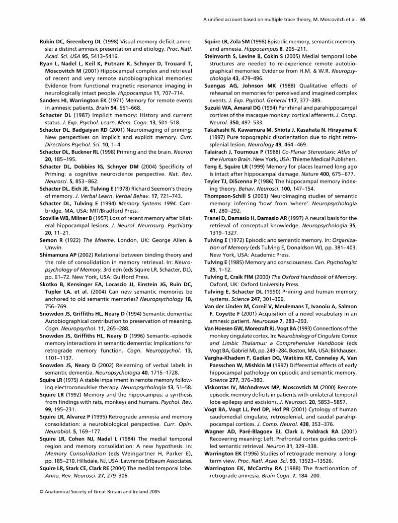
A unified account based on multiple trace theory, M. Moscovitch et al.
© Anatomical Society of Great Britain and Ireland 2005
65
Rubin DC, Greenberg DL (1998) Visual memory deficit amne-sia: a distinct amnesic presentation and etiology. Proc. Natl.Acad. Sci. USA 95, 5413–5416.
Ryan L, Nadel L, Keil K, Putnam K, Schnyer D, Trouard T,Moscovitch M (2001) Hippocampal complex and retrievalof recent and very remote autobiographical memories:Evidence from functional magnetic resonance imaging inneurologically intact people. Hippocampus 11, 707–714.
Sanders HI, Warrington EK (1971) Memory for remote eventsin amnesic patients. Brain 94, 661–668.
Schacter DL (1987) Implicit memory: History and currentstatus. J. Exp. Psychol. Learn. Mem. Cogn. 13, 501–518.
Schacter DL, Badgaiyan RD (2001) Neuroimaging of priming:New perspectives on implicit and explicit memory. Curr.Directions Psychol. Sci. 10, 1–4.
Schacter DL, Buckner RL (1998) Priming and the brain. Neuron20, 185–195.
Schacter DL, Dobbins IG, Schnyer DM (2004) Specificity ofPriming: a cognitive neuroscience perspective. Nat. Rev.Neurosci. 5, 853–862.
Schacter DL, Eich JE, Tulving E (1978) Richard Seemon’s theoryof memory. J. Verbal Learn. Verbal Behav. 17, 721–743.
Schacter DL, Tulving E (1994) Memory Systems 1994. Cam-bridge, MA, USA: MIT/Bradford Press.
Scoville WB, Milner B (1957) Loss of recent memory after bilat-eral hippocampal lesions. J. Neurol. Neurosurg. Psychiatry20, 11–21.
Semon R (1922) The Mneme. London, UK: George Allen &Unwin.
Shimamura AP (2002) Relational between binding theory andthe role of consolidation in memory retrieval. In: Neuro-psychology of Memory, 3rd edn (eds Squire LR, Schacter, DL),pp. 61–72. New York, USA: Guilford Press.
Skotko B, Kensinger EA, Locascio JJ, Einstein JG, Ruin DC,Tupler LA, et al. (2004) Can new semantic memories beanchored to old semantic memories? Neuropsychology 18,756–769.
Snowden JS, Griffiths HL, Neary D (1994) Semantic dementia:Autobiographical contribution to preservation of meaning.Cogn. Neuropsychol. 11, 265–288.
Snowden JS, Griffiths HL, Neary D (1996) Semantic–episodicmemory interactions in semantic dementia: Implications forretrograde memory function. Cogn. Neuropsychol. 13,1101–1137.
Snowden JS, Neary D (2002) Relearning of verbal labels insemantic dementia. Neuropsychologia 40, 1715–1728.
Squire LR (1975) A stable impairment in remote memory follow-ing electroconvulsive therapy. Neuropsychologia 13, 51–58.
Squire LR (1992) Memory and the hippocampus: a synthesisfrom findings with rats, monkeys and humans. Psychol. Rev.99, 195–231.
Squire LR, Alvarez P (1995) Retrograde amnesia and memoryconsolidation: a neurobiological perspective. Curr. Opin.Neurobiol. 5, 169–177.
Squire LR, Cohen NJ, Nadel L (1984) The medial temporalregion and memory consolidation: A new hypothesis. In:Memory Consolidation (eds Weingartner H, Parker E),pp. 185–210. Hillsdale, NJ, USA: Lawrence Erlbaum Associates.
Squire LR, Stark CE, Clark RE (2004) The medial temporal lobe.Annu. Rev. Neurosci. 27, 279–306.
Squire LR, Zola SM (1998) Episodic memory, semantic memory,and amnesia. Hippocampus 8, 205–211.
Steinvorth S, Levine B, Cokin S (2005) Medial temporal lobestructures are needed to re-experience remote autobio-graphical memories: Evidence from H.M. & W.R. Neuropsy-chologia 43, 479–496.
Suengas AG, Johnson MK (1988) Qualitative effects ofrehearsal on memories for perceived and imagined complexevents. J. Exp. Psychol. General 117, 377–389.
Suzuki WA, Amaral DG (1994) Perirhinal and parahippocampalcortices of the macaque monkey: cortical afferents. J. Comp.Neurol. 350, 497–533.
Takahashi N, Kawamura M, Shiota J, Kasahata N, Hirayama K(1997) Pure topographic disorientation due to right retro-splenial lesion. Neurology 49, 464–469.
Talairach J, Tournoux P (1988) Co-Planar Stereotaxic Atlas ofthe Human Brain. New York, USA: Thieme Medical Publishers.
Teng E, Squire LR (1999) Memory for places learned long agois intact after hippocampal damage. Nature 400, 675–677.
Teyler TJ, DiScenna P (1986) The hippocampal memory index-ing theory. Behav. Neurosci. 100, 147–154.
Thompson-Schill S (2003) Neuroimaging studies of semanticmemory: inferring ‘how’ from ‘where’. Neuropsychologia41, 280–292.
Tranel D, Damasio H, Damasio AR (1997) A neural basis for theretrieval of conceptual knowledge. Neuropsychologia 35,1319–1327.
Tulving E (1972) Episodic and semantic memory. In: Organiza-tion of Memory (eds Tulving E, Donaldson W), pp. 381–403.New York, USA: Academic Press.
Tulving E (1985) Memory and consciousness. Can. Psychologist25, 1–12.
Tulving E, Craik FIM (2000) The Oxford Handbook of Memory.Oxford, UK: Oxford University Press.
Tulving E, Schacter DL (1990) Priming and human memorysystems. Science 247, 301–306.
Van der Linden M, Cornil V, Meulemans T, Ivanoiu A, SalmonF, Coyette F (2001) Acquisition of a novel vocabulary in anamnesic patient. Neurocase 7, 283–293.
Van Hoesen GW, Morecraft RJ, Vogt BA (1993) Connections of themonkey cingulate cortex. In: Neurobiology of Cingulate Cortexand Limbic Thalamus: a Comprehensive Handbook (edsVogt BA, Gabriel M), pp. 249–284. Boston, MA, USA: Birkhauser.
Vargha-Khadem F, Gadian DG, Watkins KE, Conneley A, VanPaesschen W, Mishkin M (1997) Differential effects of earlyhippocampal pathology on episodic and semantic memory.Science 277, 376–380.
Viskontas IV, McAndrews MP, Moscovtich M (2000) Remoteepisodic memory deficits in patients with unilateral temporallobe epilepsy and excisions. J. Neurosci. 20, 5853–5857.
Vogt BA, Vogt LJ, Perl DP, Hof PR (2001) Cytology of humancaudomedial cingulate, retrosplenial, and caudal parahip-pocampal cortices. J. Comp. Neurol. 438, 353–376.
Wagner AD, Paré-Blagoev EJ, Clark J, Poldrack RA (2001)Recovering meaning: Left. Prefrontal cortex guides control-led semantic retrieval. Neuron 31, 329–338.
Warrington EK (1996) Studies of retrograde memory: a long-term view. Proc. Natl. Acad. Sci. 93, 13523–13526.
Warrington EK, McCarthy RA (1988) The fractionation ofretrograde amnesia. Brain Cogn. 7, 184–200.
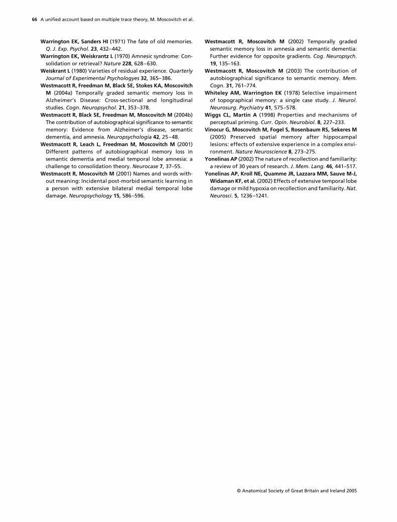
A unified account based on multiple trace theory, M. Moscovitch et al.
© Anatomical Society of Great Britain and Ireland 2005
66
Warrington EK, Sanders HI (1971) The fate of old memories.Q. J. Exp. Psychol. 23, 432–442.
Warrington EK, Weiskrantz L (1970) Amnesic syndrome: Con-solidation or retrieval? Nature 228, 628–630.
Weiskrant L (1980) Varieties of residual experience. QuarterlyJournal of Experimental Psychologyes 32, 365–386.
Westmacott R, Freedman M, Black SE, Stokes KA, MoscovitchM (2004a) Temporally graded semantic memory loss inAlzheimer’s Disease: Cross-sectional and longitudinalstudies. Cogn. Neuropsychol. 21, 353–378.
Westmacott R, Black SE, Freedman M, Moscovitch M (2004b)The contribution of autobiographical significance to semanticmemory: Evidence from Alzheimer’s disease, semanticdementia, and amnesia. Neuropsychologia 42, 25–48.
Westmacott R, Leach L, Freedman M, Moscovitch M (2001)Different patterns of autobiographical memory loss insemantic dementia and medial temporal lobe amnesia: achallenge to consolidation theory. Neurocase 7, 37–55.
Westmacott R, Moscovitch M (2001) Names and words with-out meaning: Incidental post-morbid semantic learning ina person with extensive bilateral medial temporal lobedamage. Neuropsychology 15, 586–596.
Westmacott R, Moscovitch M (2002) Temporally gradedsemantic memory loss in amnesia and semantic dementia:Further evidence for opposite gradients. Cog. Neuropsych.19, 135–163.
Westmacott R, Moscovitch M (2003) The contribution ofautobiographical significance to semantic memory. Mem.Cogn. 31, 761–774.
Whiteley AM, Warrington EK (1978) Selective impairmentof topographical memory: a single case study. J. Neurol.Neurosurg. Psychiatry 41, 575–578.
Wiggs CL, Martin A (1998) Properties and mechanisms ofperceptual priming. Curr. Opin. Neurobiol. 8, 227–233.
Vinocur G, Moscovitch M, Fogel S, Rosenbaum RS, Sekeres M(2005) Preserved spatial memory after hippocampallesions: effects of extensive experience in a complex envi-ronment. Nature Neuroscience 8, 273–275.
Yonelinas AP (2002) The nature of recollection and familiarity:a review of 30 years of research. J. Mem. Lang. 46, 441–517.
Yonelinas AP, Kroll NE, Quamme JR, Lazzara MM, Sauve M-J,Widaman KF, et al. (2002) Effects of extensive temporal lobedamage or mild hypoxia on recollection and familiarity. Nat.Neurosci. 5, 1236–1241.