french-crg prospective days auditorium – grenoble, france
TRANSCRIPT


Journées prospectives scientifiques F-CRG - 9-11 décembre 2019
French-CRG Prospective days
Auditorium – Grenoble, France
9, 10 & 11 December 2019
Programme
List of participants
Abstracts

Journées prospectives scientifiques F-CRG - 9-11 décembre 2019
Local scientific organization
BLANC Nils, D2AM beamline, [email protected]
BOUDET Nathalie, D2AM beamline, [email protected]
FERRER Jean-Luc, FIP2 beamline, [email protected]
HAZEMANN Jean-Louis, FAME & FAME-UHD beamlines, [email protected]
MICHA Jean-Sébastien, IF beamline, [email protected]
PROUX Olivier, FAME & FAME-UHD beamlines, [email protected]
RENAUD Gilles, IF beamline, [email protected]
SALLAZ-DAMAZ Yoann, FIP2 beamline, [email protected]
F-CRG Scientific Committee
BLANCHARD Juliette, Lab. de Réactivité de Surface, Ivry sur Seine, [email protected]
BRIOIS Valérie, Synchrotron SOLEIL, [email protected]
DUMESNIL Karine, Institut Jean Lamour, Nancy, [email protected]
GERGAUD Patrice, CEA-Grenoble, [email protected]
LEGRAND Pierre, Synchrotron SOLEIL, [email protected]
LEVITZ Pierre, PHENIX, Paris, [email protected]
MORIN Guillaume, IMPMC, Paris, [email protected]
PIGNOL David, CEA-Cadarache, [email protected]
THOMAS Olivier, IM2NP, Marseille, [email protected]
Coordination organization
CLEMENT Valérie, ROBERT Kimberley, CRG-office, [email protected]
MOLLIER-SABET Françoise, Institut Néel, [email protected]

Journées prospectives scientifiques F-CRG - 9-11 décembre 2019
Monday 9th December 2019
11:00 - 13:30 Registration in the ESRF Central - Building entrance hall
12:00 - 13:30 Lunch at the EPN campus restaurant
Chair : P. Legrand
13:30 - 13:45 Welcome / Introduction E. Bustarret F-CRG spokesperson
13:45 - 14:30 Beamlines presentations Project leaders
14:30 - 15:00 The EBS project J. Susini ESRF Director of Research
15:00 - 15:30 The MAGNIFIX project
15:30 - 16:00 Coffee break around the posters - ESRF Central Building entrance hall
Chair : P. Gergaud
16:00 - 16:45 Imaging strain and defects in electronic materials and devices: State of the art and future trends
O. Thomas IM2NP, Marseille
16:45 - 17:10 Diffusion des Rayons X aux Grands Angles et aux Petits Angles pour la Détermination de la Morphologie de Systèmes Polymères
G. Sudre IMP, Lyon
17:10 - 18:05 Round-table discussion on F-CRG operation
N. Boudet, O. Proux P. Legrand, P. Gergaud, R. Guinebretière
19:00 - 21:00 Cocktail at EPN Cafetaria

Journées prospectives scientifiques F-CRG - 9-11 décembre 2019
Tuesday 10th December 2019
Chair : P. Levitz
09:00 - 09:45 The past is the key to the future A. Thompson SOLEIL, Paris
09:45 - 10:10 Apports des spectroscopies optiques à la cristallographie macromoléculaire
A. Royant IBS, Grenoble
10:10 - 10:35 Expériences de cristallographie biologique à façon
D. Pignol CEA-BIAM, Marseille
10:35 - 11:05 Coffee break around the posters - ESRF Central Building entrance hall
Chair : K. Dumesnil
11:05 - 11:30
X-ray imaging and X-Ray Absorption Spectroscopy at relevant environmental concentrations : Powerful Structural Tools in Environmental Nanotechnologies
M. Auffan CEREGE, Aix-en-Provence
11:30 - 11:55 Co-crystallization and diffraction in situ for rapid drug design
G. Labesse CBS, Montpellier
11:55 - 12:40 Round-table discussion on data analysis J.-S. Micha, Y. Sallaz-Damaz, P. Levitz, A. Boulle
12:40 - 14:10 Lunch at the EPN campus restaurant
Chair : E. Lacaze
14:10 - 14:55 Synchrotron techniques for the study of batteries: the importance of X-ray spectroscopies
L. Stievano ICGM, Montpellier
14:55 - 15:20
Structure, propriétés électroniques et réactivités de catalyseurs à base de particules sub-nanométriques de platine supportées sur alumine : une approche multi-techniques
C. Chizallet IFPEN, Solaize
15:20 - 15:45 Spéciation des produits de fission dans le combustible irradié: apports des lignes de lumière
Y. Pontillon CEA-DEN, Cadarache
15:45 - 16:15 Coffee break around the posters - ESRF Central Building entrance hall

Journées prospectives scientifiques F-CRG - 9-11 décembre 2019
Chair : J. Blanchard
16:15 - 16:40 Revealing metal speciation in hydrothermal solutions through in-situ XAS and Raman spectroscopy
L. Truche ISTerre, Grenoble
16:40 - 17:05 BM32 (μLaue) - BM02 : quelques exemples pour la métallurgie
G. Geandier Inst. J. Lamour, Nancy
17:05 - 17:50 Round-table discussion on in situ, operando, coupling…
N. Blanc, G. Renaud, J. Blanchard, K. Dumesnil, A. Vlad
19:00 - 21:00 Dinner at EPN restaurant
Wednesday 11th December 2019
Chair : O. Thomas
09:00 - 09:45 Spectroscopie d'absorption X pour une approche moléculaire en sciences de l’environnement
G. Morin IMPMC, Paris
09:45 - 10:10 Benefits of coherence for imaging of complex natural and manufactured material
L. Michot PHENIX, Paris
10:10 - 10:35 Matériaux bidimensionnels sondés par rayonnement synchrotron
J. Coraux Institut Néel, Grenoble
10:35 - 11:05 Coffee break around the posters - ESRF Central Building entrance hall
Chair : G. Morin
11:05 - 11:30 Caractérisations micro/nano-structurales de matériaux céramiques par DRX et analyses numériques
A. Boulle IRCER, Limoges
11:30 - 11:55 Ion transport at the interface between highly mismatched oxide perovskites interfaces
R. Felici CNR-SPIN, Roma
11:55 - 12:40 Round-table discussion on imaging and coherence
J.-L. Hazemann, J. C. Da Silva, O. Thomas, G. Morin
12:40 - 14:10 Lunch at the EPN campus restaurant

Journées prospectives scientifiques F-CRG - 9-11 décembre 2019

Journées prospectives scientifiques F-CRG - 9-11 décembre 2019
Participants
AFANASIEV Pavel IRCELYON - UCB Lyon 1 / CNRS UMR 5256
AGUILAR Antonio Institut Neel - UGA / CNRS UPR 2940
ALBA Michel CEA Saclay [email protected]
AUFFAN Melanie CEREGE - Aix-Marseille Université / CNRS UMR 7330
BABONNEAU David Institut Pprime - Univ. Poitiers / CNRS UPR 3346
BAZARKINA Elena IGEM (RAS) & Institut Neel - CNRS UPR 2940
BERHAUT Christopher ESRF [email protected]
BERZIN Christophe ESRF [email protected]
BEUTIER Guillaume SIMAP - G-INP / UGA / CNRS UMR 5266
BIKONDOA Oier Warwick University [email protected]
BLANC Nils Institut Neel - UGA / CNRS UPR 2940
BLANCHARD Juliette Laboratoire de Réactivité de Surface - CNRS UMR 7197
BORDET Pierre Institut Neel - UGA / CNRS UPR 2940
BOREL Franck Institut de Biologie Structurale - IBS
BORSCHNECK Daniel CEREGE - Aix-Marseille Université / CNRS UMR 7330
BOUAT Sophie S.A.V.E.D. [email protected]
BOUDET Nathalie Institut Neel - UGA / CNRS UPR 2940
BOULLE Alexandre Institut de Recherche sur les Céramiques - Univ. Limoges / CNRS UMR 7315
BRUNEL Fabrice C2P2 - UCB Lyon / CNRS UMR 5265
BRUNELLI Michela DUBBLE CRG (NL) [email protected]

Journées prospectives scientifiques F-CRG - 9-11 décembre 2019
BURRIEL Monica Minatec [email protected]
BUSTARRET Etienne Institut Neel - UGA / CNRS UPR 2940
CARRIERE Marie CEA Grenoble - INAC [email protected]
CATON Francois Lab. de Rhéologie et Procédés - G-INP / UGA / CNRS UMR 5520
CAVAZZA Christine CNRS UMR 5249 - iRTSV/CEA - Grenoble
CHAURAND Perrine CEREGE - Aix-Marseille Université / CNRS UMR 7330
CHIZALLET Céline IFP energies nouvelles [email protected]
COBESSI David Institut de Biologie Structurale - IBS
COLIN Claire Institut Neel - UGA / CNRS UPR 2940
CORAUX Johann Institut Neel - UGA / CNRS UPR 2940
CORNELIUS Thomas IM2NP - CNRS UMR 7334 [email protected]
CREPIN Thibaut Institut de Biologie Structurale - IBS
D ACAPITO Francesco LISA/BM08 CRG [email protected]
DA SILVA Julio Cesar Institut Neel - UGA / CNRS UPR 2940
DAILLANT Jean Synchrotron Soleil [email protected]
DE SANTIS Maurizio Institut Neel - UGA / CNRS UPR 2940
DENIS MEYERE Auriane
Institut de Biologie Structurale - IBS
DEUTSCH Thierry IRIG [email protected]
DEVAUX Didier LEPMI - UGA / CNRS UMR 5279
DIDIERJEAN Claude CRM2 - Univ. Lorraine / CNRS UMR 7036
DUMESNIL Karine Institut Jean Lamour - Univ. Lorraine / CNRS UMR 7198
EYMERY Joel CEA Grenoble - INAC [email protected]

Journées prospectives scientifiques F-CRG - 9-11 décembre 2019
FELICI Roberto CNR-SPIN, sede di Tor Vergata
FERNANDEZ MARTINEZ Alejandro
ISTerre - UGA / CNRS UMR 5275
FERRER Jean Luc Institut de Biologie Structurale - IBS
FEVRE Mathieu LEM - ONERA / CNRS UMR 104
FOOS Nicolas Institut de Biologie Structurale - IBS
GALLET Benoit Institut de Biologie Structurale - IBS
GEANDIER Guillaume Institut Jean Lamour - Univ. Lorraine / CNRS UMR 7198
GEANTET Christophe IRCELYON - UCB Lyon 1 / CNRS UMR 5256
GERGAUD Patrice CEA Grenoble [email protected]
GRENIER Stephane Institut Neel - UGA / CNRS UPR 2940
GUILLOT Stephane ISTerre - UGA / CNRS UMR 5275
GUINEBRETIERE René Institut de Recherche sur les Céramiques - Univ. Limoges / CNRS UMR 7315
HAZEMANN Jean Louis
Institut Neel - UGA / CNRS UPR 2940
JAHO Sofia Institut de Biologie Structurale - IBS
JEANNE BROU Roselyne
LEPMI - UGA / CNRS UMR 5279
JOLY Yves Institut Neel - UGA / CNRS UPR 2940
KIEFFER Isabelle OSUG - UGA / CNRS UMS 832
LABESSE Gilles CNRS UMR 5048 - UM 1 - INSERM UMR 1054
LACAZE Emmanuelle CNRS INP [email protected]
LAHERA Eric OSUG - UGA / CNRS UMS 832
LEGRAND Pierre Synchrotron SOLEIL [email protected]
LEVITZ Pierre PHENIX – Sorbonne Université / CNRS UMR 8234

Journées prospectives scientifiques F-CRG - 9-11 décembre 2019
LIVET Frederic INP Grenoble - CNRS - UGA [email protected]
LYONNARD Sandrine CEA Grenoble - INAC [email protected]
MARTINELLI Lucio CNRS UMR 7643 - Ecole Polytechnique
MATHIEU Eric Bernard Institut de Biologie Structurale - IBS
MENUT Denis Synchrotron Soleil [email protected]
MICHA Jean Sebastien CEA Grenoble - INAC [email protected]
MICHAUD Laurent CEA Grenoble [email protected]
MICHOT Laurent PHENIX – Sorbonne Université / CNRS UMR 8234
MORFIN Isabelle CNRS UMR 5588 - Univ. Grenoble Alpes
MORIN Guillaume Sorbonne Universite - IMPMC [email protected]
MORONI Martine ESRF [email protected]
NAVARRO Etienne IM2NP - CNRS UMR 7334 [email protected]
PERARD Julien CNRS UMR 5249 - iRTSV/CEA - Grenoble
PIGNOL David CEA cadarache [email protected]
PONTILLON Yves CEA cadarache [email protected]
POUGET Stephanie CEA Grenoble - INAC [email protected]
PRANGE Thierry CNRS UMR 8015 [email protected]
PROUX Olivier OSUG - UGA / CNRS UMS 832
RAOUX Denis [email protected]
RENAUD Gilles CEA Grenoble - INAC [email protected]
RENEVIER Hubert CNRS Grenoble - INP [email protected]
RIEUTORD Francois CEA Grenoble - INAC [email protected]

Journées prospectives scientifiques F-CRG - 9-11 décembre 2019
ROBACH Odile CEA Grenoble - INAC [email protected]
ROSA Angelika ESRF [email protected]
ROSE Jerome CEREGE - Aix-Marseille Université / CNRS UMR 7330
ROVEZZI Mauro OSUG - UGA / CNRS UMS 832
ROYANT Antoine Institut de Biologie Structurale - IBS
SALLAZ DAMAZ Yoann
Institut de Biologie Structurale - IBS
SARRET Geraldine ISTerre - UGA / CNRS UMR 5275
SIBERT Eric LEPMI - UGA / CNRS UMR 5279
SITJA Georges CINaM - Aix-Marseille Universite / CNRS UMR 7325
SOLDO OLIVIER Yvonne
Institut Neel - UGA / CNRS UPR 2940
SPANO BUDAYOVA Monika
Institut de Biologie Structurale - IBS
STIEVANO Lorenzo CNRS UMR 5253 - Universite Montpellier II
SUDRE Guillaume UCB Lyon 1 - CNRS UMR 5223
SUSINI Jean ESRF [email protected]
TARDIF Samuel CEA Grenoble - INAC [email protected]
TESTEMALE Denis Institut Neel - UGA / CNRS UPR 2940
THEPAUT Michel Institut de Biologie Structurale - IBS
THOMAS Olivier IM2NP - CNRS UMR 7334 [email protected]
THOMPSON Andrew SYNCHROTRON SOLEIL [email protected]
TIMMINS Joanna Institut de Biologie Structurale - IBS
TRUCHE Laurent ISTerre - UGA / CNRS UMR 5275
ULRICH Olivier CEA Grenoble - INAC [email protected]

Journées prospectives scientifiques F-CRG - 9-11 décembre 2019
VARROT Annabelle CERMAV - UGA / CNRS UPR 5301
VAXELAIRE Nicolas CEA Grenoble [email protected]
VERNEDE Xavier Institut de Biologie Structurale - IBS
VERONESI Giulia iRTSV - CEA-Grenoble / CNRS UMR 5249
VILLEVIEILLE Claire LEPMI - UGA / CNRS UMR 5279
VLAD Alina Synchrotron Soleil [email protected]
VOLBEDA Anne Institut de Biologie Structurale - IBS
ZAPATA DOMINGUEZ Diana
CEA Grenoble - INAC [email protected]
ZITOLO Andrea Synchrotron Soleil [email protected]

Journées prospectives scientifiques F-CRG - 9-11 décembre 2019
Imaging strain and defects in electronic materials and devices:
State of the art and future trends
Olivier Thomas
Aix Marseille Univ, Université de Toulon, CNRS, IM2NP, Marseille, France Courriel : [email protected]
Since the early 1960s microelectronics has made extraordinary progress that shows up in our daily lives. Ever decreasing transistor size, use of new materials or intentional strain have allowed this amazing evolution which has followed Gordon Moore’s predictions [1] until very recently. Based on a complex assembly of materials with vastly different thermo-mechanical properties, microelectronic devices are a fantastic playground for investigating elastic strains and defects in a large range of scales (from millimetres to nanometres). Because of their high penetrating power X-rays are perfectly suited for investigating elastic strains in buried materials within electronic chips. X-ray diffraction is moreover extremely sensitive to lattice deformations. One should mention also the flexibility of bringing various environments and complementary measurements around the sample. On the other hand, X-ray beams are difficult to focus, which has limited the spatial resolution of X-ray strain imaging until recently.
Strain and defect imaging with X-rays have made very impressive progress lately. Progress in X-ray focusing optics allows nowadays scanning x-ray diffraction mapping to be performed with a resolution in the 50-100 nm range [2]. Full field X-ray microscopy is improving a lot too with resolutions in the 100 nm range [3]. Micro-Laue diffraction is very sensitive to lattice rotations and plasticity-induced defects [4]. By far the best spatial resolution is obtained with coherent diffraction imaging, which is a lensless imaging technique, with a typical resolution of 8-10 nm [5,6].
These recent progresses, which have been made possible thanks to the development of brighter and more coherent synchrotron sources, are revolutionizing the field of strain and defect imaging at small scales. I will show various examples in the field of electronic materials and devices, where X-ray diffraction and strain imaging can bring very detailed information, often critical for understanding and improving device properties: strain-engineered Si and SixGe1-x, GaN nanowires, Cu-filled though Si vias (TSVs), phase change materials for memories, …
Operando experiments and time resolution are critical for understanding reliability issues or, from a more fundamental viewpoint, to investigate the dynamics of defects. They maybe within reach with the new “4th generation” synchrotron sources that are becoming available.
[1] G. Moore, Electronics 1965, 38, 114. [2] G. Chahine, M.-I. Richard, R. A. Homs-Regojo, T.-N. Tran-Caliste, D. Carbone, V. L. R. Jacques, R. Grifone, P. Boesecke, J. Katzer, I. Costin, H. Djazouli, T. Shroeder, T. U. Schulli, J. Appl. Cryst. 2014, 47, 762. [3] H. Simons, A. King, W. Ludwig, C. Detlefs, W. Pantleon, S. Schmidt, F.Stohr, I. Snigireva, A. Snigirev, H.F. Poulsen, Nature Comm. 2015, 6, 6098. [4] C. Leclere, T.W. Cornelius, Z. Ren, A. Davydok, J.-S. Micha, O. Robach, G. Richter, L. Belliard, O. Thomas, J. Appl. Cryst. 2015, 48, 291. [5] M. Pfeifer, G. Williams, I. Vartanyants, R. Harder, I. Robinson, Nature 2006, 442, 63. [6] S. Labat, M.-I. Richard, M. Dupraz, M. Gailhanou, G. Beutier, M. Verdier, F. Mastropietro, T.W. Cornelius, T.U. Schülli, J. Eymery, O. Thomas, ACS Nano 2015, 9, 9210.
Journées prospectives scientifiques F-CRG - 9-11 décembre 2019
Diffusion des Rayons X aux Grands Angles et aux Petits Angles pour la
Détermination de la Morphologie de Systèmes Polymères
G. Sudre1, K. Lamnawar2, I. Morfin3, L. David1
1 Ingénierie des Matériaux Polymères – UMR 5223, Univ Lyon, CNRS, 69100, Villeurbanne, France
2 Ingénierie des Matériaux Polymères – UMR 5223, INSA de Lyon, 69100, Villeurbanne, France 3 Laboratoire Interdisciplinaire de Physique, Univ Grenoble Alpes, CNRS, 38000, Grenoble,
France Courriel : [email protected]
Le laboratoire Ingénierie des Matériaux Polymères est une unité mixte de recherche dont les activités portent sur la synthèse de nouvelles macromolécules, la préparation d’architectures macromoléculaires, la formulation des polymères, leur élaboration, la détermination de propriétés spécifiques ou encore leur caractérisation chimique, physique ou morphologique. Au cours de cet exposé, nous décrirons quelques études morphologiques menées grâce à des résultats obtenus sur la ligne de lumière BM02-D2AM de l’ESRF et nous montrerons l’utilité de ces résultats pour comprendre les propriétés macroscopiques de systèmes polymères.
En particulier, nous mettrons en évidence l’intérêt de mesure de diffusion aux petits angles pour la résolution de la structure d’hydrogels hautement étirables formés par des interactions polyélectrolytes (Lalevée et al. 2016 ; Lalevée et al., 2017). Formés à partir de polysaccharides polyélectrolytes faibles (acide hyaluronique et chitosane), ces hydrogels sont formés par complexation à différents pH, dont dépendent à la fois la conformation des composants et la nature des interactions.
Comme seconde illustration, la combinaison de mesures réalisées aux grands angles et aux petits angles sera illustrée sur la problématique de formation de films multicouches par co-extrusion. Différents polymères co-extrudés permettront de mettre en avant l’importance du couple de polymères choisi pour la co-extrusion afin de conserver la morphologie multicouche (Lu et al., 2018 ; Lu et al., Submitted). Nous verrons notamment les changements de morphologies observées lorsque le couple de polymère est compatible, ou lorsque le couple de polymère peut réagir aux interfaces.
1) Yan, X.; Alcouffe, P.; Sudre, G.; David, L.; Bernard, J.; Ganachaud, F. Chemical Communications, 2017, 53, 1401-1404. 2) Lalevée, G.; Sudre, G.; Montembault, A.; Meadows, J.; Malaise, S.; Crépet, A.; David, L.; Delair, T. Carbohydrate Polymers, 2016, 154, 86-95. 3) Lalevée, G.; David, L.; Montembault, A.; Blanchard, K.; Meadows, J.; Malaise, S.; Crépet, A.; Grillo, I.; Morfin, I.; Delair, T.; Sudre, G. Soft Matter, 2017, 13, 6594-6605. 4) Lu, B.; Lamnawar, K.; Maazouz, A.; Sudre, G.; ACS Applied Materials & Interfaces, 2018, 10, 29019-29037. 5) Lu, B.; Alcouffe, P.; Sudre, G.; Pruvost, S.; Serghei, A.; Liu, C.; Maazouz, A.; Lamnawar, K. Submitted.

Journées prospectives scientifiques F-CRG - 9-11 décembre 2019
Table ronde Fonctionnement des F-CRG
Animateurs : : N. Boudet, O. Proux (CRG), P. Legrand P. Gergaud (CS), R. Guinebretière
Cette période charnière pour l’ensemble des lignes à l’ESRF nous semble être l’opportunité de repenser à l’organisation et au fonctionnement des 5 instruments français, que ce soit en terme d’accès (temps de faisceau), du déroulement de l’expérience et de l’interaction laboratoire – ligne.
Les principaux points que nous aimerions aborder sont :
1) Le mode d’accès au ligne via les comités de programme ESRF et CRG-SOLEIL, la possibilité d’avoir des accès plus rapide, une attribution sur une durée plus longue (1 an, 2 ans…)
2) Expériences de routine ou sur-mesure : ou mettre le curseur, quelles implications de part et d’autre ?
3) Extension de l’expérience et du travail du local contact à sa préparation et à l’analyse des données ?
4) Activités de service ou collaboration ? (projets communs, accueil de scientifique, supports de laboratoires, reconnaissance des missions de chacun…)
5) Quel doit être le positionnement des lignes par rapport aux mesures en laboratoires, aux autres synchrotrons… ?

Journées prospectives scientifiques F-CRG - 9-11 décembre 2019
The past is the key to the future
A. Thompson1, D. Diffraction2 et C. Diffusion3
1Synchrotron SOLEIL, St Aubin, France
Courriel : [email protected]
The principle evolutions of macromolecular crystallography will be discussed from a personal, historical, perspective, identifying the advances in source, detection, sample environment or scientific perspective that have led to major "disruptive" advances in the method. Recent disruptive advances will then be discussed in order to try to identify future directions of MX post "Diffraction limited storage ring" upgrades. Many references will be taken from work at SOLEIL, but the tendancies of "travel" are generic.

Journées prospectives scientifiques F-CRG - 9-11 décembre 2019
Apports des spectroscopies optiques à la cristallographie macromoléculaire
A. Royant1,2
1Institut de Biologie Structurale, 38000 Grenoble, France 2European Synchrotron Radiation Facility, 38000 Grenoble, France
Courriel : [email protected]
L’analyse de données structurales obtenues sur des macromolécules biologiques à l’aide de la cristallographie aux rayons X peut bénéficier d’informations obtenues par des techniques complémentaires. En particulier, l’utilisation d’un certain nombre de spectroscopies optiques (électroniques : absorption de lumière UV-visible, émission de fluorescence ou vibrationnelles : Raman) est déterminante pour établir la nature de l’état physiologique adopté par certaines macromolécules dans l’état cristallin, ou contrôler le développement de l’endommagement radiatif spécifique au sein du site actif de protéines (von Stetten et al, 2015 ; von Stetten et al, 2017). Les systèmes biologiques d’intérêt sont généralement colorés de par des groupements chimiques absorbant la lumière (cofacteur, centre métallique, chromophore), mais pas forcément. Le laboratoire Cryobench, tout juste renommé icOS pour ‘in crystallo Optical Spectroscopy’) est développé conjointement par l’IBS et l’ESRF pour faciliter l’utilisation de ces techniques spectroscopiques complémentaires soit directement sur les lignes de lumière (expériences de type ‘on-line’), soit dans un laboratoire dédié (expériences de type ‘off-line’). Ce dernier vient d’être reconstruit dans le hall Chartreuse de l’ESRF (Secteur 30) et tire avantage d’un dispositif expérimental fortement automatisé (Figure 1). Cet exposé abordera l’utilisation de ce laboratoire et des instruments associés dans le cadre de FIP2-BM07.
Figure 1: Le laboratoire icOS à l’ESRF : la cabine de contrôle (à gauche) et la station expérimentale (à droite).
1) von Stetten, D.; Giraud, T.; Carpentier, P.; Sever, F.; Terrien, M.; Dobias, F.; Juers, D. H.; Flot, D.; Mueller-Dieckmann, C.; Leonard, G. A.; de Sanctis, D.; Royant, A. Acta Crystallogr. D, 2015, 71, 15-26.
2) von Stetten D; Giraud T; Bui S; Steiner RA; Fihman F; de Sanctis D; Royant A. J. Struct. Biol., 2017, 200, 124-127.

Journées prospectives scientifiques F-CRG - 9-11 décembre 2019
Expériences de cristallographie biologique à façon
D. Pignol1
1BIAM, Institut de Biosciences et Biotechnologies d’Aix-Marseille, CEA-CNRS-AMU Courriel : [email protected]
La nouvelle ligne FIP2 accueillera à la fin 2020 ses premières expériences dans une configuration complémentaire de celle des lignes dédiées à la cristallographie biologique à haut débit, permettant des expérimentations "à façon" qui seront détaillées et discutées sous forme de table ronde avec ses utilisateurs.

Journées prospectives scientifiques F-CRG - 9-11 décembre 2019
X-ray imaging and X-Ray Absorption Spectroscopy at relevant environmental
concentrations : Powerful Structural Tools in Environmental Nanotechnologies
M. Auffan1, J. Rose1, O. Proux2, I. Kieffer2, J-L Hazemann2
1 CEREGE, CNRS, Aix Marseille Univ, IRD, INRA, Coll France, Aix-en-Provence, France
2 FAME-UHD, FAME, ESRF, Grenoble, France Courriel : [email protected]
Engineered nanomaterials (ENMs) have become a fast growing economic sector. As a consequence of the many debates concerning their safety, efforts are developed at international and national levels to develop a code of ethics for a safe and responsible development of ENMs. A sustained growth of the nanotechnology industry will rely heavily on the characterization of risks to the environment (water and soil resources, trophic transfers, biodiversity) and human health that may be posed by ENMs in relevant exposure conditions (low doses, mid-/long-term, trophic and transgenerational transfers, etc.) In this regard, physical-chemists, (micro)biologists, and ecologists need to conduct meaningful experiments to study the environmental risk of ENMs with access to relevant mechanistic data across several spatial and temporal scales (Auffan et al. 2019). Experimental devices as mesocosms that can be tailored to virtually mimic any ecosystem appear as particularly well-suited (Auffan et al. 2014) for the determination of the (bio)degradability, (bio)distribution, (bio)transformation, and impacts of ENMs. However, adhering to environmentally relevant exposure scenarios implicitly represents a technical challenge since it requires to explore the localization and the speciation of a target chemical element at relevant and consequently low doses in complex matrices, which is critical in the fields of environmental and biogeochemistry sciences. These past few years, the significant improvement of X-ray imaging (2D and 3D) and X-Ray Absorption Spectroscopy techniques in term of detection limit and resolution (spectroscopic and spatial) helped us to determine unambiguously and with greater precision the speciation and distribution of the probed metal composing ENMs in sediment, biota, nanomaterials... The positive impact of these techniques will be discussed based on examples dealing with the behavior and fate of TiO2-, CeO2- and Ag-based ENMs in ecologically relevant conditions (Tella et al. 2014, 2015) and obtained both on synchrotron beamlines and laboratory apparatus.
References
1) Auffan M, Masion A, Mouneyrac C, de Garidel-Thoron C, Hendren CO, Thiery A, Santaella C, Giamberini L, Bottero J-Y, Wiesner MR, Rose J NanoImpact 2019 13: 66-69.
2) Auffan M, Tella M, Santaella C, Brousset L, Pailles C, Barakat M, Espinasse B, Artells E, Issartel J, Masion A, Rose J, Wiesner M, Achouak W, Thiery A, Bottero J-Y Scientific reports 2014 4: 5608.
3) Tella M, Auffan M, Brousset L, Issartel J, Kieffer I, Pailles C, Morel E, Santaella C, Angeletti B, Artells E, Rose J, Thiery A, Bottero J-Y Environmental Science & Technology 2014 48: 9004–9013.
4) Tella M, Auffan M, Brousset L, Morel E, Proux O, Chaneac C, Angeletti B, Pailles C, Artells E, Santaella C, Rose J, Thiery A, Bottero J-Y Environmental Science: Nano 2015 2: 653-663.

Journées prospectives scientifiques F-CRG - 9-11 décembre 2019
Combining “dry” cocrystallization with in situ diffraction to facilitate ligand
screening by X-ray crystallography
G. Labesse1
1 Centre de Biochimie Structurale de Montpellier CNRS, Univ. De Montpellier, INSERM, Montpellier, France
Courriel : [email protected]

Journées prospectives scientifiques F-CRG - 9-11 décembre 2019
Table ronde Numérique et analyse
Animateurs : : J.-S. Micha, Y. Sallaz-Damaz (CRG), A. Boulle, P. Levitz (CS)
L’avancée continuelle des performances des lignes de lumière synchrotron conduit à produire des données toujours plus massives en raison de l’accroissement des cadences de mesures et de la taille des détecteurs. L’ensemble des données collectées est plus riche d’informations et il offre des capacités inédites de cartographie systématique et suivre une approche statistique pour des résultats plus représentatifs. Il peut permettre ainsi d’explorer un plus grand nombre de paramètres expérimentaux et de découvrir des signaux non envisagés a priori.
Toutefois, les données à l’état brut deviennent plus complexes à manipuler et à se représenter (car elles appartiennent à un espace à plusieurs dimensions). Le bénéfice tiré de cette intensification du flux de données est dépendant de la capacité à traiter et manipuler les données numériques. Quand les moyens d’analyse sont insuffisants ou mal dimensionnés, l’énergie de l’utilisateur (même expérimenté) est détournée de l’objectif premier de son expérience et de l’interprétation des mesures.
Le verrou numérique peut ainsi sévèrement ralentir toute la chaîne de mesures (depuis la préparation de l’expérience, l’acquisition des données exploitables jusqu’à la production de connaissance et la publication), et voire rendre une installation stérile, malgré la qualité intrinsèque de l’instrumentation et des expertises scientifiques du personnel de ligne.
Afin d’améliorer le service à la communauté scientifique, et rapprocher l’utilisateur de ses données les lignes F-CRG mettent en place de nouveaux outils numériques adaptés. Ceux-ci vont tout ou en partie s’appuyer sur les solutions développées à l’ESRF ou dans les grands instruments (rayons X et autres…). De nombreux points pourront être abordés car ils formeront probablement le futur environnement d’accueil des utilisateurs sur synchrotron :
Avant l’expérience : formation, simulation d’expérience virtuelle, prédiction de faisabilité… Pendant l’expérience : rédaction cahier électronique, rédaction des métadonnées
(minimales), visualisation des données, traitement de données en ligne (partiel ou complet), logiciels de contrôle-commande, enregistrement partiel des données, …
Après l’expérience : localisation des moyens d’analyse, accès à distance aux données, localisation des données (stockage, archivage), données publiques (data policy ESRF), alimentation base de données matériaux/caractérisation, niveau de compression, définition des données brutes/traitées, traitement de données hors ligne, …
Pendant ces trois étapes, se posent les questions du rôle, des compétences et des responsabilités de chacun dans le succès d’une expérience dans la production d’une connaissance scientifique fiable et pérenne, ainsi que celles sur les ressources nécessaires, humaines, en infrastructure (informatique) et algorithmique (dont intelligence artificielle).
Nous souhaitons recueillir vos avis et vos expériences sur les points qui vous apparaissent essentiels à améliorer et développer afin d’établir des priorités de développement.

Journées prospectives scientifiques F-CRG - 9-11 décembre 2019
Synchrotron techniques for the study of batteries: the importance
of X-ray spectroscopies
L. Stievano1,2
1 Institut Charles Gerhardt Montpellier - UMR 5253, CNRS, Univ. Montpellier, 34095
Montpellier, France 2Laboratoire 2, code postal, ville, pays
Courriel : [email protected] The research of new high performance Li-ion and post Li-ion batteries requires a thorough understanding of the electrochemical reaction mechanisms that take place during the charge/discharge processes both in the electrode materials and at the interfaces between the electrodes and the other components. Only in this way, in fact, it is possible to optimise the performance of existing systems and to design new improved methodologies. For this goal, a large panel of techniques is applied either ex situ or operando, providing complementary information on both bulk reactions and surfaces/interfaces. Among them, synchrotron X-ray-based techniques play a key role. In fact, thanks to the high brilliance, coherence and resolution available at synchrotron light sources, it is possible to obtain time and space-resolved information which is vital for the understanding of the different physico-chemical processes governing the working and aging mechanisms in batteries. In this presentation, several case studies of different electrochemical systems based on alkali and alkaline-earth metals performed using different ex situ and operando tools will be shown. In particular, the complementarity of typical bulk techniques, such as X-ray diffraction and X-ray absorption spectroscopy, with other space resolved imaging methods will be highlighted in the presentation of several case studies. Moreover, the recent development of innovating chemometric approaches specifically conceived to extract all possible information from the large datasets usually collected during synchrotron-based studies will also be presented.[1]
1) M. Fehse, A. Iadecola, M. T. Sougrati, P. Conti, M. Giorgetti, L. Stievano. Energy Storage Mater., 2019 , 18,
328-337.

Journées prospectives scientifiques F-CRG - 9-11 décembre 2019
Structure, propriétés électroniques et réactivité de catalyseurs à base de particules sub-nanométriques de platine supportées sur
alumine : une approche multi techniques
Céline Chizallet
IFP Energies nouvelles, Rond-point de l’échangeur de Solaize, BP3, 69360 Solaize
Courriel : [email protected]
Les catalyseurs à base de particules
sub-nanométriques de platine supportées sur
alumine sont centraux en catalyse hétérogène,
notamment en reformage catalytique, en
déshydrogénation d’alcanes, et dépollution
automobile. L’identification de la structure et
des propriétés électroniques des sites actifs des
particules est un enjeu important, pour lequel
l’association de calculs DFT, de spectroscopies
d’absorption X et de microscopie électronique
en transmission s’avère fructueuse. Les
agrégats métalliques Pt supportés sur γ-Al2O3
chlorée présentent un diamètre proche de 0.9
nm (Fig. b)[1] cohérent avec des modèles de
type Pt13.[2] Des modèles DFT ont été
développés,[3,4] incluant l’effet de l’hydrogène
adsorbé, à l’origine d’une reconstruction des
agrégats. Ils permettent également l’attribution
des spectres HERFD-XANES in situ enregistrés
sur la ligne FAME,[1,4] et une quantification des
taux de recouvrement en hydrogène présents en condition réactionnelle (Fig. a). Grâce à la
tomographie HR-HAADF-STEM, la localisation préférentielle des plaquettes d’alumine a pu
être établie,[1] alors que le chlore se trouve aussi de manière préférentielle sur les arêtes, en
accord avec la DFT.[5] L’effet de la bimétallicité (association platine-étain) est également pris
en compte, avec un effet dominant de la ductilité des agrégats alliés sur leur interaction avec
l’hydrogène (Fig. c).[6] Cette même ductilité joue un rôle central dans la capacité des
agrégats de platine à stabiliser les intermédiaires de déshydrogénation du
methyl-cyclohexane.[7] Le rôle en catalyse de déshydrogénation des atomes de platine
demeurant isolés sur le support (flèches sur la fig. b), dont la DFT démontre que s’ils
constituent l’état le plus stable en conditions oxydantes, ils tendent thermodynamiquement à
s’agréger en conditions réductrices,[8] demeure une question ouverte. [1] A.T.F. Batista et al., in preparation ; [2] C. H. Hu, C. Chizallet, C. Mager-Maury, M. Corral Valero, P. Sautet, H. Toulhoat, P. Raybaud, J. Catal. 2010, 274, 99-110; [3] C. Mager-Maury, G. Bonnard, C. Chizallet, P. Sautet, P. Raybaud, ChemCatChem 2011, 3, 200-207 ; [4] A. Gorczyca, V. Moizan, C. Chizallet, O. Proux, W. Del Net, E. Lahera, J. L. Hazemann, P. Raybaud, Y. Joly, Angew. Chem., Int. Ed 2014, 53, 12426 –12429; [5] A.T.F. Batista et al., J. Catal. 2019, 378, 140-143; [6] A. Gorczyca, P. Raybaud, V. Moizan, Y. Joly, C. Chizallet, ChemCatChem 2019, 11, 3941–3951; [7] W. Zhao, C. Chizallet, P. Sautet, P. Raybaud, J. Catal. 2019, 370, 118-129; [8] C. Dessal et al., Nanoscale 2019, 11, 6897-6904.
H/Pt
Dilution effect:
Pt13 > Pt10Sn3
saturation
Ductility effect:
Pt10Sn3> Pt13
Pt 1
3P
t 10S
n3
a)
11560 11570 11580 11590
0
1
2
3
4
5
No
rma
lize
d A
bs
orp
tio
n (
A. U
.)
Energy (eV)Pt13H18
25°C, P(H2) = 1bar
500°C, P(H2) = 1bar
25°C, P(H2) = 10-5 bar
500°C, P(H2) = 10-5 bar
Pt13H16
Pt13H4
Pt13H10
Pt13H20
Pt13H14
Pt13H8
Pt13H2
(100)(110)
Sim
Exp
Pt Al O H
b)c)

Journées prospectives scientifiques F-CRG - 9-11 décembre 2019
Spéciation des produits de fission dans le combustible nucléaire
irradié : Apports des lignes de lumière
Y. Pontillon, F. Audubert, V. Klosek, J. Lechelle, J. Noirot
CEA, DEN, DEC, F-13108 Saint-Paul-lez-Durance, France
Courriel : [email protected]
À l’heure où le secteur de l’énergie rencontre un certain nombre de défis majeurs, sa relation avec l'environnement est crucial. L’électricité occupe une place particulière dans la filière énergétique : à la fois vecteur d’énergie et forme d’énergie. Actuellement, en France, la majeure partie de l'électricité, environ 70% à 75%, est produite par des centrales nucléaires à eau pressurisée. Les céramiques de type UO2 ou (U,Pu)O2 sont les combustibles nucléaires utilisés dans ces centrales. Sous irradiation neutronique, la composition de cette céramique évolue fortement du fait de la création des produits de fission. Outre les gaz de fission, des Produits de Fission (PF) dits volatils comme le césium, l’iode, le tellure ou encore comme le baryum, le molybdène et le ruthénium sont formés au sein même du combustible, modifiant ainsi ses propriétés physico-chimiques. La spéciation de ces PF pilote de nombreux mécanismes impliqués dans le terme source radiologique (c’est-à-dire la nature et la quantité de radionucléides potentiellement relâchés à l’extérieur de la céramique) et ce tant en fonctionnement de base qu’en situation accidentelle. De par la nature même du matériau, et notamment sa haute radiotoxicité, les études de la chimie du solide sont rendues particulièrement complexes. Cette difficulté est encore accrue par les niveaux de température d’intérêt (de 1000°C en fonctionnement normal à plus de 2400°C en situation d’accident grave, comme à Fukushima en 2011) et le caractère très dilué de ces PF dans la matrice oxyde. Au cours de cette présentation, nous aborderons les apports essentiels des études par spectroscopie d’absorption des rayons X (SAX) pour ce domaine de la recherche. Couplée aux techniques de caractérisation plus standards (type MET, MEB, SIMS, …) nous verrons comment la SAX appliquée aux combustibles avant et après traitements thermiques spécifiques, mais aussi in situ (T et PO2 contrôlées) conduit à l’identification des mécanismes réactionnels impliqués dans le relâchement des PF lors de situations accidentelles données et à la détermination des diagrammes de phase PF-UO2.

Journées prospectives scientifiques F-CRG - 9-11 décembre 2019
Revealing metal speciation in hydrothermal solutions through in-situ
XAS and Raman spectroscopy
L. Truche1
1ISTerre, 38041, Grenoble, France
Courriel : [email protected]
In this presentation, I will present resent investigations on metal behavior in aqueous fluid under high temperature and pressure, using either XAS, Raman, or a combination of theses two techniques. I will show, how analytical breakthrough made both on the spectroscopic cells, and on the instruments themself, have revolutionize our view on metal speciation in hydrothermal fluids. I will also emphasize what will be the futur scientific directions in this field of research: detection limits, in-situ fluid inclusion analysis, organo metallic complexes, and control on redox states.

Journées prospectives scientifiques F-CRG - 9-11 décembre 2019
BM32 (μLaue) - BM02 : quelques exemples pour la métallurgie
G. Geandier1, G. Altinkurt1,2, M. Fèvre2, M. Dehmas1,3, P.O. Renault4, P. Goudeau4, J.L.
Grosseau-Poussard5, M. Guerain5, F. Rakotovao5, B. Panicaud6, B. Malard3
1Institut Jean Lamour, UMR 7198 CNRS-UL, Nancy, France 2LEM – ONERA, UMR 104, Châtillon, France
3CIRIMAT, UMR 5085 CNRS, Toulouse, France 4PPRIME, UPR 3346 CNRS, Futuroscope Chasseneuil, France
5LASIE, UMR 7356 CNRS, La Rochelle, France 6Institut Charles Delaunay, UTT, Troyes, France Courriel : [email protected]
Les lignes BM02 et le microLaue de BM32 sont depuis longtemps utilisés par la communauté des métallurgistes pour des analyses de différents alliages avec des techniques de pointes fournies par les lignes. À travers quelques exemples d’expériences réalisées sur les lignes BM02 et BM32, les particularités de ces lignes et leurs apports pour les études en métallurgie seront montrés. Un cas sera développé pour des couches d’oxyde (chromine) utilisés pour la protection des métaux (Grosseau-Poussard et al., 2014) car les deux lignes BM32 (Guerain et al, 2017) et BM02 (Rakotovao et al., 2018) ont été utilisées pour analyser le développement des contraintes dans l’ensemble oxyde / métal (voir figure 1).
Figure 1: (gauche) cartographie de contraintes autour d’une cloque d’oxyde réalisé en microLaue (Guerain et al.,
2017) (droite) évolution des contraintes interne dans une couche d’oxyde au cours d’un traitement thermique in situ (BM02) (Rakotovao et al., 2018)
Les attentes de la communauté des métallurgistes pour le développement de ces lignes seront également discutées.
1) Grosseau-Poussard, J. L.; Guerain, M.; Goudeau, P.; Geandier, G.; Panicaud, B.; Tamura, N.; Kunz, M.; Dejoie, C. & Micha, J. S., Contributions Advances in Science and Technology, 2014, 91, 100-107
2) Guerain, M.; Grosseau-Poussard, J.-L.; Geandier, G.; Panicaud, B.; Tamura, N.; Kunz, M.; Dejoie, C.; Micha, J.-S.; Thiaudière, D. & Goudeau, P., Journal of Applied Physics, 2017, 122, 195105
3) Rakotovao, F.; Panicaud, B.; Grosseau-Poussard, J.; Tao, Z.; Geandier, G.; Renault, P.; Bonnet, G.; Girault, P.; Goudeau, P. & Boudet, N., Journal of Alloys and Compounds, 2018, 744, 591-599

Journées prospectives scientifiques F-CRG - 9-11 décembre 2019
Table ronde Expériences in situ, operando, couplages de techniques
Animateurs : : N. Blanc, G. Renaud (CRG), J. Blanchard, K. Dumesnil (CS) , A. Vlad (SIXS)
Est-ce mieux d’avoir des environnements échantillons en local ou de pouvoir installer ceux des
utilisateurs ? Pour être efficace au mieux : qui doit être compétent sur l’utilisation de l’environnement :
le staff CRG, les utilisateurs ou une tierce personne ? Tisser des collaborations entre les lignes et les
IT/chercheurs de laboratoires qui ont développé des environnements particuliers... Autant de points à
aborder au cours de cette table ronde. Ces points peuvent être schématiquement classés en trois items,
pour lesquels nous vous proposons une liste non-exhaustive de propositions que nous aimerions
développer avec vous.
1. Positionnement des expériences synchrotron par rapport aux expériences de laboratoire
Environnement échantillon (operando, in situ), environnement de l’expérience, compatibilité
avec techniques labo : mesures complémentaires vs mesures préparatoires ?
Standardisation des porte-échantillons ?
Échange d’appareillages ou de technologie ?
2. Que faut-il mettre en place pour que cela marche ?
Préparation des expériences in situ plus longues (calibration, tests, interfaçage…), sur site et hors faisceau, pour anticiper au maximum.
Étude préalable de faisabilité, de dégradation sous faisceau. Encourager le dépôt de proposal comprenant une partie développement instrumental pour
l’environnement échantillon sensibiliser les comités de programme. Au niveau mécanique : donner l’accès à un support pour l’installation et le développement des
montages expérimentaux (fournir les plans d’interface, étudier réalisation de des pièces d’interface si projet défini suffisamment en amont), collaborations ligne-utilisateurs à plus ou moins long terme, co-développements…
Au niveau informatique : définir le type d’étude pour anticiper les algorithmes de traitement des données et la réduction des données sur la ligne.
Pour les expériences in situ, prévoir un plan B ex situ…
3. Nouvelles expériences ?
Environnements échantillons : besoins récurrents et non pourvus ?
Besoins en terme de résolution temporelle ?
Quels couplages en ligne ? d’autres outils de caractérisation structurale, type spectromètre
RAMAN, FT-IR, UV-VIS, spectromètre de masse, visualisation, température… ?
Besoin de coupler les différents types de mesure (un proposal sur plusieurs lignes) ? Promouvoir les collaborations industrielles.

Journées prospectives scientifiques F-CRG - 9-11 décembre 2019
Spectroscopie d'absorption X pour une approche moléculaire en sciences de
l’environnement
Morin G.1, Le Pape P.1, Juillot F.1, Ona-Nguema G.1, Noël V.1,2, Stetten L.1,3,4, Wang Y.5, Merrot P.1, Ikogou M.1,6, Fernandez-Rojo L.7, Mangeret A.3, Casiot C.7, Battaglia-Brunet F.8,
Landrot G.6, Brown Jr. G.E.2,9, Bargar J.R.2, Olivi L.10, Proux O.11
1IMPMC, CNRS-SU-MNHN-IRD, Paris, France ; 2SSRL, SLAC, Menlo-Park, CA, USA; 3IRSN, Fontenay-aux-Roses, France ; 4University of Vienna, Vienne, Autriche ; 5EPFL, Lausanne,
Suisse ; 6Soleil Synchrotron, Saint-Aubin, France ; 7HSM, CNRS-IRD-UM, Montpellier, France ; 8BRGM, Orléans, France ; 9Stanford University, Palo-Alto, CA, USA : 10Elettra Synchrotrone,
Trieste, Italie ; 11ESRF-CRG, OSUG CNRS, Grenoble, France Courriel: [email protected]
La compréhension du fonctionnement biogéochimique de notre environnement et la mise en place de moyens pour le préserver nécessitent d’identifier les mécanismes responsables des échanges d’éléments chimiques depuis les échelles moléculaires jusqu’à celles des interfaces terre-eau-biota-atmosphère, des molécules aux paysages. La spectroscopie d’absorption X, qui sonde sélectivement l’état redox, la coordinance, les premiers ligands et l’environnement moléculaire d’un élément chimique donné dans des matrices complexes, est devenue incontournable en sciences de l’environnement. Ceci concerne notamment la compréhension des réactions biogéochimiques qui contrôlent la dispersion, l’immobilisation, l’atténuation naturelle et la bioremédiation des polluants. Nous présenterons, entre-autres, quelques avancées récentes dans la compréhension du devenir à long terme des éléments métalliques [1-5], des métalloïdes toxiques [6-10] et de l’uranium [11-15] dans des contextes variés, dans les milieux côtiers tropicaux [1-5,10] et en aval d’anciens sites miniers [2,7,8,11-15]. Nous montrerons également comment les approches moléculaires permettent de mieux comprendre certains mécanismes en jeu dans la bioremédiation et l’atténuation naturelle de ces contaminations [4,7,8,13]. Un bref aperçu des possibilités actuelles et quelques enjeux méthodologiques pour le futur seront présentés.
1. Noël et al. 2015 Ni cycling in mangrove sediments from New Caledonia. GCA 169, 82–98. 2. Noël et al. 2017 Oxidation of Ni-rich mangrove sediments after isolation from the sea (Dumbea Bay, New Caledonia): Fe and Ni behavior and environmental implications. ACSE&SC 3. Morin et al. 2017 Nickel accelerates pyrite nucleation at ambient temperature. GPL 5, 6-11. 4. Ikogou et al. 2017 Long-term sequestration of nickel in mackinawite formed by Desulfovibrio capillatus upon Fe(III)-citrate reduction in the presence of thiosulfate. App. Geochem. 80, 143-154. 5. Merrot et al. 2019 Nickel and iron partitioning between clay minerals, Fe-oxides and Fe-sulfides in lagoon sediments from New Caledonia. STOTEN 689, 1212–1227. 6. Le Pape et al. 2017, Arsenic incorporation in pyrite at ambient temperature at both tetrahedral S-I and octahedral Fe+II sites: evidence from EXAFS-DFT analysis. ES&T 51, 150−158. 7. Le Pape et al. 2017, Complete removal of arsenic and zinc from a heavily contaminated Acid Mine Drainage via an indigenous SRB consortium. JHM 321, 764-772. 8. Fernandez-Rojo et al. 2018 Hydraulic retention time affects bacterial community structure in an As-rich acid mine drainage (AMD) biotreatment process. Appl Microb. Biotech. 102, 9803–9813. DOI : 10.1007/s00253-018-9290-0 9. Le Pape et al. 2018 Local environment of arsenic in sulfide minerals: insight from high-resolution X-Ray spectroscopies and first-principle calculations at the As K-edge. JAAS 33, 2070-2082. 10. Wang et al. 2018 Arsenic speciation in Mekong Delta sediments depends on their depositional environment. ES&T 52, 3431-3439. 11. Morin et al. 2016 Mononuclear U(IV) complexes and ningyoite as major uranium species in lake sediments. GPL 2, 95-105. 12. Stetten et al. 2018 Geochemical control on the reduction of U(VI) to mononuclear U(IV) species in lacustrine sediment. GCA 222, 171-186. 13. Stetten et al. 2018 Redox fluctuations and organic complexation govern uranium redistribution from U(IV)-phosphate minerals in a mining-polluted wetland soil, Brittany, France. ES&T 52, 13099-13109. 14. Stetten et al. 2019 Experimental redox transformations of uranium phosphate minerals and mononuclear species in a contaminated wetland. JHM 384, 121362. 15. Seder-Colomina et al. 2018 Carbonate facilitated mobilization of uranium from lacustrine sediments under anoxic conditions. ES&T 52, 9615-9624x.

Journées prospectives scientifiques F-CRG - 9-11 décembre 2019
Benefits of coherence for imaging of complex natural and
manufactured materials.
Laurent J. Michot
Laboratoire Phenix CNRS Sorbonne Université UMR 8234, 4, Place Jussieu, 75252 Paris. Courriel : [email protected]
Most environmental materials as well as numerous building materials are multi-scale objects that are highly variable and heterogeneous. Understanding and predicting reactivity, transport, mechanical properties, etc… of such systems therefore requires the use of a combination of various simulation methods and characterization tools. Among those, X-ray imaging techniques in both the hard and soft X-ray range are particularly valuable. Furthermore, the increase in coherence provided by the new sources under development could open new and unique opportunities in the field. Based on a few selected examples, we will try to assess the potential of novel synchrotron beamlines and in particular of French CRGs at the ESRF for studying complex multiscale natural and manufactured materials.

Journées prospectives scientifiques F-CRG - 9-11 décembre 2019
Matériaux bidimensionnels sondés par rayonnement synchrotron
J. Coraux1
1Univ. Grenoble Alpes, CNRS, Grenoble INP, Institut Néel, 38000 Grenoble, France
Courriel : [email protected] Les matériaux bidimensionnels (2D) forment aujourd’hui une très large famille. Le graphène, le nitrure de bore hexagonal, les dichalocogénures de métaux de transition sont quelques exemples. Ces matériaux peuvent s’empiler en couches, faites chacune d’un réseau atomique régulier dans leur plan, via des interactions van der Waals. Combinés ainsi, ou individuellement, ils couvrent un ensemble de propriétés très vaste. Les sources synchrotron qu’on trouve sur les CRG, avec leur brillance adaptée, leurs faisceaux focalisés, et leur sensibilité chimique, permettent la structure et la composition des matériaux 2D, et dans certains cas leurs modifications lors d’un processus physique ou chimique particulier. Diffraction, diffusion et spectroscopie d’absorption sont des outils précieux pour aborder un certain nombre de thématiques très actives dans la “communauté 2D”, par exemple concernant l’étude de transformations structurales (induites par une réaction catalytique, une déformation, l’intercalation), du dopage par des atomes substituants (contrôle des densités de porteurs de charge, magnétisme), et d’hétérostructures faites d’empilements de matériaux 2D qui s’influencent les uns les autres, tout d’abord structuralement. Dans cet exposé je tenterai de donner un aperçu de l’utilisation qui pourrait être faite à plus ou moins court terme des lignes de lumières CRG pour étudier des matériaux 2D — dans la continuité et au delà de ce qui a été fait jusqu’alors.
Figure : Quelques-uns des représentants de la famille des matériaux 2D, sous la forme d’une ou plusieurs couches.
D’après Chandra Deb Nath et al. Micro and Nano Techno. p.165 (2019)

Journées prospectives scientifiques F-CRG - 9-11 décembre 2019
Caractérisations micro/nano-structurales de matériaux céramiques
par DRX et analyses numériques
A. Boulle1
1 Institut de Recherche sur les Céramiques (IRCER), Limoges, France
Courriel : [email protected]

Ion transport at the interface between highly mismatched oxide perovskites interfaces
R. Fellici1, F. Zarotti1,2, V. Foglietti1, Nan Yang3 et G. Balestrino1,2
1CNR-SPIN, c/o DICII, University of Tor Vergata, Via del Politecnico 1, I-00133 Rome, Italy2DICII, University of Tor Vergata, Via del Politecnico 1, I-00133 Rome, Italy
3School of Physical Science and Technology, ShanghaiTech University, Shanghai, People’s Republic of China
Courriel : [email protected]
It has been recently shown that oxide interfaces made of perovskites oxides with different lattice parameters show peculiar ion transport properties1. In particular the interface between Yttrium doped BaZrO3 and NdGaO3 shows an ionic conductivity more than one order of magnitude higher than the bare constituents.
X-ray diffraction intensity maps from the interface show that presence of a lattice of misfit dislocations (see figure). The intensity of the peaks of the misfit dislocation strongly depends on the atmosphere during annealing carried out at about 400 °C. In particular after annealing in wet atmosphere the intensity of the dislocation peaks increase and it reduces after annealing in dry atmosphere or in vacuum2.
This experimental evidence gave us the idea to study the structural and transport properties of the heterostructure made of insulating BaZrO3 grown on NdGaO3. In this case both components do not show any ionic transport. The system shows however a very high ionic transport supporting the idea that interface misfit dislocation network is responsible for the increase in ionic conductivity3.
Figure 1: X-ray diffraction intensity map measured from a 10nm BaZr-1xYxO3-y deposited onto NdGaO3. In the maps we can observe peaks from the substrate, at K integer and H half integer positions, the peaks from the film at about 0.9*H and 0.9*K. Other peaks coming from the misfit dislocation network are also observable. The intensity of them is very sensitive to the strain induced in both the lattice of the film and in the one of the substrate (from ref. 2).
1. Nan Yang et al., Nano Lett. 2015, 15, 4, 2343-23492. R. Felici et al., physica status solidi 2019, 256, 18002173. F. Zarotti et al.,Physical Review Materials 2019, 3, 103606
2.0
1.5
1.0
0.5
0.0
0.0 0.5 1.0 1.5 2.0H [r.l.u]
K [r.
l.u]
103
104
cts
(a)
(b)
1.7 1.8 1.9 2 2.10
5000
10000
15000
20000
25000
30000
1.7 1.8 1.9 2 2.10
5000
10000
15000
20000
25000
30000
1.7 1.8 1.9 2.0 2.1
1.7 1.8 1.9 2.0 2.1
10
20
30
0
10
20
30
0
H [r.l.u.]
K [r.l.u.]
cts *
10-3
cts *
10-3
(a)
(b)

Journées prospectives scientifiques F-CRG - 9-11 décembre 2019
Table ronde Imagerie et cohérence
Animateurs : : J.-L. Hazemann, J. C. Da Silva (CRG), O. Thomas, G. Morin (CS)
Avec l’upgrade de la nouvelle source ESRF, les CRGs françaises vont disposer d’une nouvelle source de faible émittance et d’une grande cohérence. Le but de cette table ronde est de bien cerner les besoins des utilisateurs, leur attente et la cohérence avec les projets d’évolution de chaque ligne.
Nous débuterons par un rappel de ce qui se faisait jusqu’à présent (3D Laue, Kmap, tomographie, cohérence …) suivi par la présentation des projets d’évolution de chaque ligne dans le domaine de l’imagerie et de la cohérence.
En retour nous aimerions identifier et connaître les besoins spécifiques de l’ensemble de la communauté afin de prioriser nos objectifs, tels que :
la résolution spatiale, la taille d’échantillon, l’environnement échantillon, l’analyse des données sur place et à postériori.

Journées prospectives scientifiques F-CRG - 9-11 décembre 2019
Posters
1. A. Aguilar Tapia, S. Ould-Chikh, E. Lahera, A. Prat, W. Delnet, O. Proux, I. Kieffer, J.-L.
Hazemann, Reaction cells for XAS and XES operando characterization
2. A. Aguilar-Tapia, E. Bazarkina, W. Del Net, I. Kieffer, E. Lahera, A. Prat, O. Proux, A. Ramos,
M. Rovezzi, D. Testemale, O. Ulrich, J.-L. Hazemann, Les lignes FAME et FAME-UHD
3. S. Arnaud, N. Blanc, N. Boudet, G. Chahine, La ligne D2AM
4. Bazarkina E.F., Truche L., Akinfiev N.N., Magnin V., Proux O., Lahera E., Del-Net W.,
Hazemann J.L., In situ XAS-HERFD experimental study of zinc transport by hydrothermal fluids
5. L. Berhaut, D. Zapata Dominguez, P. Kumar, P.-H. Jouneau, W. Porcher, D. Aradilla, S.
Tardif, S. Pouget, S. Lyonnard, Multiscale Multiphase Lithiation and Delithiation Mechanisms in
a Composite Electrode Unraveled by Simultaneous Operando Small-Angle and Wide-Angle X-
Ray Scattering
6. C. Berzin, F. Borel, D. Cobessi, J.–L. Ferrer, P. Israel-Gouy, E. Mathieu, A. Royant, Y. Sallaz-
Damaz, La ligne FIP2
7. F. Brunel, E. Bourgeat-Lami, M. Lansalot, V. Monteil, Synthesis and characterization of
polyethylene nanoparticles
8. T.W. Cornelius, Z. Ren, J.-S. Micha, O. Robach, G. Richter, O. Thomas, Geometrically
necessary dislocations in three-point bent Au nanowires studied by in-situ Laue microdiffraction
9. M. De Santis, V. Langlais, K. Schneider and X. Torrelles, Growth-mode and interface structure
of epitaxial ultrathin MgO/Ag(001) films: a SXRD study performed with the new INS2 set-up on IF
beamline
10. M. De Santis, O. Geaymond, L. Martinelli, J.-S. Micha, G. Renaud, O. Robach, S. Tardif, O.
Ulrich, La ligne IF
11. C. Didierjean, T. Perrot, S. Dumarçay, E. Gelhaye, F. Favier, M. Schwartz., Identification of
natural ligands of glutathione transferases by biochemical and structural approaches, including
affinity crystallography
12. F. Dussert, G. Sarret, A. Tarantini, D. Beal, K. D. Wegner, D. Truffier-Boutry, S. Motellier, M.
Carriere, Towards safer-by-design quantum dots
13. J. Eymery, O. Robach, J.-S. Micha, O. Ulrich, Combining structural and light emission
measurements with X-rays

Journées prospectives scientifiques F-CRG - 9-11 décembre 2019
14. J.-L. Ferrer, Y. Sallaz-Damaz, C. Berzin, E. Mathieu, P. Jacquet, P. Israel-Gouy, D. Cobessi,
F. Borel, M. Budayova-Spano, X. Vernede, FIP2-BM07: a post-EBS automated beamline
designed for a large range of macromelecular crystallography experiments
15. O. Geaymond, M. De Santis, L. Martinelli, J.-S. Micha, G. Renaud, O. Robach, S. Tardif, O.
Ulrich, INS2: Instrument d'études in situ UHV de Nanostructures et Surfaces sur la ligne CRG-IF
BM32 à l'ESRF
16. I. Kieffer, D. Testemale, Ph. Bollard, A. Garenne, D. Albert, L. Bonal, O. Poch, B. Schmitt,
SSHADE, la base de données européenne de spectroscopie du solide
17. L.G. Michaud, P. Montmeat, F. Fournel, C. Castan, S. Tardif, F. Rieutord, Transfer and
mechanics of silicon thin films on polymer
18. I. Morfin, N. Boudet, N. Blanc, G. Chahine et F. de Geuser, WAXS-SAXS for soft and hard
condensed matter on D2AM
19. J. Rêche, T. Nguyen-Thanh, G. Freychet, P. Gergaud, Analyse dimensionnelle, forme et
rugosité, de nanostructures organisées par diffusion des rayons X aux petits angles
20. F. Rieutord, F. Fournel, V. Larrey, M. Tedjini, S. Tardif, C. Morales, C. Bridoux, D. Landru,
O. Kononchuk, Water Transport along Si/Si Direct Wafer Bonding Interfaces
21. O. Robach, J.-S. Micha, O. Ulrich, S. Tardif, A. Vivo, F. Perrin, R. Barrett, S. Irisawa, Un
écran multi-longueur-d'ondes pour visualiser le fond de basse intensité autour d'un microfaisceau
de rayons X et son emploi pour cartographier in situ les traces d'irradiation sur des miroirs
focalisants 'Kirkpatrick-Baez'.
22. E. Sibert, M. De Santis, Y. Soldo-Olivier, Suivi d’un dépôt électrochimique simultanément par
diffraction X de surface in situ et par chrono-ampérométrie
23. Y. Soldo-Olivier, E. Sibert, M. De Santis, Y. Joly, Surface Resonant X-Ray Diffraction to probe
the electronic structure at the electrochemical interface
24. N. Vaxelaire, Etudes in-situ électriques de films et dispositifs piézo et ferroélectriques: challenges
et perspectives expérimentales
25. Claire Villevieille, Operando synchrotron and neutron-based techniques to probe battery
materials
26. D. Zapata Dominguez, C. L. Berhaut, P. Kumar, P.-H. Jouneau, W. Porcher, D. Aradilla, S.
Tardif, S. Pouget, S. Lyonnard, Investigation of the aging mechanism in nanostructured silicon-
based lithium-ion batteries by in-situ and operando scattering techniques

Journées prospectives scientifiques F-CRG - 9-11 décembre 2019
Reaction cells for XAS and XES operando characterization
A. Aguilar Tapia1, S. Ould-Chikh2, E. Lahera3, A. Prat1, W. Delnet3, O. Proux3, I. Kieffer3 and J-L Hazemann1
1 Institut Néel, UPR 2940 CNRS - Université Grenoble Alpes, Grenoble, France 2 King Abdullah University of Science and Technology, KAUST Catalysis Center (KCC) and
Physical Sciences and Engineering Division (PSE), Thuwal, Saudi Arabia 3 OSUG, UMS 832 CNRS - Univ. Grenoble Alpes, F-38041 Grenoble, France
Courriel : [email protected]
Heterogeneous catalysts commonly exhibit a variety of different surface sites that are difficult to identify. Identification of the active sites is critical for the design and development of improved catalytic materials. Ideally, characterization of a catalyst involves the measurement of its corresponding properties during the catalytic reaction, i.e., operando conditions. However, performing analytic measurements of the reaction process under realistic conditions is highly challenging. Currently, X-ray absorption spectroscopy (XAS) is one of the most widely used techniques for analysis of catalysts under reaction conditions due to the penetration depth of the high-energy X-rays, enabling adequate analysis of the electronic and structural properties of heterogeneous catalysts. Our setup is derived from the previous high-pressure/high-temperature cell available on FAME.1 Therefore, it is possible to operate at high temperatures (up to 1000 °C). The design offers the capability of using fluorescence and transmission detection modes. The reaction cell includes a plug-flow reactor made from glassy carbon which allows almost all of the X-rays to be transmitted to the sample2 (Fig. 1 left). The cell is available in BM30 beamline. Further modifications of the reaction cell left to a new cell with a 70° aperture, which allows irradiation of the 14-crystal analyzer spectrometer (CAS) available in BM16. This new HT cell allow the collection of HERFD-XANES and XES spectra in operando conditions (Fig. 1 right). Both reaction cells count with a completely automated gas distribution system, which is used to deliver a mixture of gases through the reaction cell, and the venting. The gas composition from the reactor is monitored on-line by an EcoCat-P portable mass spectrometer system operated remotely. The system is equipped with two identical capillary inlets for the on-line analysis of the reactor outlet and a bypass line. The system also allows quantitative analysis and has the capability to monitor up to 64 species in real time. It offers detection levels down to ppb levels.
Figure 2: HT operando reaction cells available in BM30 (left) and BM16 (right)
1) Testemale, D.; Argoud, R.; Geaymond, O.; and Hazemann J.L. Rev. Sci. Instrum. 2005, 76, 1-5 2) Aguilar-Tapia, A.; Ould-Chikh, S.; Lahera, E.; Prat, A.; Delnet, W.; Proux, O.; Kieffer, I.; Basset, J.M.; Takanabe, K.
and Hazemann, J.L. Rev. Sci. Instrum. 2018, 89, 1-9

Journées prospectives scientifiques F-CRG - 9-11 décembre 2019
In situ HERFD-XAS experimental study of zinc transport by sulfur-rich
hydrothermal fluids
E.F. Bazarkina1,2, N.N. Akinfiev2, L. Truche3, V. Magnin3 , O. Proux4, E. Lahera4, W. Del Net4, D. Testemale1, J.-L. Hazemann1
1Néel Institute, 38000, Grenoble, France ; 2IGEM RAS, 119017, Moscow, Russia 3ISTerre, 38000, Grenoble, France ; 4OSUG, 38000, Grenoble, France
Courriel : [email protected]
In geochemistry, zinc is classified as a chalcophile element, i.e. it has a low affinity for oxygen and prefer to bond with reduced sulfur (i.e. sulfides). Being highly dispersed in the crust, Zn is typically concentrated in hydrothermal sulfide ores. Sphalerite (ZnS) is common in many types of hydrothermal ore deposits from porphyry to epithermal. These ores are formed by hot (100-600°C) aqueous solutions, in which sulfur is ubiquitous. At such conditions, sulfur can be an important complexing ligand. However, to date, comparatively little attention has been paid to the identity, structure and stability of S-bearing complexes in S-rich hydrothermal fluids. The goal of this study is to characterize these species in situ by X-ray absorption spectroscopy (XAS) using high energy resolution fluorescence detection (HERFD) via crystal analyzers on BM16 at ESRF. To measure Zn speciation in S-bearing solutions we used thiosulfate solutions in equilibrium with sphalerite ZnS(s). Above 200°C, the thiosulfate becomes unstable and decomposes to SO4
2-, H2S0
(aq)/HS- and other intermediate-valence S species such as S3- (Pokrovski and Dubessy,
2015). In fluids with high S concentrations (4m S), dissolved Zn is surrounded by 3.5±0.5 S atoms at 2.33±0.01Å as found from EXAFS modelling. The conventional and HERFD XANES spectra of solutions with different S and Zn concentrations are shown in Figure 1. Due to the fact that hydrosulfide HS- and S3
- are the major reduced S species in such solution at 350°C, we interpret these XAS data by the mixture of trigonal planar Zn(HS)3
-, and tetrahedral Zn(HS)42- or Zn(HS)2S3
-
. The work is in progress to settle thermodynamic properties of these Zn aqueous species configurations.
Figure 3: Conventional and HERFD XANES spectra of Zn in S-bearing fluids in comparison with solid standards.
1) Pokrovski, G.S.; Dubessy, J. Earth Planet. Sci. Lett., 2015, 411, 298-309.

Journées prospectives scientifiques F-CRG - 9-11 décembre 2019
Multiscale Multiphase Lithiation and Delithiation Mechanisms in a Composite
Electrode Unraveled by Simultaneous Operando Small-Angle and Wide-Angle X-
Ray Scattering
Christopher L. Berhaut1, Diana Zapata Dominguez1, Praveen Kumar1, Pierre-Henri Jouneau1,
Willy Porcher2, David Aradilla1, Samuel Tardif1, Stéphanie Pouget1 and Sandrine Lyonnard1
1University Grenoble Alpes, CEA, IRIG, F-38054 Grenoble, France 2University Grenoble Alpes, Liten, F-38054 Grenoble, France
Courriel : [email protected]
The (de)lithiation process and resulting atomic and nano-scale morphological changes of a a-Si/c-FeSi2/graphite composite negative electrode are investigated within a Li-ion full-cell at several current-rates (C-rates) and after prolonged cycling by simultaneous operando synchrotron wide-angle and small-angle X-ray scattering (WAXS and SAXS). WAXS allows the probing of the local crystalline structure. In particular, the observation of the graphite (de)lithiation process, revealed by the LixC6 Bragg reflections, enables access to the respective capacities of both graphite and active silicon. Simultaneously and independently, information on the silicon state of (de)lithiation and nano-scale morphology (1 nm to 60 nm) is obtained through SAXS. During lithiation, the SAXS intensity in the region corresponding to characteristic distances within the a-Si/c-FeSi2 domains increases. The combination of the SAXS/WAXS measurements over the course of several charge/discharge cycles, in pristine and aged electrodes, provides a complete picture of the current-rate (C-rate) dependent sequential (de)lithiation mechanism of the a-Si/c-FeSi2/graphite anode. Our results1) indicate that, within the composite electrode, the active silicon volume does not increase linearly with lithium insertion and point toward the important role of the electrode morphology to accommodate the nanoscale silicon expansion, an effect that remains beneficial after cell ageing and most probably explains the excellent performance of the composite material.
Figure 4. Simultaneous (a) electrochemistry, (b) WAXS (Graphite 002) and (c) SAXS intensity profile variations upon cell charge, and (d) schematic representation of the BM02 beamline setup at ESRF.
1) C. Berhaut et al., ACS Nano, 2019, 13, 10, 11538-11551
Journées prospectives scientifiques F-CRG - 9-11 décembre 2019
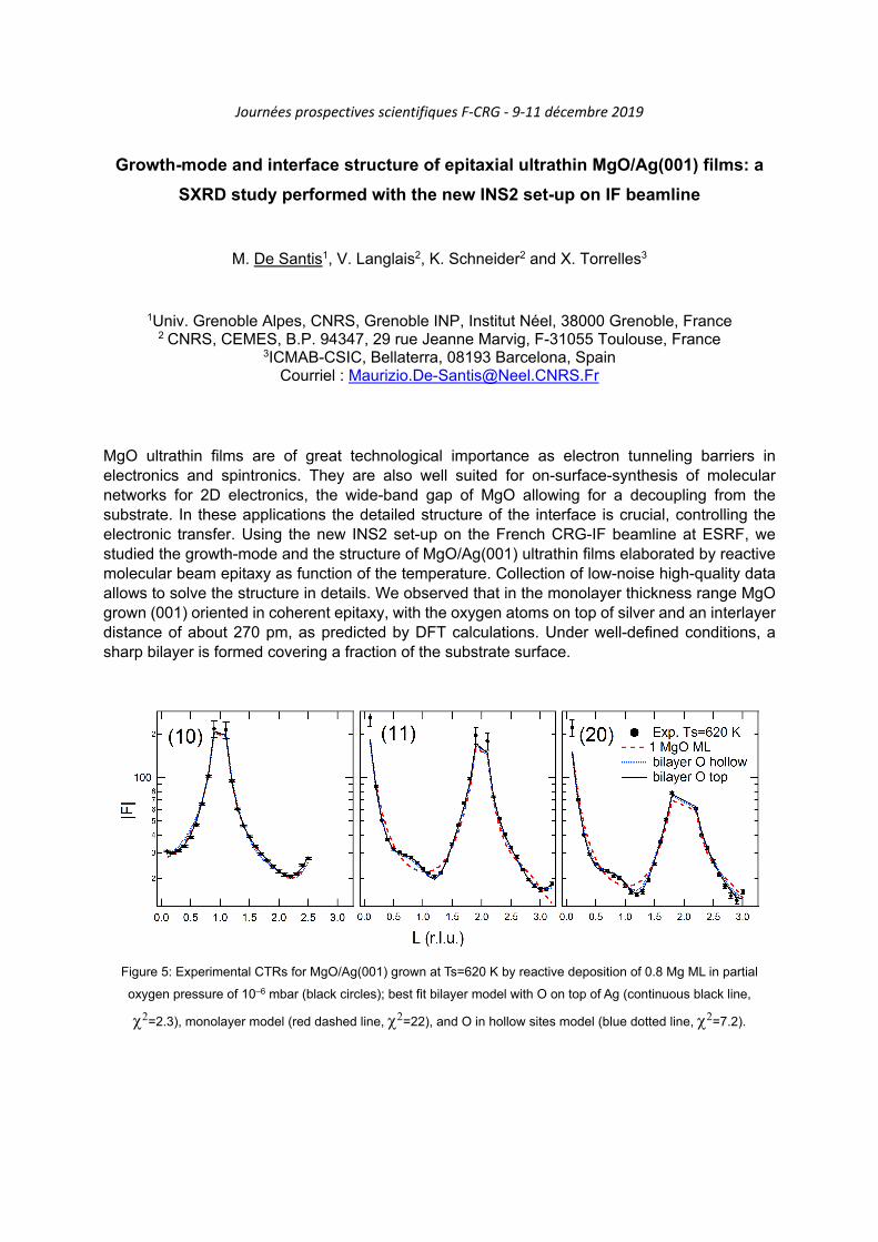
Growth-mode and interface structure of epitaxial ultrathin MgO/Ag(001) films: a
SXRD study performed with the new INS2 set-up on IF beamline
M. De Santis1, V. Langlais2, K. Schneider2 and X. Torrelles3
1Univ. Grenoble Alpes, CNRS, Grenoble INP, Institut Néel, 38000 Grenoble, France 2 CNRS, CEMES, B.P. 94347, 29 rue Jeanne Marvig, F-31055 Toulouse, France
3ICMAB-CSIC, Bellaterra, 08193 Barcelona, Spain Courriel : [email protected]
MgO ultrathin films are of great technological importance as electron tunneling barriers in electronics and spintronics. They are also well suited for on-surface-synthesis of molecular networks for 2D electronics, the wide-band gap of MgO allowing for a decoupling from the substrate. In these applications the detailed structure of the interface is crucial, controlling the electronic transfer. Using the new INS2 set-up on the French CRG-IF beamline at ESRF, we studied the growth-mode and the structure of MgO/Ag(001) ultrathin films elaborated by reactive molecular beam epitaxy as function of the temperature. Collection of low-noise high-quality data allows to solve the structure in details. We observed that in the monolayer thickness range MgO grown (001) oriented in coherent epitaxy, with the oxygen atoms on top of silver and an interlayer distance of about 270 pm, as predicted by DFT calculations. Under well-defined conditions, a sharp bilayer is formed covering a fraction of the substrate surface.
Figure 5: Experimental CTRs for MgO/Ag(001) grown at Ts=620 K by reactive deposition of 0.8 Mg ML in partial
oxygen pressure of 10–6 mbar (black circles); best fit bilayer model with O on top of Ag (continuous black line,
=2.3), monolayer model (red dashed line, =22), and O in hollow sites model (blue dotted line, =7.2).

Combining structural and light emission measurements with X-rays Joël Eymery1, Odile Robach1, Jean-Sébastien Micha2 and Olivier Ulrich1
1 Univ. Grenoble Alpes, CEA, IRIG, MEM, NRS, 38000 Grenoble Cedex, France 2 Univ. Grenoble Alpes, CEA, IRIG, SYMMES, 38000 Grenoble Cedex, France
corresponding author – Joël Eymery: [email protected]
Nitride heterostructures demonstrated novel optical and electronic properties making use of quantum confinement effects and strain engineering. The emergence of disruptive functionalities is strongly related to the growth and technology controls, but also to the development of advanced characterization techniques having high spatial resolution. Focused X-ray beams provide innovative solutions to analyse quantitatively the morphology, defects, strain and composition of these materials. We will present recent breakthroughs obtained at the BM32 beamline of the European synchrotron radiation facility on nitride wires and µLEDS grown by MOVPE. Installed at the European synchrotron (ESRF) in Grenoble (BM32 beamline), the Laue microdiffraction instrument (µLaue) is unique in Europe and probes the matter by diffracting a polychromatic beam of a few hundred nanometers. The acquisition of the Laue diffraction diagram is very fast and allows scanning the samples to get the structural parameters of mono or polycrystalline materials in terms of orientation, crystallographic lattice parameters and state of deformations with a high precision. We added recently to this technique the possibility to record the emitted visible and near IR light excited from X-ray with the so-called XEOL technique (X-ray Excited Optical Luminescence). The acquisition of XEOL spectra (typically 1 s) can be easily synchronized to data collection of the Laue pattern so that to measure on the same specimen’s location. This new setup, just tested before the major upgrade of ESRF, has demonstrated its ability to correlate the crystal structural properties and the visible emission efficiency in nanoscale nitride heterostructures emitters (nanowires and µLEDs) and phosphors used for color management (i.e. white lightning). Some new results will be presented in this poster.
Fig. 1: Left: Studied samples: (a) self-organized wires dispersed on glass, (d) standing wires grown on sapphire. (c) shows the MQW heterostructures and (d) an example of photoluminescence spectra. (e) is a µDisplay structure with its technological integration shown in (f). Right: Evolution of the scattering patterns collected by Laue Microdiffraction as a function of beam location along the nanowire. At the wire tip (right hand side) the multiple components shape of Laue peaks correspond to the periodic structure of the MQWs.

Journées prospectives scientifiques F-CRG - 9-11 décembre 2019
Analyse dimensionnelle, forme et rugosité, de nanostructures
organisées par diffusion des rayons X aux petits angles
J. Rêche1, T. Nguyen-Thanh1, G. Freychet2 et P. Gergaud1
1 Univ. Grenoble Alpes, F-38000 Grenoble, France.
CEA, LETI, MINATEC Campus, F-38054 Grenoble, France. 2 NSLS II, Brookhaven, NY, USA Courriel : [email protected]
Les composants développés en microélectronique doivent être toujours plus performants, tout en étant toujours plus petits. Cette réduction nécessite un contrôle dimensionnel (CD) toujours plus poussé et précis. La diminution continue des tailles de nanostructures repousse les limites des techniques classiques, SEM, AFM, scattérométrie. La diffusion des rayons X aux petits angles (T-SAXS ou GISAXS) est une technique de caractérisation considérée comme prometteuse pour le contrôle dimensionnel par la dernière feuille de route de l’IRDS [1, avec de nombreuses études autour de la technique dite de CD-SAXS [2, 3]. Nous présenterons les capacités du CD-SAXS à travers plusieurs exemples (réseaux de lignes avec des périodicités, largeurs de lignes, profil de lignes et rugosité latérale différents) [4,5]. Un focus particulier sera fait sur l’extraction de la rugosité latérale des lignes. L’amplitude de rugosité mesurée peut être inférieure au nm. Nous montrerons aussi qu’il est possible d’extraire la PSD (Dispersion Spectrale de Puissance) de la rugosité LWR (Line Width Roughness) [5]. Les mesures ont été effectuées sur les lignes de lumière BM05, BM02 et BM32 à l'ESRF. Ces résultats s’appuient aussi sur des simulations inverses de type Monte Carlo Markov pour la reconstruction de la forme de ligne. Les résultats de rugosité sont obtenus soit sur des simulations par transformée de Fourier d’échantillons rugueux virtuels mais aussi sur des échantillons réels élaborés avec des rugosités « aléatoires », i.e. non périodiques mais contrôlées. Remerciements : Ces études ont été financées par le programme ANR/RTB “Recherche Technologique de Base” le projet FUI/Minalogic Nanolytix et le programme IRT Nanoelec. Nous remercions N. Boudet, N. Blanc, S. Tardif et E. Ziegler de l’ESRF pour leur support sur les lignes de lumière CRG-BM02, CRG-BM32 et BM05. Les mesures de laboratoire ont été réalisées sur la plateforme de Nano-Caractérisation de Minatec, Grenoble. [1] IRDS (2017). International Technology Roadmap for Semiconductors, https://irds.ieee.org/. [2] Hu, T., Jones, et al., Journal of Applied Physics, 96, 1983-1987. [3] Jones, R. et al, J. P. (2006) MOEMS, 5, 013001-013007. [4] G. Freychet, PhD Univ. Grenoble Alpes, 2016. [5] J. Rêche, PhD Univ. Grenoble Alpes, 2019. [6] J. Reche, et al, in 34th European Mask and Lithography Conference, Grenoble, France, 2018
Figure 1. Détermination par CD-SAXS de la forme de lignes (a) et de la rugosité latérale de lignes (b).

Journées prospectives scientifiques F-CRG - 9-11 décembre 2019
SSHADE, la base de données européenne de spectroscopie du solide
I. Kieffer1, D. Testemale2, Ph. Bollard3, A. Garenne3, D. Albert3, L. Bonal3, O. Poch3 et B. Schmitt3
1OSUG, FAME, Univ. Grenoble Alpes/CNRS/IRD/Irstea/Météo France, 38000 Grenoble, France
2Institut Néel, Univ. Grenoble Alpes/CNRS/Grenoble INP, 38000 Grenoble, France 3Institut de Planétologie et Astrophysique de Grenoble (IPAG), Univ. Grenoble Alpes/CNRS,
Grenoble, France Courriel : [email protected], [email protected]
L’infrastructure de base de données SSHADE (http://www.sshade.eu) héberge des données spectrales issues de nombreux types de matériaux: minéraux, météorites, matières organiques, …, ainsi que des spectres simulés, couvrant tout le spectre électromagnétique allant des rayons gamma aux ondes radio. Son interface de recherche, de visualisation et d’export est ouverte aux utilisateurs depuis février 2018. Le consortium SSHADE regroupe actuellement 21 laboratoires de 11 pays différents.
Dans ce cadre, les lignes FAME et FAME-UHD de l’ESRF ont intégré SSHADE en créant la base de données SSHADE/FAME avec 2 objectifs principaux:
combler le manque de base de données dans le domaine de la spectroscopie d’absorption X. En effet, peu de bases existent et la description des échantillons y est souvent trop vague pour que les spectres puissent effectivement être réutilisés, amenant les utilisateurs à mesurer très régulièrement les mêmes références et conduisant donc à une perte de temps de faisceau globale.
anticiper la tendance politique actuelle exigeant de rendre les données publiques, mais en allant au-delà, de telle sorte que les données soient effectivement accessibles mais aussi réutilisables, indexées et contenant suffisamment de détails pour qu’elles puissent à terme servir de dépôt de jeux de spectres (utiles pour des codes de calculs utilisant des méthodes d’intelligence artificielle par exemple).
Bénéficiant de l’infrastructure SSHADE, la base de données SSHADE/FAME est pérenne (labellisation INSU en tant que Service National d'Observation). Elle s’appuie sur un data model très élaboré qui permet de décrire très précisément tous les éléments (de l'échantillon à la mesure) et de faire une recherche efficace. De plus, en attribuant un DOI (Digital Object Identifier) à chaque jeu de données, elle offre la possibilité de diffuser les spectres de façon robuste et en accord avec les exigences actuelles de publication. Elle compte déjà plus de 200 spectres XAS (et plus de 1800 spectres en tout).
Le poster présentera la base, ses objectifs, ses caractéristiques et son utilisation, tant du point de vue du « consommateur » que du « fournisseur » de spectres.

Journées prospectives scientifiques F-CRG - 9-11 décembre 2019
Transfer and mechanics of silicon thin films on polymer
L.G. Michaud1, P. Montmeat1, F. Fournel1, C. Castan1, S. Tardif2, F. Rieutord2
1 Univ. Grenoble Alpes, CEA, LETI, 38000 Grenoble, France
2 Univ. Grenoble Alpes, CEA, IRIG, MEM, 38000 Grenoble, France Courriel : [email protected]
Strain engineering is a widespread and effective method in microelectronic to tune semi-conductors properties. For example, applying elastic strain can increase charge carriers’ mobility in silicon based transistors (Li et al., 2014). Our group has developed a new approach to prepare strainable Si thin-films, using transfer on a flexible polymer substrate. Typical Si film thickness range between 20 and 200 nm, over a 200 mm diameter (Montméat et al., 2016). The flexible polymer substrate enables mechanical deformation of the silicon thin film. Moreover, at the nanometric scale, silicon can reach several percent of elastic strain (Zhang et al., 2016). The work presented here describes how ultra-thin monocrystalline silicon films are transferred on different polymer substrates. We also performed tensile tests on resulting samples using uniaxial tensile stage and bulge test. Full strain tensor and crystal orientation were measured using X-rays Laue micro-diffraction at ESRF beamline BM32. In situ measurements over macroscopic areas showed that a high homogeneous strain (>1%) was achievable on a macroscopic area in a silicon on polymer film using external uniaxial (figure 1) or biaxial load. In addition, mechanical coupling between the flexible substrate and silicon film was studied. These promising results should allow us to reach even higher strain level in silicon or in other semi-conductor materials.
Figure 6: Strain components in the crystal reference frame during a uniaxial tensile test along a <110> direction on a 100 nm silicon single crystal, measured by Laue microdiffraction. (left) Diagonal strain component as a function of macroscopic strain (right) Strain tensor mapping on a 2x2 mm² surface at around 0.95 % macroscopic strain (blue vertical line on the left curve)
1) J. Li, Z. Shan, and E. Ma, MRS Bulletin, 2014, 39, 108. 2) P. Montméat, I.D.N. Brandolisi, S. Tardif, T. Enot, G. Enyedi, et al., ECS Trans., 2016, 75, 247. 3) H. Zhang, J. Tersoff, S. Xu, H. Chen, Q. Zhang, et al., Science Advances, 2016, 2, e1501382.

Journées prospectives scientifiques F-CRG - 9-11 décembre 2019
Un écran multi-longueur-d'ondes pour visualiser le fond de basse intensité autour
d'un microfaisceau de rayons X et son emploi pour cartographier in situ les
traces d'irradiation sur des miroirs focalisants "Kirkpatrick-Baez".
O. Robach1,2, J.-S. Micha2,3, O. Ulrich1,2, S. Tardif1,2, A. Vivo4, F. Perrin4, R. Barrett4, S. Irisawa5
1Univ. Grenoble Alpes, CEA, IRIG, DEPHY, MEM, NRS, Grenoble, France ; 2CEA-CNRS CRG-IF BM32 beamline at ESRF, Grenoble, France ; 3Univ. Grenoble Alpes, CNRS, CEA, IRIG,
SYMMES, Grenoble, France ; 4 ESRF, Grenoble, France 5JTEC Corporation, Ibaraki Osaka, Japan
Courriel : [email protected] Le prérequis d'un instrument de microdiffraction est un microfaisceau stable en taille et position. La qualité de la focalisation est habituellement décrite au premier ordre par la largeur à mi-hauteur de la tache focale, et au second ordre par le front d'onde. Un autre critère de qualité est l'absence de fond "longue distance" autour du microfaisceau. Un tel fond, même peu intense, nuit en effet à la mesure de signaux faibles, par exemple pour sonder des cristaux petits ou très déformés au voisinage de cristaux gros et bien cristallisés. En pratique, sur l'instrument de microdiffraction de Laue de la ligne BM32 (Ulrich et al, 2011, Leclere et al., 2016), un fond "longue distance" en forme de croix de taille millimétrique est progressivement apparu autour du microfaisceau. Ce fond a été attribué à une dégradation non uniforme des miroirs de focalisation "Kirkpatrick Baez", sous l'effet de l'irradiation par les rayons X, avec des dépôts visibles de contaminants. L'objectif ici était de développer une technique de diagnostic in situ pour cartographier la surface des miroirs en terme de fond produit. Ceci en vue de tester l'efficacité des différentes techniques de préservation des miroirs, qui seront mises en place au redémarrage de l'instrument, après remise à neuf des miroirs par le fabricant. En réduisant l'ouverture des fentes avant KB et en déplaçant l'hexapode placé sous chacun des miroirs, nous avons pu examiner la tache focale issue de la réflexion d'un rayon fin par les deux miroirs, pour différentes positions x,y d'impact du rayon sur la surface des deux miroirs. L'observation directe de la tache focale avec le microscope optique in situ de l'instrument, via la fluorescence visible d’un cristal en couche mince (YAG), s'est avérée peu pratique, du fait du champ réduit du microscope (300 microns) et des mouvements parasites du microfaisceau lors du scan x,y des miroirs. Nous avons finalement utilisé comme "écran résolu en longueur d'onde" le diagramme de diffraction de Laue d'un cristal de silicium. En effet, autour de chaque spot de Laue du cristal, une croix peu intense fournit une image "mono-longueur d'onde" des pieds de faisceau. Afin d'éloigner sur le détecteur les signaux provenant des différents x (resp. y) sur l'échantillon (à distance échantillon détecteur fixée), nous avons utilisé des cristaux de silicium courbés (diamètre 36 mm) autour de y (resp. x). Nous décrivons dans ce poster les premiers résultats obtenus. En certains points des miroirs, les branches de la croix comportent des oscillations d'intensité, d'origine inexpliquée. Après démontage des miroirs, des profils de topographie optique "sans contact" (A. Vivo et al., 2019) ont été mesurés à l'ESRF. Chez le fabricant, la topographie optique après enlèvement de la couche de métal réfléchissant (S. Irisawa, 2019) a montré un endommagement de l'interface silice/platine. Plusieurs modifications de l'enceinte sous azote des miroirs sont prévues afin de préserver au mieux les miroirs remis à neuf.
1) Ulrich et al., Rev. Sci. Instrum., 2011, 82, 033908 2) Leclere et al., J. Synchr. Rad., 2016, 23, 1395-1400 3) A. Vivo, F. Perrin, ESRF, "Surface metrology measurements report : Fixed curvature BM32 KB mirrors from JTEC
contaminated after almost 7 years of use", April 4th 2019 4) S. Irisawa, JTEC, "Report of surface measurement after de-coating for ESRF mirrors", May 17th 2019

Journées prospectives scientifiques F-CRG - 9-11 décembre 2019
Surface Resonant X-Ray Diffraction to probe the electronic structure at the electrochemical interface
Yvonne Soldo-Olivier1, Eric Sibert2, M. De Santis1 and Y. Joly1
1 Institut Néel (CNRS & Université Grenoble Alpes), 25 rue des Martyrs, Grenoble, France 2 LEPMI St. Martin d’Hères, France
Courriel : [email protected] The “heart” of electrochemistry, science describing the mutual properties of coupled material/electricity in presence of transformation and/or transportation of chemical species, is in the space called “electrochemical interface”. In this singular domain of some Ångstrom of thickness occurs the charge exchange between the conducting electrode and the electrolyte. The description of the electrochemical interface and the comprehension of the related electro-catalytic mechanisms is first and foremost a question of material of electrode and more specifically a problem of surface of this material. However, there is currently no experimental method to specifically probe the electronic structure of the surface directly under electrochemical operation and thus experimental evidence for the theoretical predictions regarding charge distribution is still lacking. In this context, we aim at developing the surface resonant x-ray diffraction (SRXRD) into a standard technique to probe charge distribution and electronic densities for operando electrochemical systems. Indeed, SRXRD couples surface X-Ray diffraction, widely used to solve the atomic structure at the surface of single crystals, to X-Ray absorption near edge spectroscopy (XANES), highly sensitive to the oxidation states. The experimental approach is supported by the theoretical ab initio calculations of the FDMNES home-made software, recently implemented with the simulation of surface diffraction experiments1 and further developed for electrochemical interfaces description. We present here an exploratory in situ SRXRD study (CRG-D2AM) coupled to FDMNES calculations of Pt(111) in 0.1M H2S04, a particularly interesting model surface for the investigation of competitive anions adsorption processes hindering the kinetics of the catalytic processes. Although this system has been extensively studied, the description of adsorbed sulfates in acidic media as a function of the applied potential and of the related charge exchange is still controversial.
1) Y. Joly et al. Journal of Chemical Theory and Computation, 2018, 14( 2),461-1170

Journées prospectives scientifiques F-CRG - 9-11 décembre 2019
Etudes in-situ électriques de films et dispositifs piézo et ferroélectriques:
challenges et perspectives expérimentales
N. Vaxelaire1
1Univ. Grenoble Alpes, CEA, LETI, F-38000 Grenoble. Courriel : [email protected]
Les films piézoélectriques et/ou ferroélectriques connaissent de nombreuses applications dans les capteurs, les transducteurs (PbZrTiO3, KNaNbO3), les filtres radio-fréquences (AlN, LiNbO3) ou les mémoires non volatiles (HfO2). Le CEA/LETI possède une expertise ancienne sur ces thématiques et les développements matériaux actuels portent sur la technologie 5G, les nouvelles générations d’accéléromètres ou sur les mémoires ferroélectriques. Dans chacun des cas, une compréhension fine de la structure et des évolutions (wake-up, vieillissement, dégradation) du film actif lors de cycles électriques est nécessaire pour qualifier les procédés et les designs. Les études in-situ électriques sont alors particulièrement pertinentes pour compléter les caractérisations électriques standards. Dans la pratique, s’il est possible de réaliser des mesures de diffraction in-situ électriques avec des sources de laboratoire [1], il est avantageux d’utiliser les rayonnements synchrotron pour effectuer des expériences plus rapides, et obtenir des faisceau plus petit permettant de ‘’viser’’ les zones d’intérêts où le champ électrique est appliqué. Des mesures de diffraction en mode stroboscopiques [2] en synchronisant le détecteur de rayons X avec le stimulus électrique sont également disponible à la communauté par exemple sur les lignes Diffabs@Soleil ou BM28@ESRF. Dans ce cas particulier, le flux disponible sur les lignes BM est un facteur limitant et reste uniquement envisageable des films diffractant ‘bien’. Les films pour capteurs relativement épais (1µm) sont éligibles à ces caractérisations. En revanche, dans le cas des applications mémoires (films actifs de 10nm), le signal de diffraction est très faible particulièrement lorsque l’on vise un objet unique de quelques dizaines de µm² [3].
Figure 7: (a) Pic de diffraction d’un film de PbZrTiO3 (épaisseur 1µm). Evolution relative de l’intensité (c) et largeur(b) des trois familles de domaine en fonction du champ électrique appliqué.
L’upgrade des CRG ouvrira ces caractérisations in-situ à de nouvelles familles d’échantillons grâce à l’augmentant du flux incident. Une diminution de la divergence de la source pourra faciliter l’obtention de faisceau de petites dimensions sans perte de flux. Des approches pseudo-grazing ou in-plane localisées sur une petite zone d’intérêt pourront alors être envisagées.
1) Allouche et al, Materials & Design (2018) 154, 340 2) Vaxelaire et al (in preparation) 3) François et al, (2019) IEEE 11th International Memory Workshop

Journées prospectives scientifiques F-CRG - 9-11 décembre 2019
Operando synchrotron and neutron-based techniques to probe battery materials
C. Villevieille1
1Université Grenoble-Alpes, Grenoble INP, Laboratoire LEPMI, Saint Martin d’Hères, France Courriel : [email protected]
The Li-ion chemistry is thus far the most advanced chemistry employed in battery technology. To date, Li-ion batteries dominate the market of the electronics and portables devices.1 However, in the field of electric and hybrid vehicles further improvements are required in terms of performance, safety, and cost. The same set of criteria concerns other systems based on alternative chemistries such as Na-ion and solid-state batteries. Advanced Li-ion batteries and the pre-cited novel systems utilize less understood electroactive materials and thus show new reaction mechanisms during electrochemical cycling, the understanding of which requires new characterization tools and techniques. 2 Unfortunately, in most cases the electrochemical cells used for operando measurement are not ideal and suffer from low internal pressure (i.e. poor contact between the electrodes).3 It shorts the lifespan of the cell and as a consequence most of the studies presented in the literature focus on the first/second cycle. Herein we present different cell designs developed in our laboratory and used for operando/in situ studies.4 Having overcome earlier mentioned obstacle our operando/in situ cells are able to sustain more than 100 cycles and simultaneously to perform structural studies such as X-ray and neutron diffraction.5-7 For the latter one, we also developed a new set-up called stroboscopic mode. It allows operando study of the batteries that are cycling at very high rates (e.g. 10C) with a neutron patterns collected each 1 s along 200 cycles and more. Additionally, we will use bulk, surface and imaging techniques to better understand the limitation of all solid state batteries. As an example, Operando neutron imaging has been employed to understand the Li diffusion within the electrode and inside an all-solid state batteries employing Li3PS4 as solid electrolyte whereas operando X-ray microscopic tomography was employed to follow the possible electro-mechanical fracture occurring during cycling and the possible impact on the electrochemical performance.8 Other examples based on different operando/in situ techniques such as X-ray diffraction, neutron diffraction, neutron imaging, and X-ray tomography microscopic used to characterize batteries will be presented here.
1. M. Winter, B. Barnett and K. Xu, Chemical Reviews, 2018, 118, 11433-11456. 2. D. Liu, Z. Shadike, R. Lin, K. Qian, H. Li, K. Li, S. Wang, Q. Yu, M. Liu, S. Ganapathy, X. Qin, Q.-H. Yang, M.
Wagemaker, F. Kang, X.-Q. Yang and B. Li, Advanced Materials, 2019, 31, 1806620. 3. C. Villevieille, T. Sasaki and P. Novak, Rsc Advances, 2014, 4, 6782-6789. 4. J. Conder, R. Bouchet, S. Trabesinger, C. Marino, L. Gubler and C. Villevieille, Nature Energy, 2017, 2. 5. L. Vitoux, M. Reichardt, S. Sallard, P. Novak, D. Sheptyakov and C. Villevieille, Frontiers in Energy Research,
2018, 6. 6. L. Boulet-Roblin, D. Sheptyakov, P. Borel, C. Tessier, P. Novak and C. Villevieille, Journal of Materials
Chemistry A, 2017, 5, 25574-25582. 7. L. Boulet-Roblin, P. Borel, D. Sheptyakov, C. Tessier, P. Novak and C. Villevieille, Journal of Physical Chemistry
C, 2016, 120, 17268-17273. 8. X. Wu, J. Billaud, I. Jerjen, F. Marone, Y. Ishihara, M. Adachi, Y. Adachi, C. Villevieille and Y. Kato, Advanced
Energy Materials, 2019, 9, 1901547.

Journées prospectives scientifiques F-CRG - 9-11 décembre 2019
Investigation of the aging mechanism in nanostructured silicon-based lithium-ion
batteries by in-situ and operando scattering techniques
Diana Zapata Dominguez1, Christopher L. Berhaut1, Praveen Kumar1, Pierre-Henri Jouneau1,
Willy Porcher2, David Aradilla1, Samuel Tardif1, Stéphanie Pouget1 and Sandrine Lyonnard1
1University Grenoble Alpes, CEA, IRIG, F-38054 Grenoble, France
2University Grenoble Alpes, Liten, F-38054 Grenoble, France
Courriel : [email protected]
Lithium ion batteries are one of the best solutions for energy storage mainly due to the lighter weight of the lithium conferring a high energy density. Silicon is an interesting Li-ion host material with a high capacity (3579 mAh/g, 10x larger than graphite). However, it suffers from large volume expansion upon the insertion-disinsertion of the lithium ions, which results in the continuous formation of SEI, leading to Li trapping and to irreversible capacity and electrical contact losses. One way of limiting volume expansion is to use silicon nanoparticles, where the small size enables Li transportation and strain relaxation. At the same time, it is may be interesting to also alloy the silicon with germanium to improve the electronic and ionic conductivity, giving better lithium diffusion1), or to blend it in composite electrodes with graphite2). Yet, the mechanisms of aging (i.e. performance decrease over cycling) are not well known and require detailed analysis to understand their origin and to limit their effects. Here we report on the study of (Si,Ge) nanoparticles-based electrodes with excellent rate capabilities and capacity retention using a combined operando SAXS/WAXS technique on beamline D2AM (BM02) and other complementary techniques. In particular we discuss the respective contribution of Si and Ge upon lithiation and delithiation.
Figure 1. (a) Operando WAXS, (b) zoom on the (Si,Ge) 111 reflection and (c) decomposition of the peak in Si-rich and Ge-rich sub-components, (d) electrochemical measurements during the experiment over several partial cycles
and (e) evolution of the Si and Ge contribution.
1) A. Desrues et al., Batteries & Supercaps 2019, 2, 1 – 10 2) C. Berhaut et al., ACS Nano, 2019, 13, 10, 11538-11551

Journées prospectives scientifiques F-CRG - 9-11 décembre 2019
Monday 9th December 2019
11:00 - 13:30 Registration in the ESRF Central - Building entrance hall
12:00 - 13:30 Lunch at the EPN campus restaurant
Chair : P. Legrand
13:30 - 13:45 Welcome / Introduction
E. Bustarret, F-CRG spokesperson
13:45 - 14:30 Beamlines presentations
Project leaders
14:30 - 15:00 The EBS project
J. Susini, ESRF Director of Research
15:00 - 15:30 The MAGNIFIX project
15:30 - 16:00 Coffee break around the posters
ESRF Central Building entrance hall
Chair : P. Gergaud
16:00 - 16:45
Imaging strain and defects in electronic materials and devices: State of the art and future trends
O. Thomas, IM2NP, Marseille
16:45 - 17:10
Diffusion des Rayons X aux Grands Angles et aux Petits Angles pour la Détermination de la Morphologie de Systèmes Polymères
G. Sudre, IMP, Lyon
17:10 - 18:05
Round-table discussion on F-CRG operation
N. Boudet, O. Proux
P. Legrand, P. Gergaud, R. Guinebretière
19:00 - 21:00 Cocktail at EPN Cafetaria
Tuesday 10th December 2019
Chair : P. Levitz
09:00 - 09:45 The past is the key to the future
A. Thompson, SOLEIL, Paris
09:45 - 10:10
Apports des spectroscopies optiques à la cristallographie macromoléculaire
A. Royant, IBS, Grenoble
10:10 - 10:35
Expériences de cristallographie biologique à façon
D. Pignol, CEA-BIAM, Marseille
10:35 - 11:05 Coffee break around the posters
ESRF Central Building entrance hall
Chair : K. Dumesnil
11:05 - 11:30
X-ray imaging and X-Ray Absorption Spectroscopy at relevant environmental concentrations : Powerful Structural Tools in Environmental Nanotechnologies
M. Auffan, CEREGE, Aix-en-Provence
11:30 - 11:55
Co-crystallization and diffraction in situ for rapid drug design
G. Labesse, CBS, Montpellier
11:55 - 12:40
Round-table discussion on data analysis
J.-S. Micha, Y. Sallaz-Damaz
P. Levitz, A. Boulle
12:40 - 14:10 Lunch at the EPN campus restaurant

Journées prospectives scientifiques F-CRG - 9-11 décembre 2019
Tuesday 10th December 2019
Chair : E. Lacaze
14:10 - 14:55
Synchrotron techniques for the study of batteries: the importance of X-ray spectroscopies
L. Stievano, ICGM, Montpellier
14:55 - 15:20
Structure, propriétés électroniques et réactivités de catalyseurs à base de particules sub-nanométriques de platine supportées sur alumine : une approche multi-techniques
C. Chizallet, IFPEN, Solaize
15:20 - 15:45
Spéciation des produits de fission dans le combustible irradié: apports des lignes de lumière
Y. Pontillon, CEA-DEN, Cadarache
15:45 - 16:15 Coffee break around the posters
ESRF Central Building entrance hall
Chair : J. Blanchard
16:15 - 16:40
Revealing metal speciation in hydrothermal solutions through in-situ XAS and Raman spectroscopy
L. Truche, ISTerre, Grenoble
16:40 - 17:05
BM32 (μLaue) - BM02 : quelques exemples pour la métallurgie
G. Geandier, Inst. J. Lamour, Nancy
17:05 - 17:50
Round-table discussion on in situ, operando, coupling…
N. Blanc, G. Renaud
J. Blanchard, K. Dumesnil, A. Vlad
19:00 - 21:00 Dinner at EPN restaurant
Wednesday 11th December 2019
Chair : O. Thomas
09:00 - 09:45
Spectroscopie d'absorption X pour une approche moléculaire en sciences de l’environnement
G. Morin, IMPMC, Paris
09:45 - 10:10
Benefits of coherence for imaging of complex natural and manufactured material
L. Michot, PHENIX, Paris
10:10 - 10:35
Matériaux bidimensionnels sondés par rayonnement synchrotron
J. Coraux, Institut Néel, Grenoble
10:35 - 11:05 Coffee break around the posters
ESRF Central Building entrance hall
Chair : G. Morin
11:05 - 11:30
Caractérisations micro/nano-structurales de matériaux céramiques par DRX et analyses numériques
A. Boulle, IRCER, Limoges
11:30 - 11:55
Ion transport at the interface between highly mismatched oxide perovskites interfaces
R. Felici, CNR-SPIN, Roma
11:55 - 12:40
Round-table discussion on imaging and coherence
J.-L. Hazemann, J. C. Da Silva
O. Thomas, G. Morin
12:40 - 14:10 Lunch at the EPN campus restaurant