frank labortory of neutron physics-jinr- introduction
DESCRIPTION
This is the introduction about the laboratory of neutron physics belong to JINR- Joint institute of nuclear research. It is located in dubna city, russia. This insitute is one of most center for high energy physics in the world.TRANSCRIPT

FRANK LABORTORY OF NEUTRON PHYSICS
Vu Duc Phu
Institute of Physics ( IOP)
10 Dao Tan-Ba Dinh-Ha Noi-Viet Nam
JOINT INSTITUTE FOR NUCLEAR RESEARCH

ContentsStructure of Frank Laboratory
of Neutron Physics
Three main facilities of FLNP
Three main techniques: RBS, ERDA, PIXE

Structure of Frank Laboratory of Neutron Physics
- FLNP -
Directorate
• Director: Valery Nickolaevich Shvetsov• Laboratory Scientific Leader: Viktor Lazarevich
Aksenov
Scientific Departments
• Division of Nuclear Physics - DNP• Division of Condensed Matter Research and
Development - DCMRDTechnical Departments
• Reactor IBR -2• IREN facility• EG -5

Division of Nuclear Physics - DNP
Leader of department: Valery Nickolaevich Shvetsov
Sector 1
• Investigations of Neutron-nuclear Interactions
Sector 2
• Investigations of Neutron Fundamental Properties
Sector 3
• Neutron Activation Analysis and Applied Research

Division of Condensed Matter Research and Development - DCMRD
Department of Neutron Investigations of
Condensed Matter –DNICM
• Studies of the structure and dynamics of solids, liquids and other condensed systems using and developing neutron scattering methods.
Department of Spectrometers Complex – DSC
• Provide electronics for neutron experiments on the EG-5 and reactor IBR-2

Three main facilities of FLNP
IBR-2 Pulsed Reactor
IREN Facility
EG-5 Accelerator
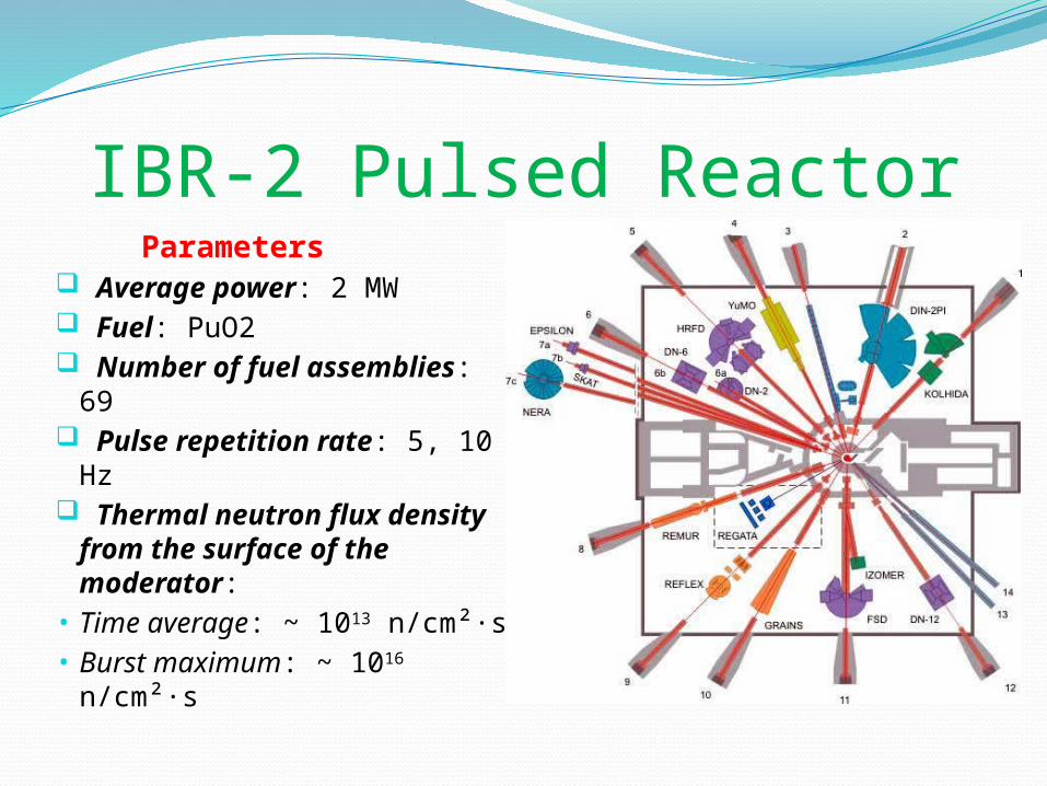
IBR-2 Pulsed Reactor Parameters Average power: 2 MW Fuel: PuO2 Number of fuel
assemblies: 69 Pulse repetition rate: 5,
10 Hz Thermal neutron flux density
from the surface of the moderator:• Time average: ~ 1013
n/cm²·s• Burst maximum: ~ 1016
n/cm²·s

IREN FacilityIntense Resonance Neutron Source
Parameters Peak current: 3 A Repetition rate: 50 Hz Electron pulse duration: 100 ns Electron energy: 30 MeV Beam power: 0.4 kW Multiplication: 1 Neutron intensity: 1011 n/s

EG-5 Van De Graaff Accelerator It was built in 1965 in
Dubna The beam of the accelerated
ions is applied for investigation of the depth distributions of all elements in different samples, using analytical methods : RBS/NR, ERDA and PIXE.
Typical Structure of Van De Graaff Accelerator

EG-5 Van De Graaff Accelerator

Parameters of EG-5:Energy Region : 0.9-
3.5 MeV Beam intensity for H+ : 30μA Beam intensity for He+ :
10μA Energy Spread <500
eV Number of beam lines : 6
EG-5 Van De Graaff Accelerator

RBS-Rutherford Backscattering Spectrometry

HistoryThe Geiger–Marsden
experiment (1909 – 1914)
E. Rutherford give the atom model (1911)
Discovered the nucleus
RBS-Rutherford Backscattering Spectrometry
Thomson model and Rutherford model

RBS-Rutherford Backscattering Spectrometry
• Elastic collision between incident light particle (projectile) with the energy E0 and stationary nucleus (target)
• Scattering kinematic factor :
(apply the conversation of energy and momentum)

• Scattering cross section:
(CM)
- Rutherford’s Formula -
RBS-Rutherford Backscattering Spectrometry

Characteristics of RBS:Near-surface layer analysis of
solids Very sensitive for heavy
elements Elemental composition Depth profiling of individual
elements
RBS-Rutherford Backscattering Spectrometry
A typical spectra of RBS

ERDA – Elastic Recoil Detection Analysis

ERDA was first demonstrated by L’Ecuyer et al. in 1976
ERDA – Elastic Recoil Detection Analysis

ERDA – Elastic Recoil Detection Analysis
Recoil Kinematic Factor :
Recoil cross section

Characteristics of ERDA: Depth profiling of lightest
elements ( H, D,T) High resolution
ERDA – Elastic Recoil Detection Analysis
A typical spectra of ERDA

PIXE – Particle Induced X-ray Emision
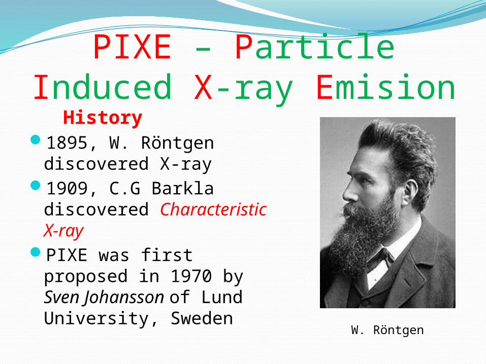
History1895, W. Röntgen discovered X-
ray1909, C.G Barkla discovered
Characteristic X-rayPIXE was first proposed in 1970
by Sven Johansson of Lund University, Sweden
PIXE – Particle Induced X-ray Emision
W. Röntgen

PIXE – Particle Induced X-ray Emision
Bohr Model : introduced by Niels Bohr in 1913
quantum physical interpretation
Explained the spectral emission lines of atomic hydrogen
Then in 1913, H. Moseley give the empirical law of Characteristic X-ray - Moseley’s Law
Bohr Model

PIXE – Particle Induced X-ray Emision
Moseley’s Law:
– Rydberg’s constant Z – atomic number – screening constant n – main quantum number frequency of X-ray quantum

Characteristics of PIXE: High Sensitivity The setup of experiment is
simular one of RBS
PIXE – Particle Induced X-ray Emision

Thank for your attention !!!