forefront - 2012 cancer center publication - mc6783-2012
TRANSCRIPT
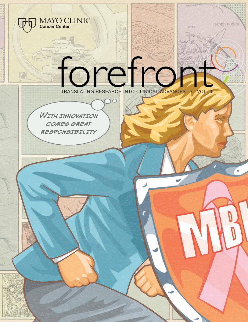
With innovation comes great
responsibility
TranslaTing research inTo clinical advances • vol. 3

Available wherever books are sold or at:
MAyo CliniC CAnCER CEnTER
ouR MissionTo relieve the burdens of cancer by promoting basic and clinical research on the incidence, causes and progression of cancer and translating discoveries into improved methods for prevention, detection, diagnosis, prognosis and therapy.
ouR PRiMARy VAluEThe needs of the patient come first.
VoluME 3
Forefront is the magazine of Mayo Clinic Cancer Center, a leader in translational cancer research and the effort to discover better ways to prevent, detect and treat cancer for patients around the globe.
Managing Editor Joe Dangor
Please address comments to [email protected] or Forefront Magazine, Mayo Clinic, 200 First Street SW Rochester, MN 55905.
Mayo is a nonprofit 501 (c)(3) charitable organization, and contributions are tax-deductible to the extent allowed by law. Contributions support Mayo programs in patient care, medical education and research, which improve the quality of medical care that benefits people everywhere.
© 2012 Mayo Foundation for Medical Education and Research MC6783-1012
The essential guide to breast cancer …
from the most trusted name in medicine.
www.Store.MayoClinic.com
By the Numbers
Director’s MessageFueling discovery
With Innovation Comes Great ResponsibilityThe story of molecular breast imaging
The Secret Life of ResearchersBehind the scenes of one cycle of discovery
Treatment and Love Triumph Over CancerStomach cancer can’t stop a wedding
Meet the InvestigatorProfiles in discovery
Discovering the Link Between Cancer and ObesityThere’s more than meets the eye
Patients Overcome Cancer ChallengesArizona patient symposium educates and inspires
Labnotes 27 Spotlights 28 Directory 29 Parting Shot Back Cover
01
02
04
10
16
18
22
28
Page 4
Page 10
Page 18
Page 28

Articles that advance our understanding of cancer published in peer reviewed journals by Mayo Clinic Cancer Center researchers in 2011.
2227
Number of patients Mayo Clinic Proton BeamTherapy Program will treat per year when centers open in Minnesota and Arizona in 2015 and 2016.
Clinical trials led by Mayo Clinic Cancer Center researchers available to patients in 2011.
Cancer patients in the U.S. on waiting lists for the proton beam therapy in 2011.11,000
2,400
39Years since Mayo Clinic Cancer Center was first named a NCI-designated Comprehensive Cancer Center.
1056

Fueling Discovery
P.S. Earlier this year, we launched a quarterly email newsletter edition of Forefront to provide more frequent updates on research and administrative news. You can explore this new publication online at cancercenter.mayo.edu/forefront.
Welcome to the 2012 edition of Forefront, the annual magazine of the Mayo Clinic Cancer Center.New treatments to control and cure cancer are fueled by the discoveries of researchers and
practitioners. At Mayo Clinic Cancer Center, we bring together more than 1,000 of the nation’s top researchers and practitioners in an integrated setting that provides synergistic benefits far beyond what either group could accomplish alone. The stories in this year’s edition of Forefront celebrate that synergy. Here are a few examples.
Our cover story takes a closer look at molecular breast imaging (MBI). MBI is a new breast cancer imaging tool developed at Mayo Clinic that detects breast cancer in women who have dense breast tissue more effectively than mammography.
On page 10 we’ll give you a behind-the-scenes look at how our researchers and physicians took the discovery of a single protein, nearly two decades ago, and turned it into a series of promising new cancer drugs.
And on page 22, we highlight the groundbreaking work of several Mayo researchers who are identifying the links between excess weight and specific cancers.
Finally, some of you know I’m a big fan of numbers. Opposite this page you’ll see a regular column we call “By the Numbers,” which takes a numeric approach to highlighting key activities at Mayo Clinic Cancer Center over the past year.
I hope you enjoy this edition of Forefront.
Robert B. Diasio, M.D.DirectorMayo Clinic Cancer Center William J. and Charles H. Mayo Professor

4 f o r e f r o n t
How can we use this technology?

Mayo team taps gamma rays for new imaging machine to detect tumors in dense breast tissue.
How can we help this patient?

eborah Rhodes, M.D. had a lot on her mind. She was thinking about a patient who had come to see her about a lump in her breast. The
patient was pregnant and worried. Her sister had been diagnosed with breast cancer in her 40s and the patient, who had very dense breast tissue, wanted to know — if she was to develop breast cancer — how confident was Dr. Rhodes that a mammogram could detect tumors early.
Dr. Rhodes had studied the patient’s mammogram, reviewed the medical literature and was shocked to discover that the chances of finding a tumor early on for a patient with dense breast tissue were less than 50 percent. It was 2003 and magnetic resonance imaging (MRI) was not yet used for screening women at high risk for breast cancer, so the patient’s options seemed limited.
Dr. Rhodes spoke to a radiologist who suggested she speak to another Mayo colleague, Michael O’Connor, Ph.D., a nuclear physicist, who was on his way back from a cardiology conference in Israel.
Dr. O’Connor was interested in a new type of gamma detector he had seen that was made of a thin layer of semiconductor material — a material flexible enough to possibly be used for breast imaging. Unlike X-rays, gamma rays are not influenced by breast density.
Wanting the best for her patient, Dr. Rhodes called Dr. O’Connor and arranged to meet to discuss the gamma detector. That meeting turned out to be prescient.
It wasn’t long before the two of them set to work fashioning a crude version of a new breast imaging technology they dubbed Molecular Breast Imaging, or MBI. The pair was eventually joined by Mayo Clinic biomedical engineer Carrie Hruska, Ph.D., and by radiologists Amy Conners, M.D. and Katie Jones, M.D.
This is a humble group of individuals that eschews attention, and not one of them would ever cast themself as a superhero. But perhaps they should. For even though they don’t possess super human strength, wear suits with built-in weapons, control the weather or shoot spider webs from their wrists, they are on a mission to develop a new tool in the battle against breast cancer.
Carrie Hruska, Ph.D., Michael O’Connor, Ph.D.
and Deborah Rhodes, M.D.
6 f o r e f r o n t

When it comes to breast cancer screening one size does not fit allFor decades, mammography has been the gold standard for diagnosing breast cancer and it remains a proven and powerful tool.
But the MBI team has developed a new technology with the potential to revolutionize breast cancer screening for a specific group of women with dense breast tissue. In preliminary studies, MBI is three times more effective than mammography at finding tumors in dense breast tissue, which is common among premenopausal women.
Dense breast tissue comprises the breast’s milk glands and milk ducts, says Sandhya Pruthi, M.D., a consultant in Mayo’s Breast Clinic in Minnesota. On a mammogram, the dense tissue appears white against the darker fatty tissue. But cancer can also appear white on a mammogram. “That’s why dense breast tissue can obscure the detection of a mass,” Dr. Pruthi says. “We might not see the mass as well through the white part of the mammogram.”
According to Dr. Pruthi, all women have a mixture of dense and fatty breast tissue. The proportion of dense versus fatty tissue depends on factors such as genetics, hormones and age. Before menopause, when a woman’s body is still making estrogen, breast tissue tends to be denser. After hormone production declines with menopause, breast tissue becomes less dense.
Custom breast cancer imaging“Providing personalized, patient-specific care has always been a focus at Mayo Clinic,” says Dr. Rhodes. “Our hope is that MBI will become one of the tools, along with mammography, that we can use to help tailor breast imaging based on the characteristics of a patient’s breast tissue.”
Unlike a mammogram which is an X-ray of breast tissue. MBI, uses a radioisotope tracer which is injected into the patient’s arm before the breast is imaged using a special camera. Breast cells that are behaving abnormally absorb the tracer, and appear on the image as bright white spots.
“A mammogram looks at how tissue appears. MBI tells us what the cells in breast tissue are actually doing — whether they’re actively growing and changing,” explains Dr. Hruska.
Dr. Connors says MBI provides clearer, more detailed images of dense breast tissue. She points out that in a recent Mayo study of more than 1,600 women with dense breast tissue, MBI found 11 tumors per 1,000 breasts imaged compared with 3 per 1,000 with mammography. A previous Mayo study yielded similar
results. “In this group of women with dense breast tissue, we can reliably find about three times more cancers than mammography can,” Dr. Conners says.
She also points out that MBI does not cause as many false positive exams as other techniques used for breast cancer screening such as ultrasound. She adds that it’s a comfortable exam for patients, and relatively straightforward and fast for the radiologist to interpret.
MBI is approved by the Food and Drug Administration (FDA), and is currently used at Mayo Clinic in Minnesota for research and diagnostics. For patients, the test is fairly simple. After the tracer is injected, the breast is compressed in the camera, with two-thirds less pressure than a mammogram. Two images are taken of each breast. The patient is seated during the 40-minute procedure.
n addition to Molecular Breast Imaging (MBI), other alternative methods of breast imaging may be useful
for women with dense breast tissue. These methods are generally used in addition to mammography. Alternative methods include:
Magnetic Resonance Imaging (MRI) uses magnets and computers to create about 3,000 images, each showing a thin, horizontal slice of breast tissue. This highly sensitive test is generally used to diagnose women at severe risk of breast cancer, and to guide treatment of known breast cancer. MRI is considered too complex and expensive for routine breast imaging. “It would be like using an Indy race car to drive to work,” Dr. Whaley says.
Ultrasound uses sound waves to generate images and involves no radiation. The Food and Drug Administration (FDA) recently approved an automated whole-breast ultrasound system. Ultrasound can yield false-positive test results — images that look suspicious but are benign on biopsy. “If automated screening ultrasound causes a lot of unnecessary biopsies, it may prove too costly for routine screening,” Dr. Whaley says.
Tomosynthesis uses advanced digital mammography technology to generate multiple mammogram images showing slices of breast tissue. Tomosynthesis reduces overlapping of dense breast tissue, which can hide or simulate breast cancers.
c a n c e r c e n t e r . m a y o . e d u 7

With MBI, the quality of the image depends on the number of gamma rays detected by the camera. In 2003, the prototype MBI system required a 20 millicurie (mCi) radiation dose — five times the level of a mammogram — to obtain good images. “That was clearly too high,” Dr. Rhodes says.
The Mayo team undertook painstaking efforts to modify the camera and lower the dosage. First, physicists redesigned the collimator — the device that filters gamma rays into the camera — to increase the number of gamma rays captured. That work involved complex computer simulations. “We created models of the gamma camera and the collimator. Then we changed characteristics of the system to see what would happen to the images,” Dr. O’Connor explains. “It’s fairly computationally intense, but Mayo’s computer cluster can run a simulation in a few hours rather than days.”
amy Conners, M.D. and Katie Jones, M.D.
The researchers made further refinements based on the amount of energy used in the system, and then used software algorithms to sharpen images. “We played all these technical tricks, and found we can make a beautiful image at a lower radiation dose,” Dr. Hruska says.
That dose was 8 mCi. In addition, unlike the preliminary study, participants in the recent study had no breast cancer risk factors except dense breast tissue. Yet, the refined MBI system was still three times more effective than mammography in dense breast tissue. “That astonishing series of technical innovations has allowed us to maintain the same high detection rate, at a fraction of the radiation exposure,” Dr. Rhodes says.
8 f o r e f r o n t

hen her mom died of breast cancer at the age of 56 in 1994, Cindy Quaale knew she also was
at high risk for the disease. Her doctor recommended that Cindy start having annual mammograms, and she took comfort in the fact that she was being diligent about screening for the disease. At her mammogram appointment in 2007, Cindy noticed a poster seeking patients for a clinical trial testing a new breast imaging technology called molecular breast imaging (MBI). “My doctor had told me I had dense breast tissue, so I decided to sign up for the trial,” she says.
Cindy’s decision to sign up for the trial may have saved her life. She had an MBI in 2007 and it was negative, then last year before her annual mammogram, Cindy received a letter from Mayo Clinic asking if she would like to have a second MBI. While Cindy’s mammogram came back negative, her MBI revealed a four-centi-meter breast cancer tumor. Cindy sought treatment and today is cancer free. “I was stunned,” she says. “Thank goodness I signed up for the trial.”
The team believes it can lower the radiation dose further. The 8 mCi data were acquired using a format that would allow the physicists to simulate how the images would look with a 4 mCi dose. The results are still being compiled. “But we’re fairly sure 4 mCi will work,” Dr. O’Connor says. “That is the point where we can use MBI routinely for screening.”
MBI is proving to be a powerful research tool, helping Mayo investigate cancer risk in subgroups of women with dense breast tissue. “We know that, as a group, women with dense breast tissue are more likely to get cancer,” Dr. Hruska says. “But the mechanisms are poorly understood. There’s no way of knowing if an individual woman has the type of dense breast tissue that’s going to develop cancer.”
Dr. Hruska’s interest in cancer risk was sparked when she noted intriguing variations in MBIs of healthy dense breast tissue. Some images show uptake of the radiotracer — an indicator of abnormal cell behavior — but in a manner different from uptake by cancer cells. “We sometimes see this radiotracer uptake in a totally benign pattern,” she says. “We don’t under-stand why the tracer is going to the benign tissue. What is
Thank goodness I signed up for
that trial.
different about this dense tissue compared to the dense tissue that doesn’t take up the tracer? And is that the difference that might be able to predict individual risk for cancer?”
To find out, Dr. Hruska is working with other Mayo researchers to study 25 patients with dense breast tissue. After MBI, the patients’ healthy breast tissue is biopsied so the researchers can analyze it for differences that might explain the varying patterns of tracer uptake.
This effort to characterize subtypes of dense breast tissue could result in further individualized breast screening. “If you were a patient with dense breast tissue, you could have a MBI after your mammogram. If cancer wasn’t identified, but MBI showed your dense tissue is highly active, you might benefit from additional screening,” Dr. Hruska says.
Assessing a woman’s breast cancer risk accurately is central to improving early detection and prevention. Eventually, MBI may be used to predict a woman’s risk without having to biopsy her breast tissue. “As a noninvasive imaging technology, MBI is a very powerful tool at an individual level,” Dr. Hruska says. “We’re just getting started on all the possibilities.”n
c a n c e r c e n t e r . m a y o . e d u 9

The secreT
life of researchers
It’s not a stretch to say that the research ecosystem
at Mayo Clinic Cancer Center, shares something in
common with a colony of honey bees. Just as honey
bees forage in the environment to discover pollen
and nectar to maintain their hive and sustain plant life,
Mayo cancer researchers are involved in a continu-
ous cycle of discovery to transform breakthroughs
in the lab into effective treatments in clinical practice.
This is the behind-the-scenes story of one cycle of discovery.
The secreT
life of researchers1 0 f o r e f r o n t

Upon returning to the hive, scout bees
inform the colony about the location of
the food source.
c a n c e r c e n t e r . m a y o . e d u 1 1

eearlier this year at a national medical conference, Mayo researchers created a buzz by sharing some promising news. In preliminary results of a clinical trial, they’d found a novel combination of two cancer treatment drugs — different from chemotherapy — that had effectively reduced the size of neuro-endocrine tumors, the type reported to have taken the life of Apple founder Steve Jobs.
In early findings, 52 percent of the first 25 patients treated with the drug combination had significant regression of their disease. “Even though the study is still ongoing, we saw a surprising number of responses in pancreatic neuroendocrine tumor patients,” says Timothy J. Hobday, M.D., the trial’s director. “The difference we saw was tumor shrinkage, not just stability.” The trial has been extended to treat more patients and better establish how well the new drug combination works.
The same two-drug combo therapy is relevant to a wide range of cancers and by applying the drugs involved in the trial, Mayo researchers have been responsible for propagating a series of important discoveries, all aimed at improving treatment for patients. “From the start, it’s been an ongoing back and forth between the lab and the clinic,” says oncologist Scott Kaufmann, M.D., Ph.D., consultant in medical oncology, hematology and pharmacology and the Helen C. Levitt Professor.
Discover, share and informThe story began nearly two decades ago at the bench of a basic science lab at Mayo. In the late 1980s, a researcher in pharma-cology, Robert Abraham, Ph.D., discovered and characterized a protein that’s present throughout the body and also happens to be particularly critical to the growth of cancer cells. He found that the protein bound to a drug called rapamycin, used to suppress the immune response of patients who have received a transplanted organ.
Inspired by the dance, other bees leave the hive to travel to the food. There, they collect nectar and pollen to bring back to the hive.
Through a simple dance, they communicate the specific distance and direction to fly to find the food.
1 2 f o r e f r o n t

c a n c e r c e n t e r . m a y o . e d u 1 3
The discovery of the protein, called mTOR (mammalian target of rapamycin), provided new insight about how to block its function. Dr. Abraham and colleagues at Mayo contacted Wyeth, the drug company that produced rapamycin. “We showed them what this could do in the way of anti-tumor effects in experi-mental systems,” recalls Charles Erlichman, M.D., consultant in medical oncology and the Peter and Frances Georgeson Professor of Gastroenterology Cancer Research.
The findings led Wyeth to develop a new drug, called temsiro-limus, an analog or relative of rapamycin. Though Dr. Abraham moved on to another research center, Dr. Erlichman and other collaborators at Mayo pushed the research ahead, initiating the first safety tests of temsirolimus in human patients.
Inspire, study, testThe buzz about mTOR piqued the interest of several cancer researchers, and new clinical trials were soon under way. Because mTOR works in cancer cells by prompting growth factors that help cells multiply and grow blood vessels, it made sense to test an mTOR inhibitor in cancers — such as renal cell cancer — where those growth factors appear in abundance. The thinking about temsirolimus bore out. Initial studies at Mayo ultimately led to large clinical trials performed worldwide that resulted in Food and Drug Administration (FDA) approval of the drug for renal cell cancer.
One idea that seemed like a stretch was testing temsirolimus for patients with mantle cell lymphoma, a particularly hard-to-treat subset of the cancer that occurs in the immune cells. Thomas Witzig, M.D., Mayo’s chair of research in hematology, applied to the National Cancer Institute (NCI) for permission to investigate whether the drug would affect the similar path-ways turned on in mantle cell lymphoma. But Dr. Witzig says the proposal encountered an unexpected glitch. “The NCI was looking for drugs for solid tumors,” he says.
“Somehow the grant found its way into a drawer,” recalls Dr. Kaufmann, who was involved in early discussions about how the drug might be used. A few years later, however, the NCI asked Dr. Witzig to begin those trials. The results were impressive. “Temsirolimus had a 38 percent single agent response rate, which at the time was unprecedented for that subtype of lymphoma,” Dr. Kaufmann says.
“ from the start, it’s been an ongoing back and forth between the lab and the clinic.” — Scott Kaufmann, M.D., Ph.D.

Dr. Witzig has pushed the research forward, testing other rapamycin analogs, and he’s now finding analogs active in a wide range of lymphomas.
At the same time in another Mayo lab, biochemist and molecu-lar biologist Debabrata Mukhopadhyay, Ph.D., was investigating rapamycin from another perspective. He showed in animal models and in tissue cultures that it may be possible to shrink pancreatic tumors if rapamycin was combined with a particular antibody capable of blocking blood vessel growth, known as angiogenesis. “If you put the two together, you could have a greater effect than either drug alone,” Dr. Mukhopadhyay says.
With those findings, Dr. Erlichman and others began thinking about how a mTOR inhibitor might work as part of a combina-tion therapy for renal cell cancer. They launched studies testing the combination of temsirolimus and the antibody bevacizumab. Those findings, presented at several national cancer meetings, have shown the combination is capable of significant activity. A formal manuscript is in progress and two large multicenter clinical trials, one led by Wyeth and the other by the Eastern Cooperative Research Group, a clinical research organization, are under way.
CollaborateAs Mayo researchers continue to scout mTOR inhibitors, they are opening doors to new potential drug therapies.
For rare neuroendocrine tumors, like those Dr. Hobday specializes in treating, mTOR inhibitors may be critical. Dr. Hobday led Mayo’s participation in a multicenter phase III randomized clinical trial of the oral mTOR inhibitor everolimus. The study was funded and conducted by the drug company Novartis. The results, published last year in the New England Journal of Medicine, showed the drug was more effective than a placebo at slowing the progression of the disease. The studies led to FDA approval of everolimus for these rare malignancies.
Another Mayo researcher, oncologist Jan Buckner, M.D., is studying the effectiveness of mTOR inhibitors in brain cancers.
Dr. Erlichman is leading a study investigating the effective-ness of the two-drug combo, temsirolimus and the antibody bevacizumab, in five different tumor types: pancreatic neuroendocrine tumors, carcinoid tumors, ovarian cancer, endometrial cancer, and hepatocellular carcinoma. All have similar molecular pathways that may respond well to the two-drug combo.
1 4 f o r e f r o n t
“ it really hits home how we depend on basic science research to move the field forward. i’m very hopeful about this drug combination. i think ultimately it may provide another important treatment option for our patients.”
— Charles Erlichman, M.D.

Patient impactWith the great variation among types of cancer and the tremendous complexity of molecular pathways, more work lies ahead. Reflecting on the initial discovery of mTOR, Dr. Erlichman says, “It really hits home how we depend on basic science research to move the field forward. I’m very hopeful about this drug combination. I think ultimately it may provide another important treatment option for our patients.” n
This continuous cycle of discovery is essential for the pollination, flowering,
and fruiting of the food source, thus sustaining
growth of plant life.
c a n c e r c e n t e r . m a y o . e d u 1 5

In June 2011, 33-year-old Regan Roloff was a month away from marrying her sweetheart. She had a great job as a 911 dispatcher in Adrian, Minn.; she was planning her wedding
and looking toward the future with excitement and anticipation. Her world was good.
But in an instant everything changed. Symptoms of a life-threatening disease hit her with such force that she had to call 911 to get help from her colleagues. That event sent her on a journey no one expected.
Regan began to experience severe abdominal pain late in the day. She was hunched over and could barely move or talk. Her colleague, dispatcher Larry Rogers, was on duty the night Regan called 911 for help. “I knew it was Larry,” says Regan. “I knew he was going to take care of me. He’s kind of like a papa bear at work.”
Within minutes, paramedics reached Regan and transported her to a nearby hospital.
By that time, Regan’s fiancée, Lonnie, a local sheriff’s deputy, was at her side. He had been out of town preparing for their upcoming wedding. Both he and Regan figured maybe she had a bad ulcer. About a week later they met with a doctor.
“I could tell right away when he walked in that this was going to be bad news,” she says. The couple was stunned as the doctor shared the diagnosis. Cancer had perforated the wall of her
stomach, and there was a chance it had spread throughout her abdomen. “I remember thinking, ‘I have a wedding in four weeks — we’re getting married in four weeks.’”
The couple vowed not to let cancer destroy their dream. They decided to keep their wedding date, regardless of the circum-stances. They went to Mayo Clinic in Rochester, Minn., where surgeon John Donohue, M.D., led the medical team who cared for Regan.
“We started off with a look in the abdomen to see if there was any evidence of spread of the tumor away from the stomach itself,” says Dr. Donohue. The good news was that the tumor had not spread. To prevent it from spreading, Regan started treatment with chemotherapy. “There are probably seeds or cancer cells out there that we can’t see that the chemotherapy can treat,” says Robert McWilliams, M.D., a Mayo Clinic oncologist. “The idea is if you give a little bit of chemotherapy before the surgery, you might decrease the size of the surgery you might have to perform.”
A week after her first chemotherapy treatment, the couple was married. They decided not to talk about Regan’s health on that day. It was a day to celebrate their love and their future together. “The wedding ended up being fantastic,” she says.
But shortly after the wedding, they renewed their focused on her treatment, with two more rounds of chemotherapy and then a surgery that involved removing two-thirds of her stomach. A section of small intestine was then attached to the remaining stomach, allowing her to eat.
Even after surgery, her doctors were concerned that cancer could still be present, but the pathologist looked at all the slides and didn’t find any residual cancer, she says.
With treatment behind her, Regan continues to get stronger. She’s now cancer free and back at work with her colleagues who she says are part of her work family. Regan sees her doctors every three months for follow-up tests to make sure she remains cancer free. And, together with Lonnie she looks forward to the future. Her story of triumph, determination and love serves as an inspiration to many. n
Treatment and love triumph
Regan and Lonnie on their wedding day.
1 6 f o r e f r o n t

c a n c e r c e n t e r . m a y o . e d u 1 7
over cancer

At Mayo Clinic Cancer Center, hundreds of researchers dedicate their professional lives to lessening the burden of cancer on the world. Each one has a unique story to tell. Here are the stories of three.
MEET THE INvESTIgAToR
1 8 f o r e f r o n t

c a n c e r c e n t e r . m a y o . e d u 1 9
Listening to Panos Anastasiadis, Ph.D., describe cancer cell behavior is a bit like listening to a psycholo-gist describing the actions of a wayward teen. Why? Because at their core, cancer cells are anti-social, Dr. Anastasiadis says. “In any multi cell organism, the cells have to adhere together, to communicate with each other, to work together in order to form tissues and organs.”
As chair of cancer biology at Mayo Clinic in Florida and director of a lab that focuses on metastasis and brain cancer, Dr. Anastasiadis leads a team of researchers who are investigating the role of cell behavior in the spread of cancer.
“Very early in my career I became interested in the way cells conn-ected with each other,” he says. “I started looking at that in the brain, first in Parkinson’s disease.”
Dr. Anastasiadis says that in Parkinson’s, neurons lose their connections with each other and die, but how these connections, impact cell survival was a mystery. He thought that some type of communication must be responsible for maintaining proper cell connections. He started looking at the connectors — adhesion
molecules — and found that in cancer, the signals which emanated from these molecules was irregular. When cancer cells lose the function of these molecules during tumor progression, they disorganize, change shape and become more migratory and invasive.
Signaling is important in cancer because it’s a key event in metastasis, a process that causes cells to migrate and spread throughout the body. “Almost without exception, what kills cancer patients is metastasis, or cancer spread, not the primary tumor,” Dr. Anastasiadis says.
“Mayo Clinic is one of the largest treatment centers in the country for brain cancers,” he says. “That gives us the ability to do research that few other places can perform, because they just don’t see enough patients.”
“I have several people working in my lab every day, and every day one of them is going to give me some good news,” Dr. Anastasiadis says. “Every day, one of them is going to tell me something that we didn’t know yesterday. The greatest feeling is when you can translate that knowledge into something that can help patients. That’s what we’re here for, to make a difference for patients.” n
Panos Anastasiadis, Ph.D.Uncovering the role of cell behavior in the spread of cancer
2 0 f o r e f r o n t
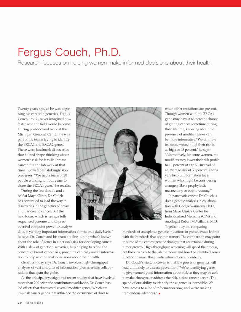
Fergus Couch, Ph.D.Research focuses on helping women make informed decisions about their health
Twenty years ago, as he was begin-ning his career in genetics, Fergus Couch, Ph.D., never imagined how fast-paced the field would become. During postdoctoral work at the Michigan Genome Center, he was part of the teams trying to identify the BRCA1 and BRCA2 genes. These were landmark discoveries that helped shape thinking about women’s risk for familial breast cancer. But the lab work at that time involved painstakingly slow processes. “We had a team of 20 people working for four years to clone the BRCA1 gene,” he recalls.
During the last decade and a half at Mayo Clinic, Dr. Couch has continued to lead the way in discoveries in the genetics of breast and pancreatic cancer. But the field today, which is using a fully sequenced genome and unprec-edented computer power to analyze data, is yielding important information almost on a daily basis,” he says. Dr. Couch and his team are fine-tuning what’s known about the role of genes in a person’s risk for developing cancer. With a slew of genetic discoveries, he’s helping to refine the concept of breast cancer risk, providing clinically useful informa-tion to help women make decisions about their health.
Genetics today, says Dr. Couch, involves high-throughput analyses of vast amounts of information, plus scientific collabo-rations that span the globe.
As the principal investigator of recent studies that have involved more than 200 scientific contributors worldwide, Dr. Couch has led efforts that discovered several “modifier genes,” which are low-risk cancer genes that influence the occurrence of disease
when other mutations are present. Though women with the BRCA1 gene may have a 65 percent chance of getting cancer sometime during their lifetime, knowing about the presence of modifier genes can be more informative. “We can now tell some women that their risk is as high as 95 percent,” he says. “Alternatively, for some women, the modifiers may lower their risk profile to 10 percent at age 50, instead of an average risk of 30 percent. That’s very helpful information for a woman who might be considering a surgery like a prophylactic mastectomy or oophorectomy.”
In pancreatic cancer, Dr. Couch is doing genetic analyses in collabora-tion with George Vasmatzis, Ph.D., from Mayo Clinic’s Center for Individualized Medicine (CIM) and oncologist Robert McWilliams, M.D. Together they are comparing
hundreds of unexplored genetic mutations in precancerous lesions with the hundreds that occur in tumors. The comparison may point to some of the earliest genetic changes that are retained during tumor growth. High-throughput screening will speed the process, but then it’s back to the lab to understand how the identified genes function to make therapeutic intervention a possibility.
Dr. Couch’s view, however, is that the power of genetics will lead ultimately to disease prevention. “We’re identifying genes to give women good information about risk so they may be able to make changes, or address the risk, before cancer occurs. The speed of our ability to identify these genes is incredible. We have access to a lot of information now, and we’re making tremendous advances.” n

c a n c e r c e n t e r . m a y o . e d u 2 1
Raoul Tibes, M.D., Ph.D.Looking for leukemia’s achilles heel, one gene at a time
If such a drug does not yet exist, Dr. Tibes and his research team look for new molecules – ones that could become drugs in the future. Dr. Tibes is leading several early stage clinical trials in looking for new therapies for acute leukemia and MDS.
As both a practitioner and researcher, Dr. Tibes is able to take discoveries made in the laboratory directly into patient care. He is involved in about 35 Phase 1 or 2 clinical trials aimed at developing new cancer therapies. “If patients, who have not responded to previous therapies, do not want to take highly intensive chemo-therapy or be in the hospital for several weeks, we offer them alternative therapies,” he says. “My goal at Mayo Clinic is to offer the latest and most inn-
ovative medications now and to develop new treatments for leukemia and MDS patients in the future.”
Dr. Tibes trained in Germany before coming to the U.S. to complete his medical residency and fellowship programs. After becoming interested in hematological malignancies, those that affect blood and bone marrow, Dr. Tibes dedicated himself to both academic research and clinical practice. These dual passions made Mayo Clinic a perfect fit for him. He can improve treat-ment for cancer patients as both a physician and researcher.
“Medicine is the ideal profession for me,” Dr. Tibes says. “It is based on scientific knowledge and human disease and it is embedded in a very social environment. I really cherish and enjoy the combination of each aspect.” n
As an oncologist and translational researcher, Raoul Tibes, M.D., Ph.D. sees his patients’ unwavering spirit as they battle leukemia. “All of my patients have a tremendous great-ness and dignity, even when they suffer from advanced disease,” he says, and he wants to make a differ-ence for these patients right now.
At Mayo Clinic in Arizona, Dr. Tibes spearheads the Acute and Chronic Leukemia Program, which brings Mayo Clinic leukemia and blood cancer specialists together to provide comprehensive patient care and access to new drugs undergoing clinical development for leukemia and myelodysplastic syndromes (MDS).
“There’s a great sense of urgency when treating leukemia,” Dr. Tibes says. “The limited effectiveness of available drugs makes it difficult for doctors to treat leukemia and MDS.” As a result, Dr. Tibes’ research focuses on discovering molecular abnormalities in leukemia in order to target them with new drug combinations that will lead to better treatment options.
“In the laboratory, I developed a way to inhibit—or switch off—each gene in leukemia cells, one gene at a time,” Dr. Tibes says. “We can do this simultaneously for hundreds to thousands of genes. This approach allows us to identify each gene’s function and learn whether removing a certain function will kill the leukemia cell.
To make the quickest impact possible, as soon as Dr. Tibes and his team identify potential gene targets, they search for existing drugs to alter their function. “Once we find a gene that may be deadly to leukemia cells, we test whether this gene could be the Achilles’ heel of leukemia.”
At Mayo Clinic Cancer Center, several leading researchers are providing a clearer picture of how
obesity affects cancer risks and survival rates.
Although the overall rate of new cancer cases is declining, a recent
report co-authored by researchers from the Centers for Disease Control,
the North American Association of Central Cancer Registries, the National
Cancer Institute and the American Cancer Society confirms research
showing that excess weight and a sedentary lifestyle are risk factors for
1/4 to 1/3 of common cancers. About one-third of adults in the United
States — almost 78 million — are obese which is defined as being
more than 30 pounds over healthy weight.
Research going on at Mayo Clinic Cancer is paving the way for new
treatments and preventive strategies to address this confounding and
ubiquitous twist to the cancer puzzle.
Discovering the Link between
2 2 f o r e f r o n t
cancer obesity

c a n c e r c e n t e r . m a y o . e d u 2 3
uring the last several years, hematologist-oncologist Edith Perez, M.D., has become increasingly emphatic about the importance of exercise, balanced dietary intake, and prevention of obesity in the battle against cancer. It may seem like a simple prescription, but Dr. Perez, the Serene M. and Frances C. Durling Professor, is at the fore of a new wave of research evaluating how the nation’s changing physique is intersecting with cancer care.
“There’s no doubt obesity is becoming more prevalent in the United States and many other countries,” she says. “What’s now becoming evident is that it’s an urgent issue that pertains to the types of tumors people get and their survival after treatment.
A study recently presented by Dr. Perez’s group evaluated women with early stage HER2-positive breast cancer,
which accounts for a 1/3 of all invasive breast tumors. The study found that obese women—which is defined by the World Health Organization as a body mass index greater than 30 percent—tended to develop larger tumors than women with normal BMI. In addition, they were more likely to have cancer-ous cells in their lymph nodes.
While medications, including the anti-HER2 therapy known as Herceptin, were effective in treating breast cancer in obese women and those of normal weight, the study found obese
women had slightly worse outcomes overall. The same is turning out to be true in other subtypes of breast cancer, says Dr. Perez. In a current study she’s looking at the highly aggressive triple nega-tive breast cancer, and she adds, “we’re seeing it all over again. Obesity is associated with increased risk and worse outcomes.”
Among her goals is to identify new treatments that might improve the course of treatment for obese patients. Little is known about how obesity alters tumor growth. Obese women tend to have abnormally high levels of estrogen, which can promote some tumors. Excessive weight can also lead to dysregulated insulin, which also may provide an environment that helps cancer grow.
Dr. Perez’s group is currently participating with investiga-tors throughout North America in a large, randomized clini-cal trial, investigating the effectiveness of adding the drug metformin to breast cancer chemotherapy for obese patients. Already approved by the Food and Drug Administration and other regulatory agencies for management of diabetes-related glucose intolerance, metformin has shown preliminary effec-tiveness against cancers. “We don’t know exactly why,” she says, “but it appears to have properties that influence the biology of breast cancer.”
“ we’re seeing it all over again. obesity is associated with increased risk and worse outcomes.”
— Edith Perez, M.D.
Fat cells
InsulIn
Pancreas
Insulin may play a role in fueling cancer. The more carbohydrates you consume, the faster your blood sugar
rises. In response, your pancreas pumps out lots of insulin to move that sugar into cells...but insulin also promotes fat storage.
Excessive fat – obesity – can lead to problems in insulin production. Plus, all of this sugar and insulin generates
large numbers of hazardous free radicals and creates an environment in which cancer could develop.

2 4 f o r e f r o n t
Esophageal CancerBecause acid reflux and excessive weight are known risk factors for highly lethal esophageal cancer, and because cases are dramatically on the rise, oncologist Harry Yoon, M.D., has been interested in how well obese patients fare after treatment. He studied the outcomes of nearly 800 patients with esophageal adenocarcinoma, all of whom had undergone surgery at Mayo Clinic and had their tumors successfully removed. The results showed that, among nonsmokers, obese patients (with
BMI of 30 or higher) were twice as likely to die of their
cancer than patients of normal weight: The five-year
survival in obese patients was 18%, compared to 36% in patients of normal weight. “This was the first study to show that being obese has an adverse effect on survival in this tumor type,” he says.
One of the biggest problems patients face after surgery is micrometastatic disease, undetectable cancerous cells that have seeded distant organs and ultimately give rise to new tumors. Yet it’s not entirely clear why obese patients might be more
susceptible to the development of these remote tumors. “There’s some thinking that adipose cells themselves may have a pro-tumor effect on micrometastatic disease,” Dr. Yoon says. In addition, insulin dysregulation and other pathways may contribute to cancer’s inflammatory pathways.
While the exact mechanisms remain to be clarified, Yoon has begun discussing weight management among esophageal cancer survivors: “In the past, I didn’t routinely talk with esophageal cancer patients about excess weight, because the more common issue is their regaining weight that had been lost around the time of surgery, swallowing and nutrition, and recurrent cancer. Plus, there wasn’t evidence in this disease that being obese was worse. Now the issue of excess weight is on my radar, and I discuss weight management with patients.”
Dr. Yoon hopes to expand the outcomes study to even larger cohorts of patients and to other institutions, and to seek ways to intervene in the disease process.
“ this was the first study to show that being obese has an adverse effect on survival in this tumor type.”
— Harry Yoon, M.D.
Fat cells may have a pro-tumor effect that assists in the spread of cancer. Cancer
cells can survive after treatment and “seed” in distant organs, giving rise to new tumors. Having excess fat may make a patient more
susceptible to the spread of cancer.Fat cells
cancer cells
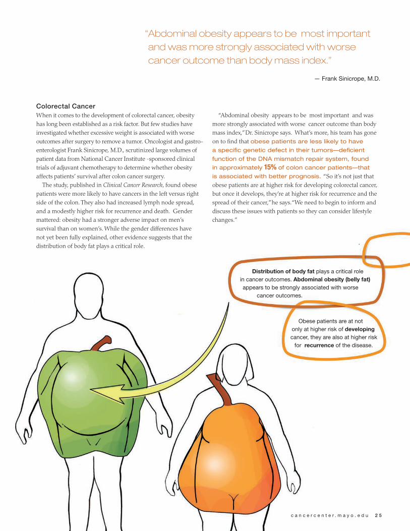
Colorectal CancerWhen it comes to the development of colorectal cancer, obesity has long been established as a risk factor. But few studies have investigated whether excessive weight is associated with worse outcomes after surgery to remove a tumor. Oncologist and gastro-enterologist Frank Sinicrope, M.D., scrutinized large volumes of patient data from National Cancer Institute -sponsored clinical trials of adjuvant chemotherapy to determine whether obesity affects patients’ survival after colon cancer surgery.
The study, published in Clinical Cancer Research, found obese patients were more likely to have cancers in the left versus right side of the colon. They also had increased lymph node spread, and a modestly higher risk for recurrence and death. Gender mattered: obesity had a stronger adverse impact on men’s survival than on women’s. While the gender differences have not yet been fully explained, other evidence suggests that the distribution of body fat plays a critical role.
“Abdominal obesity appears to be most important and was more strongly associated with worse cancer outcome than body mass index,” Dr. Sinicrope says. What’s more, his team has gone on to find that obese patients are less likely to have
a specific genetic defect in their tumors—deficient
function of the DNA mismatch repair system, found
in approximately 15% of colon cancer patients—that
is associated with better prognosis. “So it’s not just that obese patients are at higher risk for developing colorectal cancer, but once it develops, they’re at higher risk for recurrence and the spread of their cancer,” he says. “We need to begin to inform and discuss these issues with patients so they can consider lifestyle changes.”
“ Abdominal obesity appears to be most important and was more strongly associated with worse cancer outcome than body mass index.”
— Frank Sinicrope, M.D.
Distribution of body fat plays a critical role in cancer outcomes. abdominal obesity (belly fat) appears to be strongly associated with worse
cancer outcomes.
Obese patients are at not only at higher risk of developing cancer, they are also at higher risk
for recurrence of the disease.
c a n c e r c e n t e r . m a y o . e d u 2 5

2 6 f o r e f r o n t
Liver CancerEven indirectly, obesity may be leading to the rise of certain cancers. A study by Mayo Clinic gastroenterologist and hepa-tologist W. Ray Kim, M.D., suggested such a trend is in the works in liver cancer, which has tripled in the U.S. in the last three decades and has only a 10 to 12 percent five-year survival rate when it’s detected in later stages. Looking at the effect of hepatitis C on liver cancer within Olmsted County, Minn. Dr. Kim found an increase in cases of fatty liver disease, which can result from obesity and can develop into liver-scarring cirrhosis. “If you have cirrhosis,” he says, “that’s an automatic risk factor for hepatocellular carcinoma.” While only 11 percent of cases of HCC were linked to obesity, Dr. Kim says they may foreshadow a larger problem. “It may be a small percentage of cases overall,” Dr. Kim says. “But with the nationwide obesity epidemic, we believe the rates of liver cancer may dramatically increase in the foreseeable future.”
What doctors are quick to point out is that weight happens to be one cancer risk factor that’s pretty much within patients’ control. Dr. Perez hopes that all the emerging data will prompt more individual awareness about the importance of eating well-balanced meals and getting exercise. “Maintaining adequate weight and exercising are two things that people can take charge of,” she points out. She’s working to get the word out through education and by encouraging all exercise regimens that will get people off the couch. The path to decrease risk and improve outcomes after breast cancer is well outlined, “While we continue to conduct laboratory and clinical research to unravel the molecular biology of cancer and come up with even better therapies than we have today all people can also actively participate by influencing factors that are within their control. There are some healthier lifestyle factors that can help improve people’s lives.” n
“ with the nationwide obesity epidemic, we believe the rates of liver cancer may dramatically increase in the foreseeable future.”
— W. Ray Kim, M.D.
Obesity may lead to other conditions that are risk factors for developing cancer. One of those
conditions is fatty liver, which can in some cases develop into cirrhosis (scar tissue in the liver). Cirrhosis has in turn
been associated with increased risk of liver tumors.
Fatty lIver
cIrrotIc lIver
lIver cancer

c a n c e r c e n t e r . m a y o . e d u 2 7
Proton Beam Therapy Program Receives Benefactor gift
In June, Mayo Clinic announced that Lawrence W. and Marilyn W. Matteson of Moline, Ill., had given $10 million to help launch the Mayo Clinic Proton Beam Therapy Program and that it would use matching gift funds to establish the Lawrence W. and Marilyn W. Matteson Fund in Cancer Research.
When available to patients in 2015 in Rochester, Minn., and in 2016 in Phoenix, Ariz., Mayo’s Proton Beam Therapy Program will use the most advanced intensity-modulated technol-ogy known as pencil beam scanning, which few centers currently use.
Unlike conventional radiation therapy that can damage healthy tissue while it destroys the tumor, proton beam therapy delivers nearly its entire dose within the tumor, sparing healthy tissue surround-ing the cancer. This form of therapy is vital in sensitive areas such as the brain and lungs and is especially useful for treating children, who are at higher risk for radiation damage because their bones and tissues are still growing.
“Mr. Matteson is a grateful patient who was very impressed with the care he received at Mayo Clinic,” says Robert Foote, M.D., chair of the Mayo Clinic Department of Radiation Oncology in
Rochester. “Mr. and Mrs. Matteson wanted a way to show their gratitude and chose to donate funds that will help us ease the burden cancer has on patients, especially young children.”
“This gift is going to benefit children who are going through terrible cancers to not have the residual effects they would have with regular treatment,” said Marilyn Matteson. “The side effects won’t be something that follows them for the rest of their lives.”
Lawrence Matteson and his son, Larry, of Burlington, Iowa, are the founders of L.W. Matteson, Inc., a marine construction and dredging company in Burlington. The company maintained one of the largest fleets of dredging and marine construction equipment on the Mississippi River. The company was sold in 2010. Matteson and his son maintain ownership of Matteson Marine, which operates switch boats on the Mississippi. Marilyn Matteson is retired from John Deere.
The Mayo Matching Gift Program recognizes that endowment of priority programs is critical for the future. In 2005, Mayo Clinic’s Board of Trustees created a matching gift program to build and enhance specific endowments represent-ing Mayo’s highest research priorities. n
Mayo Clinic, ASU collaborate on biomedical informatics
Arizona State University (ASU) recently relocated its Biomedical Informatics department to the Scottsdale campus of Mayo Clinic. This move is part of a formal commitment to deepen collab-orative efforts between Mayo Clinic and ASU in health care, medical research and education.
Biomedical informatics is the science that translates information into knowl-edge. It is a key part of the burgeoning field of individualized medicine, which aims to more precisely treat patients with cancer.
Housing ASU’s Biomedical Informatics department on the Mayo campus will allow ASU students enrolled in the program to work side by side with prac-ticing Mayo Clinic physicians and will create a greater opportunity to advance biomedical informatics research and technology.
“We welcome the BMI program to our Mayo Clinic campus and are excited about the potential to merge the best minds in research and clinical disciplines in the pursuit of health care solutions in the age of personalized medicine,” says Wyatt Decker, M.D., CEO of Mayo Clinic in Arizona. “This collaboration under-scores the dramatic growth in the field of biomedical informatics and its impor-tance to unraveling the mysteries of human diseases.” n

2 8 f o r e f r o n t
Study aims to individualize care for breast cancer patients
Doctors at Mayo Clinic Cancer Center are currently enrolling women in the Breast Cancer Genome Guided Therapy Study, or BEAUTY. The goal of the study, which is funded by the Mayo Clinic Center for Individualized Medicine, is to revolutionize routine cancer care by building drugs and prescription dosages that are specifi-cally engineered to eliminate a patient’s tumor.
Patients enrolled in the study -newly diagnosed breast cancer patients whose cancer has not spread - will have billions of letters of both their normal and tumor DNA sequenced to identify the relatively small number of genetic changes that allow breast cancers to develop and thrive. Patients will undergo sequenc-ing of their healthy cells (germline) as well as the tumor genome (somatic), both before and after treatment. This will allow researchers to identify the
genetic changes occurring in the cancer cells and, perhaps more importantly, the genetic changes that allow cancers cells to thrive despite chemotherapy.
Those three whole genome sequences will generate several billion pieces of information for each patient. With 200 patients participating in part I of the study, researchers will have trillions of data points to compare.
This is where the rapidly growing field of bioinformatics comes into action. Teams of mathematicians, with expertise in biology and software engineering, build sophisticated algorithms to layer the information gleaned from each patient - one on top of the other. Custom-made computer programs then begin sifting through those layers, filter-ing out the irrelevant pieces until they’re left with the handful of single genetic mutations that have implications for each patient’s tumor. Researchers then
test different drugs against patient tumors and pair existing chemo- therapy drugs with the unique DNA of each patient.
As important, researchers are also growing the patient’s tumors in immunocompromised mice “avatars.” Armed with this store of individual-ized genomic information, researchers will use the “avatars” to study both existing and new anti-cancer drugs on living samples of the individual patient’s tumors. The goal is to accelerate drug development by bringing forward the most promising new drugs which have been identified to work in tumors resis-tant to standard chemotherapy.
In part II of BEAUTY, physicians will be able to apply this genomic information and new drug develop-ment as personalized care for study participants. n
forefront / labnotes
forefront / by the numbers
forefront / spotlights
forefront / meet the investigator

c a n c e r c e n t e r . m a y o . e d u 2 9
Mouse avatars supporting role in individualized treatment revolution
While the name may evoke images of blue extraterrestrial rodents that belong in a James Cameron 3D epic, “mouse avatar” is the term used by cancer researchers to describe a mouse with a compromised immune system into which human cancer cells are implanted to create a personalized model of a human patient’s disease. The use of mouse avatars in cancer research is providing important live information that may help researchers develop better, more individualized, treatments for cancer patients.
“The idea is to be able to mimic the patient’s cancer in the mouse avatar in order to test new cancer treatments that may or may not be effective for an individual human patient,” says Paul Haluska, M.D. an oncologist and ovarian cancer researcher at Mayo Clinic Cancer Center.
Dr. Haluska says despite advances in chemotherapy, options for treating ovarian cancer have remained much the same over the last four decades due to a lack of untreated, living epithelial ovarian cancer cells for research purposes.
To help address this problem, Dr. Haluska and his team are using mouse avatars in their research. Each mouse carries the implanted tissue of a specific patient. The idea is to use a patient’s mouse avatar to develop the most effective treatment plan for the patient.
Similarly, another group of Mayo Clinic researchers are using mouse avatars to help them understand why standard chemotherapy works better in some breast cancer patients than others.
Matthew Goetz, M.D. and his team on the Breast Cancer Genome Guided Therapy Study, are working with 200 participants with non-metastatic breast cancer who are receiving chemotherapy before surgery. Before chemotherapy, researchers will map the genes of the patient and their tumor. For patients whose cancer cannot be treated with standard chemotherapy researchers will look for common genetic mutations that allow the tumors to adapt and thrive during chemotherapy.
At the same time, each patient in the study will have their tumor tissue “immortalized” by having their tumor tissue implanted in their own mouse avatar — one avatar for tissue taken before chemotherapy and another avatar for tissue taken after chemotherapy. The “avatars’’ will allow researchers to study the effects of chemotherapy on individual tumors and identify the best treatment, without harm to the patient. n
“ The idea is to be able to
mimic the patient’s cancer
in the mouse avatar in
order to test new cancer
treatments that may or
may not be effective for an
individual human patient.”
— Paul Haluska, M.D., oncologist and ovarian cancer
researcher at Mayo Clinic Cancer Center

3 0 f o r e f r o n t
vaccine cures prostate cancer in mice
Mayo Clinic investigators and collabo-rators from the United Kingdom have cured well-established prostate tumors in mice using a human vaccine with no apparent side effects. This novel cancer treatment approach encourages the immune system to rid itself of prostate tumors without assistance from toxic chemotherapies and radiation treat-ments. Such a treatment model could someday help people to live tumor free with fewer side effects than those experienced from current therapies.
“We are hopeful that this will over-come some of the major hurdles which we have seen with immunotherapy cancer research,” says Richard Vile, Ph.D., Mayo Clinic immunologist, Richard M. Schulze Family Foundation Professor and a lead author of the study.
Mayo’s immunotherapy research led by Dr. Vile already shows promise in treating prostate cancer and melanoma. It also is a prime candidate for treatment of many more aggressive cancers, such as lung, brain and pancreatic cancer.
Among the team’s findings: no trace of autoimmune diseases in the mice. The murine T-cells attacked only cancerous prostate cells, leaving the healthy tissue unharmed.
To develop this new approach, geneti-cists assembled snippets of genetic code from healthy human prostate tissue into a complementary DNA library. These bits were then inserted into a swarm of common laboratory viruses, which were cultured and reintroduced into the test mice as a vaccine during a series of intravenous injections.
Development of comprehensive complementary DNA libraries from
healthy human prostate tissue represents the key to successful immunotherapy. All infections, allergens and tissues, including tumors, have a unique finger-print called an antigen — a molecular protein tag that triggers a response from the body’s immune system. Dr. Vile deployed the human vaccine prostate cancer antigens through the mutated viruses to raise a full-on assault from the mice’s T-cells. After exposure to the mutated viruses, the animals’ immune systems recognized the antigens expressed in the virus and produced a potent immune response to attack the prostate tumors.
“Nobody knows how many antigens the immune system can really see on tumor cells,” says Dr. Vile. “By express-ing all of these proteins in highly immunogenic viruses, we increased their visibility to the immune system. The immune system now thinks it is being invaded by the viruses, which are expressing cancer-related antigens that should be eliminated.”
Previous attempts to vaccinate against prostate and other types of cancerous tumors have been hampered largely by researchers’ inability to isolate a sufficiently diverse and robust collection of antigens in tumor cells. Because of this, tumors often mutate and re-establish themselves in spite of the body’s immune system.
The use of viruses as vectors for complementary DNA libraries overcomes the difficulty of isolating antigens in tumor cells by giving the immune system a more complete picture of the cancerous invader. n
“ We are hopeful that this
will overcome some of
the major hurdles which
we have seen with
immunotherapy cancer
research.”
— Richard Vile, Ph.D., Mayo Clinic immunologist, Richard M. Schulze
Family Foundation Professor and a lead author of the study

gene may hold key to cancer causing mutations
For several decades, researchers have been linking genetic mutations to diseases ranging from cancer to devel-opmental abnormalities. What hasn’t been clear, however, is how the body’s genome sustains such destructive glitches in the first place. Now a team of Mayo Clinic scientists and collaborators provide an unprecedented glimpse of a little-understood gene, called MMSET, revealing how it enables disease-causing mutations to occur. The findings were published in the journal Nature.
“MMSET had been known for many years, and had been shown to be mutated in several diseases, but its func-tion had never quite been pinpointed,” says lead author Zhenkun Lou, Ph.D., Mayo Clinic pharmacologist and senior author of the study.
The researchers found that normally-functioning MMSET is usually help-ful. It plays a restorative role within the genome, recruiting proteins like p53-binding protein 1 to repair breaks that occur in DNA and to maintain genetic stability. But when MMSET malfunctions, the protective pathway falls short, and a cascade of mutations take place that can lead to disease processes.
“It was not clear before the study how p53-binding protein 1 was targeted to sites of DNA damage. We found MMSET regulates this critical pathway,” Dr. Lou says. “But when the gene is impaired, cells don’t have the correct response to DNA damage.” Misregulation of MMSET has been implicated in cancers like multiple myeloma as well as the inherited disorder of severe retardation known as Wolf-Hirschhorn syndrome, even though the MMSET mutation looks different in the two diseases.
While the study answers a long baffling mystery about the function of the gene, it also suggests avenues for new therapeutic approaches for several disorders, Dr. Lou says. One possible route for clinical investigation is for patients with MMSET mutations, which keep DNA from undergoing efficient repair, to be given treatment that will help minimize genetic damage. For instance, patients with defects in DNA damage-maintenance machinery often succumb to neurological disorders (e.g., ataxia telangiectasia and Wolf-Hirschhorn syndrome), since neurons are very sensitive to DNA damage spontaneously occurring in cells. These patients could be given anti-oxidative treatment to help maintain the health of DNA and preserve neurons.
The finding also suggests new think-ing about treating certain cancers. MMSET protein has been found in abundance in hard-to-treat malignan-cies such as multiple myeloma and glioblastoma, a devastating brain tumor.
“It may be that these cancers don’t respond well to chemotherapy treat-ment, which works by interrupting DNA, because the [MMSET produc-ing] cancer cells are more efficient at repairing themselves,” Dr. Lou says. Dr. Lou is currently working with the National Institutes of Health-Mayo Brain Tumor SPORE (Specialized Program of Research Excellence) to investigate whether MMSET levels will be a biomarker to guide glioblastoma treatment. Future investigations may involve inhibiting MMSET in prolif-erating cancer cells, which may make cancers more responsive to cell-killing chemotherapies. n
c a n c e r c e n t e r . m a y o . e d u 3 1
While the study answers a
long baffling mystery about
the function of the gene, it
also suggests avenues for
new therapeutic approaches
for several disorders.
— Zhenkun Lou, Ph.D., Mayo Clinic pharmacologist and
senior author of the study

overcoming cancer challengesArizona patient symposium provides answers
A cancer diagnosis often triggers a long list of questions. Was my cancer inherited? Should I change my diet? What can I do to improve my quality of life?
Mayo Clinic Cancer Center staff in Arizona hosted a compre-hensive two-day symposium for patients earlier this year to help shed light on a number of medical and lifestyle issues for patients diagnosed with cancer.
More than 500 attendees came to the Westin Kierland Resort and Spa in Scottsdale for “Living with Cancer, a Mayo Clinic Symposium for Patients and Their Loved Ones.” The event was led by Ruben Mesa, M.D., deputy director of the Mayo Clinic Cancer Center and chair of Mayo Clinic’s Division of Hematology and Medical Oncology in Arizona.
“The symposium is about fighting back against disease with knowledge,” Dr. Mesa says. The now annual event debuted as a biennial event in Chicago in 2008 and was held in Rochester, Minn., in 2010.
“The symposium provides patients with the latest methods available for the diagnosis and treatment of their disease as well as information on how to overcome the other challenges cancer brings to patients — financial, legal, nutritional, emotional, and spiritual issues,” he adds.
The first day of sessions covered topics ranging from chemo-therapy and radiation to genetics and alternative medicine. The second day continued with lifestyle seminars that ran the gamut of financial planning when ill, employment and insurance, to intimacy and spirituality.
The agenda also included disease-specific breakout sessions covering breast cancer, prostate cancer, lung cancer, colorectal cancer and blood-based cancers including leukemia, lymphoma, myeloma and myeloproliferative disorders.
The next symposium will be held January 19 – 20 at Mayo Clinic’s Phoenix campus. To get advance notice of the 2013 symposium and watch video from the 2012 event visit: www.mayoclinic.org/arizona/living-with-cancer.html. n
“ The symposium provides patients with the latest methods available
for the diagnosis and treatment of their disease as well as information
on how to overcome the other challenges cancer brings to patients
— financial, legal, nutritional, emotional, and spiritual issues”
— Ruben Mesa, M.D.
3 2 f o r e f r o n t
Administration TitleRobert Diasio, M.D. DirectorEdith Perez, M.D. Deputy Director at Large Charles Erlichman, M.D. Deputy Director Clinical AffairsRuben Mesa, M.D. Deputy Director ArizonaRobert Smallridge, M.D. Deputy Director FloridaBrian Nelson Deputy Director AdministrationDaniel Billadeau, Ph.D. Associate Director Basic SciencesStephen Russell, M.D. Ph.D. Associate Director Translational Research Gloria Petersen, Ph.D. Associate Director Population SciencesAminah Jatoi, M.D. Associate Director Education and TrainingDebabrata Mukhopadhyay, Ph.D. Associate Director Global Collaborations Jan Buckner, M.D. Associate Director Cooperative Groups Kenneth Saling Associate Director Administration Cooperative Groups Sharri Hackbarth Associate Director Administration ArizonaDrew Memmott Associate Director Administration Rochester Thomas Keith Associate Director Administration Florida
CAnCer reseArCh ProgrAms
Cancer educationAminah Jatoi, M.D. Associate Director
Cancer Prevention and ControlScott Leischow, Ph.D. Program Co-Leader Charles Loprinzi, M.D. Program Co-Leader
Cell BiologyPanagiotis Anastasiadis, Ph.D. Program Co-Leader Jan van Deursen, Ph.D. Program Co-Leader
Developmental TherapeuticsScott Kaufmann, M.D., Ph.D. Program Co-Leader Zhenkun Lou, Ph.D. Program Co-Leader
gastrointestinal CancerGregory Gores, M.D. Program Co-Leader Mark McNiven, Ph.D. Program Co-Leader
genetic epidemiology and risk AssessmentJames Cerhan, M.D., Ph.D. Program Co-Leader Alexander Parker, Ph.D. Program Co-Leader
gene and Virus TherapyEvanthia Galanis, M.D. Program Co-Leader
hematologic malignanciesLeif Bergsagel, M.D. Program Co-Leader Thomas Witzig, M.D. Program Co-Leader
Immunology and ImmunotherapyKeith Knutson, Ph.D. Program Co-Leader Larry Pease, Ph.D. Program Co-Leader
neuro-oncologyJoseph Loftus, Ph.D. Program Co-Leader Brian O’Neill, M.D. Program Co-Leader
Women’s CancerSean Dowdy, M.D. Program Co-Leader James Ingle, M.D. Program Co-Leader
Arizona13400 East Shea BoulevardScottsdale, AZ 85259
Florida4500 San Pablo RoadJacksonville, FL 32224
minnesota200 First Street S.W. Rochester, MN 55905
cancercenter.mayo.edue-mail: [email protected]
mAyo ClInIC CAnCer CenTer directory
sPeCIAlIzeD ProgrAms oF reseArCh exCellenCe (sPores) Brain Tumor sPoreBrian Patrick O’Neill, M.D. Principal InvestigatorRobert Jenkins, M.D., Ph.D. Co-Principal Investigator
Breast Cancer sPoreJames Ingle, M.D. Principal Investigator
lymphoma sPore(Shared with the University of Iowa)Thomas Witzig, M.D. Co-Principal Investigator
multiple myeloma sPore(Shared with Dana-Farber Cancer Center)Leif Bergsagel, M.D. Principal Mayo Project InvestigatorRafael Fonseca, M.D. Principal Mayo Project Investigator
ovarian Cancer sPoreLynn Hartmann, M.D. Principal InvestigatorScott Kaufmann, M.D., Ph.D. Co-Principal Investigator
Pancreatic Cancer sPoreGloria Petersen, Ph.D. Principal Investigator
Prostate Cancer sPoreDonald Tindal, Ph.D. Co-Principal InvestigatorBrian Davis, M.D. Ph.D. Co-Principal Investigator
AFFIlIATIons AnD CollABorATIons*Academic and Community Cancer Researchers United Alliance for Clinical Trials in OncologyAmerican Cancer SocietyAmerican College of Surgeons Oncology GroupBiodesign Institute - Arizona State UniversityCancer and Leukemia Group B *Cancer Prevention Network Children’s Oncology GroupCoalition of National Cancer Cooperative Groups Eastern Cooperative Oncology GroupGLIOGENEGynecologic Oncology GroupHormel Institute - University of MinnesotaIndian Health ServiceMinnesota Partnership for Biotechnology and Medical GenomicsNative Programs National Cancer Institute Ontario Institute for Cancer ResearchPancreatic Cancer Genetic EpidemiologyPharmacogenetics Research Network *Phase I Program*Phase II ConsortiumRadiation Therapy Oncology GroupTranslational Genomics Research Institute (TGen)*Based at Mayo Clinic
mayo Clinic Department of Development Toll-free: 1-800-297-1185 e-mail: [email protected] www.mayoclinic.org/development

1 f o r e f r o n t
forefront / parting shot
Ma
yo
Cl
iniC
200 First Street SW
Rochester, M
N 55905
Ad
dr
ess se
rv
ice
re
qu
est
ed
No
N-Pr
ofit o
rg
.U
.S. Po
Stag
ePA
IDM
ayo C
li NiC
Mandalas are an ancient art form known for their deep spiritual meaning and representation of wholeness. The idea is that each of us is made up of tiny cells that form our body which exists in the world and in the larger universe. Today, mandalas are recognized as tools for well-being, helping to release tension, anxiety and fears. At the Mayo Clinic Cancer Education Center in Rochester, Minn., visitors can participate individually or collectively in creating a mandela.
Man
dala
on
disp
lay
at th
e M
ayo
Clin
ic C
ance
r Edu
catio
n C
ente
r in
Roch
este
r, M
inn.